the digestive system - weeblydrmanatomy.weebly.com/uploads/1/5/4/7/15477822/25_-_digestive... ·...
TRANSCRIPT
1
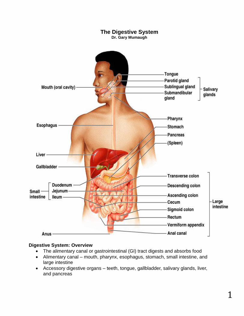
The Digestive System Dr. Gary Mumaugh
Digestive System: Overview
The alimentary canal or gastrointestinal (GI) tract digests and absorbs food
Alimentary canal – mouth, pharynx, esophagus, stomach, small intestine, and large intestine
Accessory digestive organs – teeth, tongue, gallbladder, salivary glands, liver, and pancreas

2
Digestive Process
The GI tract is a “disassembly” line o Nutrients become more available to the body in each step
There are six essential activities: o Ingestion, propulsion, and mechanical digestion o Chemical digestion, absorption, and defecation
Gastrointestinal Tract Activities
Ingestion – taking food into the digestive tract
Propulsion – swallowing and peristalsis o Peristalsis – waves of contraction and relaxation of muscles in the organ
walls
Mechanical digestion – chewing, mixing, and churning food
Chemical digestion – catabolic breakdown of food
Absorption – movement of nutrients from the GI tract to the blood or lymph
Defecation – elimination of indigestible solid wastes
3
Regulation of digestion involves:
Mechanical and chemical stimuli, stretch receptors, osmolarity, and presence of substrate in the lumen
Extrinsic control by CNS centers
Intrinsic control by local centers Receptors of the GI Tract
Mechano- and chemoreceptors respond to: o Stretch by the presence of food o Osmolarity – solute concentration o pH of contents o Presence of end products of digestion
They initiate reflexes that: o Activate or inhibit digestive glands to secrete digestive juices o Mix lumen contents and move them along
Nervous Control of the GI Tract
Intrinsic controls o Nerve plexuses near the GI tract initiate short reflexes o Short reflexes are mediated by local enteric plexuses (gut brain)
Extrinsic controls o Long reflexes arising within or outside the GI tract o Involve CNS centers and extrinsic autonomic nerves
Peritoneum and Peritoneal Cavity
Peritoneum – serous membrane of the abdominal cavity o Visceral – covers external surface of most digestive organs o Parietal – lines the body wall
Peritoneal cavity o Lubricates digestive organs o Allows them to slide across one another
Mesentery – double layer of peritoneum that provides: o Vascular and nerve supplies to the viscera o A means to hold digestive organs in place and store fat
Layers of the Alimentary Canal
Mucosa o Secretes mucus, enzymes and hormones o Absorption of end products of digestion into blood o Protection against disease
Submucosa o Dense connective tissue with blood, lymph and nerves
Muscularis externa or muscularis o Responsible for peristalsis and segmentation
Serosa o Actually the visceral peritoneum
4
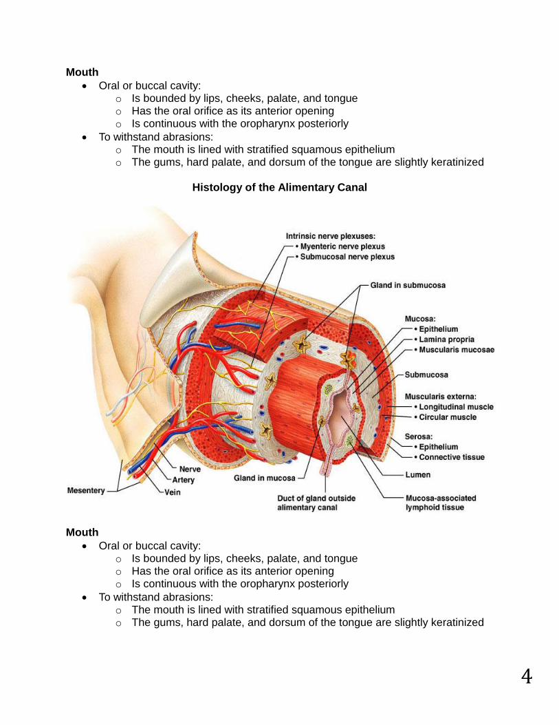
Mouth
Oral or buccal cavity: o Is bounded by lips, cheeks, palate, and tongue o Has the oral orifice as its anterior opening o Is continuous with the oropharynx posteriorly
To withstand abrasions: o The mouth is lined with stratified squamous epithelium o The gums, hard palate, and dorsum of the tongue are slightly keratinized
Histology of the Alimentary Canal
Mouth
Oral or buccal cavity: o Is bounded by lips, cheeks, palate, and tongue o Has the oral orifice as its anterior opening o Is continuous with the oropharynx posteriorly
To withstand abrasions: o The mouth is lined with stratified squamous epithelium o The gums, hard palate, and dorsum of the tongue are slightly keratinized

5
Lips and Cheeks
Have a core of skeletal muscles o Lips: orbicularis oris o Cheeks: buccinators
Vestibule – bounded by the lips and cheeks externally, and teeth and gums internally
Oral cavity proper – area that lies within the teeth and gums
Labial frenulum – median fold that joins the internal aspect of each lip to the gum
Palate
Hard palate o Assists the tongue in chewing o Slightly corrugated on either side of the raphe (midline ridge) which helps
to create friction
Soft palate – mobile fold formed mostly of skeletal muscle o Closes off the nasopharynx during swallowing
Tongue
Occupies the floor of the mouth and fills the oral cavity when mouth is closed
Functions include: o Gripping and repositioning food during chewing o Mixing food with saliva and forming the bolus o Initiation of swallowing, and speech
Intrinsic muscles change the shape of the tongue
Extrinsic muscles alter the tongue’s position
Lingual frenulum secures the tongue to the floor of the mouth

6

7
Salivary Glands
Produce and secrete saliva that: o Cleanses the mouth o Moistens and dissolves food chemicals o Aids in bolus formation o Contains enzymes that break down starch
Three pairs of extrinsic glands – parotid, submandibular, and sublingual
Intrinsic salivary glands (buccal glands) – scattered throughout the oral mucosa
Parotid – lies anterior to the ear between the masseter muscle and skin o Parotid duct – opens into the vestibule next to the second upper molar
Submandibular – lies along the medial aspect of the mandibular body o Its ducts open at the base of the lingual frenulum
Sublingual – lies anterior to the submandibular gland under the tongue o It opens via 10-12 ducts into the floor of the mouth

8
Saliva
Secreted from serous and mucous cells of salivary glands
A 97-99.5% water, hypo-osmotic, slightly acidic solution containing o Electrolytes o Digestive enzyme – salivary amylase o Proteins – mucin, lysozyme, defensins o Metabolic wastes – urea and uric acid
Control of Salivation o Intrinsic glands keep the mouth moist o Extrinsic salivary glands secrete serous, enzyme-rich saliva in response
to: Ingested food which stimulates chemoreceptors and
pressoreceptors The thought of food
Strong sympathetic stimulation inhibits salivation and results in dry mouth Teeth
Primary and permanent dentitions have formed by age 21
Primary – 20 deciduous teeth that erupt at intervals between 6 and 24 months
Permanent – enlarge and develop causing the root of deciduous teeth to be resorbed and fall out between the ages of 6 and 12 years
o All but the third molars have erupted by the end of adolescence o There are usually 32 permanent teeth
Teeth are classified according to their shape and function o Incisors – chisel-shaped teeth adapted for cutting or nipping o Canines – conical or fanglike teeth that tear or pierce o Premolars (bicuspids) and molars – have broad crowns with rounded tips
and are best suited for grinding or crushing
During chewing, upper and lower molars lock together generating crushing force
Tooth Structure o Two main regions – crown and the root
Crown – exposed part of the tooth above the gingiva (gum) Enamel – acellular, brittle material composed of calcium salts and
hydroxyapatite crystals is the hardest substance in the body
Encapsules the crown of the tooth Root – portion of the tooth embedded in the jawbone
Tooth and Gum Disease: Periodontitis
Dental caries – gradual demineralization of enamel and dentin by bacterial action o Dental plaque, a film of sugar, bacteria, and mouth debris, adheres to
teeth o Acid produced by the bacteria in the plaque dissolves calcium salts o Without these salts, organic matter is digested by proteolytic enzymes o Daily flossing and brushing help prevent caries by removing forming
plaque

9
Tooth and Gum Disease: Periodontitis - continued
Gingivitis – as plaque accumulates, it calcifies and forms calculus, or tartar
Accumulation of calculus: o Disrupts the seal between the gingivae and the teeth o Puts the gums at risk for infection
Periodontitis – serious gum disease resulting from an immune response o Risk factors include smoking, diabetes, and oral or tongue or lip piercing
Pharynx
From the mouth, the oro- and laryngopharynx allow passage of: o Food and fluids to the esophagus o Air to the trachea
Lined with stratified squamous epithelium and mucus glands
Has two skeletal muscle layers o Inner longitudinal o Outer pharyngeal constrictors
Esophagus
Muscular tube going from the laryngopharynx to the stomach
Travels through the mediastinum and pierces the diaphragm
Joins the stomach at the cardiac orifice
Glands secrete mucus as a bolus moves through the esophagus

10
Digestive Processes in the Mouth
Food is ingested
Mechanical digestion begins (chewing)
Propulsion is initiated by swallowing
Salivary amylase begins chemical breakdown of starch
The pharynx and esophagus serve as conduits to pass food from the mouth to the stomach
Deglutition (Swallowing)
Involves the coordinated activity of the tongue, soft palate, pharynx, esophagus and 22 separate muscle groups
Buccal phase – bolus is forced into the oropharynx
Pharyngeal-esophageal phase – controlled by the medulla and lower pons o All routes except into the digestive tract are sealed off
Peristalsis moves food through the pharynx to the esophagus
Stomach – Gross Anatomy
Chemical breakdown of proteins begins and food is converted to chyme
Cardiac region – surrounds the cardiac orifice
Fundus – dome-shaped region beneath the diaphragm
Body – midportion of the stomach
Pyloric region – made up of the antrum and canal which terminates at the pylorus
The pylorus is continuous with the duodenum through the pyloric sphincter

11
Stomach – Gross Anatomy
Greater curvature – entire extent of the convex lateral surface
Lesser curvature – concave medial surface
Lesser omentum – runs from the liver to the lesser curvature
Greater omentum – drapes inferiorly from the greater curvature to the small intestine
Microscopic Anatomy of the Stomach
Muscularis – has an additional oblique layer that: o Allows the stomach to churn, mix, and pummel food physically o Breaks down food into smaller fragments
Gastric pits contain gastric glands that secrete gastric juice, mucus, and gastrin Glands of the Stomach
Gastric glands of the fundus and body have a variety of secretory cells o Mucous neck cells – secrete acid mucus o Parietal cells – secrete HCl and intrinsic factor
Stomach Lining
The stomach is exposed to the harshest conditions in the digestive tract
To keep from digesting itself, the stomach has a mucosal barrier with: o A thick coat of bicarbonate-rich mucus on the stomach wall o Epithelial cells that are joined by tight junctions o Gastric glands that have cells impermeable to HCl
Damaged epithelial cells are quickly replaced Digestion in the Stomach - The stomach:
Holds ingested food
Degrades this food both physically and chemically
Delivers chyme to the small intestine
Enzymatically digests proteins with pepsin
Secretes intrinsic factor required for absorption of vitamin B12 Regulation of Gastric Secretion
Neural and hormonal mechanisms regulate the release of gastric juice
Stimulatory and inhibitory events occur in three phases o Cephalic (reflex) phase: prior to food entry o Gastric phase: once food enters the stomach o Intestinal phase: as partially digested food enters the duodenum
Cephalic Phase o Excitatory events include:
Sight or thought of food Stimulation of taste or smell receptors
o Inhibitory events include: Loss of appetite or depression Decrease in stimulation of the parasympathetic division

12
Regulation of Gastric Secretion - continued
Gastric Phase o Excitatory events include:
Stomach distension Activation of stretch receptors (neural activation) Activation of chemoreceptors Release of gastrin to the blood
o Inhibitory events include: A pH lower than 2 Emotional upset that overrides the parasympathetic division
Intestinal Phase o Excitatory phase – low pH; partially digested food enters the duodenum
and encourages gastric gland activity o Inhibitory phase – distension of duodenum, presence of fatty, acidic, or
hypertonic chyme, and/or irritants in the duodenum Initiates inhibition of local reflexes and vagal nuclei Closes the pyloric sphincter Releases enterogastrones that inhibit gastric secretion

13
Gastric Contractile Activity
Peristaltic waves move toward the pylorus at the rate of 3 per minute
Most vigorous peristalsis and mixing occurs near the pylorus
Chyme is either: o Delivered in small amounts to the duodenum or o Forced backward into the stomach for further mixing
Regulation of Gastric Emptying
Gastric emptying is regulated by: o The neural enterogastric reflex o Hormonal (enterogastrone) mechanisms
These mechanisms inhibit gastric secretion and duodenal filling
Carbohydrate-rich chyme quickly moves through the duodenum
Fat-laden chyme is digested more slowly causing food to remain in the stomach longer
Small Intestine
Gross Anatomy o Runs from pyloric sphincter to the ileocecal valve o Has three subdivisions: duodenum, jejunum, and ileum o The jejunum extends from the duodenum to the ileum o The ileum joins the large intestine at the ileocecal valve
Microscopic Anatomy o Structural modifications of the small intestine wall increase surface area
Plicae circulares: deep circular folds of the mucosa and submucosa Villi – fingerlike extensions of the mucosa Microvilli – tiny projections of absorptive mucosal cells’ plasma
membranes

14
Intestinal Juice
Secreted by intestinal glands in response to distension or irritation of the mucosa
Slightly alkaline and isotonic with blood plasma
Largely water, enzyme-poor, but contains mucus Liver
The largest gland in the body
Superficially has four lobes – right, left, caudate, and quadrate
The falciform ligament: o Separates the right and left lobes anteriorly o Suspends the liver from the diaphragm and anterior abdominal wall
Liver: Associated Structures o Bile leaves the liver via:
Bile ducts, which fuse into the common hepatic duct The common hepatic duct, which fuses with the cystic duct
These two ducts form the bile duct
Liver: Microscopic Anatomy o Hexagonal-shaped liver lobules are the structural and functional units of
the liver Composed of hepatocyte (liver cell) plates radiating outward from a
central vein Portal triads are found at each of the six corners of each liver lobule
o Portal triads consist of a bile duct and Hepatic artery – supplies oxygen-rich blood to the liver Hepatic portal vein – carries venous blood with nutrients from
digestive viscera o Hepatocytes’ functions include:
Production of bile Processing bloodborne nutrients Storage of fat-soluble vitamins Detoxification
Composition of Bile
A yellow-green, alkaline solution containing bile salts, bile pigments, cholesterol, neutral fats, phospholipids, and electrolytes
Bile salts are cholesterol derivatives that: o Emulsify fat o Facilitate fat and cholesterol absorption o Help solubilize cholesterol
The chief bile pigment is bilirubin, a waste product of heme

15

16
The Gallbladder
Thin-walled, green muscular sac on the ventral surface of the liver
Stores and concentrates bile by absorbing its water and ions
Releases bile via the cystic duct, which flows into the bile duct Regulation of Bile Release
Acidic, fatty chyme causes the duodenum to release: o Cholecystokinin (CCK) and secretin into the bloodstream
Bile salts and secretin transported in blood stimulate the liver to produce bile
Cholecystokinin causes: o The gallbladder to contract o The hepatopancreatic sphincter to relax
As a result, bile enters the duodenum

17
Pancreas
Location o Lies deep to the greater curvature of the stomach o Encircled by the duodenum and the tail abuts the spleen
Exocrine function o Secretes pancreatic juice which breaks down food o Acini (clusters of secretory cells) contain zymogen granules with digestive
enzymes
Endocrine function o Release of insulin and glucagon
Pancreatic Juice
Water solution of enzymes and electrolytes o Neutralizes acid chyme o Provides environment for pancreatic enzymes
Enzymes are released in inactive form and activated in the duodenum
Active enzymes secreted o Amylase, lipases, and nucleases o These enzymes require ions or bile for optimal activity

18
Regulation of Pancreatic Secretion
Secretin and CCK are released when fatty or acidic chyme enters the duodenum
CCK and secretin enter the bloodstream
Upon reaching the pancreas: o CCK induces the secretion of enzyme-rich pancreatic juice
Vagal stimulation also causes release of pancreatic juice Digestion in the Small Intestine
As chyme enters the duodenum: o Carbohydrates and proteins are partially digested o No fat digestion has taken place o Chyme is released slowly into the duodenum o Mixing is required for proper digestion o Virtually all nutrient absorption takes place in the small intestine
Motility in the Small Intestine
The most common motion of the small intestine is segmentation o Initiated by intrinsic pacemaker cells o Moves contents steadily toward the ileocecal valve
After nutrients have been absorbed: o Peristalsis begins with each wave starting distal to the previous o Meal remnants, bacteria, mucosal cells, and debris are moved into the
large intestine
Control of Motility o Local enteric neurons of the GI tract coordinate intestinal motility o Cholinergic neurons cause:
Contraction and shortening of muscle layer Distension of the intestine
o The gastroileal reflex and gastrin: Relax the ileocecal sphincter Allow chyme to pass into the large intestine
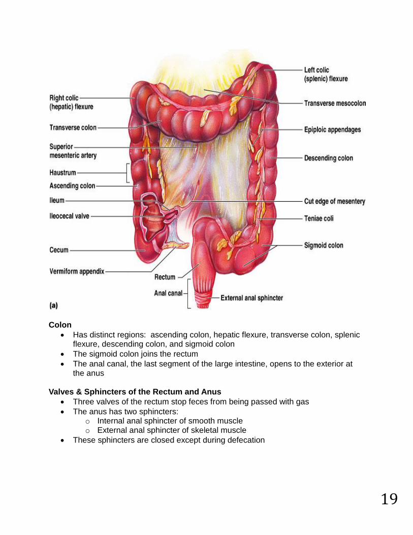
Large Intestine
Has three unique features: o Teniae coli – three bands of smooth muscle o Haustra – pocketlike sacs caused by muscle tone o Epiploic appendages – fat-filled pouches of visceral peritoneum
Is subdivided into the cecum, appendix, colon, rectum, and anal canal
The saclike cecum: o Lies below the ileocecal valve in the right iliac fossa o Contains a wormlike vermiform appendix
19
Colon
Has distinct regions: ascending colon, hepatic flexure, transverse colon, splenic flexure, descending colon, and sigmoid colon
The sigmoid colon joins the rectum
The anal canal, the last segment of the large intestine, opens to the exterior at the anus
Valves & Sphincters of the Rectum and Anus
Three valves of the rectum stop feces from being passed with gas
The anus has two sphincters: o Internal anal sphincter of smooth muscle o External anal sphincter of skeletal muscle
These sphincters are closed except during defecation

20
Bacterial Flora
The bacterial flora of the large intestine consist of: o Bacteria surviving the small intestine that enter the cecum and o Those entering via the anus
These bacteria: o Colonize the colon o Ferment indigestible carbohydrates o Release irritating acids and gases (flatus) o Synthesize B complex vitamins and vitamin K
Functions of the Large Intestine
Other than digestion of enteric bacteria, no further digestion takes place
Vitamins, water, and electrolytes are reclaimed
Its major function is propulsion of fecal material toward the anus
Though essential for comfort, the colon is not essential for life Motility of the Large Intestine
Haustral contractions o Slow segmenting movements that move the contents of the colon o Haustra sequentially contract as they are stimulated by distension
Presence of food in the stomach: o Activates the gastrocolic reflex o Initiates peristalsis that forces contents toward the rectum

21
Defecation
Distension of rectal walls caused by feces o Stimulates contraction of the rectal walls o Relaxes the internal anal sphincter
Voluntary signals stimulate relaxation of the external anal sphincter and defecation occurs
Absorption
Up to 10 L of food, drink, and GI secretions enter the GI tract daily
Only 1 L or less reaches the large intestine
Virtually all food, 80% of electrolytes and water absorb in the small intestine
It is nearly impossible to exceed the absorptive capacity if the GI tract
At the end of the ileum, all that remains is some water, indigestible food materials, and millions of bacteria
The debris is passed on into the large intestine Water Absorption
Approximately 9 L of water, mostly derived from GI tract secretions, enter the small intestine daily
Water is the most abundant substance in chyme
95% of water is absorbed in the small intestines by osmosis
Normal rate of water absorption is 300-400 ml/hour
Water moves in both directions across intestinal mucosa Malabsorption of Nutrients
Results from anything that interferes with delivery of bile or pancreatic juice
Factors that damage the intestinal mucosa (e.g., bacterial infection)
Gluten enteropathy (adult celiac disease) – gluten damages the intestinal villi and reduces the length of microvilli
o Treated by eliminating gluten from the diet (all grains but rice and corn)

22
Developmental Aspects and Lifespan Changes
During fetal life, nutrition is via the placenta, but the GI tract is stimulated toward maturity by amniotic fluid swallowed in utero
At birth, feeding is an infant’s most important function and is enhanced by o Rooting reflex (helps infant find the nipple) and sucking reflex (aids in
swallowing)
Digestive system has few problems until the onset of old age
During old age the GI tract activity declines, absorption is less efficient, and peristalsis is slowed
Changes to the digestive system are slow and slight, and eventually include:
Teeth may become sensitive
Gums may recede
Teeth may loosen, break or fall out
Heartburn may become more frequent
Constipation may become more frequent
Nutrient absorption decreases
Accessory organs age but typically not necessarily in ways that effect health
Cancer
GI cancers rarely have early signs or symptoms
Metastasized colon cancers frequently cause secondary liver cancer
Prevention is by regular dental and medical examinations
Colon cancer is the 2nd largest cause of cancer deaths in males (lung cancer is 1st)
Regular colon examination should be done for all those over 50
Colon cancer is the 2nd largest cause of cancer deaths in males (lung cancer is 1st)
Forms from benign mucosal tumors called polyps whose formation increases with age
Regular colon examination should be done for all those over 50