the development of an aquatic bivalve model: evaluating ... · gametogenesis. 2,3,7,8-tcdd at...
TRANSCRIPT

A
sauga2oToTg©
K
1
oa(lnm1CWaTm
0d
Aquatic Toxicology 81 (2007) 10–26
The development of an aquatic bivalve model: Evaluating the toxic effectson gametogenesis following 2,3,7,8-tetrachlorodibenzo-p-dioxin
(2,3,7,8-TCDD) exposure in the eastern oyster (Crassostrea virginica)
M.L. Wintermyer, K.R. Cooper ∗Rutgers, The State University of New Jersey, Cook Campus, 76 Lipman Drive, New Brunswick, NJ 08901, USA
Received 21 April 2006; received in revised form 12 October 2006; accepted 19 October 2006
bstract
The objective of this study is to develop a gametogenesis protocol to serve as a model for evaluating the toxic effects of chemicals on oogenesis andpermatogenesis in the eastern oyster (Crassostrea virginica). The compound 2,3,7,8-tetrachlorodibenzo-p-dioxin (2,3,7,8-TCDD) was selecteds a “proof of principle” toxicant to examine developmental toxicity in this invertebrate system. The studies were designed to: (1) test the modelsing 2,3,7,8-TCDD and (2) to use histopathological evaluations to characterize the effects on oocyte and sperm development during stages ofametogenesis. 2,3,7,8-TCDD at 10 pg/g resulted in significant histopathological gonadal lesions by day 14 of gametogenesis in both femalend male oysters. These lesions resulted in complete inhibition of gonadogenesis. Studies also showed that a total body dose of 2 and 10 pg/g,3,7,8-TCDD caused adverse responses resulting in abnormal gametogenesis in female and male oysters, respectively, such as: (1) incompleteocyte division, (2) inhibition of oocyte growth and maturation, (3) unsynchronized sperm development, and (4) inhibition of spermatogenesis.
he eastern oyster is one of the most responsive invertebrate models tested to date for reproductive effects of chemicals. Therefore, the easternyster can be used as a sensitive toxicological model for examining the effects of dioxin-like compounds and other xenobiotics on gametogenesis.he reported studies show that environmentally relevant concentrations of 2,3,7,8-TCDD (2–10 pg/g) have a significant adverse effect on oysterametogenesis.2006 Elsevier B.V. All rights reserved.
D; Hi
toTJ112r(ogO
eywords: Crassostrea virginica; Oyster model; Gametogenesis; 2,3,7,8-TCD
. Introduction
Chemicals found in the environment as industrial byproductsr pollutants can have multiple adverse effects on organisms. Annthropogenic chemical, 2,3,7,8-tetrachlorodibenzo-p-dioxin2,3,7,8-TCDD), is one of the most toxic environmental pol-utants. Public concern about 2,3,7,8-TCDD has stimulatedumerous studies to assess TCDD’s behavior in the environ-ent and its effect on living organisms (Isensee and Jones,
975; Brown, 1991; Rune et al., 1991; Bergen et al., 1993;hevreuil et al., 1996; Rhodes et al., 1997; Chen et al., 2002;intermyer and Cooper, 2003; Schultz et al., 2003; Miller et
l., 2004). Mammalian models have demonstrated that 2,3,7,8-CDD significantly impairs and, in a few species, inhibitsale and female reproductive capabilities. Studies have shown
∗ Corresponding author. Tel.: +1 732 932 1000x500.E-mail address: [email protected] (K.R. Cooper).
at2dsri
166-445X/$ – see front matter © 2006 Elsevier B.V. All rights reserved.oi:10.1016/j.aquatox.2006.10.005
stopathology
hat 2,3,7,8-TCDD causes a decrease in spermatogenesis andvulation rate indicating that the most sensitive organs to 2,3,7,8-CDD exposure are the testis and ovaries (Rune et al., 1991;ohnson et al., 1992; Mably et al., 1992; Bjerke and Peterson,994; Sommer et al., 1996; Gray et al., 1997; Heimler et al.,998; Schultz et al., 2003; Miller et al., 2004; Moon et al.,004). For example, research has shown, in various female mice,ats, and hamsters, that exposure to low doses of 2,3,7,8-TCDD0.8–2.5 �g/kg) has resulted in: (1) reduced ovarian weights, (2)varian neoplasms, (3) delayed pregnancies, (4) decreased estro-en levels, and (5) reduced or eliminated ovulation (Gray andstby, 1995; Chaffin et al., 1996; Gray et al., 1997; Salisbury
nd Marcinkiewicz, 2002; Miller et al., 2004). Male reproduc-ive studies have shown that in utero and in vivo exposure of,3,7,8-TCDD has resulted in: (1) altered sperm maturation, (2)
ecreased reproductive organ weights, (3) decreased epididymalperm reserves, (4) decreased or inhibited spermatogenesis, (6)educed Leydig cell function and Leydig cell volumes, and (7)ncreased vacuolar degeneration in germs cells with apoptosis
Aqua
ic1Pe
Tdeoi1eWt(mfIg2nWmdl(earTmolhrs1mmss
dtb1vniAoA2fiei
tv2aig1mamasSlrcMa
ietrrea
st2eos
2
2
2ntrbeL
2
fAn
M.L. Wintermyer, K.R. Cooper /
n tubules using morphological, histochemical, phase contractytometry, biochemical, and molecular techniques (Rune et al.,991; Mably et al., 1992; Johnson et al., 1992; Bjerke andeterson, 1994; Sommer et al., 1996; Gray et al., 1997; Moont al., 2004).
Studies involving aquatic organisms have shown that 2,3,7,8-CDD can bioaccumulate in tissues and adversely effect repro-uction. Field and laboratory studies have demonstrated thatxposure to 2,3,7,8-TCDD can result in altered gonadal devel-pment, egg fertilization success, and embryonic developmentn fish and invertebrate species (Isensee and Jones, 1975; Brown,991; Birnbaum and Tusmisto, 2000; Toomey et al., 2001; Giesyt al., 2002; Butler et al., 2004; Wintermyer and Cooper, 2003;intermyer et al., 2005). For example, in adult female rainbow
rout (Oncorhynchus mykiss), exposure to dietary 2,3,7,8-TCDD1.8 ng/kg) during the reproductive season resulted in accu-ulation of 2,3,7,8-TCDD into tissues and eggs, decreased
ry survival, and decreased adult survival (Giesy et al., 2002).n adult female bivalves exposure to 2,3,7,8-TCDD duringametogenesis resulted in partially undifferentiated gonads,,3,7,8-TCDD accumulation into tissues and oocytes, and a sig-ificant reduction in egg fertilization success (Butler et al., 2004;intermyer and Cooper, 2003; Wintermyer et al., 2005). In adultale bivalves, it has been shown that 2,3,7,8-TCDD exposure
uring gametogenesis resulted in 2,3,7,8-TCDD tissue accumu-ations, reduced spermatogenesis, and reduced sperm viabilityWintermyer and Cooper, 2003). The array of 2,3,7,8-TCDDffects demonstrate some species variability but, many effectsre seen in multiple wildlife, domestic, and laboratory speciesanging from fish through birds and mammals (Birnbaum andusmisto, 2000). The most sensitive adverse effects observed inultiple species appear to be developmental, including effects
n the reproductive system. At relatively low dioxin exposuresevels, structural malformations are not common in species;owever, functional alterations such as in the female and maleeproductive systems and in reproductive behaviors are the mostensitive signs of developmental toxicology (Peterson et al.,993). Cross-species comparisons have shown both female andale reproductive toxicity to have some species variability, butany effects are analogous confirming that 2,3,7,8-TCDD expo-
ure can have a significant effect on the developing reproductiveystem regardless of species (Peterson et al., 1993).
The mechanism of action (MOA) for 2,3,7,8-TCDD is wellocumented in mammalian literature and is mediated throughhe AhR/ARNT receptor complex in mammals and most verte-rate species (Poland and Knutson, 1982; Pohjanvirta and Jouko,994; Fernandez-Salguero et al., 1996; Miller et al., 2004). Con-ersely, in invertebrate species the MOA for 2,3,7,8-TCDD isot understood. Evidence of an Ah-like receptor (96.7 kDa) innvertebrate species has been reported; however, the invertebratehR lacks specific, high-affinity binding for 2,3,7,8-TCDD andther prototypical AhR ligands (Butler et al., 2001). BivalvehR, as well as other invertebrate AhR homologues do not bind
,3,7,8-TCDD unlike the mammalian AhR (95–130 kDa) andsh AhR (105–146 kDa) homologues (Hahn et al., 1994; Butlert al., 2001). The difference in AhR binding affinity distinguishesnvertebrate from vertebrate AhRs but, as in mammalian tissues,iwal
tic Toxicology 81 (2007) 10–26 11
he amount of dioxin that accumulates in aquatic invertebratesaries with the lipid content of tissues (Institute of Medicine,003). In oysters, unlike vertebrate species, complete gonadalnd gamete resorption is repeated seasonally and recrudescencenvolves formation of a new gonad prior to the formation ofametes (Loosanoff, 1942; Galtsoff, 1964; Kennedy and Battle,964; Eble and Scro, 1996). It is during this time of new develop-ent (i.e. gonadogenesis and gametogenesis) that bivalves have
n increase in gonadal lipid content for the purpose of gameteaturation. Increased body burdens of organic contaminants areresult of tissue lipid content, low phase II metabolism, and
low clearance rates in bivalves (Livingstone and Farrar, 1984;tegeman, 1985; Rhodes et al., 1997). The accumulation of pol-
utants in bivalves and fish species can lower their biochemicaleserves (i.e. protein, carbohydrate, lipid, and glycogen) and canontribute to poor egg quality and fertilization rates (CapuzzocDowell, 1996; Van, 2002; Sepulveda et al., 2003; Wintermyer
nd Cooper, 2003).The reproductive systems of terrestrial and aquatic organ-
sms, both male and female, are sensitive to 2,3,7,8-TCDDxposure during early stages of development (i.e. gonadal, game-ogenic, embryonic, fetal, and neonatal) suggesting that theeproductive organs may be a target for 2,3,7,8-TCDD-inducedesponses (ASTM, 1994; Bjerke and Peterson, 1994; Sommert al., 1996; Gray et al., 1997; Schultz et al., 2003; Wintermyernd Cooper, 2003; Miller et al., 2004; Moon et al., 2004).
This research demonstrated that the eastern oyster is aensitive toxicological model for evaluating invertebrate game-ogenesis. The results indicate that a total body dose as low aspg/g 2,3,7,8-TCDD can adversely affect early stages of east-rn oyster gametogenesis. The data presented are the first reportf environmentally relevant levels of 2,3,7,8-TCDD having aignificant adverse effect on oyster reproduction.
. Material and methods
.1. Chemicals
The following chemicals were used in the studies performed:,3,7,8-tetrachlorodibenzo-p-dioxin (98% pure, 50 ± 5 �g/ml in-nonane) was purchased from Cambridge Isotope Diagnos-ics (Woburn, MA). Toluene (>99% pure) and all histologicaleagents and chemicals (i.e. histological stains, 10% phosphate-uffered formalin, ethanol, glutaraldehyde, OsO4, acetone,pon, and araldite) were purchased from Fisher Scientific (Fairawn, NJ).
.2. Animals
Adult eastern oysters (Crassostrea virginica) were purchasedrom the Blue Mussel Company, Prince Edward Sound, Canada.ll oysters used in these studies (N = 605) were weighed (g),umbered, and notched. The average total oyster weight (includ-
ng shells) was 45 ± 2.5 g and the average soft tissue bodyeight (wet weight, w/w) was 5.0 ± 0.60 g (mean ± S.D.). Theverage oyster dimensions (mm) were: height = 62.1 ± 5.36,ength = 38.2 ± 3.61, and width = 17.2 ± 2.68 (mean ± S.D.).

1 Aqua
Etmw
2
rmrwic(wg
fTsawodgto
2
sbwswarnniotip
2
fitaaxH
atG
2
gcaiaapetfic
cTlwZ
2
vt
3
3
2 M.L. Wintermyer, K.R. Cooper /
ach oyster was notched on the left side of the valves for accesso the adductor muscle. Oysters were injected via the adductoruscle regardless of group treatment to ensure that injectionsere not the cause of adverse physiological effects.
.3. Gametogenesis protocol
Oysters were maintained in a holding phase at 15 ◦C in aecirculating seawater system with no additional food supple-ented. Under these conditions, oysters remained in a dormant
eproductive phase. Oysters, regardless of treatment group,ere maintained under optimal holding conditions (i.e. salin-
ty (26–27 ppt), light (12 h cycle), water flow (7.57 l/min), waterhemistry (pH, nitrite, nitrate, and ammonia), and water changesDavis and Chanley, 1956; ASTM, 1994). Optimal conditionsere held constant during both the holding phase and gameto-enic phase.
Gametogenesis was initiated in each oyster immediatelyollowing treatment injections (i.e. seawater, toluene, or 2,3,7,8-CDD) by transferring oysters to a 20 ◦C recirculating seawaterystem and supplementing existing food with 2 l of live culturedlgae (C. isochrisis; 15 million cells/ml) per day. The studiesere conducted over 28 days to allow for full gonadal devel-pment. The 28 days time period for gonadal development wasetermined in previous work (Wintermyer, 1998). Under theseametogenic conditions, the gender of the oysters could be iden-ified as early as 72 h after induction. The 28 days time frame foryster gametogenesis was used in all of the studies performed.
.4. Dosing regime
Adult control oysters were injected with 100 �l of filteredeawater (0.45 � filter mesh) or 100 �l of 4 ppb; ng/g (parts perillion) toluene (solvent carrier). Treated oysters were injectedith 2 or 10 pg/g (parts per trillion) 2,3,7,8-TCDD based on
oft tissue body weight (w/w). Following injections, all oystersere placed on absorbent paper for 1 h before being returned torecirculating seawater system. This allowed discharging and
ecirculation of the injected dose by the oysters with out a sig-ificant loss to the recirculating seawater system. Oysters wereot fed 24 h before or 24 h after the injections. Each oyster wasnjected with the respective treatment dose prior to the inductionf gametogenesis and re-injected on day 14 (t1/2 for TCDD) ofhe study according to the procedure described above. This dos-ng regiment was based on the t1/2 of elimination determined inrevious work (Wintermyer et al., 2005).
.5. Light microscopy (LM)
Oyster samples were preserved in a10% phosphate-bufferedormalin (pH 6.9–7.1) for several days and then transferrednto 70% ethanol. Transverse cuts were made with a scalpelhrough the mid-visceral region of the oyster to obtain a segment
pproximately 5 mm thick. Segments were embedded in paraffinfter processing (dehydration and clearing through an alcohol:ylene series). Sections (6–12 �m) were cut and stained witharris hematoxylin and eosin (H&E). Sections were examinedor
tic Toxicology 81 (2007) 10–26
t 10× and 20× magnifications using a Carl Ziess, compactransmitted-light microscope KM (Howard and Smith, 1983).onad condition was graded according to Kennedy (1977):
Stage 0 = resting stage• Dormant phase of gametogenesisStage I = early development• Pre-vitellogenic oocytes (6–7 �m in diameter) located
within the germinal epithelial wall• Spermatogonia present in the tubulesStage II = later development• Vitellogenic oocytes (11–49 �m in diameter) attached to the
follicular wall• Primary and secondary spermatocytesStage III = sexual maturityStage IIIa = maturity• Post-vitellogenic oocytes (50–55 �m in diameter)
unattached from the follicular wall• Spermatids and mature spermStage IIIb = spawningStage IIIc = redevelopmentStage IIId = recently spent
.6. Electron microscopy (EM)
Gonadal samples (1 mm × 1 mm) were fixed for 24 h in 2%lutaraldehyde in 0.1 M phosphate buffer. Post-fixation wasompleted in 1% OsO4 using the same buffer for 60 min onrotating plate at room temperature. The samples were rinsed
n buffer, and then dehydrated in ethanol solutions (70, 80, 95,nd 100%). All samples were placed on a rotating plate fortotal of 30 min for each progressive ethanol solution. Sam-
les were: (1) cleared in acetone, (2) placed in a 1:1 mixture ofpon:araldite in ethanol (100%) for 1 h, (3) placed in a 1:3 mix-ure of epon:araldite in acetone for 1 h, and (4) samples werenally placed in 100% epon:araldite and polymerization wasompleted at 60 ◦C for 24 h.
Semi-thin (1 �m) and ultra-thin (60–70 nm) sections wereut with glass and diamond ultramicrotome knives, respectively.he semi-thin sections were stained with 0.5% toluidine blue for
ight microscopy (LM). The ultra-thin sections were contrastedith uranyl acetate and lead citrate stains and examined using aiess 10CH electron microscope (Howard and Smith, 1983).
.7. Statistical analysis of data
Results were analyzed using 1-way and 2-way analysis ofariance (ANOVA) and expressed as the percentage related tohe controls (seawater and toluene).
. Results
.1. Gametogenesis protocol
The 28 days gametogenesis protocol developed in our lab-ratory using untreated eastern oysters (C. virginica) (N = 270)esulted in the time correlated gonadal and gametogenic devel-

Aqua
otro1dvgodnt1a
c(lddmpdr2
F(ga
M.L. Wintermyer, K.R. Cooper /
pment as follows: (1) the branching of follicles and tubuleshroughout the gonadal connective tissue in females and males,espectively, by days 1–7, (2) gender identification of individualysters at day 7, (3) the peak of gamete development by day4, and (4) the maturation of gametes by day 28. Within theeveloping follicles at day 7, there were various sizes of pre-itellogenic oocytes, 6–10 �m in diameter, located within theerminal epithelial (follicular) wall indicative of early stages inocyte development (Stage I; Fig. 1a). The male gonads hadeveloping/branching tubules with the presence of spermatogo-
ia lining the outer periphery of the inner tubules correlatingo early spermatogenic development (Stage I; Fig. 1b). At day4 of the gametogenesis protocol, the gonads had well definednd differentiated follicles in the female and tubules in the maleeaof
ig. 1. Gonadogenesis at days 7, 14, and 28 in non-treated eastern oysters (CrassostrStage 1) at day 7. (c) A female and (d) a male gonad illustrating late development tonad illustrating mature gametes at day 28 (Stage IIIb). Thin arrows show folliculand the bar scale indicates oocyte sizes. H&E stain. Similar results were obtained fro
tic Toxicology 81 (2007) 10–26 13
omprising greater than 90% of the gonadal connective tissueFig. 1c and d). Within the developed follicles, there were vitel-ogenic oocytes ranging from approximately 11 to 50 �m iniameter and attached to the follicular wall correlating to lateevelopment and early maturation stages (Stage II; Fig. 1c). Theale gonads had well developed and expanded tubules with the
resence of primary and secondary spermatocytes and the earlyevelopmental stages of spermatids (Stage II; Fig. 1d) also cor-elating to the maturation stages of spermatogenesis. By day8, the gonads were fully developed and compact with differ-
ntiated follicles in the female and tubules in the male (Fig. 1end f). With in the densely packed follicles post-vitellogenicocytes, approximately 50–55 �m in diameter, were unattachedrom the follicular wall concluding the maturation phase (Stageea virginica): (a) a female and (b) a male gonad illustrating early developmento early maturation stages (Stage II/IIIa) at day 14. (e) A female and (f) a maler and tubular development, thick arrows show oocyte and sperm developmentm three independent studies.

1 Aqua
IwbmoF
daaeS8Smwmg
3g
o(staao(tsdtpfd2ecdstf(
blo(e
tSdsfmtstepo1ttistsetdlgoap
sIdd2csp
TT
N
FMT
Rs
4 M.L. Wintermyer, K.R. Cooper /
IIb; Fig. 1e). In the male, densely packed tubules were observedith spermatogonia lining the periphery of the tubules followedy primary spermatocytes, secondary spermatocytes, and sper-atids. Mature sperm were observed gathered toward the center
f the tubules indicating the readiness for spawning (Stage IIIb;ig. 1f).
This protocol has been validated for gonadal and gametogenicevelopment over 28 days according to specific conditions suchs, but not limited to, water temperature and flow, water quality,nd live algal food quality and quantity. At day 7, all oystersvaluated (N = 90; 45 females, 45 males) were at developmentaltage I (100%). At day 14, 86.7% of the females (N = 39) and0.0% of the males (N = 36) evaluated were at developmentaltage II. At day 28, all oysters evaluated (N = 90; 45 females, 45ales) were at developmental Stage IIIa (100%) (Table 1). Thereas no significant difference between staged gonadal develop-ent in female and male groups at day 7, 14, or 28. This 28 days
ametogenesis protocol was used in all the studies performed.
.2. The effects of 2,3,7,8-TCDD on gonadogenesis andametogenesis in C. virginica
Gonadal and gametogenic development is shown in Fig. 2a–hf female and male oysters treated with seawater, toluene4 ng/g), 2 and 10 pg/g 2,3,7,8-TCDD during early gametogene-is (days 7 and 14 of the gametogenesis protocol). Fig. 2a showshe gonadal development of a control seawater female oystert day 7. Seawater control females (N = 20) had well-definednd differentiated follicular development with pre-vitellogenicocytes closely attached to the follicular epithelial wall at day 7Stage I). Fig. 2b shows the gonadal development for a femaleoluene solvent control (4 ng/g) oyster at day 7. The tolueneolvent control females (N = 20) had a slight delay in gonadalevelopment compared to the seawater control females illus-rated by the lack of differentiated follicular structures; however,re-vitellogenic oocytes were present in the follicles of allemales examined (Stage I). Fig. 2c shows the day 7 gonadalevelopment for a 2 pg/g 2,3,7,8-TCDD exposed oyster. Thepg/g 2,3,7,8-TCDD females (N = 20) had moderately differ-ntiated follicular structures at day 7 compared to the seawaterontrol group with pre-vitellogenic oocytes showing delayedevelopment indicated by the small (≤6 �m), compact, darkly
tained oocytes present in the follicles (Stage I). Fig. 2d showshe gonadal development for a 10 pg/g 2,3,7,8-TCDD exposedemale oyster at day 7. The 10 pg/g 2,3,7,8-TCDD femalesN = 20) had abnormal gonadal development at day 7 indicated(
go
able 1he staging of gonadal and gametogenic development in non-treated oysters (C. virg
on-treated oysters Day 7 (Stage I)
emale oysters (N = 135) 45/45 (100%)ale oysters (N = 135) 45/45 (100%)
otal oysters evaluated (N = 270) N = 90
esults were obtained in three independent studies. At each time point (day 7, 14, anignificant difference between or among female and male groups at days 7, 14 and 28a Day 14 females, six oysters had advanced oogenesis; Day 14 males, nine oysters
tic Toxicology 81 (2007) 10–26
y: (1) delayed follicular growth/branching, (2) lack of follicu-ar definition (thin epithelial wall), (3) delayed pre-vitellogenicocyte growth (≤6 �m), and (4) the appearance of macrophagesi.e. brown cells) infiltration in the gonadal tissue of all femalesxamined as early as day 7 of gametogenesis.
Fig. 2e shows the gonadal development of a seawater con-rol male at day 14. The seawater control males (N = 20) hadtage II gonadal development at day 14 indicated by the well-ifferentiated and expanded tubules with primary and secondarypermatocytes present (individual spermatocytes not visiblerom photomicrograph). Fig. 2f shows the gonadal develop-ent of a toluene solvent control (4 ng/g) male at day 14. The
oluene control males (N = 20) had Stage II gonadal developmentimilar to the seawater control males. As with the seawater con-rol males, the toluene-treated males had well-differentiated andxpanded tubules with advanced stages of spermatogenic cellsresent in the tubules at day 14. Fig. 2g shows the gonadal devel-pment of a 2 pg/g 2,3,7,8-TCDD exposed male oyster at day4. The 2 pg/g 2,3,7,8-TCDD treated males (N = 20) appearedo be undergoing normal gonadal development similar to bothhe seawater and toluene controls. The outer peripheries of thenner tubules appeared to have an enlarged space between thepermatogenic cells and the tubule epithelium (*); however, theubules were well differentiated and expanded at day 14. Fig. 2hhows the gonadal development of a 10 pg/g 2,3,7,8-TCDDxposed male oyster at day 14. The 10 pg/g 2,3,7,8-TCDDreated males (N = 20) had abnormal gonadal development atay 14 indicated by: (1) delayed tubular growth/branching, (2)ack of differentiated tubular structures, (3) delayed spermato-enesis compared to control males, and (4) the appearancef macrophages (i.e. brown cells) in the gonadal tissue ofll males examined on day 14 (macrophage infiltration notictured).
The percentage of female and male oysters which displayedpecific morphological lesions at days 7 (Stage I) and 14 (StageI), respectively, are shown in Table 2. In the female oysters atay 7, differential follicular development and delayed gonadalevelopment (Stage I) were significantly different in the toluene,and 10 pg/g 2,3,7,8-TCDD groups compared to the seawater
ontrol group (ANOVA, P < 0.05). Macrophage infiltration wasignificantly different in the 10 pg/g 2,3,7,8-TCDD group com-ared to the seawater, toluene, and 2 pg/g 2,3,7,8-TCDD groups
ANOVA, P < 0.01).The male oysters at day 14 in the 10 pg/g 2,3,7,8-TCDDroup had a significant difference in differential tubule devel-pment, delayed spermatogenesis, and macrophage infiltration
inica) using the 28 days gametogenesis protocol
Day 14a (Stage II) Day 28 (Stage IIIa)
39/45 (86.7%) 45/45 (100%)36/45 (80.0%) 45/45 (100%)N = 90 N = 90
d 28), 30 untreated oysters were randomly selected (15 females, 15 males). No(ANOVA, P > 0.05).
had delayed spermatogenesis.

M.L. Wintermyer, K.R. Cooper / Aquatic Toxicology 81 (2007) 10–26 15
Fig. 2. Female gametogenesis at day 7 and male gametogenesis at day 14 showing treatment groups: seawater, 4 ng/g toluene, 2 and 10 pg/g 2,3,7,8-TCDD in C.virginica. (a) A seawater control female illustrating early stages of gametogenesis at day 7 (Stage I). (b) A toluene solvent control female illustrating early stages ofdevelopment at day 7 (Stage I). (c) A 2 pg/g TCDD female illustrating early gametogenesis at day 7 (Stage I). (d) A 10 pg/g TCDD female illustrating a delay in earlygonadogenesis with a delay in oocyte growth (early Stage 1), and invading macrophages (*) at day 7. (e) A seawater control male illustrating normal spermatogenesisat day 14 (Stage II). (f) A toluene solvent control male illustrating normal spermatogenesis at day 14 (Stage II). (g) A 2 pg/g TCDD male illustrating an enlargedintercellular space within tubule (*) at day 14 (Stage II). (h) A 10 pg/g TCDD male showing a delay in gonadogenesis and spermatogenesis at day 14 (Stage 1). Thinarrows indicate development of follicles and tubules, thick arrows indicate oocytes and sperm development, and bar scale represent oocyte sizes. H&E stain. Similarresults were obtained in three independent studies.

16 M.L. Wintermyer, K.R. Cooper / Aquatic Toxicology 81 (2007) 10–26
Table 2Histological (LM) evaluation of morphological lesions during gonadogenesis in female oysters at day 7 and male oysters at day 14 exposed to seawater, toluene(4 ng/g), 2 and 10 pg/g 2,3,7,8-TCDD
LM Females day 7 (Stage I) Males day 14a (Stage II)
Seawater control, N = 20 females, 20 males *&$ Differentiated follicles, 100% & Differentiated tubules, 100%!£ Delayed development (early Stage I), 0% Delayed development (Stage II), 0%Pre-vitellogenic oocytes, 100% « Enlarged intercellular space, 0%Macrophages, 0% × Macrophages, 0%
Toluene control, (4 ng/g) N = 20 females, 20 males * Differentiated follicles, 40% $ Differentiated tubules, 100%Delayed development (early Stage I), 60% ? Delayed development (Stage II), 0%Pre-vitellogenic oocytes, 100% ‡ Enlarged intercellular space, 0%? Macrophages, 0% ¿ Macrophages, 0%
2 pg/g TCDD, N = 20 females, 20 males & Differentiated follicles, 20% * Differentiated tubules, 100%! Delayed development (early Stage I), 100% # Delayed development (Stage II), 0%Pre-vitellogenic oocytes, 100% ‡« Enlarged intercellular space, 100%M̂acrophages, 0% Macrophages, 0%
10 pg/g TCDD, N = 20 females, 20 males $ Differentiated follicles, 0% *$& Differentiated tubules, 0%£ Delayed development (early Stage I), 100% #? Delayed development (Stage II), 100%Pre-vitellogenic oocytes, 100% Enlarged intercellular space, NA?̂ Macrophages, 100% ¿× Macrophages, 100%
Results were obtained from three independent studies. Symbols indicate significant difference between treatment groups within females at day 7 (ANOVA, P < 0.01)a
aces
cgttdP
TatattttoeootgoltS
3d
icg
1Ttigsd(sosgFfiSaotfSatfibfoa
nd within the males at day 14 (ANOVA, P < 0.05).a Males day 14, enlarged intercellular spaces refer to enlarged intercellular sp
ompared to the seawater, toluene, and 2 pg/g 2,3,7,8-TCDDroups (ANOVA, P < 0.01). The 2 pg/g 2,3,7,8-TCDD male oys-ers had tubules with an enlarged intercellular space between theubule epithelium and spermatogenic cells that was significantlyifferent from the seawater and toluene control males (ANOVA,< 0.01) (Table 2).Gonadal development in oysters exposed to 2 pg/g 2,3,7,8-
CDD early in gonadogenesis did not appear to be as adverselyffected, histologically, compared to the 10 pg/g 2,3,7,8-TCDDreated oysters; however, gametogenesis was impaired at days 7nd 14 in both the female and male oysters, respectively. His-ologically, the 2 pg/g 2,3,7,8-TCDD treated females appearedo have similar follicular development (i.e. follicular defini-ion and expansion) compared to the controls at day 7 andhe males appeared to be undergoing normal gonadal devel-pment at day 14 (Fig. 2c and g). Female and male oystersxposed to 10 pg/g 2,3,7,8-TCDD during early gonadal devel-pment underwent altered gametogenesis illustrated by the lackf gamete development and maturation by days 7 and 14, respec-ively (Fig. 2d and h). The end result was an inhibition ofonadogenesis and gametogenesis in both the female and maleysters by day 14 indicated by the significant delay in fol-icular and tubular branching/expansion, lack of follicular andubule differentiation, and inhibition of gamete development attage I.
.3. Female gonadogenesis and gametogenesis over 28ays using 10 pg/g 2,3,7,8-TCDD
Female oyster gonadogenesis and gametogenesis was exam-ned in this study due to an alteration in the gender ratioorrelated to 10 pg/g 2,3,7,8-TCDD exposure. Inhibition ofametogenesis in the female eastern oyster was observed by day
(tdt
within tubules.
4 of the gametogenesis protocol as a result of 10 pg/g 2,3,7,8-CDD exposure during early gonadogenesis. Fig. 3 shows
he photomicrographs for follicular and oocyte developmentn the seawater, toluene (4 ng/g), and 10 pg/g 2,3,7,8-TCDDroups at days 1, 14, and 28 of gametogenesis (female oystershown only). Fig. 3a shows the follicular as well as the oocyteevelopment of the female seawater control group. At day 1N = 15 oysters; 9 females, 6 males), the female gonads showedigns of early development (Stage I) indicated by the presencef branching follicles through out the gonadal connective tis-ue with various sizes of oocytes (5–7 �m) located within theerminal epithelial (follicular) wall (pre-vitellogenic oocytes).ig. 3b shows at day 14 (N = 15 oysters; 8 females, 7 male), theollicular structures were less defined and various sizes of matur-ng oocytes (11–50 �m) (vitellogenic/post-vitellogenic oocytes;tage II/IIIa) were present in all females evaluated. Fig. 3c showst day 28 (N = 15 oysters; 6 females, 9 males), the majorityf the oocytes were fully matured in each female denoted byhe oocyte sizes (50–55 �m) and detachment of the oocytesrom the germinal epithelial walls (post-vitellogenic oocytes;tage IIIa). Fig. 3d–f shows the female gonadal developments well as the oocyte development for the toluene solvent con-rol (4 ng/g) oysters. Fig. 3d shows at day 1 (N = 15 oysters; 6emales, 9 males), the gonadal development was slightly delayedn the females compared to the seawater controls illustratedy the delay in follicular development and moderately undif-erentiated follicular membranes (Stage 0/I). An early stage ofocyte development was indicated by the oocyte sizes (6 �m)nd close adherence to the germinal epithelial base membrane
pre-vitellogenic oocytes). Fig. 3e shows at day 14 (N = 15 oys-ers; 9 females, 6 males), the follicles were well developed andifferentiated with various sizes of oocytes (11–50 �m) attachedo the germinal base membranes (vitellogenic oocytes; Stage
M.L. Wintermyer, K.R. Cooper / Aquatic Toxicology 81 (2007) 10–26 17
Fig. 3. Female gametogenesis at days 7, 14, and 28 of the 28 days gametogenesis protocol showing treatment groups: seawater, toluene (4 ng/g), and 10 pg/g2,3,7,8-TCDD. (a) Seawater control group at day 1 illustrating pre-vitellogenic oocytes (Stage I), (b) day 14 illustrating vitellogenic oocytes (Stage II), (c) day 28illustrating post-vitellogenic oocytes (Stage IIIa), (d) toluene solvent control group at day 1 illustrating pre-vitellogenic oocytes (Stage I), (e) day 14 illustratingvitellogenic oocytes (Stage II), (f) day 28 illustrating vitellogenic and post-vitellogenic oocytes (Stage II/IIIa), (g) 10 pg/g TCDD treatment group at day 1 illustratingpre-vitellogenic oocytes (Stage I), (h) day 14 illustrating pre-vitellogenic oocytes (Stage I), and (i) day 28 illustrating inhibition of oocyte grow and development( ick ars
Itsmow(esfdg
o(dodftd
pre-vitellogenic oocytes; Stage I). Thin arrows show follicular development, thtain. Similar results were obtained from three independent studies.
I). Development was slightly delayed compared to the seawa-er control females denoted by the majority (>50%) of smallerized oocytes and closer adherence of the oocytes to the ger-inal epithelial wall. Fig. 3f shows that by day 28 (N = 15
ysters; 9 females, 6 males), a majority (>50%) of the oocytesere fully matured in all females indicated by the oocyte sizes
50–55 �m) and detachment of the oocytes from the germinalpithelial wall (post-vitellogenic oocytes; Stage IIIa). Fig. 3g–i
hows the follicular structure as well as the oocyte developmentor the 10 pg/g 2,3,7,8-TCDD treated oysters. Fig. 3g shows atay 1 (N = 15 oysters; 6 females, 9 males), there was a delay inonadogenesis compared to controls as illustrated by the lackcscb
row show oocyte development, and the bar scale represents oocyte sizes. H&E
f follicular branching and undifferentiated follicular structuresStage I). The oocytes were dense and compact denoted by theark staining, irregular shapes, and small size (6 �m) in all thebserved females (pre-vitellogenic oocytes). Fig. 3h shows atay 14 (N = 15 oysters; 2 female, 13 males), the follicles in theemales were not well differentiated indicated by the thinness ofhe follicular membranes and lack of follicular expansion. Theelay in oocyte development and maturation (Stage I) was severe
ompared to the seawater and toluene controls at day 14. Fig. 3ihows at day 28 (N = 15 oysters; 2 females, 13 males), the folli-les in both females were moderately differentiated in structure,ut oocyte growth and maturation was inhibited as indicated by
18 M.L. Wintermyer, K.R. Cooper / Aquatic Toxicology 81 (2007) 10–26
Table 3Female oyster staging of gonadal development at days 1, 14, and 28 following exposure to seawater, toluene (4 ng/g) and 10 pg/g 2,3,7,8-TCDD
Female oysters Day 1 (Stages 0 and I) Day 14a (Stage II) Day 28 (Stage IIIa)
(*) Seawater control (N = 23) 9/9 (100%) 6/8 (75.0%) 6/6 (100%)(§) Toluene solvent control (N = 24) 6/6 (100%) 5/9 (55.5%) 9/9 (100%)(*§) 10 pg/g TCDD (N = 10) 6/6 (100%) 0/2 (0%) 0/2 (0%)
Results were obtained in three independent studies. At each time point (days 1, 14, and 28), five oysters were randomly sampled from each control and treatmentgroup in which only the females were histologically evaluated. Symbols indicate significant difference in female development between treatment groups (day 1–28)(ANOVA, P < 0.05). Significant difference in female development between treatment groups (days 1–28) (ANOVA, P < 0.05).
a Day 14 females, seawater control group had two oysters with advanced oogenesis; toluene solvent control group had four females with delayed oogenesis; 10 pg/gT
tgo
dwSshtadhst
isdtgadmfofta1
3s1
gatm2p5saptdt
gmstacsw
TTu
T
ST1
Rg
CDD group had two females with inhibited oogenesis.
he oocyte sizes (6 �m) and lack of oocyte detachment from theerminal epithelium walls by day 28 (Stage I, pre-vitellogenicocytes).
The percentage of female oysters at Stages I, II, and III atays 1, 7, and 28, respectively, are shown in Table 3. The sea-ater, toluene, and 10 pg/g 2,3,7,8-TCDD female oysters hadtage I gamete development at day 1 (100%). At day 14, theeawater-treated females had 75.0%, the toluene-treated femalesad 55.5%, and the 10 pg/g 2,3,7,8-TCDD-treated female oys-ers had 0% Stage II oocyte development. At day 28, the seawaternd toluene-treated female oysters had 100% Stage IIIa oocyteevelopment while the 10 pg/g 2,3,7,8-TCDD-treated femalesad 0% Stage IIIa oocyte maturation. There was a statisticalignificance between the controls and the 10 pg/g 2,3,7,8-TCDDreatment group development over 28 days (ANOVA, P < 0.05).
The percentage of oysters that developed as a female follow-ng 10 pg/g 2,3,7,8-TCDD exposure are shown in Table 4. Theeawater control group had a higher percentage of oysters thateveloped as females (51%) compared to the percentage of oys-ers that developed as males (49%). The toluene (solvent control)roup also had a higher percentage of oysters that developeds females (53%) compared to the percentage of oysters thateveloped as males (47%), and the 10 pg/g 2,3,7,8-TCDD treat-ent group had a lower percentage of oysters that developed as
emales (22%) compared to the percentage of oysters that devel-ped as male (78%). The percent of oysters that developed intoemales in the control groups was significantly different from
he 10 pg/g 2,3,7,8-TCDD group (ANOVA, P < 0.05). There waslso a significant difference between the gender of oysters in the0 pg/g group (ANOVA, P < 0.001).ttt
able 4he percent of oysters that developed as females in the seawater control group, toluesing the 28 days gametogenesis protocol
reatment groups # Females
Day 1 Day 14 Day
eawater control (N = 45) 9 8 6oluene control (4 ng/g) (N = 45) 6 9 90 pg/g TCDD* (N = 45) 6 2 2
esults were obtained in three independent studies. At each time point (days 1, 14,roup. Symbols indicate significant difference between percent females (ANOVA, P* Significant difference between 10 pg/g TCDD # females and # males over 28 day
.4. Electron microscopy of gametogenesis during earlytages of gonadogenesis following exposure to 2 and0 pg/g 2,3,7,8-TCDD
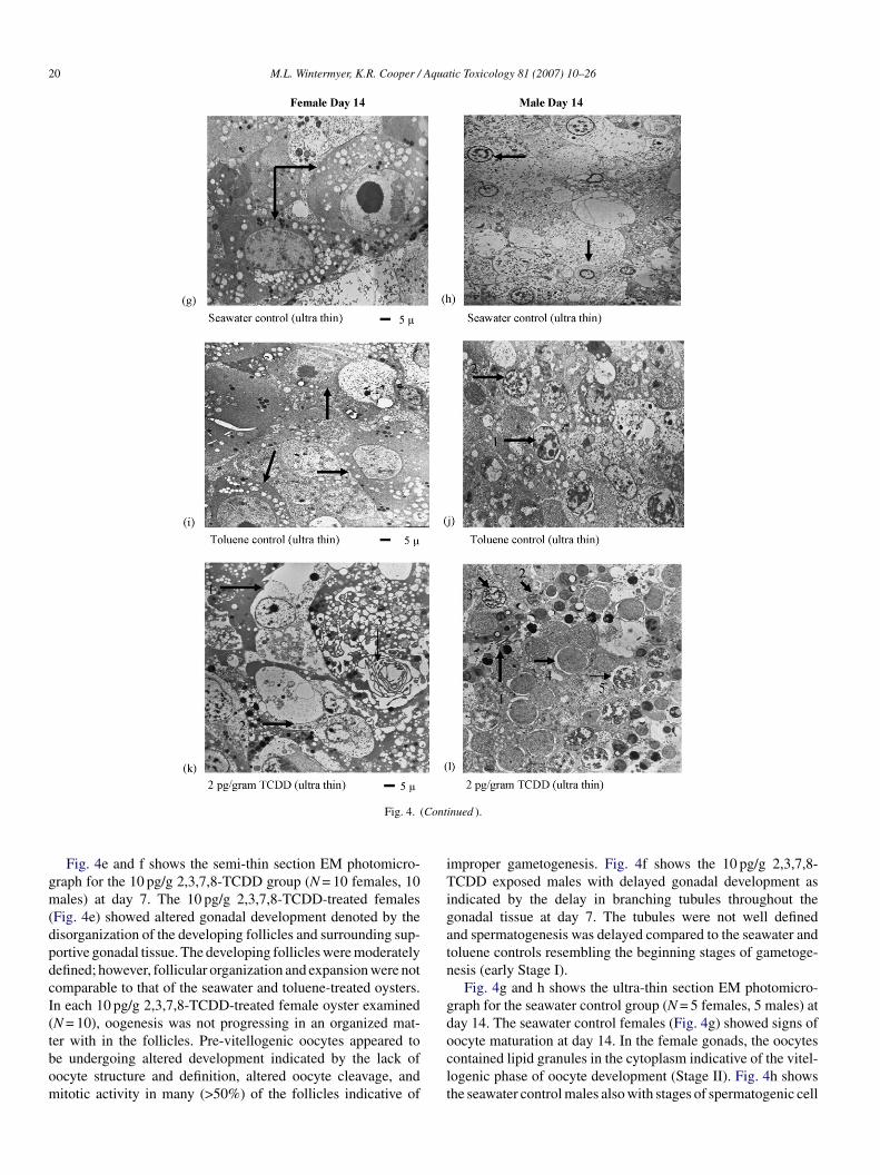
To further examine the effects of 2,3,7,8-TCDD on earlyametogenesis, gonadal tissues were evaluated using semi-thinnd ultra-thin electron microscopy. Fig. 4 shows the EM pho-omicrographs of gametogenesis at days 7 and 14 in female and
ale oysters exposed to seawater, toluene (4 ng/g), 2, and 10 pg/g,3,7,8-TCDD. Fig. 4a and b shows the semi-thin section EMhotomicrographs for the seawater control group (N = 5 females,males). The seawater control females (Fig. 4a) showed early
tages of gametogenesis at day 7 denoted by the well definednd differentiated follicular development and the presence ofre-vitellogenic oocytes (early Stage I). Fig. 4b seawater con-rol males also showed early stages of gonadal development atay 7 (Stage I) denoted by the well-differentiated tubules andhe presence of early spermatogenic cells (Stage I).
Fig. 4c and d shows the semi-thin section EM photomicro-raph for the toluene solvent control group (N = 5 females, 5ales). The toluene control females (Fig. 4c) showed early
tages of gametogenesis at day 7 similar to the seawater con-rol group; however, the toluene females appeared to be moredvanced in development (late Stage I) compared to the seawaterontrol females (early Stage I) illustrated by the larger oocyteizes and expanded follicles. Fig. 4d shows that the toluene malesere also in an early stage of gonadal development similar to
he seawater control males exhibiting well defined and expandedubules. Spermatogenesis (Stage I) appeared to be comparableo the control males at day 7.
ne solvent control group (4 ng/g), and 10 pg/g 2,3,7,8-TCDD treatment group
# Males %Females
28 Day 1 Day 14 Day 28
6 7 9 51 (**)
9 6 6 53 (§)
9 13 13 22 (** §)
and 28), five oysters were randomly selected from each control and treatment< 0.05). Significant difference between percent females (ANOVA, P < 0.05).s (ANOVA, P < 0.001).

M.L. Wintermyer, K.R. Cooper / Aquatic Toxicology 81 (2007) 10–26 19
Fig. 4. Electron microscopy (EM) of gametogenesis at days 7 and 14 in the eastern oyster Crassostrea virginica showing treatment groups: seawater, toluene (4 ng/g),2 and 10 pg/g 2,3,7,8-TCDD. Semi-thin sections: (a) a seawater control female and (b) male illustrating early stages of development (Stage I) at day 7. (c) Atoluene solvent control female (Stage I/II) and (d) male (Stage I) illustrating early stages of development at day 7. (e) A 10 pg/g TCDD exposed female illustratingaltered gonadogenesis and altered pre-vitellogenic oocyte development (Stage I) and (f) a male illustrating delayed gonadogenesis and early stages of spermatogoniadevelopment (Stage I) at day 7. Ultra-thin sections: (g) a seawater control female and (h) male illustrating oocyte and sperm maturation (Stage II) at day 14. Anoocyte containing lipid granules in the cytoplasm (arrow) and a male tubule containing secondary spermatocytes (arrow). (i) A toluene solvent control female and (j)male illustrating early to late stages of development at day 14 (late Stage I). Oocytes contained few lipid granules in the cytoplasm (arrow) and in the male tubule: (1)spermatogonia with peri-nuclear vacuolization and (2) primary spermatocytes were present (Stage II). (k) A 2 pg/g TCDD exposed female and (l) male at day 14. Afollicle with: (1) altered oocyte division and (2) development, and a (3) membranous swirl within the follicle. A male tubule with unsynchronized spermatogenesis(i.e. (1) sperm, (2) spermatid, (3) primary spermatocyte, and (4) spermatogonia) in same area of tubule and spermatocytes exhibiting (5) peri-nuclear vacuolizationat day 14. Thin arrows indicate follicular and tubular development, thick arrows indicate oocyte and sperm developmental stages, and bar scales represent gametesizes. Semi-thin sections were stained with toluidine blue and the ultra-thin sections were contrasted with uranyl acetate and lead citrate stains. Similar results wereobtained in three independent studies.
20 M.L. Wintermyer, K.R. Cooper / Aquatic Toxicology 81 (2007) 10–26
Cont
gm(dpdcI(tbom
iTigatn
gd
Fig. 4. (
Fig. 4e and f shows the semi-thin section EM photomicro-raph for the 10 pg/g 2,3,7,8-TCDD group (N = 10 females, 10ales) at day 7. The 10 pg/g 2,3,7,8-TCDD-treated females
Fig. 4e) showed altered gonadal development denoted by theisorganization of the developing follicles and surrounding sup-ortive gonadal tissue. The developing follicles were moderatelyefined; however, follicular organization and expansion were notomparable to that of the seawater and toluene-treated oysters.n each 10 pg/g 2,3,7,8-TCDD-treated female oyster examinedN = 10), oogenesis was not progressing in an organized mat-
er with in the follicles. Pre-vitellogenic oocytes appeared toe undergoing altered development indicated by the lack ofocyte structure and definition, altered oocyte cleavage, anditotic activity in many (>50%) of the follicles indicative ofoclt
inued ).
mproper gametogenesis. Fig. 4f shows the 10 pg/g 2,3,7,8-CDD exposed males with delayed gonadal development as
ndicated by the delay in branching tubules throughout theonadal tissue at day 7. The tubules were not well definednd spermatogenesis was delayed compared to the seawater andoluene controls resembling the beginning stages of gametoge-esis (early Stage I).
Fig. 4g and h shows the ultra-thin section EM photomicro-raph for the seawater control group (N = 5 females, 5 males) atay 14. The seawater control females (Fig. 4g) showed signs of
ocyte maturation at day 14. In the female gonads, the oocytesontained lipid granules in the cytoplasm indicative of the vitel-ogenic phase of oocyte development (Stage II). Fig. 4h showshe seawater control males also with stages of spermatogenic cell
Aqua
ms(
fddtaot((gnd
gmsFcaaIcbowwut
1egfghp2agocaeailff
sItl
ttis2i1s
4
4
vTcdttobgimeatbuSSb1e2tcglasear
4g
t2v
M.L. Wintermyer, K.R. Cooper /
aturation at day 14. In the male gonads, the tubules containedecondary spermatocytes evidence of advanced spermatogenesisStage II).
Fig. 4i and j shows the ultra-thin section EM photomicrographor the toluene solvent control group (N = 5 females, 5 males) atay 14. The toluene exposed females (Fig. 4i) showed a slightelay in oocyte maturation at day 14 (late Stage I) compared tohe seawater controls. The oocytes appeared to have less lipidccumulation compared to controls indicating an earlier stage ofocyte maturation at day 14 (early Stage II). Fig. 4j shows in theoluene-treated male gonads, spermatogonia were the majority>50%) of sperm present indicative of early sperm developmentlate Stage I; early Stage II) compared to the seawater controlroup. In addition, the toluene male group also displayed peri-uclear vacuolization around spermatogonia in the tubules atay 14 compared to the seawater control males.
Fig. 4k and l shows the ultra-thin section EM photomicro-raph for the 2 pg/g 2,3,7,8-TCDD group (N = 10 females, 10ales). The 2 pg/g 2,3,7,8-TCDD exposed females and males
howed delayed and altered gamete maturation at day 14. Inig. 4k, the female gonads showed improper oogenesis indi-ated by altered cleavage resulting in oocytes with two nucleind two nucleoli, and a significant alteration in oocyte sizesnd shapes giving the appearance of disfigured oocytes (StageI). In the male gonads (Fig. 4l), sperm development appearedomparable to the seawater and toluene controls as indicatedy the well structured spermatogenic cells and advanced stagesf spermatogenesis; however, several stages of spermatogenesisere observed within the same area, implying that developmentas not synchronized at day 14 (late Stage II). Peri-nuclear vac-olization was also observed around the spermatogonia similaro what was observed in the toluene treatment group.
The results of the EM photomicrographs illustrate that 2 and0 pg/g 2,3,7,8-TCDD can interfere with gamete developmentarly in gonadogenesis (days 1–7) according to the 28 daysametogenesis protocol. At day 7, the 10 pg/g 2,3,7,8-TCDDemale and male oysters each exhibited altered and delayedametogenesis with the females displaying an altered affect,istologically, in gamete development and organization com-ared to the 2,3,7,8-TCDD males at day 7 (Fig. 4e and f). Thepg/g 2,3,7,8-TCDD female and male oysters exhibited alterednd unsynchronized gamete development at day 14. The femaleonads exhibited altered oocyte cleavage resulting in improperocyte growth and development, and the males displayed unsyn-hronized sperm development with peri-nuclear vacuolizationround the developing spermatogonia (Fig. 4k and l). Oogen-sis in the 2 pg/g 2,3,7,8-TCDD females appeared to the moredversely affected, histologically, compared to spermatogenesisn the 2 pg/g 2,3,7,8-TCDD male gonads at day 14 (Fig. 4k and). This observation was similar in the 10 pg/g 2,3,7,8-TCDDemales and 10 pg/g 2,3,7,8-TCDD males at day 7 (Fig. 4e and).
The percentage of female and male oysters, which displayed
pecific morphological lesions at days 7 (Stage I) and 14 (StageI) using EM, are shown in Table 5. The 10 pg/g 2,3,7,8-TCDDreated females did not show a significant difference in fol-icular and oocyte development compared to the seawater andptte
tic Toxicology 81 (2007) 10–26 21
oluene controls at day 7 (Table 5). The 10 pg/g 2,3,7,8-TCDDreated males at day 7 did not show a significant differencen tubule development and spermatogenesis compared to theeawater and toluene controls (Table 5). At day 14, the 2 pg/g,3,7,8-TCDD treated females were not significantly differentn follicular development and oogenesis, and the males at day4 did not have a significant difference in spermatogenesis andynchronized development compared to controls (Table 5).
. Discussion
.1. Gametogenesis protocol study
The 28 days gametogenesis protocol was developed andalidated using untreated oysters and then tested using 2,3,7,8-CDD exposed oysters. Studies were performed using theharacterized 28 days gametogenic cycle with time points atays 1, 7, 14, and 28. Untreated oysters, as well as seawater andoluene exposed oysters, displayed similar gonadal and game-ogenic development. At day 7 of gametogenesis, the genderf the oyster, as well as developmental stage (Stage I), coulde identified using light microscopy. Day 14 was the peak ofonadal development; the developing gametes began to maturento primary oocytes in the female and mature sperm in the
ales. This stage (Stage II) could be identified best by usinglectron microscopy. Day 28 of the gametogenic cycle resulted infully developed gonad ready for spawning under normal, non-
reatment conditions. This stage (Stage IIIa) could be identifiedy light or electron microscopy. Validating this protocol withntreated oysters showed that day 7 represented developmentaltage I, day 14 represented Stage II, and day 28 representedtage IIIa. Typically, untreated female oysters were observed toe slightly more advanced in gonadal development at days 7 and4 compared the male oysters under the same conditions; how-ver, at day 28, both genders were fully matured. Testing this8 days protocol using 2,3,7,8-TCDD exposed oysters showedhat: (1) a total body dose of 2 and 10 pg/g 2,3,7,8-TCDD couldause abnormal gonadal development resulting in inhibition ofamete development and maturation by day 14, (2) morpho-ogical lesions in the gonads were permanent through day 28,nd (3) morphological lesions were repeatable and proven con-istent with 2,3,7,8-TCDD exposure in both female and maleastern oysters. This 28 days gametogenesis protocol can be useds a dependable and sensitive toxicological tool in evaluatingeproductive toxicology in bivalve species.
.2. The effects of 2,3,7,8-TCDD on gonadogenesis andametogenesis in C. virginica
Studies using the 28 days gametogenesis protocol revealedhat oogenesis in the oyster appeared to be more sensitive to,3,7,8-TCDD exposure compared to spermatogenesis wheniewed using light microscopy (Fig. 2c, d, g, and h). Mor-
hologically, oocyte development was easier to evaluate dueo the rapid increase in oocyte sizes during maturation andheir distinctive shapes. Although the stages of spermatogen-sis in the male appeared to be delayed/inhibited as a result of
22 M.L. Wintermyer, K.R. Cooper / Aquatic Toxicology 81 (2007) 10–26
Table 5Histological (EM) evaluation of morphological lesions during gonadogenesis in female and male oysters at days 7 and 14 exposed to seawater, toluene (4 ng/g), 2and 10 pg/g 2,3,7,8-TCDD
EM Day 7 (Stage I)
Female Male
Seawater control, N = 5 females, 5 males Differentiated follicles, 100% Differentiated tubules, 100%Undifferentiated follicles, 0% Undifferentiated tubules, 0%Pre-vitellogenic oocytes, 100% Stage I (spermatogonia), 100%Delayed development, 0% Delayed gonadogenesis, 0%
Toluene control (4 ng/g), N = 5 females, 5 males Differentiated follicles, 100% Differentiated tubules, 100%Undifferentiated follicles, 0% Undifferentiated tubules, 0%Pre-vitellogenic oocytes, 100% Stage I (spermatogonia), 100%Delayed development, 0% Delayed gonadogenesis, 0%
10 pg/g TCDD, N = 10 females, 10 males Differentiated follicles, 0% Differentiated tubules, 0%Undifferentiated follicles, 100% Undifferentiated tubules, 100%Pre-vitellogenic oocytes, 100% Stage I (spermatogonia), 100%Delayed development, 100% Delayed gonadogenesis, 100%
EM Day 14 (Stage II)
Female Male
Seawater control, N = 5 females, 5 males Differentiated follicles, 100% Differentiated tubules, 100%Vitellogenic oocytes, 100% Primary and secondary spermatocytes, 100%Abnormal oocyte development, 0% Unsynchronized sperm development, 0%Delayed development, 0%
Toluene control (4 ng/g), N = 5 females, 5 males Differentiated follicles, 100% Differentiated tubules, 100%Vitellogenic oocytes, 100% Primary and secondary spermatocytes, 0%Abnormal oocyte development, 0% Unsynchronized sperm development, 0%Delayed development, 0%
2 pg/g TCDD, N = 10 females, 10 males Differentiated follicles, 20% Differentiated tubules, 100%Vitellogenic oocytes, 0% Primary and secondary spermatocytes, 100%Abnormal oocyte development, 100% Unsynchronized sperm development, 100%Delayed development, 100%
R sampn tment
2ngnsrsaatcm2teG
a2gsT
dtgi(2CIsssclilaat
esults were obtained from three independent studies. Oysters were randomlyumber of females and males per group. No significant difference between trea
,3,7,8-TCDD exposure, the developing tubules and sperm didot appear disfigured as with the oocytes. Upon viewing maleametogenesis using electron microscopy, it was evident thatot only were spermatogenic cells morphologically altered, butperm development was not synchronized. Peterson et al. (1993)eported that structural malformations in the male reproductiveystem were not common in species; however, functional alter-tions such as in Leydig cell function, daily sperm production,nd sperm maturation were sensitive signs of developmentaloxicity. There are reports of altered sperm maturation, germell vacuolization, sperm phagocytosis, and inhibition of sper-atogenesis occurring in mammalian male species exposed to
,3,7,8-TCDD, in agreement with the findings in the male oys-ers of this study (Rune et al., 1991; Johnson et al., 1992; Mablyt al., 1992; Bjerke and Peterson, 1994; Sommer et al., 1996;ray et al., 1997; Moon et al., 2004).These studies have shown using both light microscopy
nd electron microscopy that the morphological lesions in the
,3,7,8-TCDD-treated groups occur as early as day 7 of gonado-enesis and gametogenesis and persist through day 14. Ourtudies have shown that at day 7 the females exposed to 2,3,7,8-CDD (2 and 10 pg/g) had a significant delay in gonadalatis
led at days 7 and 14 from each control and treated group to obtain the samegroups within females or within males at day 7 or day 14 (ANOVA, P > 0.05).
evelopment and oocyte growth as well as macrophage infiltra-ion. These findings are comparable to reports of undifferentiatedonads, decreased oocyte number, and reduced oocyte viabilityn fish and other invertebrate species exposed to 2,3,7,8-TCDDIsensee and Jones, 1975; Brown, 1991; Birnbaum and Tusmisto,000; Toomey et al., 2001; Giesy et al., 2002; Wintermyer andooper, 2003; Butler et al., 2004; Wintermyer et al., 2005).
n the 2,3,7,8-TCDD exposed males at day 14, there was aignificant delay in gonadal development and spermatogene-is, increased macrophage infiltration, and enlarged intercellularpaces between the germinal epithelium and the spermatogenicells. Rune et al. (1991) reported that in the marmoset (Cal-ithrx jacchus), there was a decrease in the intercellular contactn the germinal epithelium, as indicated by the enlarged intercel-ular spaces between the Sertoli’s cells and the spermatogoniafter 2,3,7,8-TCDD treatment. They also reported that there wasn accumulation of premature spermatocytes and spermatids inhe tubular lumen as a result of decreased intercellular contact
nd an inhibition of sperm maturation caused by 2,3,7,8-TCDDreatment (Rune et al., 1991). The observations of enlargedntercellular spaces with in the tubules and unsynchronizedpermatogenesis in the monkey are similar to our findings in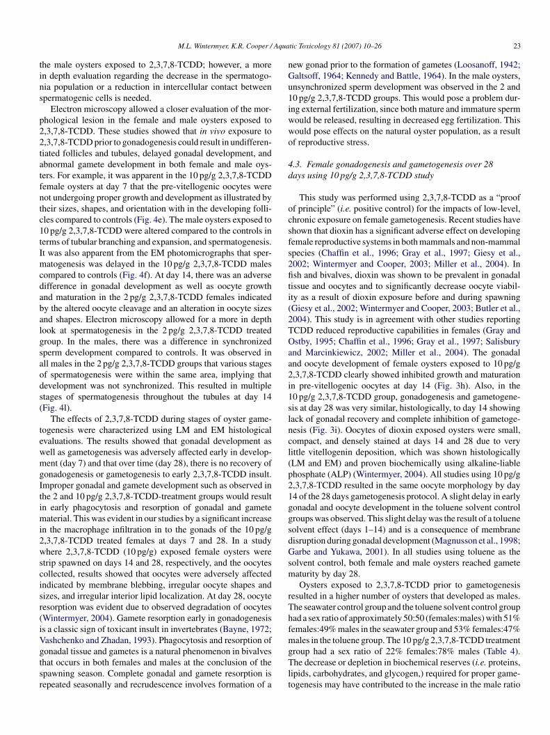
Aqua
tins
p22tatfntc1tImcdabalgsaods(
tewmgItimi2wscisr(iVgtsr
nGu1iwwo
4d
ocsfs2fiti(2TOaa2i1slncl(p21ggsdGsm
rThfm
M.L. Wintermyer, K.R. Cooper /
he male oysters exposed to 2,3,7,8-TCDD; however, a moren depth evaluation regarding the decrease in the spermatogo-ia population or a reduction in intercellular contact betweenpermatogenic cells is needed.
Electron microscopy allowed a closer evaluation of the mor-hological lesion in the female and male oysters exposed to,3,7,8-TCDD. These studies showed that in vivo exposure to,3,7,8-TCDD prior to gonadogenesis could result in undifferen-iated follicles and tubules, delayed gonadal development, andbnormal gamete development in both female and male oys-ers. For example, it was apparent in the 10 pg/g 2,3,7,8-TCDDemale oysters at day 7 that the pre-vitellogenic oocytes wereot undergoing proper growth and development as illustrated byheir sizes, shapes, and orientation with in the developing folli-les compared to controls (Fig. 4e). The male oysters exposed to0 pg/g 2,3,7,8-TCDD were altered compared to the controls inerms of tubular branching and expansion, and spermatogenesis.t was also apparent from the EM photomicrographs that sper-atogenesis was delayed in the 10 pg/g 2,3,7,8-TCDD males
ompared to controls (Fig. 4f). At day 14, there was an adverseifference in gonadal development as well as oocyte growthnd maturation in the 2 pg/g 2,3,7,8-TCDD females indicatedy the altered oocyte cleavage and an alteration in oocyte sizesnd shapes. Electron microscopy allowed for a more in depthook at spermatogenesis in the 2 pg/g 2,3,7,8-TCDD treatedroup. In the males, there was a difference in synchronizedperm development compared to controls. It was observed inll males in the 2 pg/g 2,3,7,8-TCDD groups that various stagesf spermatogenesis were within the same area, implying thatevelopment was not synchronized. This resulted in multipletages of spermatogenesis throughout the tubules at day 14Fig. 4l).
The effects of 2,3,7,8-TCDD during stages of oyster game-ogenesis were characterized using LM and EM histologicalvaluations. The results showed that gonadal development asell as gametogenesis was adversely affected early in develop-ent (day 7) and that over time (day 28), there is no recovery of
onadogenesis or gametogenesis to early 2,3,7,8-TCDD insult.mproper gonadal and gamete development such as observed inhe 2 and 10 pg/g 2,3,7,8-TCDD-treatment groups would resultn early phagocytosis and resorption of gonadal and gamete
aterial. This was evident in our studies by a significant increasen the macrophage infiltration in to the gonads of the 10 pg/g,3,7,8-TCDD treated females at days 7 and 28. In a studyhere 2,3,7,8-TCDD (10 pg/g) exposed female oysters were
trip spawned on days 14 and 28, respectively, and the oocytesollected, results showed that oocytes were adversely affectedndicated by membrane blebbing, irregular oocyte shapes andizes, and irregular interior lipid localization. At day 28, oocyteesorption was evident due to observed degradation of oocytesWintermyer, 2004). Gamete resorption early in gonadogenesiss a classic sign of toxicant insult in invertebrates (Bayne, 1972;ashchenko and Zhadan, 1993). Phagocytosis and resorption of
onadal tissue and gametes is a natural phenomenon in bivalveshat occurs in both females and males at the conclusion of thepawning season. Complete gonadal and gamete resorption isepeated seasonally and recrudescence involves formation of agTlt
tic Toxicology 81 (2007) 10–26 23
ew gonad prior to the formation of gametes (Loosanoff, 1942;altsoff, 1964; Kennedy and Battle, 1964). In the male oysters,nsynchronized sperm development was observed in the 2 and0 pg/g 2,3,7,8-TCDD groups. This would pose a problem dur-ng external fertilization, since both mature and immature spermould be released, resulting in decreased egg fertilization. Thisould pose effects on the natural oyster population, as a resultf reproductive stress.
.3. Female gonadogenesis and gametogenesis over 28ays using 10 pg/g 2,3,7,8-TCDD study
This study was performed using 2,3,7,8-TCDD as a “prooff principle” (i.e. positive control) for the impacts of low-level,hronic exposure on female gametogenesis. Recent studies havehown that dioxin has a significant adverse effect on developingemale reproductive systems in both mammals and non-mammalpecies (Chaffin et al., 1996; Gray et al., 1997; Giesy et al.,002; Wintermyer and Cooper, 2003; Miller et al., 2004). Insh and bivalves, dioxin was shown to be prevalent in gonadal
issue and oocytes and to significantly decrease oocyte viabil-ty as a result of dioxin exposure before and during spawningGiesy et al., 2002; Wintermyer and Cooper, 2003; Butler et al.,004). This study is in agreement with other studies reportingCDD reduced reproductive capabilities in females (Gray andstby, 1995; Chaffin et al., 1996; Gray et al., 1997; Salisbury
nd Marcinkiewicz, 2002; Miller et al., 2004). The gonadalnd oocyte development of female oysters exposed to 10 pg/g,3,7,8-TCDD clearly showed inhibited growth and maturationn pre-vitellogenic oocytes at day 14 (Fig. 3h). Also, in the0 pg/g 2,3,7,8-TCDD group, gonadogenesis and gametogene-is at day 28 was very similar, histologically, to day 14 showingack of gonadal recovery and complete inhibition of gametoge-esis (Fig. 3i). Oocytes of dioxin exposed oysters were small,ompact, and densely stained at days 14 and 28 due to veryittle vitellogenin deposition, which was shown histologicallyLM and EM) and proven biochemically using alkaline-liablehosphate (ALP) (Wintermyer, 2004). All studies using 10 pg/g,3,7,8-TCDD resulted in the same oocyte morphology by day4 of the 28 days gametogenesis protocol. A slight delay in earlyonadal and oocyte development in the toluene solvent controlroups was observed. This slight delay was the result of a tolueneolvent effect (days 1–14) and is a consequence of membraneisruption during gonadal development (Magnusson et al., 1998;arbe and Yukawa, 2001). In all studies using toluene as the
olvent control, both female and male oysters reached gameteaturity by day 28.Oysters exposed to 2,3,7,8-TCDD prior to gametogenesis
esulted in a higher number of oysters that developed as males.he seawater control group and the toluene solvent control groupad a sex ratio of approximately 50:50 (females:males) with 51%emales:49% males in the seawater group and 53% females:47%ales in the toluene group. The 10 pg/g 2,3,7,8-TCDD treatment
roup had a sex ratio of 22% females:78% males (Table 4).he decrease or depletion in biochemical reserves (i.e. proteins,
ipids, carbohydrates, and glycogen,) required for proper game-ogenesis may have contributed to the increase in the male ratio

2 Aqua
oiewtd1Mtar
5
oisnttT(i1r(i1
ahafcethtTctfida2amfoaH2rs2d
peirheaou(aoAcub
6
geedtdfrd7gmiae2a(a(
ega21bsit(a
4 M.L. Wintermyer, K.R. Cooper /
bserved in the 10 pg/g 2,3,7,8-TCDD group. Invertebrate stud-es have shown that due to nutritional stress, wound repair, andnvironmental stressors such as pollution, biochemical reservesill go toward survival and; therefore, result in an alteration in
he sex ratio toward males due to the high energy cost of pro-ucing oocytes (Coe, 1932; Tranter, 1958; Bahr and Hillman,967; Davis and Hillman, 1971; Russell-Hunter, 1979; CapuzzocDowell, 1996). This bivalve model can be used to confirm
hat 2,3,7,8-TCDD, at low environmental doses, can adverselylter oocyte development and maturation as well as alter the sexatio by using histopathology (LM).
. Summary
The results presented in this study provide evidence that theyster can be used as a sensitive toxicological tool in evaluat-ng reproductive toxicity. We have shown through laboratorytudies that gonadal development as well as egg viability is sig-ificantly reduced as a result of 2,3,7,8-TCDD exposure prioro and during gametogenesis. The dogma in literature has beenhat invertebrates have little to no adverse effects to 2,3,7,8-CDD exposure. This assumption is based on several factors:
1) invertebrate species lack an Ah receptor like that observedn mammals and fish (Denison et al., 1985, 1986; Hahn et al.,994), (2) lack of low-dose, chronic exposure studies evaluatingeproductive toxicity in bivalves or other invertebrate speciesCooper, 1989), and (3) the lack of structural malformations innvertebrate species exposed to 2,3,7,8-TCDD (Peterson et al.,993; West et al., 1997).
There are several hypotheses relating to how 2,3,7,8-TCDDdversely affects gametogenesis in the literature. The principalypothesis is that 2,3,7,8-TCDD disrupts the cell cycle causinglterations in cell division and ultimately development (i.e. dif-erentiation) through inhibition of various cell cycle factors andell cycle phases (Weber et al., 1997; Puga et al., 2000; Oikawat al., 2001; Buchanan et al., 2002). Although cell cycle fac-ors have not been studied in the oyster, it is likely that they areighly conserved and would be affected in a similar fashion tohese observed in amphibian and mammalian systems. 2,3,7,8-CDD toxicity is mediated through the AhR/ARNT receptoromplex in mammals; however, in lieu of the present evidencehat bivalves do not have an Ah receptor similar to mammals orsh, it has been suggested that the changes in reproductive tissueue to inhibition of cell differentiation and/or development is byn AhR-independent mechanism (Lin et al., 2001; Butler et al.,004; Wintermyer et al., 2005). In the mammalian system, therere proteins that are multi-functional and can be used to aid inechanisms unrelated to their intended physiological purpose;
or example, cytokines, peptides, lipoprotein receptors, xenobi-tics receptors (i.e. nuclear receptors), and the Ah receptor (Mand Whitlock, 1996; Weib et al., 1996; Kornmann et al., 1999;erz et al., 2000; Herz and Bock, 2002; DiCicco-Bloom et al.,004; Puga et al., 2000). It is not unreasonable to consider that a
eceptor (i.e. xenobiotic receptor) or perhaps a protein (i.e. heathock protein) may be capable of mediating the toxic effects of,3,7,8-TCDD in a region-and stage-specific role during gonadalevelopment.fiifo
tic Toxicology 81 (2007) 10–26
Based both on our findings and the reports in the literature, weropose that the oyster is a useful model to use for studying theffects of 2,3,7,8-TCDD on gametogenesis. We base our reason-ng on five factors: (1) the model is sensitive to environmentallyelevant low levels of dioxin (2–10 pg/g TCDD), (2) the modelas displayed the same gonadal lesions as a result of dioxinxposure both in field studies and in the laboratory (Wintermyernd Cooper, 2003; Wintermyer, 2004), (3) gametogenesis in theyster can be induced repeatedly and without chemical manip-lation (Davis and Chanley, 1956; Loosanoff and Davis, 1963),4) the model is reliable in terms of the histopathological alter-tions displayed, and (5) toxicity test manuals and protocols foryster maintenance/care are well documented (Galtsoff, 1964;STM, 1994). Future advancements in molecular techniques
ritical for invertebrate research will enable this bivalve to besed both as a sentinel organism and an essential laboratory-ased model for the effects of toxicants on gametogenesis.
. Conclusion
In conclusion, these studies were designed to undertake twooals: (1) test the bivalve model using low-level, sub-chronicxposures of 2,3,7,8-TCDD and (2) to use histopathologicalvaluations to characterize the effects on gamete developmenturing selected stages of oyster gametogenesis. The samplingime points for histologically evaluating gametogenesis wereays 7, 14, and 28. The advantage of using the oyster modelor reproductive studies is that gametogenesis can be monitoredepeatedly from the very early stages of primordial germ cellivision to a fully developed gonad in a short period of time (i.e.days; Loosanoff and Davis, 1963). Our protocol for oyster
ametogenesis was 28 days based on standard bioaccumulationethods for aquatic and terrestrial monitoring, sub-chronic test-
ng and metabolism studies, an oyster pharmacokinetic model,nd standard safety assessment protocols (Brown, 1991; Bergent al., 1993; Eaton and Klaassen, 1996; Johnson, 2001; Mussel,003; Steensma et al., 2004; Yu et al., 2004; Wintermyer etl., 2005). The levels of 2,3,7,8-TCDD used in this research2–10 pg/g) were within the environmental range of sedimentsnd bivalve tissue body burdens (Newark Bay, NJ; 11–20 pg/g)NJEPA, 1993; NJDEP, 1996).
The effects of 2,3,7,8-TCDD on oyster reproduction occurarly in gonadogenesis and persist throughout the 28 daysametogenic cycle. 2,3,7,8-TCDD at 2 pg/g showed delayednd altered gamete development and maturation, and 10 pg/g,3,7,8-TCDD showed inhibition of gametogenesis by days4–28. The data presented suggest that the eastern oyster cane used as a toxicological model for evaluating gametogene-is by utilizing the characterized 28 days gametogenic cyclen conjunction with histopathological evaluations to describehe effects on gamete development. This model can be used to:1) examine early to late stages of gametogenesis by using LMnd/or EM techniques and (2) the model can be used for both
eld studies and controlled laboratory studies. By incorporat-ng the characterized 28 days gametogenic cycle as an endpointor reproductive toxicity in invertebrates, the usefulness of theyster as an aquatic model is markedly expanded.

Aqua
R
A
B
B
B
B
B
B
B
B
B
C
C
C
C
C
C
D
D
D
D
D
E
E
F
G
G
G
G
G
H
H
H
H
H
I
I
J
J
K
K
K
L
M.L. Wintermyer, K.R. Cooper /
eferences
merican Society for Testing Material (ASTM), 1994. Standard Methods forthe Examination of Water and Wastewater, 14th ed. American Public HealthAssociation, New York.
ahr, L.M., Hillman, R.E., 1967. Effects of repeated shell damage on gameto-genesis in the American oyster Crassostrea virginica. Proc. Natl. ShellfishAssoc. 57, 59–62.
ayne, B.L., 1972. Some affects of stress in the adult on the larval developmentof Mytilus edulis. Nature 237, 459–462.
ergen, B., Nelson, W., Pruell, R., 1993. Bioaccumulation of PCB congenersby blue mussels (Mytilus edulis) deployed in New Bedford Harbor, Mas-sachusetts. Environ. Toxicol. Chem. 12, 1671–1681.
irnbaum, L.S., Tusmisto, J., 2000. Non-carcinogenic effects of 2,3,7,8-TCDDin animals. Food Addit. Contam. 17 (4), 275–288.
jerke, D.L., Peterson, R.E., 1994. Reproductive toxicity of 2,3,7,8-tetracholordibenzo-p-dioxin in male rats: different effects of in utero versuslactational exposure. Toxicol. Appl. Pharmacol. 127 (2), 241–249.
rown, R., 1991. The toxicokinetics and histological effects of 2,3,7,8-TCDDon the soft-shell clam, Mya arenaria. Ph.D. Dissertation, Rutgers University,25–29.
uchanan, D., Ohsako, S., Tohyama, C., Cooke, P., Iguchi, T., 2002. Dioxininhibition of estrogen-induced mouse uterine epithelial mitogenesis involveschanges in cyclin and transforming growth factor-beta expression. Toxicol.Sci. 66, 62–88.
utler, R.A., Kelly, M.L., Powell, W.H., Hahn, M.E., Van Beneden, R.J., 2001.An aryl hydrocarbon receptor (AHR) homologue from the soft-shelled clam,Mya arenaria: evidence that invertebrate AHR homologues lack 2,3,7,8-tetrachloro-p-dioxin and �-naphthoflavone binding. Gene 278, 223–234.
utler, R.A., Kelley, M.L., Olberding, K.E., Gardner, G.R., Van Bene-den, R.J., 2004. Aryl hydrocarbon receptor (AhR)-independent effects of2,3,7,8-tetrachlorodibenzo-p-dioxin (2,3,7,8-TCDD) on softshell clam (Myaarenaria) reproductive tissue. Comp. Biochem. Physiol. C: Toxicol. Phar-macol. 138 (3), 375–381.
apuzzo McDowell, J., 1996. The bioaccumulation and biological effects oflipophilic organic contaminants. In: Kennedy, V.S., Roger, N., Albert, E.(Eds.), The Eastern Oyster Crassostrea virginica. Maryland Sea Grant Col-lege Publication, University of Maryland System, College Park.
haffin, C., Peterson, R., Hutz, R., 1996. In utero and lactational exposure offemale Holtzman rats to 2,3,7,8-tetrachlorodibenzo-p-dioxin: modulation ofthe estrogen signal. Biol. Reprod. 55, 62–67.
hen, W., Luoping, Z., Li, X., Xinong, W., Liyu, H., Huasheng, H., 2002.Residue levels of HCHs, DDTs and PCBs in shellfish from coastal areasof east Xiamen Island and Minjiang Estuary, China. Mar. Pollut. Bull. 45,385–390.
hevreuil, M., Blanchard, M., Teil, M., Carru, A., Testard, P., Chesterikoff, A.,1996. Evaluation of the pollution by organochlorinated compounds (poly-chlorobiphenyls and pesticides) and metals Cd, Cr, Cu, and Pb in the waterand in the zebra mussel (Dreissena-polymorpha pallas) of the River Seine.Water Air Soil Pollut. 88, 371–381.
oe, W., 1932. Sexual phases in the American oyster (Ostrea virginica). Biol.Bull. 63, 419–441.
ooper, K.R., 1989. Effects of polychlorinated dibenzo-p-dioxins and polychlo-rinated dibenzofuran on aquatic organisms. Aquat. Sci. 1, 227–242.
avis, H.C., Chanley, P.E., 1956. Spawning and egg production of oysters andclams. Proc. Natl. Shellfish Assoc. 46, 40–58.
avis, N.W., Hillman, R.E., 1971. Effect of artificial shell damage on sex deter-mination in oysters. Proc. Natl. Shellfish Assoc. 61, 2.
enison, M., Hamilton, J., Wilkinson, C., 1985. Comparative studies of arylhydrocarbon hydrolase and the Ah receptor in nonmammalian species.Comp. Biochem. Physiol. 80C, 319–324.
enison, M., Wilkinson, C., Okey, A., 1986. Ah receptor for 2,3,7,8-TCDD:comparative studies in mammalian and nonmammalian species. Chemo-
sphere 15, 1665–1672.iCicco-Bloom, E., Lelievre, V., Zhou, X., Rodriguez, W., Tam, J., Waschek, J.,2004. Embryonic expression and multifunctional actions of the natriureticpeptides and receptors in the developing nervous system. Dev. Biol. 271 (1),161–175.
L
tic Toxicology 81 (2007) 10–26 25
aton, D., Klaassen, C., 1996. Principles of toxicology. In: Klaassen, C.M.A.,Doull, J. (Eds.), Casarett and Doull’s Toxicology, fifth ed. Press, New York,p. 1994.
ble, A., Scro, R., 1996. General anatomy. In: Kennedy, V.S., Roger, N., Albert,E. (Eds.), The Eastern Oyster (Crassostrea virginica). Maryland Sea GrantCollege Publication, University of Maryland System, College Park.
ernandez-Salguero, P., Hilbert, D., Rudikoff, S., Ward, J., Gonzalez, F.,1996. Aryl-hydrocarbon receptor-deficient mice are resistant to 2,3,7,8-tetrachlorodibenzo-p-dioxin induced toxicity. Toxicol. Appl. Pharmacol.140, 173–179.
arbe, T., Yukawa, H., 2001. Common solvent toxicity: auto-oxidation of res-piratory redox-cyclers enforced by membrane derangement. Z. Naturforsch.56, 483–491.
altsoff, P.S., 1964. The American oyster Crassostrea virginica (Gmelin). Fish.Bull. 64, 1–456.
iesy, J.P., Jones, P.D., Kannan, K., Newsted, J.L., Tillitt, D.E., Williams,L.L., 2002. Effects of chronic dietary exposure to environmentally rel-evant concentrations to 2,3,7,8-tetrachlorodibenzo-p-dioxin on survival,growth, reproduction and biochemical responses of female rainbow trout(Oncorhynchus mykiss). Aquat. Toxicol. 59 (1/2), 35–53.
ray, L.E., Ostby, J.S., 1995. In utero 2,3,7,8-tetrachlorodibenzo-p-dioxin(2,3,7,8-TCDD) alters reproductive morphology and function in female ratoffspring. Toxicol. Appl. Pharmacol. 133, 285–294.
ray, L.E., Ostby, J.S., Kelce, W.R., 1997. A dose–response analysisof the reproductive effects of a single gestational dose of 2,3,7,8-tetrachlorodibenzo-p-dioxin in male Long Evans Hooded rat offspring.Toxicol. Appl. Pharmacol. 146 (1), 11–20.
ahn, M.E., Poland, A., Glover, E., Stegeman, J.J., 1994. Photoaffinity labelingof the Ah receptor: phylogenetic survey of diverse vertebrae and invertebratespecies. Arch. Biochem. Biophys. 310 (1), 218–228.
eimler, I., Rawlings, R., Owen, H., Hutz, R.J., 1998. Dioxin perturbs, in adose- and time-dependent fashion, steroid secretion, and induces apoptosisof human luteinized granulose cells. Endocrinology 139 (10), 4373–4379.
erz, J., Gotthardt, M., Willnow, T., 2000. Cellular signaling by lipoproteinreceptors. Curr. Opin. Lipidol. 11 (2), 161–166.
erz, J., Bock, H., 2002. Lipoprotein receptors in the nervous system. Annu.Rev. Biochem. 71, 405–434.
oward, D., Smith, C., 1983. Histological techniques for marine bivalve mol-lusks. In: Howard, D., Smith, C. (Eds.), NOAA Technical MemorandumNMFS-F/NEC-25. U.S. Department of Commerce, Woods Hole, MA, pp.1–100.
nstitute of Medicine, 2003. Dioxins and dioxin-like compounds in the foodsupply. Report of the Institute of Medicine of the National Academies. TheNational Academies Press, Washington, DC.
sensee, A.R., Jones, G.E., 1975. Distribution of 2,3,7,8-tetrachlorodibenzo-p-dioxin (2,3,7,8-TCDD) in an aquatic model ecosystem. Environ. Sci.Technol. 9, 668–672.
ohnson, L., Dickerson, R., Safe, S., Nyberg, C., Lewis, R., Welsh Jr., T., 1992.Reduced Leydig cell volume and function in adult rats exposed to 2,3,7,8-tetrachlorodibenzo-p-dioxin without a significant effect on spermatogenesis.Toxicology 76 (2), 103–118.
ohnson, R.S., 2001. Final report on the safety assessment of cetethyl morpholin-ium ethosulfate. Int. J. Toxicol. 20 (3), 99–102.
ennedy, V.S., 1977. Reproduction in Mytilus edulis aoteanus and Aula-comya maorians (Mollusca: Bivalvia). N. Z. J. Mar. Freshwater Res. 11,255–267.
ennedy, A., Battle, H., 1964. Cyclic changes in the gonad of the Americanoyster, Crassostrea virginica (Gmelin). Can. J. Zool. 42, 305–321.
ornmann, M., Kleeff, J., Debinski, W., Korc, M., 1999. Pancreatic cancer cellsexpress interleukin-13 and -4 receptors, and their growth is inhibited byPseudomonas exotoxin coupled interleukin-13 and -4. Anticancer Res. 19(1A), 125–131.
in, T., Ko, K., Moore, R., Buchanan, D., Cooke, P., Peterson, R., 2001. Role
of the aryl hydrocarbon receptor in the development of control and 2,3,7,8-tetrachlorodibenzo-p-dioxin exposed male mice. J. Toxicol. Environ. HealthA 64, 327–342.oosanoff, V., 1942. Seasonal gonadal changes in the adult oysters, Ostreavirginica, of Long Island Sound. Biol. Bull. 83, 195–206.

2 Aqua
L
L
M
M
M
M
M
M
N
N
O
P
P
P
P
R
R
RS
S
S
S
S
S
T
T
V
V
W
W
W
W
W
W
W
6 M.L. Wintermyer, K.R. Cooper /
oosanoff, V., Davis, H., 1963. Rearing of bivalve mollusks. Adv. Mar. Biol.32, 82–91.
ivingstone, D., Farrar, S., 1984. Tissue and sub-cellular distribution of enzymeactivities of mixed function oxygenase and benzo[a]pyrene metabolism inthe common mussel Mytilus edulis L. Sci. Total Environ. 39, 209–235.
a, Q., Whitlock, J., 1996. The aromatic hydrocarbon receptor modulates theHepa 1c1c7 cell cycle and differentiated state independently of dioxin. Mol.Cell. Biol. 6 (5), 2144–2150.
ably, T.A., Bjerke, D.L., Moore, R.W., Gendron-Fitzpatrick, A., Peterson,R., 1992. In utero and lactational exposure of male rats to 2,3,7,8-tetrachlorodibenzo-p-dioxin: effects on spermatogenesis and reproductivecapacity. Toxicol. Appl. Pharmacol. 114 (1), 118–126.
agnusson, A., Sulaiman, M., Dutia, M., Tham, R., 1998. Effects of tolueneon tonic firing and membrane properties of rat medial vestibular nucleusneurons in vitro. Brain Res. 779, 334–337.
iller, K.P., Borgeest, C., Greenfeld, C., Tomic, D., Flaws, J.A., 2004. In uteroeffects of chemicals on reproductive tissues in females. Toxicol. Appl. Phar-macol. 198, 111–131.
oon, D.G., Lee, K.C., Kim, Y.W., Park, H.S., Cho, H.Y., Kim, J.J., 2004.Effect of 2,3,7,8-TCDD on corpus cavernosum histology and smooth musclephysiology. Int. J. Impot. Res. 16 (3), 224–230.
ussel Watch, 2003. Ref. No. 1 50930 NOAA Coastal Monitoring of America.The Mussel Watch Program, NOAA, Coastal Monitoring and Bio-effectsAssessment Division, Costal Monitoring Branch. Silver Spring, MD 20901,www.gsf.de/UNEP/mussel.html.
ew Jersey Department of Environmental Protection (NJEPA) and Energy, Divi-sion of Science and Research, 1993. Polychlorinated Biphenyls (PCBs),Chlordane, and DDTs in Selected Fish and Shellfish from New Jersey Waters,1988–1991: Results from New Jersey’s Toxics in Biota Monitoring Program.http://www.state.nj.us/dep/dsr/pub.htm.
ew Jersey Department of Environmental Protection (NJDEP), 1996. NewJersey Coastline Shapefile. Office of Information Resources Management(OIRM), Bureau of Geographic Information and Analysis (BGIA), Trenton,NJ, http://www.sate.nj.us/dep/gis/digidownload/zips/statewide/coast.zip.
ikawa, K., Ohbayashi, T., Mimura, J., Iwata, R., Kameta, A., Evine, K.,Fujii-Kuriyama, Y., Kuroda, M., Mukai, K., 2001. Dioxin suppresses thecheckpoint protein, MAD2, by an aryl hydrocarbon receptor-independentpathway. Cancer Res. 61, 5707–5709.
eterson, R.E., Theobald, H.M., Kimmel, G.L., 1993. Developmental andreproductive toxicity of dioxins and related compounds: cross-species com-parisons. Crit. Rev. Toxicol. 23 (3), 283–335.
ohjanvirta, R., Jouko, T., 1994. Short-term toxicity of 2,3,7,8-TCDD in labo-ratory animals: effects, mechanisms, and animal models. Pharmacol. Rev.46, 483–549.
oland, A., Knutson, J., 1982. 2,3,7,8-TCDD and related halogenated aromatichydrocarbons: examination of the mechanism of toxicity. Annu. Rev. Phar-macol. Toxicol. 22, 517–554.
uga, A., Barnes, S., Dalton, T., Chang, C., Knudsen, E., Maier, M., 2000.Aromatic hydrocarbon receptor interaction with the retinoblast proteinpotentiates repression of E2F-dependent transcription a cell cycle arrest.J. Biol. Chem. 275, 2943–2950.
hodes, L., Gardner, G., Van Beneden, R., 1997. Short-term tissue distribu-tion, depuration and possible gene expression effects of [H3]-2,3,7,8-TCDDexposure in soft-shell clams (Mya arenaria). Environ. Toxicol. Chem. 16,1888–1894.
une, G.M., deSouza, P., Krowke, R., Merker, H.J., Neubert, D., 1991. Mor-phological and histochemical effects of 2,3,7,8-tetrachlorodibenzo-p-dioxin
(2,3,7,8-TCDD) on marmoset (Callithrix jacchus) testes. Arch. Androl. 26(3), 143–154.ussell-Hunter, W.D., 1979. A Life of Invertebrates. Macmillan, New York.alisbury, T., Marcinkiewicz, J., 2002. In utero and lactational exposure to
2,3,7,8-tetracholordibenzo-p-dioxin and 2,3,7,8-pentachlorodibenzofuran
Y
tic Toxicology 81 (2007) 10–26
reduces growth and disrupts reproductive parameters in female rats. Biol.Reprod. 66, 1621–1626.
chultz, R., Suominen, J., Varre, T., Hakovirta, H., Parvinen, M., Toppari, J.,Pelto-Huikko, M., 2003. Expression of aryl hydrocarbon receptor and arylhydrocarbon receptor nuclear translocator messenger ribonucleic acids andproteins in rat and human testis. Endocrinology 144 (3), 767–776.
epulveda, M., Quinn, B., Denslow, N., Holm, S., Gross, T., 2003. Effects ofpulp and paper mill effluents on reproductive success of largemouth bass.Environ. Toxicol. Chem. 1, 205–213.
ommer, R.J., Ippolito, D.L., Peterson, R.E., 1996. In utero and lactationalexposure of the male Holtzman rat to 2,3,7,8-tetrachlorodibenzo-p-dioxin:decreased epididymal and ejaculated sperm numbers without alteration insperm transit rate. Toxicol. Appl. Pharmacol. 140 (1), 146–153.
teensma, A., van Dijck, P.W., Hempenius, R.A., 2004. Safety evaluation ofphosphodiesterase derived from Leptographium procerum. Food Chem.Toxicol. 42 (6), 935–944.
tegeman, J., 1985. Benzo[a]pyrene oxidation and microsomal enzyme activityin the mussel (Mytilus edulis) and other bivalve mollusc species from thewestern North Atlantic. Mar. Biol. 89, 21–30.
oomey, B., Bello, S., Hahn, M., Cantrell, S., Wright, P., Tillitt, D., Di Giulio,R., 2001. 2,3,7,8-Tetrachlorodibenzo-p-dioxin induces apoptotic cell deathand cytochrome P4501A expression in developing Fundulus heteroclitusembryos. Aquat. Toxicol. 53 (2), 127–138.
ranter, D.J., 1958. Reproduction in Australian pearl oysters (Lamellibranchia)III. Pinctada albina: breeding season and sexuality. J. Mar. Freshwater Res.9, 191–216.
an, E., 2002. Timing of exposure to a pulp and paper effluent influences themanifestation of reproductive effects in rainbow trout. Environ. Toxicol.Chem. 21, 2338–2347.
ashchenko, M., Zhadan, P., 1993. Ecological assessment of the marine environ-ment using two sea urchin tests: disturbance of reproduction and sedimentembryo-toxicity. Sci. Total Environ. Suppl., 1235–1245.
eber, T., Fan, Y., Chapkin, R., Ramos, K., 1997. Growth-related signaling invascular smooth muscle cells is deregulated by 2,3,7,8-TCDD during theG0/G1 transition. J. Toxicol. Environ. Health 51, 369–386.
eib, C., Kumar Kolluri, S., Kiefer, F., Gottlicher, M., 1996. Complementationof Ah receptor deficiency in hepatoma cells: negative feedback regulationand cell cycle control by the Ah receptor. Exp. Cell Res. 226 (1), 154–163.
est, C., Ankley, G., Nichols, J., Elonen, G., Nessa, D., 1997. Toxicity and bioac-cumulation of 2,3,7,8-tetrachlorodibenzo-p-dioxin in long-term tests withthe freshwater benthic invertebrates Chironomus tentans and Lumbriculusvariegatus. Environ. Toxicol. Chem. 16, 1287–1294.
intermyer, M., 1998. Tissue distribution of 2,37,8-Tetrachlorobenzo-p-dioxin(TCDD) in the adult eastern oyster (Crassostrea virginica) and its effectonegg and larval survival. M.S. Thesis Dissertation. Rutgers University, NewBrunswick, NJ.
intermyer, M., 2004. The development of an aquatic bivalve modelfor evaluating the toxic effects on gametogenesis following 2,3,7,8-tetrachlorodibenzo-p-dioxin (2,3,7,8-TCDD) exposure in the eastern oyster(Crassostrea virginica). Thesis Dissertation. Rutgers University, NewBrunswick, NJ.
intermyer, M., Cooper, K., 2003. Dioxin/furan and PCB concentrations in east-ern oyster (Crassostrea virginica) tissues and the effects on egg fertilizationand development. J. Shellfish Res. 22, 737–746.
intermyer, M., Skaidas, A., Roy, A., Yang, Y., Georgapoulos, P., Burger,J., Cooper, K., 2005. The development of a physiologically-basedpharmacokinetic (PBPK) model using the distribution of 2,3,7,8-
tetrachlorodibenzo-p-dioxin (2,3,7,8-TCDD) in the tissues of the easternoyster (Crassostrea virginica). Mar. Environ. Res. 60 (2), 133–152.u, S., Mei, J., Ahren, B., 2004. Basic toxicology and metabolism studies of 1,5-anhydro-d-fructose using bacteria, cultured mammalian cells, and rodents.Food Chem. Toxicol. 42 (10), 1677–1686.