the development of amputation
TRANSCRIPT

The Development of Amputation
By NORMAN T. KIRK, M.D.
"War is the only proper school for the surgeon."-HIPPOCRATES.
AMPUTATION is practiced by many primitive peoples, and somearchaeologists think it has been in use at least since neolithictimes. Neolithic knives and saws of stone and bone have been
found, and the presence of what look like amputated bone stumps inskeletons of the period suggests that this may have been one of theirmany uses. Hollinderl has shown the possibility by performing amputa-tions in six or seven minutes with these primitive instruments. Moodie2describes an ancient Peruvian pottery figure as showing a leg amputatedat the tibiotarsal junction, and holding in the right hand a cap of paintedbone (or wood or metal) to be adjusted to the stump of the leg (Fig. 1) .The murals at La Tene and elsewhere show many hands with amputatedfingers. But even if the evidence for prehistoric amputations were moreextensive and conclusive than it is, it would still not be certain that theseamputations had a surgical intent; there were probably motives of magic,ritual sacrifice, riddance from demoniacal possession, appeasement ofthe gods, punishment, or beautification.
In the Edwin Smith Surgical Papyrus we have a copy, made in theseventeenth century before Christ, of the first part of an Egyptian surgi-cal treatise which was already ancient at that time. Proceeding from headto foot, the extant part finishes the thorax and starts on the spinal col-umn; the remaining part doubtless continued with the abdomen and thelimbs and may have included amputation cases; we do not know.
Among the 250 wounds mentioned in Homer, with a mortality of 75per cent, there is not a single case of loss of limb. Herodotus, however,has a story of Hegesistratus of Elis, who was captured by the Spartans,thrown into prison and condemned to death. He cut off his chainedfoot at the instep, burrowed his way out of prison, and escaped, after-wards correcting his Chopart amputation by a wooden foot.
The earliest extant scientific account of amputation is in the Hip-pocratic treatise On Joints,3 which probably belongs to the latter halfof the fifth century B.C., when Herodotus also was writing. The author,who may have been Hippocrates, recommends amputation in gangrene.
132

FIG. 1. Peruvian jar showing an amputation and cap cover for stump. Courtesy AmericanMuseum of Natural History, New York.

NORMAN T. KIRK
Gangrene, he says, takes place in wounds where large blood vesselshave been strongly compressed, in fractures of bones which have beenbound too tight, and in other cases of immoderate constriction. Strangu-lated parts generally drop off and most such cases recover, even when aportion of the thigh, arm, forearm or leg has come away. The prognosisis better in leg and forearm cases than in those of the thigh and arm.In fractures complicated by gangrene, separation of the dead from theliving parts occurs quickly and the dead parts drop off speedily, becausethe bone has already given way. In gangrene without bone injury, thefleshy parts soon die here also, but the bone separates slowly at the"boundary of the blackening." The author recommends amputation ofthe gangrenous extremity at the joint below "the boundaries of theblackening" as soon as it is "fairly dead and has lost its sensibility,"and he advises that care must be taken not to wound the living part, lestthe patient swoon away and die from pain. He also recommends theexpectant treatment, that is, to await the line of demarcation and self-amputation of the extremity.
Amputation between healthy and diseased tissues is first encounteredin Celsus,4 who lived about the time of Tiberius. When gangrene doesnot yield to other treatment, he says:Between the sound and the diseased part, the flesh is to be cut through witha scalpel down to the bone, but this must not be done actually over a joint,and it is better that some of the sound part should be cut away than that anyof the diseased part should be left behind. When the bone is reached, thesound flesh is drawn back from the bone and undercut from around it, sothat in that part also some bone is bared; the bone is then to be cut throughwith a small saw as near as possible to the sound flesh which still adheresto it; next the face of the bone, which the saw has roughened, is smootheddown, and the skin drawn over it; this must be sufficiently loosened in anoperation of this sort to cover the bone all over as completely as possible.The part where the skin has not been brought over is to be covered with lint;and over that a sponge soaked in vinegar is to be bandaged on.
Ligature is not mentioned in this connection, but speaking elsewhere ofhemorrhage from recent wounds Celsus says that when styptics fail "theblood-vessels which are pouring out the blood are to be seized and tiedwith two ligatures, one on each side of the wounded part, after whichthe vessels are to be divided between the ligatures, so that they mayretract and still have their openings closed. If the case does not admitof this, the actual cautery may be used." In castration, he says, "the veinsand arteries towards the groin are to be ligatured with linen thread andcut away below the ligature."
From Hippocrates through Celsus, amputation seems to be indicatedonly in gangrene, and only as a last resort; but by the time of Archigenesand Heliodorus, under Trajan (c. 100 A.D.), it had become a recognized
134

THE DEVELOPMENT OF AMPUTATION
procedure for ulcers, growths, injuries, and even for deformities. Archi-genes5 advises placing a tight circular bandage above the line of sever-ance, and cutting down upon and typing or sewing the chief blood-vessels. If bleeding is nevertheless profuse, one must cauterize. Amputa-tion should be through healthy tissue, but not at a joint. Heliodorus alsouses the circular bandage to reduce bleeding, to retract the skin andflesh and prevent the saw from tearing them, and to guide the knife.He advises against making a complete circular incision before sawing;one should rather cut down to the bone on that side first on which thereis least flesh, then saw through the bone, and finally cut through theremainder of the flesh at a stroke to sever the limb; in this way there willbe less bleeding. For the same reason, one should not amputate too closeto the elbow or knee. After the amputation, a dressing consisting oftufts of lint, a folding compress, and sponges, is bound on tightly. Indescribing the operation for hernia, Heliodorus says the larger blood-vessels should be tied and the smaller ones transfixed with a sharphook and twisted round and round to close them. Perhaps he employedthe same technique in amputation. In the surviving fragments on majoramputations there is no reference to the flap method, but in describingthe amputation of superfluous digits, he writes:
A circular incision is made round the digit near its base. From this twovertical incisions are made opposite one another, and the flaps so formeddissected up. The base being thus laid bare, the digit is removed by cuttingforceps, the exposed head of the metacarpal is scraped smooth with araspatory, and the flaps are then brought together and sutured.6
Antyllus, in the first half of the second century A.D., gives a fulland clear description of double ligature to prevent hemorrhage in ar-teriotomy for aneurysm. Galen, half a century later, adds that the liga-tures should be of a material that does not rot too quickly, and to doctorsat Rome he recommends a shop in the Via Sacra between the Templeof Rome and the Forum, where aseptic ligaments from Gaul are plenti-ful and cheap.7 In view of the frequency with which ligatures are ad-vised in other operations from Celsus onward, it is remarkable that noancient writer mentions them in connection with amputation. Thereseems to have been a gradual decline in amputative technique, and it hadalready set in by the time of Galen, whose practice was apparently thesame as that of "Hippocrates."
Paul of Aegina8 (c. 640 A.D.) describes with approval the pro-cedure of Leonides of Alexandria, a Greek surgeon at Rome (c. 200A.D.), who first cut down to the bone on that side on which the blood-vessels were fewest and smallest, then applied linen rags to the incisedparts to prevent the saw from tearing them, and after sawing throughthe bone and cutting through the flesh on the other side, applied red-
135

NORMAN T. KIRK
hot irons to the vessels to stop bleeding, dressed the stump, and tookmeasures to induce suppuration.
Albucasis was the ablest of the Muslim surgeons. His great medicalencyclopedia written toward the end of the eleventh century containsa surgical treatise in three books. The first of these is devoted to cauteriza-tion, with a different form of instrument for each part of the body andfor each disease. He does not, however, as is commonly asserted, pre-scribe the use of a cauterizing knife for amputation. When gangrene ofthe lower arms and legs does not yield to medication, he says" oneshould amputate to prevent its extending above the elbow or knee, whenit becomes fatal. The limb should be tightly bound above and belowthe line of severance. While an assistant pulls upward on the upperbandage to retract the skin and flesh, one should make a circular incisiondown to the bone, insert linen pads on both sides to prevent laceration,then cut or saw through the bone. If hemorrhage occurs during theoperation, cauterize promptly or apply some hemostatic powder beforeproceeding. Apply a suitable dressing and tend until cured.
About the middle of the thirteenth century a crude anesthetic tech-nique was developed by Hugh of Lucca and adopted by his son andpupil Theodoric. A sponge was saturated with a soporific solution con-taining opium and mandrake, and was dried in the sun. Before an op-eration, it was soaked in hot water for an hour, then held under thepatient's nose. After the operation the patient was revived by applyinga sponge soaked in vinegar.10 Guy de Chauliac" cites Theodoric's pro-cedure in connection with amputation, but its use seems to have beenmore general.
Henri de Mondeville12 (c. 1300), like Albucasis, used constrictingcircular bandages above and below the site of amputation. These weredrawn in opposite directions by assistants to facilitate the cutting, di-minish pain, and lay the bone bare. The fleshy parts were cut with abroad thin red-hot iron or gold cautery, then covered with a damp clothor leather pad to prevent laceration by the saw.
Guy de Chauliacl3 (c. 1340) describes a similar procedure, attributesit to Albucasis and Avicenna, and gives the impression that it was incommon use. Amputation was performed through the mid-portion ofthe limb or through the joint, depending upon the site of the gangrene.The cutting was by the knife, not by cautery; but the actual cautery orboiling oil was applied afterwards to stop bleeding. Guy then goes on todescribe the method he himself preferred. This consisted in lacing abandage tightly at the joint above the gangrene and allowing mortifica-tion to bring about spontaneous amputation. It is more to the physician'scredit, he says, to have the limb fall off of itself than to cut it off, forwhen it has been cut off the patient is forever mourning its loss andthinking it might have been saved.
136

THE DEVELOPMENT OF AMPUTATION
In many of the writers we have mentioned, it is not clear what thesite of amputation is in relation to the gangrene; but most of them seemto intend a simple circular incision through viable tissue just above thegangrene. Vigo14 (1514), however, says it is better to cut near thehealthy tissue but so as to leave a margin of gangrenous tissue behind,and this for three reasons: (1) to avoid pain; (2) to avoid bleeding;(3) to lessen the pain of the post-amputative cautery. Both flesh andbone are to be cauterized up to the sound tissue. Anesthesia is inadvis-able because of the risk to the patient.
The use of firearms in warfare was now increasing the demand formilitary surgeons, and the nature and treatment of gunshot wounds wasbecoming a controversial subject second only to syphilis. Brunswick(1497), Vigo (1514) and Gersdorff (1517) thought that gunshotwounds were poisoned by the gunpowder used in firing the shot. As inother poisonous wounds, the actual cautery was indicated where possible,otherwise boiling oil. In the latter half of the sixteenth century it wasshown by Maggi, Pare, Gale and others that gunshot wounds are notburns and that they are not poisonous.
To stop bleeding from the stumps of criminals who have had theirhands or feet cut off by the executioner, Brunswick advises that thestump be stuck into the body of a black hen that has been slit open.Fragments of bone should be removed, jagged ends sawed smooth, andthe skin drawn over the stump and sutured. Rules are also given for theguidance of the executioner himself. The flesh should be bound tightlyabove the site of amputation and retracted upward, the knife poised overthe joint and driven through with a club. Ashes of burnt paper shouldbe sprinkled over the wound, the band then loosened and the skindrawn over the stump as above.15 In a separate chapter'6 on the amputa-tion of superfluous or gangrenous members, added in later editions,Brunswick says that a supernumerary finger or thumb should be ampu-tated at the root and the wound cauterized with hot oil. If gangreneextends to a joint, disarticulate; otherwise amputate below the joint.In both cases the skin must be retracted as far as possible by an assistantand a circular bandage tightly bound a finger's breadth above the gan-grene. Then incise the soft parts to the bone, saw through the latter,apply styptic powder mixed with egg white, or cauterize with the hotiron or oil. Brunswick also suggests the use of Theodoric's anesthesia onrecalcitrant patients.
The use of firearms also multiplied many times the number ofamputation cases that came to the hands of the military surgeon. Gers-dorff (1517), whose publication was the first to contain a woodcut illus-tration of an amputation (Fig. 2), claims to have performed a hundredor two hundred amputations for gangrene and erysipelas.'7 He used twoconstricting circular bands a finger's breadth apart and amputated be-
137

2JlAein abficlttitn!tffehI IIV/ s3 tot 4UCr flitenmd b.tm 1tb cf1npti2 enbrUfoc b . fdtcF fiI hL 'ZCictO un) O P 21n p ai b_tf.f~ il f ci4i b
S ra.hn.. >4.
Arl (
4,
I
I
I.
*'I
I I llIi
L_
FIG. 2. First printed illustration of an amputation. From Gersdorff's Feldtbuch derWundt-Artzney (1517). From the copy in the Army Medical Library.
!
-4"

THE DEVELOPMENT OF AMPUTATION
tween them. He has an elaborate recipe for a styptic to be mixed with eggwhite and applied to the wound to check bleeding. If that fails, he saysthe cautery should be used. "Bind now over the styptic a good thickpad, take the bladder of a bull, ox, or hog, one which is strong, cutthe neck open so that it will go over the pad and stump, and the bladdershould be wet but not too soft; draw it then over all, tie it hard witha band and you need have no care about the bleeding.''18 Neither Gers-dorff nor Brunswick nor Vigo mentions the ligature in this connection.
It was Pare, the great French army surgeon, who reintroduced in thesixteenth century the ligation of vessels in amputation (see Frontis-piece), and other forgotten teachings of Celsus, and who discontinuedthe use of the cautery and burning oil for checking hemorrhage and fortreatment of gunshot wounds.
In 1536, Pare as a junior surgeon in his first military campaign onthe Straits of Susa, following the teaching of Vigo and the example ofhis seniors, used pledgets and tents dipped in burning hot oil as a dress-ing for the wounded. He was of the opinion that gunshot wounds werenot poisoned, that these caustics caused pain and increased the inflam-matory reaction, so one night, his oil giving out, he made a "digestive"dressing of the yolk of an egg, oil of rose and turpentine, which heapplied to several wounded. He could not sleep that night, fearing hewould find his patients dead in the morning. But, on arising early, hefound'that they had rested well and the wounds were not inflamed. Hestates that this was his first discovery and that he thereupon resolvednever to burn a gunshot wound again.19
Prior to 1552 Par,e still advocated and used the cautery, after ampu-tation. But about this time, being accustomed to tying vessels in severewounds, he suggested to his colleagues that the practice might be ex-tended to vessels exposed during amputations of extremities. He tellsus how, "at first in my budding practice thereof,,I always had my cau-teries and hot irons in readiness, so that if anything happened otherwisethan I expected in this new work, I might fetch succour from the ancientpractice, until at length confirmed by the happy experience of almost aninfinite number of particulars, I bid adieu to allf hot irons and cauterieswhich were commonly used in this work.'" 20
He quotes Celsus among others in his argument for the use of theligature rather than cautery, stating that the latter is of value whenamputation is performed through dead tissue, but,in living tissue it istoo painful and leaves an eschar, which, on sloughing off is frequentlyfollowed by secondary hemorrhage and the bone becomes bare and anincurable ulcer remains.2'
In his Voyage of Danvilliers, 1552,22 Pare records the successfulamputation of a "gentleman's leg" through the upper third withoutthe use of the cautery. Elsewhere he describes a re-amputation at the
139

PourtraiEl des iambes drtificielles.
FIG. 3. Artificial leg invented by Pare, from his Oeuvres, 1575. From the copy in theArmy Medical Library.

,, i.t:'...:
I
il' ., l~~~Pr-jS |0
t:rl-'s~~-/ nrd~~

NORMAN T. KIRK
same level in the case of an officer whose foot had been shot off withan "iron bullet" and whose stump had healed but was too long forproper prosthesis.
He emphasized the importance of removal of all dead tissue in ampu-tating for gangrene, according to the advice of Celsus, and he alsointroduced the modern surgical doctrine of the site of election. Forexample, the leg was to be cut off five fingers' breadth under the kneeso that the patient could more easily use a wooden leg; but in the armhe states we must do otherwise and cut off as little of the sound partas possible.23 He describes the pain referred to the amputated extremity,which was often grievously complained of by the patient, for monthsafter its removal, and he points out that this should not hinder theintended amputation necessary to save life in cases of gangrene.
In addition to these marked advances in the technique of surgery,Pare also invented many useful surgical instruments and made thefirst artificial limbs for the upper and lower extremities (Fig. 3).
The first description of the use of the ligature in amputationsoriginally written in English, was published in 1596 by Peter Lowe,a Scotch army surgeon.24
Fabry, the leading German surgeon of his period, in his monographon gangrene in 1593, like Pare, recommended amputation above thediseased part, and is credited with being first to amputate through thethigh. He controlled the circulation by means of a band tightened bytwisting with a stick. An illustration showing an amputation beingperformed in the home is reproduced from Fabry's book (Fig. 4).
The reintroduction of the ligature by Pare, and the discovery of thecirculation of the blood in 1616 by Harvey led to the invention ofMorel's tourniquet in 1674, commonly known as the Spanish windlass,and Petit's tourniquet, invented in 1718, perfected the means of con-trolling hemorrhage in amputation.
The control of hemorrhage by the tourniquet and ligature havingbeen accomplished, the next requirement was speed and simplicity toreduce pain in amputation.
Until the eighteenth century, with the exception of Pare, littlethought was given to the condition of the stump resulting from theoperation. In fact, the ideal stump for prosthetic appliances was notattained until after the introduction of anesthesia in 1846-47, and ofaseptic surgery following the antiseptic period of Lister in 1870.
The commonest of all amputations was the transverse circulartype. It consisted of a simple transverse incision through healthy tissues.This method, as can readily be seen, did not provide adequate coveringfor the bone end after healing. It was, however, the quickest and simplestmethod for the removal of a useless or diseased extremity. This procedurewas modified by Petit of Paris in 1718, to afford more bone covering.
142

THE DEVELOPMENT OF AMPUTATION
The skin and fascia were first divided circularly and retracted andthen the muscles were cut through at the highest possible level.
Louis of Paris, in 1752, returned to the simple transverse incision,but he sawed through the bone at a level higher than the retractedsoft parts.
Technical improvements were added by Alanson of Liverpool, in1779, and by Hey of Leeds in 1803. Hey, who called his method the"triple incision" described it as follows: The first incision divided theskin and fascia, which were dissected up for a sufficient distance. Asecond incision divided the muscles to the bone. The muscles were thenretracted and the bone sawed through at a still higher level.25
The oblique or oval method was first described by Sharpe of Londonin 1739. This method, as the name implies, is a modification of thecircular-cut in the same fashion-except that instead of the incisionbeing transverse in direction it is oblique, forming a lateral flap. Itallows a higher access to the bone and a somewhat wider range ofadaptability in selected cases.
The ratchet incision, introduced by Malgaigne of Paris, 1837, isformed by adding a longitudinal incision to the oblique or circular.This incision is particularly adaptable for amputations through joints.
The flap methods were developed from the transverse circular oroblique by adding two longitudinal incisions. Verd'un of Amsterdam(1696) employed musculo-cutaneous flaps, cut by tranfixion. Ravaton(1739) cut double square flaps equal to half the thickness of the limb.Vermale (1765) improved the latter's technique by fashioning roundedor oval flaps by the transfixation or dissection method. Transfixationwas originally used as it required less time, causing less pain to thepatient, but the resultant flaps could not be as neatly fashioned.
This was the status of the technique of amputations, developedsince the time of Pare, mainly in civil surgery, at the beginning of thenineteenth century.
It was at this period, due to the effort and writings of the greatBaron Larrey of France in the Napoleonic Wars, and the BritishMedical Corps ably headed by Guthrie in the Wars of Great Britain,that amputations reached the peak of advancement and perfection priorto the days of anesthesia and asepsis.
Larrey26 was the first to disarticulate the hip joint successfully. Heperformed this operation twice in 1803, both cases terminated favorably.He developed a method for disarticulation of the shoulder joint, whichhe stated had been done twice previously with fair success, and per-formed the operation eleven times in twenty-four hours-followingthe capture of Smolensk in the Russian campaign. Nine of these patientsrecovered completely, the other two dying of "dysentery." In this samecampaign he performed two hundred amputations in a twenty-four
143

NORMAN T. KIRK
hour period. In connection with the treatment of a shoulder girdle aftera traumatic amputation of the arm by a cannon ball, he describes thetechnique of debridement as we understand it today.
Guthrie successfully disarticulated the hip joint during the Battleof Waterloo. In his Treatise on gunshot wounds he reviews twentycases of this amputation as performed by the leading surgeons of thetimes, including one of his own, three of which were successful, andseven of which died of causes independent of operation. Ten died as aresult of the operation or by infection. This operation was not in goodfavor with the profession at the time, due to the high mortality, andhe here makes a plea for its acceptance. Larrey considered its danger,when properly performed, not particularly greater than disarticulationat the shoulder.
Amputations were then classified by military surgeons as follows:Primary-the operation being performed as soon after the receipt ofthe injury as the condition of the patient permitted-ordinarily in thefirst twenty-four hours. Secondary-performed later than three weeksafter the receipt of injury and after the development of suppuration.
Prior to this time many authorities, including Faure (1756), Hunter(1794), Baron Percy (1792), the latter quoting the opinions of LaMortimere, Louis Sabatier and Dessault, favored and taught that sec-ondary amputation was preferable.
Larrey and Guthrie both found that primary amputation gave alower operative mortality, the stumps healed more quickly with lessinfection, and fewer cases of recurrent hemorrhage occurred. They alsofound that many cases succumbed to infection, after long periods ofsuffering, while awaiting amputation, and that hospital gangrene andmetastatic abscess of internal viscera were more prevalent followingthe secondary operation. They both practiced immediate amputationon the field of battle. This we now know lessened the shock and absorp-tion of proteids and infection in the wound. They advocated primaryoperation in all cases where amputation was indicated.
In general, the indications for immediate primary amputation were:all compound fractures of long bones the result of gunshot wounds,whether produced by shell or rifle bullet; all fractures entering joints;all gunshot wounds of joints; non-fracture cases with extensive loss ofsoft parts, including the main nerve supply, and in destruction of themain blood supply of an extremity with or without fracture. This appliedto all thigh injuries, since the results of the treatment of compoundfractures of the femur were particularly unsatisfactory, as illustrated byforty-three of the most promising cases after the Battle of Toulousethat were treated conservatively in a well equipped nearby hospital.Of this number, thirteen died forthwith, and twelve were amputatedsecondarily, with seven deaths. Of the remaining eighteen, three months
144

THE DEVELOPMENT OF AMPUTATION
later, five were considered well, two thought their legs better than anamputation stump and the remaining eleven wished they had sufferedprimary amputation.
Fewer amputations were required in the leg than in the thigh,because of the superficial position of the tibia and the presence of thetwo bones, and still fewer in the forearm and arm. Guthrie believedthat in uncomplicated compound fractures of the leg, the result ofbullet wounds, primary amputation was rarely indicated.
The circular method of amputation was the method of choice andwas generally performed. In high thigh amputations and occasionallyin the leg and at the shoulder and hip, the flap method was used.
The technique of the amputation of the thigh by the circularmethod as described by Guthrie was as follows.27 After the applicationof the tourniquet, or of digital pressure of the femoral on the pelvis,the skin is retracted upwards by the hands of one assistant and puton the stretch downward by another, making its division more easilyaccomplished, giving less pain to the patient and providing the maximumretraction of the integument. The incision was made through healthytissue, above the injury. The skin and fascia were cut through withone circular incision of the knife, this being the most painful part ofthe operation. The fascia was completely divided and separated frommuscle to allow the maximum contraction. The outer muscles were cutthrough at the retracted integument by circular incision, and, after theirretraction, the deeper muscles attached to the bone were likewiseincised, at the level of the retracted superficial muscles. The muscleattached to the bone was then separated for a distance of at least threeinches, so that the bone end would be well imbedded. In secondaryamputations, due to the lessened elasticity of the skin and muscles frominflammatory infiltration, the bone was sectioned an inch or more higher.The muscles were then retracted with a linen retractor, the periosteumcut through circularly at the saw line and the bone sawed through. Theperiosteum was not removed above the bone end. Guthrie found thatan exfoliation of bone usually followed if the periosteum was removed.The arteries were secured and tied with a strong thread ligature, oneend being cut short and the other long, unless dental silk was used,when both were cut short. Veins were not ligated unless absolutelynecessary, as it was thought this produced sepsis. The nerves were cutshort and allowed to retract. The wound was sponged with cold waterand dried. The soft parts were then brought down over the bone end,placed in apposition horizontally across it, and retained by adhesiveplaster strapping. The long ends of the ligatures were brought outbetween the adhesive strapping so that they could be removed whenthey separated, on the sixth to tenth day. The stump was then bandaged.Adhesive plaster strapping was used by the British in primary ampu-
145

C:
0
0
E0
C.
rz

THE DEVELOPMENT OF AMPUTATION
tations, in the hope that primary union would follow. It was notemployed in secondary amputations, bandages only being used, a rollerbeing applied from the upper part of the thigh to the stump end.
Baron Larrey directed that healing by first intention and the useof adhesive plaster be not attempted, having found that death too oftenoccurred following this method. He was of the opinion that the stumpend should be left open, covered only with a light dressing, and thatthe soft part could be properly held in place by circular bandaging.
Larrey found in his campaign in Saxony that the Saxons dividedthe skin and muscles by means of a curved knife (see Fig. 5) at onestroke, and sawed the bone on a level with the sectioned soft parts. Thewounds were sutured tightly without ligation of vessels, before theremoval of the tourniquet. Exact and close apposition generally pre-vented wound hemorrhage but the pain the patients suffered wassevere. He reports that all cases, without exception, which he saw, diedof gangrene in the interior of the wound after the fourth day. In hisown wounded in his two months' campaign in Saxony, he reports ninehundred and seventy-two amputations, including twenty-two successfuldisarticulations of the shoulder and twenty double amputations, withtwo hundred and forty-one deaths. Tetanus was responsible for anumber of these. His mortality was lower than the British and lowerthan that in our own War of the Rebellion.
Guthrie originally recommended a flap operation in amputationthrough the upper part of the thigh. Later he was of the opinion thatthe circular method was preferable, unless the soft parts were sodestroyed that the circular method could not be used to advantage, inwhich case he performed the flap amputation.28 The circulation wascontrolled by digital pressure on the femoral over the pelvis. Lateralskin and fascia flaps were fashioned and retracted by the hands of theassistant, the inner flap being cut longer than the outer to afford bettercovering for the bone end. The muscle was then divided to the bonefollowing the outline of the skin incision from without inward, thefemoral and profunda arteries being secured after section of the internalflap. The muscles were retracted by linen retractors and the hands ofthe assistant while the bone was sawed through. After control of hemor-rhage the flaps were bound together laterally and fixed with one suturein the center reinforced by adhesive strapping. The cutting of lateralrather than anterior-posterior flaps allowed much better dependent drain-age of the stump end. The bone was sawed through just below thelesser trochanter. It was believed that additional length gave no betterstump unless amputation was possible through the middle third, andshortening to this level allowed more covering for the bone end.
Guthrie quotes and agrees with Petit that the following preceptshould 'govern the surgeon in amputating: "To cut away as much ofthe bone, but as little of the flesh, as possible."29
147

NORMAN T. KIRK
The sites of election for amputations in the thigh, at this period,were at its middle third, or just below the lesser trochanter.
In the leg the site of election was four inches below the patella(Guthrie), or four fingers' breadth below the knee (Larrey). Bothagreed that amputations in the lower third of the leg healed poorly,and that these long stumps were a detriment when fitted with awooden leg (flexed knee peg leg), as the stump end protruded toofar posteriorly. Few patients could afford to have an artificial footattached which was less serviceable, but when this was desired the stumpshould measure seven inches in length.
Where it was impracticable to amputate lower and in order to geta satisfactory weight bearing stump about the knee, Larrey recom-mended amputation by the circular method, just below the tibial tubercle,with removal of the head of the fibula, rather than amputation throughthe thigh.
The circular method was generally employed at the site of election,the two bones being sawed of equal length and the crest of the tibiacut away if particularly sharp. The soft parts were approximated laterallyrather than transversely, as in the thigh, to prevent the tibia fromcutting through. This method only was practiced by the French, andthey never amputated below the point of election, regardless of thesite of injury in the leg.
Guthrie, in selected cases of war wounds and civil practice, advo-cated the long posterior musculo-cutaneous method of Hey, but statedthat considerably more attention was necessary in dressing. In thismethod a long posterior flap of skin, fascia and muscle, from the calfwas cut by the transfixation method. A short anterior skin flap wasfashioned and after section of the bones at the middle third of the leg,the posterior flap was brought up in position covering the bone ends,fixed with two lateral sutures and reinforced with adhesive. The onlyobjection to this method was that care had to be exercised in dressing,or the heavy posterior flap would pull away and expose the bone. Thismethod of forming the posterior flap by dissection of the skin and fasciafrom without, is still described and accepted in our textbooks on surgery.
This period should not be passed over without a brief descriptionof Larrey's method of hip and shoulder joint amputations, since he wasthe first to perform successfully the former and since he developed andperformed the latter so many times, with comparatively low mortality.Larrey first ligated the femoral artery and vein, just below Poupart'sligament, while digital pressure was made by an assistant. A straightsharp pointed knife was then plunged perpendicularly through thethigh at a level of the lesser trochanter and, by cutting downward andinward, the internal flap was formed. The capsular ligament was dividedby a single stroke of the bistoury, the head of the femur dislocated, the
148

THE DEVELOPMENT OF AMPUTATION
ligamentum teres divided, and an outer flap formed by cutting fromwithin outward, from the brim of the acetabulum over the greattrochanter, downward and outward to the level of the tuberosity. Theassistant held the flaps to control hemorrhage until the vessels wereligated. If the amputation was through healthy tissue, a few interruptedstitches were placed through the fascia and skin (none through muscle)to approximate the flaps. The operation required but fourteen to fifteenseconds, after the primary ligation.
Larrey's description of the amputation of the arm at the shoulderjoint is as follows. The patient is seated at a suitable height, and theoperation commenced by a longitudinal incision beginning at the edgeof the acromion and descending about one inch below the level of theneck of the humerus. By this act the integuments are cut through andthe fibres of the deltoid muscle divided into two equal parts. Then theskin of the arm is drawn toward the shoulder by an assistant and twoflaps formed, an interior and posterior one, by two oblique cuts, fromwithin outwards and downwards, so that the tendons of the pectoralismajor and latissimus dorsi are comprised in each section. No apprehen-sion is to be entertained as to injuring the axillary vessels, since theyare out of reach of the point of the instrument. The cellular adhesionsof the two flaps are then divided and raised up by an assistant, who atthe same time stops the cut circumflex arteries, and the whole shoulderjoint is laid bare. By a third circular incision around the neck of thehumerus the capsule and articular tendons are divided; the head of thebone is drawn a little outwards and the knife passed to its posteriorpart, in order to complete the section of the tendonous and ligamentousattachments on this side. The assistant immediately directs the firstfingers of his two hands to the brachial plexus, in order to compressthe artery and render himself master of the blood. The cutting edge ofthe knife is, in time, turned backwards and the bundle of the axillaryvessels divided on a level with the inferior angles of the two flaps, andin front of the assistant's fingers. The patient does not lose a drop ofblood, and, the pressure being continued, the extremity of the axillaryartery is easily discovered. It is then seized with a pair of dissectingforceps, in order that a ligature may be immediately thrown around it.The circumflex vessels merely remain to be tied, and the operationis terminated.
The wound having been cleansed, the flaps are approximated, andmaintained in contact by means of two or three adhesive straps, mod-erately tight, and the whole stump covered with fine linen dipped insome tonic liquid, such as warm wine. The dressing is concluded bythe application of charpee or fine tow over the linen, with simplequadrilateral compresses, and an appropriate bandage.
Robert Liston30 (1837) popularized the flap method of amputation,
149

NORMAN T. KIRK
considering that it gave a superior stump to the circular. He used thetransfixation method, fashioning the antero-posterior flap for disarticu-lation of the hip and in amputation through the thigh and a longposterior flap in amputation through the leg. He believed the sites ofelection to be, in the thigh, the middle third and in the leg, the upperthird for the laborer, and the middle third if an artificial foot wasdesired as a part of the prosthesis. Digital pressure was preferred tothe tourniquet; hemorrhage from the flaps was controlled by theassistant seizing and holding them with his hands until the vessels weresecured.
Syme31 (1845) found that the mortality of amputation throughthe shaft of the femur in the best civilian hospitals was from fifty toseventy-five per cent, and the resultant stumps of those that survivedwere unsatisfactory. He considered these results to be due to the openingof and inflammation in the large medullary canal, to trauma of thecortical bone, and to interference with the periosteum, with the resultantexfoliation of the femur. He recommended that amputation for com-pound fractures, tumors of the leg or disease of the knee joint bemade only through the cancellous bone of the condyles and that ampu-tations for compound fractures and tumors of the thigh be made onlythrough the trochanters. This was the attempted adaptation of hissuccessful operative procedure through the cancellous bone of the legjust above the level of the ankle.
With the introduction of sulphuric ether at the Massachusetts Gen-eral Hospital by Morton and Warren in 1846, a period of rapid progressin general surgery began. Speed to lessen pain during the struggles anddistress of the patient was no longer necessary. There was time forthought and improvement of technique, for study of the resultingstumps in amputation, and for application of the theories of Pasteurand Lister, adopted and put into practice in the late eighteen seventies.
Lister, in his early hospital experience (1864-66), had been con-cerned by his own high mortality in amputations. He had a mortalityof forty-five per cent in Syme amputations, a mortality higher by farthan those of Larrey and Guthrie. Lister attempted the suture of flapsand the closure of wounds by silver wire and drainage, hoping, afterthe teaching of Syme, for primary union.
The antiseptic teachings of Lister came too late for our War ofthe Rebellion, and were first used by the military surgeon late in theFranco-Prussian War. The closed method of amputation was not asafe procedure until the advent of general asepsis, introduced by VonBergman (1886-1891).
In the history of the War of the Rebellion32 are recorded a greatmass of data, statistics, references and case reports of amputations which
150

THE DEVELOPMENT OF AMPUTATION
had occurred in this and in former wars. The Medical Department ofthe Union Army recorded 12,106 amputations of the lower extremitiesfor 27,010 gunshot fractures, excluding 1,518 partial amputations ofthe foot. At this period amputations were classified, accordingly to timeof performance following receipt of injury, as Primary, within twenty-four hours; Intermediary, following the twenty-four hour period andincluding the time of inflammation; Secondary, two weeks or more afterthe injury and during the suppurative process; and Reamputation, wherethe extremity was removed later and at a higher level.
Chloroform seems to have been the anesthetic of choice. Infectionand suppuration were the rule after operation, and healing was usuallyby granulation. Hospital gangrene of the stump, a frequent complica-tion, was attributed to overcrowding. The mortality in primary amputa-tions was lower than in the intermediary, but was higher in nearly allgroups in primary amputations than in secondary.
Primary disarticulation of the hip was practiced twenty-five times,three cases being successful, one dying from infection and the otherssuccumbing to shock and hemorrhage during or almost immediatelyfollowing operation. The following are the results of the sixty-six casesoperated upon for amputation of the hip.
Primary amputation 25 Mortality 88%Intermediary amputation 23 Mortality 100%Secondary amputation 9 Mortality 77.7%Re-amputation 9 Mortality 33.3%
Total 66 74.75%
The expectant treatment of compound intra-capsular fractures ofthe hip gave a mortality of 98.8%. Fifty-five excisions gave a mortalityof 90.9%o. Amputation, therefore, was considered indicated: (1) Whenthe thigh is torn off or the upper extremity of the femur comminutedwith great laceration of soft parts, in such proximity to the trunk thatoperation in the continuity is impracticable. (2) When a fracture ofthe head, neck or trochanter is complicated with a wound of the femoralvessels. (3) When a gunshot wound of the hip joint is complicatedby a severe fracture, compound, comminuted, of the limb below or bya wound of the knee joint.
Of the many methods used, the anterior-posterior flap method wasemployed most often. Larrey's method was used eight times.
At this time there were 254 cases collected and reported from theliterature with a mortality of 89.1%. Only two re-amputations had beenreported prior to the nine performed during the Civil War. With thehigh mortality of the expectant treatment, the primary amputation seemedjustified, though this was still questioned by many.
151

NORMAN T. KIRK
There were 6,738 gunshot wounds of the shaft of the femur. Ofthese, 3,467 were treated conservatively, with a mortality of 51.1%and 2,900 amputated through the thigh, with an average mortality of5 5.4%. In primary amputation, however, the mortality was lower 49.87%f.Prior to 1850 only 250 gunshot wounds of the femur were reportedas treated by conservative methods, 3,474 were so treated to 1876.There was a grand total of 6,228 amputations through the thigh, witha mortality of 53.8%. This mortality varied as to time and site ofoperation, as .ollows:Upper Third 73.6% Primary 49.8%Middle Third 55.3% Intermediary 63.7%Lower Third 45% Secondary 45.9%
Shock due to operation played a small part in the mortality ratein amputations at and below the middle third of the thigh, but infectionwith general sepsis was usually the cause of death in these cases. Shockwas found to be severe at the hip and to a less extent in those of theupper third of the thigh. In 9,017 amputations of the thigh, collectedfrom the literature of preceding wars (1689 to 1876), there was amortality of 83.2%o.
The results of conservative treatment of gunshot fractures of thefemur did not accord with Guthrie's observations and writings in 1827.He wrote: "Upon a review of the many cases I have seen I do notbelieve that more than one-sixth recovered so as to have useful limbs;two-thirds of the whole died either with or without amputation; andthe limbs of the remaining sixth were not only nearly useless but acause of much uneasiness to them the remainder of their lives."
As has been indicated, all types of methods were used but nodefinite conclusions as to the best method of treatment of soft partsin the formation of the stump seem to have been reached. The flapmethod was used 1,141 times, the circular method 863 times, and theoval method 18 times. The straight circular and the unmodified flapmethod were recommended. The Gritti-Stokes operation was employedtwice.
Compound fractures of the knee joint were responsible for 2,399amputations through the thigh, almost equalling those required forgunshot fractures of the shaft of the femur.
There were 3,355 reported cases of gunshot fractures involvingthis joint, 868 of which, under conservative treatment, gave a mortalityof 60.6%o. The mortality following resection in 57 cases was 81%0.There were 54 cases treated by disarticulation with a mortality of 56.6%.
The operation of disarticulation was performed 135 times becauseof gunshot injuries of the leg. The mortality of amputations throughthe thigh in these cases was 51.1%. When primary amputation was
152

THE DEVELOPMENT OF AMPUTATION
practiced the mortality was reduced to 43.8%/c. It was 63.4% in inter-mediary and secondary amputation. From these statistics, it appearedthat by primary amputation in all cases of compound fractures of theknee joint, the mortality could have been lowered by 16.8% and suchoperation was recommended for all cases.
In 52%o of the 8,988 gunshot fractures of the bones of the legtreated by amputation, 807 were performed through the thigh and 112by disarticulation at the knee joint.
Of the 3,939 cases receiving conservative treatment there was amortality of only 13.1%7. The mortality rate in the 5,452 amputationsof the leg averaged 32.9%. The cases were about equally divided as tothirds, and, in cases not specified, as to site of amputation. The mortalitywas highest in the lower third, and lowest in the middle third. In one-half of the cases the type of amputation was not stated. Those amputatedby the flap method exceeded those by the circular by about 400 cases.It was found that sloughing of the soft parts was less frequent whenthe crest of the tibia was bevelled and this was recommended in allamputations.
Of the 1,711 gunshot fractures of the ankle, one-third were treatedby conservative methods with a mortality of 19.5%. 1,083 were ampu-tated through the leg, and 19 through the thigh. Amputation throughthe ankle was practiced in 161 cases with a mortality of 25%o. Ofthese there were:
Pirogoff's method 49 28.5% mortalitySyme's method 83 25.8% mortalityOther methods 29 17% mortality
Amputation through the leg was done 1,612 times (21.3%) forinjuries of the ankle and foot and disarticulation of the ankle joint161 times (2.1%c) for the same condition. Although the mortalitywas a trifle lower in the cases of disarticulation, 18%o of the 161 requiredre-amputation through the leg, while only 4%o of the leg stumpsrequired re-amputation.
At this period in amputations it would appear that amputationthrough the middle third of the leg was preferable to one throughthe ankle joint. This level was considered the site of election, as themortality was 7% lower than in either the upper or lower third. Halfof the re-amputations necessitated in the leg followed original amputa-tions in the lower third. Considering this fact and the type and costof the prosthesis when finished, this level was selected as the ideal,regardless of the fact that the stump end protruded backward whenfitted with a peg leg.
The foregoing statistics clearly demonstrate the high morbidityand resultant mortality in the treatment of compound fractures by
153

NORMAN T. KIRK
amputation before the advent of aseptic surgery. The technical proceduresin amputation, such as the treatment of the soft parts by using themanifold modifications of the circular and flap methods, using trans-fixation, dissection, or some special method, and treatment of the boneand its periosteum, either by cutting it flush at the saw line or coveringthe bone end with a musculo-periosteal flap, one and all failed to lessenthe death rate from sepsis.
With the development of aseptic surgery in the latter part of thenineteenth century, the primary debridement of wounds, the improve-ment in the treatment of compound fractures by immediate splinting(for transportation), the suspension traction methods, and the steriliza-tion and control of infection by derivatives of the halogen group, fewersuch cases, both in peace and war, are found to require amputation.The mortality rate has been greatly lowered.
It is of interest to note the difference in the mortality rate inamputations of the lower extremity as determined by Estes33 in hispersonal experience in civil industrial surgery, when compared withthe above tables of the pre-aseptic days of military surgery.
Cases Deaths % MortalityAmputations of foot 75 0 0Amputations of leg, lower third 123 2 1.62Amputations of leg, middle third 69 0 0Amputations of leg, upper third 29 1 3.44Amputations of thigh, lower third 112 8 7.14Amputations of thigh, middle third 49 8 16.32Amputations of thigh, upper third 19 4 21.00Amputations of knee joint 41 1 2.43Amputations of hip joint 12 4 33.33
529 28 5.29
The following tables from The Surgeon General's Office, U. S.Army, show the amputations of the lower extremity in France for battlecasualties during World War I. Injury to soft parts with its incidentinfection was responsible for more than half as many amputations aswere compound fractures and the mortality rate was higher.
These tables are comparative only, and exceed the total numberof amputations since there is a certain amount of duplication in thatone amputation may be listed under both headings.
Amputations necessitated by compound fractures of:Number Deaths % Mortality
Tibia and Fibula 1,564 274 17Knee 8 3 371/2Femur 770 227 29Pelvis 5 3 60
2,347 507 21
154

THE DEVELOPMENT OF AMPUTATION
Amputations necessitated by injury to soft parts:Number Deaths % Mortality
Leg 664 156 23Thigh 555 134 24Knee 130 34 26Hip 13 3 23
1,362 327 24
In my own personal experience34 of some 1,500 primary amputations,repair of guillotine stumps and re-amputations in army hospitals follow-ing the first World War, there have been two operative deaths, bothfrom shock. One was a guillotine disarticulation at the hip joint for achronic septic condition; and the other a primary amputation of thethigh. Included in this number are ten disarticulations of the hip joint.
Agents of modern living have, on the other hand, become muchmore effective in their destructive powers. The automobile, the railroad,the airplane, accidents incident to mining, and heavy powerful machineryused in industry, all produce greater mutilation and destruction thanagents used for transportation, and in manufacture half a century ago.Wounds from shells have always been more severe and more complicatedthan those due to bullets. Shells cause greater comminution of boneand destruction of soft parts and blood supply. In the Civil War 10%of the wounds were caused by shell and 90o% by bullets. In the firstWorld War this ratio was reversed. The high explosive shell, replacingthe shrapnel in the trench warfare of the World War, was withoutparallel in its destructive action.
Larrey found that the closure of flaps, even loosely, by suture, inhis time meant gangrene of the stump and death from sepsis. Guthrieand the British closed their flaps with adhesive plaster, leaving largespaces for drainage, an occasional suture being used to hold heavy flapsin apposition. The same principle was used during the Civil War, exceptin particular amputations where flaps were closed loosely by suture.With aseptic surgery, suture of amputation flaps became safe. At firstonly the skin and fascia were closed but later the large muscle flapswere quilted over the bone end loosely and the skin and fascia closedwith drainage. Flaps were cut long and sewed up loosely, as a bag ofmeal is closed, to allow for post-operative swelling and drainage.
The experiences of the World War afforded an opportunity un-equaled in recent days for our Allies and ourselves alike to study thetechnique and results and after care of amputations. Certain new methodswere developed, many old ones discarded, the whole surgery of ampu-tations was simplified and the functional value and composition ofthe various stumps from a prosthetic standpoint determined.
Prior to World War I, during a period of it, and even after-wards, it was still accepted that suture following amputations necessi-
155

NORMAN T. KIRK
tated by trauma or for the control of infection was a correct procedure.It was also accepted doctrine that amputations should be performed wellabove the lesion, through healthy tissue, if such tissue exists in a badlyinfected extremity.
Our Allies used the open or guillotine amputation in compoundfractures and for the control of virulent infections following extensivelacerated wounds of soft parts and bones before our entrance into thefirst World War.
The large number of deaths and re-amputations that occurred duringevacuation or after arrival at stationary hospitals because of the gasbacillus and other infections in our Army in France caused the ChiefSurgeon of the Expeditionary Forces to issue orders forbidding primarysuture after amputation.
In the highly fertilized fields of France early and severe infectionswith gas producing bacilli (Welch and others) and the hemolytic strep-tococcus occurred. These cases required amputation when debridementand mechanical sterilization failed.
With certain modifications in technique the general surgical prin-ciples practiced by Larrey in amputations over a century before, onthe same battlefields, came again into vogue.
Today amputations are classified as to type, as follows: (1) ClosedMethod: The amputation is performed through healthy tissue; thesoft parts are closed; primary union is expected; the surgery of thestump is completed, and it is usually an operation of election. (2) TheGuillotine, or Open Method: The extremity is removed by a circulartype amputation, and the whole cross section is left open for dressing.Further surgical repair is necessary before the stump is suitable forprosthesis. The reader is referred to the recent literature for presentday technique.
Conditions requiring amputation are more numerous in the lowerthan they are in the upper extremity. The lower extremity, as well asits stump, is subject to much harder usage and greater work of weightbearing and locomotion than is the upper. It is also more subject tocirculatory disturbances, new growths, deformities not amenable tocorrection, uncontrollable infections, traumatic injuries, and so on, thanis the upper. The amputation stump in the lower extremity must be soconstructed as to carry the body weight in whole or in part and to actas a lever in producing locomotion in walking. It must, to be useful,perform these functions painlessly. Ideally, the stump should bear thewhole body weight upon its end as nature intended the long bones tofunction. Practically, however, few stumps are fitted with a prosthesisto allow for full weight bearing, either because better function can begained otherwise or because the stump does not permit full weightto be borne upon the stump end. Nevertheless, from a strictly surgical
156
THE DEVELOPMENT OF AMPUTATION
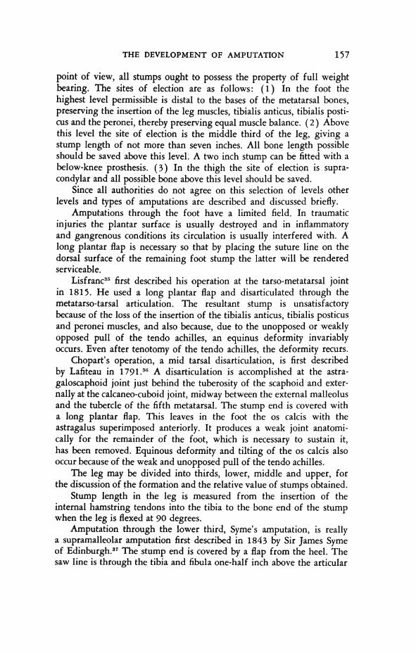
point of view, all stumps ought to possess the property of full weightbearing. The sites of election are as follows: (1) In the foot thehighest level permissible is distal to the bases of the metatarsal bones,preserving the insertion of the leg muscles, tibialis anticus, tibialis posti-cus and the peronei, thereby preserving equal muscle balance. (2) Abovethis level the site of election is the middle third of the leg, giving astump length of not more than seven inches. All bone length possibleshould be saved above this level; A two inch stump can be fitted with abelow-knee prosthesis. (3) In the thigh the site of election is supra-condylar and all possible bone above this level should be saved.
Since all authorities do not agree on this selection of levels otherlevels and types of amputations are described and discussed briefly.
Amputations through the foot have a limited field. In traumaticinjuries the plantar surface is usually destroyed and in inflammatoryand gangrenous conditions its circulation is usually interfered with. Along plantar flap is necessary so that by placing the suture line on thedorsal surface of the remaining foot stump the latter will be renderedserviceable.
Lisfranc35 first described his operation at the tarso-metatarsal jointin 1815. He used a long plantar flap and disarticulated through themetatarso-tarsal articulation. The resultant stump is unsatisfactorybecause of the loss of the insertion of the tibialis anticus, tibialis posticusand peronei muscles, and also because, due to the unopposed or weaklyopposed pull of the tendo achilles, an equinus deformity invariablyoccurs. Even after tenotomy of the tendo achilles, the deformity recurs.
Chopart's operation, a mid tarsal disarticulation, is first describedby Lafiteau in 1791.36 A disarticulation is accomplished at the astra-galoscaphoid joint just behind the tuberosity of the scaphoid and exter-nally at the calcaneo-cuboid joint, midway between the external malleolusand the tubercle of the fifth metatarsal. The stump end is covered witha long plantar flap. This leaves in the foot the os calcis with theastragalus superimposed anteriorly. It produces a weak joint anatomi-cally for the remainder of the foot, which is necessary to sustain it,has been removed. Equinous deformity and tilting of the os calcis alsooccur because of the weak and unopposed pull of the tendo achilles.
The leg may be divided into thirds, lower, middle and upper, forthe discussion of the formation and the relative value of stumps obtained.
Stump length in the leg is measured from the insertion of theinternal hamstring tendons into the tibia to the bone end of the stumpwhen the leg is flexed at 90 degrees.
Amputation through the lower third, Syme's amputation, is reallya supramalleolar amputation first described in 1843 by Sir James Symeof Edinburgh.37 The stump end is covered by a flap from the heel. Thesaw line is through the tibia and fibula one-half inch above the articular
157

NORMAN T. KIRK
surface of the tibia. The os calcis is dissected from the heel and thisflap is brought up over the bone ends. This amputation has remainedpopular in England and in Canada.
Wilson, of Boston, in his excellent description of the operation,38states its chief merits as follows: (1) It provides a stump capable oftransmitting an entire body weight to the ground by direct end pressurewithout pain. (2) It removes only the foot and therefore leaves thestump with greatest leverage power. (3) It is less crippling, in thatless tissue is removed than in an amputation through the leg. Thepatient, therefore, is able to walk about his room without an appliance.Wilson states further that the skin on the plantar surface of the heelmust be healthy and non-damaged, and that wound infection practicallyalways causes failure.
Huggins, who also advocates this amputation, states in his excellentmanual on amputations39 that a Syme's stump which is not entirelyend bearing is inferior for functional purposes to an amputation throughthe leg. He enumerates some four or five reasons for failure, and statesthat unsatisfactory Syme's stumps are not uncommon. Too frequentlythese stumps are incapable of direct end bearing without pain, whichmay be the fault of the operator. The stump end is bulbous and moreor less insecure, because of the sub-plantar fat in the heel flap coveringit, making its prosthesis very apparent and unsightly. It can be fittedonly with difficulty by the average prosthetist. The leg is encased in aleather laced bucket extending to the knee, reinforced with metal,either in front or behind or on the two lateral surfaces. This appliance,however, does not necessitate the objectionable thigh corset and shoulderstrap required by an amputation through the leg. Care must be exercisedin dissecting the heel flap or its blood supply may be interfered with,causing necrosis and failure. This stump when wholly end bearingpossesses certain advantages for the laborer.
Pirogoff, in 1853, modified Syme's amputation, making an osteo-plastic stump.40 His idea in doing this was not primarily to do anosteoplastic amputation but to overcome the difficulty he encounteredin dissecting the heel flap according to Syme's method. He broughtup a heel flap containing the posterior surface of the os calcis andunited it to the sawed section of the tibia and fibula just above thearticular cartilage of the tibia. This stump has the disadvantages of theosteoplastic amputation, as stated above. In addition, it is too long tobe properly fitted with a prosthesis allowing ankle motion. The Syme'sstump is superior.
Amputation through the lower third of the leg gives unsatisfactorystumps in comparison with those through the middle third. The lowerthird of the tibia has no muscular insertion and is covered mainly bytendons. The circulation of the skin is poor, the stump tends to become
158
THE DEVELOPMENT OF AMPUTATION
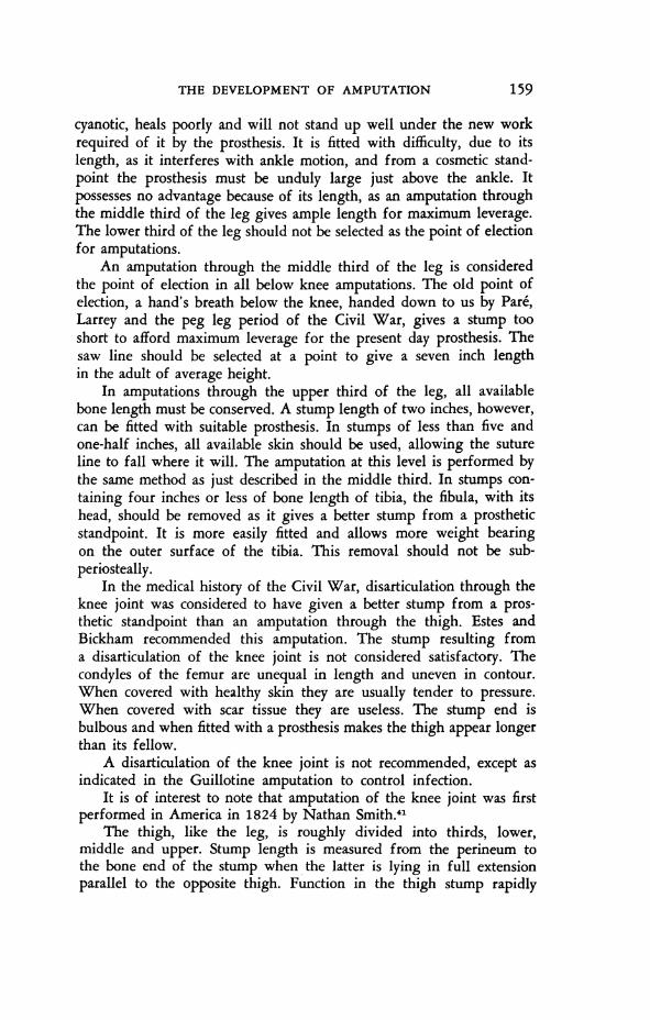
cyanotic, heals poorly and will not stand up well under the new workrequired of it by the prosthesis. It is fitted with difficulty, due to itslength, as it interferes with ankle motion, and from a cosmetic stand-point the prosthesis must be unduly large just above the ankle. Itpossesses no advantage because of its length, as an amputation throughthe middle third of the leg gives ample length for maximum leverage.The lower third of the leg should not be selected as the point of electionfor amputations.
An amputation through the middle third of the leg is consideredthe point of election in all below knee amputations. The old point ofelection, a hand's breath below the knee, handed down to us by Pare,Larrey and the peg leg period of the Civil War, gives a stump tooshort to afford maximum leverage for the present day prosthesis. Thesaw line should be selected at a point to give a seven inch lengthin the adult of average height.
In amputations through the upper third of the leg, all availablebone length must be conserved. A stump length of two inches, however,can be fitted with suitable prosthesis. In stumps of less than five andone-half inches, all available skin should be used, allowing the sutureline to fall where it will. The amputation at this level is performed bythe same method as just described in the middle third. In stumps con-taining four inches or less of bone length of tibia, the fibula, with itshead, should be removed as it gives a better stump from a prostheticstandpoint. It is more easily fitted and allows more weight bearingon the outer surface of the tibia. This removal should not be sub-periosteally.
In the medical history of the Civil War, disarticulation through theknee joint was considered to have given a better stump from a pros-thetic standpoint than an amputation through the thigh. Estes andBickham recommended this amputation. The stump resulting froma disarticulation of the knee joint is not considered satisfactory. Thecondyles of the femur are unequal in length and uneven in contour.When covered with healthy skin they are usually tender to pressure.When covered with scar tissue they are useless. The stump end isbulbous and when fitted with a prosthesis makes the thigh appear longerthan its fellow.
A disarticulation of the knee joint is not recommended, except asindicated in the Guillotine amputation to control infection.
It is of interest to note that amputation of the knee joint was firstperformed in America in 1824 by Nathan Smith.41
The thigh, like the leg, is roughly divided into thirds, lower,middle and upper. Stump length is measured from the perineum tothe bone end of the stump when the latter is lying in full extensionparallel to the opposite thigh. Function in the thigh stump rapidly
159

NORMAN T. KIRK
diminishes above the lower third. The site of election is in the lowerthird of the thigh through the cancellous bone just above the condylesof the femur. Owing to the strong contractility of the massive musclesof the thigh, conical stumps are more prone to develop in amputationsthrough it than in the upper extremity of the leg.
All stumps in the lower third of the thigh should be wholly weightbearing and should be so fitted as to give the maximum functionalvalue.
Gritti,42 in 1857, described his supracondylar osteoplastic amputa-tion, which was later modified by Stokes,43 in 1870. Stokes used thesame method except for slight modifications of the flaps. This stumphas the objections of all osteoplastic amputations. When successful itgives an ideal end bearing thigh stump.
Sabanaieff44 described a transcondyloid femoro-tibial method inwhich the saw line was transcondylar. To this sawed surface he applieda bone flap with adherent soft parts which he cut from the anteriorsurface of the tibia. This stump contained the whole knee joint andthe patella. To form the anterior flap, skin of three fingers' breadthbelow the tubercle of the tibia is required. This operation is difficultto perform, infection and reaction in the retained synovial membraneof the knee joint is always possible, the stump end is bulbous and whenfitted with a prosthesis, causes the knee to fall below its fellow. It isnot considered an operation of election.
In 1902 Wilms,45 after sawing through the end of the femur,covered it with the quadriceps extensor tendon, which he sutured tothe periosteum on the posterior surface of the femur.
The supracondylar tendoplastic or aperiosteal method of amputationis through the supracondylar portion of the femur, just below the shaft,and through the cancellous bone of the condyles. The quadriceps tendon,which is included with the long anterior skin flap, is cut close to itsinsertion into the patella, brought over the end of the sectioned femurand sutured to the fascia posteriorly.
The hip joint should present a buttock equal in size and contour toits fellow, without redundant or excessive muscle below the level ofthe tuberosity of the ischium. The tuberosity of the ischium coveredwith its normal soft parts, forms the base and weight bearing pointof the hip stump. Excessive muscle produces a flabby, insecure seatwhich becomes pinched by the prosthesis. The skin should be roundedout and free from sulci or redundancy. The suture line should be as faras possible from the rectum to prevent post-operative soiling.
This amputation, as already stated, was first successfully performedby Larrey in 1803. In America it was done by Brashear of Marylandin 1806.46
Shock in this operation is usually severe, regardless of the amount
160

THE DEVELOPMENT OF AMPUTATION
of blood lost. It can be lessened by blocking the sciatic and anteriorcanal nerves. Intravenous salt solution and plasma and often bloodtransfusion are used to combat shock. It will, of course, be increased inthose cases where trauma has necessitated the amputation because of theshock incident to the injury itself.
Larrey practiced primary ligation of the common femoral. He thencut the flaps and disarticulated by transfixion. Guthrie controlled thecirculation by digital pressure of the femoral under Poupart's ligament,and cut the flap from without inward.
Wyeth47 in 1890 introduced his method of control of blood supply,using his famous pins which held a rubber tube tourniquet in place.He did a circular amputation of the soft parts and bone well below theperineum and later disarticulated the joint and removed the remainingbone through an external incision, leaving large flabby muscle flapswhich were quilted together. His method is more or less followed inthe external ratchet incision, in which the trochanter and neck of thefemur are removed subperiosteally and a large stump produced. Thisprocedure will not give the ideal stump.
The anterior ratchet method with the long postero-internal skinfascia flap, with primary ligation of the common femoral, as describedby Huggins, is considered the ideal.
The femur may be disarticulated, sawed through the neck or ampu-tated below the great trochanter, as elected. Either method gives anequally satisfactory stump, requiring only slight modification of theskin flaps.
A discussion of amputation of the upper extremity is omitted exceptfor mention of Vanghetti's work in 1897-98 in cineplastic amputations.48It was as a result of his observations in the Italian-Abyssinian War thatVanghetti's theory was first conceived. In his experiments on chickenshe found that if the distal end of a muscle or tendon were freed fromits insertion and covered with skin, the muscle would retain its voluntarypower of contraction. His operation was first performed on man in1900 by Ceci of Pisa, a surgeon and friend of Vanghetti. This workwas followed by Sauerbruch in Germany and by DeFrancesso and Gal-leazzi in Italy. Some five different types of "motors" have been developedby this method to activate the prosthesis of the upper extremity.49
Baer and Wilson,50 in compliance with orders from the ChiefSurgeon A. E. F. in France, made a complete study of the subject andvisited several hospitals in Italy where this work was being done. Intheir conclusion they stated that cineplastic amputation was then inthe experimental stage and that the results so far obtained were oflittle practical value in improving the working efficiency of the ampte'.
In the present paper, a vast amount of pertinent detail has beenomitted. The writer has been concerned, mainly, in sketching the gradual
161

NORMAN T. KIRK
evolution of the art of amputation from earliest times to a period endingsome years after the last war when our experience was perhaps at itsmaximum. There has been no attempt to cover the subject as it isrelated to the present war nor to examine into the merits of chemo-therapy, the use of dried blood cells in non-healing stumps, refrigerationanesthesia or other recent procedures.
In closing it should be mentioned that a definite procedure in ampu-tations has been outlined in Circular Letter No. 91 of April 26, 1943from the Office of The Surgeon General, to serve as a guide on thesubject of amputation to the end that at the earliest possible moment,a sound and definite procedure would be followed, eliminating unap-proved practices on the part of medical officers who are not fullyacquainted with the cumulative experience of the past twenty-five years.
REFERENCES
1. HOLLANDER, EUGEN. Die chirurgische Sage. Archiv fiur klinische Chirurgie 106:316-339, 1915.
2. MOODIE, RoY L. Paleopathology; An Introduction to the Study of Ancient Evidencesof Disease. Urbana, University of Illinois Press, 1923. Facing p. 532.
3. Ch. 69. Loeb Classical Library ed., vol. III, pp. 361 ff.4. De medicina, bk. VII, ch. 33; cf. bk. V, ch. 26 (Loeb Classical Library ed., vol.
II, p. 81), and bk. VII, ch. 22 at end.5. In: Oribasius. Collectiones medicae. Bk. XLVII, ch. 13.6. Ibid., ch. 15.7. Methodus medendi, bk. 13 near end (Kuhn ed., vol. X, p. 942).8. Bk. VI, ch. 84.9. Albucasis de Chirurgia, ed. J. Channing, Oxford, 1778, pp. 419 if. (Bk. II, ch. 87.)
10. THEODORIC. Chirurgia. Bk. IV, ch. 8. (Collectio chirurgica Veneta, 1498, fol. 146recto.)
11. La Grande Chirurgie, ed. E. Nicaise, Paris, 1890, p. 436.12. Chirurgia, Tract. III, Doct. I, ch. 7.13. Cf. n. 11.14. Practica in chirurgia, Rome, 1514, bk. IV, ch. 7.15. Das Buch der Chirutrgia, Strasburg, 1497, Tract. III, ch. 18.16. Tract. IV, ch. 6 (ed. 1508).17. Feldtbuch der Wundtartzney, Strasburg, 1517, fol. lxviii2 verso.18. Tract. V, ch. 4 in later editions.19. Works, tr. T. Johnson, London, 1634, p. 1143.20. Ibid., p. 463.21. Ibid., pp. 462, 1135.22. Ibid., p. 1148.23. Ibid., p. 458.24. A Discourse of the Whole Art of Chyrurgery. London, 1596 and many later editions.25. HEY, WILLIAM. Practical Observations in Surgery . ed. 3. London, Cadell and
Davies, 1814. Pp. 527 f.26. LARREY, DoMINIQUE; JEAN. AMemoires de chirurgie militaire. Paris, J. Smith. Vol.
II, 1812, pp. 180-195.27. GUTHRIE, G. J. A Treatise on Gun-shot Wounds . ed. 2. London, Burgess and
Hill, 1820. Pp. 374-378.28. Ibid., p. 380.29. Ibid., p. 381.30. Practical Surgery . London, John Churchill, 1837. Pp. 317 f.31. SYME, JAMES. Treatise on the Excision of Diseased Joints. Edinburgh, Adam Black,
1831.
162

THE DEVELOPMENT OF AMPUTATION 163
32. Medical and surgical history of the War of the Rebellion. Part 3, vol. 2. Surgicalhistory. Washington, 1883.
33. ESTES, WM. L. "Amputations in industrial surgery." Annals of Surgery 81:164-190(Jan.) 1925.
34. KIRK, NORMAN THOMAS, Amputations. Hagerstown, Md., W. F. Prior Co., 1943."Amputations in War." J.A.M.A. 120:13-16 (Sept. 5) 1942."The guillotine amputation." I.A.M.A. 124:1027-1030 (April 8) 1944
(with F. M. McKeever).35. LISFRANC, JACQUES. Nouvelle tnmthode operatoire pour l'amputation partielle du
pied dans son articulation tarso-metatarsienne. Paris, Gabon, 1815.36. In: FOURCROY, A. F. La medecine eclairee par les sciences physiques. . . . Vol. 4,
Paris, Buisson, 1792, pp. 85-88.37. SYME, JAMES. "Amputation at the ankle-joint." Lond. and Edinb. Month. J. Med.
Sci. 3:93-96, 1843.38. WILSON, PHILIP D. "The Syme operation." Surgical Clinics of North America
1:711-728, 1921.39. HUGGINS, G. Amputation Stumps; Their Care and Treatment. London, 1918.40. PIROGOFF, NIKOLAI IVANOVICH. "Kostno-plasticheskoye udlineniye kostei goleni
pri vilushtshenii stopi." Voyenno-med. J. (St. Petersburg) 632:83-100, 1854.41. SMITH, NATHAN. "On amputation at the knee-joint." Amer. Med. Rev. and J.
2:370-371, 1825.42. GRIrrI, Rocco. "Dell' amputazione del femore al terzo inferiore e della dis-
articolazione del ginocchio." Ann. Univ. di Med. 161:5-32 (Milano) 1857.43. STOKES, SIR WILLIAM. "On supra-condyloid amputation of the thigh." Med.-Chir.
Trans. 53:175-186 (London) 1870.44. SABANIEFF, I. F. "Amputatio femoris inter-condyloidea osteoplastica." Chir. Vestnik
6:14-26 (St. Petersburg) 1890.45. WILMS, MAX. "Tragfahiger Amputationsstumpf; Bedeckung mit der Achillessehne."
Centralbl. f. Chir. 29:721 (Leipzig) 1902.46. BRASHEAR, WALTER. ["First case of amputation at the hip-joint in the United
States.]" Trans. Kentucky Med. Soc. 2:265-266, 1852 (pub. 1853).47. WYETH, JOHN A. "Bloodless amputation at the hip joint." N. Y. Med. J. 51:528,
1890.48. VANGHETTI, G. Plastica e protesi cinematiche; nuova teoria sulle amputazioni.
Empoli, 1906.49. Bibliography in Index-Catalogue, 4th ser., vol. I, pp. 327-329.50. "A report to the Chief Surgeon, A.E.F., by Major Williams S. Baer, M.R.C., and
Capt. Philip D. Wilson, M.R.C. Subject: Cinematic amputation in Italian hospi-tals." War Medicine (pub. by the American Red Cross) 2:218 (Paris) 1918.
ADDITIONAL REFERENCES
The best accounts of the earlier history of amputation are still the following:WERNHER, ADOLPH. "Geschichte der Gliederablosungen, I: Von den altesten Zeiten
bis zur Griindung der Academie royale de Chirurgie." Deutsches Archiv f. Gesch. d.Med. 1: 139-182, 1878.
GURLT, E. Geschichte der Chirurgie. 3 vols. Berlin, August Hirschwald, 1898. Vol. 3,pp. 793-805 and the previous passages there summarized.