the development of a wide range crispr-cas9 gene editing
TRANSCRIPT
The development of a wide range CRISPR-Cas9 gene
editing system
by
Eduvan Bisschoff
Submitted in fulfilment of the requirements in respect of the degree
Magister Scientiae
in the
Department of Microbial, Biochemical and Food Biotechnology; Faculty of Natural and
Agricultural Sciences
at the
University of the Free State
P.O. Box 339
Bloemfontein
9301
South Africa
Supervisor: Prof. J. Albertyn
Co-supervisors: Prof. C. H. Pohl-Albertyn
January 2019

Chapter 1
II
Acknowledgements
I would like to thank and acknowledge the following:
Prof. Jacobus Albertyn for all the guidance, support, patience, motivation and
hard work throughout this study; as well as for being a role model and
inspiration as a scientist.
Prof. Carlien Pohl for all the motivation, valuable input and everyday support.
Marianka de Jonge for all the immense support, patience, motivation and
inspiration.
Marnus Du Plooy, Ruan Fourie, JTR Brink, Bianca Pieterse and Johan Klinck
for valuable input as well as discussions.
The Pathogenic Yeast Research Group for all the support and help.
Friends and family for all the love and support throughout the last few years.
Financial assistance:
The financial assistance of the National Research Foundation (NRF) towards this
research is hereby acknowledged. Opinions expressed and conclusions arrived at, are
those of the author and are not necessarily to be attributed to the NRF.

III
Table of Contents
Declaration .............................................................................................................. VII
Dissertation Summary .......................................................................................... VIII
Chapter 1 ................................................................................................................... 1
Literature review....................................................................................................... 1
CRISPR-CAS9 strategies used for efficient gene targeting as well as gene
disruption in yeast. .................................................................................................. 1
1. Introduction ....................................................................................................... 1
2. Meganucleases .................................................................................................. 3
3. Zinc finger nucleases ....................................................................................... 4
4. Transcription activator-like effector nucleases .............................................. 4
5. CRISPR-Cas9 ..................................................................................................... 5
5.1. Mechanism ..................................................................................................... 6
5.2. History ............................................................................................................ 7
5.3. CRISPR –Cas9 strategies ............................................................................. 8
5.4. Double stranded break (DSB) repair .......................................................... 10
5.5. CRISPR interference (CRISPRi) .................................................................. 11
6. CRISPR-Cas9 strategies in various yeast ..................................................... 11
6.1. Saccharomyces cerevisiae ......................................................................... 12
6.1.1. Cas9 .......................................................................................................... 12
6.1.2. gRNA ......................................................................................................... 12
6.1.3. Multiplexing .............................................................................................. 14
6.1.4. CRISPRi .................................................................................................... 14
6.2. Kluyveromyces lactis .................................................................................. 15
6.3. Yarrowia lipolitica ........................................................................................ 16
6.4. Komagataella phaffii (formerly Pichia pastoris) ....................................... 17

IV
6.5. Wide range CRISPR-Cas9 for Kluyveromyces and Ogataea. .................. 18
7. Conclusion....................................................................................................... 18
8. Purpose of study ............................................................................................. 19
9. References ....................................................................................................... 20
Chapter 2 ................................................................................................................. 33
Expression of three different codon optimized Cas9 Genes using the wide
range pKM180 vector in seven different yeast species ...................................... 33
1. Abstract ........................................................................................................... 34
2. Introduction ..................................................................................................... 34
3. Materials and methods ................................................................................... 37
3.1. Rubidium chloride competent cells ........................................................... 37
3.2. Bacterial Transformation ............................................................................ 38
3.3. Restriction digestion ................................................................................... 38
3.4. Agarose gel electrophoresis ...................................................................... 39
3.5. Gel extraction ............................................................................................... 39
3.6. Ligation and cloning ................................................................................... 39
3.7. Plasmid extraction ....................................................................................... 40
3.8. Construction of the pKM180 containing the codon optimized CAS9
vectors .................................................................................................................... 41
3.9. Yeast transformation ................................................................................... 42
3.10. Plasmid integration confirmation ............................................................... 45
3.11. SDS PAGE analysis ..................................................................................... 46
3.12. Western blot analysis .................................................................................. 47
4. Results and discussion .................................................................................. 48
4.1. Construction of pKM180::CAS9 ................................................................. 48
4.2. Yeast Transformation .................................................................................. 54

V
4.3. Western blot analysis .................................................................................. 56
5. Conclusion....................................................................................................... 58
6. References ....................................................................................................... 59
Chapter 3 ................................................................................................................. 64
Development of the wide range CRISPR-Cas9 system by incorporating a RGR-
gRNA construct and validation through ADE2 disruption ................................. 64
1. Abstract ........................................................................................................... 65
2. Introduction ..................................................................................................... 65
3. Material and methods ..................................................................................... 68
3.1. In silico work ................................................................................................ 68
3.2. E. coli Rubidium chloride competent cells................................................ 68
3.3. Bacterial Transformation ............................................................................ 68
3.4. Restriction digestion ................................................................................... 68
3.5. Polymerase Chain Reaction ....................................................................... 68
3.6. Agarose gel electrophoresis ...................................................................... 73
3.7. DNA extractions ........................................................................................... 73
3.8. Construction of the CRISPR-Cas9 vector.................................................. 73
3.9. Construction of the donor DNA and validation of the CRISPR-Cas9
vector. ..................................................................................................................... 75
3.10. Ligation and cloning reactions ................................................................... 76
3.10.1. NEBuilder ligation and cloning (New England Biolabs 2017) .............. 76
3.10.2. T4 ligation and cloning (Devi et al. 2009) ............................................... 76
3.10.3. pMiniT 2.0 Ligation and cloning (New England BioLabs Inc. 2017) .... 77
3.11. Yeast transformation ................................................................................... 77
3.11.2. One-step transformation (Chen et al. 1992) . Error! Bookmark not defined.
3.11.3. Bicine transformation (Klebe et al. 1983) ..... Error! Bookmark not defined.

VI
4. Results and discussion .................................................................................. 78
4.1. Constructing the CRISPR-Cas9 vectors. ................................................... 78
4.2. Transformation ............................................................................................ 89
4.3. Saccharomyces cerevisiae ......................................................................... 92
4.4. Kluyveromyces lactis .................................................................................. 93
4.5. Yarrowia lipolytica ....................................................................................... 94
4.6. Debaryomyces hansenii ............................................................................. 95
4.7. Arxula adeninivorans .................................................................................. 96
4.8. Ogataea polymorpha ................................................................................... 99
4.9. Komagataella phaffii ................................................................................. 100
5. Conclusion..................................................................................................... 104
6. References ..................................................................................................... 106
Chapter 4 ............................................................................................................... 111
Summary and concluding remarks ..................................................................... 111
1. Summary and concluding remarks ............................................................. 111
2. References ..................................................................................................... 113

VII
Declaration
I, Eduvan Bisschoff, declare that the Master’s Degree research dissertation or
publishable, interrelated articles, or coursework Master’s Degree mini-dissertation that
I herewith submit for the Master’s Degree qualification at the University of the Free
State is my independent work, and that I have not previously submitted it for a
qualification at another institution of higher education.
I, Eduvan Bisschoff, hereby declare that I am aware that the copyright is vested in the
University of the Free State.
I, Eduvan Bisschoff, hereby declare that all royalties as regards intellectual property
that was developed during the course of and/or in connection with the study at the
University of the Free State, will accrue to the University.
I, Eduvan Bisschoff, hereby declare that I am aware that the research may only be
published with the dean’s approval.
Signature Date
05/ 02/ 2019

VIII
Dissertation Summary
CRISPR is a revolutionary method to effectively and efficiently alter the genomic
make-up of an organism. Unlike any other genetic engineering tool or technique,
CRISPR is remarkably cheaper, simpler and faster to perform. In biotechnology, the
best eukaryotic organism for research is yeast, due to their fast growth rate and ease
of manipulation compared to multicellular organisms. Hence the aim of the study was
the development of a wide range CRISPR-Cas9 system for a wide variety of different
yeasts for easy and fast gene editing. The system were validated in Saccharomyces
cerevisiae and six other non-conventional yeast. System construction began with the
incorporation (separately) of three different optimized CAS9 (optimized for expression
in Pichia pastoris, Candida albicans and Homo sapiens) genes into the wide range
pKM180 vector. The three different CAS9 construct were then tested for correct
expression of the Cas9 protein and the effects thereof in all the yeasts. Through
western blot analysis it was observed that all three of the different Cas9 proteins were
expressed successfully in the different yeasts. However, all of the Cas9 proteins had
a negative effect on the growth of the yeast. For the completion of the CRISPR-Cas9
system, a Ribozyme‐gRNA‐Ribozyme cassette was incorporated into the wide range
CAS9 vector, containing the C. albicans optimized CAS9. The system was then
validated with successful disruption of the ADE2 gene in all of the yeasts. This proved
that the wide range CRISPR-Cas9 system was applicable in a wide variety of different
yeasts, thus allowing for rapid, cost-effective genetic manipulation of
biotechnologically relevant yeast strains.
Keywords: Arxula adeninivorans ; Debaryomyces hansenii; Kluyveromyces lactis;
Ogataea polymorpha; Komagataella phaffii; Saccharomyces cerevisiae; Yarrowia
lipolytica; Pichia pastoris; Candida albicans optimized CAS9; CRISPR-Cas9; Wide
range CRISPR-Cas9 system.

Chapter 1
Literature review
CRISPR-CAS9 strategies used for efficient gene targeting as well
as gene disruption in yeast.
1. Introduction
The direct manipulation of an organism's genome by the usage of certain
biotechnology techniques is referred to as genetic manipulation. These techniques are
used to alter the genetic makeup of cells by the transference or deletion of genes
within or across species boundaries, which may result in improved or novel organisms
(Lamont & Lacey 2013). New DNA may be introduced into the hosts’ genome by
making use of molecular cloning methods, which entails isolating and copying
techniques to generate a DNA sequence (Berg & Mertz 2010). The DNA can also be
synthesized for the purpose of inserting the construct into the host (Kaufman & Nixon
1996). The DNA can be incorporated either indirectly with the use of a vector system
or directly through micro-injection and micro-encapsulation techniques (Lacal et al.
1999; Chen & Dubnau 2004; Singh et al. 2010; Zaretsky et al. 2011). Genes may be
altered by either gene knockout (removal) or by gene knockdown (reduced
expression) using a nuclease and may also be changed through the use of gene
targeting (Westphal & Leder 1997; Summerton 2007). This technique (gene trageting)
is different in that it uses homologous recombination to alter a gene, and can thus be
used in the removal of a gene or exons, addition of a gene, or to introduce point
mutations. An example is the Cre-Lox recombination. This system consists of a single
enzyme, the Cre recombinase, which recombines a pair of short target sequences
called the Lox sequences. Placing Lox sequences appropriately, allow genes to be
activated, repressed, or exchanged for other genes (Sauer 1987; Egener et al. 2002).
Genetic engineering techniques have been applied in numerous fields including
research, agriculture, industrial biotechnology and medicine. Genetic engineering can
be used to produce desirable traits in organisms, which can be used in many different
application areas to benefit humanity. For instance genetic engineering has been used
in medical applications to mass-produce insulin, human growth hormones, follistim,
Chapter 1
2
human albumin, monoclonal antibodies, antihemophilic factors, vaccines and many
other drugs (Walsh 2006; Waegeman et al. 2013; Berlec & Štrukelj 2013). Gene
therapy is also an application where in (only one) defective genes in humans can be
replaced with effective ones (Knudson 1967; Krauss 1992).
Genetic engineering techniques can be used to manipulate most plants, animals and
microorganisms. Bacteria, the first organisms to be genetically modified in 1973, can
have plasmid DNA inserted containing new genes that code for pharmaceuticals (such
as insulin and human growth hormone) or enzymes such as in laundry detergent,
enzymes that process food and other substrates (Jones et al. 1968; Cohen & Chang
1973; Williams et al. 1982; Gray et al. 1985). Plants have been modified forinsect
protection, herbicide resistance, virus resistance, enhanced nutrition, tolerance to
environmental pressures and the production of edible vaccines. In addition modified
crops have been commercialized (Dookun 2001; Li et al. 2001; ISAAA 2017).
Experimental genetically modified (GM) cell lines and GM animals, such as mice or
zebrafish, have been used for research, model animals and the production of
agricultural or pharmaceutical products. The genetically modified animals include
animals with genes deleted, increased susceptibility to disease, hormones for extra
growth and the ability to express proteins in their milk (Dodd et al. 2000; Olswang et
al. 2002; Yoder et al. 2002; Hanson & Hakimi 2008).
In biotechnology, one of the best eukaryotic model organism for research is yeast due
to their fast growth rate and ease of manipulation compared to multicellular organisms
(Madzak et al. 2004; Gerngross 2004; Nevoigt 2008; Stovicek et al. 2015; Skrzypek
et al. 2017). Saccharomyces cerevisiae is characterized as the most well-known
eukaryotic system due to the amount of data available regarding the yeasts’ genetics,
physiology and molecular biology (Nevoigt 2008). Due to this, S. cerevisiae is widely
used as a model organism for research. The fast growth rate, ease of use,
maintenance and manipulation of this yeast makes it ideal for the development of new
genetically modified strains for research and industrial purposes. However, non‐
conventional yeasts, i.e. non S. cerevisiae species, have also attracted much attention
and have been widely used as cell factories to produce a number of different
recombinant proteins and biomolecules for various research and industrial purposes
(Van der Walt et al. 1990; Hsieh & Da Silva 1998; van Ooyen et al. 2006; Gasser et
al. 2013). However, compared to S. cerevisiae, engineering many of these yeast
Chapter 1
3
species are challenging, because of the limited data available on the genetics,
physiology and molecular biology and/or less defined molecular tools. Thus, improved
knowledge on the biology and engineering tool development for many of the non-
conventional yeasts could greatly improve industrial and research applications. This
review will briefly present the key developments in genome engineering and major
genetic engineering tools, which include Meganucleasese, zink finger nucleases
(ZFN), transcription activator-like effector nucleases (TALEN). However, most of this
review will focus on the CRISPR technology and its development for use in S.
cerevisiae and non-conventional yeast.
2. Meganucleases
Meganucleases are found in a large number of organisms and are
endodeoxyribonucleases characterized by a large recognition site (double-stranded
DNA sequences of 12 to 40 base pairs) (Chevalier 2001; Chevalier et al. 2002). Due
to the exceptional long length of the recognition sequence, this region generally occurs
only once in any given genome (Chevalier 2001). This property therefore characterizes
meganucleases to be the most specific, natural occuring restriction enzyme.
Meganucleases also known as homing endonucleases currently have six known
structural families, which are classified on their structure (LAGLIDADG; GIY-YIG; His-
Cys box; H-N-H; PD-(D/E)xK and Vsr-like) (Stoddard 2011). Among the homing
endonucleases, the LAGLIDADG family has become a valuable tool for genome
engineering and - studying (Chevalier et al. 2005). By using protein engineering,
through mutagenesis, to change the recognition sequence, any target in any genome
can be cleaved by the enzyme (Seligman 2002). This however can pose a drawback,
meaning protein-engineering needs to be performed to allow recognition of the
nuclease for a specific nucleotide sequence and therefore to obtain the desired target
site. In addition, protein engineering can be difficult and time consuming (Seligman
2002; Sussman et al. 2004; Rosen et al. 2006). The best characterized
meganucleases, which are mostly used for genome engineering, include I-SceI, I-CreI
and I-DmoI (Chevalier et al. 2005).
Chapter 1
4
3. Zinc finger nucleases
Zinc fingers are defined as zinc ion-regulated small protein motifs, which bind in a
sequence-specific manner to DNA (Laity et al. 2001). Each one of the zinc fingers
recognize a 3 bp DNA sequence (Klug & Rhodes 1987). Therefore, unlike
meganucleases, multiple zinc fingers (about 6-8) could be combined to achieve a
larger complex which contributes to a more specific DNA binding capability.
Zinc-finger nucleases are synthetic restriction endonucleases generated by combining
a zinc finger domain (DNA-binding domain) to a restriction endonuclease domain
(DNA-cleavage domain). The zinc finger DNA-binding domains commonly consist of
three to six separate zinc finger repeats, which can recognize between 9 and 18 bp
each (Gupta & Musunuru 2014). The non-specific cleavage domain from FokI (type IIs
restriction endonuclease) is usually used for the ZFNs’ cleavage domain (Kim et al.
1996). Dimerization of the cleavage domain must occur to ensure the cleavage of the
targeted DNA and thus a pair of ZFNs are needed for the targeted sites (Bitinaite et
al. 1998). ZFNs fuse the cleavage domain to the C-terminus of each zinc finger
domain. In order to allow for the dimerization of the two cleavage domains to perform
DNA cleavage, the two individual ZFNs must bind to the complementary DNA strands.
Zinc finger (DNA binding) domains can be modified to allow targeting of a specific
desired DNA sequence (Gupta & Musunuru 2014). Thus, enabling the modified ZFNs
to recognize and cleave any target sequence within a desired genome. This however
is time consuming due to the protein engineering that needs to be performed to alter
the binding site.
4. Transcription activator-like effector nucleases
The TALEs (Transcription Activator-Like Effector) are proteins, which are secreted by
the bacteria Xanthomonas to help them infect plants (Boch & Bonas 2010). The
bacteria inject the TALEs directly into plant cells by using a type III secretion system,
followed by the specific binding of the TALEs to the plant genes, which regulates it to
facilitate the bacterial colonization. Transcription activator-like effector nucleases
(TALEN) are restriction endonucleases that can be modified to perform cleavage on a
specific target DNA sequence (Boch 2011). They are generated by combining a
transcription activator-like effector (TALE) DNA-binding domain to a DNA cleavage

Chapter 1
5
domain (FokI) (Boch & Bonas 2010). The TALE DNA binding domain commonly
consists of 33 to 34 highly conserved repeated amino acid sequence (Boch et al. 2009;
Moscou & Bogdanove 2009). TALEs can be modified, at the 12th and 13th amino acid
to bind to nearly any target DNA sequence. Thus when combined with the restriction
endonuclease domain, the DNA can be cleaved at a specific location (Boch 2011).
TALEN also has the same drawbacks as ZFN and meganucleases, which includes
protein engineering.
5. CRISPR-Cas9
Although the discovery of the three genetic engineering tools, meganucleases
followed by ZFNs and TALENs, continuously increased the efficacy of genome editing,
the targeting of different sites in the host genome required redesigning and rebuilding
of new sets of proteins (Seligman 2002; Boch 2011; Gupta & Musunuru 2014). This
drawback partially prevented ZFNs and TALENs to be broadly accepted by the
scientific community (Sakuma & Woltjen 2014). In this respect, CRISPR has reformed
the genetic engineering field due to its phenomenal editing efficiency compared to the
existing tools (Figure 1) (Adli 2018). More importantly, CRISPR is also much simpler,
more flexible, faster, and cheaper to use. The CRISPR gene-editing tool consists of a
programmable endonuclease whose DNA-targeting specificity and cutting activity
depends on a short guide RNA.
Figure1: Feasibility of the major genetic engineering tools (Adli 2018).
CRISPR (Clustered Regularly Interspaced Short Palindromic Repeats) is a sequence
in the bacterial genome which contains parts of foreign genetic material, such as those
found within plasmids and bacteriophages, which have invaded a bacterium (Terns &
Terns 2011; Bhaya et al. 2011; Wiedenheft et al. 2012). These foreign DNA parts
(spacer) are then used by the host bacterium to target DNA from similar viruses during

Chapter 1
6
following attacks by using Cas (CRISPR Associated Systems) proteins. This system
is known as CRISPR-Cas and it plays a key role in the bacterial defence system. The
application of the recently discovered CRISPR-Cas9 system is an innovative, genetic
engineering tool that allows scientists to change, at will, any DNA sequence of any
living organism in a specific manner (Jinek et al. 2012; Barrangou 2015). The Cas9
protein can be modified with a single guide RNA (sgRNA) to generate site-specific
DNA breaks.
5.1. Mechanism
The CRISPR-Cas9 system helps to protect bacteria in three steps, namely adaptation,
production of gRNA/Cas complex and finally targeting (Figure 2) (Terns & Terns 2011;
Bhaya et al. 2011; Wiedenheft et al. 2012). Adaptation or spacer acquisition is the
process where the Cas enzymes (Cas1 and Cas2) recognize the invading DNA and
cleave it to produce a protospacer (Figure 2A). This protospacer is then ligated and
extended into the CRISPR array adjacent to the leader sequence, to the direct repeat
thus creating a new CRISPR part to serve as a tool for targeting similar foreign DNA
in later infections.
The production of the gRNA/Cas complex step starts with the transcription of the
CRISPR array to yield the pre-crRNA as indicated by Figure 2B (CRISPR RNA)
(Marraffini & Sontheimer 2010; Dugar et al. 2013). This is followed by the binding of
various trans-activating (tracr) RNAs to the pre-crRNA, resulting in double stranded
RNAs. The Cas9 protein then associates with the tracrRNA-pre-crRNA strand, which
is then recognised and cleaved, to generate small crRNAs, by RNaseIII. The crRNAs
then undergo secondary trimming, at either the 5-prime or 3-prime ends to produce
mature crRNAs (Marraffini & Sontheimer 2010; Dugar et al. 2013; Karvelis et al. 2013).
Both the mature crRNAs and tracrRNA must associate with the Cas9 protein to form
an active interference complex (Jinek et al. 2012). The crRNA-Cas9 complex then
finds the invading DNA (Figure 2C), binds to it, recognises the PAM (Protospacer
adjacent motif) sequence downstream of the protospacer, and cleave the invading
DNA (Garneau et al. 2010; Jinek et al. 2012). The PAM site is an essential targeting
component, which is not found in the bacterial genome (Mali et al. 2013a). This
enables the bacteria to distinguish between his own DNA from foreign DNA, hence
avoiding the possibility of self-targeting and degradation of the CRISPR locus by Cas

Chapter 1
7
enzymes. Researchers saw the mechanism and modified it to serve as a genetic
engineering tool (Jinek et al. 2012). However this prosess took approximately 30 years
from the discovery of its first components (Ishino et al. 1987).
Figure 2: CRISPR-Cas9 adaptive immunity (Samson et al. 2013). The three crucial
steps are (A) adaptation, (B) production of gRNA/Cas complex and then (C) targeting
5.2. History
Notably, CRISPR had been simply known as prokaryotic DNA repeat elements, from
the genome of Escherichia coli, since it was first discovered in the late 1980s (Ishino
et al. 1987). Later it was detected that CRISPRs are present in numerous bacteria and
archaea (Mojica et al. 2000). Shortly after, the association of CAS genes with CRISPR
were identified (Jansen et al. 2002). In 2005 an observation was made, which
describes that many spacer sequences within CRISPR are derived from viral and/or
plasmid origins (Mojica et al. 2005; Pourcel et al. 2005). These findings later led to the
discovery that recognised CRISPR-Cas as a bacterial immune system (Barrangou et
al. 2007). Later, in 2008, it was revealed that the Cas proteins are guided by small
mature CRISPR RNAs (crRNAs), transcribed from spacer sequences, towards the
A
B
C
Chapter 1
8
invading DNA to interfere with proliferation in E. coli (Brouns et al. 2008; Marraffini &
Sontheimer 2008).
Around 2010, Garneau and colleagues discovered that Cas9 is the only enzyme within
the gene cluster that can cleave DNA (Garneau et al. 2010). Next a key component in
the biogenesis and processing of crRNA in a CRISPR system was revealed,
describing a noncoding trans-activating crRNA (tracrRNA) hybridizing with crRNA to
facilitate RNA-guided targeting of Cas9 (Deltcheva et al. 2011). Therefore, the Cas9
and RNase III enzymes are crucial for the processing of mature crRNA. These two
above mentioned findings suggested that the Cas9, together with the mature crRNA
and trancrRNA are crucial elements of the CRISPR-Cas9 system (Garneau et al.
2010; Deltcheva et al. 2011). The idea subsequently arose that this system, if
engineered correctly, may be utilised for genome editing. In 2011, Sapranauskas and
co-workers proved that the type II CRISPR system from Streptococcus thermophiles
can be transferred to Escherichia coli and still perform its function (Sapranauskas et
al. 2011). In 2012 the groups of Charpentier, Doudna, and Siksnys showed that the
CAS9 can be purified from Streptococcus thermophilus or Streptococcus pyogenes
and can be guided by a site-specific crRNA fused to a tracrRNA to cleave a target
DNA in vitro (Jinek et al. 2012; Gasiunas et al. 2012). In 2013 CRISPR was used to
accomplish genome editing in human and mouse cells (Cong et al. 2013; Mali et al.
2013b). Since these initial studies, the CRISPR-Cas9 tool has been widely used for
genome engineering in various model systems.
5.3. CRISPR –Cas9 strategies
The Cas9 enzyme consist of two nuclease domains, namely RuvC and HNH, which is
based on homology of known nuclease structures (Haft et al. 2005; Makarova et al.
2006). The RuvC domain cleaves the non-target DNA strand, where the PAM site are
located, and the HNH domain cleaves the target strand of DNA, strand to which the
gRNA binds (Barrangou et al. 2007; Gasiunas et al. 2012).
Normally the CAS9 and the gRNA are expressed in different expression cassettes.
The DNA sequence of CAS9 gene has been optimized by various authors to test for
optimal expression and function. The CAS9 gene have showed some success when
it was either native (from Streptococcus pyogenes), Homo sapiens codon-optimized
or yeast codon-optimized over a variety of different yeast, respectively (DiCarlo et al.

Chapter 1
9
2013; Gao & Zhao 2014; Ryan et al. 2014; Zhang et al. 2014; Bao et al. 2015; Horwitz
et al. 2015; Generoso et al. 2016). The CAS9 expression cassette need to consist of
a compatible promoter and terminator, to drive and terminate expression respectively,
and a NLS (nuclear localisation signal), which can be linked to the 3-prime or both 5-
and 3-prime side of the CAS9, to transport the Cas9 enzyme to the nucleus.
The gRNA is a short synthetic RNA that consist of scaffold part, which is necessary
for the association with the Cas9 enzyme, and a user-specified targeting sequence
(approximately 20 bp of length) which allows targeting of the desired DNA region in
the host genome to be modified (Jinek et al. 2012). Therefore, by just changing the
approximately 20 bp target sequence in the gRNA, one can simply change the
genomic target. The gRNA expression cassette is usually driven by a non-mRNA
polymerase promoter, for example, RNA polymerase III (Pol III) promoters (U3 and
U6) (DiCarlo et al. 2013; Zhang et al. 2014; Jakočiūnas et al. 2015; Mans et al. 2015;
Bao et al. 2015). The promoter is followed by a specific target sequence, the gRNA
scaffold and lastly the terminator. However the Pol III promoters have limitations when
it comes to the expression of gRNA in different hosts (Gao & Zhao 2014). They are
very specific for each organism and not all these promoters have been characterized
in many organisms, thus making it difficult to choose the correct Pol III promoters for
CRISPR. Furthermore, the Pol III promoters limit the CRISPR target sequences to G
(N20) GG and A (N20) GG. Therefore, another strategy was developed and used
successfully for the correct expression of the gRNA. Gao and Zhao developed a
strategy where they took advantage of the nuclease activity of ribozymes (Gao & Zhao
2014). They designed an artificial gene, RGR (Ribozyme‐gRNA‐Ribozyme), where the
RGR gene undergoes self‐catalysed cleavage to yield the gRNA without any
modifications. The RGR gene was expressed under an alcohol dehydrogenase 1
(ADH1) promoter and was successful in guiding the Cas9 to the target site. In addition,
this RGR strategy diminished the limit to what the target sequence must contain. The
gRNA expression construct can thus contain any RNA polymerase II (Pol II) promoter
followed by a Hammerhead (HH) self-splicing ribozyme, the target sequence, the
gRNA scaffold, a Hepatitis delta virus (HDV) self-splicing ribozyme and the terminator.
Hence, the self-splicing ribozymes were used in the construct to remove any post-
transcriptional modifications on both ends. The HH promotes cleavage on its 3-prime
side and the HDV promotes cleavage on its 5-prime side. On the 5-prime side of the

Chapter 1
10
Hammerhead, six base pairs are added that are the reverse complement of the first
six base pair of the CRISPR site. These two regions will subsequently combine
allowing the spicing of the gRNA, resulting in a RNA strand that does not contain any
post transcriptional modifications.
5.4. Double stranded break (DSB) repair
After cleavage of the DNA by the Cas9, the host cell will attempt to repair the DSB
(Figure 3). Due to the fact that the DSBs are harmful to the host cell, the cell contains
mainly two classes of DNA repair mechanisms (Pâques & Haber 1999; Sung & Klein
2006; Bétermier et al. 2014). These include non-homologous end joining (NHEJ) and
homology directed repair (HDR) (Figure 3). NHEJ can be used to knockout genes
when the aim is to introduce a mutation (Pitcher et al. 2007; Bétermier et al. 2014).
These mutations can be of variable lengths due to the insertion and/or deletion of
bases (indels). This repair method is by far the most common when it comes to DSB
(when no repair template is available) and it also has an advantage in nuclease-
induced breaks. The effectiveness of mutations are more likely to happen seeing that
perfectly re-joined breaks will most likely be cleaved again, until they obtain an indel
(DiCarlo et al. 2013; Bétermier et al. 2014; Mans et al. 2015). Insertion of an indel
results in the change of the sequence, which in turn can no longer be cleaved.
However, this repair method decreases the survival of the cells.
Figure 3: Double strand break repair via HDR and NHEJ (Adli 2018).

Chapter 1
11
HDR relies on homologous recombination when a homologous donor DNA template
is provided (Pâques & Haber 1999; Sung & Klein 2006). The donor template can be
of various lengths (with homology to the target sequence) and can be modified to
contain any sequence to disrupt or repair the gene of interest (DiCarlo et al. 2013;
Blazeck et al. 2014; Horwitz et al. 2015; Richardson et al. 2016; Schwartz et al. 2016).
Therefore, this can lead to the introduction of precise alterations to the genome, which
are specified by the template. However, the donor template should not contain the
target sequence or the PAM site, to prevent cleavage from the Cas9 (DiCarlo et al.
2013). In addition, the template can be introduced as a separate entity or transported
as part of an expression vector (DiCarlo et al. 2013; Bao et al. 2015; Horwitz et al.
2015; Garst et al. 2016).
5.5. CRISPR interference (CRISPRi)
Another technique, CRISPR interference (CRISPRi), was reported by Qi and co-
workers 2013 (Qi et al. 2013). They generated an inactive version of the Cas9 enzyme,
which does not contain any nuclease activity. They mutated both the nuclease sites
(D10A and H840A), and observed that this inactive Cas9 enzyme can still bind to a
target sequence in the genome. Thus, the idea arose to use this inactive Cas9
(dCas9), when expressed with a gRNA, to generate a DNA complex to interfere with
transcription, whether it be elongation, binding of the RNA polymerase or the
transcription factor. CRISPRi can be modified to be used for both activation and
repression of multiple targets simultaneously, and its effects are reversible (Qi et al.
2013; Farzadfard et al. 2013; Zalatan et al. 2015; Chavez et al. 2015). Thus, this
system provides an ideal approach to disturb gene expression for study and have also
been adapted for the use in yeast. In this sense, CRISPRi shares traits with the small
interfering RNA (siRNA) tool for gene silencing in eukaryotes (Unniyampurath et al.
2016). However, instead of preventing the transcription factors and enzymes from
binding, the siRNA binds to and cleaves the mature mRNA within the cytosol.
Nevertheless both tools serves as valid candidates for gene silencing.
6. CRISPR-Cas9 strategies in various yeast
Due to aforementioned reasons, various strategies have been tested in various yeast
to develop a functional CRISPR-Cas9 gene editing system. Here we will discuss

Chapter 1
12
strategies used in Saccharomyces cerevisiae, Kluyveromyces lactis, Yarrowia
lipolytica, Komagataella phaffii, Kluyveromyces marxianus, Ogataea polymorpha and
Ogataea parapolymorpha.
6.1. Saccharomyces cerevisiae
6.1.1. Cas9
In S. cerevisiae, researchers preferentially use the CAS9 gene from S. pyogenes
(DiCarlo et al. 2013; Gao & Zhao 2014; Ryan et al. 2014; Zhang et al. 2014;
Jakočiūnas et al. 2015; Mans et al. 2015; Bao et al. 2015; Horwitz et al. 2015;
Generoso et al. 2016). Only Xu and co-workers described the use of Streptococcus
thermophilus CRISPR3 loci-encoded CAS9 (recognizing a different PAM site
,NGGNG), albeit with much lower engineering efficiency (Xu et al. 2015). Most
commonly, the expression of CAS9 was placed under the control of different strength
constitutive promoters from either self-replicating low-copy centromeric -, high-copy
2µ - or integration vectors (DiCarlo et al. 2013; Gao & Zhao 2014; Ryan et al. 2014;
Zhang et al. 2014; Jakočiūnas et al. 2015; Mans et al. 2015; Bao et al. 2015; Horwitz
et al. 2015; Generoso et al. 2016). However, expression by a high-copy vector, using
strong constitutive promoter, resulted in a poor growth of some yeast strains (Ryan et
al. 2014; Generoso et al. 2016). However, this problem was not observed in other
studies when the same strategy for CAS9 expression was used (Gao & Zhao 2014;
Bao et al. 2015). Nonetheless the toxicity problem the Cas9 protein poses could be
avoided by expressing with weaker promoters (Ryan et al. 2014; Generoso et al.
2016). Apart from the toxicity, the way of expression and optimization of CAS9 does
not seem to be crucial for CRISPR-Cas9 engineering in S. cerevisiae.
6.1.2. gRNA
Design, expression, and delivery of the gRNA components are crucial parameters for
successful CRISPR-Cas9 engineering (Jinek et al. 2012). For this yeast, to ensure
abundant expression of the chimeric gRNA molecule, the gRNA construct was most
commonly expressed using a high-copy vector (DiCarlo et al. 2013; Gao and Zhao
2014; Ryan et al. 2014; Zhang et al. 2014; Jakočinas et al. 2015; Mans et al. 2015;
Bao et al. 2015; Horwitz et al. 2015; Generoso et al. 2016). A requirement for a
functional Cas9-gRNA complex is that both ends of the gRNA molecule must be highly

Chapter 1
13
specific (Jinek et al. 2012). Three most common strategies has been reported to
successfully transcribe the gRNA molecule. Firstly a Pol III promoter were provided
with a transcript containing a leader sequence, which are cleaved during gRNA
maturation, were used (DiCarlo et al. 2013; Farzadfard et al. 2013). This strategy of
expressing the gRNA cassette using a Pol III promoter was demonstrated when
expression were driven using a SNR52 promoter and terminated with a SUP4
terminator. This resulted in a RNA molecule containing no post transcriptional
modifications, similar to that found in prokaryotes (Wang & Wang 2008). This was
used for the successful targeting of a single gene in a haploid or diploid laboratory
strains (engineering efficiencies up to 100%), various industrial strains (engineering
efficiencies from 65-78%) and polyploid strains (between 15-60% engineering
efficiencies) (DiCarlo et al. 2013; Zhang et al. 2014; Jakočiūnas et al. 2015; Mans et
al. 2015; Horwitz et al. 2015; Laughery et al. 2015; Generoso et al. 2016). It is
noteworthy that engineering efficiencies discussed are defined as the number of
clones with the desired edit compared to the number of transformants obtained. The
second strategy includes the use of a Pol III promoter, which contains cis-regulatory
elements within the mature RNA molecule (tRNA) combined with a ribozyme (Ryan et
al. 2014). This results in the cleavage of the transcript on its 5-prime end. This was
performed with the expression of a gRNA molecule fused to a Hepatitis delta virus
(HDV) self-splicing ribozyme, which is driven by a tRNA promoter and SNR52
terminator. This construct resulted in almost 100% gene knock-out efficiency in a
diploid strain (laboratory), and for the polyploid strain (industrial), more than 90%
efficiency was achieved. For the third mentioned strategy, a Pol II promoter followed
by the flanking of the gRNA by two self-splicing ribozymes was used (Gao & Zhao
2014). This was demonstrated in a laboratory strain, with efficient gene disruption, with
a construct containing a gRNA molecule flanked with a Hammerhead (HH) on its 5-
prime side and HDV ribozymes on the 3-prime end. In addition, an ADH1 promoter
was used to regulate expression. Besides the chimeric gRNA approach, separate
expression of a targeting crRNA array driven by a Pol III promoter, processed by native
RNA processing enzymes, and tracrRNA transcribed from another Pol III promoter
has been reported (Bao et al. 2015). This method resulted in engineering efficiencies
from 76%-100% in a laboratory strain.

Chapter 1
14
6.1.3. Multiplexing
The efficient HDR mechanism in S. cerevisiae allows for multiple CRISPR-Cas9
targets simultaneously (Ryan et al. 2014; Jakočiūnas et al. 2015; Mans et al. 2015;
Bao et al. 2015; Horwitz et al. 2015; Lee et al. 2015). However, for each genome
target, an individual gRNA and a donor template are required for successful editing.
There were several strategies demonstrated to express multiple gRNAs. First Mans
and co-workers constructed three different vectors, each containing a different
selection marker and up to two different gRNA expression cassettes (Mans et al.
2015). This was co-transformed with donor DNA, which resulted in 100%, 70% and
65% gene knock out efficiencies of two, four or six genes targeted, respectively. The
second strategy demonstrated was a single expression construct, containing several
gRNA cassettes (Ryan et al. 2014; Jakočiūnas et al. 2015). A single expression vector
containing 5 separate gRNAs resulted in efficiencies ranging between 50-100%
(Jakočiūnas et al. 2015). Ryan and co-workers reported efficiencies of 86% and 81%
in haploid and 43% and 19% in diploid strains when two or three genes were targeted,
respectively, with a HDV-gRNA expression cassette (Ryan et al. 2014). Note that the
expression vector was co-transformed with donor DNA, which had 50 bp homologous
overlaps corresponding to the target locus. Bao and colleagues also demonstrated a
multiplexing method where an array of different interspaced crRNAs were expressed
(Bao et al. 2015). They targeted three different genes, which resulted in engineering
efficiencies ranging from 27-100%. Lastly Horwitz and co-workers used a method
where they transform different linear gRNA expression cassettes together with a single
gapped expression vector (Horwitz et al. 2015). They used the gap repair method to
transform three different gRNA cassettes together with the single open vector and
donor DNA, with 500 bp homologous ends, which resulted in three-gene deletion
mutants (64% efficiency).
6.1.4. CRISPRi
In the context of metabolic engineering and functional genomics, targeted regulation
of gene expression is important. Since the initial development of the CRISPR method
advances have been made to adapt this system for activation and repression of gene
transcription in S. cerevisiae. Gilbert et al. 2013 showed that repression can be further
enhanced by combining a repressor domain to the dCas9 (Gilbert et al. 2013). They

Chapter 1
15
tested the method when they targeted a TEF1 promoter driving GFP expression and
observed an 18-fold reduction in GFP fluorescence. When they fused the dCas9 to
the mammalian transcriptional repressor domain Mxi1, a 53-fold reduction in
fluorescence was observed. Another group instead fused an activator domain (VP64)
to the dCas9 (Farzadfard et al. 2013). This allowed for both repression and activation
of the gene targeted, depending on the targeting site in the promoter region. When
this complex targeted a region upstream the TATA box of the minimal CYC1m
promoter, the promoter was activated to achieve an activation level of max 2.5 fold.
For a higher activation level, a synthetic promoter was created by arraying a number
of operators upstream of the CYC1m promoter. The activation level increased
proportionally to the number of operators. When 12 operators were used, the
activation reached a level of 70 fold. On the other hand, when the target sites changed
to adjacent to the TATA box or the transcriptional start site, the expression of the
CYC1m promoter was repressed. Chavez and co-workers fused a tripartite activator
consisting of VP64, p65, and Rta (VRP) with the dCas9 (Chavez et al. 2015). This
fusion was tested on the HED1 and GAL7 promoters and resulted in a 38- and 78 fold
increase in activation, respectively. Fusion of dCas9 with VP64 only gave 9- and 14
fold activation of the same promoters. Zalatan and co-workers approached to targeted
up- and down-regulation, using the dCas9, differently (Zalatan et al. 2015). They
combined the gRNA with effector protein recruitment domains and expressed the
dCas9 and regulation proteins, which are fused to RNA-binding domains. They called
this gRNA complex, containing protein recruitment properties, the scaffold RNA
(scRNA). This complex resulted in a 20-50 fold increase, when scRNA binding VP64
activation domain was used. This is much higher compared to the achieved level when
the dCas9- VP64 fusion complex was used. They also showed that when several
hairpins are combined in a single scRNA, it could amplify activation or combine
activation and repression of different sites.
6.2. Kluyveromyces lactis
Kluyveromyces lactis is well known for its ability to produce β-galactosidase and has
also been used as an expression host for the production of the milk clotting enzyme
bovine chymosin (van den Berg et al. 1990). This yeast is also used to commercially
produce the native enzyme lactase and some metabolites (van Ooyen et al. 2006).

Chapter 1
16
Horwitz and co-workers validated CRISPR-Cas9 editing in an industrial strain of K.
lactis. The 2µ element present in an S. cerevisiae expression vector was exchanged
for the K. lactis specific pKD1 vector-stabilizing element (Horwitz et al. 2015). They
deleted the KU80 gene in the yeast to reduce the NHEJ activity. Numerous studies
have shown that when the KU70 and KU80 (critical genes for NHEJ) genes are
deleted, a decrease in NHEJ and an increase in HDR are observed (Boulton 1996;
Daley et al. 2005; Verbeke et al. 2013; Kretzschmar et al. 2013; Juergens et al. 2018a).
The method allowed for integration of three six-gene-DNA parts, with low efficiency,
into three separate chromosomal loci (Horwitz et al. 2015).
6.3. Yarrowia lipolitica
Yarrowia lipolytica is the most studied oleaginous yeast and has attracted the attention
of industry and researchers due to its extraordinary biotechnological potential and its
application in the biotechnology industry with the production of several types of
metabolites, such as mannitol, γ-decalactone citric acid, intracellular lipids, and lipase
(Gonçalves et al. 2014).
Several recent studies have revealed the great potential the yeast has in CRISPR-
Cas9 technology (Schwartz et al. 2016, 2017a; Gao et al. 2016). Schwartz and
colleagues constructed a centromeric vector containing a hybrid SCR1´-tRNA
promoter for gRNA expression and a Yarrowia codon-optimized CAS9 (Schwartz et
al. 2016). This vector successfully deleted the KU80 gene with high efficiency. HDR-
mediated deletions, with high efficiency, were also obtained when donor DNA
homologous (1000 bp homologous overlapping ends) to the gene of interest were
transformed with the vector into a KU70 mutant strain, lacking the ability to perform
NHEJ (Schwartz et al. 2016). Multiplex gene deletion in Y. lipolytica was also
demonstrated (Gao et al. 2016). A vector was designed to carry a Yarrowia codon-
optimized CAS9 gene, driven by a TEF1 promoter, together with gRNAs flanked with
the HH and HDV ribozymes, which was also driven by a TEF1 promoter. However, in
the absence of donor DNA, NHEJ-mediated gene mutations occurred with decreasing
efficiencies for the increasing number of targeted genes. When a donor template was
included on the CAS9/gRNA vector, HDR-mediated gene disruption was shown to be
successful, with higher rates in the ku80 mutants.

Chapter 1
17
CRISPR-Cas9 technology also resulted in the development of a toolkit (Schwartz et
al. 2017b). This toolkit allows for integration of donor cassettes, which are delivered
into the host yeast with the usage of a separate replicative vector that requires
separate selection during the transformation. In a strain that contains the NHEJ repair
mechanism, 17 locations were tested. Five, three, and nine sites showed integration
efficiencies of 48-69%, 6% and 0%, respectively. Sequential marker-less integration
of a metabolic pathway into the described loci was shown.
Another CRISPR tool (CRISPRi) was created by Schwartz and colleagues to repress
several genes, which is involved in NHEJ, to increase the rate of HDR (Schwartz et al.
2017a). They showed that when using their CRISPRi tool to repress expression of the
KU70 and KU80 genes, separately (56% and 73%) and together (approx. 90%),
noteworthy increases in HDR are obtained when a donor fragment containing 1 kb
homology was used (Schwartz et al. 2017a). They achieved these levels of HDR when
they repressed the genes with a dCas9 fused to an Mxi1 repressor. In addition, the
HDR rates obtained were comparable to the rates of a Y. lipolitica ku70 mutant and
their system proved to be successful in the repression of multiple, up to eight, genes.
6.4. Komagataella phaffii (formerly Pichia pastoris)
K. phaffii belong to a group of methylotrophic yeasts and is extensively used in protein
production, due to its excellent folding and secretion capability, by means of
recombinant DNA techniques. Although this yeast has a major limitation namely poor
HDR, which makes it very difficult to engineer it is still extensively used in genetic and
biochemical research as well as in the biotechnical industry (Gasser et al. 2013).
Weninger and co-workers tested a wide variety of differently expressed optimized
CAS9 genes and gRNA (Weninger et al. 2016). They tested over 90 constructs
containing different, codon optimized CAS9 genes, various gRNA sequences and
various RNA polymerase promoters. They established that when changing a single
feature, for example the codon optimization of the CAS9 gene, CRISPR-Cas9
functionality could be completely abolished. They concluded this after only 6% of the
constructs tested successful and resulted in single gene non-sense mutations.
When using a vector containing a low copy ARS element together with a native bi-
directional HXT1 promoter, which drives the expression of the H. sapiens codon-

Chapter 1
18
optimized CAS9 and gRNA flanked by the HH- and HDV-ribozymes, the transcript
mostly (90%) resulted in single gene non-sense mutations (Weninger et al. 2016).
When two genes were targeted, they observed non-sense mutations in both of the
open reading frames (ORF) at a high frequency. However, when they included donor
DNA, very low integration efficiency occurred. Thus, suggesting that NHEJ remained
the dominant way of DSB repair.
6.5. Wide range CRISPR-Cas9 for Kluyveromyces and Ogataea.
Juergens and co-workers developed a wide range CRISPR-Cas9 system to edit the
genome of two Kluyveromyces and Ogataea spieces (Kluyveromyces lactis,
Kluyveromyces marxianus, Ogataea polymorpha and Ogataea parapolymorpha)
(Juergens et al. 2018a). They constructed a plasmid vector containing two constitutive
expression cassettes for the CAS9 gene and the gRNAs, flanked by HH and HDV
ribozymes, and a pangenomic origin of replication (Ori). The system was validated
with the disruption of the ADE2 gene in each of the yeast species. In both
Kluyveromyces species, very high (≥96%) targeting efficiencies were obtained,
however only about 23% (K. marxianus) and 31% (K. lactis) of the colonies contained
the repair fragment. In the two Ogataea species, after a prolonged incubation period,
targeting efficiencies of 9% (O. polymorpha) and approx. 63% (O. parapolymorpha)
were observed mediated by NHEJ. When an O. parapolymorpha KU80 mutant was
transformed with a 960 bp donor fragment to disrupt the ADE2 gene, <1% targeting
efficiency was observed.
7. Conclusion
Several genetic engineering tools have been developed over the last few decades.
Some of which include meganucleases, ZFN and TALEN. However, with the
continuously improvement of the tools to obtain better engineering efficiencies, they
nevertheless were difficult to use due to the protein engineering required to target
different sites. This limitation was the main reason why these tools were never broadly
accepted by the scientific community. In this respect, in 2013 immerged the CRISPR-
Cas9 genetic engineering tool, which can simply target different sites in the genome
by just changing the gRNA target sequence.

Chapter 1
19
The CRISPR-Cas9 tool consist of two main components, the Cas9 enzyme (binds to
the target DNA and cleaves it) and a gRNA (binds to the Cas9 enzyme and guides it
to the target site). In yeast, the expression of the CAS9 has been indicated to not be
a crucial element for the functionality of the system. However, some CAS9 genes that
have been optimized displayed enhanced editing efficiencies in certain yeast. For the
gRNA, expression is very important to obtain the correct functionality, due to the fact
that any Pol II promoters used will result in post-translational modifications (Gao &
Zhao 2014). Thus, for correct expression a non-mRNA producing promoter needs to
be used for expression. However, a recently discovered strategy displayed that any
promoter can be used, by just making use of the self-catalysing characteristic of
ribozymes. This will remove any post-transcriptional modifications, such as a poly(A)
tail and a 7-methylguanylate cap, resulting in a functional gRNA.
Most of the yeasts tested have limitations when it comes to the deletion or repairment
of genes. Thus, understanding different strategies performed of the different yeast and
also the resulted efficiencies of the systems, may aid in the development of new
systems to improve targeting as well as engineering efficiencies. Although the vast
majority of CRISPR-Cas9 systems available for yeast, none can efficiently function in
a wide variety of different yeast. Hence, using these strategies, which include the
optimization of the CAS9 and the gRNA expressing using the ribozymes, a wide range
CRISPR-Cas9 system can be develop. This will provide a cost-effective system which
can be used to test various aspects of different yeast through gene editing.
In this project the aim will be to construct such a system, which will be tested in a wide
variety of different yeast. Three different CRISPR-Cas9 systems will be constructed
by using the wide range pKM180 vector (Smit et al. 2012) for the backbone and a
different optimized CAS9 gene (optimized for Pichia pastoris, Candida albicans or
Homo sapiens). A gRNA will also be incorporated in the vector. The RGR-gRNA
(gRNA flanked with ribozymes) strategy will be used for the expression of the gRNA.
The ribozyme strategy is important to use due to the wide range property of the
system. Hence, the usage of a single promoter for the use in a variety of different
yeast.
8. Purpose of study
Aim:

Chapter 1
20
To construct a single CRISPR-Cas9 system for easy and efficient gene editing in a
wide variety of different yeast species and to explore the systems’ potential for gene
disruption in numerous yeast including Saccharomyces cerevisiae and six other non-
conventional yeasts.
Objective 1:
To perform an extensive literature study on the different CRISPR-Cas9 strategies used
for efficient gene targeting as well as gene disruption in non-pathogenic yeast.
Objective 2:
To construct a set of wide range vectors containing three different optimized CAS9
genes and to test these for correct expression of the Cas9 protein and the effects
thereof on the selected yeasts.
Objective 3:
To incorporate a Ribozyme‐gRNA‐Ribozyme -gRNA construct into the CAS9 vectors
to yield the complete CRISPR-Cas9 systems. This will provide the vector with gene
targeting characteristics, which will allow for gene targeting in the selected yeasts.
Objective 4:
To validate the CRISPR-Cas9 system by disrupting the ADE2 gene in Saccharomyces
cerevisiae and six other non-conventional yeast.
9. References
Adli, M., (2018) 'The CRISPR tool kit for genome editing and beyond'. Nat Commun
9, 1911. doi: 10.1038/s41467-018-04252-2
Bao, Z.,Xiao, H.,Liang, J.,et al., (2015) 'Homology-Integrated CRISPR–Cas (HI-
CRISPR) System for One-Step Multigene Disruption in Saccharomyces
cerevisiae'. ACS Synth Biol 4, 585–594. doi: 10.1021/sb500255k
Barrangou, R., (2015) 'The roles of CRISPR–Cas systems in adaptive immunity and
beyond'. Curr Opin Immunol 32, 36–41. doi: 10.1016/j.coi.2014.12.008

Chapter 1
21
Barrangou, R.,Fremaux, C.,Deveau, H.,et al., (2007) 'CRISPR Provides Acquired
Resistance Against Viruses in Prokaryotes'. Science (80- ) 315, 1709–1712. doi:
10.1126/science.1138140
Berg, P.,& Mertz, J.E., (2010) 'Personal Reflections on the Origins and Emergence of
Recombinant DNA Technology'. Genetics 184, 9–17. doi:
10.1534/genetics.109.112144
Berlec, A.,& Štrukelj, B., (2013) 'Current state and recent advances in
biopharmaceutical production in Escherichia coli, yeasts and mammalian cells'. J
Ind Microbiol Biotechnol 40, 257–274. doi: 10.1007/s10295-013-1235-0
Bétermier, M.,Bertrand, P.,& Lopez, B.S., (2014) 'Is Non-Homologous End-Joining
Really an Inherently Error-Prone Process?'. PLoS Genet 10, e1004086. doi:
10.1371/journal.pgen.1004086
Bhaya, D.,Davison, M.,& Barrangou, R., (2011) 'CRISPR-Cas Systems in Bacteria and
Archaea: Versatile Small RNAs for Adaptive Defense and Regulation'. Annu Rev
Genet 45, 273–297. doi: 10.1146/annurev-genet-110410-132430
Bitinaite, J.,Wah, D.A.,Aggarwal, A.K.,& Schildkraut, I., (1998) 'FokI dimerization is
required for DNA cleavage'. Proc Natl Acad Sci 95, 10570–10575. doi:
10.1073/pnas.95.18.10570
Blazeck, J.,Hill, A.,Liu, L.,et al., (2014) 'Harnessing Yarrowia lipolytica lipogenesis to
create a platform for lipid and biofuel production'. Nat Commun 5, 3131. doi:
10.1038/ncomms4131
Boch, J., (2011) 'TALEs of genome targeting'. Nat Biotechnol 29, 135–136. doi:
10.1038/nbt.1767
Boch, J.,& Bonas, U., (2010) 'Xanthomonas AvrBs3 Family-Type III Effectors:
Discovery and Function'. Annu Rev Phytopathol 48, 419–436. doi:
10.1146/annurev-phyto-080508-081936
Boch, J.,Scholze, H.,Schornack, S.,et al., (2009) 'Breaking the Code of DNA Binding
Specificity of TAL-Type III Effectors'. Science (80- ) 326, 1509–1512. doi:
10.1126/science.1178811
Boulton, S., (1996) 'Identification of a Saccharomyces cerevisiae Ku80 homologue:
roles in DNA double strand break rejoining and in telomeric maintenance'. Nucleic

Chapter 1
22
Acids Res 24, 4639–4648. doi: 10.1093/nar/24.23.4639
Brouns, S.J.J.,Jore, M.M.,Lundgren, M.,et al., (2008) 'Small CRISPR RNAs Guide
Antiviral Defense in Prokaryotes'. Science (80- ) 321, 960–964. doi:
10.1126/science.1159689
Chavez, A.,Scheiman, J.,Vora, S.,et al., (2015) 'Highly efficient Cas9-mediated
transcriptional programming'. Nat Methods 12, 326–328. doi:
10.1038/nmeth.3312
Chen, I.,& Dubnau, D., (2004) 'DNA uptake during bacterial transformation'. Nat Rev
Microbiol 2, 241–249. doi: 10.1038/nrmicro844
Chevalier, B.,Monnat, R.J.,& Stoddard, B.L., (2005) 'The LAGLIDADG Homing
Endonuclease Family'. Homing Endonucleases and Inteins 16, 33–47. doi:
10.1007/3-540-29474-0_3
Chevalier, B.S.,Kortemme, T.,Chadsey, M.S.,et al., (2002) 'Design, Activity, and
Structure of a Highly Specific Artificial Endonuclease'. Mol Cell 10, 895–905. doi:
10.1016/S1097-2765(02)00690-1
Chevalier, B.S.,& Stoddard, B.L., (2001) 'Homing endonucleases: structural and
functional insight into the catalysts of intron/intein mobility.'. Nucleic Acids Res 29,
3757–74. doi: 10.1093/nar/29.18.3757
Cohen, S.N.,& Chang, A.C.Y., (1973) 'Recircularization and Autonomous Replication
of a Sheared R-Factor DNA Segment in Escherichia coli Transformants'. Proc
Natl Acad Sci 70, 1293–1297. doi: 10.1073/pnas.70.5.1293
Cong, L.,Ran, F.A.,Cox, D.,et al., (2013) 'Multiplex genome engineering using
CRISPR/Cas systems'. Science (80- ) 339, 819–823. doi:
10.1126/science.1231143
Daley, J.M.,Palmbos, P.L.,Wu, D.,& Wilson, T.E., (2005) 'Nonhomologous End Joining
in Yeast'. Annu Rev Genet 39, 431–451. doi:
10.1146/annurev.genet.39.073003.113340
Deltcheva, E.,Chylinski, K.,Sharma, C.M.,et al., (2011) 'CRISPR RNA maturation by
trans-encoded small RNA and host factor RNase III'. Nature 471, 602–607. doi:
10.1038/nature09886

Chapter 1
23
DiCarlo, J.E.,Norville, J.E.,Mali, P.,et al., (2013) 'Genome engineering in
Saccharomyces cerevisiae using CRISPR-Cas systems'. Nucleic Acids Res 41,
4336–4343. doi: 10.1093/nar/gkt135
Dodd, A.,Curtis, P.M.,Williams, L.C.,& Love, D.R., (2000) 'Zebrafish: bridging the gap
between development and disease.'. Hum Mol Genet 9, 2443–2449. doi:
10.1093/hmg/9.16.2443
Dookun, A., (2001) 'Agricultural biotechnology in developing countries'. Biotechnol
Annu Rev 7, 261–285. doi: 10.1016/S1387-2656(01)07040-5
Dugar, G.,Herbig, A.,Förstner, K.U.,et al., (2013) 'High-Resolution Transcriptome
Maps Reveal Strain-Specific Regulatory Features of Multiple Campylobacter
jejuni Isolates'. PLoS Genet 9, e1003495. doi: 10.1371/journal.pgen.1003495
Egener, T.,Granado, J.,Guitton, M.-C.,et al., (2002) 'High frequency of phenotypic
deviations in Physcomitrella patens plants transformed with a gene-disruption
library.'. BMC Plant Biol 2, 1–6. doi: 10.1186/1471-2229-2-6
Farzadfard, F.,Perli, S.D.,& Lu, T.K., (2013) 'Tunable and Multifunctional Eukaryotic
Transcription Factors Based on CRISPR/Cas'. ACS Synth Biol 2, 604–613. doi:
10.1021/sb400081r
Gao, S.,Tong, Y.,Wen, Z.,et al., (2016) 'Multiplex gene editing of the Yarrowia lipolytica
genome using the CRISPR-Cas9 system'. J Ind Microbiol Biotechnol 43, 1085–
1093. doi: 10.1007/s10295-016-1789-8
Gao, Y.,& Zhao, Y., (2014) 'Self-processing of ribozyme-flanked RNAs into guide
RNAs in vitro and in vivo for CRISPR-mediated genome editing'. J Integr Plant
Biol 56, 343–349. doi: 10.1111/jipb.12152
Garneau, J.E.,Dupuis, M.-È.,Villion, M.,et al., (2010) 'The CRISPR/Cas bacterial
immune system cleaves bacteriophage and plasmid DNA'. Nature 468, 67–71.
doi: 10.1038/nature09523
Garst, A.D.,Bassalo, M.C.,Pines, G.,et al., (2016) 'Genome-wide mapping of
mutations at single-nucleotide resolution for protein, metabolic and genome
engineering'. Nat Biotechnol 35, 48–55. doi: 10.1038/nbt.3718
Gasiunas, G.,Barrangou, R.,Horvath, P.,& Siksnys, V., (2012) 'Cas9-crRNA
ribonucleoprotein complex mediates specific DNA cleavage for adaptive immunity
Chapter 1
24
in bacteria'. Proc Natl Acad Sci 109, E2579–E2586. doi:
10.1073/pnas.1208507109
Gasser, B.,Prielhofer, R.,Marx, H.,et al., (2013) 'Pichia pastoris : protein production
host and model organism for biomedical research'. Future Microbiol 8, 191–208.
doi: 10.2217/fmb.12.133
Generoso, W.C.,Gottardi, M.,Oreb, M.,& Boles, E., (2016) 'Simplified CRISPR-Cas
genome editing for Saccharomyces cerevisiae'. J Microbiol Methods 127, 203–
205. doi: 10.1016/j.mimet.2016.06.020
Gerngross, T.U., (2004) 'Advances in the production of human therapeutic proteins in
yeasts and filamentous fungi.'. Nat Biotechnol 22, 1409–1414. doi:
10.1038/nbt1204-1589e
Gilbert, L.A.,Larson, M.H.,Morsut, L.,et al., (2013) 'CRISPR-Mediated Modular RNA-
Guided Regulation of Transcription in Eukaryotes'. Cell 154, 442–451. doi:
10.1016/j.cell.2013.06.044
Gonçalves, F.A.G.,Colen, G.,& Takahashi, J.A., (2014) 'Yarrowia lipolytica and its
multiple applications in the biotechnological industry'. Sci World J 2014, 1–14. doi:
10.1155/2014/476207
Gray, G.L.,Baldridge, J.S.,McKeown, K.S.,et al., (1985) 'Periplasmic production of
correctly processed human growth hormone in Escherichia coli: natural and
bacterial signal sequences are interchangeable'. Gene 39, 247–254. doi:
10.1016/0378-1119(85)90319-1
Gupta, R.M.,& Musunuru, K., (2014) 'Expanding the genetic editing tool kit: ZFNs,
TALENs, and CRISPR-Cas9'. J Clin Invest 124, 4154–4161. doi:
10.1172/JCI72992
Haft, D.H.,Selengut, J.,Mongodin, E.F.,& Nelson, K.E., (2005) 'A guild of 45 CRISPR-
associated (Cas) protein families and multiple CRISPR/Cas subtypes exist in
prokaryotic genomes.'. PLoS Comput Biol 1, e60. doi:
10.1371/journal.pcbi.0010060
Hanson, R.W.,& Hakimi, P., (2008) 'Born to run; the story of the PEPCK-Cmus
mouse.'. Biochimie 90, 838–42. doi: 10.1016/j.biochi.2008.03.009
Horwitz, A.A.,Walter, J.M.,Schubert, M.G.,et al., (2015) 'Efficient Multiplexed

Chapter 1
25
Integration of Synergistic Alleles and Metabolic Pathways in Yeasts via CRISPR-
Cas'. Cell Syst 1, 88–96. doi: 10.1016/j.cels.2015.02.001
Hsieh, H.-P.,& Da Silva, N.A., (1998) 'Partial-pKD1 plasmids provide enhanced
structural stability for heterologous protein production in Kluyveromyces lactis'.
Appl Microbiol Biotechnol 49, 411–416. doi: 10.1007/s002530051191
ISAAA., (2017) 'GM traits list'.
http://www.isaaa.org/gmapprovaldatabase/gmtraitslist/default.asp. Accessed,
May 10, (2017)
Ishino, Y.,Shinagawa, H.,Makino, K.,et al., (1987) 'Nucleotide sequence of the iap
gene, responsible for alkaline phosphatase isozyme conversion in Escherichia
coli, and identification of the gene product.'. J Bacteriol 169, 5429–5433. doi:
10.1128/jb.169.12.5429-5433.1987
Jakočiūnas, T.,Bonde, I.,Herrgård, M.,et al., (2015) 'Multiplex metabolic pathway
engineering using CRISPR/Cas9 in Saccharomyces cerevisiae'. Metab Eng 28,
213–222. doi: 10.1016/j.ymben.2015.01.008
Jansen, R.,Embden, J.D.A. van.,Gaastra, W.,& Schouls, L.M., (2002) 'Identification of
genes that are associated with DNA repeats in prokaryotes'. Mol Microbiol 43,
1565–1575. doi: 10.1046/j.1365-2958.2002.02839.x
Jinek, M.,Chylinski, K.,Fonfara, I.,et al., (2012) 'A Programmable Dual-RNA-Guided
DNA Endonuclease in Adaptive Bacterial Immunity'. Science (80- ) 337, 816–821.
doi: 10.1126/science.1225829
Jones, J.,Collier, E.,& Siebert, J., (1968) 'Enzyme-Containing Detergent
Compositions'.
https://docs.google.com/viewer?url=patentimages.storage.googleapis.com/pdfs/
US3635828.pdf%0Ahttps://docs.google.com/viewer?url=patentimages.storage.g
oogleapis.com/pdfs/US3790482.pdf. Accessed, May 10, (2017)
Juergens, H.,Varela, J.A.,Gorter de Vries, A.R.,et al., (2018) 'Genome editing in
Kluyveromyces and Ogataea yeasts using a broad-host-range Cas9/gRNA co-
expression plasmid'. FEMS Yeast Res 18, . doi: 10.1093/femsyr/foy012
Karvelis, T.,Gasiunas, G.,Miksys, A.,et al., (2013) 'crRNA and tracrRNA guide Cas9-
mediated DNA interference in Streptococcus thermophilus'. RNA Biol 10, 841–

Chapter 1
26
851. doi: 10.4161/rna.24203
Kaufman, R.I.,& Nixon, B.T., (1996) 'Use of PCR to isolate genes encoding sigma54-
dependent activators from diverse bacteria.'. J Bacteriol 178, 3967–3970. doi:
10.1128/jb.178.13.3967-3970.1996
Kim, Y.G.,Cha, J.,& Chandrasegaran, S., (1996) 'Hybrid restriction enzymes: zinc
finger fusions to Fok I cleavage domain.'. Proc Natl Acad Sci U S A 93, 1156–60.
doi: 10.1128/AEM.03246-13
Klug, A.,& Rhodes, D., (1987) '“Zinc fingers”: a novel protein motif for nucleic acid
recognition'. Trends Biochem Sci 12, 464–469. doi: 10.1016/0968-
0004(87)90231-3
Knudson, A.G., (1967) 'Genetics and the lipidoses'. J Am Oil Chem Soc 44, 623–627.
doi: 10.1007/BF02680030
Krauss, J., (1992) 'Hematopoietic stem cell gene replacement therapy'. Biochim
Biophys Acta - Rev Cancer 1114, 193–207. doi: 10.1016/0304-419X(92)90015-Q
Kretzschmar, A.,Otto, C.,Holz, M.,et al., (2013) 'Increased homologous integration
frequency in Yarrowia lipolytica strains defective in non-homologous end-joining'.
Curr Genet 59, 63–72. doi: 10.1007/s00294-013-0389-7
Lacal, J.C.,Rosario, P.,& Feramisco, J., (1999) 'Microinjection'.
https://books.google.co.za/books?id=2QVmrx_CpVUC&pg=PA9&redir_esc=y#v
=onepage&q&f=false. Accessed, May 08, (2017)
Laity, J.H.,Lee, B.M.,& Wright, P.E., (2001) 'Zinc finger proteins: new insights into
structural and functional diversity'. Curr Opin Struct Biol 11, 39–46. doi:
10.1016/S0959-440X(00)00167-6
Lamont, J.,& Lacey, J., (2013) “Genetically Modified Organisms.” Blackwell Publishing
Ltd, Oxford, UK
Laughery, M.F.,Hunter, T.,Brown, A.,et al., (2015) 'New vectors for simple and
streamlined CRISPR-Cas9 genome editing in Saccharomyces cerevisiae'. Yeast
32, 711–720. doi: 10.1002/yea.3098
Lee, M.E.,DeLoache, W.C.,Cervantes, B.,& Dueber, J.E., (2015) 'A Highly
Characterized Yeast Toolkit for Modular, Multipart Assembly'. ACS Synth Biol 4,

Chapter 1
27
975–986. doi: 10.1021/sb500366v
Li, Y.,Wu, Y.H.,McAvoy, R.,& Duan, H., (2001) 'Transgenics in crops'. Biotechnol Annu
Rev 7, 239–260. doi: 10.1016/S1387-2656(01)07039-9
Madzak, C.,Gaillardin, C.,& Beckerich, J.-M., (2004) 'Heterologous protein expression
and secretion in the non-conventional yeast Yarrowia lipolytica : a review'. J
Biotechnol 109, 63–81. doi: 10.1016/j.jbiotec.2003.10.027
Makarova, K.S.,Grishin, N. V.,Shabalina, S.A.,et al., (2006) 'A putative RNA-
interference-based immune system in prokaryotes: Computational analysis of the
predicted enzymatic machinery, functional analogies with eukaryotic RNAi, and
hypothetical mechanisms of action'. Biol Direct 1, 1–7. doi: 10.1186/1745-6150-
1-7
Mali, P.,Esvelt, K.M.,& Church, G.M., (2013a) 'Cas9 as a versatile tool for engineering
biology'. Nat Methods 10, 957–963. doi: 10.1038/nmeth.2649
Mali, P.,Yang, L.,Esvelt, K.M.,et al., (2013b) 'RNA-Guided Human Genome
Engineering via Cas9'. Science (80- ) 339, 823–826. doi:
10.1126/science.1232033
Mans, R.,van Rossum, H.M.,Wijsman, M.,et al., (2015) 'CRISPR/Cas9: A molecular
Swiss army knife for simultaneous introduction of multiple genetic modifications
in Saccharomyces cerevisiae'. FEMS Yeast Res 15, 1–15. doi:
10.1093/femsyr/fov004
Marraffini, L.A.,& Sontheimer, E.J., (2010) 'CRISPR interference: RNA-directed
adaptive immunity in bacteria and archaea'. Nat Rev Genet 11, 181–190. doi:
10.1038/nrg2749
Marraffini, L.A.,& Sontheimer, E.J., (2008) 'CRISPR Interference Limits Horizontal
Gene Transfer in Staphylococci by Targeting DNA'. Science (80- ) 322, 1843–
1845. doi: 10.1126/science.1165771
Mojica, F.J.M.,Díez-Villaseñor, C.,García-Martínez, J.,& Soria, E., (2005) 'Intervening
sequences of regularly spaced prokaryotic repeats derive from foreign genetic
elements'. J Mol Evol 60, 174–182. doi: 10.1007/s00239-004-0046-3
Mojica, F.J.M.,Diez-Villasenor, C.,Soria, E.,& Juez, G., (2000) 'Biological significance
of a family of regularly spaced repeats in the genomes of Archaea, Bacteria and

Chapter 1
28
mitochondria'. Mol Microbiol 36, 244–246. doi: 10.1046/j.1365-
2958.2000.01838.x
Moscou, M.J.,& Bogdanove, A.J., (2009) 'A Simple Cipher Governs DNA Recognition
by TAL Effectors'. Science (80- ) 326, 1501–1501. doi: 10.1126/science.1178817
Nevoigt, E., (2008) 'Progress in Metabolic Engineering of Saccharomyces cerevisiae'.
Microbiol Mol Biol Rev 72, 379–412. doi: 10.1128/MMBR.00025-07
Olswang, Y.,Cohen, H.,Papo, O.,et al., (2002) 'A mutation in the peroxisome
proliferator-activated receptor gamma-binding site in the gene for the cytosolic
form of phosphoenolpyruvate carboxykinase reduces adipose tissue size and fat
content in mice.'. Proc Natl Acad Sci U S A 99, 625–30. doi:
10.1073/pnas.022616299
Pâques, F.,& Haber, J.E., (1999) 'Multiple pathways of recombination induced by
double-strand breaks in Saccharomyces cerevisiae.'. Microbiol Mol Biol Rev 63,
349–404
Pitcher, R.S.,Brissett, N.C.,& Doherty, A.J., (2007) 'Nonhomologous End-Joining in
Bacteria: A Microbial Perspective'. Annu Rev Microbiol 61, 259–282. doi:
10.1146/annurev.micro.61.080706.093354
Pourcel, C.,Salvignol, G.,& Vergnaud, G., (2005) 'CRISPR elements in Yersinia pestis
acquire new repeats by preferential uptake of bacteriophage DNA, and provide
additional tools for evolutionary studies'. Microbiology 151, 653–663. doi:
10.1099/mic.0.27437-0
Qi, L.S.,Larson, M.H.,Gilbert, L.A.,et al., (2013) 'Repurposing CRISPR as an RNA-
Guided Platform for Sequence-Specific Control of Gene Expression'. Cell 152,
1173–1183. doi: 10.1016/j.cell.2013.02.022
Richardson, C.D.,Ray, G.J.,DeWitt, M.A.,et al., (2016) 'Enhancing homology-directed
genome editing by catalytically active and inactive CRISPR-Cas9 using
asymmetric donor DNA'. Nat Biotechnol 34, 339–344. doi: 10.1038/nbt.3481
Rosen, L.E.,Morrison, H.A.,Masri, S.,et al., (2006) 'Homing endonuclease I-CreI
derivatives with novel DNA target specificities'. Nucleic Acids Res 34, 4791–4800.
doi: 10.1093/nar/gkl645
Ryan, O.W.,Skerker, J.M.,Maurer, M.J.,et al., (2014) 'Selection of chromosomal DNA

Chapter 1
29
libraries using a multiplex CRISPR system.'. Elife 3, 1–15. doi:
10.7554/eLife.03703
Sakuma, T.,& Woltjen, K., (2014) 'Nuclease-mediated genome editing: At the front-
line of functional genomics technology'. Dev Growth Differ 56, 2–13. doi:
10.1111/dgd.12111
Samson, J.E.,Magadán, A.H.,Sabri, M.,& Moineau, S., (2013) 'Revenge of the phages:
Defeating bacterial defences'. Nat Rev Microbiol 11, 675–687. doi:
10.1038/nrmicro3096
Sapranauskas, R.,Gasiunas, G.,Fremaux, C.,et al., (2011) 'The Streptococcus
thermophilus CRISPR/Cas system provides immunity in Escherichia coli'. Nucleic
Acids Res 39, 9275–9282. doi: 10.1093/nar/gkr606
Sauer, B., (1987) 'Functional expression of the cre-lox site-specific recombination
system in the yeast Saccharomyces cerevisiae.'. Mol Cell Biol 7, 2087–2096. doi:
10.1128/MCB.7.6.2087.Updated
Schwartz, C.,Frogue, K.,Ramesh, A.,et al., (2017a) 'CRISPRi repression of
nonhomologous end-joining for enhanced genome engineering via homologous
recombination in Yarrowia lipolytica'. Biotechnol Bioeng 114, 2896–2906. doi:
10.1002/bit.26404
Schwartz, C.,Shabbir-Hussain, M.,Frogue, K.,et al., (2017b) 'Standardized Markerless
Gene Integration for Pathway Engineering in Yarrowia lipolytica'. ACS Synth Biol
6, 402–409. doi: 10.1021/acssynbio.6b00285
Schwartz, C.M.,Hussain, M.S.,Blenner, M.,& Wheeldon, I., (2016) 'Synthetic RNA
Polymerase III Promoters Facilitate High-Efficiency CRISPR–Cas9-Mediated
Genome Editing in Yarrowia lipolytica'. ACS Synth Biol 5, 356–359. doi:
10.1021/acssynbio.5b00162
Seligman, L.M., (2002) 'Mutations altering the cleavage specificity of a homing
endonuclease'. Nucleic Acids Res 30, 3870–3879. doi: 10.1093/nar/gkf495
Singh, M.N.,Hemant, K.S.Y.,Ram, M.,& Shivakumar, H.G., (2010)
'Microencapsulation: A promising technique for controlled drug delivery'. Res
Pharm Sci 5, 65–77
Skrzypek, M.S.,Binkley, J.,Binkley, G.,et al., (2017) 'The Candida Genome Database

Chapter 1
30
(CGD): Incorporation of Assembly 22, systematic identifiers and visualization of
high throughput sequencing data'. Nucleic Acids Res 45, D592–D596. doi:
10.1093/nar/gkw924
Smit, M.S.,Theron, C.W.,Albertyn, J.,& Labuschagné, M., (2012) 'Novel expression
construct'. 04652
Stoddard, B.L., (2011) 'Homing endonucleases: From microbial genetic invaders to
reagents for targeted DNA modification'. Structure 19, 7–15. doi:
10.1016/j.str.2010.12.003
Stovicek, V.,Borja, G.M.,Forster, J.,& Borodina, I., (2015) 'EasyClone 2.0: expanded
toolkit of integrative vectors for stable gene expression in industrial
Saccharomyces cerevisiae strains'. J Ind Microbiol Biotechnol 42, 1519–1531.
doi: 10.1007/s10295-015-1684-8
Summerton, J.E., (2007) 'Morpholino, siRNA, and S-DNA compared: impact of
structure and mechanism of action on off-target effects and sequence specificity.'.
Curr Top Med Chem 7, 651–660. doi: 10.2174/156802607780487740
Sung, P.,& Klein, H., (2006) 'Mechanism of homologous recombination: mediators and
helicases take on regulatory functions'. Nat Rev Mol Cell Biol 7, 739–750. doi:
10.1038/nrm2008
Sussman, D.,Chadsey, M.,Fauce, S.,et al., (2004) 'Isolation and Characterization of
New Homing Endonuclease Specificities at Individual Target Site Positions'. J Mol
Biol 342, 31–41. doi: 10.1016/j.jmb.2004.07.031
Terns, M.P.,& Terns, R.M., (2011) 'CRISPR-based adaptive immune systems'. Curr
Opin Microbiol 14, 321–327. doi: 10.1016/j.mib.2011.03.005
Unniyampurath, U.,Pilankatta, R.,& Krishnan, M.N., (2016) 'RNA interference in the
age of CRISPR: Will CRISPR interfere with RNAI?'. Int. J. Mol. Sci. 17:291
van den Berg, J.A.,van der Laken, K.J.,van Ooyen, A.J.,et al., (1990) 'Kluyveromyces
as a host for heterologous gene expression: expression and secretion of
prochymosin.'. Biotechnology (N Y) 8, 135–139. doi: 10.1038/nbt0290-135
Van der Walt, J.P.,Smith, M.T.,& Yamada, Y., (1990) 'Arxula gen. nov. (Candidaceae),
a new anamorphic, arthroconidial yeast genus'. Antonie van Leeuwenhoek, Int J
Gen Mol Microbiol 57, 59–61. doi: 10.1007/BF00400338

Chapter 1
31
van Ooyen, A.J.J.,Dekker, P.,Huang, M.,et al., (2006) 'Heterologous protein
production in the yeast Kluyveromyces lactis'. FEMS Yeast Res 6, 381–392. doi:
10.1111/j.1567-1364.2006.00049.x
Verbeke, J.,Beopoulos, A.,& Nicaud, J.-M., (2013) 'Efficient homologous
recombination with short length flanking fragments in Ku70 deficient Yarrowia
lipolytica strains'. Biotechnol Lett 35, 571–576. doi: 10.1007/s10529-012-1107-0
Waegeman, H.,De Lausnay, S.,Beauprez, J.,et al., (2013) 'Increasing recombinant
protein production in Escherichia coli K12 through metabolic engineering'. N
Biotechnol 30, 255–261. doi: 10.1016/j.nbt.2011.11.008
Walsh, G., (2006) 'Biopharmaceutical benchmarks 2006'. Nat Biotechnol 24, 769–776.
doi: 10.1038/nbt0706-769
Wang, Q.,& Wang, L., (2008) 'New Methods Enabling Efficient Incorporation of
Unnatural Amino Acids in Yeast'. J Am Chem Soc 130, 6066–6067. doi:
10.1021/ja800894n
Weninger, A.,Hatzl, A.-M.,Schmid, C.,et al., (2016) 'Combinatorial optimization of
CRISPR/Cas9 expression enables precision genome engineering in the
methylotrophic yeast Pichia pastoris'. J Biotechnol 235, 139–149. doi:
10.1016/j.jbiotec.2016.03.027
Westphal, C.H.,& Leder, P., (1997) 'Transposon-generated “knock-out” and “knock-in”
gene-targeting constructs for use in mice'. Curr Biol 7, 530–533. doi:
10.1016/S0960-9822(06)00224-7
Wiedenheft, B.,Sternberg, S.H.,& Doudna, J.A., (2012) 'RNA-guided genetic silencing
systems in bacteria and archaea'. Nature 482, 331–338. doi:
10.1038/nature10886
Williams, D.C.,Van Frank, R.M.,Muth, W.L.,& Burnett, J.P., (1982) 'Cytoplasmic
inclusion bodies in Escherichia coli producing biosynthetic human insulin
proteins'. Science (80- ) 215, 687–689. doi: 10.1126/science.7036343
Xu, K.,Ren, C.,Liu, Z.,et al., (2015) 'Efficient genome engineering in eukaryotes using
Cas9 from Streptococcus thermophilus'. Cell Mol Life Sci 72, 383–399. doi:
10.1007/s00018-014-1679-z
Yoder, J.A.,Nielsen, M.E.,Amemiya, C.T.,& Litman, G.W., (2002) 'Zebrafish as an

Chapter 1
32
immunological model system'. Microbes Infect 4, 1469–1478. doi:
10.1016/S1286-4579(02)00029-1
Zalatan, J.G.,Lee, M.E.,Almeida, R.,et al., (2015) 'Engineering complex synthetic
transcriptional programs with CRISPR RNA scaffolds'. Cell 160, 339–350. doi:
10.1016/j.cell.2014.11.052
Zaretsky, D. V.,Zaretskaia, M. V.,Rusyniak, D.E.,& DiMicco, J.A., (2011) 'Stress-free
microinjections in conscious rats'. J Neurosci Methods 199, 199–207. doi:
10.1016/j.jneumeth.2011.05.004
Zhang, G.-C.,Kong, I.I.,Kim, H.,et al., (2014) 'Construction of a Quadruple Auxotrophic
Mutant of an Industrial Polyploid Saccharomyces cerevisiae Strain by Using RNA-
Guided Cas9 Nuclease'. Appl Environ Microbiol 80, 7694–7701. doi:
10.1128/AEM.02310-14

Chapter 2
Expression of three different codon optimized Cas9 Genes using
the wide range pKM180 vector in seven different yeast species

Chapter 2
34
1. Abstract
This study describes new adaptations to CRISPR-Cas9 systems to develop a new
CRISPR-Cas9 system for easy and fast gene editing in a wide variety of different yeast
species and to explore the systems’ potential for numerous gene edits,
simultaneously. Here we present three different pKM180- Cas9 expression constructs,
each with a different codon optimized (Pichia pastoris, Candida albicans or Homo
sapiens), constitutively driven, CAS9. Each of the CAS9 genes are fused with a
nuclear localization sequence to ensure transportation of the expressed protein into
the nucleus. These expression constructs make use of the rRNA locus from
Kluyveromyces marxianus for integration into the yeasts and selection of positive
transformants is facilitated by the dominant selection of Hygromycin via the
constitutively driven Hygromycin resistant (HPH) gene. The three different Cas9
constructs were transformed into seven different biotechnologically relevant yeasts
using two different chemical transformation protocols. Integration of the expression
vectors into the yeast genome were confirmed through PCR and gel electrophoresis.
Protein expression of the Cas9 were confirmed via western blotting using a Cas9-
antibody. This system would allow for rapid, cost-effective genetic manipulation of
biotechnologically relevant yeast.
2. Introduction
In biotechnology the best eukaryotic organism for research is yeast, due to their fast
growth rate and ease of manipulation compared to multicellular organisms (Madzak et
al. 2004; Gerngross 2004; Nevoigt 2008; Stovicek et al. 2015; Skrzypek et al. 2017).
Prokaryotic organisms are very easy to handle, but they have limitations when it
comes to the production of eukaryotic proteins due to post-translational modifications.
However, unlike bacteria, not much CRISPR-Cas9 research have been done on yeast.
CRISPR-Cas9 is the recently discovered, innovative, genetic engineering tool that
would allow scientists to change, at will, any DNA sequence of, presumably, any living
organism in a specific manner (Jinek et al. 2012). The Cas9 can be programmed with
a single guide RNA (sgRNA) to generate site-specific DNA breaks. Unlike any other
genetic engineering tools or techniques, CRISPR is remarkably simpler, faster and
cheaper to perform (Jinek et al. 2012; Adli 2018).
Chapter 2
35
Cas9 (CRISPR associated systems) is an RNA-guided DNA endonuclease enzyme
which is associated with the adaptive immunity, CRISPR (Clustered Regularly
Interspaced Short Palindromic Repeats), of bacteria (Wang et al. 2011; Terns & Terns
2011; Bhaya et al. 2011; Jinek et al. 2012). The bacteria use Cas9 in the adaptation,
CRISPR processing and interference steps in the adaptive immunity. The Cas9
enzymes binds to a guide RNA to form the interference complex, which will then check
for sites complementary to the 20 bp spacer region of the guide RNA. If the foreign
DNA is complementary to the guide RNA, the Cas9 binds to it, recognises the PAM
site (NGG) and then cleaves the invading DNA.
Apart from the purpose Cas9 serves in bacterial immunity, the Cas9 enzyme has been
modified to be utilized as a genome engineering tool, which can induce site-directed
double strand DNA breaks when combined with a guide RNA (Jinek et al. 2012). While
native Cas9 (Streptococcus pyogenes) requires a guide RNA composed of two
different RNAs [CRISPR RNA (crRNA), and the trans-activating RNA (tracrRNA)] that
interact and undergo processing to yield the mature guide RNA, Cas9 targeting has
been simplified through the combination and modification of the crRNA and tracrRNA
to create a chimeric single guide RNA. In addition, Cas9 has gained much popularity
due to its property to cleave any sequence as long as it is complementary to the
bounded guide RNA. Hence making CRISPR-Cas9 engineering very easy compared
to other genetic engineering tools. The Cas9 nuclease binds to the DNA followed by
the cleavage of the complementary DNA-strand (target strand) by the HNH domain of
the Cas9 enzyme (Barrangou et al. 2007; Gasiunas et al. 2012). The other DNA strand
(containing the PAM sequence) is cleaved by the RuvC-like domain, thus resulting in
a DSB. This cleavage action is performed three nucleotides upstream of the PAM site.
Recently, numerous different approaches for CRISPR-Cas9 engineering have been
described to alter genes in various yeasts (DiCarlo et al. 2013; Jacobs et al. 2014;
Horwitz et al. 2015; Schwartz et al. 2016; Weninger et al. 2016; Juergens et al. 2018b).
However, for the application in yeast, CRISPR-Cas9 engineering is not as
straightforward as in bacteria. Unlike bacteria, yeast have a nuclease and other
membrane enclosed organelles. Therefore, the CAS9 needs to be transported from
the cytoplasm to the nuclease to perform its function. This localization of the Cas9
protein can be done by linking the CAS9 gene to a nuclear localization sequence

Chapter 2
36
(NLS), which allows the transportation of the mature Cas9 protein to the nucleus of
the host cell. This is needed for the Cas9 to get accessibility to the genomic material.
The CAS9 gene can not only be native but can, and in many cases have to be
optimized according to the yeast codon bias to obtain the correct expression and
optimal function (DiCarlo et al. 2013; Gao & Zhao 2014; Ryan et al. 2014; Zhang et al.
2014; Jakočiūnas et al. 2015; Mans et al. 2015; Bao et al. 2015; Horwitz et al. 2015;
Generoso et al. 2016). In addition, some yeasts also showed enhanced function when
the CAS9 gene were H. sapiens optimized.
Usually, the expression of CAS9 is under the control of constitutive promoters of
different strength originating from either self-replicating low-copy centromeric -, high-
copy 2µ - or integration vectors (DiCarlo et al. 2013; Gao & Zhao 2014; Ryan et al.
2014; Zhang et al. 2014; Jakočiūnas et al. 2015; Mans et al. 2015; Bao et al. 2015;
Horwitz et al. 2015; Generoso et al. 2016). However, expression in a high-copy vector
(if available), using a strong constitutive promoter may result in poor growth due to the
toxic effect linked to the Cas9 protein in yeast (Ryan et al. 2014; Generoso et al. 2016).
In some studies it was however observed that this toxicity does not pose a big problem
and can be overcome with the usage of weaker promoters (Gao & Zhao 2014; Bao et
al. 2015).
Therefore, the aim of this part of the study was the construction of a wide range vector
containing either a Pichia pastoris, Candida albicans or Homo sapiens optimized
CAS9 gene. These constructs were tested for correct expression of the Cas9 and the
effects thereof on Saccharomyces cerevisiae and six other non-conventional yeast.
Please note that although the name of Pichia pastoris was changed to Komagataella
phaffi for the purpose of this study the codon optimised CAS9 gene is referred to as a
Pichia pastoris optimised CAS9 (PCAS9) seeing that the sequence of the optimised
gene was referred in the same way by Weninger et al. (2016) from whom this
sequence was obtained.

Chapter 2
37
3. Materials and methods
3.1. Rubidium chloride competent cells
Escherichia coli cells were inoculated in 5 ml Luria-Bertani (LB) (Table 1) media and
grown over night (O.N.) at 37˚C to yield the pre-inoculum (Turvey 1997).
Table 1. Media used for all bacterial transformations
Media or buffer Composition
LB (Luria-Bertani) 1% typtone, 1% NaCl, 0.5% Yeast extract, pH 7 (adjusted with
5N NaOH), 1.5% agar (only for plates)
One millilitre of the pre-inoculum was transferred into 100 ml of Psi broth (Table 2) and
grown at 37˚C to an OD600 of 0.6. The cells were transferred to two 50 ml centrifuge
tubes, chilled on ice for 15 minutes and centrifuged at 4 000 x g for 5 minutes at 4˚C.
After centrifugation the pellet was resuspended in 40 ml of ice cold transformation
buffer 1 (TFB1) and incubated on ice for 15 minutes. The suspension was pelleted at
4 000 x g for 5 minutes at 4˚C. The cells were resuspended in 4ml transformation
buffer 2 (TFB2), incubated on ice for 60 minutes, aliquoted (50 µl per tube), snapped
freezed and stored at -80˚C.
Table 2. Media and buffers used for the making of Rubidium chloride competent cells
Media or Buffer Composition
PSI broth 2% tryptone, 0.5% yeast extract, 0.5% MgSO4, pH 7.6
TFB1 1.22% RbCl2, 1% MgCl2, 0.3% KOAc, 0.15% CaCl2, 15%
Glycerol, ph 5.8
TFB2 0.21% MOPS, 0.12% RbCl2, 2.1% CaCl2, 15% Glycerol,
pH 6.5

Chapter 2
38
3.2. Bacterial Transformation
All the bacterial transformations were performed as follows: 5 µl of the DNA
suspension were added to the chemically-competent cells and incubated on ice for 1
hour, the cells were heat shocked at 42˚C for 30 seconds, incubated on ice for 2
minutes and incubated for 1 hour (with shaking) at 37˚C after 1 ml of LB media was
added (Chung et al. 1989). The cells were then spun down, resuspended in 300 µl of
LB media and plated out on LB plates containing [30 µg/ml] kanamycin, using the
spread plate technique and incubated overnight at 37 ˚C. The colonies were picked
up, inoculated in 5 ml LB media containing [50 µg/ml] kanamycin and incubated at
37˚C, with shaking, overnight. The plasmids were extracted and screening followed
using restriction analysis.
3.3. Restriction digestion
Restriction enzymes where either obtained from New England Biolabs Inc or Thermo
Fisher Scientific Inc (New England Biolabs Inc. 2014; Thermo Fisher Scientific Inc
2015a). For all restriction digestion reaction the mixtures, as depicted in Table 3, were
prepared, depending on the enzyme manufacturer.
Table 3. The reaction composition for all restriction digestion reactions from both
Thermo science and NEB
Component
Reaction mixture
Thermo science NEB
DNA 500- 1000 ng 500- 1000 ng
Enzyme 5% (10 U – 20 U) 2% (10 U- 40 U)
Enzyme specific buffer 10% 10%
dH2O Up to 100% Up to 100%

Chapter 2
39
The mixture were incubated for at least 60 minutes at 37˚C, followed by analysis using
agarose gel electrophoresis. Prior to the loading of the samples onto the gel, the
samples were mixed with 6 x DNA loading dye in a ratio of 5:1.
3.4. Agarose gel electrophoresis
For DNA analysis using agarose gel electrophoresis a 0.8% agarose gel was prepared
with 1 x TAE buffer (Tris base, acetic acid and EDTA) and stained with SYBR Safe
DNA Stain at a dilution of 1:10,000 (Thermo Fisher Scientific). DNA samples were
separated for 30 minutes at 90 volts and visualisation of gels were performed in a Bio-
Rad Gel Doc™ XR+. For the molecular weight marker the O’GeneRuler DNA 10 kb
ladder mix was used for each of the gels in this study.
3.5. Gel extraction
The gel piece containing the fragment of interest was excised from the gel and
collected in a tube (Thermo Fisher Scientific Inc 2015b). The gel was then incubated
after the addition of Binding Buffer, in a ratio of 1:1 (volume: weight), at 60˚C until the
gel was completely dissolved. This was followed by the addition of 1 volume 100%
isopropanol. Eight hundred microliters of the mixture was then transferred to a
GeneJET purification column and centrifuged for 1 min at 14 000 x g. the column was
washed with 700 µl of wash buffer and transferred to a new 1.5 ml microcentrifuge
tube. The DNA was eluted after the wash step with the addition of 50 µl elution buffer
(10 mM Tris-HCl, pH 8.5).
3.6. Ligation and cloning
For the purpose of cloning and ligation the DNA vectors and fragments were prepared
with the desired restriction enzymes (Devi et al. 2009). Ligation was then performed
with the preparation of the reaction mixture (Table 4) and incubated O.N at 4˚C.
Table 4. The T4 ligation sticky end protocol (Thermo Fisher Scientific).
Component Reaction mixture
Linear vector DNA 20-100 ng

Chapter 2
40
Insert DNA 5:1 molar ratio over vector
10x T4 DNA Ligase buffer 10%
Thermo Scientific T4 DNA Ligase 5% (1 Weiss U)
Water, nuclease-free To 100%
Following ligation, 5 µl reaction product was transformed into competent E. coli XL10-
Gold (Stratagene) cells. The transformed cells were then plated out on LB plates
(Table 5) containing [30 µg/ml] kanamycin and screened for positive transformants.
3.7. Plasmid extraction
The plasmid extraction method used is based on the alkaline lysis principle (Table 5)
(Birnboim & Doly 1979; Ish-Horowicz & Burke 1981)
Table 5. The buffers used for the plasmid extraction for the purpose of screening or
cloning
Buffer Composition
Resuspension Buffer 50mM glucose, 25mM Tris-HCl (pH 8), EDTA (pH
8)
Lysis Buffer 0.2M NaOH, 1% SDS
Neutralization Buffer 60% 5 M potassium acetate, 11.5% acetic acid.
TE buffer 10mM Tris-HCl (pH 8), 1mM EDTA (pH 8).

Chapter 2
41
Cells were harvested by centrifugation at top speed (12 000 x g) for 30 seconds, after
which it was resuspended in a 100 μl of ice-cold resuspension buffer. A lysis buffer
(200 μl) were added, inverted to mix, left for 5 minutes and then neutralized with the
addition of 150 μl ice-cold neutralization buffer. The suspension was centrifuged at top
speed (12000 x g) for 10 minutes at 4˚C and the supernatant was transferred to a new
1.5ml tube. The DNA was then precipitated with the addition of 1 ml, 100% ethanol
and incubated for 2 minutes at room temperature. The DNA suspension was then
centrifuged at 12 000 x g for 5 minutes at 4˚C, followed by the removal of the
supernatant, washing with 1ml of 70% ethanol and resuspension in TE buffer
containing [20 µg/ml] RNase A. The DNA concentration was then determined using a
NanoDrop™ One/OneC Microvolume UV Spectrophotometer.
3.8. Construction of the pKM180 containing the codon optimized CAS9 genes
For the construction of the expression system, three different codon optimised CAS9
genes (as reported by other authors) were obtained from Genscript (GenScript 2002).
The Candida albicans optimized CAS9 (CCAS9) (Vyas et al. 2015) were obtained in
plasmid pESC-URA. The Pichia pastoris (Weninger et al. 2016) and Homo sapiens
(Human) optimized CAS9 (HCAS9) (Gao & Zhao 2014) were obtained in a pAO815
vector.
For the purpose of successful ligation and cloning the different optimised CAS9 genes
as well as the pKM180 were cleaved with different restriction enzymes. For the
PCAS9, NcoI and HindIII were used to remove the CAS9 from the pAO815. For
CCAS9 and HCAS9, NcoI + BamHI were used to liberate it from pESC-URA and
pAO815 vectors, respectively. After the restriction digestion, the fragments were
purified through gel extraction. The purified fragments were then ligated into pKM180
that was digested with the same restriction enzymes, transformed into competent E.
coli cells and screened for successful transformants containing the desired plasmids.
For screening the plasmids were digested with restriction enzymes from thermo
scientific. The pKM::PCAS9 were digested with AflII, pKM::CCAS9 with AclI and
pKM::HCAS9 with NruI and SalI.

Chapter 2
42
3.9. Yeast transformation
In a previous study (E. Bisschoff; B.Sc. Honours research project performed in 2016)
the optimal transformation protocol was determined for each yeast specie (Table 6).
The different yeasts were therefore transformed using one of two different alkali-ion
based protocols namely the bicine or a one-step (lithium acetate) transformation
protocols (Klebe et al. 1983; Chen et al. 1992) depending on which of these two
methods showed the highest transformation efficiency for each yeast specie. Optimal
hygromycin concentration was also determined for each yeast respective to the
optimized CAS9 (Table 6).
Table 6. The different yeast used in this study and also the hygromycin concentration
used for each yeast respective to the vector used for transformation
Yeast
Transformation Hygromycin [mg/L]
pKM180* PCAS9 CCAS9 HCAS9
Saccharomyces cerevisiae CENPK42 600 600 600 150
Kluyveromyces lactis UOFS-Y1167 600 600 600 150
Yarrowia lipolytica Po1f 600 600 600 600
Debaryomyces hansenii UOFS-Y0610 600 300 150 150
Arxula adeninivorans UOFS-Y1220 600 600 300 300
Arxula adeninivorans UOFS-Y1219 600 300 300 300
Arxula adeninivorans Ls3 600 600 600 150
Ogataea polymorpha UOFS Y1507 600 300 300 150
Komagataella phaffii UOFS-Y1552 600 600 600 600
Komagataella phaffii SMD 1168 600 600 600 150

Chapter 2
43
Komagataella phaffii Km.71 600 600 600 150
Komagataella phaffii GS115 600 600 600 150
*Empty plasmid acting positive control.
Before transformation, the three different CAS9 plasmids were linearized by NotI
(NEB) digestion to remove the bacterial moiety and therefore exposing the rDNA
regions to allow homologous recombination to take place. For a positive control the
normal pKM180 were transformed into all the yeast. For both transformation methods
used, transformed yeast were incubated for up to 7 days at 30˚C.
For transformation using the one-step transformation method (Chen et al. 1992), cells
were grown overnight in 5 ml YPD media (Table 7). The cell suspension (0.8 ml) was
centrifuged at 12 000 x g for 4 seconds, the supernatant were aspirated and 0.5 μg
plasmid and 50 μg carrier DNA (salmon sperm DNA) were added. The mixture were
resuspended in 100 µl One-step buffer (Table 7) and left to incubate for 30 minutes at
42˚C.
Table 7. The media and buffers used for the One-step transformation protocol
Media or buffer Composition
YPD (Yeast Extract–
Peptone–Dextrose)
1% Yeast, 2% peptone, 2% glucose, 1.5% Agar (only for
plates)
One-step buffer 2 M lithium acetate, 50 % PEG, 1 M DTT
After incubation the suspension were centrifuged at 12 000 x g for 8 seconds, the
supernatant was aspirated and the cells were resuspended in 1 ml of YPD media and
incubated at 30˚C, with shaking, for 3 hours. After incubation, the cell suspension were
centrifuged at 5 000 x g for 2 minutes and resuspended in 300 µl of YPD. The cell
suspension were then plated, spread-plate technique, on YPD plates containing
hygromycin.

Chapter 2
44
The bicine transformation method (Klebe et al. 1983) benefits in that one can prepare
competent yeast cells and these can be stored at -80˚C for later use. This is the only
yeast protocol that allows the preparation of competent cells that can be stored for
later use. The preparation of competent cells was done by the harvesting of the cells,
grown in YPD media (Table 8), at an OD600 of 0.6- 0.8. Cells were centrifuged at 1 000
x g for 5 minutes to pellet the cells. The supernatant was then aspirated and the cells
were resuspended in 50 ml of Buffer A (Table 8). The cells were then again pelleted
at 1 000 x g for 5 minutes, resuspended in 2 ml of Buffer A, aliquoted in 200 µl per
microcentrifuge tube, placed at -20˚C for 4 hours and then stored at -80˚C .
Table 8. The media and buffers used for the bicine transformation protocol
Media or buffer Composition
YPD (Yeast Extract–
Peptone–Dextrose)
1% Yeast, 2% peptone, 2% glucose, 1.5% Agar (only for
plates)
Buffer A 1 M Sorbitol, 10 mM Bicine (pH 8.35), 3% ethylene glycol,
5% DMSO
Buffer B 40% PEG, 200 mM Bicine (pH 8)
Buffer C 0.15 M NaCl, 10 mM Bicine (pH 8.35)
For transformation the competent cells were incubated with 0.43 μg plasmid and 50
μg carrier DNA (salmon sperm DNA) for 15 seconds at 37 ˚C. The mixture was then
suspended in 1.4 ml of Buffer B (Table 9) and incubated at 30˚C for 60 minutes. After
incubation the cells were centrifuged at 3 000 x g for 5 seconds to form a pellet. The
supernatant was aspirated and the pellet was suspended in 1 ml of Buffer C. The
suspension were then centrifuged at 3 000 x g for 5 seconds, the supernatant were
aspirated and the pellet was resuspended in 1 ml YPD and incubated at 30˚C, with
shaking, for 3 hours. After incubation, the cells were spun down at 3 000 x g for 2 min
and resuspended in 300 µl of YPD. The cell suspension was then plated out, spread-
plate technique, on YPD plates containing hygromycin.

Chapter 2
45
3.10. Plasmid integration confirmation
After transformation, plasmid integration was confirmed through amplification of a part
of the CAS9 gene. For the pKM::PCAS9 and pKM::CCAS9 transformed yeast, primers
cacas9-1 forward and reverse were used for amplification and for the pKM::HCAS9,
primers hscas9-1 forward and reverse was used for amplification (Table 9).
Table 9. Primers used with their corresponding properties
Primer name Length
(bp)
GC
(%)
Tm
(˚C)
Sequence
cacas9-1 forward 21 42.9 57.2 TTGCCAGAAAAAAGGATTGGG
cacas9-1 reverse 23 43.5 58.2 GATTGATGAATTAAGGTGGCGTC
hscas9-1 forward 20 50 57.3 TGGAGGAGTCCTTTTTGGTG
hscas9-1 reverse 20 45 57.7 AAAATTCCTCCTGGCTTGCT
The KAPA Taq PCR kit was used for the amplification with the reaction mixture
prepared according to Table 10 and the reaction conditions as depicted in Table 11
(KAPA Biosystems Inc. 2014).
Table 10. The reaction mixture for the KAPA Taq PCR kit
Component Reaction mixture
Template DNA Up to 250 ng
10mM dNTP Mix 2%
10uM Forward primer 4%
10uM Reverse primer 4%
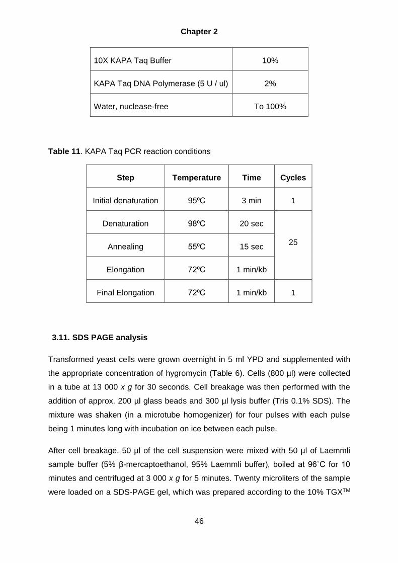
Chapter 2
46
10X KAPA Taq Buffer 10%
KAPA Taq DNA Polymerase (5 U / ul) 2%
Water, nuclease-free To 100%
Table 11. KAPA Taq PCR reaction conditions
Step Temperature Time Cycles
Initial denaturation 95⁰C 3 min 1
Denaturation 98⁰C 20 sec
25 Annealing 55⁰C 15 sec
Elongation 72⁰C 1 min/kb
Final Elongation 72⁰C 1 min/kb 1
3.11. SDS PAGE analysis
Transformed yeast cells were grown overnight in 5 ml YPD and supplemented with
the appropriate concentration of hygromycin (Table 6). Cells (800 µl) were collected
in a tube at 13 000 x g for 30 seconds. Cell breakage was then performed with the
addition of approx. 200 µl glass beads and 300 µl lysis buffer (Tris 0.1% SDS). The
mixture was shaken (in a microtube homogenizer) for four pulses with each pulse
being 1 minutes long with incubation on ice between each pulse.
After cell breakage, 50 µl of the cell suspension were mixed with 50 µl of Laemmli
sample buffer (5% β-mercaptoethanol, 95% Laemmli buffer), boiled at 96˚C for 10
minutes and centrifuged at 3 000 x g for 5 minutes. Twenty microliters of the sample
were loaded on a SDS-PAGE gel, which was prepared according to the 10% TGXTM

Chapter 2
47
FastCastTM Acrylamide Kit ,BIO-RAD protocol (Bio-Rad Laboratories 2019a). Protein
separation was performed for approximately 40 minutes at 200 V.
3.12. Western blot analysis
Following SDS-PAGE separation of proteins, these were analysed through western
blot analyses using a Cas9 monoclonal HRP-conjugated antibody (Abcam).
All the buffers used for the preparation and performance of the western blot analysis
are shown in Table 12.
Table 12. The buffers used for western blot analysis
Buffer Composition
TGS Tris glycine SDS
PBS (Phosphate Buffered
Saline)
0.8% NaCl, 0.02% KCl, 0.144% Na2HPO4,
0.024% KH2PO4 (pH 7.4)
Towbin (Transfer) buffer 20% methanol, 10% 10 x TGS.
Wash buffer 0.05% Tween 20, 99.95% PBS
Blocking buffer 0.05% Tween 20, 5% milk powder, 200 ml PBS
Antibody solution buffer blocking buffer, Cas9 monoclonal HRP-
conjugated (1: 5 000 of antibody: blocking buffer)
Antibody substrate
solution (Clarity Western
ECI Substrate)
Luminol Enhancer Solution, Peroxide Solution
(1:1 ratio)
The SDS page gel was equilibrated in Towbin transfer buffer (Bio-Rad Laboratories
2010). While the gel equilibration took place, 6 pieces of thick (0.8 mm) filter paper
were soaked in Towbin buffer. For the PVDF membrane, the membrane was briefly
transferred to 100% methanol for 30 seconds, then rinsed in water for 2 minutes
Chapter 2
48
followed by the incubation in Towbin buffer for 10 minutes. The sandwich was staked
on the anode in the order of: three filter papers, the membrane, the SDS PAGE gel
and again three filter papers. The cathode were placed on top and the transferring of
proteins took place at 25 V and 1 A for 30 minutes.
After the run, the membrane was briefly wetted in methanol and incubated in wash
buffer for 2 minutes (Bio-Rad Laboratories 2019b). The membrane was then
transferred to blocking buffer, protein side up, and incubated for 1 hour with continuous
agitation. After the 1 hour incubation, the membrane was transferred to blocking buffer,
containing the HRP-conjugated Cas9 antibody. The membrane was incubated
overnight in the antibody solution followed by washing six times, with each wash
consisting of a 5 minutes incubation step, in wash buffer with continuous agitation. The
antibody substrate solution was prepared and added to the membrane and incubated
at room temperature for 5 minutes. The solution was discarded, and the membrane
was imaged and analysed using a Gel Doc™ XR+ (Bio-Rad).
4. Results and discussion
4.1. Construction of pKM180::CAS9
The wide range expression system pKM180 (Smit et al. 2012), was used as backbone
for the development of a single CRISPR-Cas9 system (Figure 1A). The pKM180 uses
a region of the rRNA locus from Kluyveromyces marxianus for integration into the
yeast’s genome. Selection of positive transformants is facilitated by the dominant
selection of Hygromycin by the presence of the HPH gene, which is also modified to
function in CTG-clade yeasts (e.g. C. albicans), which is driven and terminated by the
Saccharomyces cerevisiae TEF1 promoter and -terminator. The plasmid has NotI
restriction sites to linearize the plasmid. For the cloning in bacteria, the pKM180
contains the normal bacterial moiety with the kanamycin resistant gene as a selection
marker. The yeast part of the vector consists of a promoter region derived from the
Yarrowia lipolytica transcription elongation factor (TEF1) gene that drives expression
of a gene inserted in the pET28 derived multiple cloning site (MCS). Expression is
terminated by the K. marxianus inulase terminator (KmINUt) sequence.

Chapter 2
49
Figure 1: (A) the pKM 180 vector (7 598bp) that was used as the backbone for the
construction of the (B) pKM180 + optimized CAS9 vector (~11 700bp). (C) The general
structure of the CAS9 gene obtained consisting of two zones on both ends comprising
several restriction sites, a Kozak sequence, the gene and a Nuclear localisation
sequence (NLS).
For the construction of the expression cassettes the three different codons optimised
CAS9 genes were obtained from Genscript (GenScript 2002; Gao & Zhao 2014; Vyas
et al. 2015; Weninger et al. 2016). The basis of each of the CAS9 genes consist out
of a region on both the 5- and 3-prime ends which contains various restriction sites for
cloning purposes (Figure 1C). Following the restriction site region, on the 5-prime side
of the gene, is a Kozak sequence to assist in translation initiation, this is followed by
the CAS9 gene and several SV40 nuclear localisation sequences (NLS), (1x NLS, 2x
NLS and 3x NLS for the P. pastoris, C. albicans and H. sapiens optimised genes
respectively). The purpose of the NLS is to target Cas9 localisation to the nucleus. In
the case of the CCAS9, the gene is optimized to function in both the CTG and non-

Chapter 2
50
CTG clade. In this case all the CTG codons were replaced with CTC codons. The
reason for this is because some yeast including a number of Candida species have
adopted a specific codon usage bias, where the CTG codon encodes serine instead
of leucine (Translation table 12: Alternative Yeast Nuclear Code). This can be
problematic due to the different properties the amino acids contain. Serine is
commonly characterised as a polar amino acid, whereas leucine is characterized as a
non-polar (hydrophobic) amino acid. In the formation of proteins, the hydrophobic
amino acids tend to form bonds in the inside of the protein, where the environment is
favourable for them, and the polar amino acids tends to be on the outside of the
protein. This change can thus lead to the incorrect folding of the protein and thus
altering its function. This modified version of the CAS9 gene therefore allows selection
in yeasts using the standard translation table as well as in yeast that use the alternative
translation code. After the three different optimized Cas9 amino acid sequences were
aligned to the native Cas9, using Geneious R10, (Figure 2) it was observed that there
are several amino acids from the P. pastoris optimized Cas9 [which was obtained from
Weninger and co-workers (2016)] that did not correspond to the native Cas9. This can
be problematic when protein folding occurs within the cell and could influence the
structure and function of the Cas9.

Chapter 2
51
Figure 2: The alignment of the three different optimized CAS9 translation (PCAS9 =
P. pastoris CCAS9 = C, albicans and HCAS9 = H. sapiens) sequence to the native
CAS9 (SPCAS9= Streptococcus pyogenes) translation sequence. As can be seen
from the alignment there are several amino acids (red boxes) which are different from
the P. pastoris optimized Cas9 compared to the three other Cas9 proteins.

Chapter 2
52
The 4 414 bp PCAS9, obtained in a pAO815 vector, was removed by restriction
digestion by NcoI and HindIII (Figure 3B). The CCAS9 were obtained from the pESC-
URA vector when cleaved by NcoI + BamHI (Figure 4B), which yielded a fragment size
of 4 213 bp. The HCAS9 (4 189bp) were removed from pAO815 with NcoI + BamHI
(Figure 5B). For the purpose of successful ligation and cloning, the pKM180 were
cleaved with similar restriction enzymes respective to each optimized CAS9 to yield
fragment sizes of 7 506bp, 7 531bp and 7 531 bp for the PCAS9, CCAS9 and HCAS9,
respectively (Figure 3A; 4A and 5A). DNA fragments were gel purified, pKM180 and
the various CAS9 genes ligated and the mixture transformed (as mentioned in chapter
2 section 3.8).
The colonies obtained from the transformation were screened with restriction enzymes
which specifically cuts in the CAS9 gene. pKM::PCAS9 were digested with AflII, this
yielded fragment sizes of 6 695bp, 3 779bp and 1 183bp for successful transformants
and 7 598bp for negative transformants. As seen in Figure 3, three clones [(Figure 3C)
sample 2, 3, 4] were obtained where all of them indicated the correct restriction profile.
Figure 3: A) The pKM180 linearized with NcoI and HindIII (7 506bp). B) PCAS9, which
was cleaved from the pAO815 vector with NcoI and HindIII (4 414bp). C) After ligation
the colonies were screened with AflII. For successful ligations fragment sizes of 6 695

Chapter 2
53
bp, 3 779 bp and 1 183 bp were obtained. L represents the DNA ladder; 1 is the
negative control (pKM180 without insert) and 2 to 4 pKM::PCAS9.
For the pKM::CCAS9 vector the transformants were screened with HindIII, which
resulted in 8 530bp, 2 463bp and 755bp bands if successful and 7 598bp if
unsuccessful. As seen in Figure 4C, several colonies were obtained after
transformation, after which 14 were randomly screened. Samples 5, 6, 9 and 12 to 15
tested successful for ligation to obtain the pKM::CCAS9 vector
Figure 4: A) The pKM180 vector linearized with NcoI + BamHI (7 531bp). B) CCAS9,
which was cleaved from the pESC-URA vector with NcoI + BamHI (4 213bp). C) After
ligation the colonies were screened with HindIII. For successful ligations fragment
sizes of 8 530bp, 2 463bp and 755bp have been obtained. L represents the DNA
ladder; 1 is the negative control and 2 to 15 is the screened colonies.
Lastly, HCAS9 was digested with Nrul and SalI to yield 10 766bp and 950bp when
successful and 7 598bp if unsuccessful. As seen in Figure 5C, several colonies were
obtained after transformation, after which 14 were randomly screened. Samples 5 to
7, 9,12, 13 and 15 tested successful for ligation to obtain the HCAS9 vector

Chapter 2
54
Figure 5: A) The pKM180 vector linearized with NcoI + BamHI (7 531bp). B) HCAS9,
which was cleaved from the pAO815 vector with NcoI + BamHI (4 189bp). C) After
ligation the colonies were screened with Nrul and SalI. For successful ligations
fragment sizes of 10 766bp and 950bp have been obtained. L represents the DNA
ladder; 1 is the negative control and 2 to 15 is the screened colonies.
4.2. Yeast Transformation
After construction of the pKM180::CAS9 (Figure 1B), yielding the pKM::PCAS9 (11
645 bp), pKM::CCAS9 (11 736 bp) and pKM::HCAS9 (11 712bp), these (as well as
the empty pKM180 plasmid as positive control) was transformed into 12 different
yeasts (Table 7). Before transformation each of the plasmids were linearized by NotI
digestion to remove the bacterial moiety and therefore exposing the rDNA regions to
allow homologous recombination to take place. For Saccharomyces cerevisiae
CENPK42, Kluyveromyces lactis UOFS-Y1167, Yarrowia lipolytica Po1f,
Debaryomyces hansenii UOFS-Y0610, Arxula adeninivorans UOFS-Y1219, Arxula
adeninivorans Ls3 and Ogataea polymorpha UOFS Y1507 the one-step
transformation method was used for maximal transformation efficiency. For Arxula
adeninivorans UOFS-Y1220, Komagataella phaffi UOFS-Y1552, Komagataella phaffi

Chapter 2
55
SMD 1168, Komagataella phaffi Km.71 and Komagataella phaffi GS115
transformation, the bicine method was used to achieve maximal transformation
efficiency (Klebe et al.). Table 7 indicates potential toxicity of the CAS9 vectors through
the increased susceptibility to hygromycin. Some of the yeast strains become more
sensitive to Hygromycin following transformation of the pKM180::CAS9 gene. This
was most evident when the HCAS9 was transformed. For these strains, the
Hygromycin was reduced to allow growth of transformed colonies (Table 7). The
reason for this increased sensitivity is however not known.
After transformation, integration of the expression vectors into the transformed yeast
genome were confirmed through PCR analysis (Figure 6). For the pKM::PCAS9 and
pKM::CCAS9 primers cacas9-1 forward and reverse were used to amplify a 694bp
part of the CAS9. For the pKM::HCAS9 primers hscas9-1 forward and reverse was
used to amplify an 816 bp part of the CAS9.

Chapter 2
56
Figure 6: Vector integration into the host genome were successful in all the
transformed yeast as displayed on each gel respective to the CAS9 vector used. L
represents the ladder, 1 represents the positive control (plasmid DNA) and 2 to 13 are
the yeast samples, on all three of the gels. The primers binding site on each of the
three different optimized CAS9 genes are also depicted. The large amplicon in each
lane may possibly represent impurities.
4.3. Western blot analysis
Western blot analysis was performed to confirm successful expression of the Cas9
protein. In the initial protocol, a centrifugation step (14 000 x g for 5 minute) was
included to remove the cellular debris after the cell breakage with glass beads. It was
determined that the glass beads used did not disrupt the nucleus of cells, and thus the
nucleus was removed along with the cellular debris during the initial centrifugation
step. This resulted in the removal of the Cas9 protein from the suspension and
therefore no bands were obtained on the western blot membranes. Note that these
membranes were not included due to the negative results. Therefore, the
centrifugation step was removed and protein preparation for SDS PAGE was
performed with the cellular debris.
Western blot analysis with the HRP conjugated Cas9 antibody confirmed that all three
of the different optimized Cas9 proteins were expressed at their respective molecular
weights when compared to the protein standard (Figure 7). However, as can be seen,
in Figure 7C, first lane in panel 1 to 5; 9 and 12 (see Table 14 for the yeast referencing
number) the pKM::PCAS9 had more than one band. However, the reason for this
phenomenon is not known and difficult to determine. A possible reason may be that
the yeasts started translation at a different ATG codon downstream, which resulted in
different size proteins. This may also contribute to the reason for the zero targeting
efficiency obtained by Weninger and co-workers, when they used the PCAS9
(Weninger et al. 2016).

Chapter 2
57
Figure 7: A) Protein ladder (colour prestained protein standard, broad range (11-245
kDa)) used for the determination of the protein band sizes. B) The calculated molecular
weight of the three different optimized Cas9 proteins. (C) The different optimized Cas9
proteins visualised through western blot analysis with the HRP-conjugated Cas9
antibody, in each yeast (see Table 13 for the yeast reference number). WT represents
wild type, P represents PCAS9, C represents CCAS9 and H represents HCAS9.

Chapter 2
58
Table 13 The different yeast used in this study as well as their reference number.
Reference number
Yeast
1 Saccharomyces cerevisiae CENPK42
2 Kluyveromyces lactis UOFS-Y1167
3 Yarrowia lipolytica Po1f
4 Debaryomyces hansenii UOFS-Y0610
5 Arxula adeninivorans UOFS-Y1220
6 Arxula adeninivorans UOFS-Y1219
7 Arxula adeninivorans Ls3
8 Ogataea polymorpha UOFS Y1507
9 Komagataella phaffii UOFS-Y1552
10 Komagataella phaffii SMD 1168
11 Komagataella phaffii Km.71
12 Komagataella phaffii GS115
5. Conclusion
A number of yeast CRISPR-Cas9 systems have been developed and applied
successfully (DiCarlo et al. 2013; Jacobs et al. 2014; Horwitz et al. 2015; Schwartz et
al. 2016; Weninger et al. 2016; Juergens et al. 2018b). These systems are designed
to satisfy the special demands of a specific yeast and they follow similar construction
principles. The aim of this study was to construct a wide range yeast CRISPR-Cas9

Chapter 2
59
system for the application in a wide variety of different yeasts and to explore the
system’s potential in regards to gene editing.
For the first part of the study three different pKM 180::Cas9 expression constructs
were developed, each with a different codon optimized (P. pastoris, C. albicans and
H. sapiens), constitutively expressed CAS9. Each of the CAS9 genes were fused with
a nuclear localization sequence (1x NLS, 2x NLS and 3x NLS for P. pastoris, C.
albicans and H. sapiens, respectively), to ensure transportation of the expressed
protein into the nucleus. These expression constructs make use of the rRNA locus
from Kluyveromyces marxianus for integration into the yeasts and selection of positive
transformants is facilitated by the dominant selection of Hygromycin. The three
different CAS9 constructs were transformed into seven different biotechnologically
relevant yeasts (12 strains in total) using two different chemical transformation
protocols. Some yeast showed reduced growth likely due to a toxic effect of the Cas9
protein, thus a lower hygromycin concentration was used to accommodate for the
reduction in growth. Integration of the expression vectors into the yeast genome were
confirmed through PCR and gel electrophoresis. This was followed by confirming
successful expression of the Cas9 via western blot analysis using a Cas9 specific
monoclonal HRP-conjugated antibody. In addition, the PCAS9 showed extra bands on
some of the western blot membranes, which are possibly due to either incorrect
translation or folding or possibly proteolysis of the PCas9 protein.
6. References
Adli, M., (2018) 'The CRISPR tool kit for genome editing and beyond'. Nat Commun
9, 1911. doi: 10.1038/s41467-018-04252-2
Bao, Z.,Xiao, H.,Liang, J.,et al., (2015) 'Homology-Integrated CRISPR–Cas (HI-
CRISPR) System for One-Step Multigene Disruption in Saccharomyces
cerevisiae'. ACS Synth Biol 4, 585–594. doi: 10.1021/sb500255k
Barrangou, R.,Fremaux, C.,Deveau, H.,et al., (2007) 'CRISPR Provides Acquired
Resistance Against Viruses in Prokaryotes'. Science (80- ) 315, 1709–1712. doi:
10.1126/science.1138140

Chapter 2
60
Bhaya, D.,Davison, M.,& Barrangou, R., (2011) 'CRISPR-Cas Systems in Bacteria and
Archaea: Versatile Small RNAs for Adaptive Defense and Regulation'. Annu Rev
Genet 45, 273–297. doi: 10.1146/annurev-genet-110410-132430
Bio-Rad Laboratories., (2019a) 'TGX TM and TGX Stain-Free TM FastCast TM
Acrylamide Kit and Starter Kit Instruction Manual'. http://www.bio-rad.com/.
Accessed, August 23, (2018)
Bio-Rad Laboratories., (2010) 'Trans-Blot ® Turbo TM Blotting System'. http://www.bio-
rad.com/. Accessed, August 23, (2018)
Bio-Rad Laboratories., (2019b) 'Western ECL Substrates'. http://www.bio-rad.com/.
Accessed, August 23, (2018)
Birnboim, H.C.,& Doly, J., (1979) 'Nucleic Acids Res. 1979 Birnboim'. Nucleic Acid
Res 7, 1–11. doi: 10.1093/nar/7.6.1513
Chen, D.C.,Yang, B.C.,& Kuo, T.T., (1992) 'One-step transformation of yeast in
stationary phase'. Curr Genet 21, 83–84. doi: 10.1007/BF00318659
Chung, C.T.,Niemela, S.L.,& Miller, R.H., (1989) 'One-step preparation of competent
Escherichia coli: transformation and storage of bacterial cells in the same
solution.'. Proc Natl Acad Sci 86, 2172–2175. doi: 10.1073/pnas.86.7.2172
Devi, S.,Kedlaya, R.,Maddodi, N.,et al., (2009) 'Calcium homeostasis in human
melanocytes: role of transient receptor potential melastatin 1 (TRPM1) and its
regulation by ultraviolet light'. Am J Physiol Physiol 297, C679–C687. doi:
10.1152/ajpcell.00092.2009
DiCarlo, J.E.,Norville, J.E.,Mali, P.,et al., (2013) 'Genome engineering in
Saccharomyces cerevisiae using CRISPR-Cas systems'. Nucleic Acids Res 41,
4336–4343. doi: 10.1093/nar/gkt135
Gao, Y.,& Zhao, Y., (2014) 'Self-processing of ribozyme-flanked RNAs into guide
RNAs in vitro and in vivo for CRISPR-mediated genome editing'. J Integr Plant
Biol 56, 343–349. doi: 10.1111/jipb.12152
Gasiunas, G.,Barrangou, R.,Horvath, P.,& Siksnys, V., (2012) 'Cas9-crRNA
ribonucleoprotein complex mediates specific DNA cleavage for adaptive immunity
in bacteria'. Proc Natl Acad Sci 109, E2579–E2586. doi:

Chapter 2
61
10.1073/pnas.1208507109
Generoso, W.C.,Gottardi, M.,Oreb, M.,& Boles, E., (2016) 'Simplified CRISPR-Cas
genome editing for Saccharomyces cerevisiae'. J Microbiol Methods 127, 203–
205. doi: 10.1016/j.mimet.2016.06.020
GenScript., (2002) 'GenScript'. https://www.genscript.com/. Accessed, January 30,
(2019)
Gerngross, T.U., (2004) 'Advances in the production of human therapeutic proteins in
yeasts and filamentous fungi.'. Nat Biotechnol 22, 1409–1414. doi:
10.1038/nbt1204-1589e
Horwitz, A.A.,Walter, J.M.,Schubert, M.G.,et al., (2015) 'Efficient Multiplexed
Integration of Synergistic Alleles and Metabolic Pathways in Yeasts via CRISPR-
Cas'. Cell Syst 1, 88–96. doi: 10.1016/j.cels.2015.02.001
Ish-Horowicz, D.,& Burke, J.F., (1981) 'Rapid and efficient cosmid cloning'. Nucleic
Acids Res 9, 2989–2898. doi: 10.1093/nar/9.13.2989
Jacobs, J.Z.,Ciccaglione, K.M.,Tournier, V.,& Zaratiegui, M., (2014) 'Implementation
of the CRISPR-Cas9 system in fission yeast'. Nat Commun 5, 5344. doi:
10.1038/ncomms6344
Jakočiūnas, T.,Bonde, I.,Herrgård, M.,et al., (2015) 'Multiplex metabolic pathway
engineering using CRISPR/Cas9 in Saccharomyces cerevisiae'. Metab Eng 28,
213–222. doi: 10.1016/j.ymben.2015.01.008
Jinek, M.,Chylinski, K.,Fonfara, I.,et al., (2012) 'A Programmable Dual-RNA-Guided
DNA Endonuclease in Adaptive Bacterial Immunity'. Science (80- ) 337, 816–821.
doi: 10.1126/science.1225829
Juergens, H.,Varela, J.A.,Gorter de Vries, A.R.,et al., (2018) 'Genome editing in
Kluyveromyces and Ogataea yeasts using a broad-host-range Cas9/gRNA co-
expression plasmid'. FEMS Yeast Res 18, . doi: 10.1093/femsyr/foy012
KAPA Biosystems Inc., (2014) 'KAPA HiFi HotStart ReadyMix PCR Kit Technical Data
Sheet'. https://www.kapabiosystems.com/product-applications/products/pcr-
2/kapa-hifi-pcr-kits/. Accessed, January 28, (2019)

Chapter 2
62
Klebe, R.J.,Harriss, J. V.,Sharp, Z.D.,& Douglas, M.G., (1983) 'A general method for
polyethylene-glycol-induced genetic transformation of bacteria and yeast.'. Gene
25, 333–341
Madzak, C.,Gaillardin, C.,& Beckerich, J.-M., (2004) 'Heterologous protein expression
and secretion in the non-conventional yeast Yarrowia lipolytica : a review'. J
Biotechnol 109, 63–81. doi: 10.1016/j.jbiotec.2003.10.027
Mans, R.,van Rossum, H.M.,Wijsman, M.,et al., (2015) 'CRISPR/Cas9: A molecular
Swiss army knife for simultaneous introduction of multiple genetic modifications
in Saccharomyces cerevisiae'. FEMS Yeast Res 15, 1–15. doi:
10.1093/femsyr/fov004
Nevoigt, E., (2008) 'Progress in Metabolic Engineering of Saccharomyces cerevisiae'.
Microbiol Mol Biol Rev 72, 379–412. doi: 10.1128/MMBR.00025-07
New England Biolabs Inc., (2014) 'NEBcloner'. http://nebcloner.neb.com/#!/redigest.
Accessed, January 07, (2019)
Ryan, O.W.,Skerker, J.M.,Maurer, M.J.,et al., (2014) 'Selection of chromosomal DNA
libraries using a multiplex CRISPR system.'. Elife 3, 1–15. doi:
10.7554/eLife.03703
Schwartz, C.M.,Hussain, M.S.,Blenner, M.,& Wheeldon, I., (2016) 'Synthetic RNA
Polymerase III Promoters Facilitate High-Efficiency CRISPR–Cas9-Mediated
Genome Editing in Yarrowia lipolytica'. ACS Synth Biol 5, 356–359. doi:
10.1021/acssynbio.5b00162
Skrzypek, M.S.,Binkley, J.,Binkley, G.,et al., (2017) 'The Candida Genome Database
(CGD): Incorporation of Assembly 22, systematic identifiers and visualization of
high throughput sequencing data'. Nucleic Acids Res 45, D592–D596. doi:
10.1093/nar/gkw924
Smit, M.S.,Theron, C.W.,Albertyn, J.,& Labuschagné, M., (2012) 'Novel expression
construct'. 04652
Stovicek, V.,Borja, G.M.,Forster, J.,& Borodina, I., (2015) 'EasyClone 2.0: expanded
toolkit of integrative vectors for stable gene expression in industrial
Saccharomyces cerevisiae strains'. J Ind Microbiol Biotechnol 42, 1519–1531.

Chapter 2
63
doi: 10.1007/s10295-015-1684-8
Terns, M.P.,& Terns, R.M., (2011) 'CRISPR-based adaptive immune systems'. Curr
Opin Microbiol 14, 321–327. doi: 10.1016/j.mib.2011.03.005
Thermo Fisher Scientific Inc., (2015a) 'Restriction Enzymes–Thermo Scientific - ZA'.
https://www.thermofisher.com/za/en/home/brands/thermo-scientific/molecular-
biology/thermo-scientific-restriction-modifying-enzymes/restriction-enzymes-
thermo-scientific.html. Accessed, January 07, (2019)
Thermo Fisher Scientific Inc., (2015b) 'Thermo Scientific GeneJET Gel Extraction Kit'.
https://assets.thermofisher.com/TFS-
Assets/LSG/manuals/MAN0012661_GeneJET_Gel_Extraction_UG.pdf.
Accessed, January 07, (2019)
Turvey, S., (1997) 'Competent cells : Rubidium Chloride'. 1, 4000
Vyas, V.K.,Barrasa, M.I.,& Fink, G.R., (2015) 'A Candida albicans CRISPR system
permits genetic engineering of essential genes and gene families'. Sci Adv 1,
e1500248. doi: 10.1126/sciadv.1500248
Wang, R.,Preamplume, G.,Terns, M.P.,et al., (2011) 'Interaction of the Cas6
Riboendonuclease with CRISPR RNAs: Recognition and Cleavage'. Structure 19,
257–264. doi: 10.1016/j.str.2010.11.014
Weninger, A.,Hatzl, A.-M.,Schmid, C.,et al., (2016) 'Combinatorial optimization of
CRISPR/Cas9 expression enables precision genome engineering in the
methylotrophic yeast Pichia pastoris'. J Biotechnol 235, 139–149. doi:
10.1016/j.jbiotec.2016.03.027
Zhang, G.-C.,Kong, I.I.,Kim, H.,et al., (2014) 'Construction of a Quadruple Auxotrophic
Mutant of an Industrial Polyploid Saccharomyces cerevisiae Strain by Using RNA-
Guided Cas9 Nuclease'. Appl Environ Microbiol 80, 7694–7701. doi:
10.1128/AEM.02310-14

Chapter 3
Development of the wide range CRISPR-Cas9 system by
incorporating a RGR-gRNA construct and validation through ADE2
disruption

Chapter 3
65
1. Abstract
The wide range pKM180 consisting of a C. albicans optimized CAS9 (CCAS9) gene
and a gRNA construct flanked by ribozymes was developed. Both the CAS9 and the
gRNA expression were constitutively driven by a Yarrowia lipolytica TEF1 promoter.
The integration of the vector is facilitated by the rRNA locus from Kluyveromyces
marxianus and selection of positive transformants is through hygromycin resistance.
The vector was transformed into the different yeasts and the efficacy of the CRISPR-
Cas9 gene editing system was determined by targeting the ADE2 gene which if
disrupted results in a red phenotype of colonies. When the vector was transformed
without donor DNA, most of the yeast displayed moderate to high disruption efficiency
through non-homologous end joining (NHEJ). When donor DNA was included in the
transformation, most of the yeast displayed gene disruption through homologous
recombination (HR), however at low to moderate efficiency. This proved that the wide
range CRISPR-Cas9 system was applicable in a wide variety of different yeast thus
allowing for rapid, cost-effective genetic manipulation of biotechnologically relevant
yeast.
2. Introduction
With the application of CRISPR-Cas9 in yeast, there are two main strategies used for
gRNA expression (Gao & Zhao 2014). The first strategy is the use of an RNA
polymerase III (Pol III) promoter for example the U3 or U6 promoters. The second
strategy makes use of the self-cleavage ability of ribozymes. Using the latter strategy
allows the expression of the gRNA construct using any promoter. In this system the
gRNA construct is flanked by self-catalysed ribozymes, which promotes self-cleavage
and remove any modifications (poly(A) tail or 7-methylguanylate cap) that have been
added during transcription. Here the application of this strategy is described in the
development of a wide range CRISPR-Cas9 system where the use of a single
promoter for the expression of the gRNA in all of the yeast is essential.
The CRISPR-Cas9 engineering tool has been successfully tested in a number of
organisms (Jinek et al. 2012; Hwang et al. 2013; DiCarlo et al. 2013; Wang et al. 2013;
Gratz et al. 2013; Friedland et al. 2013; Jiang et al. 2013; Vyas et al. 2015; Kistler et
al. 2015; Yan et al. 2017). The system creates double stranded breaks through RNA‐
Chapter 3
66
guided DNA cleavage, resulting in a very easy method to achieve targeted gene
disruption and insertion (Jinek et al. 2012; Horwitz et al. 2015). This system commonly
consists of the Cas9 enzyme and a guide RNA (gRNA). The Cas9 enzyme performs
the cleavage action of the double stranded DNA and consists of two nuclease
domains, namely RuvC and HNH (Haft et al. 2005; Makarova et al. 2006). The RuvC
domain cleaves the DNA strand that contains the PAM sequence and the HNH domain
cleaves the target strand. The gRNA is a short synthetic RNA which originated when
the site-specific crRNA was fused to a tracrRNA (scaffold part) (Jinek et al. 2012). The
scaffold part is necessary for the association with the Cas9 enzyme and the user-
specified crRNA part (approximately 20 bp of length), which states the target DNA in
the host genome to be modified. Therefore, by just changing the target sequence in
the gRNA, one can simply target any gene for modification.
The gRNA binds to the Cas9 nuclease and guides it to the specific target (Jinek et al.
2012). The Cas9 then binds to, and cleaves the target DNA to generate a double
stranded break (DSB). In yeast, this DSB can lead to gene alteration that can inactivate
the gene through non-homologous end joining (NHEJ) with the creation of indels
(insertion or deletion of nucleotides) (Pitcher et al. 2007; Bétermier et al. 2014). These
indels may create a frame shift in bases, which can create different amino acid
sequence and thus will diminish proper protein folding and activity. This DSB can also
introduce an opportunity for homologous recombination (HR) to insert a donor
fragment (if provided) specified for the user’s purpose (Pâques & Haber 1999; Sung
& Klein 2006). However, there is one major challenge in using CRISPR for targeted
gene editing, which is the production of the gRNAs (Gao & Zhao 2014).
The first 20‐ nucleotide sequence of the gRNA is used to guide targeted DNA cleavage
(Jinek et al. 2012). Any additional bases or modifications at the ends of gRNA will
abolish the gRNA’s ability to guide DNA cleavage by Cas9 (Haurwitz et al. 2010; Jinek
et al. 2012). Hence, the expression of the gRNA needs to be closely monitored for
successful gene targeting. The most widely used and well‐characterized promoters
are RNA polymerase II promotors (Pol II) (Gao & Zhao 2014). However, these
promoters cannot be used directly to produce gRNA for CRISPR, due to the fact that
mRNAs that are transcribed through Pol II promoters undergo extensive processing
and modification at both ends. Therefore, the gRNA expression cassette is usually
driven by a non-mRNA polymerase promoter for example (Pol III) promoters (U3 and

Chapter 3
67
U6), the promoter is followed by a specific target sequence, the gRNA scaffold and
then the terminator. However, the Pol III promoters have limitations when it comes to
the gRNA expression in different host. They are very specific for each organism and
not all these promoters have been characterized in every organism, thus, making it
difficult to choose the correct RNA polymerase promoters for CRISPR. Furthermore,
the RNA polymerase promoters limit the CRISPR target sequences to G (N20) GG
and A (N20) GG, respectively. Therefore, another strategy has been developed and
used successfully for the correct expression of the gRNA. Gao and Zhao developed a
strategy where they took advantage of the nuclease activity of ribozymes. They
designed an artificial gene, RGR (Ribozyme‐gRNA‐Ribozyme), where the RGR gene
undergoes self‐catalysed cleavage to yield the gRNA without any modifications. This
strategy leads to the successful expression of the gRNA using any promoter, thus
resulting in successful guidance of the Cas9 enzyme to the target gene. In addition,
this RGR strategy diminished the limitation to what the target sequence (in the gRNA)
must contain. The gRNA expression construct can thus contain any RNA polymerase
II promoter followed by a Hammerhead (HH) self-splicing ribozyme, the target
sequence, the gRNA scaffold, a Hepatitis delta virus (HDV) self-splicing ribozyme and
the terminator.
The RGR strategy needs to be applied to design an integrative wide range CRISPR-
Cas9 system for the application in a wide variety of different yeast (Gao & Zhao 2014).
In the previous chapter, three different optimized CAS9 vectors (pKM::PCAS9,
pKM::CCAS9 and pKM::HCAS9) were constructed using the wide range pKM180
vector and three different optimized CAS9 [P. pastoris (PCAS9), C. albicans (CCAS9)
and H. sapiens (HCAS9)]. The vectors were transformed into the yeast and expression
of each of the different optimized CAS9 were tested to observe if the Cas9 protein is
being expressed. For further development of the wide range CRISPR-Cas9 system, a
gRNA needs to be included together with the CAS9. This will provide the vector with
gene targeting characteristics, which will allow for gene targeting in the different yeast
tested. Therefore, the aim of this part of the study was to construct and validate a wide
range CRISPR-Cas9 vector. This vector contained the optimized CAS9 as well as an
RGR ADE2 CRISPR target site and was used to test for successful and efficient gene
disruption in Saccharomyces cerevisiae and six other non-conventional yeast.

Chapter 3
68
3. Material and methods
3.1. In silico work
All in silico design and construction of plasmids were performed using Geneious R10
(Geneious 2017).
3.2. E. coli Rubidium chloride competent cells
Refer to chapter 2 section 3.1
3.3. Bacterial Transformation
Refer to chapter 2 section 3.2
3.4. Restriction digestion
Refer to chapter 2 section 3.3
3.5. Polymerase Chain Reaction
All PCR reactions performed were either perform using the KAPA Taq PCR kit or the
KOD Hot Start PCR kit (Novagen 2011; KAPA Biosystems Inc. 2014). The PCR
reaction mixtures were prepared according to either Table 1 or 3 and with reaction
conditions as stated in either Table 2 or 4, respectively.
Table 1. The reaction mixture for the KAPA Taq PCR kit (KAPA Biosystems Inc. 2014)
Component Reaction mixture
Template DNA Up to 250 ng
10mM dNTP Mix 2%
10µM Forward primer 4%
10µM Reverse primer 4%

Chapter 3
69
10X KAPA Taq Buffer 10%
KAPA Taq DNA Polymerase (5 U
/ µl)
2%
Water, nuclease-free To 100%
Table 2. KAPA Taq PCR reaction conditions (KAPA Biosystems Inc. 2014)
Step Temperature Time Cycles
Initial denaturation 95⁰C 3 min 1
Denaturation 98⁰C 20 sec
25 Annealing Lowest Primer
Tm⁰C for 10
sec
15 sec
Elongation 72⁰C 1 min/kb
Final Elongation 72⁰C 1 min/kb 1
Table 3. The reaction mixture for the KOD Hot Start PCR kit (Novagen 2011)
Component Reaction mixture
Template DNA Up to 250 ng
2mM dNTP Mix 10%
25mM MgSO4 6%
10µM Forward primer 3%
10µM Reverse primer 3%

Chapter 3
70
10X KOD Hot Start Buffer 10%
KOD Hot Start Polymerase (1 U /
µl)
2%
Water, nuclease-free To 100%
Table 4. KOD Hot Start PCR reaction conditions (Novagen 2011)
Step Target size Cycles
Pol. activation 95⁰C 1
Denaturation 95⁰C for 20 sec
30 Annealing Lowest Primer
Tm⁰C for 10 sec
Extension 70⁰C 25 sec/kb
All the primers used in this study are presented in Table 5. The first two letters
represents the yeast that the primers were designed for, Aa= Arxula adeninivorans ;
Dh= Debaryomyces hansenii; Kl= Kluyveromyces lactis; Op= Ogataea polymorpha;
Pp= Komagataella phaffii; Sc= Saccharomyces cerevisiae; Yl= Yarrowia lipolytica.
The sequences that are in bold represent the ADE2 CRISPR site. For the identification
of the CRISPR target sites the CRISPR tool in Geneious R10 (www.geneious.com)
was used. The ADE2 ORF of each of the selected yeast species was analysed for a
GN(20)NGG motif. Off target sites were eliminated by including the full genome
sequence (if available) of the relevant yeast. Only CRISPR sites with an on-target
activity of higher than 70% were selected.
Table 5. The primers used in this study with their corresponding properties
Name Sequence Tm⁰C GC
content Length
Aa donor 1F GACTCAAAAACGGTGGGAAT 56.2 45 20

Chapter 3
71
Aa donor 1R GACAATGGAAATTCACATGCTCGATCTCAAT 64.4 38.7 31
Aa donor 2F GCATGTGAATTTCCATTGTCCAGATGCCTC 66.6 46.7 30
Aa donor 2R GTTTTTATATTCTTCATATCCAACCTTC 55.6 28.6 28
Aa gRNA ADE2
1F
GTTAAGCATTTCCTTCTGAGAAAGTTCTGAT
GAGTCCGTGAGGACGAAACGAGTAAGCT N/A* 44.1 59
Aa gRNA ADE2
1R
CTATTTCTAGCTCTAAAACTCGCTCTCTACTA
CAAAGTTGACGAGCTTACTCGTTTCGT N/A 40.7 59
Dh donor 1F GACGGAAAAACTATAGGTATATTAGG 55.1 34.6 26
Dh donor 1R TTTGCACAATAATATCACATTTTCTGGCTAAT
TCC 63.6 31.4 35
Dh donor 2F ATGTGATATTATTGTGCAAATGCCAAGGGG 64.6 40 30
Dh donor 2R TTGTAGTTTTCGTAGCCCAC 55.7 45 20
Dh gRNA ADE2
1F
GTTAAGCATTTCCTTCTGAGTCTTGACTGAT
GAGTCCGTGAGGACGAAACGAGTAAGCT N/A 45.8 59
Dh gRNA ADE2
1R
CTATTTCTAGCTCTAAAACTCGTATGCTAAA
GTTCTTGAGACGAGCTTACTCGTTTCGT N/A 39 59
Kl donor 1F GATCAAAGAACTGTCGGTATTTTA 55.2 33.3 24
Kl donor 1R GAATGTAGAGTTTCAACAGTTAAGACATCAC
AAA 62.8 32.4 34
Kl donor 2F ACTGTTGAAACTCTACATTCCATTGTACAAAT
GC 64.7 35.3 34
Kl donor 2R ATTCTTGATATCCGATAGTTTCTAATT 54.9 35.9 27
Kl gRNA ADE2
1F
GTTAAGCATTTCCTTCTGAGATGGGCCTGAT
GAGTCCGTGAGGACGAAACGAGTAAGCT N/A 49.2 59
Kl gRNA ADE2
1R
CTATTTCTAGCTCTAAAACTTGTATGCCGAG
AAATGGGCGACGAGCTTACTCGTTTCGT N/A 44.1 59
Op donor 1F AAGGTCGTTGGAATTTTGGG 56.5 45 20
Op donor 1R ATCGAATACAAACATCAACGTGCTCAATCT 63.9 36.7 30

Chapter 3
72
Op donor 2F CGTTGATGTTTGTATTCGATAGTTCAGATGC
C 64.7 40.6 32
Op donor 2R TTTATTAAGGTATTCTTCATAGCCAA 54.8 26.9 26
Op gRNA ADE2
1F
GTTAAGCATTTCCTTCTGAGTAGATGCTGAT
GAGTCCGTGAGGACGAAACGAGTAAGCT N/A 45.8 59
Op gRNA ADE2
1R
CTATTTCTAGCTCTAAAACTCGTCTAGAGTA
CATAGATGGACGAGCTTACTCGTTTCGT N/A 40.7 59
Pp donor 1F GATTCTCAGGTAATAGGTATTCTAGG 56 38.5 26
Pp donor 1R GTTGTTGATATCAATCTCTACAGTGAGAACA
TC 62.3 36.4 33
Pp donor 2F TAGAGATTGATATCAACAACGCCACCAATG 63.8 40 30
Pp donor 2R AATTCAAAGACGATTCTTCAAATAGG 56.6 30.8 26
Pp gRNA ADE2
1F
GTTAAGCATTTCCTTCTGAGCCGTACCTGAT
GAGTCCGTGAGGACGAAACGAGTAAGCT N/A 49.2 59
Pp gRNA ADE2
1R
CTATTTCTAGCTCTAAAACATCGGGTGGTCT
TTCCGTACGACGAGCTTACTCGTTTCGT N/A 45.8 59
Sc donor 1F GATTCTAGAACAGTTGGTATATTAGG 55 34.6 26
Sc donor 1R GACAAGAACCGTTAGCACATCACATTTTTCA
G 65.3 40.6 32
Sc donor 2F ATGTGCTAACGGTTCTTGTCTAGATGGAGTA
G 65.7 43.8 32
Sc donor 2R GTTTTCTAGATAAGCTTCGTAACC 55.7 37.5 24
Sc gRNA ADE2
1F
GTTAAGCATTTCCTTCTGAGACAATTCTGAT
GAGTCCGTGAGGACGAAACGAGTAAGCT N/A 44.1 59
Sc gRNA ADE2
1R
CTATTTCTAGCTCTAAAACTGTGGATAGTCT
CTACAATTGACGAGCTTACTCGTTTCGT N/A 39 59
yl donor 1F GACTCGAAAACAATTGGTATTCT 55.3 34.8 23
yl donor 1R GTTGTTGACAAATTTCAACGGTTATGACGTC 64.2 38.7 31
yl donor 2F CGTTGAAATTTGTCAACAACTCCACCAATG 64.3 40 30

Chapter 3
73
yl donor 2R TTGAGGTACTGCTCGTATCC 56.5 50 20
Yl gRNA ADE2
1F
GTTAAGCATTTCCTTCTGAGCCATGCCTGAT
GAGTCCGTGAGGACGAAACGAGTAAGCT N/A 49.2 59
Yl gRNA ADE2
1R
CTATTTCTAGCTCTAAAACTCGCTCGCATTC
GTCCATGCGACGAGCTTACTCGTTTCGT N/A 47.5 59
pKM 180
(linearize)- F AATGACGTCAGAATTCTCGAGATCCC 62.6 46.2 26
pKM 180
(linearize)- R ATTATCAGTCGTACTGTTATTAAGTGCTGT 61 33.3 30
pKM 180
(gRNA)- F
AGTACGACTGATAATCCAGAGACCGGGTTG
GCG N/A 54.5 33
pKM 180
(gRNA)- R
AATTCTGACGTCATTGAATTCTGACGTCATTA
TTATCAGTGTG N/A 34.9 43
*Length of primer do not permit accurate Tm determination
3.6. Agarose gel electrophoresis
Refer to chapter 2 section 3.4
3.7. DNA extractions
For DNA gel extraction see chapter 2 section 3.5 and for DNA plasmid extraction
chapter 2 section 3.7
3.8. Construction of the CRISPR-Cas9 vector
The TEF1 gene was amplified from the Yarrowia lipolytica Po1f genome with primers
ylTEF1-2F and - 2R using the Kapa PCR kit reaction protocol (KAPA Biosystems Inc.
2014). The PCR fragment was gel purified and inserted into the pMiniT 2.0 vector via
the NEB ligation protocol to yield the pMini + TEF vector (Thermo Fisher Scientific Inc
2015b; New England BioLabs Inc. 2017). The ligated product (5 µl) were then
transformed into NEB 10-Beta cells (Chung et al. 1989), plated on LB+ [100 µg/ml]

Chapter 3
74
ampicillin and grew over night. The transformed colonies were then screened with BglI
for confirmation of successful ligation.
The pRS316-RGR-GFP vector containing the Ribozyme‐gRNA‐Ribozyme (RGR-
gRNA) was obtained from Addgene (Gao & Zhao 2014; Addgene 2015). The RGR-
gRNA construct was isolated by amplifying the fragment with the primer pair hammer
1F:ylTEF1 overlap and hammer 1R:ylTEF1 overlap (KAPA Biosystems Inc. 2014).
The pMini + TEF1 vector was also linearized through amplification to remove the ORF
of the TEF1 gene with primers ylTEF1-3F and - 3R. Both the gRNA and vector
fragments were gel purified and ligated using NEBuilder to yield the pMini+TEF1-
RGR-gRNA vector (Thermo Fisher Scientific Inc 2015b; New England Biolabs 2017).
NEB product (5 µl) was then transformed and plated out on LB+ [100 µg/ml] ampicillin
plates and allowed to grow O.N. at 37˚C (Chung et al. 1989). The colonies obtained
from the transformation were screened with BglI, for confirmation of successful ligation
(Thermo Fisher Scientific Inc 2015a).
For the pKM::PCAS9+CRISPR vector, the TEF1-RGR-gRNA construct was cleaved
from the pMini+TEF1-RGR-gRNA vector with XhoI and SalI and each of the CAS9
vectors were cleaved with XhoI (Thermo Fisher Scientific Inc 2015a). The fragments
were gel extracted, ligated, transformed and screened for positive transformants with
BglII (Chung et al. 1989; Thermo Fisher Scientific Inc 2015a, b; New England BioLabs
Inc. 2017). Alternatively, for the pKM::HCAS9 and the pKM::CCAS9, the vectors were
linearized with primers (PKM180 (linearize)- F and -R) (Novagen 2011). The gRNA
construct was also amplified out of the pMini+TEF1-RGR-gRNA vector with primers
(pKM 180 (gRNA)-F and -R) (KAPA Biosystems Inc. 2014). The fragments (vectors as
well as the gRNA construct) were ligated using NEBuilder, 5 µl of the product was then
transformed into NEB 10-Beta competent cells and screening for successful
transformants were done with the digestion using BglII (Chung et al. 1989; Thermo
Fisher Scientific Inc 2015a; New England Biolabs 2017).
The CCAS9 was isolated from the pKM::CCAS9 with BglII and AvrII (New England
Biolabs Inc. 2014; Thermo Fisher Scientific Inc 2015a). The PCAS9 were digested out
of the pKM::PCAS9+CRISPR with Acc65I and HindIII. The fragments were then gel
extracted and connected using NEBuilder. Five microliters of the product were
transformed into NEB 10-Beta cells and the transformed colonies were then screened

Chapter 3
75
with PstI to find successful colonies (Chung et al. 1989; Thermo Fisher Scientific Inc
2015a, b; New England Biolabs 2017).
After the pKM::CCAS9+CRISPR was obtained, the ADE2 target gene of each yeast
were inserted into the pKM::CCAS9+CRISPR vector (Chung et al. 1989; KAPA
Biosystems Inc. 2014; Thermo Fisher Scientific Inc 2015b; New England Biolabs
2017). For this purpose two 50 bp oligonucleotides were designed to insert the
CRISPR site for each of the seven yeast species. The oligonucleotides were added
together and elongated in a PCR reaction to obtain a 100 bp double stranded
fragment. The pKM::CCAS9+CRISPR vector were linearized with primers ylTEF1-3R
and AHO1099. The fragments were then purified through gel extraction, ligated using
NEBuilder and transformed (5 µl of the NEBuilder product). This was done for each
yeasts’ ADE2 target site, respectively, to yield pKM::CCAS9+ADE2.
3.9. Construction of the donor DNA and validation of the CRISPR-Cas9 vector.
The donor DNA was obtained by amplifying two separate regions of the ADE2 gene
from each yeast. These two regions encompass approximately 200 bp on the 5’-side
and the 3’-side of the ADE2 ORF. For fragment 1: a PCR was performed using each
yeast genomic DNA with the respective donor 1f and donor 1r primers listed in Table
8. For fragment 2, a PCR was performed using genomic DNA from each yeast with
the respective donor 2f and donor 1r primers listed in Table 5. In every case the
reverse primer of fragment 1 had an overlap with the forward primer of fragment 2.
The final donor fragment was then obtained when a PCR was performed using
fragment 1 and 2 with the respective donor 1f and donor 2r primers from Table 5.
After obtaining the plasmids and donor DNA, transformation were performed on each
yeast, with and without donor DNA (Klebe et al. 1983; Chen et al. 1992). After
transformation single colonies were picked (from all the transformed yeast) and
streaked out on YNB dropout plates (Table 6), lacking adenine, to confirm ADE2
disruption mutants. Colony PCR were then performed on the successful mutants from
the donor DNA transformed yeast to confirm successful integration of the donor
fragment.
Table 6: The media used to test for ADE2 mutants
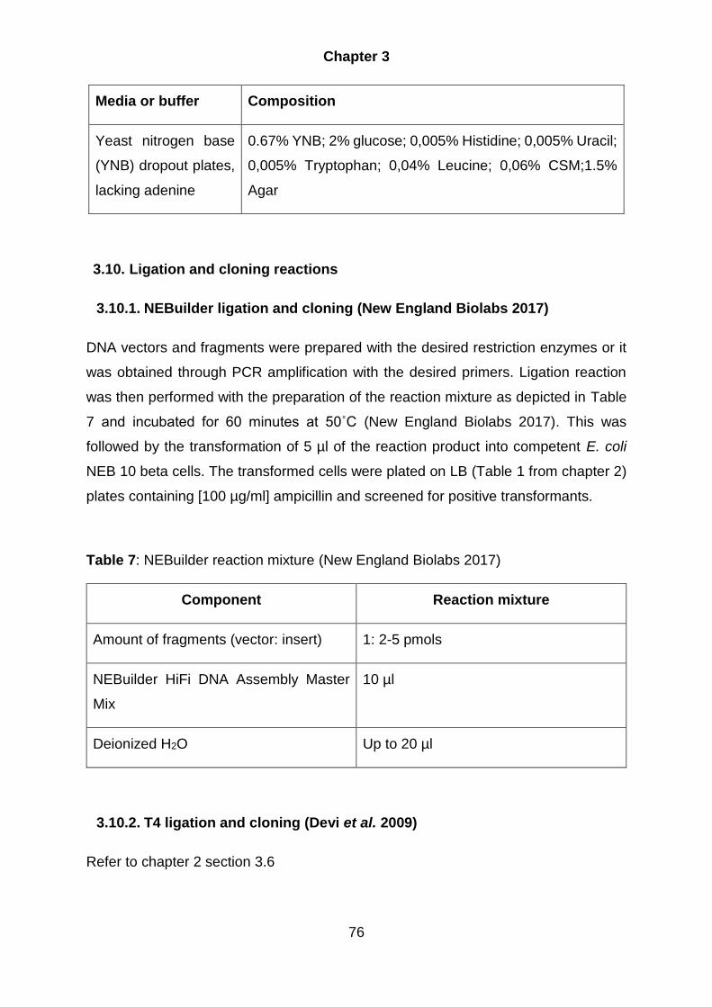
Chapter 3
76
Media or buffer Composition
Yeast nitrogen base
(YNB) dropout plates,
lacking adenine
0.67% YNB; 2% glucose; 0,005% Histidine; 0,005% Uracil;
0,005% Tryptophan; 0,04% Leucine; 0,06% CSM;1.5%
Agar
3.10. Ligation and cloning reactions
3.10.1. NEBuilder ligation and cloning (New England Biolabs 2017)
DNA vectors and fragments were prepared with the desired restriction enzymes or it
was obtained through PCR amplification with the desired primers. Ligation reaction
was then performed with the preparation of the reaction mixture as depicted in Table
7 and incubated for 60 minutes at 50˚C (New England Biolabs 2017). This was
followed by the transformation of 5 µl of the reaction product into competent E. coli
NEB 10 beta cells. The transformed cells were plated on LB (Table 1 from chapter 2)
plates containing [100 µg/ml] ampicillin and screened for positive transformants.
Table 7: NEBuilder reaction mixture (New England Biolabs 2017)
Component Reaction mixture
Amount of fragments (vector: insert) 1: 2-5 pmols
NEBuilder HiFi DNA Assembly Master
Mix
10 µl
Deionized H2O Up to 20 µl
3.10.2. T4 ligation and cloning (Devi et al. 2009)
Refer to chapter 2 section 3.6
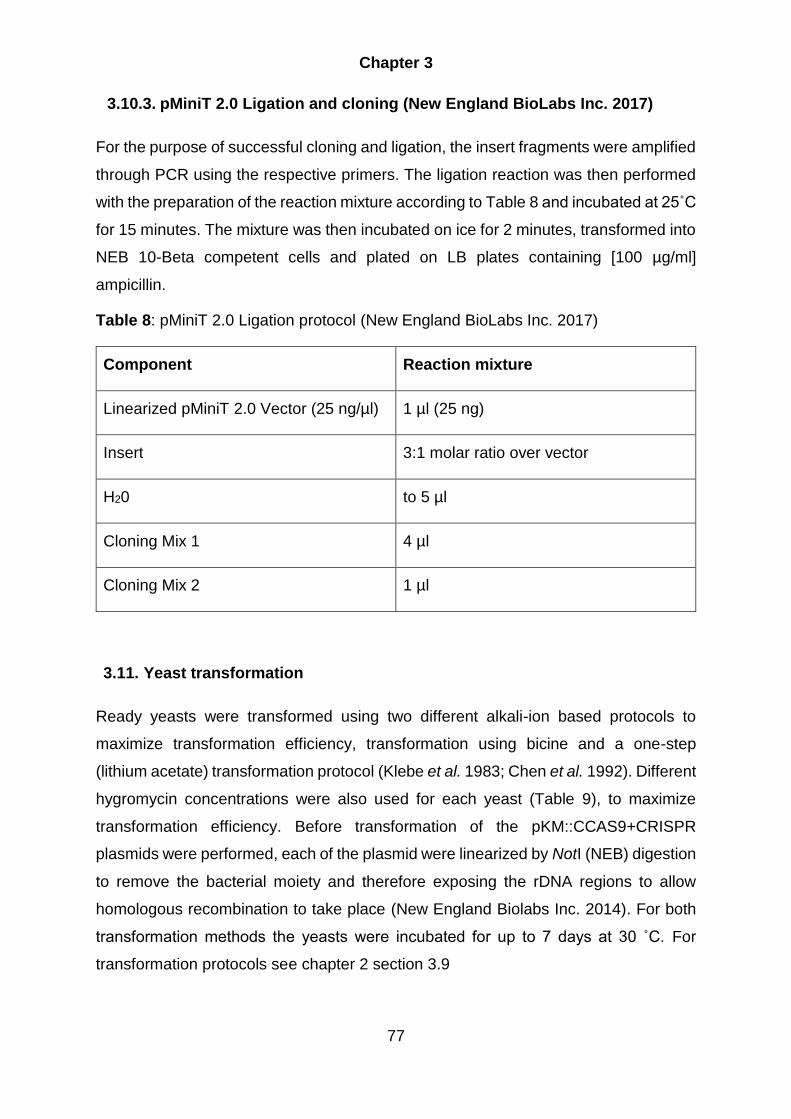
Chapter 3
77
3.10.3. pMiniT 2.0 Ligation and cloning (New England BioLabs Inc. 2017)
For the purpose of successful cloning and ligation, the insert fragments were amplified
through PCR using the respective primers. The ligation reaction was then performed
with the preparation of the reaction mixture according to Table 8 and incubated at 25˚C
for 15 minutes. The mixture was then incubated on ice for 2 minutes, transformed into
NEB 10-Beta competent cells and plated on LB plates containing [100 µg/ml]
ampicillin.
Table 8: pMiniT 2.0 Ligation protocol (New England BioLabs Inc. 2017)
Component Reaction mixture
Linearized pMiniT 2.0 Vector (25 ng/µl) 1 µl (25 ng)
Insert 3:1 molar ratio over vector
H20 to 5 µl
Cloning Mix 1 4 µl
Cloning Mix 2 1 µl
3.11. Yeast transformation
Ready yeasts were transformed using two different alkali-ion based protocols to
maximize transformation efficiency, transformation using bicine and a one-step
(lithium acetate) transformation protocol (Klebe et al. 1983; Chen et al. 1992). Different
hygromycin concentrations were also used for each yeast (Table 9), to maximize
transformation efficiency. Before transformation of the pKM::CCAS9+CRISPR
plasmids were performed, each of the plasmid were linearized by NotI (NEB) digestion
to remove the bacterial moiety and therefore exposing the rDNA regions to allow
homologous recombination to take place (New England Biolabs Inc. 2014). For both
transformation methods the yeasts were incubated for up to 7 days at 30 ˚C. For
transformation protocols see chapter 2 section 3.9
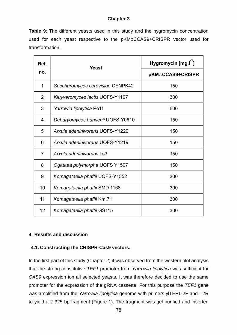
Chapter 3
78
Table 9: The different yeasts used in this study and the hygromycin concentration
used for each yeast respective to the pKM::CCAS9+CRISPR vector used for
transformation.
Ref.
no. Yeast
Hygromycin [mg.l-1
]
pKM::CCAS9+CRISPR
1 Saccharomyces cerevisiae CENPK42 150
2 Kluyveromyces lactis UOFS-Y1167 300
3 Yarrowia lipolytica Po1f 600
4 Debaryomyces hansenii UOFS-Y0610 150
5 Arxula adeninivorans UOFS-Y1220 150
6 Arxula adeninivorans UOFS-Y1219 150
7 Arxula adeninivorans Ls3 150
8 Ogataea polymorpha UOFS Y1507 150
9 Komagataella phaffii UOFS-Y1552 300
10 Komagataella phaffii SMD 1168 300
11 Komagataella phaffii Km.71 300
12 Komagataella phaffii GS115 300
4. Results and discussion
4.1. Constructing the CRISPR-Cas9 vectors.
In the first part of this study (Chapter 2) it was observed from the western blot analysis
that the strong constitutive TEF1 promoter from Yarrowia lipolytica was sufficient for
CAS9 expression ion all selected yeasts. It was therefore decided to use the same
promoter for the expression of the gRNA cassette. For this purpose the TEF1 gene
was amplified from the Yarrowia lipolytica genome with primers ylTEF1-2F and - 2R
to yield a 2 325 bp fragment (Figure 1). The fragment was gel purified and inserted

Chapter 3
79
into the pMiniT 2.0 cloning vector (New England Biolabs) via ligation to yield the
pMini+TEF vector (4 871 bp). The transformed colonies were screened with BglI to
yield fragment sizes of 3 642 bp and 1 235 bp if successful for ligation (Figure 1C).
Figure 1: A) Yarowia lipolytica TEF1 gene (2 325 bp), which was amplified from the
yeast genome with primers ylTEF1-2F and - 2R. The TEF1 gene, was ligated into the
B) pMiniT 2.0 vector (2 588 bp). C) The transformed colonies were then screened with
BglI. Successful ligation fragment sizes of 3 642 bp and 1 235 bp have been obtained.
L represents the DNA ladder and 1 to 4 are the screened colonies with lane four
indicating the correct fragments.
Plasmid pRS316-RGR-GFP (Gao & Zhao 2014; Addgene 2015) was obtained from
Addgene (www.addgene.com). This plasmid contain the hammerhead (HH) ribozyme,
gRNA scaffold and a HDV ribozyme as well as a CRISPR site specific for GFP (Figure
2A). The HH ribozyme promotes cleavage on its 3-prime side and the HDV ribozyme
promotes cleavage on its 5-prime side. For the HH ribozyme to perform its function,
the first 6 nucleotides must be reverse complementary to the first 6 bp of the CRISPR
target sequence region (Figure 2B). Thus, for each of the different target sites it is
necessary to modify the first 6 bp of the HH sequence, for the sole reason to maintain

Chapter 3
80
ribozyme functionality. The reason for the cleavage is that the gRNA is an essential
part of the CRISPR-Cas9 system, it must be highly specific without any post
transcriptional modifications (i.e. a 5’-Cap and/or 3’-Poly A tail), otherwise the gRNA
with the CRISPR site would not be able to bind to the Cas9 protein and therefore the
interference complex won’t form, thus abolishing its function. The post transcriptional
modifications can be prevented by using a non mRNA producing polymerase
promoter, which will ensure that no post transcriptional modifications take place,
however, the use of these promoters have limitations. They are very specific for each
organism and also only a limited number of all these promoters has been
characterized in the selected yeasts. Importantly, these drawbacks prevent the use in
a wide range system, with a universal promoter. For this reason, instead of using the
more simpler approach by only using a Pol III promoter the more complex approach
was used, where a self-splicing Ribozymes were used which promotes self-cleavage
to remove any post transcriptional modifications on both ends.
Figure 2: A) The generic RGR-gRNA, containing the CRISPR target site and the
gRNA scaffold, which is flanked by self-splicing ribozymes (HH and HDV). B) The
ADE2 CRISPR sites for each yeast were created using two overlapping

Chapter 3
81
oligonucleotide (gRNA 1F and 1R). The forward oligonucleotide contains the first 6 bp
(that needs to be the reverse complementary of the first 6 bp of the ADE2 CRISPR
target site for each yeast). The reverse oligonucleotide contained the specific ADE2
CRISPR site.
The RGR gRNA was removed from the pRS316-RGR-GFP plasmid by amplifying the
fragment with primer pair hammer 1F:ylTEF1 overlap and hammer 1R:ylTEF1 overlap
(Figure 3). This yielded a fragment size of 212 bp (RGR-gRNA). The primers were
designed to create a 15 bp overlap that corresponds with the TEF1 promotor’s 3-prime
end and the TEF1 terminators 5-prime end. This overlap is necessary to perform the
NEBuilder ligation reaction. The pMini+TEF vector was linearized through PCR
amplification to remove the ORF of the TEF1 gene but leaving the promoter and
terminator regions intact. The vector was amplified with primers ylTEF1-3F and -3R to
yield a 3 353 bp fragment. Both the gRNA and vector fragments were gel purified and
ligated with NEBuilder to yield the pMini+TEF1-RGR-gRNA vector. The colonies
obtained from the transformation were screened with BglI, where 12 out of the 15
colonies were positive when restriction analysis displayed band sizes of 2 296 bp and
1253 bp. Some of the lanes do display three bands, this is a result of incomplete
digestion, meaning the top band represent undigested plasmid (3 543 bp).

Chapter 3
82
Figure 3: A) pMini + TEF1, where the TEF1 ORF frame was removed to obtain a 3
353 bp fragment. B) The RGR gRNA was amplified from plasmid pRS316-RGR-GFP
to obtain a 212 bp fragment. C) The two fragments were ligated and screening for
positive transformants were done with the BglI enzyme to obtain sizes of 2 296 bp and
1 253 bp if successful. L represents the ladder and 1 to 15 the screened samples.
The next step comprised the construction of the complete pKM::CAS9+CRISPR vector
containing either the PCAS9, CCAS9 or the HCAS9 gene (designated
pKM::PCAS9+CRISPR, pKM::CCAS9+CRISPR or pKM::HCAS9+CRISPR,
respectively). This was done by inserting the TEF1-RGR-gRNA into each of the
pKM180::CAS9 vectors resulting in either pKM::PCAS9+CRISPR,
pKM::CCAS9+CRISPR or pKM::HCAS9+CRISPR. Initially the CAS9 vectors was
linearize by digesting it with XhoI, resulting in a ~11 700 bp linear fragment. The TEF1-
RGR-gRNA were obtained by digesting the pMini + TEF1-RGR-gRNA vector with XhoI
and SalI to yield a 1 036 bp fragment. The different restriction enzymes used to obtain
the TEF1-RGR-gRNA fragment would not pose any problem in the ligation reaction,
because they share compatible ends to one another. The TEF1-RGR-gRNA fragment
were then ligated into each of the different optimized linear CAS9 fragments to yield
the CRISPR-Cas9 vector (~12 700 bp). However when the transformed colonies were
screened with BglII, only the pKM::PCAS9+CRISPR were obtained (Figure 4). For
successful ligation there needed to be three bands, due to the TEF1-RGR-gRNA
containing one of the BglII cleaving sites. For pKM::PCAS9+CRISPR the band sizes
needed to be 5 950 bp, 4 219 bp and 2 508 bp for successful ligation and this profile
was obtained for one of the colonies tested (Figure 4C, Lane 1).

Chapter 3
83
Figure 4: A) pKM::PCAS9 vector linearized with XhoI (11 649 bp). B) TEF1-RGR-
gRNA fragment removed from pMini with XhoI and SalI (1 036 bp). C) The fragments
were ligated and screened for positive transfomants with BglII, which should yield
fragment sizes of 5 950 bp, 4 219 bp and 2 508 bp if successful. Only one clone, Lane
1, was obtained with the correct restriction profile. L represents the DNA ladder and 1
to 12 the screened samples.
To obtain the pKM::CCAS9+CRISPR or pKM::HCAS9+CRISPR an alternative
approach was followed (Figure 5). Linearization of the pKM::HCAS9 (11 712 bp) and
the pKM::CCAS9 (11 736), linearizing of the vectors were performed with primers
(pKM180 (linearize) - F and -R) and amplifying the TEF1-RGR-gRNA construct out of
the pMini with primers that has 15nt extensions (pKM 180 (gRNA)-F and -R) to yield
a 1 062 bp fragment. This gave the TEF1-RGR-gRNA construct overlapping end which
corresponded with the ends of the linearized pKM::CAS9 vector. This is a requirement
so that NEBuilder can be performed. NEBuilder reactions were then performed on the
fragments, however no positive colonies were obtained after screening with BglII. The
reason for the unsuccessful ligation is likely due to the NEBuilder reaction that is
performed at 50˚C and a crucial requirement is that the overlapping regions must have
a Tm of at least 48˚C. In this case the Tm of the overlapping regions of the gRNA were
40˚C, therefore the overlaps of the gRNA did possible not hybridize to the ends of the
vectors.

Chapter 3
84
Figure 5: A) pKM::CCAS9 (1 = 11 736 bp) and pKM::HCAS9 (2 = 11 712 bp) vectors
which were linearized through amplifying with primers pKM180 (linearize) - F and –R.
B) The TEF1-RGR-gRNA (1 062 bp) was amplified with primers pKM 180 (gRNA)-F
and –R form the pMini. C) The fragments were ligated and the screened for positive
transformants with BglII, that should yield product sizes of 5 950 bp, 4 219 bp and 2
508 bp if successful. L represents the ladder, 1 to 7 the pKM::CCAS9+CRISPR
screening and 8 to 14 the pKM::HCAS9+CRISPR screening.
At this stage of the study it was decided to only use the pKM::CCAS9 for completion
of the wide range CRISPR-Cas9 system. The main reason was due to the fact it was
observed that the pKM::HCAS9 had a negative effect on the yeasts in term of both
growth as well as hygromycin resistance in comparison to the other two Cas9 proteins.

Chapter 3
85
In addition it was also observed that the codon optimization as reported by (Weninger
et al. 2016) of the PCAS was not optimal. This was evident when the amino acid
sequences of the three Cas9 proteins were aligned with the native CAS9 (Chapter 2;
Figure 2). This can be crucial seeing that the different amino acids can abolish or
change the function of the Cas9 protein.
At this stage only the pKM::PCAS9+CRISPR plasmid were successfully constructed.
For completion of the pKM::CCAS9+CRISPR the PCAS9 was replaced (swapped)
with the CCAS9 gene (Figure 6).
The CCAS9 was isolated from the pKM::CCAS9 with BglII and AvrII, which resulted in
a 4 203 bp fragment containing the CAS9 gene. The PCAS9 were removed by
restriction digestion from the pKM::PCAS9+CRISPR with Acc65I and HindIII to yield
an 8 160 bp fragment. This resulted in an overlap between the CAS9 fragment
(CCAS9) and the CRISPR-Cas9 backbone containing the gRNA construct. The
fragments were then gel extracted and fused using NEBuilder. In addition, this time
the Tm of the overlapping regions were well above 50˚C. The transformed colonies
were then screened with PstI to yield fragment sizes of 5 136 bp, 4 226 bp, 3 016 bp
and 406 bp if successful (Figure 6C, Lane 1 to 6 and 8).

Chapter 3
86
Figure 6: A) Isolation of the CCAS9 (4 203 bp). B) PCAS9 were removed from the
pKM::PCAS9+CRISPR by restriction enzyme digestion to yield an 8 160 bp
(backbone) fragment. C) The fragments were ligated and screened for positive clones
with PstI. Expected bans sizes for positive clones were 5 136 bp, 4 226 bp, 3 016 bp
and 406 bp. L represents the ladder and 1 to 8 the pKM::CCAS9+CRISPR that were
screened. All clones screened (apart for lane 7) indicated correct band sizes. D)
Plasmid map depicting the complete wide range pKM::CCAS9+CRISPR plasmid .

Chapter 3
87
A plasmid map of the complete pKM::CCAS9+CRISPR expression vector is depicted
in Figure 6D. As described in Chapter 2 (section 3.8) the pKM180 contains a region of
the rRNA locus from Kluyveromyces marxianus for integration into the yeast’s host
genome. Selection is facilitated through Hygromycin by the presence of the HPH gene
which is regulated and terminated by the Saccharomyces cerevisiae TEF1 promoter
and -terminator. The cloning region contains the Yarrowia lipolytica transcription
elongation factor (TEF1) gene. This promoter will drive expression of any gene that is
inserted in the pET28 derived multiple cloning site (MCS). For this study the CAS9
gene was inserted into this multiple cloning site (Chapter 2). Expression is terminated
by the K. marxianus inulase terminator (KmINUt) sequence. For the complete CRISPR
system, the hammerhead (HH) ribozyme, CRISPR site, gRNA scaffold and a HDV
ribozyme was placed under the regulation and termination of the Y. lipolytica TEF1
promoter. This region was inserted in the pKM180::CAS9 plasmid between the
KmINUt and the 1rRNA region as indicated in Figure 6D.
For the final part of the study the wide range CRISPR-Cas9 system was validated by
targeting disruption of the ADE2 gene in each of the selected yeast strains. When the
ADE2 gene is disrupted, the product P-ribosylaminoimidazole accumulates, due to the
disruption in the adenine pathway (Figure 7) (Ugolini & Bruschi 1996). This leads to a
phenotypic change in the colony’s color changing from creamy to red. P-
ribosylaminoimidazole itself is not red, the color emerges when this intermediate
product is oxidized by the cells resulting in the accumulation of a red pigment.

Chapter 3
88
Figure 7: Displayed here is the adenine biosynthesis pathway (Ugolini & Bruschi
1996). The red Ade2 enzyme indicates where the pathway is targeted, meaning the
ADE2 gene is going to be disrupted. This will allow for the accumulation of P-
ribosylaminoimidazole, which forms a red pigment when oxidised by the cells.
To insert the ADE2 CRISPR site for each of the seven yeast species, two 50 bp
oligonucleotides were designed (see Figure 2B). The forward oligonucleotide has a 20
bp overlap with the Yarrowia lipolytica TEF1 followed by a part of the HH ribozyme
sequence. In between these two region are an additional 6 bp that is the reverse
complement of the first six base pairs of the ADE2 CRISPR site. The reverse
oligonucleotide has a 20 bp overlap with the gRNA scaffold part, the ADE2 CRISPR
site and 3’-side of the HH ribozyme sequence. The forward oligonucleotide and the
reverse oligonucleotide contain a 16 bp overlap with each other and when combined
in a PCR reaction will result in a 100bp double strand DNA fragment. The
pKM::CCAS9+CRISPR vector were linearized with primers ylTEF1-3R and AHO1099.
The oligonucleotides have an overlap of approximately 20 bp with the ends of the
linear plasmid allowing the use of NEBuilder to connect the two fragments. For each
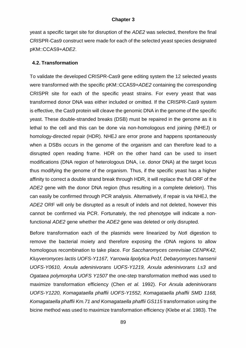
Chapter 3
89
yeast a specific target site for disruption of the ADE2 was selected, therefore the final
CRISPR-Cas9 construct were made for each of the selected yeast species designated
pKM::CCAS9+ADE2.
4.2. Transformation
To validate the developed CRISPR-Cas9 gene editing system the 12 selected yeasts
were transformed with the specific pKM::CCAS9+ADE2 containing the corresponding
CRISPR site for each of the specific yeast strains. For every yeast that was
transformed donor DNA was either included or omitted. If the CRISPR-Cas9 system
is effective, the Cas9 protein will cleave the genomic DNA in the genome of the specific
yeast. These double-stranded breaks (DSB) must be repaired in the genome as it is
lethal to the cell and this can be done via non-homologous end joining (NHEJ) or
homology-directed repair (HDR). NHEJ are error prone and happens spontaneously
when a DSBs occurs in the genome of the organism and can therefore lead to a
disrupted open reading frame. HDR on the other hand can be used to insert
modifications (DNA region of heterologous DNA, i.e. donor DNA) at the target locus
thus modifying the genome of the organism. Thus, if the specific yeast has a higher
affinity to correct a double strand break through HDR, it will replace the full ORF of the
ADE2 gene with the donor DNA region (thus resulting in a complete deletion). This
can easily be confirmed through PCR analysis. Alternatively, if repair is via NHEJ, the
ADE2 ORF will only be disrupted as a result of indels and not deleted, however this
cannot be confirmed via PCR. Fortunately, the red phenotype will indicate a non-
functional ADE2 gene whether the ADE2 gene was deleted or only disrupted.
Before transformation each of the plasmids were linearized by NotI digestion to
remove the bacterial moiety and therefore exposing the rDNA regions to allow
homologous recombination to take place. For Saccharomyces cerevisiae CENPK42,
Kluyveromyces lactis UOFS-Y1167, Yarrowia lipolytica Po1f, Debaryomyces hansenii
UOFS-Y0610, Arxula adeninivorans UOFS-Y1219, Arxula adeninivorans Ls3 and
Ogataea polymorpha UOFS Y1507 the one-step transformation method was used to
maximize transformation efficiency (Chen et al. 1992). For Arxula adeninivorans
UOFS-Y1220, Komagataella phaffii UOFS-Y1552, Komagataella phaffii SMD 1168,
Komagataella phaffii Km.71 and Komagataella phaffii GS115 transformation using the
bicine method was used to maximize transformation efficiency (Klebe et al. 1983). The

Chapter 3
90
concentration of hygromycin was reduced for transformation for a number of yeast to
accommodate for the reduction in growth due to the introduction of the CAS9 vector
as well as the gene disruption. This reduction in growth may have led to increased
susceptibility to hygromycin for some yeast, even when the yeast strains possessed
the necessary hygromycin resistance gene carried on the pKM::CCAS9+ADE2 vector.
The results of the transformation with or without donor DNA are listed in Table 10. In
this table the number of colonies obtained are indicated. For each yeast, 20 colonies
were randomly selected (except for A. adeninivorans UOFS-Y1220, where 10 were
selected) and streaked on YNB dropout plates, lacking adenine to observe how many
colonies turns red. This indicated if the CRISPR system were effective as well as the
level of efficiency in terms of deletion or disruption of the ADE2 gene. The number of
colonies that turned red versus those that stayed white are also indicated in Table 17.
As can be observed from Table 10, a moderate to high (i.e. 50% to 100%) efficiency
was obtained for most of the yeasts tested.
Table 10: Results for the total colonies obtained after transformation (with or without
donor DNA) and the results for the screening of ade2 mutants when streaked out on
the YNB dropout plates, lacking adenine.
Yeast
Transformed Colonies
With donor DNA Without donor DNA
Total Red/White Total Red/White
Saccharomyces cerevisiae CENPK42 27 15/20 64 18/20
Kluyveromyces lactis UOFS-Y1167 26 14/20 23 12/20
Yarrowia lipolytica Po1f 23 20/20 43 20/20
Debaryomyces hansenii UOFS-Y0610 27 20/20 206 20/20
Arxula adeninivorans UOFS-Y1220 13 4/10 15 1/10
Arxula adeninivorans UOFS-Y1219 126 20/20 TMTC 20/20
Arxula adeninivorans Ls3 TMTC* 18/20 TMTC 18/20

Chapter 3
91
Ogataea polymorpha UOFS Y1507 TMTC 10/20 TMTC 13/20
Komagataella phaffii UOFS-Y1552 64 4/20 154 6/20
Komagataella phaffii SMD 1168 55 10/20 165 11/20
Komagataella phaffii Km.71 56 14/20 104 11/20
Komagataella phaffii GS115 49 20/20 80 20/20
*Too many to count
Table 11 represents the HDR and NHEJ disruption efficiencies of the red colonies
obtained, for each yeast, when transformation with donor DNA was performed.
Table 11: HDR and NHEJ disruption results for the red colonies obtained after
transformation with donor DNA.
Yeast Transformed Colonies With donor DNA
HR (%) NHEJ (%)
Saccharomyces cerevisiae CENPK42 40 60
Kluyveromyces lactis UOFS-Y1167 36 64
Yarrowia lipolytica Po1f 50 50
Debaryomyces hansenii UOFS-Y0610 60 40
Arxula adeninivorans UOFS-Y1220 25 75
Arxula adeninivorans UOFS-Y1219 40 60
Arxula adeninivorans Ls3 50 50
Ogataea polymorpha UOFS Y1507 20 80
Komagataella phaffii UOFS-Y1552 0 100
Komagataella phaffii SMD 1168 0 100
Komagataella phaffii Km.71 0 100
Komagataella phaffii GS115 30 70

Chapter 3
92
Colonies selected from the transformations where donor DNA was added and that
turned red, were also further analysed via PCR analysis (see below results obtained
for each yeast strain). This indicated if the ADE2 ORF were replaced by the donor
DNA region, i.e. repair of the Cas9 induced double strand break via HDR resulting in
a complete deletion, or if the double stranded break was repaired via NHEJ.
4.3. Saccharomyces cerevisiae
In Saccharomyces cerevisiae, the addition of the donor DNA resulted in a reduction of
transformation efficiency and of the 20 colonies randomly selected, 15 had a red
phenotype, indicating a 75% disruption efficiency. Displayed in Figure.8 (B) are the
different colonies streaked out to indicate the ratio of successful vs unsuccessful gene
deleted mutants. The red colonies were then analysed through PCR to determine how
many of the 15 colonies contained the donor fragment, meaning the gene were
repaired through HR, and how many was repaired through NHEJ. A fragment size of
449 bp (the size of the donor fragment) will confirm the integration of the donor
fragment. A size of 1 716 bp (size of the original gene) will indicate that the gene was
disrupted through NHEJ. From Figure 8 (A) the total number of donor fragment repair
colonies obtained were 6 out of 15 red colonies (green arrow and box), resulting in a
HR-disruption efficiency of 40% when the yeast is transformed with a donor fragment.
Thus, giving the NHEJ a repair efficiency of 60%, 9 out of the 15, which are indicated
by the red arrow and box.

Chapter 3
93
Figure 8: A) ADE2 disruption results for Saccharomyces cerevisiae CENPK42 when
transformed with donor DNA. For the NHEJ repair mechanism, the PCR fragment size
will be 1 716 bp (red arrow and box) and for the HR repair mechanism, the fragment
size will be 449 bp (green arrow and box). B) On the plate are the wild type (WT) as
well as the different colonies streaked out to display the ratio of successful colonies
obtained vs unsuccessful. The pie chart also indicates the successful vs unsuccessful,
as well as the HR- vs NHEJ repair mechanisms regarding the disruption of the ADE2
gene.
4.4. Kluyveromyces lactis
In Kluyveromyces lactis the editing efficiency yielded was 70% when a donor fragment
was provided (14/20), which are indicated by the pie chart as well as the plate where
the ratio of successful vs unsuccessful ADE2 mutants have been streaked out (Figure
9 (B)). From Figure 9 (A) it can be seen that from the 14 red colonies analysed, only
5 contained the donor fragment (421 bp = green arrow and box) and the other 9 red
colonies was repaired through NHEJ (64%) due to their fragment sizes being the
original gene size (1 710 bp = red arrow and box). This indicates that when a donor
fragment was provided for editing, gene disruption via HR occurred in 36% of the red
colonies.
NHEJ Red
60%
HR Red
40%

Chapter 3
94
Figure 9: A) ADE2 disruption results for Kluyveromyces lactis UOFS-Y1167 when
transformed with donor DNA. For the NHEJ repair mechanism, the PCR fragment size
will be 1 710 bp (red arrow and box) and for the HR repair mechanism, the fragment
size will be 449 bp (green arrow and box). B) On the plate are the wild type (WT) as
well as the different colonies streaked out to display the ratio of successful vs
unsuccessful colonies obtained. The pie chart also indicates the successful vs
unsuccessful, as well as the HR- vs NHEJ repair mechanisms regarding the disruption
of the ADE2 gene.
4.5. Yarrowia lipolytica
In Yarrowia lipolytica an editing efficiency of 100% were obtained when a donor
fragment was included with transformation. This is also indicated in Figure 10 (B), by
the plated colonies in ratio of successful vs unsuccessful colonies. Figure 10(A)
indicates the gene disruption through HR (green arrow and box) occurred in 50% of
the colonies screened. The donor fragment size was 375 bp which were obtained in 9
out 18 of the red colonies screened. The other 50% were repaired via NHEJ (red arrow
and box), which was observed by PCR analysis when the other 10 colonies retained
the original gene size (1 698 bp).
NHEJ Red
64%
HR Red
36%

Chapter 3
95
Figure 10: A) ADE2 disruption results for Yarrowia lipolytica Po1f when transformed
with donor DNA. For the NHEJ repair mechanism, the PCR fragment size will be 1
716 bp (red arrow and box) and for the HR repair mechanism, the fragment size will
be 449 bp (green arrow and box). B) On the plate are the wild type (WT) as well as
the different colonies streaked out to display the ratio of successful colonies obtained
vs unsuccessful. The pie chart also indicates the successful vs unsuccessful, as well
as the HR- vs NHEJ repair mechanisms regarding the disruption of the ADE2 gene.
4.6. Debaryomyces hansenii
For Debaryomyces hansenii an editing efficiency of 100% were obtained when a donor
fragment was included with transformation. From Figure 11(A) can be seen that the
donor fragment has a size of 401 bp (green arrow and box), this fragment size was
obtained in 6 out of 15 of the red colonies when screened with PCR analysis. The
original gene size is 1 686 bp (red arrow and box) and it was obtained in the other 9
colonies screened. In Figure 11 (B) are the different colonies streaked out to indicate
the ratio of successful vs unsuccessful gene deleted mutants.
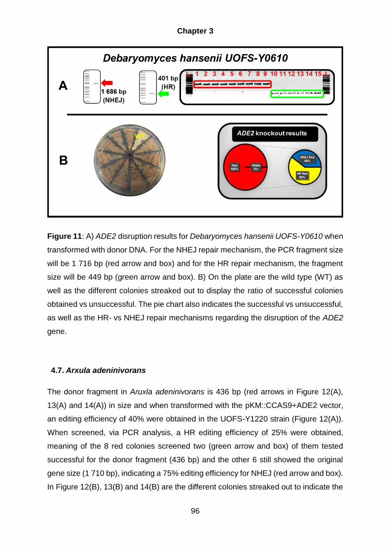
Chapter 3
96
Figure 11: A) ADE2 disruption results for Debaryomyces hansenii UOFS-Y0610 when
transformed with donor DNA. For the NHEJ repair mechanism, the PCR fragment size
will be 1 716 bp (red arrow and box) and for the HR repair mechanism, the fragment
size will be 449 bp (green arrow and box). B) On the plate are the wild type (WT) as
well as the different colonies streaked out to display the ratio of successful colonies
obtained vs unsuccessful. The pie chart also indicates the successful vs unsuccessful,
as well as the HR- vs NHEJ repair mechanisms regarding the disruption of the ADE2
gene.
4.7. Arxula adeninivorans
The donor fragment in Aruxla adeninivorans is 436 bp (red arrows in Figure 12(A),
13(A) and 14(A)) in size and when transformed with the pKM::CCAS9+ADE2 vector,
an editing efficiency of 40% were obtained in the UOFS-Y1220 strain (Figure 12(A)).
When screened, via PCR analysis, a HR editing efficiency of 25% were obtained,
meaning of the 8 red colonies screened two (green arrow and box) of them tested
successful for the donor fragment (436 bp) and the other 6 still showed the original
gene size (1 710 bp), indicating a 75% editing efficiency for NHEJ (red arrow and box).
In Figure 12(B), 13(B) and 14(B) are the different colonies streaked out to indicate the

Chapter 3
97
ratio of successful vs unsuccessful gene deleted mutants. The UOFS-1219 strain
[Figure 13(A)] yielded in a 40% HR editing efficiency (4 out of 10 indicated by the green
arrow and box) and a NHEJ efficiency of 60% (6 out of 10 indicated by the red arrow
and box).Lastly the Ls3 strain [Figure 14(A)] yielded 50% HR editing efficiency (9 out
of 18, indicated by the green arrow boxes) when fragment sizes of 436 bp was
observed. The other 9 out of 18 (50%) red colonies screened tested positive for NHEJ,
due to the original gene size (1 710 bp) that was obtained through PCR analysis.
Figure 12: A) ADE2 disruption results for Arxula adeninivorans UOFS-Y1220 when
transformed with donor DNA. For the NHEJ repair mechanism, the PCR fragment size
will be 1 716 bp (red arrow and box) and for the HR repair mechanism, the fragment
size will be 449 bp (green arrow and box). (B) On the plate are the wild type (WT) as
well as the different colonies streaked out to display the ratio of successful colonies
obtained vs unsuccessful. The pie chart also indicates the successful vs unsuccessful,
as well as the HR- vs NHEJ repair mechanisms regarding the disruption of the ADE2
gene.
NHEJ Red
75%
HR Red
25%
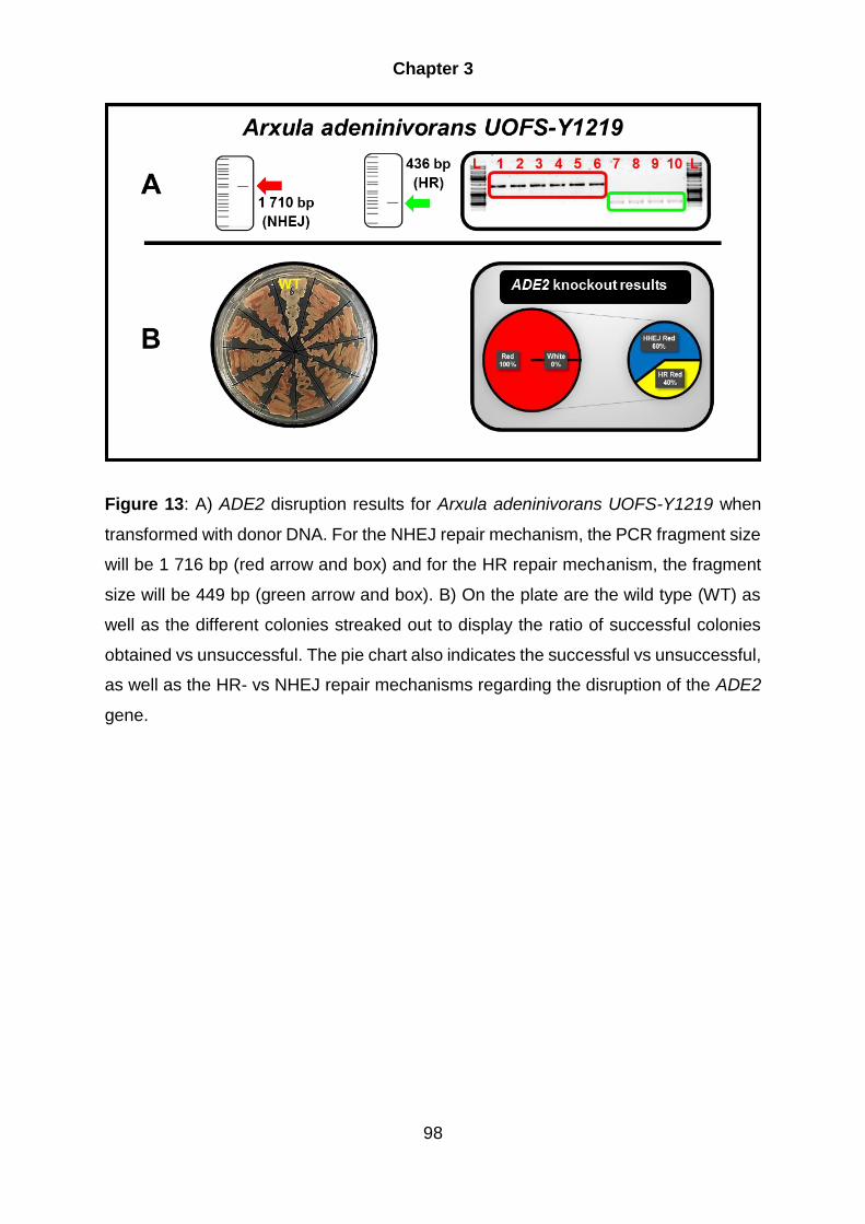
Chapter 3
98
Figure 13: A) ADE2 disruption results for Arxula adeninivorans UOFS-Y1219 when
transformed with donor DNA. For the NHEJ repair mechanism, the PCR fragment size
will be 1 716 bp (red arrow and box) and for the HR repair mechanism, the fragment
size will be 449 bp (green arrow and box). B) On the plate are the wild type (WT) as
well as the different colonies streaked out to display the ratio of successful colonies
obtained vs unsuccessful. The pie chart also indicates the successful vs unsuccessful,
as well as the HR- vs NHEJ repair mechanisms regarding the disruption of the ADE2
gene.

Chapter 3
99
Figure 14: A) ADE2 disruption results for Arxula adeninivorans Ls3 when transformed
with donor DNA. For the NHEJ repair mechanism, the PCR fragment size will be 1
716 bp (red arrow and box) and for the HR repair mechanism, the fragment size will
be 449 bp (green arrow and box). B) On the plate are the wild type (WT) as well as
the different colonies streaked out to display the ratio of successful colonies obtained
vs unsuccessful. The pie chart also indicates the successful vs unsuccessful, as well
as the HR- vs NHEJ repair mechanisms regarding the disruption of the ADE2 gene.
4.8. Ogataea polymorpha
Ogataea polymorpha displayed a 90% disruption efficiency, indicated by Figure 15(B)
with the streaked-out colonies in ratio of successful vs unsuccessful colonies. In
regards to HR, the editing efficiency obtained (green arrow and box) was 20%, due to
only 2 out of the 10 red colonies containing the donor fragment (443 bp) and the other
8 the original gene size (1 704 bp). Meaning the NHEJ efficiency was at 80%. In Figure
15 (B) are the different colonies streaked out to indicate the ratio of successful vs
unsuccessful gene deleted mutants, when donor DNA is included with the
transformation.
NHEJ Red
50%
HR Red
50%

Chapter 3
100
Figure 15: A) ADE2 disruption results for Ogataea polymorpha UOFS Y1507 when
transformed with donor DNA. For the NHEJ repair mechanism, the PCR fragment size
will be 1 716 bp (red arrow and box) and for the HR repair mechanism, the fragment
size will be 449 bp (green arrow and box). B) On the plate are the wild type (WT) as
well as the different colonies streaked out to display the ratio of successful colonies
obtained vs unsuccessful. The pie chart also indicates the successful vs unsuccessful,
as well as the HR- vs NHEJ repair mechanisms regarding the disruption of the ADE2
gene.
4.9. Komagataella phaffii
The Komagataella phaffii donor fragment is 396bp in size. However, in Figure 16(A),
strain UOFS-Y1552 yielded no donor fragments and only displayed the original size
(1 698 bp) of the gene. Meaning the 10% disruption efficiency obtained were through
NHEJ (red arrow and box). The same trend was observed in the SMD 1168 strain
(Figure 17(A), indicated by the red arrow and box), where all of the 10 red colonies
obtained portrayed the original size (1 698 bp) of the gene. This also happened in the
Km 71 strain (Figure 18 (A) indicated by the red arrow and box), the 14 red colonies
all had the original gene size.
NHEJ Red
80%
HR Red
20%

Chapter 3
101
However, successful HR disruption efficiency (30%) was obtained in the Gs 115 strain
where 10 red colonies were tested and 3 (indicated in Figure 19 (A) by the green arrow
and box) of the 10 displayed the donor fragment (396 bp), after PCR analysis. Also,
the other 7 colonies showed the original gene size (1 698 bp) resulting in a 70% NHEJ
efficiency.
In Figure 16 (B), 17(B), 18(B) and 19(B) are the different colonies streaked out to
indicate the ratio of successful vs unsuccessful gene deleted mutants, when the donor
DNA is included with the transformation.
Figure 16: A) ADE2 disruption results for Komagataella phaffii UOFS-Y1552 when
transformed with donor DNA. For the NHEJ repair mechanism, the PCR fragment size
will be 1 716 bp (red arrow and box) and for the HR repair mechanism, the fragment
size will be 449 bp (green arrow). B) On the plate are the wild type (WT) as well as the
different colonies streaked out to display the ratio of successful colonies obtained vs
unsuccessful. The pie chart also indicates the successful vs unsuccessful, as well as
the HR- vs NHEJ repair mechanisms regarding the disruption of the ADE2 gene.
NHEJ
100%
NHEJ Red
100%

Chapter 3
102
Figure 17: A) ADE2 disruption results for Komagataella phaffii SMD 1168 when
transformed with donor DNA. For the NHEJ repair mechanism, the PCR fragment size
will be 1 716 bp (red arrow and box) and for the HR repair mechanism, the fragment
size will be 449 bp (green arrow). B) On the plate are the wild type (WT) as well as the
different colonies streaked out to display the ratio of successful colonies obtained vs
unsuccessful. The pie chart also indicates the successful vs unsuccessful, as well as
the HR- vs NHEJ repair mechanisms regarding the disruption of the ADE2 gene.
NHEJ Red
100%
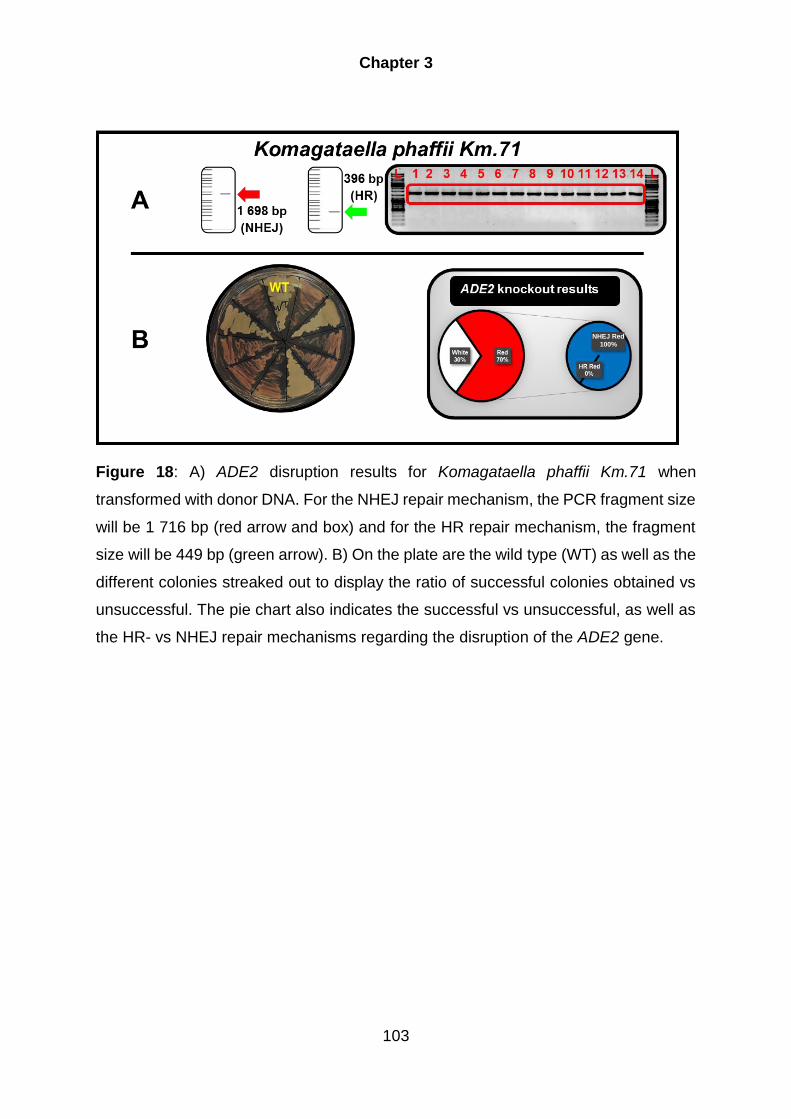
Chapter 3
103
Figure 18: A) ADE2 disruption results for Komagataella phaffii Km.71 when
transformed with donor DNA. For the NHEJ repair mechanism, the PCR fragment size
will be 1 716 bp (red arrow and box) and for the HR repair mechanism, the fragment
size will be 449 bp (green arrow). B) On the plate are the wild type (WT) as well as the
different colonies streaked out to display the ratio of successful colonies obtained vs
unsuccessful. The pie chart also indicates the successful vs unsuccessful, as well as
the HR- vs NHEJ repair mechanisms regarding the disruption of the ADE2 gene.
NHEJ Red
100%

Chapter 3
104
Figure 19: A) ADE2 disruption results for Komagataella phaffii GS115 when
transformed with donor DNA. For the NHEJ repair mechanism, the PCR fragment size
will be 1 716 bp (red arrow and box) and for the HR repair mechanism, the fragment
size will be 449 bp (green arrow and box). B) On the plate are the wild type (WT) as
well as the different colonies streaked out to display the ratio of successful colonies
obtained vs unsuccessful. The pie chart also indicates the successful vs unsuccessful,
as well as the HR- vs NHEJ repair mechanisms regarding the disruption of the ADE2
gene.
5. Conclusion
Although gRNA constructs, as part of an effective CRISPR-CAS9 gene editing system,
are designed to satisfy the special demands of the specific yeast, they nevertheless
follow similar construction principles. For the expression of the gRNA, Pol III promoters
have to be used, due to the extensive modification and processing that is applied when
Pol II promoters are used for transcription. However, Pol III promoters are very specific
for each organism. Thus, for the system to be classified as wide range, the expression
of the gRNA needs to be expressed using the same promoter in all of the yeast, hence
the development of a gRNA construct containing ribozymes on both the ends of the
gRNA. The ribozymes promote self-cleavage, thus removing any post transcriptional
modifications. In this study a gRNA construct were developed, which consisted of a
Yarrowia lipolytica TEF1 promoter and terminator, the gRNA (which consist of the

Chapter 3
105
CRISPR site followed by the gRNA scaffold) and ribozymes (HH and HDV) flanking
the gRNA.
After the development of the TEF1-RGR-gRNA construct, it was incorporated into the
pKM::CCAS9 vector to yield the pKM::CCAS9+CRISPR plasmid. Each of the ADE2
target sites of each yeast were then ligated into the pKM::CCAS9+CRISPR to yield
the pKM::CCAS9+ADE2. The vectors were successfully transformed into the different
yeasts either in the presence or absence of donor DNA. The transformed colonies
were tested for ADE2 inactivation using YNB dropout plates lacking adenine. If
successful for gene disruption/deletion the colonies obtained a red phenotype due to
the cells that oxidizes the accrued P-ribosylaminoimidazole. The results indicated that
the wide range CRISPR-Cas9 system were successful in disrupting the ADE2 gene in
all of the yeasts tested. When the donor DNA were included in the transformation, it
was shown that NHEJ were the preferred repair mechanism. However, the HR
efficiency can be increased in most yeast by modifying and optimizing the donor
fragment. A more direct approach can be taken and the NHEJ mechanism can be
disrupted through inactivation of the KU70 and/or KU80 gene(s) (Boulton 1996; Daley
et al. 2005; Verbeke et al. 2013; Kretzschmar et al. 2013; Juergens et al. 2018). These
genes are crucial in the NHEJ repair pathway and inactivation of these genes results
in a decrease in NHEJ repair mechanism.
This proven efficacy of the developed wide range CRISPR-CAS9 gene editing system
provides an efficient and precise method to target any gene. This is the first reported
system to be tested in such a broad range of yeasts, which include seven
biotechnologically relevant yeast, with some of the yeasts tested consisting of more
than one strain. For the yeasts included in this study, moderate to high (50% to 100%)
disruption or deletion efficiencies were obtained, with the exception of one strain of A.
adeninivorans (40%) and one strain of K. phaffii (30%).
To conclude, this system’s gene editing efficiency indicated comparable results to any
developed CRISPR-Cas9 system, proving that this system serves as a valuable, cost
efficient tool for easy and fast gene disruption in a number of yeast species.
Chapter 3
106
6. References
Addgene., (2015) 'Addgene'. https://www.addgene.org/. Accessed, January 30,
(2019)
Bétermier, M.,Bertrand, P.,& Lopez, B.S., (2014) 'Is Non-Homologous End-Joining
Really an Inherently Error-Prone Process?'. PLoS Genet 10, e1004086. doi:
10.1371/journal.pgen.1004086
Boulton, S., (1996) 'Identification of a Saccharomyces cerevisiae Ku80 homologue:
roles in DNA double strand break rejoining and in telomeric maintenance'. Nucleic
Acids Res 24, 4639–4648. doi: 10.1093/nar/24.23.4639
Chen, D.C.,Yang, B.C.,& Kuo, T.T., (1992) 'One-step transformation of yeast in
stationary phase'. Curr Genet 21, 83–84. doi: 10.1007/BF00318659
Chung, C.T.,Niemela, S.L.,& Miller, R.H., (1989) 'One-step preparation of competent
Escherichia coli: transformation and storage of bacterial cells in the same
solution.'. Proc Natl Acad Sci 86, 2172–2175. doi: 10.1073/pnas.86.7.2172
Daley, J.M.,Palmbos, P.L.,Wu, D.,& Wilson, T.E., (2005) 'Nonhomologous End Joining
in Yeast'. Annu Rev Genet 39, 431–451. doi:
10.1146/annurev.genet.39.073003.113340
Devi, S.,Kedlaya, R.,Maddodi, N.,et al., (2009) 'Calcium homeostasis in human
melanocytes: role of transient receptor potential melastatin 1 (TRPM1) and its
regulation by ultraviolet light'. Am J Physiol Physiol 297, C679–C687. doi:
10.1152/ajpcell.00092.2009
DiCarlo, J.E.,Norville, J.E.,Mali, P.,et al., (2013) 'Genome engineering in
Saccharomyces cerevisiae using CRISPR-Cas systems'. Nucleic Acids Res 41,
4336–4343. doi: 10.1093/nar/gkt135
Friedland, A.E.,Tzur, Y.B.,Esvelt, K.M.,et al., (2013) 'Heritable genome editing in C.
elegans via a CRISPR-Cas9 system'. Nat Methods 10, 741–743. doi:
10.1038/nmeth.2532
Gao, Y.,& Zhao, Y., (2014) 'Self-processing of ribozyme-flanked RNAs into guide
RNAs in vitro and in vivo for CRISPR-mediated genome editing'. J Integr Plant

Chapter 3
107
Biol 56, 343–349. doi: 10.1111/jipb.12152
Geneious., (2017) 'Geneious R10'. http://www.geneious.com/. Accessed, January 30,
(2019)
Gratz, S.J.,Cummings, A.M.,Nguyen, J.N.,et al., (2013) 'Genome Engineering of
Drosophila with the CRISPR RNA-Guided Cas9 Nuclease'. Genetics 194, 1029–
1035. doi: 10.1534/genetics.113.152710
Haft, D.H.,Selengut, J.,Mongodin, E.F.,& Nelson, K.E., (2005) 'A guild of 45 CRISPR-
associated (Cas) protein families and multiple CRISPR/Cas subtypes exist in
prokaryotic genomes.'. PLoS Comput Biol 1, e60. doi:
10.1371/journal.pcbi.0010060
Haurwitz, R.E.,Jinek, M.,Wiedenheft, B.,et al., (2010) 'Sequence- and Structure-
Specific RNA Processing by a CRISPR Endonuclease'. Science (80- ) 329, 1355–
1358. doi: 10.1126/science.1192272
Horwitz, A.A.,Walter, J.M.,Schubert, M.G.,et al., (2015) 'Efficient Multiplexed
Integration of Synergistic Alleles and Metabolic Pathways in Yeasts via CRISPR-
Cas'. Cell Syst 1, 88–96. doi: 10.1016/j.cels.2015.02.001
Hwang, W.Y.,Fu, Y.,Reyon, D.,et al., (2013) 'Efficient genome editing in zebrafish
using a CRISPR-Cas system'. Nat Biotechnol 31, 227–229. doi: 10.1038/nbt.2501
Jiang, W.,Zhou, H.,Bi, H.,et al., (2013) 'Demonstration of CRISPR/Cas9/sgRNA-
mediated targeted gene modification in Arabidopsis, tobacco, sorghum and rice'.
Nucleic Acids Res 41, e188–e188. doi: 10.1093/nar/gkt780
Jinek, M.,Chylinski, K.,Fonfara, I.,et al., (2012) 'A Programmable Dual-RNA-Guided
DNA Endonuclease in Adaptive Bacterial Immunity'. Science (80- ) 337, 816–821.
doi: 10.1126/science.1225829
Juergens, H.,Varela, J.A.,Gorter de Vries, A.R.,et al., (2018) 'Genome editing in
Kluyveromyces and Ogataea yeasts using a broad-host-range Cas9/gRNA co-
expression plasmid'. FEMS Yeast Res 18, . doi: 10.1093/femsyr/foy012
KAPA Biosystems Inc., (2014) 'KAPA HiFi HotStart ReadyMix PCR Kit Technical Data
Sheet'. https://www.kapabiosystems.com/product-applications/products/pcr-
2/kapa-hifi-pcr-kits/. Accessed, January 28, (2019)

Chapter 3
108
Kistler, K.E.,Vosshall, L.B.,& Matthews, B.J., (2015) 'Genome Engineering with
CRISPR-Cas9 in the Mosquito Aedes aegypti'. Cell Rep 11, 51–60. doi:
10.1016/j.celrep.2015.03.009
Klebe, R.J.,Harriss, J. V.,Sharp, Z.D.,& Douglas, M.G., (1983) 'A general method for
polyethylene-glycol-induced genetic transformation of bacteria and yeast.'. Gene
25, 333–341
Kretzschmar, A.,Otto, C.,Holz, M.,et al., (2013) 'Increased homologous integration
frequency in Yarrowia lipolytica strains defective in non-homologous end-joining'.
Curr Genet 59, 63–72. doi: 10.1007/s00294-013-0389-7
Makarova, K.S.,Grishin, N. V.,Shabalina, S.A.,et al., (2006) 'A putative RNA-
interference-based immune system in prokaryotes: Computational analysis of the
predicted enzymatic machinery, functional analogies with eukaryotic RNAi, and
hypothetical mechanisms of action'. Biol Direct 1, 1–7. doi: 10.1186/1745-6150-
1-7
New England Biolabs., (2017) 'NEBuilder HiFi'. https://www.neb.com/-
/media/catalog/datacards-or-manuals/manuale2621.pdf. Accessed, January 25,
(2019)
New England Biolabs Inc., (2014) 'NEBcloner'. http://nebcloner.neb.com/#!/redigest.
Accessed, January 07, (2019)
New England BioLabs Inc., (2017) 'NEB PCR Cloning Kit Instruction Manual'.
https://www.neb.com/-/media/catalog/datacards-or-manuals/manuale1202.pdf.
Accessed, January 25, (2019)
Novagen., (2011) 'KOD Hot Start DNA Polymerase protocol'. http://goo.gl/UWwdYi.
Accessed, January 25, (2019)
Pâques, F.,& Haber, J.E., (1999) 'Multiple pathways of recombination induced by
double-strand breaks in Saccharomyces cerevisiae.'. Microbiol Mol Biol Rev 63,
349–404
Pitcher, R.S.,Brissett, N.C.,& Doherty, A.J., (2007) 'Nonhomologous End-Joining in
Bacteria: A Microbial Perspective'. Annu Rev Microbiol 61, 259–282. doi:
10.1146/annurev.micro.61.080706.093354

Chapter 3
109
Sung, P.,& Klein, H., (2006) 'Mechanism of homologous recombination: mediators and
helicases take on regulatory functions'. Nat Rev Mol Cell Biol 7, 739–750. doi:
10.1038/nrm2008
Thermo Fisher Scientific Inc., (2015a) 'Thermo Scientific GeneJET Gel Extraction Kit'.
https://assets.thermofisher.com/TFS-
Assets/LSG/manuals/MAN0012661_GeneJET_Gel_Extraction_UG.pdf.
Accessed, January 07, (2019)
Thermo Fisher Scientific Inc., (2015b) 'Restriction Enzymes–Thermo Scientific - ZA'.
https://www.thermofisher.com/za/en/home/brands/thermo-scientific/molecular-
biology/thermo-scientific-restriction-modifying-enzymes/restriction-enzymes-
thermo-scientific.html. Accessed, January 07, (2019)
Ugolini, S.,& Bruschi, C. V., (1996) 'The red/white colony color assay in the yeast
Saccharomyces cerevisiae: Epistatic growth advantage of white ade8-18, ade2
cells over red ade2 cells'. Curr Genet 30, 485–492. doi: 10.1007/s002940050160
Verbeke, J.,Beopoulos, A.,& Nicaud, J.-M., (2013) 'Efficient homologous
recombination with short length flanking fragments in Ku70 deficient Yarrowia
lipolytica strains'. Biotechnol Lett 35, 571–576. doi: 10.1007/s10529-012-1107-0
Vyas, V.K.,Barrasa, M.I.,& Fink, G.R., (2015) 'A Candida albicans CRISPR system
permits genetic engineering of essential genes and gene families'. Sci Adv 1,
e1500248. doi: 10.1126/sciadv.1500248
Wang, H.,Yang, H.,Shivalila, C.S.,et al., (2013) 'One-step generation of mice carrying
mutations in multiple genes by CRISPR/cas-mediated genome engineering'. Cell
153, 910–918. doi: 10.1016/j.cell.2013.04.025
Weninger, A.,Hatzl, A.-M.,Schmid, C.,et al., (2016) 'Combinatorial optimization of
CRISPR/Cas9 expression enables precision genome engineering in the
methylotrophic yeast Pichia pastoris'. J Biotechnol 235, 139–149. doi:
10.1016/j.jbiotec.2016.03.027
Yan, H.,Opachaloemphan, C.,Mancini, G.,et al., (2017) 'An Engineered orco Mutation
Produces Aberrant Social Behavior and Defective Neural Development in Ants'.
Cell 170, 736–747.e9. doi: 10.1016/j.cell.2017.06.051

Chapter 3
110

Chapter 4
Summary and concluding remarks
1. Summary and concluding remarks
The CRISPR-Cas9 tool consists of the Cas9 enzyme and a gRNA (Jinek et al. 2012)
that binds to the Cas9 enzyme and guides it to the target DNA. This is then followed
by the binding of the Cas9-gRNA complex to the target DNA, where the Cas9 enzyme
then cleaves the DNA and creates double stranded breaks.
Application of the CRISPR-Cas9 system relies on the effective regulation (i.e.
homozygous promoter regions) and translation (i.e. codon optimisation) of the CAS9
gene in each yeast host (DiCarlo et al. 2013; Gao & Zhao 2014; Ryan et al. 2014;
Zhang et al. 2014; Bao et al. 2015; Horwitz et al. 2015; Generoso et al. 2016). For the
gRNA, expression is very important to obtain the correct functionality and therefore
the choice of the correct promoter is essential (Gao & Zhao 2014). If any RNA
Polymerase II promoters are used, in for example yeasts, the RNA produced will
contain elements added during post-translational modifications because these
promoters produces mRNA. Thus, for functional expression, a non-mRNA producing
promoter needs to be used. Fortunately, the use of the self-catalysing characteristic
of ribozymes can circumvent these problems. Therefore, any promoter can be used
as long as the gRNA is flanked by self-splicing ribozymes to remove any post
translational modifications, resulting in a functional gRNA.
This study describes new adaptations to CRISPR-Cas9 systems through the
development of a new CRISPR-Cas9 system for easy and fast gene editing in a wide
variety of different yeast species. Chapter 2 discussed the construction of three
different pKM::CAS9 expression constructs, each with a different codon optimized
CAS9 [P. pastoris (PCAS9), C. albicans (CCAS9) and H. sapiens (HCAS9)] (Gao &
Zhao 2014; Vyas et al. 2015; Weninger et al. 2016). Each of the CAS9 genes
contained one or more nuclear localization sequence to ensure localization to the

Chapter 4
112
nucleus. These CAS9 expression constructs consisted of the pKM180 backbone,
which makes use of a part of the rRNA locus from Kluyveromyces marxianus for
integration into the yeasts and selection of positive transformants is facilitated by the
dominant selection of Hygromycin via the constitutively driven Hygromycin resistant
(HPH) gene. The three different CAS9 constructs were transformed into seven
different biotechnologically relevant yeasts using two different chemical transformation
protocols (Klebe et al. 1983; Chen et al. 1992) to test for Cas9 expression and the
effect that each of the Cas9 proteins have on each of the yeasts. Cas9 expression
was subsequently confirmed through western blot analysis. It was observed that
expression of the P. pastoris optimised CAS9 resulted in multiple protein bands when
analysed using a western blot. Expression of the H. sapiens lead to slower growth of
transformed yeasts as well as increased sensitivity toward Hygromycin. It was
therefore apparent that the pKM::CCAS9 vector (i.e. the pKM180 containing the C.
albicans optimised CAS9 gene) was the best candidate to proceed with the testing of
complete CRISPR-CAS9 gene editing system.
For the construction of the complete wide rage CRISPR-CAS9 gene editing system,
where a single system could be used in a range of different yeasts, it was essential to
use a promoter for the expression of the gRNA that could be recognised by a range of
different yeast species. For this purpose we made use of the RGR-gRNA strategy,
where the gRNA is flanked by self-splicing ribozymes (Gao & Zhao 2014). The RGR-
gRNA was incorporated into the vector which resulted in a vector that consist of a
CCAS9 gene and a gRNA construct which is flanked by ribozymes.
To target the ADE2 gene in each of the yeast species, a specific CRISPR target
sequence was designed for each yeast and inserted in the completed
pKM::CCAS9+CRISPR vector. This vector was then transformed into the different
yeasts to determine the efficiency of inactivation (either through disruption or deletion)
of the ADE2 gene. Inactivation of the ADE2 gene results in an observable red
phenotype if plated on media that lack adenine. When the vector was transformed with
or without donor DNA, all yeast species (depending on the strain selected) showed
moderate to high deletion or disruption efficiency of the ADE2 gene. It should be noted
that when donor DNA was included in the transformation, most of the yeast displayed
gene disruption through homologous recombination (HR). Better HR disruption

Chapter 4
113
efficiency can possibly be obtained by modifying the donor fragment to obtain optimal
integration (via HR) of the donor fragment. Enhanced HR is also likely by disrupting
the KU70/80 genes (Boulton 1996; Daley et al. 2005; Verbeke et al. 2013;
Kretzschmar et al. 2013; Juergens et al. 2018b). These genes are crucial in the NHEJ
repair pathway. Thus, by disrupting these genes a decrease in NHEJ and an increase
in HR disruption efficiencies will be observed.
In conclusion, the developed wide range CRISPR-CAS9 gene editing system proved
to be successful by disrupting the ADE2 gene in all of the yeast tested. This system
therefore allows rapid, cost-effective genetic manipulation of biotechnologically
relevant yeast.
2. References
Bao, Z.,Xiao, H.,Liang, J.,et al., (2015) 'Homology-Integrated CRISPR–Cas (HI-
CRISPR) System for One-Step Multigene Disruption in Saccharomyces
cerevisiae'. ACS Synth Biol 4, 585–594. doi: 10.1021/sb500255k
Boulton, S., (1996) 'Identification of a Saccharomyces cerevisiae Ku80 homologue:
roles in DNA double strand break rejoining and in telomeric maintenance'.
Nucleic Acids Res 24, 4639–4648. doi: 10.1093/nar/24.23.4639
Chen, D.C.,Yang, B.C.,& Kuo, T.T., (1992) 'One-step transformation of yeast in
stationary phase'. Curr Genet 21, 83–84. doi: 10.1007/BF00318659
Daley, J.M.,Palmbos, P.L.,Wu, D.,& Wilson, T.E., (2005) 'Nonhomologous End
Joining in Yeast'. Annu Rev Genet 39, 431–451. doi:
10.1146/annurev.genet.39.073003.113340
DiCarlo, J.E.,Norville, J.E.,Mali, P.,et al., (2013) 'Genome engineering in
Saccharomyces cerevisiae using CRISPR-Cas systems'. Nucleic Acids Res 41,
4336–4343. doi: 10.1093/nar/gkt135
Gao, Y.,& Zhao, Y., (2014) 'Self-processing of ribozyme-flanked RNAs into guide
RNAs in vitro and in vivo for CRISPR-mediated genome editing'. J Integr Plant
Biol 56, 343–349. doi: 10.1111/jipb.12152
Generoso, W.C.,Gottardi, M.,Oreb, M.,& Boles, E., (2016) 'Simplified CRISPR-Cas
genome editing for Saccharomyces cerevisiae'. J Microbiol Methods 127, 203–

Chapter 4
114
205. doi: 10.1016/j.mimet.2016.06.020
Horwitz, A.A.,Walter, J.M.,Schubert, M.G.,et al., (2015) 'Efficient Multiplexed
Integration of Synergistic Alleles and Metabolic Pathways in Yeasts via
CRISPR-Cas'. Cell Syst 1, 88–96. doi: 10.1016/j.cels.2015.02.001
Jinek, M.,Chylinski, K.,Fonfara, I.,et al., (2012) 'A Programmable Dual-RNA-Guided
DNA Endonuclease in Adaptive Bacterial Immunity'. Science (80- ) 337, 816–
821. doi: 10.1126/science.1225829
Juergens, H.,Varela, J.A.,Gorter de Vries, A.R.,et al., (2018) 'Genome editing in
Kluyveromyces and Ogataea yeasts using a broad-host-range Cas9/gRNA co-
expression plasmid'. FEMS Yeast Res 18, . doi: 10.1093/femsyr/foy012
Klebe, R.J.,Harriss, J. V.,Sharp, Z.D.,& Douglas, M.G., (1983) 'A general method for
polyethylene-glycol-induced genetic transformation of bacteria and yeast.'. Gene
25, 333–341
Kretzschmar, A.,Otto, C.,Holz, M.,et al., (2013) 'Increased homologous integration
frequency in Yarrowia lipolytica strains defective in non-homologous end-
joining'. Curr Genet 59, 63–72. doi: 10.1007/s00294-013-0389-7
Ryan, O.W.,Skerker, J.M.,Maurer, M.J.,et al., (2014) 'Selection of chromosomal DNA
libraries using a multiplex CRISPR system.'. Elife 3, 1–15. doi:
10.7554/eLife.03703
Verbeke, J.,Beopoulos, A.,& Nicaud, J.-M., (2013) 'Efficient homologous
recombination with short length flanking fragments in Ku70 deficient Yarrowia
lipolytica strains'. Biotechnol Lett 35, 571–576. doi: 10.1007/s10529-012-1107-0
Vyas, V.K.,Barrasa, M.I.,& Fink, G.R., (2015) 'A Candida albicans CRISPR system
permits genetic engineering of essential genes and gene families'. Sci Adv 1,
e1500248. doi: 10.1126/sciadv.1500248
Weninger, A.,Hatzl, A.-M.,Schmid, C.,et al., (2016) 'Combinatorial optimization of
CRISPR/Cas9 expression enables precision genome engineering in the
methylotrophic yeast Pichia pastoris'. J Biotechnol 235, 139–149. doi:
10.1016/j.jbiotec.2016.03.027
Zhang, G.-C.,Kong, I.I.,Kim, H.,et al., (2014) 'Construction of a Quadruple
Auxotrophic Mutant of an Industrial Polyploid Saccharomyces cerevisiae Strain

Chapter 4
115
by Using RNA-Guided Cas9 Nuclease'. Appl Environ Microbiol 80, 7694–7701.
doi: 10.1128/AEM.02310-14