the development in vitro of the blood of the early chick embryo
TRANSCRIPT

The Development in vitro of the Blood of the Early Chick EmbryoAuthor(s): P. D. F. MurraySource: Proceedings of the Royal Society of London. Series B, Containing Papers of aBiological Character, Vol. 111, No. 773 (Oct. 1, 1932), pp. 497-521Published by: The Royal SocietyStable URL: http://www.jstor.org/stable/81556 .
Accessed: 08/05/2014 06:38
Your use of the JSTOR archive indicates your acceptance of the Terms & Conditions of Use, available at .http://www.jstor.org/page/info/about/policies/terms.jsp
.JSTOR is a not-for-profit service that helps scholars, researchers, and students discover, use, and build upon a wide range ofcontent in a trusted digital archive. We use information technology and tools to increase productivity and facilitate new formsof scholarship. For more information about JSTOR, please contact [email protected].
.
The Royal Society is collaborating with JSTOR to digitize, preserve and extend access to Proceedings of theRoyal Society of London. Series B, Containing Papers of a Biological Character.
http://www.jstor.org
This content downloaded from 169.229.32.137 on Thu, 8 May 2014 06:38:44 AMAll use subject to JSTOR Terms and Conditions

497
6II - OI3. 68
The Development in vitro of the Blood of the early, Chick Embryo.
By P. D. F'. MURRAY, Smithson lResearch Fellow.
(From the Strangeways Research Laboratory, Camlbridge.)
(Communicated by J. T. Wilson, F.R.S.-iReceived June 9, 1932.)
[PLATES 22-25.]
Introduction.
In a series of tissue cultures of fragments taken from different parts of young chick embryos, at various stages, it was found that differentiation frequently occurred, the characteristic cells of highly specialised tissues appearing in cultuires in which they could not possibly have been present at explantation. The most striking differentiations were the development of r ed blood corpuscles,. capillary vessels, nerve cells with axons and of rhythmically contracting cardiac muscle.
This paper describes a simple method by which may be obtained regularly and quickly the development of very large numbers of red cells, and an account is given of the histology of the cultures. It is intended to lbe introductory to a, physiological study, now in progress, of the conditions of haematopoiesis Literature. Erythropoiesis in vitro has been reported by several authors, but it has not been thoroughly investigated and, with the exceptions of the works of Slonimski (1930, a, 1931) and Shipley (1915-16), the earlier papers have con- cerned the somewhat sporadic appearance of small numbers of erythrocytes. In the earlier works, also, the explants were derived from haamatopoietic organs, or from that part of the embryo in which blood would normally have developed at latest quite soon after the time at which the experimeint was made. The present paper, on the other hand, is based principally upon cultures of fragments of the primitive streak-that is, upon explants of presumptively haematopoietic cells isolated before they had arrived at the normal heemnatopoietic region of the embryo. The literature may be very briefly summiarised as follows: Shipley (1915-16) mnade plasma cultures from the area opacca of chick embryos at a time prior to the formation of the blood islands, and obtained the differen-
tiation of erythrocytes -from amoeboid cells. Erythropoiesis is reported by N. G. and A. L. Chlopin (1925) in cultures of Axolotl spleen; by Erdmann, Eisner, and Laser (1925-26), in cultures of embryonal rat spleen; by Freifeld and Ginsburg (1927) in cultures of rabbit adrenals; by de IHaan (1928-29) in
This content downloaded from 169.229.32.137 on Thu, 8 May 2014 06:38:44 AMAll use subject to JSTOR Terms and Conditions

498 P. D. F. Murray.
cultures of blood cells of the horse; by Timofejewsky and Benewolenskaja (1929) in cultures of blood from a case of acute myeloid leukaemia, and by Benewolenskaja (1930) in cult-ures of embryonal human liver. Slonimski (1930 a, 1931), using Rana fusca and Axolotl embryos, excised the blood island zone at early stages, and kept it as a culture enclosed in a sheath of epiblastic epithelium. The little cyst became full of red blood in an abundant plasma, and there were vessels with endothelial walls.
Nomenclature.-In her study of the early stages of heematopoiesis in the area vasculosa of the living chick embryo, Sabin (1920) rightly distinguishes between the blood islands and the " angioblasts," pointing out that the one term " blood island " should not be used for these two different structures. The " angioblasts " are masses of cells or single cells formed from the mesen- chlyme, from which develop the endothelial vessels containing plasma and the blood islands ; the blood islands proper are the groups of cells or single cells derived directly from the " angioblasts," which persist attached to the endothelial walls, and whose component cells, when separated from one another by the dissolution of the blood islands, become the primitive blood cells. Such a distinction is justified, for " angioblasts " and blood islands are two distinct developmental stages differing from one another in prospective signifi- cance and in structural relationships. To " angioblast," however, I prefer the term " hoemangioblast." This expresses the fact that both endothelium and blood develop from the solid mass, whereas the term " angioblast " strictly refers only to the vessels, i.e., to the endothelium. To avoid ambiguity, to accord with His's use of the term " angioblast " to mean a layer of cells rather than individual cells, and in order to avoid the clumsy phrases " hoemangio- blastic mass " and " mass of hoemangioblasts," I restrict the term " hTmangio- blast" to the mass and use for the component cells the infrequently needed term "ha3mangioblast cell."
MATERIAL AND MTETHODS.
Material.-All the cultures with which this paper is concerned were derived from embryos having a primitive streak. The stages varied from that of the early, pear-shaped area pellucida with a well-developed primitive streak, but no head process, to that at which there was an advanced head process, and a head fold but no somites. The great majority of the explants were posterior halves of primitive streaks of embryos taken shortly before or soon after the appearance of the head process.
This content downloaded from 169.229.32.137 on Thu, 8 May 2014 06:38:44 AMAll use subject to JSTOR Terms and Conditions

Development in vitro of the Blood of early Chick Embryo. 499
Eggs incubated for periods likely to provide embryos at suitable stages (the
time varied greatly from egg to egg and in different seasons) were opened, the
albumen was poured off, and the yolks decanted into petrie dishes containing
0 * 75 per cent. saline. The blastoderm was then dissected off, cleaned of yolk,
and transferred to some smaller vessel, usually a watch glass. The part
which it was intended to cultivate was cut out of the embryo with needles
sharpened into knives.
Media and Method of Cultivation.-Two different media were used, one fluid
and the other coagulated. For the study of the later stages of erythropoiesis,
if material were required for sectioning, and for experimLents intended to
delimit the regions in the embryo occupied by haematopoietic cells, I used plasma
and embryo extract. For the early stages of haematopoiesis, up to about the
stage of dissolution of the blood islands, and if sections were not required,
I used a fluid medium which was the exudate obtained by mixing plasma and
extract in the proportions of two to three, allowing the mixture to clot, and
cutting up the clot. This medium has the advantage of being easily removable
at fixation, so not obscuring the stained specimen. In addition, it favours
the formation of thinner cultures than the coagulated medium, and such cultures
can more easily be stained as whole mounts. But in stages after the dissolu-
tion of the blood islands, serum and extract constitute an unsuitable medium,
for there is a tendency for the cells to develop abnormally or to cease develop-
ment. For this reason I have used plasma and embryo extract for all later
stages. Observations based on fluid medium cultures have all been confirmed
with the plasma medium.
Fluid medium cultures were made on 8-inch cover glasses. To obtain
attachment of the usually very small explants to the cover glass the whole
preparation was kept upside down during the first night, and then returned
to the normal position; this ensured the attachment of all but; a very few of the
explants. Those which failed to become attached and remained floating
became nodular, and nothing could be seen of their structure; they passed
through at least the early stages of hematopoiesis, producing great numbers
of small free young blood cells.
To study the structure of the blood islands and of their constituent cells,
in whole mounts, it is essential to have blood islands which are thin enough
to allow observation both during life and after staining. Primitive streak
cultures usually develop one or several large blood islands containing so many
cells that no structural details can be discerned. In order to obtain very
small, thin blood islands, I made a large number of cultures in which the piece
This content downloaded from 169.229.32.137 on Thu, 8 May 2014 06:38:44 AMAll use subject to JSTOR Terms and Conditions

500 P. D. F. Murray.
of primitive streak had been cut into a number of small fragments. This did not seem to affect subsequent growth and differentiation. In spite of the extremely small size of the tiny fragments now present on each cover glass,, inversion of the cultures overnight always ensured that some at least of the explants would become attached to the glass, and grow as very tiny and very thin sheets, in which the structure of the blood islands could be seen. It is interesting to note that even extremely tiny cultures, consisting of only a very few cells, were able to pass through at least the initial stages of haematopoiesis without abnormality.
Since dissolution of the blood islands ocecurred in the first two days or less, and since fluid medium cultures were not used for later stages, it was not necessary to change the medium. Plasma cultures intended for the study of
later stages were sometimes transferred to fresh medium about twenty-four
hours after explantation, and were not transferred again. If the change were
delayed until after the dissolution of the blood islands, all or most of the cells
floating in the liquefied medium were lost. In many cases I did not transfer to fresh medium at all, having found that the omission of the change did not
adversely affect the development of the erythroblasts.
Histological Methods.-The material obtained in the cultures was studied in
whole mounts, smears, and sections. Most of the cultures intended for whole
mounts or sections were fixed in Zenker containing 10 per cent. formol, but a
few, during the early part of the work, in Zenker containing 3 per cent. acetic
acid. As stains I used chiefly Giemsa (Gurr and Grubler), to a lesser extent. Eosin Azur II and Dominici's triple Eosin Orange G-Toluidin Blue method as
modified by Dantschakoff (1908). Giemsa proved the most useful stain.
Stained preparations were differentiated in absolute alcohol, and were then
passed through acetone: usually through graded mixtures of absolute alcohol and acetone. Acetone differentiates very slowly, and could therefore be used
for dehydration, particularly with plasma cultures in which a great thickness of
relatively impenetrable plasma made rapid dehydration impossible. Prepara-
tions mounted in Gturr's nieutral balsam showed, after a period of months, little or no fading.
The cells floating in the fluid after dissolution of the blood islands were
studied in smears, which were dried by waving vigorously in the air and fixed
rapidly in met,hyl alcohol. This was followed by staining in one or other of the above mixtures, and mounting in Gurr's neutral balsam. As smears, however carefully prepared, are liable to be a source of artefacts. I have carefully confirmed the results given by them with those given by cultures
This content downloaded from 169.229.32.137 on Thu, 8 May 2014 06:38:44 AMAll use subject to JSTOR Terms and Conditions

Development in vitro of the Blood of early Chick Embryo. 501
fixed in Zenker-formol. Discrepancies between the results of the two methods are discussed in the descriptive sectioin.
PART 1.
Gross Changes in the Cultures.
On the morning of the day after explantation the attachment of the cultures was complete, and outwandering had usually begun. By the eveniing a considerable number of cells had emigrated, and the central part of the culture consisted of a dense mass, the heemangioblast. Each culture usually formed one large haemangioblast, but sometimes produced several; especially was this so with cultures from parts of the embryo normally destined to be incorporated in the area vasculosa, but it was rare for a culture derived from the primitive streak to form more than one mass. During the night, sometimes before mid- night, dissolution of the blood islands commenced, and was usually in full progress by next morning. In a fluid medium, or in plasma cultures in which the usual liquefaction of the plasma had occurred in the neighbourhood of the explants, the greater part of the centres of the cultures disappeared, and floating about in the fluid were innumerable small round cells. I shall refer to these floating cells, because of their great number, as " clouds." Examination of dense parts of a cloud at this stage gave the impression that ha3moglobin was present, but the slight yellowish tinge was not sufficiently definite for certainty. The cells still attached to the cover glass consisted of epithelium, mesenchyme, some wandering cells, and a few primitive blood cells. By the following morning the cells in the cloud began to show a decided tendency to elongate, approaching the definitive form of red blood corpuscles, and haemo- globin was evident. The attached part of the cultures now showed an increase in the number of wandering cells. During the next day or two the erythrocyte- like form of the cloud cells became accentuated, the presence of haemoglobin frequently became obvious to the naked eye, the culture sometimes looking like a drop of rather dilute blood, and the number of wandering cells iniereased enormously.
Histological Development of the Cultures.
The primitive streak, at explantation, consists of a mass of young mnesen- chyme covered by endodermal epithelium below, and by the epiblast above. It is uncertain whether this epiblast, particularly in the posterior part, of the streak, should be regarded as presumptive ectoderm or as mesoderm, dest,ined
This content downloaded from 169.229.32.137 on Thu, 8 May 2014 06:38:44 AMAll use subject to JSTOR Terms and Conditions

502 P. D. F. Murray.
in the latter case to migrate down into the blastocoele and join the mesenchyme being formed from the primitive streak.
(1) From Explantation to the Beginning of Dissolution of the Blood Islands.- The behaviour of the tissue immediately after explantation varied, but there was always a certain amount of contraction and frequently the fragment became a small rounded nodule. There followed a latent period which might last as long as twenty-four hours, at the end of which cells began to emigrate. Some- times tlhe culture spread as a whole, producing a thin sheet, not very much thicker in the middle than near the periphery. In other cases the main mass of the culture continued as a solid or vesicular nodule, from which isolated mesenchyme cells and wandering cells emigrated, often with sheets of epithelium. Frequently the outwandering was purely epithelial, mesenchyme and wandering cells not appearing till later. Among primitive streak cultures, spreading of the fragment as a whole seemed to occur more readily, the more posterior the position in the embryo from wbich the explant was taken.
The following account refers to ctltures of posterior halves of primitive streaks in whieh the explant had spread as a whole, beeause it is only in such cultures that anything could be seen of the ehanges proceeding in the important cenitral parts of the culture.
When outwandering was well started, the eentre of the culture consisted of a dense mass in whieh the outlines of individual cells could not be seen in life. The dense mass was an haemangioblast; its strueture was studied in stained sections and in whole mounts of eultures of cut up posterior halves of primitive streaks, in whieh the tiny explants were suffieiently small and thin to allow examination of their central regions. Whole mounts of such eultures, figs. 1 and 2, Plate 22, showed that the haemangioblasts were surrounded by a flat
sheet of large and often very strongly vaeuolated epithelial cells, fig. I ; this
sheet had the appearance of a syncytium in which no cell boundaries could be
detected. The heemangioblast cells were clumped together in a dense mass.
In stained preparations they showed clear cell boundaries; these were not
visible in life, and had doubtless been rendered apparent by the shrinkage
caused in fixation. Even in the stained material areas were present in which
cell boundaries were not to be seen, fig. 1, Plate 22. The cells were much larger than the primitive blood cells to which they gave rise. The cytoplasm was
basophilic, but not as intensely so as the primitive blood cells, and areas of
many of the cells were faintly eosinophilic. The basophilia appeared to be
greater than that of the epithelial cells, but this may have been merely an
expression of the greater thickness of the hem-angioblast cells. Many of the
This content downloaded from 169.229.32.137 on Thu, 8 May 2014 06:38:44 AMAll use subject to JSTOR Terms and Conditions

Development in vitro of the Blood of early Chick Embryo. 503
cells had the form of irregular polygons, but others tended to be flattened;
this applied especially to those near or on the edge of the mass, while cells
within the mass often tended to become flattened between other, more polygonal
cells. It was a definite characteristic of the cells that they always tended to
flatten against a surface, whether the surface were the edge of the hTemangio-
blast, of the surrounding cells, or of the cover glass. This tendency is of
interest for two reasons: (1) it appears to be an important factor in the
differentiation of the first vascular endothelium; (2) its disappearance, and its
replacement by a different quality of the cells, is one of the principal changes
involved in the dissolution of the blood islands.
The nuclei of the cells were large, clear and contained one or two rather large
nucleoli of irregular form. That cell multiplication proceeded actively was
shown by the presence of many mitoses, fig. 2. The larger chromosomes had
the long slender form usual in the fowl.
The majority of haemangioblasts showed areas of ce]lular degeneration, and
in these necrotic cells the nuclei became structureless, and intensely basophil,
while the cytoplasm disintegrated. The areas of degeneration are usually
small, and seemed not to affect the remainder of the hlemangioblast; obviously
healthy heemangioblasts, with many cells in mitosis, often also contained
degenerate regions, fig. 2.
Sections, fig. 4, Plate 23, of haemangioblasts developinig in vitro confirmed the
observations made on whole mounts. In addition, they showed more clearlv
that the hIemangioblasts may be many layers thick, and that the cells tended
to be flattened against the cover glass. A point of some importance, which
was not to be discovered from whole mounts but which sections revealed, was
the presence of a thin layer of cells covering the hemmangioblast on both sides.
Since endothelium developed in many cultures, these investing layers may in
some cases represent early stages in its differentiation. Whether endothelium
was present or not, however, there was usually a mantle of epithelium investing
the hiemangioblast on both sides, and distinguishable from endothelium by its
continuity with the emigrated epithelium surrounding the ha3mangioblast, by
its thickness, and by its often having several layers. (2) The Development of Endothelium.-With the development of endothelium,
the h.emangioblast stage comes to an end and that of the blood island begins.
It is not possible to say whether every hoemangioblast developing in vitro
formed endotheliuam, buat at least a great many did so. Whole mounts of a
number of cultures provided evidence of the mode of development of the
endothelium. It has been said that the cells of the hiemangioblast tended to
This content downloaded from 169.229.32.137 on Thu, 8 May 2014 06:38:44 AMAll use subject to JSTOR Terms and Conditions
504 P. D. F. Murray.
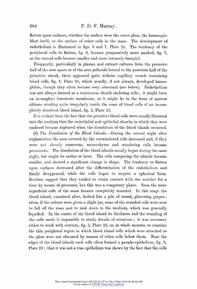
flatten upon surfaces, whether the surface were the cover glass, the haemangio- blast itself, or the surface of other cells in the mass. The development of endothelium is illustrated in figs. 6 and 7, Plate 24. The tendency of the peripheral cells to flatten, fig. 6, became progressively more marked, fig. 7, as the central cells became smaller and more intensely basophil.
Frequently, particularly in plasma and extract cultures from the posterior half of the area opaca or of the area pellucida lateral to the posterior half of the primitive streak, there appeared quite definite capillary vessels containing -blood cells, fig. 8, Plate 24, which usually, if not always, developed hbemo- globin, though they often became very abnormal (see below). Endothelium -was not avlways formed as a continuous sheath enclosing cells; it might form an incomplete fenestrate membrane, or it might be in the form of narrow ribbons winding quite irregularly inside the mass of blood cells of an incom- pletely dissolved blood island, fig. 3, Plate 23.
It is evident from the fact that the primitive blood cells were usually liberated into the nmeditim that the endothelial and epithelial sheaths in which they were enclosed becane ruptured when the dissolution of the blood islands occurred.
(3) The Dissolution of the Blood Islands.-During the second night after explantation the area covered by the outwandered cells increased and, if they were not already numerous, mesenchyme and wandering cells became prominent. The dissolution of the blood islands usually began during the same night, but might be earlier or later. The cells composing the islands became smaller, and showed a significant change in shape. The tendency to flatten upon surfaces decreased after the differentiation of the endothelium and finally disappeared, while the cells began to acquire a spherical form. Sections suggest that they tended to retain contact with one another for a -time by mweans of processes, but this was a temporary phase. Soon the more superficial cells of the mass became completely rounded. In this stage the blood island, examined alive, loolked like a pile of round, glistening grapes; often, if the culture were given a slight jar, some of the rounded cells were seen to fall off the mass and to sink down in the medium, which was generally -liquefied. In the centre of the blood island its thickness and the rounding of the cells made it impossible to study details of stru;cture; it was necessary either to work with sections, fig. 5, Plate 23, or, in whole mounts, to examine the thin peripheral region in which blood island cells which were attached to the glass were not obscured by masses of other cells below them. Near the ,edges of the blood islands such cells often formed a pseudo-epithelium, fig. 3, Plate 23; that it was not a true epithelium was shown by the fact that the cells
This content downloaded from 169.229.32.137 on Thu, 8 May 2014 06:38:44 AMAll use subject to JSTOR Terms and Conditions

Development in vitro of the Blood of early Chick Embryo. 505
were separate from one another, not forming a continuous sheet. In sections, fig. 5, Plate 23, through the centre of a dissolving blood island, it could be seen that the cells were now no longer flattened upon one another but were rounded, that cell outlines were clear, and that there were often clefts between the cells. While the process of rounding up was going on the cells seemed to become more intensely basophil, but in spite of this there were areas in their cytoplasm which were less basophilic than others, and which even stained faintly with eosin. The nLcleus had become somewhat larger compared with the cytoplasm, but its other characters had not greatly changed.
The free, spherical cells, falling away from the attached part of the cultures formed " clouds." They were now primitive blood cells.
Histogenesis of Blood and Wandering Cells.
The primitive blood cells were small and probably amceboid; at least, after fixation they frequently showed pseudopods and in life amceboid cells of similar appearance, but of uncertain origin, were frequently seen (see below, under " Wandering cells "). The primitive blood cells had a rounded nucleus enclosed in a quite small amount of basophil cytoplasm. The nuelens con- tained one or two nucleoli of somewhat irreguilar form, and usually several tiny granules of chromatin. In spite of its general basophilia, the cytoplasm ,often contained small weakly eosinophil areas resembling those already noted in earlier stages. Some cells may now show the eosinophil. structure which iDantschakof[ (1908) calls the " Hof," a term for which the nearest English gequivalent, in the sense in which the German word is used, is perhaps " foeus," as in the phrase " focus of disease." It is described by Dantschakoff as a more or less spherical region of eosinophil cytoplasm which, increasing in size, indents the nucleus, making it kidney-shaped. I confirmi Dantschakofi's description in both cultures and normal embryos, save that I find the focus to be less regular in form than is indicated by her figures, and that there may be *one large and several smaller foci in a single cell.
Camera lucida drawings showed that the primitive blood cells tended to be somewhat smaller than the corresponding cells in vivo. They proliferated -rapidly, and the chromosomes were still long and thin, as in the blood island stage; a resting cell is shown in fig. 9, Plate 25.
A.-Erythrogenesis.
The development of erythroblasts took place almost entirely among cells floating in the liquefied medium. The histogenesis was studied mainly from
This content downloaded from 169.229.32.137 on Thu, 8 May 2014 06:38:44 AMAll use subject to JSTOR Terms and Conditions

506 P. D. F. Murray.
smears, but the results so obtained were as far as possible checked by reference
to the Zenker formol fixed whole mounts and sections. In most cultures,
although nearly all the cells derived from the blood islands sank into the fluid,
some remained attached to the tissue on the cover glass, or enclosed in capillary
vessels from which they were unable to escape. These " reliet " cells were not
a reliable foundation for the study of histogenesis because, when developing in
enclosed spaces in the eulture, they tended to become abnormal, and often
remained undifferentiated or in the first stage of erythrogenesis, long after the
cells floating in the fluid had become advanced erythroblasts. They could,
however, be used for certain purposes as a check upon the cells in the smears.
The histogenesis of the red cells may be considered in three stages. As the
living cells were very translucent and gave little information, the following
account is based upon fixed and stained preparations.
Stage (1).-This stage, fig. 10, Plate 25, was characterised by increase in
size of the small primitive blood cells. The cells retained their rounded
form, and did not show pseudopods, while the cytoplasm remained basophil
but was not completely homogeneous. It was usually uneven in texture, and
contained an eosinophilic focus or foci, as described in the primitive blood
cells, but now becoming more prominent. These foci were sometimes irregular
in distribution and form, sometimes streaky and lying along cUrves concentric
with the surface of the cell, and sometimes more or less spherical. The nucleus,
probably owing to the relative and absolute increase in the quantity of
cytoplasm, was now no longer indented, but was spherical. The cytoplasm
of the relict cells in the attached cultures was uisually more homogeneous,
except when the cells were obviously unhealthy. It must be remembered,
however, that it was more difficult, on accouant of the plasma present, to
obtain a satisfactory stain of the attached culture than- of the cells in the
smears. The eosinophilic focus was not seen in all cells, but it was found in a
large number. The appearance of the nucleus at this stage depends to some extent on the
fixation. It was always round or oval, but in the smears (fixed by methyl
alcohol) it showed a brilliant red-purple colour and was full of red-purple
granules of irregular form and indefinite contour, which seemed to be united
to one another by tlhreads of the same material, forming a loose and irregular
network. Between these large granules were very numerous tiny pinkish
granules, with the result that the whole nucleuas had a smudgy appearance.
Corresponding cells in the attached parts of the cultures (Zenker formol)
showed a very different picture. The reddish granulation was not to be seen,
This content downloaded from 169.229.32.137 on Thu, 8 May 2014 06:38:44 AMAll use subject to JSTOR Terms and Conditions

Development irn vitro of the Blood of early Chtck Embryo. 507
and the nuclei were vesicular with irregularly scattered basophil chromatin
granules, and, in at least some, and probably in all cells, there were one or two
rather faintly staining basophil nucleoli. There can be no doubt that the
nuclear appearance seen in the smears was an artefact, and that the picture
shown by Zenker fixed material resembled more closely the condition in life.
During this stage cells in mitosis were frequently found, and it is evident
that differentiation may proceed through several generations; whether the
entire differentiation process was ever completed in a single cell generation is
uncertain. The chromosomes which were previously long and thin became
at this stage short and thick, and lay so close together that boundaries between
adjacent chromosomes could not be distinguished. This condition was seen
in all mitoses until cell division ceased.
Stage (2), fig. 11, Plate 25.-During the second stage the form of the cell
altered, and changes occurred in the nucleus. Elongatinig alonig one axis
the cell gradually became equi-oval in form, with blunt ends, and at the same
time, though generally after the attainment of a broad oval form, the basophilic
substance decreased in quantity, and began to be replaced by the cosinophilic
substance which is generally, and probably correctly, regarded as being
hemoglobin. It must nevertheless be noted that hoemoglobin was, present
at earlier stages than this. As this change proceeded the cells at first acquired
(after Giemsa) a curious grayish-pink-purple colour, and later became pale
pink. The texture of the nacleus appeared at first glance to be homogeneous,
but careful examination revealed a faint and loose reticulum of cytoplasmic
strands, or sometimes there mnight be several small vacuoles embedded in a
more or less homogeneous pink ground substance. The nucleus became
relatively smaller, but in many cells remained for a time large enough to
touch both sides of the elongated cell body. The chromatin took the form of a
coarse and, at first, irregular network, which became more regular in arrange-
ment and tended to concentrate at the periphery of the nucleus, where it
might resemble the spokes of a hub-less wheel. Nucleoli were still present in
some cells at least. The reddish substance, so prominent irL earlier stages, became less so, and now no longer obscured the chromatin, even in smears.
If mitoses occurred at all after the cells had begun to elongate, -they were rare; hence it is probable that the differentiation processes of the second and third
stages occurred within the lifetime of single cells, without the occurrence of
cell divisions. There was considerable variation in the time relations of the process. The
cells might lose the basophil substance before the eosinophil substance
VOL. CXI.-B. 2 M
This content downloaded from 169.229.32.137 on Thu, 8 May 2014 06:38:44 AMAll use subject to JSTOR Terms and Conditions

508 P. D. F. Murray.
appeared, so that the cytoplasm became very pale and had the appearance of a loose reticulum traversing a space filled with a fluid or some other homogeneous substance. In many cultures there were round cells which had not yet begun to elongate, with very pale or slightly eosinophil cytoplasm and small nuclei of the kind described. Whether such cells retained their circular form and never became oval, or whether they became so later, is uncertain.
Stage (3), fig. 12, Plate 25.-I have only once obtained cells which could be called mature erythrocytes; these were in a cultiire taken from the area opaca at a time when haemangioblasts probably existed. The infrequency was prob- ably due to the fact that the cultures have usually been fixed too soon, because, if kept alive longer, the cells became abnormal. In shape the differentiated erythrocyte was equi-oval, and strongly eosinophil. The nucleus was roughly oval or somewhatLa irregular in form, quite small and shrivelled, darkly basophil, and conLtained several large chromatin granuLles. The cytoplasm appeared to be perfectly homogeneous.
Comparison of erythrogenesis in vitro, as here described, with the corre- spondinig process in vivo, described by Dantschakoff (1908), shows general
agreemenit, anid minor points 6f difference are probably due in part to differences in staining.
The Development of Haemoglobin.-In older cultLures, containing advanced erythroblasts, the presence of haemoglobin is frequently obvious, and can often be detected with the naked eye. Professor Keilin examined a number of the cultures spectroscopically, and found the characteristic spectrum of hsemoglobi.n. The earliest culture in which it was recognised consisted of a dense mass of cells juist released, or just about to be released, by the dissolution of a large blood island. The cells appeared to be in all respects normal young priniitive blood cells; there was nothing suggesting that the dissolution of the blood island had been abnormally delayed.
No trace of hsematoporphyrin, or of any other hoemoglobin derivative, could be detected.
Abnormal Forms among the Erythroblasts.-Figs. 13, 14 and 15, Plate 25.- Shipley (1915-16), describing hematopoiesis in his cultures of the area opaca, states that all but a few of the erythrocytes formed were more or less abnormal. The same applies to the red blood cells formed during the course of the present experiments, but the degree of abnormality was in most cells not high, careful examination being required to reveal that a particular cell was in some respect not quite normal. Most cultures, on the other hand, contained some cells which were very abnormal indeed, and in some cultures this was true of all or nearly all.
This content downloaded from 169.229.32.137 on Thu, 8 May 2014 06:38:44 AMAll use subject to JSTOR Terms and Conditions

IDevelopment in vitro of the Blood of early Chick Embryo. 509
The commonest abnormality of form, as distinct from purely necrotic
changes, consisted in the development of vacuoles within the cytoplasm. A
large percentage of late erythroblasts, when examined closely, could be seen to
contain one or a few small vacuoles, and this comparatively trifliing deviation
from the normal was connected by all transitions with cases in which the cell
was reduced to a mere thin-walled vesicle, fig. 13, Plate 25, or complex of
vesicles enclosed by the outer wall of the cell, fig. 14, Plate 25. The nucleus
of cells in this condition was strongly compressed either against the cell wall
or in a dissepiment between vacuoles. The material in the vacfaoles did not
staini with Giemsa and so was not haemoglobin, and it had not the appearance
of fat. It is interesting that a moderate vacuolation neither prevented the
development of hmemoglobin nor seriously impeded the development of the
typical erythrocytic form. Extreme vacuolation caused the cell to remain
spherical; its effect on the development of haemoglobin is uncertain. Cells
reduced to a bag by a single large vacuole may occasionally be seen in normal
embryos. knother abnormality, often associated in the same cell with a limited
vacuolation, was the formation of erythroblasts which were drawn. out at one
or both ends into long pointed processes which tended to show beading. Both
these cells and the vacuolated cells often showed, in addition, curvature of the
whole cell inito a crescent form, fig. 15, Plate 25.
Otherwise, normal erythroblasts with two nuclei were not uncommon, and
in highly vacuolated cells there might be three or even fouir.
The various abnormalities, and especially vacuolation, show a definite
association with unfavourable conditions in the culture. Frequently, as has
been said, masses of primitive blood cells were unable to become free from the
attached part of the culture because they were enclosed in capillaries or
between sheets of cells. In these conditions it very frequenitly happened that
all the celIs in the mass became intensely vacuolated, while those which had
been ablie to escape into the fluid continued normal development.
B.-WVandering Cells (Histiocytes, Polyblasts).
The appearance of wandering cells at an early stage in the development
of the cultures has been mentioned. After the dissolution of the blood islands,
when there was a floating cloud of free cells, many of these were found to be
wancering cells. Present in only small numbers at first, the wandering cells
multiplied enormotusly in later stages, and sometimes even outnumbered the
2 M 2
This content downloaded from 169.229.32.137 on Thu, 8 May 2014 06:38:44 AMAll use subject to JSTOR Terms and Conditions

510 P. D. F. Murray.
erythroblasts. They were presenit throughout the attached part of the culture, as well as in the floating clo-ud. In later stages they became very actively phagoeytic, and had the structure shown in fig. 17, Plate 25. Seen in life, they were cells filled with glistening droplets and varying considerably in size, included among them being the largest free cells present. Studied after fixation and staining, they showed the following characters: the cytoplasm was much vacuolated, being filled with many small vacuoles, and many cells were engorged with the remains of phagocytosed material, chiefly erythroblasts and primitive blood cells. In many cells, particularly in those which had not been actively phagocytic, the nucleus was a fairly regular, small, oval, eccentrically placed structure, containing one or two large nucleoli and particles of chromatin. In other cells, and especially in the larger, more engorged, and probably older cells the nuclei had the most varied and fantastic shapes, lying compressed between the vacuoles with which the cell bodv was distended. In such cases it was difficult to make out anything of the n-uelear structure, but it seemed to resemble that of the more regular nuclei so far as its peculiar form and its compression allowed. The nucleus of the cell shown in fig. 17 is of regular form compared with that seen in many cells.
The origin of the wandering cells remains uncertain. Examination of living cultures frequently showed all transitions between large engorged and vacuolated phagocytes, and small ameeboid cells with few or no vacuoles or droplets. Some of these small cells so closely resembled primitive blood cells as to suggest that the wandering cells were derivatives of the blood islands; but whether these were one source of wandering cells or not, they were certainly not the only source, for exactly similar wandering cells appeared in large nLmbers in cultures in which there was no hematopoiesis. Further, in such cultures one sometimes finds, associated with the wandering cells, just the same
small, round, clear cells as seem to be the young stages of the wandering cells
in cultures containing blood islands and primitive blood cells. It is therefore
probable that wandering cells are not derived from the blood islands bu1t from
the small round cells which closely resemble primitive blood cells and which originate from the mesenchyme in some manner which remains unkiown.
C.-Endodergnal Wandering Cells.
Suspecting that certain cells in the cutltures were identical with the endo- dermal wandering cells, of epithelial origin, described by Dantschakofi (1908) and others, I made a series of eultures of the endodermal epithelium. These
This content downloaded from 169.229.32.137 on Thu, 8 May 2014 06:38:44 AMAll use subject to JSTOR Terms and Conditions
Development in vitro of the Blood of early Chicck Embryo. 511
eultures isually contained a certain amount of mesodermn, for it was difficult
to separate it completely from the endoderm; the suspicion was, however,
confirmAed by finding the suspected cells present in great numbers in cultures
of endoderm, while the mesodermal wandering cells were much less numerous
or altogether absent. The endodermal wandering cells had a central mass of
glistening droplets, presumably of yolk, and a lobular pseudopodiuLm of very
clear cytoplasm, there being usually a sharp line of demarcation between
the clear cytoplasm of the pseudopod and the heavily yolk-laden endoplasm.
They degenerated within two or three days, as might be expected from their
normal history in vivo.
D.-" Large Lymphocytes." (Fig. 16, Plate 25.)
Cdelis identical with those which Dantschakoff (1908) calls large lymphocytes
were undoubtedly present in the smears. While not convinced of the identity
of these cells with the large lymphocytes of later stages, the name is retained
provisionally. The number of these cells varied greatly from culture to culture; they were
always far fewer than the erythroblasts, and sometimes seemed to be absent
altogether. They were round cells, about the same size as the larger round
erytbroblasts, bubt differed from the latter in having a relatively very large
nucleus surrounded by a narrow rim of cytoplasm. The cytoplasm was
basophil, but might contain one or more areas resembling the eosinophilic
focus of erythroblasts; this is clearly shown, for the normal embryo, in
IDantsebakoff's figures. The nucleus was round and, in most cells stained in
smears, its structure was obscured by a mass of light red granules joined
together as a net. There is little doubt that this is an artefact like the similar
substance found in smear-stained early erythroblasts; it always seemed to be
present, but often was only very lightly stained so that the nuclear struLcture
could be studied. The nucleus was then seen to contain one or two rather
small nucleoli of irregular form, and often a few small fragments of
chromatin, the nucleus as a whole having a curiouisly empty appearance. I
have miade no detailed study of the histogenesis of these cells, but there is no
doubt that they, like the erythroblasts, are derivatives of the blood islands;
they might be described as an enlarged form of the primitive cell.
E.-Other Leucocytes.
Danitschakoff (1908) described cells with eosinophilic granules as appearing
in the P.xtra-vascular spaces of the yolk sac trabecule at about four to five days
This content downloaded from 169.229.32.137 on Thu, 8 May 2014 06:38:44 AMAll use subject to JSTOR Terms and Conditions

512 P. D. F. Murray.
of incubation. Cells which are almost certainly young stages in the develop- ment of these eosinophils are present in one culture and possibly in three others.
In the same paper Dantschakoff described dwarf lymphocytes and thombo- cytes as differentiating from primitive blood cells. Whether these are present in the cultures or not remains uncertain.
PART 2.
The Distribution of the Hmatopoietic Cells.
The stage of development of the embryos from which I have, in the great majority of experiments, obtained material for cultivation, was that at which there is a fully-developed primitive streak, with or without a head process, in a pear-shaped area pellucida with no head fold. I have also o'btained development of red corpuscles from stages well before the appearance of the primitive streak, blut in these early stages I have not yet made any attempt to delimit the areas of the blastoderm containing the hamnatopoietic cells.-
In the following paragr-aphs the letters in brackets refer to the areas indicated in fig. 18.
At the primitive streak stage at which most of the experiments have been made, a very large number of experiments have shown that cultuires of the posterior half of the primitive streak (ABCD) almost invariably prodouce great numbers of red blood cells.
To find the distribution of the haematopoietic cells along the length of the primitive streak the following experiment was carried out. Cultures were made of the anterior quarters (EFG) of the primitive streaks of twenty-two embryos; three of these developed a little blood, two were do-ubtful, and the remaining seventeen developed no blood at all. Eleven cultures were made of the second quarters of the priinitive streaks from the same embryos as the last group (FADG); all showed vigorous development of blood. A fragment was cultivated from the posterior end of. each of twelve primitive streaks, the fragments varying in size from eighths to quarters (HBCJ); all produced blood. It may therefore be concluded that haematopoietic cells are present in the whole of the posterior three-quarters of the primitive streak. but, that they are absent from the anterior quarter except, perhaps, in its piosterior end.
Eighteen cultures were made of that part of the area pellucidla wlaic-h lies
lateral to the posterior half of the primitive streak, betweei it and the area
This content downloaded from 169.229.32.137 on Thu, 8 May 2014 06:38:44 AMAll use subject to JSTOR Terms and Conditions

Development in vitro of the Blood of early Chick Embryo. 513
opaca (KMBA). Blood developed in twelve of these cultures, probably in three others, was absent in one and probably in another, anid one was quite doubtful. Nearly all the cultures showed appearances known to me as early stages in haeinatopoiesis, but are couanted as doubtful unless cells containing hemo- globin, or showing the oval form of the late erythroblast, were clearly recognis- able. In embryos at this stage it can frequently be seen that the posterior part of the area petlucida is divisible into a more opaque, median zone, near the posterior half of the priniitive streak, and a lightier more peripheral zone. Twelve cultures were made, six of which consisted of the inner, opaque zone (LMBA), and six of the outer clear zone (KML). Of the former, four showed de- finite development of blood and two were doubtful, but all showed some signs of hmematopoiesis. Of the second group, three showed blood, two were doubtful and one was negative, slhowing no indica- tions of hmematopoiesis. To find the dis- tribution of the ha3matopoietic cells in the more anterior part of the area pellucida, I made thirteen cultures of the area pellucida opposite the anterior quarter of the primi- tive streak (NOFE); blood appeared in four of these, but was absent from the other nine, while in five cultures made from the area pellucida opposite the head process of embryos in early head process stages, no blood was developed. Six cultures were made of the area pellucida anterior to the primitive streak or head process-that is, from the region of the pro-amnion (PNQ), buLt none of them developed blood. It may be concluded that haematopoietic cells are present opposite the posterior three-quarters of the primitive streak, and may extend a little further anteriorly. The pro-amnion, naturally, does not contain haematopoietic cells.
.... .. .............
p
emry in th stg sd o h
Theperiphera dotte FIG. 18-Daga
lefta
limity of the stagio used for cutures naoity the actual rimitftentls. Toe
broke.nh letteing its explaoinaedly the atext. brdro hergo
Ncuidb amtpitccls
Th1 eihra otdlieO1lf
Decipioni of
Fl eion 18e Diara
ofltran
emroti the stagel usedt ofo theblso majrity ofic experim eynts thes broken line inditergis ap:proiatelyi the aterirbrdrothergo
This content downloaded from 169.229.32.137 on Thu, 8 May 2014 06:38:44 AMAll use subject to JSTOR Terms and Conditions

514 P. D. F. Murray.
A series of cultures was made from the inner part of the area opaca, where the greater part of the area vasculosa later appears. This region was divided into four, as shown in the diagram, and the four quadrants were cultivated separ- ately. Thirteen cultures of anterior quadrants (RSTP) all gave negative results except one, in which blood developed, the remaining twelve being free from blood. Thirteen cultures of posterior quadrants (SUVT) gave development of blood in ten cases, while one was doubtful and two negative. Fourtee:i cultures were made of strips of area opaca from immediately behind the primitive streak (WXYZ); blood appeared in all. In order to demarcate more accurately the region occupied by the blood-formilg cells, twelve cultures were made of rather nlarrow strips of area op)aca taken from a position such that their anterior ends were opposite the widest part of the area pellucida (or.BON);
of these six produLced blood and six did not. Hence, it appears that the region of the area opaca opposite the widest part of the area pellucida, and approxi- mately opposite the anterior end of the primitive streak, contains the boundary between the region occupied by hematopoietic cells, and that into which they have not yet penietrated. To confirmn this, four cultures were made of strips of alrea opcaa from a position (f3sO) immediately behind that represented in the last series ; all forrmed blood.
It may be concluded from these experiments that the hoematopoietic cells are distributed over the whole of that part of the embryo which lies behind the broken line in fig. 18.
DIscusSION.
(1) The Determination Problem.
A.-The Haemangioblasts.-The first stage in hlematopoiesis is the formation of the hemangioblasts. Their formation is a differentiation process depending upon the activity of their component cells, and the question arises whether it is a self-differentiation.
It is evident from the culttures herein described that the diifTerentiation of the hvemangioblast is not dependent upon the specific activity of any organ of the embry-o. This conclusion follows equally from the explantation experiments of Slonimski (1930 a, 1931). In vivo, heemangioblast formation might be enforced upon the mesenchyme by some condition existing either in the primitive streak or in the area vasculosa. The first would mean that mesenchyme cells at the place, and at latest very shortly after the time, of their origin, become specifi- cally determined for hbemangioblast formnation, or at least strongly biassed in
This content downloaded from 169.229.32.137 on Thu, 8 May 2014 06:38:44 AMAll use subject to JSTOR Terms and Conditions

Development in vitro of the Blood of early Chick Embryo. 515
that dlirection. In this case the cells which form the haermangioblasts would do
so under the influence of factors intrinsic in themselves; in other words,
heemangioblast formation would be a self-differentiation. If the hypothetical
condition acted in the area vasculosa it would either not exist in the primitive
streak' cultures, or else the latter must establish an internal environment closely
resembling that in the normal area vasculosa. The internal environment
could hardly be produeed by a particular structure of the area vasculosa, because the only resemblance between its structure and that of the cultures is
the presence of the haemangioblast. If it were a particular physiological condi-
tilon, it nust be one due to the activity of a particular kind of cell. This
hypothesis must be rejected for two reasons. Firstly, it requires the assump-
tion, without evidence, of specificity in an unidentified cell to avoid making
the same assumption in the case of the presumpti-ve heemangioblast cells.
Secondly, the mesenchyme is the only tissue which belongs in common to both
primitive streak aiid area vasculosa, because it migrates from one to the other.
There is nio evidence for any similar migration of ectoderm or endoderm.
Hence, to save the hypothesis it would have to be assumed that the iinternal
environment, normally created by an unidentified cell in the area vasculosa,
was in the cultures created by a different unidentified cell whiclh normally
displays no such activity. It may therefore be concluded that the mesenchyme cells of the primitive
streak are caused to form haemangioblasts, not by conditions extrinsic to them,
but by the actioni of factors intrinsic in them. In other words, the mesenchyme
cells whichform the hcemangioblasts must be regarded, even while they are still in the
primitive streak, as self-differentiating cells, predetermined, or at least strongly biassed, towards the fomnmation of the group of cell types which are derived from the
heemangioblasts. This involves further implications. According to the view which is generally
held at present, the homatopoietic process begins in a previously uri-
diferentiated mesenchyme, all of whose cells are equivalent to one another.
This view is evidently inconsistent with the conclusion just stated, and must
therefore be abandoned, at least for primary haematopoiesis in the chick
embryo. The work of SloInimski (1930 a, 1931), to which reference has been
made, indicates that a similar conclusion must apply to the corresponding process
in Amphibia. Further support is found in the works of Federici (1926), Goss
(1928), Slonimski (1930 b) and Stohr (1931). These authors, using young
embryos of various Amphibia, performed experiments which consisted essen-
tialiv in removing the hsematopoietic region, and by this mtieans a number of
This content downloaded from 169.229.32.137 on Thu, 8 May 2014 06:38:44 AMAll use subject to JSTOR Terms and Conditions

516 P. D. F. Murray.
larvae were obtained wlhich, although without erythrocytes, in some cases lived almost to metamorphosis, but failed completely to produce red blood cells, making no attempt to compensate for the loss by any regenerative process.
It is important at this stage to mention two poinlts. Firstly, the term
"biassed" is used here in order to avoid the term " deterinination," the use of which suggests irreversible predestination. The G-erman school of experi-
mental embryologists has shown that cells which can, when isolated, self-
differentiate in the expected manner may nevertheless be caused to differentiate in quite other ways if placed in the appropriate conditions. It is possible that the presumptive helmangioblast cells of the primitive streak might similarly alter the line of their differentiation if they were subjected to suitable treatment. Secondly, the fact that some mesenchyme cells do not take part in hoematopoiesis does not mean that they, or their descendants, may in-ot do
so at later stages or under different conditions. B.-The Endothelium.-It is theoretically possible either (1) that the
presumptive endothelial cells in the hoemangioblast are constituLtionally identical with all the other hemangioblast cells, and that they suffer endothelial differentiation because of their tendency (shared with all other hoemangioblast cells) to flatten on surfaces, and because of the pressure of the developing intravascular fluid; or (2) that they were specifically biassed or even finally determined for endothelium formation prior to the development of the haemangioblasts, and have taken a superficial position because their innate constitution forces them to do so. While it is impossible to decide between these alternatives, the first accords with the histological picture and involves fewer and less improbable assumptions. But whichever is correct, it is
important to remember that the endothelium retainis, at least for a time, the power of giving rise to new erythroblasts (Sabin, 1920; Dantschakoff, 1908, 1909).
C.-The Erythroblasts.-The time of first appearanice of hemoglobin sets a later limit to the time of determination of erythroblasts. HImuoglobin is undo-ubtedly present in the elongating erythroblast, but there is no doubt that its first appearance is considerably earlier than this. Sabin (1920) found that haemoglobin could be seen as a yellow colour in the liviing cells of undissolved blood islands, but not in the earlier haemangioblast (" angioblast" stage. Slonimski (1927 b), using a micro-chemical benzidin method (1927 a) identified heamoglobin in the peripheral zone of the area vasculosa at six to seven somites. In a culture consisting of a large blood island just at the stage of
This content downloaded from 169.229.32.137 on Thu, 8 May 2014 06:38:44 AMAll use subject to JSTOR Terms and Conditions
Development in vitro of the Blood of early Chick Embryo. 517
dissolution, Professor Keilin, as stated earlier in this paper, found haemoglobin
definitely present. From these isolated facts it appears that heemoglobin
originates during the time which elapses between the development of the
endothelial walls and the dissolution of the blood islands. The final determina-
tion of the presumptive erythroblasts must therefore occur during or before
the blood island stage. No early linmit can be definitely fixed, but if the
peripheral cells of the haemangioblast, which form endothelium, are constitu-
tionally identical with all other ha-mangioblast cells, the presumptive
erythrocytes must be determined towards erythrocytogenesis after or during
the development of endothelium, since, by hypothesis, they were previously
equally liable, according to their position, to either erythrocytogenesis or to
endothelium formation. It should be noted that the erythrocytes differentiating in the cultures are
doubtless the first or primary erythrocytes (Danitschakoff, 1908), for the
definitive erythrocytes, which differ very little from the first series, do not appear
in vivo until after four or five days' incubation.
(2) Wandering Cells.
Since the wandering phagocytic cells appeared in all cultures containing
mesoderm, whether heematopoiesis occurred or not, it is clear that they are
connective tissue cells, histiocytes, rather than blood cells.
They appear first on the day after explantation, which corresponds to the
second day of incubation. Now, Dantschakoff (1909), in a very complete
study of the development of the connective tissue of the chick embryo, states
that the mesenchyme is at first a uniform reticulum of stellate cells, and that
wandering cells do not appear until the end of the fourth or the beginning of
the fifth days of incubation. It is thus probable that the wandering cells in
the cultures are precocious, their presence being not merely the result of the
emigration of already existing wandering cells from the explant, but of their
new formiation as a reaction to the conditions of life in vitro.
(3) The Histogenesis of Endothelittm.
Sabin (1920), in her account of the living area vasculosa, states that the
plasma of the first vessels is formed in the solid hoemangioblast (" angioblast ")
by the liquefaction and destruction of the cells, and that the endotheliunn is
formed, not by flattening of the peripheral cells but by the liquefaction of
This content downloaded from 169.229.32.137 on Thu, 8 May 2014 06:38:44 AMAll use subject to JSTOR Terms and Conditions

518 P. D. F. Murray.
all but their peripheral regions. From the study of fixed preparations, all other authors have coneluded that the endothelial cells are the flattened peripheral haemangioblast cells (" blood island cells "), and the cultures, as figs. 6 and 7, Plate 24, show, support this view. Nevertheless, special weight should, perhaps, be attached to direct observations of the living organism, and it seems well, the-refore, to withhold decision until Sabin's observations shall have been repeated. It is not impossible that both processes may occur.
In conclusion, it is more than a pleasure to express sincere appreciation and thanks for the assistance which I have received from several people during the course of the work, and in particular to Dr. H. B. Fell, who has throughout the work been a never-failing source of help and constructive criticism, especi- ally in the many difficulties of tissue culture methods, in histological technique, and in the preparation of the plates. Thanks are due also to Professor Keilin. of the Molteno Institute, for his spectroscopic examination of the cultures; to Dr. J. S. Niven for valuable help in the preparation of tlhe manuscript, and in other ways; to Professor J. T. Wilson; and to my wife, who worked through many a long day, and actually prepared a number of the cultures upon which this work is based. Thanks are also due to the Royal Society for a grant from the Smithson Fellowship Fund.
Summary.
(1) The differentiation in vitro of the blood of the early chick embryo was studied histologically.
(2) The media used were two: plasma and embryo extract, and serum and
embryo extract.
(3) Haematopoiesis occurred in the great majority of all cultures of any region of the blastoderm behind the level of the anterior quarter of the primitive streak. H-ematopoiesis may occur in cultures taken from levels slightly anterior to this in the area pellucida or in the area opaca. C-ultures of the entire or fragmented posterior three-quarters of the primitive streak all produce large numbers of advanced, hamoglobin-containing erythroblasts.
(4) Endothelium develops in many if not in all cultures, but particularly in fragments of the area opaca or area pellucida. It forms by the flattening of the peripheral cells of the heemangioblast, but it is not impossible that the
excavation process advocated by Sabinr may also occur.
(5) The histogenesis of the erythroblasts agrees in essentials with the same process in the norimal embryo, as described by Dantschakotf.
This content downloaded from 169.229.32.137 on Thu, 8 May 2014 06:38:44 AMAll use subject to JSTOR Terms and Conditions

Development in vitro of the Blood of early Chhick Embryo. 519
(6) Certain abnormalities which commonly occur among erythroblasts
differentiated in vitro are described. These are chiefly vacuolation, the drawing
out of the poles of the cell into long processes, and curvature of the cell as a
whole. (7) In addition to red blood cells and endothelium, cells are found which
are identical with those, found in the normal embryo, which Dantschakoff
calls " large lymphocytes." In one culture, and perhaps in three others,
cells appeared which were probably very young eosinophils.
(8) In all cultures, save those from the pro-amnion, which contained no
mesoderm, there appeared epithelium, mesenchyme, and mesodermal wander-
ing cells. (9) The wandering cells were actively phagocytic, very vacuolated cells with
a small eccentric and often distorted nucleus. They originate from mesen-
chyme, but perhaps also from the primitive blood cells derived from the blood
islands. (10) Conclusions.-A.-The primitive streak contains mesenchyme cells
which are determined, or at least strongly biassed, towards the development
of haemangioblasts, and therefore of the group of cells, considered as a whole,
to which the haemangioblasts give rise.
B.-The endothelium may be formed by cells already specifically determined
for endothelium formation, but it is more probable that it is formed by
previously indifferent hoemangioblast cells whose fate is determined by the
accident of their superficial position in the haemangioblasts.
C.-Hsemoglobin is present in the blood island before their dissolution into
primitive blood cells. Hence, the presumptive erythrocytes are determined
some time before their liberation as primitive blood cells, and probably after
the formation of endothelium.
DESCRIPTION OF PLATES.
PLATE 22.
FIG. 1.-A very small hoemaiigioblast lying on a layer of vacuolated epithelium. Note rnitoses, tendency of peripheral cells to flatten, and areas of necrosis. Explant: Longitudinal half of rather less than posterior half of primitive streak from an embryo having no head process. The explant was cut into small fragments. Serum and embryo extract, Zenker formol, Giemsa, whole mount.
FIG. 2.-Small hiemangioblast lying on a layer of vacuolated epithelium. Explant: Longitudinal half of rather less than posterior half of primitive streak of an embryo having a head process equal to between one-third and one-half of the length of the head process plus primitive streak. The explant was cut into small fragments. Serum and embryo extract, Zenker formol, Giemsa, whole mount.
This content downloaded from 169.229.32.137 on Thu, 8 May 2014 06:38:44 AMAll use subject to JSTOR Terms and Conditions
520 P. D. F. Murray.
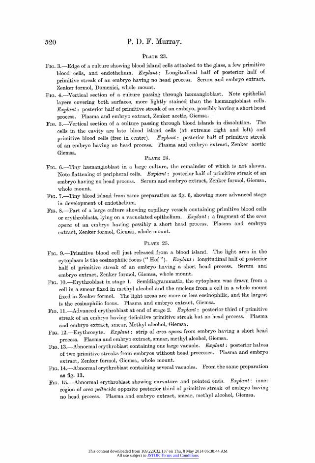
PLATE 23.
FIG. 3.-Edge of a culture showing blood island cells attached to the glass, a few primitive
blood cells, and endothelium. Explant: Longitudinal half of posterior half of
primitive streak of an embryo having no head process. Serum and embryo extract,
Zenker formol, Domenici, whole mount.
FIG. 4.-Vertical section of a culture passing through hzemangioblast. Note epithelial
layers covering both surfaces, more lightly stained than the hmmangioblast cells.
Explant: posterior half of primitive streak of an embryo, possibly having a short head
process. Plasma and embryo extract, Zenker acetic, Giemsa.
FIG. 5.-Vertical section of a culture passing through blood islands in dissolution. The
cells in the cavity are late blood island cells (at extreme right and left) and
primitive blood cells (free in centre). Explant: posterior half of primitive streak
of an embryo having no head process. Plasma and embryo extract, Zenker acetic
Giemsa. PLATE 24.
FIG. 6.-Tiny hiemangioblast in a large culture, the remainder of which is not shown.
Note flattening of peripheral cells. Explant: posterior half of primitive streak of an
embryo having no head process. Serum and embryo extract, Zenker formol, Giemsa,
whole mount. FIG. 7.-Tiny blood island from same preparation as fig. 6, showing more advanced stage
in development of endothelium.
FIG. 8.-Part of a large culture showing capillary vessels containing primitive blood cells
or erythroblasts, lying on a vacuolated epithel-ium. Explant: a fragment of the area
opaca of an embryo having possibly a short head process. Plasma and embryo
extract, Zenker formol, Giemsa, whole mount.
PLA TE 25.
FIG. 9.-Primitive blood cell just released from a blood island. The light area in the
cytoplasm is the eosinophilic focus (" Hof "). Explant: longitudinal half of posterior
half of primitive streak of an embryo having a short head process. Serum and
embryo extract, Zenker formol, Giemsa, whole mount.
FIG. 10.-Erythroblast in stage 1. Semidiagrammatic, the cytoplasm was drawn from a
cell in a smear fixed in methyl alcohol and the nucleus from a cell in a whole mount
fixed in Zenker formol. The light areas are more or less eosinophilic, and the largest
is the eosinophilic focus. Plasma and embryo extract, Giemsa.
FIG. 11.-Advanced erythroblast at end of stage 2. Explant: posterior third of primitive
streak of an embryo having definitive primitive streak but no head process. Plasma
and embryo extract, smear, Methyl alcohol, GCieisa.
FIG. 12.-Erythrocyte. Explant: strip of area opaca from embryo having a short head
process. Plasma aInd embryo extract, smear, methyl alcohol, Giemsa.
FIG. 13.-Abnormal erythroblast containing one large vacuole. Explant : posterior halves
of two primitive streaks from embryos without head processes. Plasma and embryo
extract, Zenker formnol, Giemsa, whole miiount.
FIG. 14.-Abnormal erythroblast containing several vacuoles. From the same preparation
as fig. 13. FIG. 15.-Abnormal erythroblast showing cuirvatuire and pointed ends. Explant: inner
region of area pellucida opposite posterior third of primitive streak of embryo having
no head process. Plasma and embryo extract, smear, miethyl alcohol, Giemsa.
This content downloaded from 169.229.32.137 on Thu, 8 May 2014 06:38:44 AMAll use subject to JSTOR Terms and Conditions

Murray. Proc. Roy. Soc.B, vol. -11,PI. 22.
4M,
ffi~~~~f }
>
* a.
0~~~~~~~(b % * 44;} T -
5 (4 t ' 4 *X Vi a
U, * *0 * --+ , , % , + 4 ,,- >:00
Pt~~~~~~It
2
P M deel.
This content downloaded from 169.229.32.137 on Thu, 8 May 2014 06:38:44 AMAll use subject to JSTOR Terms and Conditions

Murray. Proc. Roy, Soc. B, voL. IWl,PL. 23.
3
'00~~~~~~~~~~~~~~~~~~~~~~~~~~~~~~~~~~~~~~~1
.t < * a
'I'Iggt~~ U:X (4%.i
*4
i H u 6 )l
.,g~~~~~~~~~~Al Xg
w r aJ,,~4
~ ~P. M . de-l...,< Kc o '(
This content downloaded from 169.229.32.137 on Thu, 8 May 2014 06:38:44 AMAll use subject to JSTOR Terms and Conditions

Murray. Proc.RLoy Soe.B, vot. 11W1,Pt, 24.
I
I V.
4 tt
6~ ~ 00m1 7
'0 A'cr '4 <5;
(t~~~~ \,% 4 <
j~~~~~~~~~~
9 A~~A'
8wv'
P M .rde l. E u t h c o l l.
P.M del. Ruith coil.
This content downloaded from 169.229.32.137 on Thu, 8 May 2014 06:38:44 AMAll use subject to JSTOR Terms and Conditions

Murray. ProciRoy. SocB, vol. 111, Pt. 2 5.
14 12 13
4,
15~ ~ ~~~1
1 7
t ' S
0 01 XM,
P. M. del. Huth c olt.
This content downloaded from 169.229.32.137 on Thu, 8 May 2014 06:38:44 AMAll use subject to JSTOR Terms and Conditions

Development in vitro of the Blood of early Chick Embryo. 521
FPi. 16.-" Large lymphocyte." Explant: one-sixth of primitive streak from near its
posterior end from embryo having a short head process. Plasma and embryo
extract, smear, methyl alcohol, Giemsa.
FIG. 17.-Phagocytic wandering cell containing ingested remains. Explant: posterior
one-sixth of primitive streak of embryo having short head process. Plasma awd
enbryo extract, smear, methyl alcohol, Giemsa.
BIBLIOGRAPIHY.
Benewolenskaja, S. W. (1939). 'Arch. exp. Zellforsch,' vol. 9, p. 128.
Chlopin, N. G., and A. L. (1925). 'Arch. exp. Zellforsch,' vol. 1, p. 193. Dant-schakoff, V. (1908). 'Anat. Hefte,' vol. 37, p. 471. Dantschakoff, V. (1909). 'Arch. mikr. Anat.,' vol. 73, p. 117. Erdmann, R., Eisner, H., and Laser, H. (1926). 'Arch. exp. Zellforsch,' vol. 2, p. 361. Federici, E. (1926). 'Arch. Biol.,' vol. 36, p. 465. Freifeld, H., and Ginsburg, A. (1927). 'Arch. exp. Zellforsch,' vol. 4, p. 355. Goss, C. M. (1928). 'J. exp. Zool.,' vol. 52. de llaan, J. (1928-29). 'Arch. exp. Zellforsch.' vol. 7, p. 298. Sabin, F. (1920). " Contributions to Embryology" (Carnegie Institute Publications),
vol. 9, No. 36, p. 213. Shipley, P. G. (1915-16). 'Anat. Rec.,' vol. 10, p. 347. Sloniniski, P. (1927 a). 'C.R. Soc. Biol.,' Paris, vol. 96, p. 1496. Sloniniski, P. (1927 b). Ibid., p. 1498. Slonimski, P. (1930 a). Ibid., vol. 104, p. 823. Slonimski, P. (1930 b). Ibid., p. 1050. Slonimski, P. (1931). ' Arch. biol.,' vol. 42, p. 415. Stohr, P., junr. (193'1). 'Arch. EntwMech. Org., vol. 124, p. 707. Timnofejewski and Benewolenskaja (1929). 'Arch. exp. Zellforsch,' vol. 8, p. 1.
This content downloaded from 169.229.32.137 on Thu, 8 May 2014 06:38:44 AMAll use subject to JSTOR Terms and Conditions