the demonstration of different types of muscle fibers in human
TRANSCRIPT
The demonstration of different types ofmuscle fibers in human extraocular
muscle by electron microscopyand cholinesterase staining
Scott E. Dietert*
Surgical specimens of normal human extraocular muscle repealed in electron microscopic crosssections two distinct types of extrafusal fibers: (1) a fibrillar type (Fibrillenstniktur) and (2)an essentially afibrillar type (Felderstruktur). Fascicles of autopsy muscle, stained with amodified Koelle technique to demonstrate cholinesterase activity, showed by light microscopytwo kinds of nerve endings: (1) large, heavily staining, compact discs (typical motor end.plates or "en plaque" endings), which usually occurred singly within the distance teased, and.(2) smaller, lighter staining droplets in clusters or chains ("en grappe" endings), tuhich occurredmultiply on a single fiber. The two nerve terminal types were never seen together on thesame muscle fiber. Differential staining, with selected substrates and an inhibitor (DFP),showed, the presence of both acetyl and butyryl cholinesterases in each ending. Electronmicroscopy of muscle stained for cholinesterase correlated the fibrillar ultrastructure with thefibers possessing "en plaque" endings and the afibrillar ultrastructure with the fibers pos-sessing "en grappe" endings. These morphologic features strongly suggest that human extrinsiceye musculature is organized into two separate contractile systems similar, if not identical,to the fast and slow striated muscle systems conclusively established in the frog. A brief dis-cussion of the oculomotor implications is included.
T,he existence of separate fast and slowcontractile systems in amphibians was sug-gested in 1928 by the physiologic studiesof Sommerkamp1 on the "tonus bundle" of
From the Department of Ophthalmology, Wash-ington University School of Medicine, St. Louis,Mo.
This investigation was supported by Grant NB-04816-01 from the National Institute of Neuro-logical Diseases and Blindness, National Insti-tutes of Health, U. S. Public Health Service.
* Present address: Electron Microscopy Section,Laboratory of Viral Carcinogenesis, NationalCancer Institute, National Institutes of Health,Bethesda, Md.
frog iliofibularis muscle. Kriiger- and Fur-linger3 described two kinds of skeletal mus-cle fibers based on distinct histologic andinnervational differences between fibers lo-cated outside this "tonus bundle" and manyof those present within. These findingswere confirmed in a variety of frog musclesand extended to other submammalian ver-tebrates by Kriiger and his co-workers,'1'5
and in this country by Hess,°"s who applieda modified Koelle-Friedenwald cholinester-ase stain to characterize further the inner-vation of these two fiber types. Thesemorphologic fiber types, named Fibril! en -struktur and Felderstruktur by Kriiger, canbe most easily distinguished in transverse
51
Downloaded From: http://iovs.arvojournals.org/pdfaccess.ashx?url=/data/journals/iovs/933242/ on 03/23/2018
52 Dietert Investigative OphthalmologyFebruary 1965
sections of muscle, and are based on myo-fibrillar shape and size. Fibers exhibitingFibrillenstruktur have small, well-deline-ated fibrils, each surrounded by abundantsarcoplasm, giving an even punctate ap-pearance in the light microscope. Further-more, these fibers, when examined follow-ing nerve or cholinesterase stains, demon-strate single, large, compact "en plaque"(plaquelike) nerve endings (typical motorend plates), terminating large diametermotor nerves. Fibers are said to possessFelderstruktur when they contain largeblocks of poorly delineated fibrils (almosta mass of myofilaments) as a result ofmeager separation of fibrils by sarcoplasm,as seen in cross sections. These essentiallyafibrillar fibers are provided with numer-ous, small, "en grappe" (grapelike) nerveendings arranged linearly or in loose col-lections. These endings are derived fromefferent nerves of small diameter. Kriigerand his associates believed their investiga-tions indicated two separate contractilemechanisms possessing independent inner-vation to be the basis for the two responses.Fibrillenstruktur fibers were responsiblefor the fast, twitch, or phasic reaction,while Felderstruktur fibers were capableonly of slow, tonic, or acetylcholine con-tractions. Conclusive physiologic evidenceby Kuffler and Vaughan Williams,0 electronmicroscopic verification of these two dis-tinct muscle fiber types (fibrillar with anelaborate sarcoplasmic reticulum and afi-brillar without an extensive reticulum) byEdwards and co-workers,10 and direct cor-relation of functional response with ultra-structural type by Peachey and Huxley11
(fast fibers were fibrillar and slow fiberswere afibrillar) have clearly established theexistence of separate extrafusal, fast andslow neuromuscular systems among sub-mammalian vertebrates.
Kriiger and his co-workers1-'13 have in-sisted that a similar system existed in mam-mals, not only in the extraocular muscles,but, for example, in the diaphragm andsoleus as well. Most workers, however,such as Hess,14 are convinced that only
within the extrinsic eye muscles do mam-mals possess the twitch and tonic systemsof lower forms. Although reports of thepresence of grapelike endings on extra-ocular muscle date from Retzius15 in 1892,and extensive investigations of eye muscleinnervation have continued to the present,little attention in ophthalmic literature hasbeen directed toward these two com-plementary contractile systems, in spite ofthe clear evidence of Hess11'1C> 1T as totheir presence, both anatomically andphysiologically, in subhuman species.Recent papers by Kupfer,ls Cheng,19 andWolteiy0' -l applying combined cholin-esterase and nerve fiber stains to frozensections of human eye muscle, omitted anydirect reference to the tonic striated system,which would correlate their findings ofsmall grape- or buttonlike endings withdata regarding the structure and innerva-tion of mammalian extraocular muscle.
In the present investigation, the morpho-logic techniques used by Hess were appliedto human extrinsic eye muscles, with theintention of demonstrating the presence orabsence of the two fibril types. This wouldbe significant in any future formulation ofoculomotor function in man.
Materials and methodsNormal, adult, human striated muscle was ob-
tained from each of the six extraocular musclesat surgical enucleation and at postmortem ex-amination of individuals who had died within theprevious 18 hours. The levator palpebrae was in-cluded in the autopsy material.
The surgical specimens, limited to the distal3 to 4 mm., were utilized for routine election mi-croscopy and provided adequate tissue for ex-amination in all muscles except the superioroblique. Each muscle portion was immersed andcut into 1 mm. blocks in cold 1 per cent osmiumtetroxide in Veronal acetate buffer (pH = 7.4 to7.6), according to Palade,2'- containing 0.03 percent calcium chloride and 8.2 per cent sucrose.After IY2 hours' fixation the tissue was washedbriefly in ice cold saline, dehydrated in graded10 per cent steps of ethanol (10 minutes each),and stored until needed in warm tertiary butylalcohol. Several specimens were treated initiallywith cold 5 per cent glutaraldehyde in 0.2Msodium cacodylate buffer (pH = 7.45). After4 hours' fixation the tissue was washed for 2 hours
Downloaded From: http://iovs.arvojournals.org/pdfaccess.ashx?url=/data/journals/iovs/933242/ on 03/23/2018

Volume 4Ntimber 1
Human extraocular muscle 53
Fig. 1. Muscle spindle, transverse section. Six intrafusal muscle fiber cross sections (IF) arevisible enclosed within several sheets of connective tissue collectively called the spindle cap-sule (SC), Intrafusal capillaries (arrows). (Surgical specimen, 1 per cent osmium ter.ro.vide,azure II, and methylene blue stain, light micrograph. x650.)Fig. 2. Extrafiisal muscle fibers, transverse section. Portions of eleven muscle fibers withoutan enclosing capsule (Fig. 1) are visible. Several demonstrate two contrasting cross-sectionalpatterns: (1) Fibrillenstruktur (Fb), with a fine, uniform, smooth stippling (arrow) resultingfrom discrete myofibrils; and (2) Felderstmktur (Fl)} with a coarse, irregular, clumped ap-pearance (arrow) resulting from poorly delineated myofibrils. Other fibers (F) cannot be ac-curately classified because of overstaining. (Surgical specimen, 1 per cent osmium tetroxicle,azure II, and methylene blue stain, light micrograph. *995.)
in cold buffer, transferred to 1 per cent osmiumtetroxicle, and handled as outlined above. The glu-taraklehyde and wash used in this alternate pro-cedure contained added calcium and sucroseidentical to the osmium fixative. Following anhour in toluene, the muscle was embedded inAraldite, and polymerized in a 60° C oven over-night. Sections with a silver interference color, cutwith glass knives on an LKB Ultramicrotome,were collected on naked grids, stained with leador uranium acetate, and photographed in an RCAEMU-2E electron microscope.
To insure that the conclusions reached werederived from extrafiisal fibers, and to facilitatethe identification of Felderstruktur, each block wasoriented for transverse section and studied ini-tially with thick sections with the use of phasecontrast optics. Muscle spindles, though infre-quent in the distal area under study, were easilyidentified as two to six muscle cross sections en-cased in a thin capsule, quite similar to those inthe pictures and descriptions of Cooper and Dan-
iel-H (Fig. 1). Such structures, when isolated andthin sectioned, yielded a cross-sectional morphol-ogy in agreement with the ultrastructural findingsof Merrillers.24 At no time were conclusions drawnfrom any fiber which in the phase or electronmicroscope gave evidence of incorporation into aspindle capsule.
The autopsy material, consisting of intact mus-cle and teased preparations, was employed tocomplete the above electron microscopic survey(i.e., it was the sole source of the superioroblique) and also for conducting the followingcholinesterase studies. Following 3 per cent, icecold, glyoxal0 fixation for 2 hours in an extendedstate, a modified Koelle and Friedenwald cholin-esterase technique25 was applied in three groupsof experiments. First, a routine stain was carriedout upon each whole muscle and on small fasciclesteased from origin to insertion. In this procedurethere was a one-hour incubation at room tempera-ture with acetylthiocholine substrate (pH = 4.8).Following exposure to 2.2 per cent ammonium snl-
Downloaded From: http://iovs.arvojournals.org/pdfaccess.ashx?url=/data/journals/iovs/933242/ on 03/23/2018

54 Dietert Investigative OphthalmologyFebruary 1965
fide, the tissue was returned to 3 per cent glyoxalovernight for completion of the fixation. The wholemuscle preparations were examined under fixativein a dissecting microscope for orientation and agross impression of innervation. The small fascicleswere further teased under glycerine into groupsof one to five fibers. The lengths obtained wereno greater than 5 mm. because of the friabilityof the tissue. Such preparations were examinedin a light microscope for structure and distributionof endings. Although all areas of a fascicle wereexamined, conclusions regarding extrafusal inner-vation were derived from fibers within the middlethird of the muscle, avoiding the proximal anddistal thirds, wherein the spindles are located.23
The levator was used in this study as a control.To investigate the pharmacology of these nerveendings,25 teased portions of the lateral rectusmuscle were incubated 2 hours in either acetyl-thiocholine (ATCh) or butyrylthiocholine (BTCh)with other conditions as before. Portions requiringa butyryl cholinesterase inhibitor were immersedin Floropryl* (0.1 per cent diisopropyl fluorophos-phate [DFP] in peanut oil) for 60 minutes andwashed in saline for 5 minutes before exposure tothe substrate. Table I indicates the combinationsemployed for enzyme differentiation. Finally, tocorrelate the type of ending with ultrastructuralmorphology, teased fascicles of the inferior obliquewere stained for cholinesterase and divided underglycerine into a group of fibers possessing mainly"en grappe" endings and another mainly "enplaque" endings. (It was found most difficult totease out bundles entirely free of one or the otherending and still be grossly visible for handling.)Each group was then washed in saline and handledfor routine electron microscopy, as outlined.
Results
The observations described below (un-less previously stated otherwise) were madeon each of the rectus and oblique musclesand were in every case identical.
Extrafusal muscle fibers. Examination oftransverse sections of muscle, at the pre-liminary phase microscopic level, providedtwo contrasting muscle fiber cross sections.One type, conforming to descriptions inthe literature of Fibrillenstruktur fibers,yielded a fine, uniform, stippled appearancewith seemingly even spacing of the com-ponent myofibrils (Fig. 2, Fb). The other,possessing Felderstruktur characteristics,
Table I. Combinations employed forenzyme differentiation
DFP SubstrateEnzyme
demonstrated
00.1%, 60 minutes
00.1%, 60 minutes
ATCh AChE and BChEATCh AChEBTCh BChEBTCh Neither (control)
•Courtesy of Merck Sharp & Dohme, Philadelphia, Pa.
gave a coarse, clumped, or broken profilewith an irregular and haphazard arrange-ment of larger fiber components (Fig.2, Fl). These two fiber types were ob-served randomly and approximately equallythroughout each section studied.
Examination in the electron microscopeof such cross sections, regardless of thefixation procedure employed, confirmed theexistence of two distinctly different musclefibers. The finely punctate, uniform fibersdemonstrated small, discrete myofibrils,clearly delineated by an abundant sarco-plasm with its reticular and particulateelements (Figs. 3 and 4). Fibers having aglobular, clumped structure were foundorganized into a more or less afibrillar massof myofilaments with large, poorly de-fined, partially fusing fibrils in a sparsesarcoplasm containing an underdevelopedsarcoplasmic reticulum (Figs. 5 and 6).These ultrastructural differences correspondclosely to those confirmed by Peachey andHuxley11 in fibers of frog muscle havingtwitch and slow responses, which they iso-lated and classified physiologically andthen examined by electron microscopy.
Extrafusal nerve endings. When observedin a 20 x dissecting microscope, the wholepreparations of cholinesterase-stained extra-ocular muscle displayed two striking fea-tures. There was readily apparent a darklystaining, irregular band (1 to 2 mm. wide)which encircled the muscle and lay approxi-mately in the middle third somewhat distalto the point of union of the muscle andits nerve trunk. This area, correspondingto the terminal innervation band,20 isformed by staining of the "en plaque" end-
Downloaded From: http://iovs.arvojournals.org/pdfaccess.ashx?url=/data/journals/iovs/933242/ on 03/23/2018

Volume 4Number i
Human extraocular muscle 55
Fig. 3. Fibril] enstruktur, slightly tangential section. Selected area within a single muscle fiberdemonstrating small discrete myofibrils (/). Mitochondrion, (m); sarcoplasmic reticulum (sr).(Surgical specimen, 1 per cent osmium tetroxide, electron micrograph, x20,800.)
Fig. 4. Fibrillenstruktur, transverse section, similar to that in Fig. 3. Note the well-delineatedmyofibrils (f) with their component myofilaments (arrows) clearly visible in cross section.Mitochondrion (m); sarcoplasmic reticulum (sr). (Surgical specimen, 5 per cent glutaraldehydefollowed by 1 per cent osmium tetroxide, electron micrograph. x2O,800.)
Downloaded From: http://iovs.arvojournals.org/pdfaccess.ashx?url=/data/journals/iovs/933242/ on 03/23/2018

56 Dietert Inoestigatioe OphthalmologyFebruary 1965
Fig. 5. Felderstruktur, transverse section. Selected area within a single muscle fiber revealinglarge indistinct myofibrils (f) with the component myofilaments (arrows) blending into acontinuous mass. Mitochondrion (m); sarcoplasmic reticiilum (sr). (Surgical specimen, 1 percent osmium tetroxide, electron micrograph. x20,800.)
Fig. 6. Feldershuktur, transverse section, similar to that in Fig. 5. Large, poorly definedmyofibrils (f); myofilaments (arrows); mitochondrion (m); sarcoplasmic reticulum (sr)—themembranes are less obvious than those in Fig. 5. (Surgical specimen, 5 per cent glutaralde-hyde followed by 1 per cent osmium tetroxide, electron micrograph. x20,800.)
Downloaded From: http://iovs.arvojournals.org/pdfaccess.ashx?url=/data/journals/iovs/933242/ on 03/23/2018
Volume 4Number 1
Human extraocular muscle 57
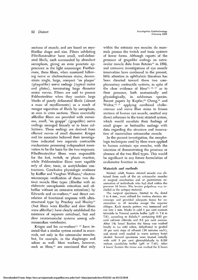
Fig. 7. Fibrillenstruktur, transverse section, Selected area within a single muscle fiber represen-tative of the vast majority in a "mainly en plaque fiber" group. The preponderance of therelatively discrete inyofibrillar pattern (f) in a bundle of fibers possessing mostly "en plaque"endings is quite suggestive that these two characteristics occur together and define a particularmuscle fiber type. Myofilaments (arrows); mitochondrion (m); sarcoplasmic reticulum (sr).(Autopsy specimen, cholinestera.se stained, and nerve endings identified, 1 per cent osmiumtetroxicle, electron micrograph. x20,800.)
ings and is found in all striated muscle.In addition, a peculiar speckled stainingwas evident over the entire muscle surfacefrom origin to insertion, suggesting a diffusewidespread innervation as well. In markedcontrast, the levator palpebrae muscle dis-played only the dense focal band of endplates.
At higher magnification, teased extra-ocular fibers displayed two kinds of stainedterminals. One was a large, intensely stain-ing, compact, often lobulated, oval endingwith an appearance quite similar to the"en plaque" ending of the classical fast ortwitch fiber (Figs. 9, 10 to 12). The secondtype appeared as smaller, lighter staining,droplets, buttons, or beads organized inloose clusters or attached chains, identicalto the "en grappe" endings associated inother species with the slow contractile re-sponse (Figs. 13 to 16). The levator pos-sessed only "en plaque" innervation.
The distribution of each type of endingwas characteristic. Although entire lengthswere not observed, the two nerve terminalclasses never could be found on the samemuscle fiber. This situation suggested twomuscle fiber types: (1) those with motorend plates and (2) those with grapelikeendings. "En plaque" terminals usually oc-curred singly on individual fibers, bothwithin the terminal innervation band andwhen encountered elsewhere. However, two"en plaque" endings were occasionally seenwithin the distances teased (Fig. 12). Sinceit was possible to find these endings scat-tered throughout the muscle from origin toinsertion, it was felt that several widelyspread "en plaque" terminals might bepresent on single fibers. This conclusion issupported by the work of Kupferis andCheng.19 In contrast, numerous "en grappe"endings were present within any teasedlength (Figs. 13 to 16). In agreement with
Downloaded From: http://iovs.arvojournals.org/pdfaccess.ashx?url=/data/journals/iovs/933242/ on 03/23/2018
58 Dietert Inoestigatioe OphthalmologyFebruary 1965
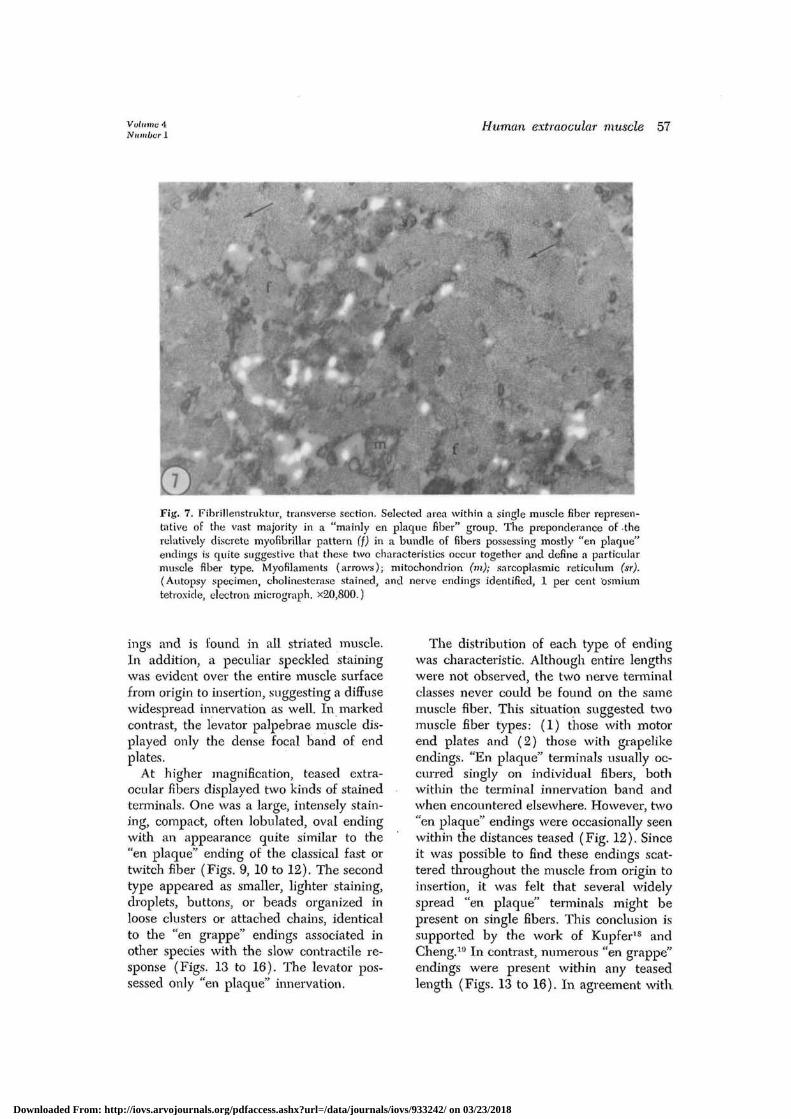
Fig. 8. Felderstruktur, transverse section. Selected area within a single muscle fiber represen-tative of the vast majority in a "mainly en grappe fiber" group. The preponderance of thisindistinct myofibrillar pattern (f) in a bundle of fibers possessing mostly "en grappe" terminalssupports the belief that these two features also occur simultaneously, and hence establish asecond muscle fiber type. Myofilaments (arrows); mitochondrion (in); sarcoplasmic reticulum(sr). (Autopsy specimen, cholinesterase stained, and nerve endings identified, 1 per centosmium tetroxide, electron micrograph. x20,800.)
Table
0.1%,
0.1%,
II. Results
DFP
060 minutes
060 minutes
of differential staining
Substrate
ATChATChBTChBTCh
Enzymedemonstrated
AChE, BChEAChEBChENeither (control)
"En plaque"endings
++ (Fig. 17)+ (Fig. 19)0
"En grappeendings
++ (Fig.+ (Fig.0
18)20)
Reactionintensity
4+1+1+0
Hess'14 studies in the monkey, great varia-bility was encountered in the distancesbetween "en grappe" endings, both on thesame fiber and on different fibers, withextremes in the range of 10 ^ to 2 or 3 mm.Isolated fibers of the levator demonstratedsingle motor end plates in the innervationband.
An investigation of the pharmacology ofthese two nerve endings was undertaken ononly one of the extrinsic eye muscles, sincethe results enumerated above had indi-
cated each to be identical within theparameters of the current investigation.Haggqvist27 has postulated recently that"en plaque" endings may contain onlyacetyl cholinesterase (AChE; true cholin-esterase), while "en grappe" endings stainfor butyryl cholinesterase (BChE; pseudo-cholinesterase). Furthermore, he demon-strated in the monkey lateral rectus onlythe acetyl fraction. The lateral rectus there-fore was selected for differential staining,with the results in Table II. Thus, both
Downloaded From: http://iovs.arvojournals.org/pdfaccess.ashx?url=/data/journals/iovs/933242/ on 03/23/2018

Volume 4Nuviber 1
Human extraocular muscle 59
F
• y \
y
Fig. 9. "En plaque" terminal, teased preparation. Selected portion of an isolated muscle fiber(F) demonstrating a single, large, oval motor end plate (arrow). (Autopsy specimen, acetyl-thiocholine substrate staining acetyl and butyryl cholinesterase, light micrograph. x390,)
Fig. 10. "En plaque" terminal, teased preparation, similar to that in Fig. 9. Another musclefiber (F) with its compact, platelike motor ending (arrow), (Autopsy specimen, acetylthio-choline substrate staining acetyl and butyryl cholinesterase, light micrograph. x390.)
Fig. 11. "En plaque" terminals, teased preparation similar to those in Fig. 9 displaying singleneuromuscular terminals (arrows) on two separate muscle fibers (F). (Autopsy specimen,acetylthiocholine substrate staining acetyl and butyryl cholinesterase, light micrograph, x390.)
Fig. 12. "En plaque" terminals, teased preparation, similar to those in Fig. 9 with the excep-tion that the rare situation' of two plaquelike endings (arrows) on a single muscle fiber (F)is demonstrated. Two other superimposed "en plaque" endings are just visible at the upperleft. (Autopsy specimen, acetylthiocholine substrate staining acetyl and butyryl cholinesterase,light micrograph. x390.)
Fig. 13. "En grappe" terminals, teased preparation. Selected portion of single muscle fiber(F), which is in marked contrast to those in Figs. 9 to 12. Numerous small drops or beads ofstain (arrows) arranged in linear array are easily visible. At no time are these grapelike end-ings found in a single isolated fashion like "en plaque" endings. (Autopsy specimen, acetylthio-choline substrate staining acetyl and butyryl cholinesterase, light micrograph. x390.)
Fig. 14. "En grappe" terminals, teased preparation, similar to those in Fig. 13. Multiple, linearstaining areas (arrows) on a single muscle fiber (F). A branching cluster arrangement seenfrequently is noted (C). (Autopsy specimen, acetylthiocholine substrate staining acetyl andbutyryl cholinesterase, light micrograph. x390.)
Downloaded From: http://iovs.arvojournals.org/pdfaccess.ashx?url=/data/journals/iovs/933242/ on 03/23/2018

60 Dietert Investigative OphthalmologyFebruary 1965
•
Fig. 15. "En grappe" terminals, teased preparation, similar to those in Fig. 13. Five smallgrapelike endings (arrows) are visible again on a single fiber (F). (Autopsy specimen, acetyl-thiocholine substrate staining acetyl and butyryl cholinesterase, light micrograph. x390.)
Fig. 16. "En grappe" terminals, teased preparation, similar to those in Fig. 13. A single fiber(F) possessing several endings, some of which (C) are in a branching cluster. (Autopsy speci-men, acetylthiocholine substrate staining acetyl and butyryl cholinesterase, light micrograph.x390.)
Introduction to Figs. 17 to 20. These figures demonstrate that "en plaque" as well as "engrappe" nerve endings are stained following the differential procedures (see text) for revealingacetyl and butyryl cholinesterase separately. The decreased reaction intensity evident uponcomparison with Figs. 9 through 16 is felt to be the result of selectively staining for onlyone of the two esterases present. (Hess11 believes maximal staining takes place only whenboth enzymes are active.)
Fig. 17. "En plaque" terminal, AChE positive, teased preparation. Selected portion of anisolated muscle fiber (F) treated with DFP and followed by acetylthiocholine substrate, whichshows only acetyl cholinesterase. A single, compact, platelike ending (arrow) is seen. (Autopsyspecimen, light micrograph. x390.)
Fig. 18. "En grappe" terminals, AChE positive, teased preparation, similar to those in Fig. 17.A single muscle fiber (F) treated with DFP and followed by acetylthioeholine substrate, whichshows only acetyl cholinesterase. Several grapelike endings (arrows) are visible. (Autopsyspecimen, light micrograph. x390.)
Fig. 19. "En plaque" terminal, BChE positive, teased preparation. Selected portion of an iso-lated muscle fiber (F) treated with butyrylthiocholine substrate, which shows only butyrylcholinesterase. One large, oval end plate (arrow) is visible. (Autopsy specimen, light micro-graph. x390.)
Fig. 20. "En grappe" terminals, BChE positive, teased preparation, similar to those in Fig. 19.A single muscle fiber (F) treated with butyrylthiocholine substrate, which shows only butyrylcholinesterase. Several small, staining areas (arrows) can be seen, (Autopsy specimen, lightmicrograph, x390.)
Downloaded From: http://iovs.arvojournals.org/pdfaccess.ashx?url=/data/journals/iovs/933242/ on 03/23/2018

Volume 4'Number 1
Human extraocular muscle 61
"en plaque" and "en grappe" endings arerevealed following each enzymatic reaction.Phis would suggest, at least at this level ofhistochemical sensitivity, that the twoterminal types are qualitatively similar inenzyme reactions. HessG> 14 has demon-strated each cholinesterase in both endingsin frog and monkey striated muscle. Thediminished reaction intensity seen duringdifferential staining was also described byHess.(i> ]1 He believed that maximal stainingtook place only when both esterases wereactive. The use of either DFP followed byacetylthiocholine, which demonstrates onlyacetyl cholinesterase (Figs. 17 and 18), orbutyrylthiocholine, which demonstratesonly butyryl cholinesterase (Figs. 19 and20), resulted in fainter staining becauseonly one of the two enzymes present wasfunctioning.
Correlation of muscle ultrastructure withkind of nerve ending. To determine if themuscle fibers with "en plaque" and "engrappe" endings were identical to thefibrillar and afibrillar muscle fiber types,respectively, an electron microscopic exam-ination was made of previously stainedfibers from postmortem inferior oblique.One group was designated "mainly enplaque fibers" and the other "mainly engrappe fibers." Each group, when studied,displayed a vast majority of its fibers tobe of one structure or the other. The "enplaque" fiber group possessed primarilyFibrillenstruktur (Fig. 7), while the "engrappe" fiber group demonstrated a pre-ponderance of Felderstruktur (Fig. 8).Although the impurity of the groups pre-vents exact correlation, these findings sug-gest that the fibrillar fibers have only "enplaque" endings, and the afibrillar fibersonly "en grappe" terminals, which is inagreement with the conclusions of Hess.10'17
Discussion
The presence of both fibrillar (presump-tive fast11) and afibrillar (presumptiveslow11) muscle fiber ultrastructure, thedemonstration of cholinesterase positive "enplaque" and "en grappe" nerve endings,
which exist on separate contractile ele-ments, and the suggestive correlation ofeach fiber class with an innervational typehave provided for human extraocularmuscle the same morphologic features asare present in frog iliofibularis muscle,13-1S
wherein the existence of separate fast andslow contractile mechanisms has beenphysiologically established.9'20 These ana-tomic findings should provide a strong im-petus for the initiation of physiologicstudies, such as nerve fiber stimulation asso-ciated with intracellular recording andtension measurements, which are clearlyrequired to establish as fact the presence offast and slow contractile systems in humanextraocular musculature. The recent in-vestigation of cat superior oblique muscleby Hess and Pilar,17 where physiologic aswell as anatomic evidence for separatetwitch and tonic responses is presented,indicates that functional studies in manwill confirm the structural impressions ofthis report. Anatomic absence of the slowmuscle fiber system in the levator of thelid, a muscle also receiving innervationfrom the oculomotor nerve, can be regardedas evidence for a selective unique organiza-tion of the rectus and oblique musculature.The presence of such a system limited pre-cisely to these six related muscles impliesa significance greater than a biologiccuriosity.
Although conclusive evidence is lacking,the belief that the "en grappe" endingsdemonstrated in this paper were somaticmotor terminals was based primarily ontheir cholinesterase positive staining, whichCoers and Woolf20 feel (at least in muscletissue) strongly implies a motor function.The work of Corbin and Oliver,30 demon-strating the cell bodies of origin of the "engrappe" endings in cat extrinsic eye muscleto lie neither in the trigeminal nucleus norin its mesencephalic root, but within thethird, fourth, and sixth cranial nervenuclei, supports this assumption. Further-more, these workers,30 through ablation andretrograde recording, showed that neuronsin the superior sympathetic ganglion and
Downloaded From: http://iovs.arvojournals.org/pdfaccess.ashx?url=/data/journals/iovs/933242/ on 03/23/2018
62 Dietert Investigative OphthalmologyFebruary 1965
Edinger-Westphal nucleus play no part insupplying "en grappe" innervation. Suchdata would seem to exclude the possibilityof attributing an autonomic function tothese endings.
The properties of the small diametermotor nerves, the junctional potential, andthe contractile response which characterizethe slow muscle system can be found in de-tail in the papers of Kuffler and VaughanWilliams.9'20 Suffice it to include here thatthe slow and fast fibers were found syner-gistic, the state of tension of the slowfibers was directly related to stimulus fre-quency, and any amount of relaxing slowfiber tension could be collapsed instantlyby superimposition of a single twitch con-traction. Should such a versatile neuro-muscular organization be established forhuman extrinsic eye muscle, a specificmechanism might be available to explain,for example, the phenomenon of simul-taneous horizontal, fast saccadic, and slowvergence movements as described by Al-pern and Wolter.31
I wish to express my appreciation to Dr. AdolphI. Cohen of the Departments of Anatomy andOphthalmology, and Dr. Sarah Luse and Mr.Paul Meyers of the Department of Anatomy,Washington University School of Medicine, fortheir donation of facilities, materials, and techni-cal advice.
REFERENCES1. Sommerkamp, H.: Das Substrat der Dauer-
verkiirzung am Froschmuskel, Arch, exper.Path. u. Pharmakol. 128: 99, 1928.
2. Kriiger, P.: Uber einen moglichen Zusam-menhang zwischen Struktur, Funktion, undchemischer Beschaffenheit der Muskeln, Biol.Zentralbl. 49: 616, 1929.
3. Fiirlinger, F.: t)ber einen Zusammenhangzwischen Struktur und Funktion von Skelett-muskeln bei Rana temporaria, Zool. Anz. 90:325, 1930.
4. Kriiger, P.: Die Innervation der tetanischenund tonischen Fasern der quergestreiftenSkelettmuskulatur der Wirbeltiere, Anat. Anz.97: 169, 1949.
5. Kriiger, P., and Giinther, P. C : Innervationund pharmakologisches Verhalten des M.gastrocnemius und M. pectoralis major derVogel, Acta Anat. 33: 325, 1958.
6. Hess, A.: The structure of extrafusal musclefibers in the frog and their innervation studiedby the cholinesterase technique, Am. J. Anat.107: 129, 1960.
7. Hess, A.: Structural differences of fast andslow extrafusal muscle fibers and their nerveendings in chickens, J. Physiol. 157: 221,1961.
8. Hess, A.: Two kinds of extrafusal musclefibers and their nerve endings in the gartersnake, Am. J. Anat. 113: 347, 1963.
9. Kuffler, S. W., and Vaughan Williams, E. M.:Small-nerve junctional potentials. The distri-bution of small motor nerves to frog skeletalmuscle, and the membrane characteristics ofthe fibers they innervate, J. Physiol. 121:289, 1953.
10. Edwards, G. A., Ruska, H., et a l : Compara-tive cytophysiology of striated muscle withspecial reference to the role of the endoplas-mic reticulum, J. Biophys. & Biochem. Cytol.2: suppl. 143, 1956.
11. Peachey, L. D., and Huxley, A. F.: Struc-tural identification of twitch and slow striatedmuscle fibers of the frog, J. Cell Biol. 13:177, 1962.
12. Siebeck, R., and Kriiger, P.: Die histologischeStruktur der ausseren Augenmuskeln als Aus-druck ihrer Funktion, von Graefes Arch.Ophth. 156: 637, 1955.
13. Kriiger, P.: Die Innervation phasisch bzw.tonisch reagierender Muskeln von Sfiuge-tieren und des Menschen, Acta Anat. 40:186, 1960.
14. Hess, A.: Further morphological observationsof "en plaque" and "en grappe" nerve end-ings on mammalian extrafusal muscle fiberswith the cholinesterase technique, Rev. Can-ad. Biol. 21: 241, 1962.
15. Retzius, G.: Zur Kenntniss der motorischenNervenendigungen, Biol. Untersuch., NeueFolge 3: 41, 1892.
16. Hess, A.: The structure of slow and fastextrafusal muscle fibers in the extraocularmuscles and their nerve endings in guineapigs, J. Cell. & Comp. Physiol. 58: 63, 1961.
17. Hess, A., and Pilar, G.: Slow fibers in theextraocular muscles of the cat, J. Physiol.169: 780, 1963.
18. Kupfer, C.: Motor innervation of extraocularmuscle, J. Physiol. 153: 522, 1960.
19. Cheng, K.: Cholinesterase activity in humanextraocular muscles, Jap. J. Ophth. 7: 174,1963.
20. Wolter, J. R., and O'Keefe, N. T.: Localiza-tion of nerve endings in relation to cholin-esterase deposits in normal human eye mus-cles, INVEST. OPHTH. 2: 558, 1963.
21. Wolter, J. R.: Thin nerves with simple end-
Downloaded From: http://iovs.arvojournals.org/pdfaccess.ashx?url=/data/journals/iovs/933242/ on 03/23/2018

Volume 4Number 1
Human extraocular muscle 63
ings containing cholinesterase in striated hu-man eye muscle, Neurology 14: 283, 1964.
22. Palade, G. E.: A study of fixation for elec-tron microscopy, J. Exper. Med. 95: 285,1952.
23. Cooper, S., and Daniel, P. M.: Muscle spin-dles in human extrinsic eye muscles, Brain72: 1, 1949.
24. Merrillers, N. C. R.: The fine structure ofmuscle spindles in the lumbrical muscles ofthe rat, J. Biophys. & Biochem. Cytol. 7: 725,1960.
25. Coupland, R. E., and Holmes, R. L.: Theuse of cholinesterase techniques for thedemonstration of peripheral nervous struc-tures, Quart. J. Micr. Sc. 98: 327, 1957.
26. Coers, C, and Woolf, A. L.: The innervationof muscle, Springfield, 111., 1959, Charles CThomas, Publisher.
27. Haggqvist, G.: On cholinesterasis in skeletalmuscles, Anat. Anz. I l l : 250, 1962.
28. Giinther, P. G.: Die Innervation der tetanis-chen und tonischen Fasem der quergestreif-ten Skelettmuskulatur der Wirbeltiere. DieInnervation des M. sartorius und des M. ileo-fibularis des Frosches, Anat. Anz. 97: 175,1949.
29. Kuffler, S. W., and Vaughan Williams, E. M.:Properties of the slow skeletal muscle fibeisof the frog, J. Physiol. 121: 318, 1953.
30. Corbin, K. B., and Oliver, R. K.: The originof fibers to the grape-like endings in theinsertional third of the extraocular muscles,J. Comp. Neurol. 77: 171, 1942.
31. Alpern, M., and Wolter, J. R.: The relationof horizontal saccadic and vergence move-ments, Arch. Ophth. 56: 685, 1956.
Downloaded From: http://iovs.arvojournals.org/pdfaccess.ashx?url=/data/journals/iovs/933242/ on 03/23/2018