the contact lens miracles-cw - eyeness.ch · 1 the contact lens miracles continuous wear michael...
TRANSCRIPT

1
The contact lens miraclesContinuous wear
Michael Wyssdipl. Augenoptiker [email protected] baertschi, Bern, Switzerland
Indication for continuous wear
• Pediatric / Geriatric Patients
Indication for continuous wear
• Bandage contact lens after surgery or injury
• Therapeutic use in cornea disorders– EBMD (Map-dot-fingerprint dystrophy) and
recurrent Erosio
– Lagophthalmus
– Open eyes during sleeping
Indication for continuous wear
• Open eyes during sleepingPre Tx Post Tx
Indication for continuous wear
• Convenience – Outback and Adventure
– Dating “hoppers”
– Lazy people – like me
Product Overview
1 Jahr1 month1 week1 month1 monthReplace
*******???0.721.521.50Modulus
???
30 days
*******
plan / -6.0
14.00
8.60
48%
160
Biofinity
<2%40%24%36%H2O
30 Tage7 days30 days30 daysCW up to
Individual+6.0 / -12.0+6.0 / -10.0+6.0 / -12.0Diopter
No restriction
**************-0.75 / -1.25 / -1.75 / -2.25
Cylinder
Lubricity
Ø
Radius
DK/t
Individual14.0013.8014.00
*******34717
Individual8.408.40 / 8.608.60
100-163154175110
GPOasysNight&DayPurevision
2
Fitting Procedure
• Initial Consultation– Information and Anamnese
– Refraction, Biomicroscopy, Topographie, Fundus, Pachimetry if available, etc.
– Insert first pair of contact lenses for 1h trial
– First check of lens fit and Vacc
– Handling instruction
Fitting Procedure
• First week, after first night– Px comes with lenses inserted, after first night
as soon as possible
– Ask about awakening (dryness, blurred vision)
– Biomicroscopy and then Rx
– If everything unaffected, start CW
Fitting Procedure
• Second week CW– Dito first week– Analysis of deposition on lens surface– eventually switch system or product
• First month CW– Dito second week, but let Px come in
afternoon– Critical for CLPC– Additionally Pachimetry if available
After Care
• Follow-up every 6 month, no selling without control
• Hand-out or Guideline about complications and “what to do” in such cases
• Hotline for emergency
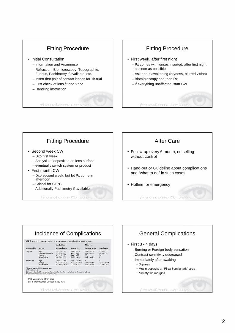
Incidence of Complications
P B Morgan, N Efron et alBr. J. Ophthalmol. 2005; 89:430-436
General Complications
• First 3 - 4 days– Burning or Foreign body sensation
– Contrast sensitivity decreased
– Immediately after awaking• Dryness• Mucin deposits at “Plica Semilunaris” area• “Crusty” lid margins
3
Complications Mucin Balls
• Incidence increases with nights wearing contacts continuous– Common in materials with high modulus
Complications CLARE
• Contact lens induced acute red eye– Incidence 1 – 3.8%
Complications CLARE
• Non-ulcerative sterile keratitis associated with colonization of gram-negative bacteria – awakening with unilateral pain, photophobia,
tearing and a red eye– Visual acuity is unaffected – Sub-epithelial infiltrates in the mid-periphery
of the cornea near the limbus, focally or diffusely
– Limbal injection circumferentially, but no anterior chamber reaction or lid edema
Complications CLPC
• Contact lens induced papillary conjunctivitis– Incidence 2-7% dependent on DW or CW
Complications CLPC
• Conjunctiva findings– Conjunctiva hyperemia & bright red/orange
colour of papilla
– Hexagonal shape change to more rounded form
– Central vascular tuft, sometimes with staining
– Can display infiltrates at apex, sometimes scarring (cream/white colour)
– Mucus can be present in severe cases
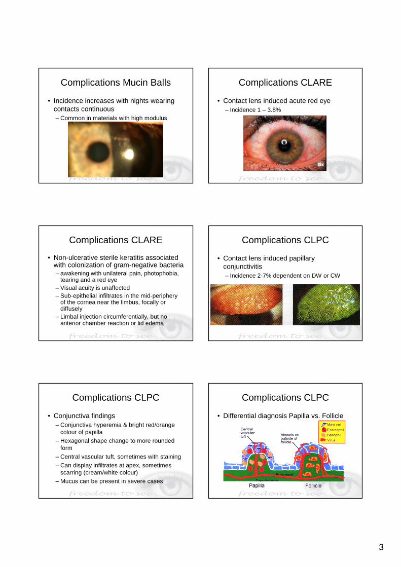
Complications CLPC
• Differential diagnosis Papilla vs. Follicle

4
Complications SEAL
• Superior Epithelial Arcuate Lesion– Incidence up to 4.5%
– most seen in Purevision Toric
Risk factors for infiltrative event
• SmokingJohn J McNally et alEye & Contact lens 29: 153-156, 2003
Risk factors for infiltrative event
• OthersJohn J McNally et alEye & Contact lens 29: 153-156, 2003
Complications IK
• Infiltrative Keratitis– Incidence 5% - 10%
Complications IK
• Infiltrates– Mild diffuse or small focal infiltrates– Infiltrates are often near the limbus, but can
be present anywhere in the cornea. There also may be mild corneal punctate staining
• Causes– Mechanical trauma – Gram-positive bacterial exotoxins– Minor foreign body trapped under the lens
Complications IK
• Symptoms– tearing, photophobia, foreign-body sensation,
injection of the bulbar conjunctiva and limbus
– Occur later in the day and may vary widely, from severe to non-existent
5
Complications AIK
• Asymptomatic Infiltrative Keratitis– Incidence 1% - 8%
Complications CLPU
• Contact lens induced peripheral ulcer– Incidence 3.3% - 5.4%
CLPU Definition
• “CLPU is an inflammatory reaction of the cornea that is characterized in its active stage by focal excavation of the epithelium, infiltration, and necrosis of the anterior stroma. However, Bowman’s layer is intact”
Complications MK
• Microbial Keratitis– Incidence 0.2% - 12.7%
Acanthamoeba Pseudomonas
MK Definition
• “MK is an infection of the cornea by microbes that is characterized by excavation of the corneal epithelium, Bowman’s layer, and stroma with infiltration and necrosis of the tissue”
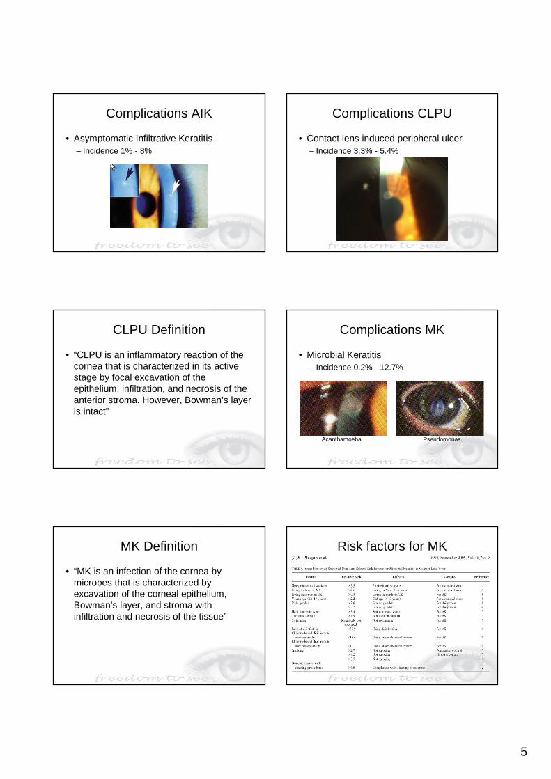
Risk factors for MK
6
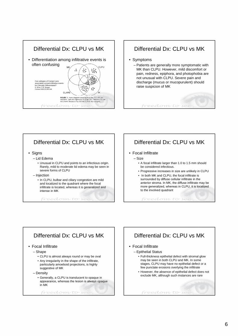
Differential Dx: CLPU vs MK
• Differentiation among infiltrative events is often confusing
Can subtypes of Contact Lens associated corneal infiltrative events be Clinically Differentiated?N. Efron, P.B. Morgan Cornea 2006;25:540-544
Differential Dx: CLPU vs MK
• Symptoms – Patients are generally more symptomatic with
MK than CLPU. However, mild discomfort or pain, redness, epiphora, and photophobia are not unusual with CLPU. Severe pain and discharge (mucus or mucopurulent) should raise suspicion of MK
Differential Dx: CLPU vs MK
• Signs – Lid Edema
• Unusual in CLPU and points to an infectious origin. Rarely, mild to moderate lid edema may be seen in severe forms of CLPU
– Injection • In CLPU, bulbar and ciliary congestion are mild
and localized to the quadrant where the focal infiltrate is located, whereas it is generalized and intense in MK
Differential Dx: CLPU vs MK
• Focal Infiltrate – Size
• A focal infiltrate larger than 1.0 to 1.5 mm should be considered infectious.
• Progressive increases in size are unlikely in CLPU• In both MK and CLPU, the focal infiltrate is
surrounded by diffuse cellular infiltrate in the anterior stroma. In MK, the diffuse infiltrate may be more generalized, whereas in CLPU, it is localized to the involved quadrant
Differential Dx: CLPU vs MK
• Focal Infiltrate – Shape
• CLPU is almost always round or may be oval• Any irregularity in the shape of the infiltrate,
particularly amoeboid projections, is highly suggestive of MK
– Density• Generally, a CLPU is translucent to opaque in
appearance, whereas the lesion is always opaque in MK
Differential Dx: CLPU vs MK
• Focal Infiltrate– Epithelial Status
• Full-thickness epithelial defect with stromal glow may be seen in both CLPU and MK. In some stages, CLPU may have no epithelial defect or a few punctate erosions overlying the infiltrate
• However, the absence of epithelial defect does not exclude MK, although such instances are rare
7
Differential Dx: CLPU vs MK
• Focal Infiltrate– Surrounding Cornea
• Mild edema or folds are seen in the cornea surrounding the focal infiltrate in MK but are not seen in CLPU
– Location • CLPU shows predilection for the peripheral cornea,
whereas MK can be central, paracentral, or peripheral
Differential Dx: CLPU vs MK
• Anterior Chamber – Is almost always present in MK. Hypopyon is
never seen in CLPU, although it is occasionally seen in the earliest stages of MK
• Clinical Course of CLPU– Mostly regress and resolve spontaneously on
discontinuation of CL wear• A CLPU usually takes approximately 3 to 4 days to
resolve
Differential Dx: CLPU vs MK
• Clinical Course in MK– Discontinuation of CL wear may result in mild
relief from symptoms, but the condition never resolves unless appropriately treated
– Worsening of symptoms despite CL removal is a strong indicator of MK
– Heals more gradually over 2 to 3 weeks • In both conditions, healing leaves a scar, which is
usually circular and faint in CLPU
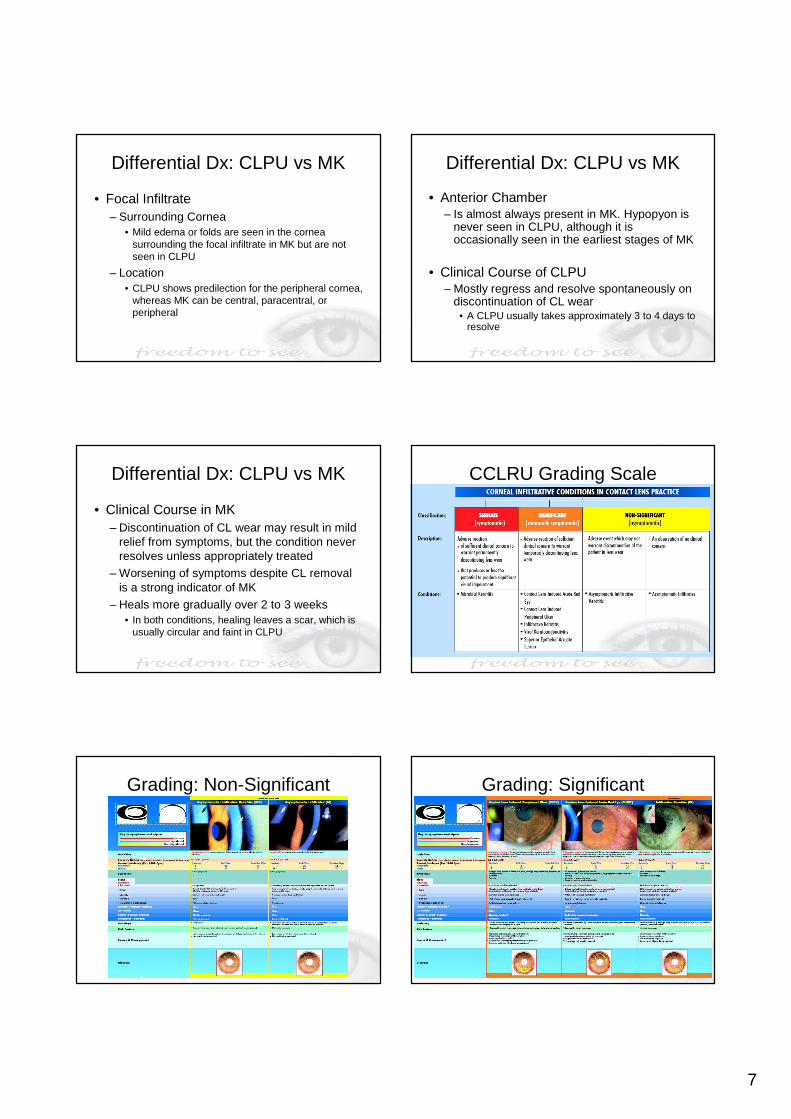
CCLRU Grading Scale
Grading: Non-Significant Grading: Significant
8
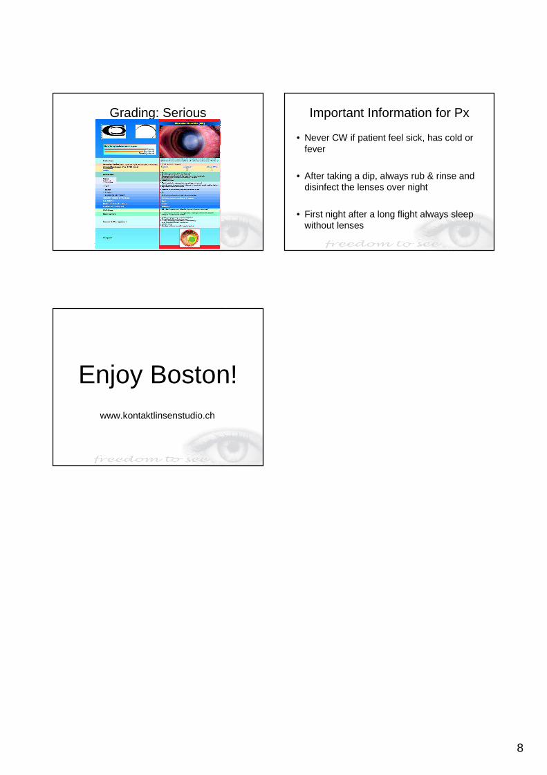
Grading: Serious Important Information for Px
• Never CW if patient feel sick, has cold or fever
• After taking a dip, always rub & rinse and disinfect the lenses over night
• First night after a long flight always sleep without lenses
Enjoy Boston!www.kontaktlinsenstudio.ch