the comparative effectiveness of ventricular shunt placement versus endoscopic third ventriculostomy...
TRANSCRIPT
J Neurosurg: Pediatrics / Volume 13 / March 2014
J Neurosurg Pediatrics 13:295–300, 2014
295
©AANS, 2014
Cerebrospinal fluid diversion for treatment of hy-drocephalus is one of the most common pediatric neurosurgery procedures performed. The annual
hospital cost of treating children with hydrocephalus is approximately $2 billion.22
Cerebrospinal fluid diversion through a shunt cathe-ter remains the preferred treatment method in hydroceph-alus. Despite the development of new shunt materials and techniques for introduction of catheters,1,3 however, the failure rate associated with a shunt within the 1st year has remained high since 1960, ranging from 25.7% to 36.8% in the year 2000.4 Shunt failure represents a significant health care expense, with an estimated cost of over $1
billion dollars annually for admissions for both pediatric and adult patients.18 It is believed that reducing the failure rate of CSF diversion within 1 year of treatment could lead to substantial improvement of the health of children with hydrocephalus and the hefty attendant health care expenditures.24
Younger patients, specifically infants under 1 year of age at the time of initial shunt placement, have the highest risk of shunt failure.17,19 Alternative and potentially more successful methods to divert CSF in infants with hydro-cephalus are being explored. Endoscopic third ventricu-lostomy (ETV), the original hydrocephalus treatment modality from the early 20th century, has regained atten-tion in recent years as an alternative to catheter-shunted CSF diversion. However, single-institutional studies re-
The comparative effectiveness of ventricular shunt placement versus endoscopic third ventriculostomy for initial treatment of hydrocephalus in infants
Clinical article
Sarah C. Jernigan, M.D., M.P.h.,1 Jay g. Berry, M.D., M.P.h.,2 Dionne a. grahaM, Ph.D.,3 anD LiLiana gouMnerova, M.D.1
1Department of Neurosurgery, 2Division of General Pediatrics, and 3Clinical Research Program, Boston Children’s Hospital, Harvard Medical School, Boston, Massachusetts
Object. The purpose of this study was to compare the effectiveness of CSF diversion with endoscopic third ventriculostomy (ETV) versus shunt therapy in infants with hydrocephalus.
Methods. The authors conducted a retrospective analysis of 5416 infants 1 year of age or younger with hydro-cephalus (congenital or acquired) in whom CSF diversion was performed using either ETV or shunt placement at 41 children’s hospitals between 2004 and 2009. Data were obtained from the Pediatric Health Information Systems database. Surgical failure was defined as the need for a repeat diversion operation within 1 year of initial surgery. The authors compared failure rates of ETV and shunt, as well as patient demographics and clinical characteristics, using hierarchical regression according to treatment group.
Results. During the period examined, 872 infants (16.1%) initially underwent ETV and 4544 (83.9%) underwent ventricular shunt placement. The median infant age was 37 days (IQR 11–122 days) for both ETV and shunt place-ment. More infants who underwent ETV rather than shunt placement were born prematurely (41.6% vs 23.9%, re-spectively; p < 0.01) and had intraventricular hemorrhage (45.4% vs 17.5%, respectively; p < 0.01). Higher operative failure rates at 1 year were observed in infants who underwent ETV as opposed to shunt surgery (64.5% vs 39.6%, respectively; OR 2.9 [95% CI 2.3–3.5], p < 0.01). After controlling for prematurity, intraventricular hemorrhage, and spina bifida, ETV remained associated with a higher risk of failure (OR 2.6 [95% CI 2.1–3.2]).
Conclusions. In infants with hydrocephalus, a greater 1-year CSF diversion failure rate may occur after ETV compared with shunt placement. This risk is most significant for procedures performed within the first 90 days of life. Further investigation of the need for multiple reoperations, cost, and impact of surgeon and hospital experience is necessary to distinguish which treatment is more effective in the long term.(http://thejns.org/doi/abs/10.3171/2013.11.PEDS13138)
Key WorDS • hydrocephalus • ETV • ventricular shunt • infants • outcome
Abbreviations used in this paper: CPC = choroid plexus cauter-ization; ETV = endoscopic third ventriculostomy; IVH = intraven-tricular hemorrhage; PHIS = Pediatric Health Information System ; VP = ventriculoperitoneal.
This article contains some figures that are displayed in color on line but in black-and-white in the print edition.
S. C. Jernigan et al.
296 J Neurosurg: Pediatrics / Volume 13 / March 2014
port that ETV is also associated with high failure rates (range 70%–80%) within 1 year in high-risk patients, such as infants under 1 year of age.10–13,18 Therefore, we compared the effectiveness of ETV and shunt placement for hydrocephalus treatment in a multicenter cohort of in-fants younger than 1 year.
MethodsStudy Design and Setting
This study was a retrospective cohort analysis of the Pediatric Health Information System (PHIS), an admin-istrative database of 39 freestanding (financially inde-pendent), not-for-profit, tertiary care pediatric hospitals in the United States.12 These hospitals are affiliated with the Children’s Hospital Association (Shawnee Mission, KS), a business alliance of children’s hospitals. The data warehouse function for the PHIS database is managed by Solucient LLC (Evanston, IL). For purposes of external benchmarking, participating hospitals provide discharge data, including demographics, diagnoses, and procedures. Data are subjected to a number of reliability and validity checks and are processed into data quality reports. Cod-ing of the PHIS database permits tracking of subsequent hospitalizations within the same hospital for individual patients using an encrypted medical record number.
Patient PopulationThe study population was infants aged less than 1
year with hydrocephalus who underwent CSF diversion via ventricular shunt placement or ETV between January 1, 2004, and December 1, 2009. Infants with hydrocepha-lus were identified using ICD-9-CM diagnosis codes of congenital hydrocephalus (742.3), communicating hydro-cephalus (331.3), obstructive hydrocephalus (331.4), or spina bifida with hydrocephalus (741.0x).1
Main ExposureThe main exposure was initial hydrocephalus treat-
ment with shunt or ETV within the 1st year of life. We examined all hospitalizations dating back to the child’s birth to determine the date of the initial treatment.
Shunt. Patients with initial ventricular shunt place-ment were identified with the ICD-9-CM procedural code of ventricular shunt placement (02.3x) during a hospital admission. All types of shunts were included for analysis. A ventriculoperitoneal (VP) shunt was defined as “ven-tricular shunt to abdominal cavity and organs” (02.34), and the “other” shunt type category included “ventricular shunt to circulatory system” (02.32), “ventricular shunt to thoracic cavity” (02.33), “ventricular shunt to urinary sys-tem” (02.35), and “other operations to establish drainage of ventricle” (02.39).1
Endoscopic Third Ventriculostomy. Patients undergo-ing ETV for their first hydrocephalus treatment were iden-tified with the ICD-9-CM procedural code for ventricu-lostomy (02.2) during a hospital admission in the absence of billing charges for implanted ventricular catheters, res-ervoirs, or shunts suggestive of external ventricular drain placement or ventriculosubgaleal shunt placement.1
Main Outcome MeasureFor patients with shunt-treated hydrocephalus, the
primary outcome of the study was treatment failure, de-fined as a revision or replacement of an existing ventricu-lar shunt within 1 year of the initial procedure. Ventricu-lar shunt revisions were identified using the ICD-9-CM procedural codes 1) for ventricular shunt replacement (02.42, 54.95) or 2) for ventricular shunt removal (02.43) and new ventricular shunt placement (02.34), or other shunt-related procedure (02.3x), in a patient with an ex-isting ventricular shunt placed during a hospital admis-sion. Patients undergoing a sequence of shunt removal and temporary ventricular drain placement followed by insertion of a new ventricular shunt were considered to have had a single episode in which an existing shunt was replaced. We assumed that a patient had no revision if there were no hospital admissions for revision.1
For patients who had undergone ETV, treatment fail-ure was defined as a subsequent surgical intervention for hydrocephalus, including repeat ETV or shunt placement within 1 year of initial ETV. The secondary outcome of survivability reflects any further surgical interventions for hydrocephalus that occurred at least 1 year after ini-tial shunt placement or ETV but within the overall study period. We assessed data for all hospitalizations within the PHIS network for the entirety of the study period, such that the first patients to be treated had follow-up for up to 5 years after their initial CSF diversion procedure but the last patients had only 1 year of follow-up.
CovariatesDemographic characteristics assessed at the time of
initial shunt placement included primary payer (private insurance, government insurance, or other), age, sex, and race (non-Hispanic Caucasian, non-Hispanic black, His-panic, Asian, and other). The category “other” primary payer includes individuals who are self-insured. The “oth-er” category of race includes all individuals who were not classified as Caucasian, black, Hispanic, or Asian.
We evaluated clinical characteristics that may be correlated with the likelihood of CSF diversion failure, including prematurity (ICD-9-CM 765.2x), low birth weight (765.0, 765.1), myelomeningocele with hydroceph-alus (741.0x), intraventricular hemorrhage (IVH; 772.1), or multiple congenital anomalies (743.xx through 759.xx). We also evaluated hospital characteristics including the annual hospital volume of ETV- and shunt-treated pa-tients, teaching status, and geographic region (Northeast, South, Midwest, and West).
Statistical AnalysisWe assessed unadjusted differences in treatment fail-
ure rates for ETV and shunt as well as risk factors for treat-ment failure with a Rao-Scott chi-square test that account-ed for the random effect of individual hospitals. To adjust for case-mix differences, we used generalized estimating equations to assess differences in failure rates for ETV and shunt surgery accounting for demographic, clinical, and hospital characteristics associated (p < 1.0) with treat-ment failure in the unadjusted analysis, and the random ef-
J Neurosurg: Pediatrics / Volume 13 / March 2014
Shunt versus ETV for initial treatment of hydrocephalus
297
fect of hospitals. We also performed partition, regression tree modeling with binary splits for continuous variables (for example, age in days) to assess which patients had the greatest success and failure with ETV and shunt treatment.
ResultsThere were 5416 infants less than 1 year of age with
hydrocephalus in the study cohort; 4544 of them (83.9%) underwent shunt placement and 872 (16.1%) underwent ETV for the initial treatment of their hydrocephalus. The 1-year failure rates for shunt and ETV treatments were 39.6% and 64.5%, respectively.
Hospital CharacteristicsThe mean annual hospital case volume was signifi-
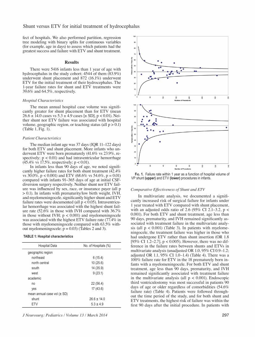
cantly greater for shunt placement than for ETV (mean 26.6 ± 14.0 cases vs 5.3 ± 4.9 cases [± SD], p < 0.01). Nei-ther shunt nor ETV failure was associated with hospital volume, geographic region, or teaching status (all p > 0.1) (Table 1, Fig. 1).
Patient CharacteristicsThe median infant age was 37 days (IQR 11–122 days)
for both ETV and shunt placement. More infants who un-derwent ETV were born prematurely (41.6% vs 23.9%, re-spectively; p < 0.01) and had intraventricular hemorrhage (45.4% vs 17.5%, respectively; p < 0.01).
In infants less than 90 days of age, we noted signifi-cantly higher failure rates for both shunt treatment (42.4% vs 30.0%, p < 0.001) and ETV (68.6% vs 54.6%, p = 0.01) compared with infants 91–365 days of age at initial CSF-diversion surgery respectively. Neither shunt nor ETV fail-ure was influenced by sex, race, or insurance payer (all p > 0.1). In infants with prematurity/low birth weight, IVH, and myelomeningocele, significantly higher shunt and ETV failure rates were documented (all p < 0.05). Intraventricu-lar hemorrhage was associated with the highest shunt fail-ure rate (52.6% in those with IVH compared with 36.7% in those without IVH; p < 0.001) and myelomeningocele was associated with the highest ETV failure rate (77.4% in those with myelomeningocele compared with 63.5% with-out myelomeningocele; p = 0.03) (Tables 2 and 3).
Comparative Effectiveness of Shunt and ETVIn multivariate analysis, we documented a signifi-
cantly increased risk of surgical failure for infants under 1 year treated with ETV compared with shunt placement, with an adjusted odds ratio of 2.6 (95% CI 2.1–3.2, p < 0.001). For both ETV and shunt treatment, age less than 90 days, prematurity, and IVH remained significantly as-sociated with treatment failure in the multivariate analy-sis (all p < 0.001) (Table 3). In patients with myelome-ningocele, the treatment failure was higher in those who had undergone ETV rather than shunt insertion (OR 1.8 [95% CI 1.2–2.7], p < 0.005). However, there was no dif-ference in the failure rates between shunts and ETVs in multivariate analysis (unadjusted OR 1.0, 95% CI 0.9–1.2; adjusted OR 1.1, 95% CI 1.0–1.4) (Table 4). There was a 100% failure rate for ETV in the 18 prematurely born in-fants with a myelomeningocele. For both ETV and shunt treatment, age less than 90 days, prematurity, and IVH remained significantly associated with treatment failure in the multivariate analysis (all p < 0.001). Endoscopic third ventriculostomy was most successful in patients 90 days of age or older regardless of comorbidities (54.6% failure rate) (Table 4). Patients were followed through-out the time period of the study, and for both shunt and ETV treatments, the highest risk of failure was within the first 90 days after the initial procedure. In patients with
TABLE 1: Hospital characteristics
Hospital Data No. of Hospitals (%)
geographic region northeast 6 (15.4) north central 10 (25.6) south 14 (35.9) west 9 (23.1)academic no 22 (56.4) yes 17 (43.6)mean annual case vol (± SD) shunt 26.6 ± 14.0 ETV 5.3 ± 4.9
Fig. 1. Failure rate within 1 year as a function of hospital volume of VP shunt (upper) and ETV (lower) procedures in infants.
S. C. Jernigan et al.
298 J Neurosurg: Pediatrics / Volume 13 / March 2014
5 years of follow-up, the shunt group still had a higher percentage of patients free from revision compared with the ETV group, but the long-term success rates for both procedures approached equality at 5 years (Fig. 2).
DiscussionOutcomes for infants with hydrocephalus treated
with ETV and shunts are different. Failure rates for ETV are higher than those for shunt in infants aged 90 days or less and for infants with a myelomeningocele. The high-est failure rate for ETV was observed in infants treated within the first 90 days of life and with diagnoses of my-elomeningocele and prematurity, whereas ETV was the most successful in infants older than 90 days, regardless of other medical comorbidities.
Success rates for ETV vary from 20% to 70% in prior studies of small groups of infants younger than 1 year of age.7–10,14 In our study, the ETV success decreased from 40%–45% to 25%–30% in infants younger than 90 days of age or with comorbidities.11–13 In another study of 11 patients under the age of 1 year who underwent a second endoscopic procedure after failed ETV, the authors found either a completely or almost completely closed stoma or new arachnoid membranes obstructing the stoma in all 11 patients and hypothesized that these young patients are at an increased risk of ETV failure because new arachnoid
membranes that block their ETV stomas are more likely to form.26 While the risk factors of age less than 3 months, prematurity, and IVH all had increased adjusted odds ratio of failure for ETV compared shunt therapy, the diagnosis of myelomeningocele was not associated with an increased risk of failure of ETV over shunt because patients with my-elomeningoceles are at an increased risk of failure of any CSF-diversion procedure compared with other children.
In many studies, age at the time of initial shunt place-ment has been shown to be an important risk factor for shunt failure in the child’s 1st year of life.1,6,15,16,24 Each of these studies included all children under the age of 1 year in a single group. We found that children aged 0–30 days, 30–60 days, and 60–90 days had a significantly increased failure rate for both VP shunts and ETVs compared with children 91–365 days of age, regardless of comorbidities, which indicates some change either in the etiology of or compensation to hydrocephalus in infants under 3 months versus those 3–12 months.
Previous studies have shown that the majority of hos-pitals perform pediatric ventriculoperitoneal shunt op-erations in relatively low volume, with 75% of hospitals performing fewer than 20 initial shunt procedures annu-ally.2,6,12 While higher hospital volume for surgical proce-dures has been correlated with better outcomes in a variety of specialties, including neurosurgery,2,11,20,25 there have been conflicting data in regard to volume and outcome for ventriculoperitoneal shunts. While 2 prior studies found a
TABLE 2: Failure rates of ventricular shunt by demographics and comorbidities*
Shunt Data No. of Cases % Surgical Failure p Valueage (days) 1–30 2050 45.2 <0.001 31–90 992 42.4 <0.001 91–365 1502 30.0 refsex male 2501 39.4 ref female 2043 39.7 0.8race† Caucasian 3050 40.1 ref African American 789 38.9 0.5 Asian 50 44 0.6 other 499 39.3 0.7prematurity no 3428 36.5 ref yes 1116 49.1 <0.001IVH no 3732 36.7 ref yes 812 52.6 <0.001MMC no 3332 38.5 ref yes 1212 42.5 0.02
* Boldface type indicates statistical significance. MMC = myelomenin-gocele; ref = reference.† Data missing for 156 patients (surgical failure rate 31.4%; p = 0.03).
TABLE 3: Failure rates of ETV by demographics and comorbidities*
ETV Data No. of Cases % Surgical Failure p Value
age (days) 1–30 266 67.9 0.001 31–90 220 70.0 0.01 91–365 260 54.6 refsex male 520 64.4 ref female 352 64.5 0.95race† Caucasian 521 63.5 ref African American 192 69.8 0.1 Asian 13 53.8 0.5 other 123 62.6 0.9prematurity no 513 59.5 ref yes 359 71.6 <0.001IVH no 479 61.4 ref yes 393 68.2 0.04MMC no 810 63.5 ref yes 62 77.4 0.03
* Boldface type indicates statistical significance.† Data missing for 23 patients (surgical failure rate 56.5%; p = 0.5).

J Neurosurg: Pediatrics / Volume 13 / March 2014
Shunt versus ETV for initial treatment of hydrocephalus
299
decrease in infection rates and mortality rates in higher-volume centers,5,21 other studies noted no correlation be-tween volume and patient survival13 or found that hospital volume only affected revision rate in 2 hospitals whose annual volume was significantly higher than that of the other 30 hospitals, with more than 80 initial shunts placed annually versus fewer than forty placed annually at most other hospitals.1 We found no correlation between hospital volume and outcomes for either shunt placement or ETV.
In a recent study Warf et al. compared the success of ETV versus ETV with choroid plexus cauterization (CPC) in infants less than 1 year of age in Uganda. They found that ETV was effective in treating hydrocephalus in only 47% of patients with myelomeningocele or non-infectious hydrocephalus compared with a success rate of 66% when ETV was combined with CPC in this same patient population. In the patients with postinfectious hy-drocephalus, no statistically significant difference was seen between ETV alone (52% success) compared with combined ETV-CPC (62% success), but there was a trend toward significance. There were very few patients with posthemorrhagic hydrocephalus in this African popu-lation (only 1% of all study patients) because very few premature infants with significant posthemorrhagic hy-drocephalus survive.27 In our study, we were unable to elucidate which patients might have undergone ETV with CPC versus ETV alone since there is no separate coding for these 2 procedures.
Although the present study is limited by its retro-spective review of an administrative database, it allowed
for a much larger number of patients than prior studies in an attempt to elucidate risk factors in a specific patient population. Coding errors could affect an administrative database, but we internally validated the PHIS patients from Boston Children’s Hospital by using our internal departmental database to ensure that our patient collec-tion algorithm was both sensitive and specific. However, we cannot account for patients who underwent a second-ary CSF-diversion procedure at an institution outside of the hospitals included in the PHIS consortium, and such patients would be included in our study as not hav-ing had a surgical failure. Also, patients who underwent initial shunt placement were not screened for subsequent endoscopic procedures, which could also indicate a po-tential failure in the initial CSF diversion. The impact of these limitations is lessened by the increased power in the study given the large patient population. One fi-nal limitation of utilizing administrative databases is the lack of clinical information available. There is no way of determining other complications of these procedures such as infection, neurological deficit, hematoma, and so on. Finally, our primary outcome of failure was defined as reoperation within 1 year, but our secondary outcome was any additional surgical interventions for hydrocepha-lus throughout the study period, allowing us to calculate shunt survival for at least a portion of the cohort popula-tion for as many as 5 years after their initial procedure.
Our rates of shunt and ETV failure correlate both with published data and the mathematical model devel-oped by Stein and Guo, which estimated a shunt failure rate of 64.2% at 1 year and 49.4% at 5 years, with a me-dian survival of 4.9 years.23
ConclusionsThis is the first study to evaluate the comparative ef-
fectiveness of shunt treatment and ETV in a multiinsti-tutional cohort of infants with hydrocephalus. Although there are limitations to studies utilizing administrative databases, the large patient populations that they allow to be studied enable an analysis of a wide variety of risk factors and patient characteristics that may contribute both positively and negatively to outcomes. The use of the administrative database allowed us to review the out-comes of over 5000 infants for our study. We found that infants with hydrocephalus had a higher treatment failure rate when they had undergone ETV compared with shunt placement, even when controlling for prematurity, IVH,
TABLE 4: Adjusted risk of surgical failure within 1 year*
FactorRisk of Surgical Failure
Unadjusted OR (95% CI) p Value Adjusted OR (95% CI) p Value
all patients: ETV vs VPS 2.9 (2.3–3.5) <0.001 2.6 (2.1–3.2) <0.0010–90 vs 91–365 days 1.9 (1.7–2.1) <0.001 1.6 (1.4–1.8) <0.001prematurity 1.8 (1.6–2.1) <0.001 1.3 (1.2–1.5) <0.001IVH 2.1 (1.8–2.4) <0.001 1.4 (1.2–1.7) <0.001MMC 1.0 (0.9–1.2) 0.75 1.1 (1.0–1.4) 0.15
* Boldface type indicates statistical significance. VPS = VP shunt.
Fig. 2. Kaplan-Meier curve for the 5-year survival of all VP shunt– and ETV-treated patients in the study.
S. C. Jernigan et al.
300 J Neurosurg: Pediatrics / Volume 13 / March 2014
and myelomeningocele, which affected the success of both shunts and ETVs.
Although prior studies in Uganda did show improved outcomes with the addition of CPC to ETV, those popula-tions did not include a significant number of infants born prematurely or infants with posthemorrhagic hydroceph-alus. Further analysis of this additional surgical proce-dure in North American patients is needed.
Future studies should evaluate which physiological changes may be present that improve survival of both shunts and ETVs that are performed after 90 days of life compared with those performed within the first 90 days of life.
Disclosure
The authors report no conflict of interest concerning the mate-rials or methods used in this study or the findings specified in this paper.
Author contributions to the study and manuscript prepara-tion include the following. Conception and design: Goumnerova, Jernigan. Acquisition of data: Jernigan, Graham. Analysis and inter-pretation of data: all authors. Drafting the article: Jernigan. Critical-ly revising the article: all authors. Reviewed submitted version of manuscript: all authors.
References
1. Berry JG, Hall MA, Sharma V, Goumnerova L, Slonim AD, Shah SS: A multi-institutional, 5-year analysis of initial and multiple ventricular shunt revisions in children. Neurosur-gery 62:445–454, 2008
2. Berry JG, Lieu TA, Forbes PW, Goldmann DA: Hospital vol-umes for common pediatric specialty operations. Arch Pedi-atr Adolesc Med 161:38–43, 2007
3. Caldarelli M, Di Rocco C, La Marca F: Shunt complications in the first postoperative year in children with meningomyelo-cele. Childs Nerv Syst 12:748–754, 1996
4. Choudhury AR: Avoidable factors that contribute to the com-plications of ventriculoperitoneal shunt in childhood hydro-cephalus. Childs Nerv Syst 6:346–349, 1990
5. Cochrane DD, Kestle JR: The influence of surgical operative experience on the duration of first ventriculoperitoneal shunt function and infection. Pediatr Neurosurg 38:295–301, 2003
6. Cochrane DD, Kestle J: Ventricular shunting for hydrocepha-lus in children: patients, procedures, surgeons and institutions in English Canada, 1989-2001. Eur J Pediatr Surg 12 Suppl 1:S6–S11, 2002
7. Egger D, Balmer B, Altermatt S, Meuli M: Third ventricu-lostomy in a single pediatric surgical unit. Childs Nerv Syst 26:93–99, 2010
8. Faggin R, Bernardo A, Stieg P, Perilongo G, d’Avella D: Hy-drocephalus in infants less than six months of age: effective-ness of endoscopic third ventriculostomy. Eur J Pediatr Surg 19:216–219, 2009
9. Fritsch MJ, Kienke S, Ankermann T, Padoin M, Mehdorn HM: Endoscopic third ventriculostomy in infants. J Neuro-surg 103 (1 Suppl):50–53, 2005
10. Goumnerova LC, Frim DM: Treatment of hydrocephalus with third ventriculocisternostomy: outcome and CSF flow pat-terns. Pediatr Neurosurg 27:149–152, 1997
11. Jenkins KJ, Newburger JW, Lock JE, Davis RB, Coffman GA, Iezzoni LI: In-hospital mortality for surgical repair of con-genital heart defects: preliminary observations of variation by hospital caseload. Pediatrics 95:323–330, 1995
12. Kelley ET, Arispe I, Holmes J: Beyond the initial indicators: lessons from the OECD Health Care Quality Indicators Proj-
ect and the US National Healthcare Quality Report. Int J Qual Health Care 18 (Suppl 1):45–51, 2006
13. Klimo P Jr, Thompson CJ, Drake J, Kestle JR: Assessing the validity of the endoscopic shunt insertion trial: did surgical experience affect the results? J Neurosurg 101 (2 Suppl): 130–133, 2004
14. Kulkarni AV, Drake JM, Mallucci CL, Sgouros S, Roth J, Constantini S, et al: Endoscopic third ventriculostomy in the treatment of childhood hydrocephalus. J Pediatr 155:254–259, 2009
15. Patwardhan RV, Nanda A: Implanted ventricular shunts in the United States: the billion-dollar-a-year cost of hydrocephalus treatment. Neurosurgery 56:139–145, 2005
16. Piatt JH Jr, Carlson CV: A search for determinants of cerebro-spinal fluid shunt survival: retrospective analysis of a 14-year institutional experience. Pediatr Neurosurg 19:233–242, 1993
17. Sainte-Rose C, Piatt JH, Renier D, Pierre-Kahn A, Hirsch JF, Hoffman HJ, et al: Mechanical complications in shunts. Pedi-atr Neurosurg 17:2–9, 1991–1992
18. Shah SS, Hall M, Slonim AD, Hornig GW, Berry JG, Sharma V: A multicenter study of factors influencing cerebrospinal fluid shunt survival in infants and children. Neurosurgery 62:1095–1103, 2008
19. Simon T, Riva-Cambrin J, Srivastava R, Bratton SL, Dean JM, Kestle JR, et al: Hospital care for children with hydrocephalus in the United States: utilization, charges, comorbidities, and deaths. J Neurosurg Pediatr 1:131–137, 2008
20. Smink DS, Fishman SJ, Kleinman K, Finkelstein JA: Effects of race, insurance status, and hospital volume on perforated appendicitis in children. Pediatrics 115:920–925, 2005
21. Smith ER, Butler WE, Barker FG II: In-hospital mortality rates after ventriculoperitoneal shunt procedures in the United States, 1998 to 2000: relation to hospital and surgeon volume of care. J Neurosurg 100 (2 Suppl Pediatrics):90–97, 2004
22. Stein SC, Guo W: Have we made progress in preventing shunt failure? A critical analysis. J Neurosurg Pediatr 1:40–47, 2008
23. Stein SC, Guo W: A mathematical model of survival in a newly inserted ventricular shunt. J Neurosurg 107 (6 Suppl): 448–454, 2007
24. Tuli S, Drake J, Lawless J, Wigg M, Lamberti-Pasculli M: Risk factors for repeated cerebrospinal shunt failures in pediatric pa-tients with hydrocephalus. J Neurosurg 92:31–38, 2000
25. Vitale MA, Arons RR, Hyman JE, Skaggs DL, Roye DP, Vi-tale MG: The contribution of hospital volume, payer status, and other factors on the surgical outcomes of scoliosis pa-tients: a review of 3,606 cases in the State of California. J Pediatr Orthop 25:393–399, 2005
26. Wagner W, Koch D: Mechanisms of failure after endoscopic third ventriculostomy in young infants. J Neurosurg 103 (1 Suppl):43–49, 2005
27. Warf BC: Comparison of endoscopic third ventriculostomy alone and combined with choroid plexus cauterization in in-fants younger than 1 year of age: a prospective study in 550 African children. J Neurosurg 103 (6 Suppl):475–481, 2005
Manuscript submitted March 22, 2013.Accepted November 4, 2013.Portions of this work were presented in abstract form as pro-
ceedings at the AANS Pediatric Section Meeting, Cleveland, OH, November 2010.
Please include this information when citing this paper: published online January 3, 2014; DOI: 10.3171/2013.11.PEDS13138.
Address correspondence to: Liliana Goumnerova, M.D., Depart-ment of Neurosurgery, Boston Children’s Hospital, Hunnewell 2, 300 Longwood Ave., Boston, MA 02115. email: Liliana.Goumnerova @childrens.harvard.edu.