the clinical and radiological features of intracranial
TRANSCRIPT

THE CLINICAL AND RADIOLOGICAL
FEATURES OF INTRACRANIAL MENINGIOMAS
IN
KENYATTA NATIONAL HOSPITAL - KENYA
A dissertation submitted in part-fulfilment
for the degree of MASTER OF MEDICINE (RADIODIAGNOSIS)
of the University of Nairobi - Kenya.
by
DR. BENJAMIN IMALINGAT
M .B .Ch.B . (NAIROBI) KENYA
JULY 1985
m c d i c a l l i b r a r yUNIVERSITY CF NAIROBI
F -.C V .-y CF MEDICINE Kc NYATTA NATION.* *_ HOSPITAL
P-O. Bex CC5CS NAIROBI • KENYA
University Of N AIR O B I Library
0324753 3

ii
DECLARATION
This dissertation is my original work and has not
been submitted for a degree in any other University.
This dissertation has been submitted for
examination with my approval as the University
Supervisor.

CONTENTS
PAGE
SUMMARY --------------------------------- 1
HISTORICAL N O T E ------------------------ 2
INTRODUCTION --------------------------- 4
MATERIALS AND METHODS------------------ 6
CAROTID ANGIOGRAPHY -------------------- 7
RESULTS-------------------------------- 10
ABNORMAL PHYSICAL FINDINGS IN
56 PATIENTS---------------------------- 18
RADIOLOGICAL FINDINGS ------------------ 21
CAROTID ANGIOGRAPHIC FINDINGS --------- 29
DIAGNOSTIC ACCURACY OF PLAIN SKULL
FILM AND CAROTID ANGIOGRAPHY IN
MENINGIOMAS---------------------------- 41
DISCUSSION----------------------------- 42
CONCLUSION----------------------------- 47
REFERENCES----------------------------- 4 8
ACKNOWLEDGEMENT 53

SUMMARY
The clinical and radiological features of
meningioma in 56 patients with a histological proof of
meningioma are presented. Generally there was no marked
difference from that presented in the literature on
this tumour. There were more female patients than male
patients and the peak age incidence was in the 5th
decade. An association between meningioma and
pregnancy was noted but trauma to the skull was found
to have no association with this tumour whatsoever.«
Most patients had symptoms lasting one year or less.
The most common symptoms were headache, impaired vision
and convulsions while reduced visual acuity and
features of raised intracranial pressure were the main
abnormal physical findings. Convexity meningioma
were the most frequent followed by sphenoid ridge
meningiomas. Hyperostosis of the skull and the typical
tumour 'blush' of meningioma were found to be the most
useful diagnostic markers. Plain skull radiography and
carotid angiography were found to be adequate diagnostic
methods in the diagnosis of meningiomas and the
introduction of other modalities of examination such as
Computerized Axial Tomography or 'Tuclear Magnetic
Resonance Imaging would probably be of little extra
benefit in as far as meningiomas are concerned.

2
Historical Note:
The earliest account on ineningiomas seems to have
been that of Louis in 1774, and Bright not only recorded
a case but also suggested that the tumour was a growth
of the duramater or perhaps of the arachnoid thus opening
up a discussion which has lasted many years ^ . Over
the successive years a multiplicity of designations were
labelled on this tumour by many different authors, for
example, Psammoma by Virchow, dural endothelioma by
Camillo Golgi and Arachnoid Mesothelioma by Harvey
Cushing, depending on what was thought to be the tissue
of origin Schmidt in 1902 drew attention to the
similarity between the microscopic appearance of the(2)cells of the tumour and those of arachnoid villi
Russell in 1950 drew attention to the diversity of
reactions which arachnoid cells exhibit as a result of
stimulation and which are reflected in the various histo
logical types of this tumour Further information on th
origins of the various histological types was shown by
Kempes in 1961 by electron microscopic studies on meningio-
mas . Stein, Opalla and Schilp in 1963 in their fatty
acid analysis of meningiomas by gas phase chromatography,
further suggested that the meningiomas are more closely
related to the leptomeninges than the dura-mater
The first major studies on this tumour were those of
Harvey Cushing in 1922 and 1928 and again by

Harvey Cushing and Einsenhardt in 1938 who outlined their
source and favoured seats of origin and also introduced
the term "Meningioma" which is now used universally.
H. Cushing considered that Arachnoid Mesothelioma was
the proper anatomic name for these tumours but he felt
that this was cumbersome and for convenience he adopted
the name "Meningioma" as a simple and at the same time
non-committal designation in as far as it indicated that
the growths in question arise from the endothelium(2)which lines the leptomeningeal spaces . It is now
generally accepted that these tumours arise from the
endothelium which lines the arachnoid membranes. Some
meningiomas are so firmly attached to the duramater
that they appear to be arising from it, but it is thought
that they are derived from sequestrated clumps of
arachnoid cells which occur within the duramater.
Intraventricular examples owe their origin to the
arachnoid contribution to the telachoroidea and
choroid plexus.

4
INTRODUCTION
The brain is completely enveloped by 3 fibrous
coverings
- the pia-mater which is closely applied to the
surface of the brain; the arachnoid membrane
separate from the pia-mater by the sub-arachnoid
space (the pia-meter and arachnoid membranes
together constitute the leptomeninges) and the
dura-mater, the outermost covering which adheres
tightly to the skull bones for which it serves
as a periosteum ^ . Meningiomas are benign
neoplasms which arise from the cells of arachnoid
membrane. Focal aggregations of these cells are to be
found especially in the form of arachnoid villi and
meningiomas tend to occur where arachnoid villi are more
plentiful. The common sites of origin are therefore
along the venous sinuses particularly the sagittal
sinus (para-sagittal meningiomas),the lateral sinuses
and the sphenoid wing. Meningiomas also arise on the
floor of the skull above the sella-turcica, in the
cerebello-pontine angle and within the ventricles.
Diffuse superficial meningiomas,(meningioma en-plaque)
may also occur. The meningioma is a benign
encapsulated tumour which may grow for many years
before producing serious symptoms. The neoplasm
invaginates the brain from which it is usually
clearly demarcated.

Loss of demarcation may imply malignant change. The tumour is easily shelled out but large feeding blood
vessels may make surgical excision hazardous through
profuse haemorrhage. The cut-surface of the tumour
is grey-pink/whorled/homogeneous and firm, sometimes
gritty owing to calcifications.
The histological pattern of meningioma may show
some variations within one tumour, quite often
however the pattern is quite uniform. Several different
patterns may occur; however in all of them the
characteristic whorling of the pale fusiform tumour is
seen. The patterns commonly seen are transitional,
syncytial, fibroblastic and angioblastic but other
patterns are found which may not be so easily
characterised. Laminated calcified particles
(Psammoma bodies) similar to those found in
arachnoid villi are common. Pleomorphism of nuclei,
which are usually ovoid and uniform is not infrequently
seen, when present in conjunction with many mitotic
figures (which are usually uncommon in meningiomas)
and infiltration of surrounding brain, there is a
likelihood of recurrence of the tumour and a possible(7)malignant transformation

6
MATERIALS AND METHODS
This study was carried out to find the incidence,
clinical features and radiological findings of
intracranial meningiomas as seen in Kenyatta National
Hospital. A retrospective and prospective study of
all the patients with intracranial meningiomas treated
at Kenyatta National Hospital over a period of six years
(1978-1983) was undertaken. Relevant clinical information
was extracted from the files of these patients which
were obtained from the Hospital Records Department.
All the 56 patients studied had plain skull
radiography and carotid angiography; while one patient
had a ventriculogram and another had Radioisotope Brain
Scanning in addition. Two plain skull radiographs, a
lateral view and a 20° occipito-frontal view were taken
in all the 56 patients. A high powered Schonander Unit
is used in our Department which makes skull radiography
faster and more accurate. The X-ray tube is mounted
on a curved cross-arm which is part of a circle, the
centre of which is also the centre of the table. Thus in
whatever position the tube is angled it is always centred
to the table. To obtain optimum definition, the smallest
possible cone is used.

7
The plain lateral films were taken with the patient
prone, the head adjusted to a true lateral position
with the median plane parallel to the film and the
inter-orbital line at right angles to the film.
The 20° occipito-frontal views were taken with the
patient prone, the head adjusted so that both the nose
and forehead touch the table and the radiographic
baseline is at right angles to the film. The tube is
angled 20° caudal and centred to the glabella.
CAROTID ANGIOGRAPHY:
Carotid angiography in all the patients was
performed by percutaneous direct puncture of the
common carotid artery, under general anaesthesia with
the patient supine. Initial premedication consisting of
atropine sulphate 0.6mg. intramuscularly for adults and
0.3mg for children was given half anhour before the onset
of the procedure. A short bevelled Lindingren needle
18 British standard gauge (SWG) (external diameter
1.2mm.) was used in adults and 20 British standard
gauge needle (external diameter 1.00mm.) was used
in children and kept flushed with normal saline. The
needles are about 9cm. long. The syringe for hand
injection is joined to the needle by a flexible tube.
f
8
This connecting tube of usually 20cm. in length is
built to withstand forced pressure injections and is
made of transparent plastic tubing. The syringes used
for hand injection were of a 10ml. capacity. The contrast
medium used in all cases was Urografin 60% w/v consis
ting of Meglumine diatrizoate 52% w/v and sodium
diatrizoate 8% w/v. Meglumine salts are known to be
less toxic to brain than sodium salts.
Using the standard Schonander table with a serial
hand changer two films are obtained in the antero
posterior position and three films in the lateral position.
For the antero-posterior views the patient's head is
supported on a circular thin wedge and positioned with
the chin tucked in, the head symmetrical and the tube
angled 15° caudal and centred 5cm. above the glabella.
With the aid of a plastic connecter and a 10ml. capacity
syringe 10ml. of contrast medium is injected by hand fast
enough to obtain an arterial phase and. a capillary phase
of the cerebral circulation. For the lateral views,
a hard paper block is placed under the head to raise
it from the table so that the’whole head is included
on the lateral film. The head is positioned symmetrically
with the median sagittal plane vertical. The X-ray
tube is then rotated 90° and centred 1cm. above the
external auditory meatus. A further hand injection of
9
10ml. of contrast medium is made and the arterial,
capillary and venous phases of cerebral circulation
obtained at two second intervals with the help of a
serial hand changer.
Only those patients with a histological confirmation
of a meningioma were included in the study. Radiographic
films were available in most of the patients and in the
few cases where they could not be traced, the Radiologist's
report in the patients file was used.
10
RESULTS
A total of 56 patients were studied of which 30
were women and 26 were men. The peak incidence was
in the -5th decade and the mean age was 35 years. The
youngest patient was 13 years and the eldest was 70
years. The ages of the patients as recorded at the
time of admission fall into successive decades as
follows:-
Table 1
Age distribution of 56 patients
Age .Group Number of patients %
0 - 1 0 Years 0 0
1 0 - 2 0 8 14
2 0 - 3 0 13 23
3 0 - 4 0 13 23
4 0 - 5 0 15 26
5 0 - 6 0 4 7
6 0 - 7 0 4 7
> 70 0 0

L
Graph I
Age in years
12
For comparison the age distribution table in Cushing's(2)Series of 97 patients is presented below
Table II
Age distribution table in Cushing's Series:
Age Group Number of Patients %
0 - 1 0 Years 0 0
10 - 20 •1 3 3.09
20 - 30 fl 11 • 11.34
30 - 40 •• 23 23.71
40 - 50 n 28 28.87
50 -60 n 21 21.65
60 - 70 •• 10 10. 30
70 - 80 •i 0 0
80 - 90 1 1.03
Although the peak incidence in this study is in the
5th decade, there is a larger proportion in the age group
20 - 50 years as compared to Cushing's Series where the
majority are in the age group 30 - 60 years. The patients
in this study tend to be a decade younger.
13
From the figures shown above, although there were
slightly more female patients than male patients the
ratio of male to female patients is approximately
1:1. What was more striking however was that in 8
out of 11 female patients where the last delivery date
was noted, the onset of symptoms coincides with the time
during which the patient was pregnant, tending to suggest
that pregnancy has some part to play in the aetiology of
a meningioma. The remaining 3 patients had delivered
2 or more years prior to the onset of symptoms of a
meningioma. Furthermore in 21 patients in whom the
parity was noted 19 of them were multiparous while only
2 were non-parous. The relationship of pregnancy and
the duration of symptoms in the 11 patients in whom the
last delivery date was recorded is shown in table III
below:-

14
Table III
PATIENT CODE NO.
AGEYEARS
PARITYTIME SINCE LAST DELIVERY IN MONTHS
DURATIONOF
SYMPTOMS IN MONTHS
2 26 1 + 1 6 6
6 45 5 + 0 168 24
11 33 5 + 1 84 144
23 45 5 + 1 3 3
25 30 5 + 0 24 6
36 33 5 + 0 36 36
40 25 4 + 0 • 12 12
41 45 5 + 0 36 12
49 38 5 + 0 18 18
52 50 8 + 0 84 84
57 28 5 + 0 h 10
Trauirta to skull:
A positive history of trauma to the skull was recorded
in only 5 patients out of a total of 34 patients in whom
the clinician specifically inquired into the history of
skull trauma

15
Table IV
History of trauma to the skull:
Number of patients
Positive History of Trauma 5
Negative History of Trauna 29
Not recorded 22
Total 56
Table V
Tribal distribution of the 56 patients:
Tribe Number of Patients %
Kikuyu 22 39
Kamba 10 17
Kalenjin 8 14
: Luo 5 9
Luhya1
4 7
Maasai 2 4
Swahili 2 4
Somali 1 2
Non Kenyan 2 4

Most of these patients were referred to Kenyatta
National Hospital and the tribal distribution and
district of residence tend to reflect their proximity
to Kenyatta National Hospital rather than an actual
tribal bias.
Symptoms;
The symptomatology of these patients is shown in
the table below:-
Table VI
Symptom Number of Patients
-Headache------------------------- 42
-Impaired vision ----------------- 38
-Convulsions (Grand Mai ) --------- 18
-Weakness upper/lower limbs ------ 15
-Mental changes ------------------ 10
-Swelling of the skull----------- 9
-Vomiting ------------------------- 8
-Dizziness ------------------------ 5
-Tinnitus ------------------------- 4
-Urine/faecal incontinence ------- 3
-Jacksonian f i t s ----------------- 3
-Dysarthria ----------------------- 2«
-Impaired hearing ---------------- 2
-Pain in the e y e ----------------- 2
-Restriction of eye movements ---- 1
-Nasal obstruction --------------- 1
-Diplopia ------------------------- 1
-Earache -------------------------- 1
-Numbness of side of face-------- 1-Difficulty in chewing food ------ 1-Polydipsia----------------------- * 1

The most common symptoms were headache, impaired
vision, convulsions and weakness of either the upper
or lower limbs or both and mental changes. The other
fairly common symptoms were those referable to a
raised intracranial pressure. The rest of the symptoms
indicated above were relatively rare. Headache and
impaired vision were reported in most patients regard
less of the specific site of the tumour. Convulsions
and symptoms referable to hemiplegia were mainly
seen in patients with convexity meningiomas while
impaired vision was the most prominent symptom in patients
with midline subfrontal meningiomas (i.e. olfactory
groove, sphenoid ridge and tuberculum sellae
meningiomas).
Duration of symptoms:
More than half of the patients, 35 of the 56
patients presented with symptoms lasting one year or
less and indeed 15 out of those 35 patients presented
with symptoms lasting 3 months or less. The shortest
duration of symptoms that was recorded was one week
which was noted in 2 patients both of whom had head
ache and convulsions and were found to have convexity
meningiomas. The longest duration of symptoms was in a

18
65 year old lady who had a left sided parietal bone swelling
and a right lower limh weakness for a period of 17 years
and was also found to have a convexity meningioma.
Below is a table showing the duration of symptoms.
Table VII
Table of duration of symptoms in 56 patients:
Duration of Symptoms Number of Patients %
0 - 1 year 35 62
1 - 3 years 10 18
Over 3 years 9 16
Unknown 2 4
ABNORMAL PHYSICAL FINDINGS IN 56 PATIENTS:
By far the most common abnormal clinical finding
was a reduced visual acuity which was noted in 37
patients (56%). This was followed by features of
raised intracranial pressure which was noted in 46%
of the patients, optic atrophy in 20 patients (36%)
and hemiplegia 15 patients (26%). All other clinical
findings as listed in the table below were relatively
rare.

19
Table VIII
Table of abnormal physical findings in 56 patients:
Physical Sign
-Impaired vision -------------
-Raised intracranial pressure
-Optic atrophy ---------------
-Hemiplegia -------------------
-Mental changes --------------
-Palpable skull swelling -----
-Abnormal deep tendon reflexes
-Exophthalmos ----------------
-Cranial nerve palsies -------
-Dysarthria ------------------
-Cerebellar ataxia -----------
-Anosmia ----------------------
Number of Patients
37
26
20
15
10
9
9
7
6 2 1
1
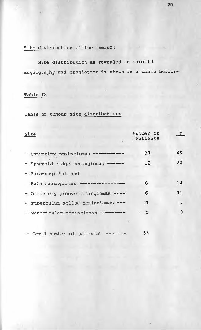
Site distribution of the tumour:
Site distribution as revealed at carotid
angiography and craniotomy is shown in a table below:
Table IX
Table of tumour site distribution:
Site Number of %Patients
- Convexity meningiomas--------------- 27 48
- Sphenoid ridge meningiomas ------ 12 22
- Para-sagittal and
Falx meningiomas-------------------- 8 14
- Olfactory groove meningiomas---- 6 11
- Tuberculum sellae meningiomas ------ 3 5
- Ventricular meningiomas ------------- 0 0
- Total number of patients 56

21
The convexity meningiomas, the most frequent in
this study constituting 48% are taken to be those which
overlie the cerebral hemispheres between the base and
parasagittal regions and have no attachment to a major
venous sinus. The sphenoid ridge is taken to be that
sharply edged shelf of bone which supports the posterior
part of the undersurface of the frontal lobe and provides
the boundary between the anterior and middle cranial
fossae. Meningiomas arising from the meninges clothing
this ridge form a clinical entity and constitute 22%
of the patients in this study. Taken as a single site,
the sphenoid ridge was the single most common site of
this tumour. No ventricular meningiomas were seen. There
was no significant laterality as 24 occurred in the left
hemisphere, 21 in the right hemisphere and 4 along the
midline.
RADIOLOGICAL FINDINGS
Plain film findings;
Of the 56 patients 43 (77%) had abnormal plain
film findings while 23 (23%) were normal. The most
frequent of these were localised hyperostosis of bone

22
adjacent to the site of the tumour, erosion or ballooning
of the pituitary fossa and increased vascular markings.
Sutural diastasis, increased convolutional markings,
localised bone resorption and tumour calcification
were also fairly common. The bone resorption was
either patchy or presented as a single large bone
defect. A combination of bone resorption and bone
sclerosis was seen quite frequently. Tumour
calcification was noted in 21% of the cases, one
of whom had an unusually large psammomatous
calcification that could have been mistaken for
a tuberculoma or a calcified haematoma. That example
is shown in photographs 3 and 4.
Marked hyperostosis with spiculation of bone was
seen in., some patients especially those where the
tumour was large, while in patients with massive
tumours there was complete bone destruction at the site
with the tumour breaking through and then only being
covered by the scalp.

23
Table of plain film findings;
Total Number of Patients - 56:Total %
Table X
Normal-------------------------------- 13 23
Abnormal------------------------------ 43 77
-Localised hyperostosis---------------- 31 76.80
-Pituitary erosion/ballooning--------- 2 9 67.04
-Increased vascular markings---------- 18 41.01
-Localised bone resorption------------ 16 36.05
-Sutural separation-------------------- 15 34.01
-Increased convolutional markings------ 11 25.05
-Mixed osteoblastic/osteolytic bone
reaction------------------------------ 11 25.05
-Tumour calcification ----------------- 9 21
-Marked bone spiculation-------------- 3 7
-Thinned cortex ------------------------ 3 7
-Localised skull elevation ----------- 2 4.06
Of all these plain film findings, hyperostosis,
increased vascular markings and psammomatous calcification
are the only features that are specifically diagnostic
of meningioma while the other x-ray findings named
above are mainly features of space occupying lesion
with raised intracranial pressure.

Photograph 1;
Plain lateral film in a 56 year old lady who
presented with 17 years history of a swelling over
the skull. It shows marked hyperostosis with
spiculation.

25
Photograph 2;
Lateral view showing an unusually large osteolytic
lesion caused by a meningioma. A region of hyperostosis
adjacent to it is noted and the pituitary fossa is eroded.

26
Photograph 3;
A Towne's view of an 18 year old girl who presented
with 3 months history of headache, reduced visual acuity
and features of raised intracranial pressure. An
unusually large calcification is noted along with
marked sutural diastasis.
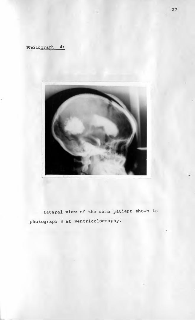
Photograph 4:
Lateral view of the same patient shown in
photograph 3 at ventriculography.

Photograph 5:
Lateral view of a patient with a massive
convexity meningioma that has completely destroyed
a portion of the parietal bone with the tumour
protruding through the skull.
I
29
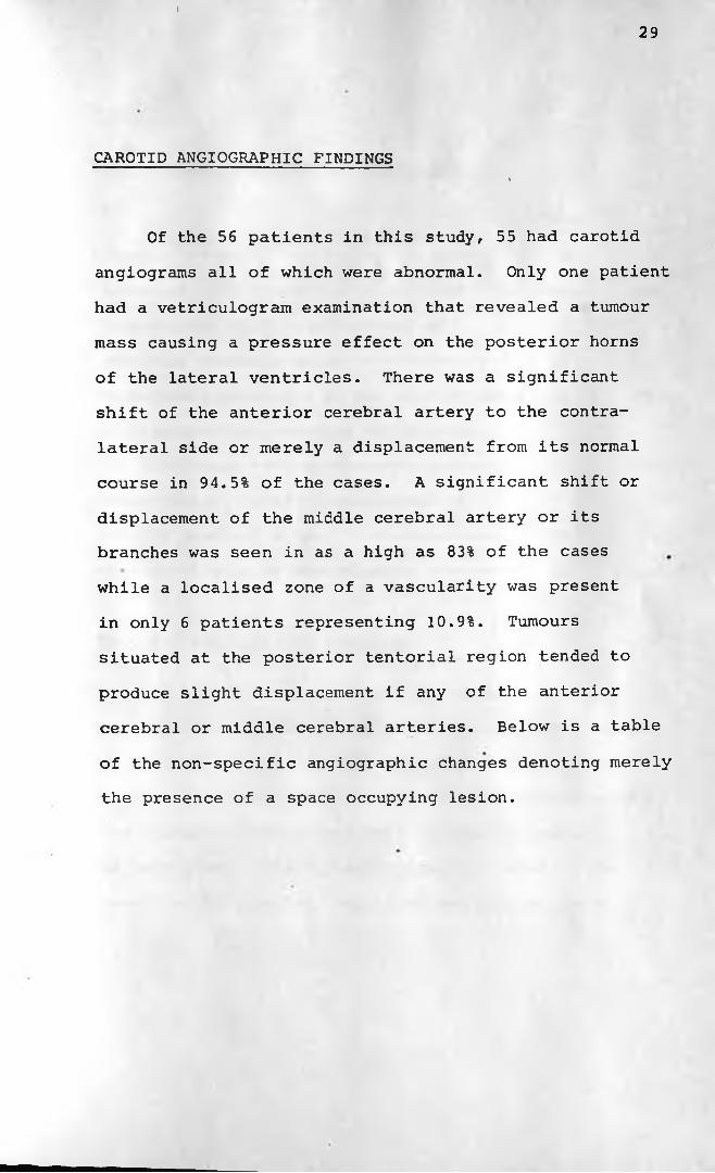
CAROTID ANGIOGRAPHIC FINDINGSt
Of the 56 patients in this study, 55 had carotid
angiograms all of which were abnormal. Only one patient
had a vetriculogram examination that revealed a tumour
mass causing a pressure effect on the posterior horns
of the lateral ventricles. There was a significant
shift of the anterior cerebral artery to the contra
lateral side or merely a displacement from its normal
course in 94.5% of the cases. A significant shift or
displacement of the middle cerebral artery or its
branches was seen in as a high as 83% of the cases
while a localised zone of a vascularity was present
in only 6 patients representing 10.9%. Tumours
situated at the posterior tentorial region tended to
produce slight displacement if any of the anterior
cerebral or middle cerebral arteries. Below is a table
of the non-specific angiographic changes denoting merely
the presence of a space occupying lesion.
30
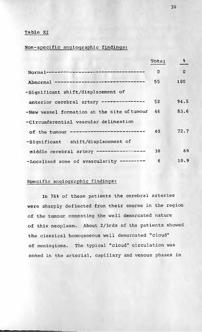
Table XI
Non-specific angiographic findings:
Total %
Normal------------------------------------ 0 0
Abnormal--------------------------------- 55 100
■Significant shift/displacement of
anterior cerebral artery ---------------- 52 94.5
-New vessel formation at the site of tumour 46 83.6
-Circumferential vascular delineation
of the tumour--------------------------- 40 72.7
-Significant shift/displacement of
middle cerebral artery ------------------ 38 69
-Localised zone of avascularity--------- 6 10.9
Specific angiographic findings:
In 78% of these patients the cerebral arteries
were sharply deflected from their course in the region
of the tumour connoting the well demarcated nature
of this neoplasm. About 2/3rds of the patients showed
the classical homogeneous well demarcated "cloud"
of meningioma. The typical "cloud" circulation was
noted in the arterial, capillary and venous phases in
31
most cases. A prominent external carotid circulation
supplying the tumour was demonstrable in 27.2% of these
patients. In a considerable number of patients the
centre of the tumour "cloud" corresponded well with
the region of hyperostosis of the skull. Only 8
patients (14.5%) had a break in the superior sagittal
sinus.
Table XII
Specific angiographic changes in 55 patients:
Total %
-Sharp deflections of arteries ------------ 43 78
-Classical "cloud" ------------------------- 35 63.6
-Central arterial zone with a peripheral
venous drainage ------------------------
-Beaded various small vessels within
tumour area -----------------------------Prominent external carotid circulation--- 15 27.2
-Fine linear, wiry arterial vessels
within tumour a r e a ------------------------ 7 12.9
-Break in superior sagittal sinus --------- 8 14.5
-Radiolucent defects within the "cloud" --- 10 18.1

32
Tumours of the parasagittal or the falx and the
convexity meningiomas in most cases tended to produce
marked and obvious changes especially the typical
tumour ."cloud" and a prominent external carotid
circulation. Meningiomas of the base of the skull
showed typical angiographic changes depending on the
specific site of the tumour. In the olfactory groove
meningiomas the lateral angiogram film (arterial phase)
revealed elevation of the proximal branches of the
anterior cerebral artery as well as bowing with a
forward concavity. The tuberculum sellae meningiomas
tended to displace of the anterior cerebral
artery upwards and posteriorly while the terminal
portion of the internal carotid artery is straightened
and displaced laterally, the two vessels thus forming
an obtuse angle, the normal angle being an acute
one. The sphenoid ridge meningiomas were
characterised by elevation of the horizontal portion
( ) of the middle cerebral artery while the anterior
cerebral artery was displaced to the contralateral
side. Together with the distal portion of the internal
carotid artery these take the shape of the letter "Y".

Photograph 6:
Lateral view of the arterial phase of an
angiogram showing a normal external carotid
circulation.

34
Photograph 7;
Lateral view of an arterial phase of a carotid
angiogram showing a prominent external carotid
circulation supplying the tumours in the parietal
region.
I
35
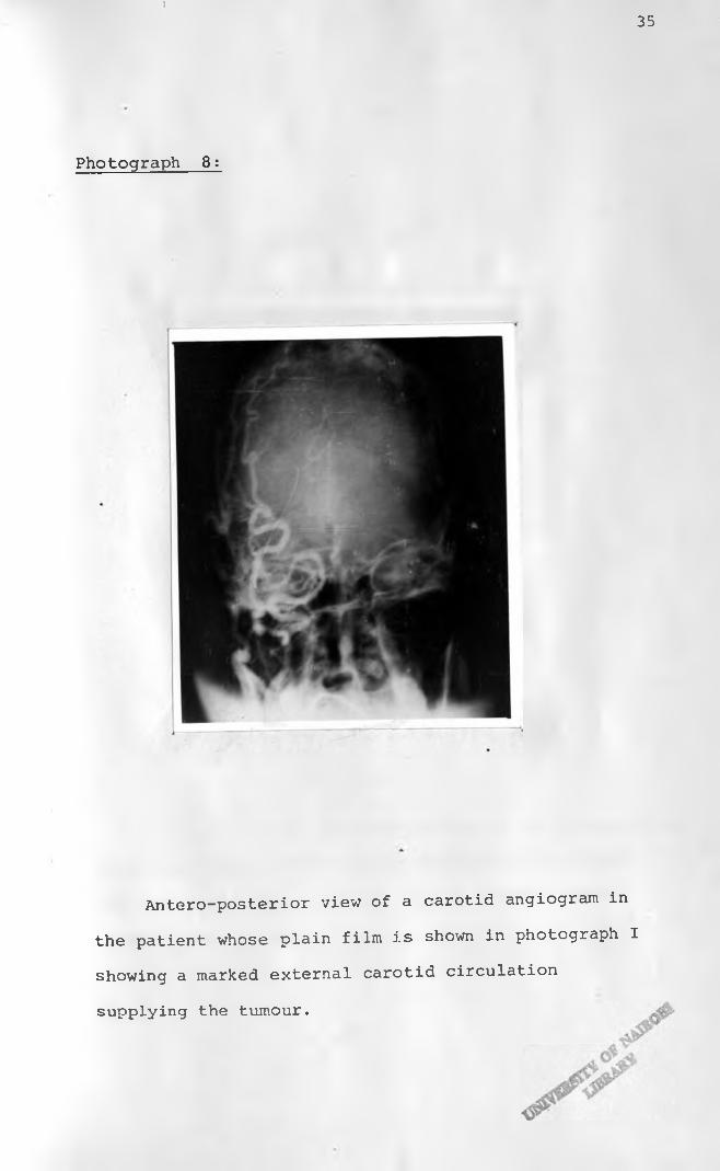
Photograph 8:
Antero-posterior view of a carotid angiogram in
the patient whose plain film is shown in photograph I
showing a marked external carotid circulation
supplying the tumour.

36
Photograph 9;
Lateral film of the same patient as in photograph
8 showing much of the contrast media in the external
carotid circulation and very little in the internal
carotid circulation.

3 7
Photographs 10, 11 and 12 below;
Show the lateral views of a carotid angiogram
showing the typical tumour blush "cloud" as seen in
the arterial, capilarry and cenous phases.
Photograph 10:
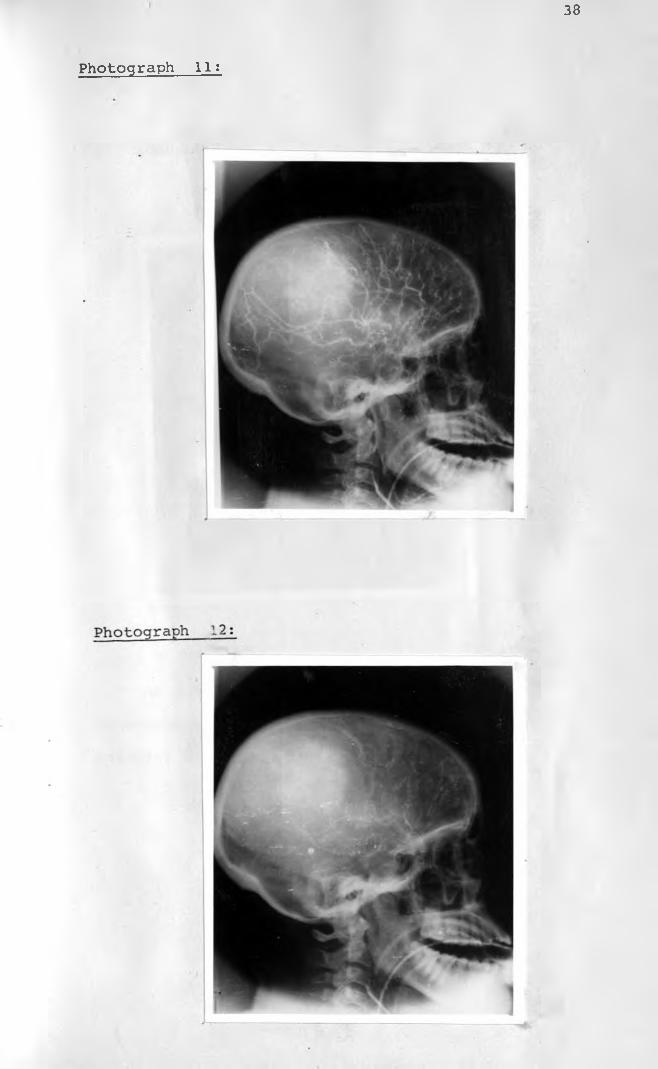
38
Photograph 11:
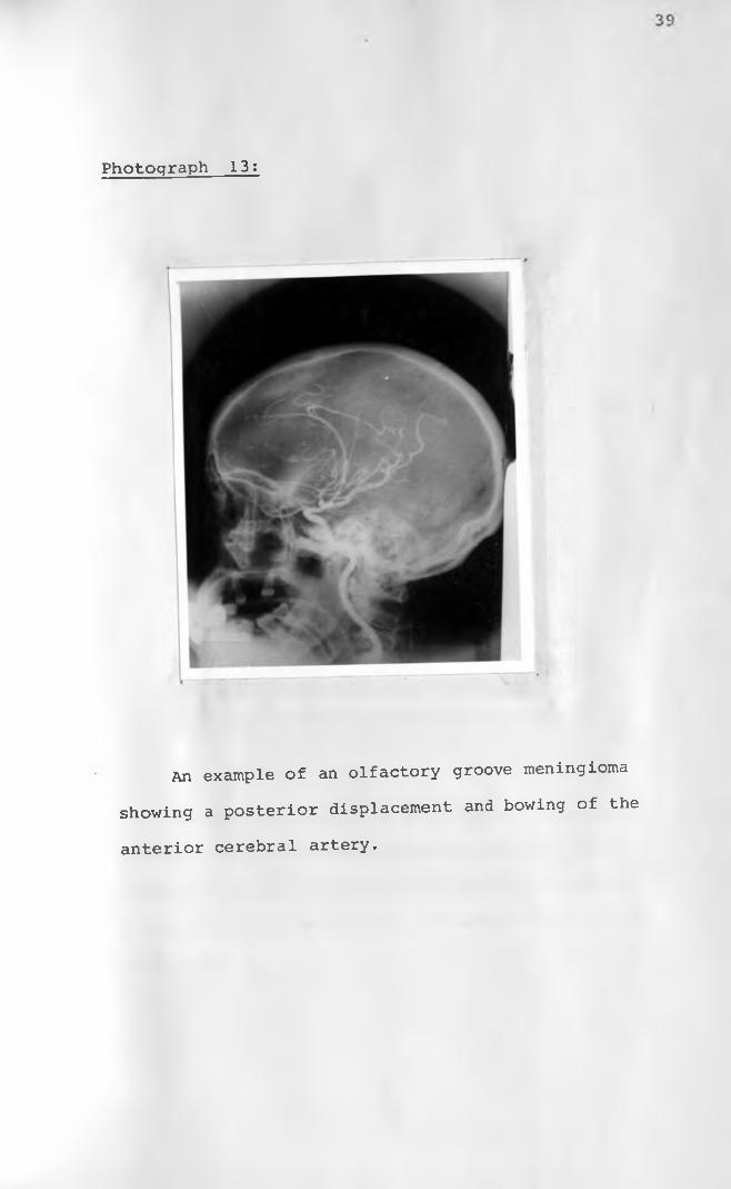
Photograph 13:
An example of an olfactory groove meningioma
showing a posterior displacement and bowing of the
anterior cerebral artery.
40
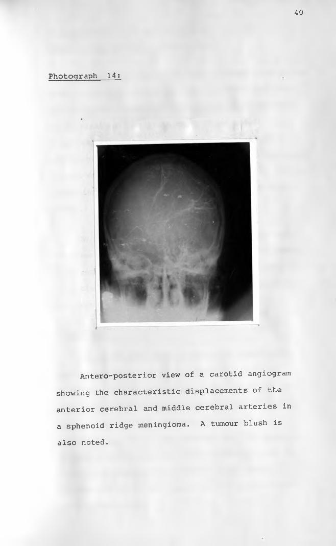
Photograph 14:
Antero-posterior view of a carotid angiogram
showing the characteristic displacements of the
anterior cerebral and middle cerebral arteries in
a sphenoid ridge meningioma. A tumour blush is
also noted.

Diagnostic Accuracy of Plain Skull Film and
Carotid Angiography in Meningiomas;
By reviewing the Radiologists's reports on plain
skull films and carotid angiograms it was possible
to assess the diagnostic accuracy of these methods
of examination. On plain skull films a definitive
diagnosis of meningioma could be made in 14 (25%)
of the patients while a diagnosis of a space occupying
lesion was made in another 22 (40%) patients. The
remaining 20 patients (35%) had normal plain skull
films.
On carotid angiography a definitive diagnosis
of a meningioma was made in 41 patients (75%) while
a diagnosis of a space occupying lesion was made on
14 patients (25%). Not a single carotid angiogram
was reported as normal.
Prognosis:
Of the 56 patients, 30 recovered completely
post-operatively which represents 53.5% of the
patients. Six patients had a permanent disability
mainly blindness or grossly reduced visual acuity
despite the surgical excision of the tumour.
Approximately 25% of the patients died at operation
or. soon after operation. Recurrence was noted in a
total of 5 patients, 2 of whom had malignant
transformation of the tumour while 2 patients were
lost to follow up.
41

42
DISCPSSIOM:
The first major studies on meningioma were carried
out by Harvey Cushing in 1922 and again by Harvey Cushing( 2 )and Eisenhardt in 1938 .Since then an enormous quantity
of literature has been written on this tumour. This
is understandable for this is a benign tumour that
rarely turns malignant and when favourably placed is
amendable to cure by excision. However nothing has
been written on this particular tumour in this hospital
and this is the first study on meningiomas in Kenyatta
National Hospital.
The incidence of intracranial meningiomas in the
Western countries, Japan and India among other neoplasms
originating within the cranium is given as 14% by Russell
and Robinstein in London 1963, 13.4% Zulch in 1967, 15.9%/ Q \
Katsura in Japan 1959 and 13.1% Dastur in India
On the African continent Adeku and Adeloye in 1973 in
their article "Cranial Meningiomas in the Nigerian
African" found meningioma to constitute 27.1% of all/ O \
primary neoplasms of the cranium . Froman and
Lipschitz in 1970 found meningiomas to constitute 30.3%
in the Bantu population in the Transvaal in South
Africa*10). in Ivory Coast Giordano et al found meningiomas
to constitute 33% of all intracranial neoplasms while
Dumas et al in Senegal West Africa put the figure at
15.9% Adeku and Jonata in a similar study
in Ibadan Nigeria, found meningiomas to constitute

43
26.7% of all intracranial primary neoplasms. A
similar study has not been done at Kenyatta National
Hospital. However, Dr. Onyango Akena reviewing 105
patients referred to Kenyatta National Hospital X-ray
Department for carotid angiography in one year found 5
meningiomas out of 17 intracranial neoplams which works(21)out to about 33% . These figures above tend to
suggest that on the whole meningiomas are more common
among the African population as compared to the Western
population, Japan and India. On the contrary however
Billinqhust J.R. in Kampala 1966, found meningiomas to(9)constitute 8.7% of all intracranial neoplasms
Furthermore Murphy N.B. reviewing .100 consecutive patients
referred for carotid angiography at Mulago Hospital
Kampala found only 2 cases of meningiomas and even then(13)in Asian women, and none in African patients . It
is doubtful whether these 2 studies by Murphy and
Billinghurst potray the true situation as regards
meningiomas in Uganda.
The presentation of meningiomas in this study is
comparable to that in other studies elsewhere. For example
the tumour was slightly more common in females and has
a peak incidence in the 5th decade. There appears to be a
relationship between pregnancy and development of a
meningioma. H. Cushing in his series found that in many
cases the tumour was found at the exact situation where
a stunning blow had been received on the skull years

( 2 )
44
before the onset of symptoms
was not found in this study.
Such an association
The symptoms in these patients were those of a
space occupying lesion and those referable to pressure
effect on the part of the tumour on adjacent structures
such as the cranial nerves. In most reports, meningiomas
have been characterised by along duration of symptoms.
This however, was not the case in this study for most
of the patients presented with a relatively short
duration of symptoms, less than 3 years in 80% of cases
while most of Cushing's patients gave a history of
5-10 years ' . Furthermore there was no relationship
between the site of the tumour and the duration of the
symptoms. It would therefore appear that this tumour
grows rapidly among the Kenyan population. An example
of this is born out in one case of a 40 year old lady
who had a normal carotid angiogram but a repeat carotid
angiogram a year later demonstrating a large spenoid
ridge meningioma.
The most common abnormal clinical finding in these
patients was decreased visual acuity, regardless of the
site of the tumour. While most patients would seek
medical attention as soon as they noticed their failing
sight, it is unfortunate in that the defect be it a
reduced visual acuity or blindness, is permanent.
The site distribution of these tumours in thist study
was similar to findings elsewhere. The most frequent
site was at the convexities of the skull followed by
the sphenoid ridge region.

The diagnostic value of hyperostosis of the
skull has been demonstrated in many articles such
as that by K. Francis in 1976 and Gold et al in 1969 (lo'22)
Hyperostosis was found to be the most common and reliable
indicator of the presence of a meningioma on plain films.
Markedly increased vascular markings on plain films
tended to support the highly vascular nature of this
tumour. The typical tumour "blush" or "cloud" was
demonstrated in a higher proportion of patients
enabling improved definite diagnosis of a meningioma.
The typical tumour "cloud" was demonstrated in 63.6%
of the patients as opposed to 42.9% in H.G. Jacobson's
series of 126 patients 1̂5 .̂ The tumour characteristically
tended to persist through the arterial, capillary and
venous phases and it is thought that this is the real
distinction between a meningioma and other tumouri U7 )circulations
It is generally accepted that plain films will
diagnose 30-63% of intracranial meningiomas though
it may raise the suspicion of the presence of a tumour
in 73-78% of the cases Jacobson et al 1959 and Gold
et al 1969^15,16 .̂ In this study however a definitive
diagnosis of meningioma was made on plain films in
very few cases, the radiologist reporting on the films
committing himself to only saying that there is a space
occupying lesion. Angiography enables a definitive
\
46
diagnosis of meningioma in 70% of cases while the
presence of a tumour should be diagnosed in 90% of the
cases according to Wickbom M I . et al 1958 and Bonna
et al 1969 a comparable definitive diagnostic
accuracy at 75% was found in this study while a diagnosis
of a space occupying lesion was made in all of the
patients. Computerized Axial Tomography (C.A.T.) is thought
to be the most accurate method of diagnosis of meningioma
and besides it is a non-invasive procedure. L.E. Claveria
in 1977 found that a C.A.T. - Scanner provides a specific
diagnosis of meningioma in 86% of cases and diagnoses
the presence of a tumour in 96% of c? --s . Given
these figures with regard to this tumour the introduction
of a C.A.T. - Scanner to Kenyatta National Hospital
would be of little extra benefit in as far as the
diagnosis of intracranial meningiomas is concerned.

47
CONCLUSION:
Meningiomas appear to constitute a higher
proportion of primary intracranial neoplasms than is
generally found in the Western countries, Japan and
India. The age group affected in the Kenyan population
appears to be a decade younger. Although the incidence
in females is not significantly higher than in males
there appears to be an association between pregnancy and the
development of meningioma and perhaps more research should
be instituted on this aspect of this tumour. The tumour
appears to grow more rapidly among the Kenyan population
than elsewhere. The symptomatology and clinical features
do not otherwise differ significantly from that found
elsewhere. Plain skull films and carotid angiography
are quite adequate in the diagnosis of a meningioma and
in demonstrating the site of the tumour as well as its
blood supply.

48
REFERENCES
1. The Surgery of tlje Central Nervous System
by D.W.C. Northfield
1st Edition 1973
PP. 229-262.
2. H. Cushing:The Meningioma (Dural Endotheliomas) : .their
Source and Favoured Seats of Origin
Brain - 1922
PP. 252-316.
3. D.S. Russell:Meningiomas - Journal of Clinical Pathology
3: 191, 1950.
4. J. Kempes:Electron Microscopic Studies of Meningioma
American Journal of Pathology
VOL. 39: 499, 1961.
5. Stein, Opalla and Schilp:Fatty Acid Analysis of Meningioma by Gas-phase
Chromatography.
Journal of Neurosurgery
Vol 20: 435, 1963.
49
6. An Atlas of Anatomy Basic to Radiology -
By I . Meshan
3rd Edition/ 1975.
PP. 350-410.
7. A Colour Atlas of Neuropathology -
By C.S. Treip
1st Edition 1978
PP. 170-171.
8. E.L. Adeku and A. Adeloye:
Cranial Meningiomas in the Nigerian African -
African Journal of Medical Sciences.
VOL. 4: 275-291, 1973.
9. J.R. Billinqhust:
Intracranial Space Occupying Lesions in African
Patients at Mulago Hospital Kampala
East African Medical Journal Vol. 43: 385, 1966.
10. C. Froman and R. Lipschitz:
Demography of Tumours of the Central Nervous
System Among the Bantu Population of the Transvaal
Republic of South Africa.
Journal of Neurosurgery Vol. 32: 660, 1970.

50
11. E.L. Adeku and Jonata:
Intracranial Masses in Ibadan
West African Medical Journal
Vol. 16: 31, 1967.
12. C. Giordano and M. Lamouche:
Meningioma in Ivory Coast
African Journal of Medical Sciences
Vol. 4: 259, 1973.
13. N . B . Murphy :
Carotid Cerebral Angiography in Ugar.
East African Medical Journal
Vol. 45: 49-52, 1968.
14. M. Dumas, P.L. Girard and H. Collomb:
Intracranial Meningiomas in Senegal
African Journal of Medical Sciences
Vol. 4: 251-158, 1973.
15. H.G. Jacobson, J.H. Shapiro, H.W. Lubetsky and
C.A. Carton:
Intracranial Meningiomas
A Roentgen Study of 126 Cases.
Radiology Vol. 72: 356, 1959.
51
16. L.H. Gold, S.A. Kieffer and H.O. Peterson:
Intracranial Meningiomas
A Retrospective Analysis of the Diagnostic
Value of the Plain Skull Films.
Neurology Vol. 19: 873, 1969.
17. Cerebral Angiography by:
H .A. Krayenbuhl
2nd Edition, 1968
PP. 295-310.
18. I. Wickbom and S. Stain:
5th Symposium Neurodialogique
Acta Radiologica
Vol. 50: 175, 1958.
19. M. Bonna and A. Appleby:
Some Observations on the Angiography of
Supratentorial Meningiomas
Clinical Radiology Vol. 20: 375-386, 1969.
20. L.E. Claveria, D. Sutton and B.M. Tress:
The Radiological Diagnosis of Meningiomas -
The Impact of E.M.I. Scanning
British Journal of Radiology
VOL. 50: 15-22, 1977.

52
21. Dr. Onyango - Akena:
Radiologist Kenyatta National Hospital
(Personal Communication) .
22. K. Francis:The Diagnostic Value of Hyperostosis in Midline
Sub-frontal Meningiomas.
Radiology 119: 121-130, Apr. 1976.

53
ACKNOWLEDGEMENTS
I wish to express my sincere thanks and appreciation
to the following
Dr. J.M.K. Kitonyi, my Supervisor for the
valuable guidance, advice and suggestions given
throughout the preparation of this paper.
Dr. R.S. Raja, formerly Chairman and Senior
Lecturer, Department of Diagnostic Radiology
for the encouragement during the initial stages
of this work.
The Clerical Staff of the Records Department
and Radiology Department for the assistance
rendered in the collection of the material.
To my Dear Wife, Dr. A.W. Imalingat and my
sweet little Son Herbert Michael Imalingat
without whose patience and encouragement
this paper may not have been completed.
Dr. Malik, Radiologist Aga Khan Hospital,
for valuable suggestions and material.
Professor Kaare Lindqvist for constantencouracement. OMlVltBSlTY of Nairobi
u b &aby
Mrs. R.I. Muturi and Miss M.B. Wangima for
their valuable secretarial services.