the chondrogenic potential of human bone-marrow- derived mesenchymal progenitor cells* by jung u....
TRANSCRIPT

The Chondrogenic Potential of Human Bone-Marrow-Derived Mesenchymal Progenitor Cells*
by JUNG U. YOO, TRACI S. BARTHEL, KEITA NISHIMURA, LUIS SOLCHAGA, ARNOLD I. CAPLAN, VICTOR M. GOLDBERG, and BRIAN JOHNSTONE
J Bone Joint Surg AmVolume 80(12):1745-57
December 1, 1998
©1998 by The Journal of Bone and Joint Surgery, Inc.

Fig. 1 Diagram showing the removal, fractionation, and culture of human bone-marrow-derived mesenchymal progenitor cells.
JUNG U. YOO et al. J Bone Joint Surg Am 1998;80:1745-57
©1998 by The Journal of Bone and Joint Surgery, Inc.

Fig. 2 Monolayer cultures of bone-marrow-derived cells without recombinant human TGF-β1 (control) (A) or with 0.1 (B) or 1.0 (C) nanogram of TGF-β1 per milliliter.
JUNG U. YOO et al. J Bone Joint Surg Am 1998;80:1745-57
©1998 by The Journal of Bone and Joint Surgery, Inc.

Fig. 3 Sections of epoxy resin-embedded cell aggregates cultured for one (A), five (B), seven (C), or fourteen (D) days (toluidine blue, x 128).
JUNG U. YOO et al. J Bone Joint Surg Am 1998;80:1745-57
©1998 by The Journal of Bone and Joint Surgery, Inc.
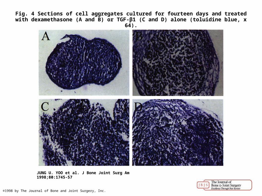
Fig. 4 Sections of cell aggregates cultured for fourteen days and treated with dexamethasone (A and B) or TGF-β1 (C and D) alone (toluidine blue, x 64).
JUNG U. YOO et al. J Bone Joint Surg Am 1998;80:1745-57
©1998 by The Journal of Bone and Joint Surgery, Inc.

Fig. 5 Sections of paraffin-embedded cell aggregates cultured for one (A), five (B), seven (C), or fourteen (D) days (toluidine blue, x 32).
JUNG U. YOO et al. J Bone Joint Surg Am 1998;80:1745-57
©1998 by The Journal of Bone and Joint Surgery, Inc.

Fig. 6 Fluorescein-conjugated immunohistochemical analysis of frozen sections of cell aggregates cultured for one (1), five (2), seven (3), or fourteen (4) days.
JUNG U. YOO et al. J Bone Joint Surg Am 1998;80:1745-57
©1998 by The Journal of Bone and Joint Surgery, Inc.

Fig. 7 Changes, with time in culture, in DNA content (A) and in incorporation of 35S-sulfate4 per cell (B) of the aggregates. cpm = counts per minute.
JUNG U. YOO et al. J Bone Joint Surg Am 1998;80:1745-57
©1998 by The Journal of Bone and Joint Surgery, Inc.

Fig. 8 Immunolocation of epitopes within the hyaluronan-binding region of aggrecan (A), decorin (B), biglycan (C), and link protein (D) in extracts of cell aggregates from the fourteenth day
separated by sodium dodecyl sulfate-polyacrylamide gel electrophor...
JUNG U. YOO et al. J Bone Joint Surg Am 1998;80:1745-57
©1998 by The Journal of Bone and Joint Surgery, Inc.

Fig. 9 Fluorescein-conjugated immunohistochemical analysis of frozen sections of cell aggregates cultured for one (A and B), seven (C and D), or fourteen (E and F) days with anti-
glycosaminoglycan antibodies 5-D-4 (A, C, and E) or 2-B-6 (B, D, and F) (x 32).
JUNG U. YOO et al. J Bone Joint Surg Am 1998;80:1745-57
©1998 by The Journal of Bone and Joint Surgery, Inc.