the chemical ecology of the soft coral-zooxanthellae ... · the chemical ecology of the soft coral...
TRANSCRIPT

This file is part of the following reference:
Michalek-Wagner, Kirsten (1999) The chemical ecology
of the soft coral-zooxanthellae association and its
signficance to the bleaching process. PhD thesis, James
Cook University.
Access to this file is available from:
http://eprints.jcu.edu.au/27503/
If you believe that this work constitutes a copyright infringement, please contact
[email protected] and quote http://eprints.jcu.edu.au/27503/
ResearchOnline@JCU
THE CHEMICAL ECOLOGY OF THE SOFT CORAL - ZOOXANTHELLAE
ASSOCIATION AND ITS SIGNIFICANCE TO THE BLEACHING PROCESS
Thesis submitted by
Kirsten Michalek-Wagner
(BSc Hons. Chem., James Cook University;
MSc Marine Biol., University of Hamburg)
September 1999
In partial fulfilment of the requirements for a Doctor of Philosophy Degree
in the Departments of Marine Biology and Chemistry of James Cook University
i

THESIS DEDICATION
This thesis is dedicated to the loving memory of my great-grandmother Josefina
and to my husband Andreas - the most wonderful man alive!

PUBLICATIONS ARISING FROM THIS THESIS
Michalek-Wagner, K. and B.L. Willis (2000). Impacts of bleaching on the soft coral
Lobophytum compactum. I. Fecundity, fertilisation and offspring viability. Coral Reefs, in press (Chapter 6).
Michalek-Wagner, K. and B.L. Willis (2000). Impacts of bleaching on the soft coral
Lobophytum compactum. II. Biochemical changes in adults and their gametes. Coral Reefs, in press (Chapter 7).
Michalek-Wagner, K. and B. F. Bowden (1999). The effects of bleaching on the secondary
metabolite chemistry of alcyonacean soft corals. Submitted to Journal of Chemical Ecology, in press (Chapter 4).
Michalek-Wagner, K., Bourne, D.J. and B. F. Bowden (1999). The impact of zooxanthellae on
secondary metabolite chemistry and development of the soft coral Lobophytum compactum. To be submitted to Limnology and Oceanography (Chapter 5).
Michalek-Wagner, K. (1999). Seasonal variation in UV-protecting mycosporine-like amino
acids of soft corals. To be submitted to Marine Biology (Chapter 2).
Michalek-Wagner, K. (1999). The role of UV-absorbing mycosporine-like amino acids in the
coral bleaching process. To be submitted to Marine Ecology Progress Series (Chapter 3).
iii

ABSTRACT
Biochemical changes in soft corals (Lobophytum compactum and Sinularia flexibilis) affected by bleaching were investigated, with the ultimate aim of understanding how molecular changes
are linked to biological responses observed in the wake of bleaching events.
Annual variation in photo-protective mycosporine-like amino acids (MAAs) in soft coral
populations provides the first evidence that MAA tissue concentrations are positively correlated
with seasonal cycles in both solar irradiance and sea-surface temperatures. The timing of peak
concentrations of MAAs in summer when exposure to solar irradiance and sea temperatures are
greatest, and in female colonies prior to spawning, further corroborate their role as photo-
protectants. In manipulative bleaching experiments elevated UVR and temperature were found
to act synergistically in the bleaching process. However, chemical degradation of MAAs during
exposure to elevated temperature is not, as previously assumed, the key to understanding the
synergistic interaction. In fact, MAA levels increased in response to simultaneous exposure to
high temperature and UVR, in both experimentally and naturally bleached colonies, suggesting
increased resource allocation towards photo-protection in corals already experiencing thermal stress.
Soft coral responded to bleaching by increasing the production of terpenoid secondary
metabolites, which aid in the prevention of microbial and algal fouling. While changes in
secondary metabolites were short-lived, a substantial increase of an anti-microbial agent in S. flexibilis may have contributed to the prevention of fouling by opportunistic bacteria. This
suggests that soft corals may alter their secondary metabolite chemistry to prevent fouling by
opportunistic bacteria following bleaching. These experimental results were validated through
analysis of soft corals affected by the 1998 mass bleaching event, where bleached colonies with
high levels of algaecides remained free of fouling, while conspecifics with substantially lower
levels were found to be overgrown. This suggests that soft corals are capable of surviving short-
term bleaching events and detrimental algal overgrowth that is often associated with bleaching,
by regulating their secondary metabolite chemistry to counteract fouling.
Understanding the role of symbiotic zooxanthellae in the production of terpenoid secondary
metabolites is integral to evaluating the full impact of bleaching disturbances on the soft coral
host. Zooxanthella cross-infection experiments with freshly metamorphosed polyps of
Lobophytum compactum demonstrated that control over the production of secondary metabolite
lies with the animal host. Moreover, the equivalence of secondary metabolite chemistry in apo-
iv
and symbiotic polyps clearly shows that the algal partner is not essential for biosynthesis.
Despite no direct algal involvement in terpene production, a strong correlation between polyp
growth and investment into terpenes suggests that, via their contribution to coral nutrition
through primary metabolism, zooxanthellae have the capacity to indirectly influence secondary
metabolism. The implications for bleached soft corals are that while the host, with or without
energetic contributions from the algal symbiont controls the production of ecologically
important terpenes, energy reserves may be insufficient to maintain the production of
ecologically significant concentrations.
Severe experimental bleaching was found to have long-term sub-lethal impacts on soft corals,
reducing overall reproductive output of Lobophytum compactum for at least two spawning
seasons. Polyp fecundity and mean egg diameters were inversely correlated with the degree of
bleaching, with complete failure of fertilisation in heavily bleached colonies in the first year and
significantly reduced fecundity in the second year after the bleaching event. Although bleached
corals recovered their zooxanthellae within 4 months, protein, lipid, MAA and carotenoid
concentrations were reduced for at least eight months in adult tissues. The reductions were
amplified when they were passed on to gametes, with the greatest reductions occurring in lipid
and protein concentrations. Although reductions in MAAs were relatively smaller when passed
on to gametes, even minor proportional reductions have significant implications for larval
survival, given that MAA levels are approximately three times higher in eggs than in maternal
tissues. By the second spawning season (20 months after experimental bleaching) the
biochemical compositions of both adult tissues and their gametes were indistinguishable from those of control (unbleached) corals.

(Signature) (date)
STATEMENT OF ACCESS
I, the undersigned, the author of this thesis, understand that James Cook University will make it
available for use within the University Library and, by microfilm or other photographic means,
allow access to users in other approved libraries. All users consulting this thesis will have to
sign the following statement:
" In consulting this thesis I agree not to copy or closely paraphrase it in whole or in part without
consent of the author; and to make proper written acknowledgement for any assistance which I
have obtained from it".
Beyond this, I do not wish to place any restriction on access to this thesis.
vi

DECLARATION
I declare that this thesis is my own work and has not been submitted in any form for another
degree or diploma at any university or other institution of tertiary' education. Information
derived from the published or unpublished work of others has been acknowledged in the text
and a list of references is given.
7 5c)qooc
(date)
vii

ACKNOWLEDGEMENTS
I am grateful to the many people who have helped me with this project in one way or another -
my heart-felt thanks and gratitude go out to all of you. To begin with, I would like to thank my
supervisors Dr. Bette Willis (Marine Biology) and A./Prof. Bruce Bowden (Chemistry) who
spent many hours covering specific aspects of this thesis. Thank you Bette for sharing your
expertise on coral reproduction with me and for your excellent editorial support, and
encouragement in the final stages of this thesis. Bruce, thank you for sharing your extensive
chemical knowledge with me and for your patience in teaching me chemistry!
The fieldwork would not have been possible without the enthusiastic and patient help of 38
volunteers who did not mind getting there fingers slimy with soft coral mucus. Particular
thanks go to David Wachenfeld and Andreas Wagner for their good sense of humour in spite of
hard work during coral spawning (good to see that you have both become soft coral lovers by now!).
I am indebted to Dr. Walt Dunlap (AIMS) for sharing his extensive expertise on MAA analysis
with me. Thank you Walt for not only teaching me how to analyse MAAs, but also for your
generous support with standards, solar radiation data, equipment and for the continued
hospitality you offered me at AIMS. Thanks to Dr. David Bourne (AIMS) for making the
impossible "possible" by managing to analyse the secondary metabolite chemistry of single polyps using mass spectrometry.
My thanks and gratitude go to many collegues and friends who have given me the gift of their
knowledge, time and experience in discussing and/or revising my work: My warmest thanks go
to Dr. Terry Done, Dr. Vicki Hall, Prof. Malcolm Shick, Dr. David Wachenfeld, Dr. Walt
Dunlap, Ken Anthony, Dr. Katherina Fabricius, A. Prof. Ove Hoegh-Guldberg, Dr. Joe Connell,
Dr. Jamie Oliver, Dr. Amanda Reichelt, Anya Salih, Jackie Wolstenholme, Dr. Chris Crossland,
Dr. Rocky de Nys, Dr. Terry Hughes, Xingjang Feng, Nadine Marshall, Dr. Mikel ten Lohius,
Dr. David Boume, Prof. John Coll, Paul Marshall, Dr. Rocky de Nys, David Stone.
I have received logistical support from Dr. David Miller and his team, who provided me with
access to much needed biochemical equipment. Thank you Zoli Florian for your assistance with
macro-photography and your good sense of humour in the hours we spent waiting till the coral
polyps were ready to be photographed. Logistical support with the coral husbandry - always
mixed with some cheerful comments and untiring efforts to help whenever possible, came from
John Morrison at the JCU aquarium. The staff at the Orpheus Island Research Station has
viii
supported me in many ways during my 34 visits to the Island — thank you all. Sincere thanks go
to Aurel Moise at the School of Mathematics, Computer Sciences and Physics at James Cook
University and the Australian Radiation Laboratory (Yallambie, Victoria) for making available
the environmental radiation data. Thanks also go to Ray Berkelmans (GBRMPA) for the use of
his temperature control set-up at Orpheus Island and for making available water temperature
data. I am indebted to Anya Salih and the Electron Microscopy Unit at the University of Sydney
for opening up the world of confocal microscopy to me and for providing me with 3D images of
coral polyps. Thanks to Ann Sharp, Eva King and Maree Hines whose efficiencies and
kindness made official paperwork as painless as possible.
I would like to extend my gratitude to John Rumney and Andy Dunstan who have supported my
work by providing a free berth on the "Undersea Explorer", and to Katherina Fabricius for
giving me the opportunity to work with her on a cross—shelf bleaching trip on board "MS Harry Messel".
This study was funded by an Overseas Postgraduate Award from JCU and a postgraduate
scholarship from the Cooperative Research Centre Reef (CRC). Additional, and greatly
acknowledged, research funding came from the Merit Research Grant Scheme at James Cook
University (1996 and 1997), the CRC Reef and the Great Barrier Reef Marine Park Authority.
On personal matters I have received direction and advice from Dr.Vicki Hall, Simon Woodley,
Prof. Howard Choat and Prof. Christian Alexander — my warmest thanks for your support.
The years would not have been the same without the friendship, laughter and support of Vicki,
Jackie, Gillianne, Ken, Lishu, Anya, Tilley, Xingjang, David, Leanne, Marc and Mandy.
Nadine, Paul, David, Vicki and Jackie your particular encouragement, smiles, understanding
and your sense of humour in putting the thesis into perspective at times made it all happen for
me — the biggest of hugs to all of you. (Paul one day we will all be doctors!). And of course
three cheers to Brett, Richard, Rob, Zac and Russell at Digital Dimensions who helped me
going through the ups and downs of thesis writing.
The love and support of my family in Spain, Germany and China always found its way down
under. Without you I could not have pursued my dreams.
Andreas — my true better half - thank you for your unconditional and continued love and
support, particularly during the final months of writing this thesis.
ix

TABLE OF CONTENTS
Title page i Thesis Dedication ii Publications arising from this thesis iii Abstract iv Statement of Access vi Declaration vii Acknowledgements viii Table of Contents x List of Tables xvii List of Figures xx
Chapter 1 General Introduction 1
1.1. Background and significance of coral bleaching 2 1.2. Causal factors and interactions of elevated solar irradiance and temperature 3
in the bleaching process
1.3. MAAs and mechanisms of biochemical defence against high irradiance 4 1.4. Potential roles of secondary metabolites in the bleaching response 6
of soft corals
1.5. Longer-term effects of bleaching — implications for reproductive output 8 1.6. Specific aims 8
Chapter 2 Seasonal variation in MAA levels of soft corals in relation 10 to annual solar irradiance and seawater temperature cycles
2.1. ABSTRACT 11 2.2. INTRODUCTION 12 2.3. MATERIALS AND METHODS 14
2.3.1. Site description 14 2.3.2. Species description 15 2.3.3. Sampling regime and preparation of experimental corals 15
x

2.3.4. Biochemical analysis of zooxanthellae densities, chlorophyll, 17 carotenoid, MAA and protein concentrations
2.3.5. Relationship between seawater temperature, solar radiation 19 and biochemical parameters
2.4. RESULTS 20 2.4.1. MAA analysis 20 2.4.2. Seasonal responses in biochemical parameters 25 2.4.3. Environmental parameters monitored during the sampling period 28
2.5. DISCUSSION 33 2.5.1. The relationship between solar radiation, temperature and MAA levels 33 2.5.2. Composition of MAAs in Lobophytum compactum and 34
Sinularia flexibilis
2.5.3. Fluctuations of MAAs in soft corals— 35 selective bioaccumulation or active regulation?
2.5.4. Photoacclimatory responses and MAAs 36 2.5.5. The role of MAAs in the reproductive cycle of soft corals 39 2.5.6. Regulation of MAA synthesis and translocation 40 2.5.7. Summary and conclusions 41
Chapter 3 The effects of high irradiance and temperature on tissue 43 concentrations of UV-absorbing MAAs
3.1. ABSTRACT 44 3.2. INTRODUCTION 45 3.3. MATERIALS AND METHODS 47
3.3.1. Effects of elevated temperature and UV radiation alone and 47 in combination on MAA levels and bleaching responses
3.3.2. Comparison of MAA levels in naturally bleached 50 (mass bleaching event 1998) and unbleached soft corals
3.3.3. Do higher MAA levels protect against bleaching? 52 3.4. RESULTS 54
3.3.4. Effects of temperature and UV radiation alone and in interaction 54 on MAA levels and bleaching
3.4.2. Controls of all parameters (experimental controls) 64
xi

3.4.3. Relationships between the biochemical parameters 65 3.4.4. Analysis of MAA levels in naturally bleached 65
and unbleached soft corals
3.4.5. Do higher MAA levels protect against bleaching? 70 3.4.6. Summary of all results gained from experimentally 73
and naturally bleached soft corals
3.5. DISCUSSION 74 3.5.1. The experimentally induced bleaching response in the soft corals 74
Lobophytum compactum and Sinularia flexibilis
3.5.2. Effects of synergistic interactions between high irradiance 75 and temperature on zooxanthellae and chlorophyll level
3.5.3. Are MAAs the key to understanding the observed 76 synergistic interaction?
3.5.4. Up-regulation of MAAs in response to thermal stress 77 3.5.5. Possible regulation of MAA induction 78 3.5.6. Summary and conclusions 79
Chapter 4 The effects of bleaching on secondary metabolite chemistry 80 of soft corals
4.1. ABSTRACT 81 4.2. INTRODUCTION 82 4.3. MATERIALS AND METHODS 85
4.3.1. Site description 85 4.3.2. Collection and preparation of experimental corals 85 4.3.3. Experimental design 85 4.3.4. Bleaching treatment 86 4.3.5. Quantification of zooxanthellae densities 87 4.3.6. Chemical analysis of secondary metabolites 87 4.3.7. Predation and algal fouling experiments 88 4.3.8. Analysis of Lobophytum compactum, Sinularia flexibilis and 90
Sarcophyton sp. affected by a natural bleaching event
in March/April 1998
4.4. RESULTS 91 4.4.1. Results of caging pilot study 91
xii

4.4.2. Changes in terpenoid secondary metabolite chemistry 91 in experimentally bleached Sinularia flexibilis and Lobophytum compactum
4.4.3. Changes in relative lipid concentrations in experimentally 96 bleached Sinularia flexibilis and Lobophytum compactum
4.4.4. Partition of secondary metabolites between animal and algal tissue 98 4.4.5. Algal recovery in experimentally bleached S. flexibilis 98
and L. compactum
4.4.6. Predation experienced by bleached and unbleached conspecifics 99 of Sinularia flexibilis and Lobophytum compactum
4.4.7. Algal overgrowth experienced by bleached and unbleached 100 conspecifics of Sinularia flexibilis and Lobophytum compactum
4.4.8. Analysis of Lobophytum compactum, Sinularia flexibilis 101 and Sarcophyton sp. affected by the 1998 natural bleaching event in the Palm Island group
4.5. DISCUSSION 104 4.4.9. Fouling of experimentally bleached soft corals 104 4.4.10. Predation of experimentally bleached soft corals 106 4.4.11. Experimentally bleached versus naturally bleached 107
soft coral conspecifics
4.4.12. Summary and conclusions 110
Chapter 5 The effects of the zooxanthellae on the secondary metabolite 111 chemistry and development of Lobophytum compactum
5.1. ABSTRACT 112 5.2. INTRODUCTION 113 5.3. MATERIAL AND METHODS 115
5.3.1. Gamete collection 115 5.3.2. Fertilisation 115 5.3.3. Larval rearing 116 5.3.4. Experimental design and symbiont infection 116 5.3.5. Offspring survivorship and polyp growth 117 5.3.6. Preparation of polyps for terpenoid chemical analysis 117 5.3.7. Statistical analysis 118

5.4. RESULTS 119 5.4.1. Fertilisation 119 5.4.2. Polyp settlement, metamorphosis and zooxanthellae infection 119 5.4.3. Polyp growth 120 5.4.4. Secondary metabolite chemistry 122 5.4.5. Correlation between growth and expression of 125
secondary metabolite chemistry
5.5. DISCUSSION 126 5.5.1. Control and production of terpenoid coral secondary metabolites 126 5.5.2. The impact of different strains of zooxanthellae on colony growth 128 5.5.3. Summary and conclusions 129
Chapter 6 The impacts of bleaching on the soft coral Lobophytum 130 compactum. I. Fecundity, fertilisation and offspring viability
6.1. ABSTRACT 131
6.2. INTRODUCTION 132
6.3. MATERIALS AND METHODS 134
6.3.1. Site description 134 6.3.2. Species description 134 6.3.3. Collection and preparation of experimental corals 134 6.3.4. Bleaching treatment 135 6.3.5. Sampling regime and analysis of biochemical 135
and reproductive parameters
6.3.6. Larval rearing 137 6.3.7. Juvenile growth 137 6.3.8. Statistical analysis 137
6.4. RESULTS 138 6.4.1. Coral survival and recovery of zooxanthellae 138 6.4.2. Oogenic cycle 140 6.4.3. Polyp fecundity 141 6.4.4. Egg size comparisons 143 6.4.5. Spawning in 1996 and 1997 145 6.4.6. Fertilisation rates 145
6.4.7. Survivorship and growth of offspring 146
xiv

6.5. DISCUSSION 148
Chapter 7 The impacts of bleaching on the soft coral Lobophytum 152 compactum. II. Biochemical changes in adults and their gametes
7.1. ABSTRACT 153 7.2. INTRODUCTION 154 7.3. MATERIALS AND METHODS 156
7.3.1. Experimental design and sampling regime 156 7.3.2. Biochemical analyses 156 7.3.3. Statistical analysis 157
7.4. RESULTS 158 7.4.1. Lipid analysis 159 7.4.2. Carotenoids 160 7.4.3. Mycosporine-like amino-acids (MAAs) 161 7.4.4. Proteins 162
7.5. DISCUSSION 164
Chapter 8 Summary and general discussion 169
8.1. Brief summary of major findings 170 8.1.1. Seasonal variations in MAA concentrations in male and female soft 170
corals in relation to annual solar irradiance and temperature cycles
(Chapter 2)
8.1.2. The effects of high irradiance and temperature on tissue 170
concentrations of UV absorbing MAAs in soft corals (Chapter 3)
8.1.3. The effects of bleaching on the secondary metabolite chemistry of 171 alcyonacean soft corals (Chapter 4)
8.1.4. The impact of different strains of zooxanthellae on the 171
secondary metabolite chemistry and growth of the soft coral host
Lobophytum compactum (Chapter 5)
xv

8.1.5. Impacts of bleaching on the soft coral Lobophytum compactum: 172 Fecundity, fertilisation and offspring viability;
Biochemical changes in adults and their gametes (Chapters 6 & 7). 8.2. General discussion 172 8.3. Final conclusions 175
References 177
xvi

LIST OF TABLES
Table 2.1. Summary of results for 2-factorial repeated measures ANOVA, testing for
differences in MAA concentrations between colonies and through time. Asterisks
denote significant differences at 0.05 level of significance.
Table 2.2. Summary of results for 2-factorial ANOVA testing for differences in MAA levels
between sexes and through time. Asterisks denote significant differences (a = 0.05).
Table 2.3. Summary of results of 2-factorial repeated measures ANOVA testing for differences
in zooxanthellae densities, total chlorophyll, carotenoid (peridinin) and protein
concentrations between individuals through time. Asterisks denote significant
differences (a = 0.05).
Table 2.4. Summary of regression analyses between coral tissue parameters (MAA,
zooxanthellae, chlorophyll, protein and peridinin concentrations) and the monthly doses
of total a) solar downflux and b) UV radiation in Lobophytum compactum and Sinularia flexibilis, (n = 23). Asterisks indicate a significant relationship (a = 0.05).
Table 2.5. Summary of regression analyses between monthly average sea temperatures at the
study site and MAA, zooxanthellae, chlorophyll, protein and peridinin tissue concentrations in Lobophytum compactum and Sinularia flexibilis (n = 24). Asterisks indicate that a relationship is significant (a = 0.05).
Table 3.1. Summary of results of 3-factorial ANOVA, testing the effects of time, experimental
treatment and tank on levels of various biochemical parameters (MAAs, zooxanthellae,
chlorophyll and protein) in two species of soft corals. Asterisks denote significant
differences from controls for an alpha level of 0.05. (N = 32 determinations, i.e. 16
colonies per treatment x 2 subsamples per species).
Table 3.2. Group means for response variables (MAAs, zooxanthellae, chlorophyll, protein) to
four experimental treatments in Lobophytum compactum and Sinularia flexibilis; (con.
= control; t UVR = increased UVR; TTemp. = increased temperature; f Temp.+ UVR
= increased temperature and UVR);means are based on 32 determinations (i.e. 16 per
treatment x 2 subsamples per species). Asterisks denote significant differences from the
control.
Table 3.3. Summary of results of 1-factorial ANOVA, testing the effect of different UVR and
temperature treatments (df = 3) on concentrations of various biochemical parameters in
Lobophytum compactum and Sinularia flexibilis. Asterisks denote significant
differences from the control (a = 0.05; n = 32 determinations (i.e. 16 per treatment x 2
subsamples per species).
xvii

Table 3.4. Results of one-way ANOVA (df = 1) comparing experimental controls of each soft
coral species with handled (tank controls) and unhandled field controls with respect to
their zooxanthellae densities and chlorophyll, protein and MAA levels. For field
controls, n = 16 determinations (i.e. 8 colonies x 2 subsamples), whereas for tanks
controls n = 32 determinations (i.e. 16 colonies x 2 subsamples).
Table 3.5. Regression analyses assessing the relationships between: 1) zooxanthella densities
and MAA tissue concentrations, 2) protein concentrations and MAA tissue levels and 3)
protein concentrations and zooxanthella levels. Asterisks denote significant
relationships (cc = 0.05) while R2 denotes the coefficient of determination, n = 512
determinations (i.e. 128 colonies x 4 sampling times).
Table 3.6. Results of Mann-Whitney tests, comparing biochemical parameters between bleached
and unbleached conspecifics of 4 soft coral species sampled during the 1998 natural
bleaching event. Asterisks denote significant differences, n = 14 determinations
(i.e. 7 colonies x 2 subsamples per species).
Table 3.7. Summary of results of 3-factorial nested ANOVA, testing the effects of MAA-level
and experimental treatment (fixed and orthogonal factors) and the nested factor tank, on
zooxanthellae and chlorophyll levels. Asterisks denote significant differences from the
control (cc= 0.05); (n = 16 determinations, i.e. 8 colonies per treatment x 2 subsamples).
Table 4.1. Summary of results of 2-factorial ANOVA testing the effects of caging and time on zooxanthellae densities Sinularia flexibilis and Lobophytum compactum during a nine-month pilot study. Asterisks denote a significant difference for an alpha level of 0.05.
Table 4.2. Summary of results of 3-factorial ANOVA testing the effects of time, bleaching and
caging on tissue concentrations of the algaecide sinulariolide, the cytotoxin flexibilide of Sinularia flexibilis and the ichthyotoxin isolobophytolide of Lobophytum compactum. Asterisks denote a significant difference (cc = 0.05).
Table 4.3. Summary of results of 3-factorial ANOVA testing the effects of time, bleaching and
caging on lipid in Sinularia flexibilis and Lobophytum compactum. Asterisks denote a significant difference for an alpha level of 0.05.
Table 4.4. Summary of results of 2-factorial ANOVA, testing for the effects of time and caging
on the recovery of zooxanthellae in experimentally bleached colonies of Lobophytum compactum and Sinularia flexibilis. Asterisks denote significant differences for an alpha
level of 0.05.
Table 4.5. Number of bite marks on test corals during the monitoring period. The figures in
brackets denote the number of corals from which the bite marks were scored
(n = 5 colonies per treatment). Shaded areas highlight bleached individuals.
Table 4.6. Results of Kruskal-Wallis tests, testing for significant differences between bleached
and unbleached conspecifics of soft coral species affected by the 1998 natural bleaching
xviii

event. Isolobophytolide only occurs in Lobophytum compactum, sinulariolide and flexibilide are found in tissues of Sinularia flexibilis, while sarcophytoxide is specific to Sarcophyton sp. Asterisks denote significant differences (n = 7 determinations per species).
Table 4.7. Results of Mann-Whitney tests, comparing levels of secondary metabolites among
bleached (unfouled), unbleached and bleached plus fouled conspecifics of soft coral
species affected by the 1998 natural bleaching event. Asterisks denote significant
differences (n = 7 determinations per species).
Table 5.1. Percentage of individuals expressing typical Lobophytum compactum chemistry
(isolobophytolide) after infection with different strains of zooxanthellae (n = 8 polyps
per treatment). (No chemical analysis was carried out with polyps that were not
successfully infected with the Zoanthus sp. and Lobophytum microlobulatum strain).
Table 6.1. Mean zooxanthellae densities/ g tissue wet weight (± SE) before and after
experimental treatments in Lobophytum compactum. N = 10 colonies (each density is a mean of 2 subsamples per colony x 4 replicate assays per subsample).
Table 6.2. Juvenile survival and growth measured as number of polyps per colony five months
after spawning. A total of 500 larvae were monitored in each treatment. Multiple polyp
stage refers to the number of polyps/ colony. Heavily bleached corals did not produce larvae.
xix

LIST OF FIGURES
Figure 2.1. Location of study site, Pioneer Bay, Orpheus Island, in the Central Great Barrier Reef
Figure 2.2. Overview of experimental design and sample preparation for the different
biochemical analyses, N = 40 determinations (i.e. 10 colonies x 4 subsamples for each
biochemical parameter).
Figure 2.3. Wavelength and absorbance maxima (Amax) of MAAs present in both the tissues of
Lobophytum compactum and Sinularia flexibilis.
Figure 2.4. Percentage contribution of palythine, shinorine and palythene to the total MAA pool in Lobophytum compactum (top) and Sinularia flexibilis (bottom). Error bars indicate standard errors for palythine only, N = 40 determinations (i.e. 10 colonies x 4
subsamples per sampling date and species).
Figure 2.5. Changes in MAA levels through time in female and male colonies of Lobophytum compactum (top) and Sinularia flexibilis (bottom). Error bars indicate standard errors, N = 20 determinations (i.e. 5 males and 5 females per species x 4 subsamples, per month).
Figure 2.6. Partitioning of MAAs between animal and plant tissue in Lobophytum compactum and Sinularia flexibilis (n = 5). Figures within the column represent the percentage of MAAs of the total pool associated with the animal tissue.
Figure 2.7. Mean concentrations of a range of biochemical parameters in the tissues of the soft coral Lobophytum compactum and Sinularia flexibilis: a) mean densities of
zooxanthellae, b) mean chlorophyll a + c2 concentrations, c) mean protein levels and d)
mean peridinin concentrations during a two-year monitoring period. Errors indicate
standard errors, N = 20 determinations (i.e. 5 male, 5 female colonies x 4 subsamples per colony, per month).
Figure 2.8. Mean total solar downflux (300-2800 nm) and UV radiation (285 - 400 nm) data
during 1996 and 1997 (courtesy A. Moise, JCU). Error bars present the standard error
(n = 144/ day). No data were available for UVR in January 1997.
Figure 2.9. Sea temperatures at 2-3m depth on the reef flat at Cattle Bay (Orpheus Island)
during the monitoring period (courtesy R. Berkelmans, GBRMPA). Full circles
represent the average monthly temperatures, while open triangles and diamonds present
the minimum and maximum temperatures respectively.
Figure 3.1. Experimental design and set-up to test the effects of UV radiation and temperature
alone and in combination on biochemical parameters (MAA, zooxanthellae and
xx

chlorophyll tissue concentrations) in L. compactum and S. flexibilis. Colonies were
sampled at time 0, 1, 2, and 3 weeks after exposure to the experimental treatments.
Figure 3.2. Experimental tank set-up under solar weave roof with UV lamps and aeration tubing
at Orpheus Island.
Figure 3.3. Bleached colonies of the four soft coral species: a) Lobophytum compactum, b) Sinularia flexibilis, c) Sinularia capitalis (on Porites sp.) and d) Sarcophyton sp. Sampled from the Palm Islands during the natural mass bleaching event in 1998.
Figure 3.4. Changes in levels of biochemical parameters through time in tissues of Lobophytum compactum and Sinularia flexibilis submitted to experimentally elevated temperature
and UVR treatments: a) + e) MAAs, b) + f) zooxanthellae, c) + g) chlorophyll and
d) + h) protein. N = 32 determinations, (i.e. 16 colonies x 2 subsamples per species).
Controls represent experimental treatment controls. Error bars denote standard errors.
Figure 3.5. Comparison of biochemical parameters between bleached and unbleached colonies
of four soft coral species collected during a natural thermal bleaching event:
a) chlorophyll; b) MAAs; c) protein and d) carotenoid (peridinin) levels. Error bars denote standard errors (n = 14 determinations, i.e. 7 colonies x 2 subsamples). The depth-adjusted MAA level in unbleached L. compactum (b) is shown in the dashed column.
Figure 3.6. Relationship between depth and MAA concentrations in Lobophytum compactum at Pioneer Bay prior to the bleaching episode (January 1998; n = 5 per depth).
Figure 3.7. Comparison of a) chlorophyll and b) MAA levels in unbleached and bleached
conspecifics of L. compactum and S. flexibilis. Data collected during March 1996 to
April 1997 represent concentrations under non-bleaching conditions, whereas data
collected in April 1998 represent bleached conditions. Each sampling period in
1996/1997 comprises n = 40 determinations (i.e. 10 colonies x 4 subsamples) whereas
in April 1998, n = 14 determinations (i.e. 7 colonies x 2 subsamples).
Figure 3.8. Bleaching response quantified as loss of a) zooxanthellae and b) chlorophyll levels
of high and low-MAA acclimatised L. compactum conspecifics after 10-day exposures
to increased temperature, solar radiation or a combination of both factors. Error bars
denote standard error, n = 16 determinations, i.e. 8 colonies x 2 subsamples per
treatment. Asterisks denote a significant difference in bleaching response between the
two groups.
Figure 3.9. Overall summary of results of Chapter 3.
Figure 4.1. Progression of algal overgrowth on various bleached soft corals in the wake of the
1998 mass-bleaching event. a). Algae colonising the tips of bleached colonies of
Sinularia sp.; b) Increased algal overgrowth on larger parts of colonies of bleached Sinularia sp.; c) Algae firmly engulf substantial parts of bleached Sinularia sp.;

d) Tissue disintegration and death of bleached and overgrown Sinularia sp. (photo courtesy K. Fabricius).
Figure 4.2. Experimental design and set-up to test the effects of bleaching on secondary
metabolites, algal overgrowth and predation in L. compactum and S. flexibilis Colonies
were sampled before the bleaching treatment (time 0), immediately after (1) and then in monthly intervals.
Figure 4.3. Caging set-up with bleached and unbleached specimen of L. compactum Figure 4.4. Bite marks inflicted on " fingers" of Lobophytum compactum by Chaetodon auriga
during a pilot experiment, testing for persistence and nature of bite marks.
Figure 4.5. Changes in tissue concentrations of the algaecide sinulariolide and the cytotoxic
antimicrobial agent flexibilide in Sinularia flexibilis through time. The bleaching
treatment was carried out between mid February and the beginning of March 1997.
Upon completion of the bleaching treatment corals were returned to the reef of origin,
and sampled for 4 months during recovery. Error bars represent standard errors
(n = 5 determinations per treatment/ per month).
Figure 4.6. Changes in tissue concentrations of the ichthyotoxin isolobophytolide in
Lobophytum compactum through time. The bleaching treatment was carried out
between mid February and the beginning of March 1997. Upon completion of the
bleaching treatment corals were returned to the reef of origin, and sampled for 4 months
during recovery (n = 5 determinations per treatment). Error bars represent standard errors.
Figure 4.7. Changes in relative lipid concentrations in a) Sinularia flexibilis and b) Lobophytum compactum through time (n = 5 determinations per treatment). Error
bars represent standard errors.
Figure 4.8. Changes in zooxanthella tissue densities in bleached and unbleached individuals of
a) Lobophytum compactum and b) Sinularia flexibilis through time. Equations set
inside the graph describe the exponential nature of recovery in bleached individuals
(n = 5 determinations per treatment).
Figure 4.9. Tissue concentrations of species-specific secondary metabolites in bleached, semi-
bleached, unbleached, and bleached and overgrown individuals of Lobophytum compactum, Sarcophyton sp. and Sinularia flexibilis (n = 14 determinations per
category per species). No semi-bleached or bleached and overgrown individuals were
available for Lobophytum compactum. Error bars represent the standard error.
Figure 4.10. Cembranolide diterpene secondary metabolites derived from Sinularia flexibilis
and possible biosynthetic pathways linking them, as suggested by Maida et al., 1993.
Figure 5.1. Experimental design for infection of coral polyps with zooxanthellae. While control
polyps were not offered any zooxanthellae, all other treatments received zooxanthellae

derived from various invertebrates of similar and different geographic origin: Treatment
A: no zooxanthellae, B: Lobophytum compactum (maternal strain), C: Lobophytum microlobulatum, D: Sarcophyton trocheliophorum, E: Sinularia flexibilis, F: Zoanthus sociatus., G: Cassiopeia xamachana, H: Tridacna squamosa. Each treatment was replicated in 3 tanks fitted with 500 planulae at the start of the experiment.
Figure 5.2. Confocal microscopy images of a single three months old Lobophytum compactum polyp, infected with zooxanthellae derived from Cassiopeia xamachana. This technique allows a separation of animal and plant tissue, with the green colour denoting animal
tissue (a), while red represents the symbiotic algal cells (b). The superimposed image
(c) clearly shows that zooxanthellae have populated most of the polyp after 3 months,
with highest densities found in the light-exposed tentacles. (Confocal micrograph by
courtesy of A. Salih and Electron Microscopy Unit, University of Sydney).
Figure 5.3. Mean number of polyps per colony in Lobophytum compactum inoculated with different strains of zooxanthellae five months after settlement. Figures on top of
columns represent the respective number of surviving colonies at five months. Error bars represent the standard error.
Figure 5.4. 'H - n.m.r. spectra a of a) six week old inoculated polyps of Lobophytum compactum (pooled sample, n = 8) showing no traces of isolobophytolide or other
methylene lactones and that b) of adult polyps with signals for the methylene lactone
protons of isolobophytolide (see structure within) clearly visible at 65.94 and 66.23
(n = 8). Signals observed in the six weeks old polyp extract were assigned to lipids.
Figure 5.5. Relationship between growth of colonies inoculated with different strains of
zooxanthellae (as mean number of polyps/colony) and the percentage of polyps
expressing isolobophytolide. Zooxanthellae strains were derived from Tridacna squamosa (= T.s.), Cassiopeia xamachana (= C.s.), Lobophytum compactum (maternal strain, = L.c), Sarcophyton trocheliophorum (= S. t.) and Sinularia flexibilis (= S.f.). Error bars denote the standard error.
Figure 6.1. Mean zooxanthellae densities (± SE) in three control and two experimental groups of Lobophytum compactum between November 1995 and November 1997. N = 10
colonies (i.e. each density is a mean of 2 subsamples per colony x 4 replicate assays per
subsample). Vertical bars on the time scale denote the initiation of the experiment, the
beginning (1 March 96) and end (15 March 96) of the bleaching treatment and the
spawnings in November 1996 and 1997 respectively.
Figure 6.2. Oocytes in the tissue of Lobophytum compactum. a) Histological section showing
stage II (immature) and stage III (mature) oocytes, b) Dissected colony from the
control group showing stage III oocytes prior to spawning

Figure 6.3. Mean polyp fecundity (± SE) in bleached and unbleached colonies of Lobophytum compactum, one (1996) and two (1997) spawning seasons after experimental treatment.
Fecundity is expressed as total number of mature oocytes (> 580[km)/ polyp. N = 100
polyps per group. Asterisks denote significantly different means for comparisons
between treatments and controls in each year.
Figure 6.4. Relationship between polyp fecundity and zooxanthellae densities in Lobophytum compactum. Mean values were used to calculate the correlation coefficient, N = 10
colonies) for determinations of zooxanthellae densities (i.e. each density is a mean of 2
subsamples per colony x 4 replicate assays per subsample); N = 100 polyps for
fecundity estimates.
Figure 6.5. Size - frequency distributions of stage III eggs derived from bleached and
unbleached colonies of Lobophytum compactum immediately prior to the 1996 and
1997 spawning seasons. N = 1000 eggs per treatment (i.e. 10 eggs x 10 polyps x 10
colonies). Numbers in the top right corner of each graph denote mean egg diameter
(um) ± SE. Asterisks denote a significant difference from the field control.
Figure 6.6. Mean fertilisation success (± SE) for eggs retrieved from bleached and unbleached
colonies of Lobophytum compactum in the first spawning season following
experimental treatment. N = 400 eggs per treatment. The asterisk denotes significantly different fertilisation success.
Figure 6.7. Developmental stages of Lobophytum compactum: a) planulae 48 hours after fertilisation; b) one-month old primary polyp; c) four-month old juvenile (4-polyp stage).
Figure 7.1. Mean tissue concentrations (± SE) of various biochemical parameters immediately
prior to spawning in 1996 (white histograms) and 1997 (solid histograms) for adult
females of Lobophytum compactum (left hand column) and their eggs (right hand
column): a-b lipids, c-d carotenoids, e-f total MAAs, g-h proteins. N = 20 or 40
determinations, i.e. 10 colonies per treatment x 2 subsamples per colony (for lipid,
protein and carotenoid determinations) or x 4 subsamples (for MAA analysis). Asterisks
denote significant differences.
Figure 7.2. Relationship between zooxanthellae densities in adult females immediately
following experimental bleaching and tissue concentrations of lipids eight months later
(1996 spawning), in both eggs and females of Lobophytum compactum, n = 100.
Figure 7.3. Typical HPLC spectrum of MAAs extracted from eggs of Lobophytum compactum.
Figure 7.4. Relationship between zooxanthellae densities in adult females following
experimental bleaching and protein concentrations eight months later in both eggs and
females of Lobophytum compactum, n = 100.
xxiv
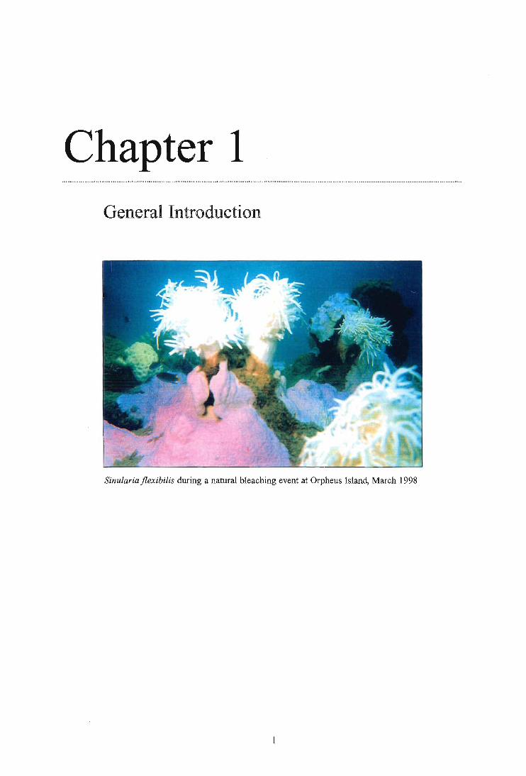
Chapter 1 General Introduction
Sinularia flexibilis during a natural bleaching event at Orpheus Island, March 1998

Chapter I: General Introduction
1. GENERAL INTRODUCTION
1.1. Background and significance of coral bleaching
The focal point of this thesis is the disruption of the symbiotic relationship between soft coral
hosts and their dinoflagellate algal partner due to stress, a phenomenon known as coral
bleaching. The animal—plant relationship, which evolved in scleractinian corals during the
Triassic, is believed to have greatly contributed to the radiation and evolutionary success of
today's tropical corals (Stanley, 1981; Stanley and Swart, 1995). Their symbiotic success is
primarily based on provision of photosynthetically-derived carbon to the animal host (Cook,
1983) and efficient uptake and recycling of nitrogen and phosphorus in the association (Miller
and Yellowlees, 1989; Sutton and Hoegh-Guldberg, 1990; Wang and Douglas, 1998),
particularly in highly oligotrophic systems. Bleaching, which can occur via loss of
zooxanthellae from the association and/or the degradation of photosynthetic pigments within the
algal partner (Hoegh-Guldberg and Smith, 1989a), therefore has the potential to compromise the fitness and survival of corals.
Coral bleaching has been described for decades and is a well-established disturbance on reefs
(Yonge and Nicholls, 1931; Goreau et al., 1964). Before the 1980s, however, bleaching episodes were generally confined to small areas that had been subjected to identifiable local
stresses, such as heavy rainfalls or river discharges which lowered salinity (Yonge and Nicholls,
1931; Goreau et al. 1964). Sudden temperature drops due to upwelling phenomena (Glynn and
Steward, 1973), sedimentation, aerial exposure due to low tides and resulting long exposure to
high irradiances or contact with pollutants are also known to lead to coral bleaching (reviewed
in Williams and Bunkley-Williams, 1990; Brown, 1997a; Hoegh-Guldberg 1999). During the
past two decades, however, acute and chronic bleaching of tropical corals has occurred on both
dramatically increased scale and intensity and these have predominantly been correlated with
high sea surface temperature anomalies (Brown and Ogden, 1993; Gleason, 1993; Hoegh-
Guldberg and Salvat, 1995; Brown et al., 1996; Jones et al., 1997; Berkelmans and Oliver,
1999). Among the recognised multiple biological effects of bleaching are reduced coral
growth, calcification, and possibly reduced reproductive output and increased mortality
(Clausen and Roth, 1975; Szmant and Gassman, 1990; Leder et al., 1991; Goreau and
Macfarlane, 1990). Ecological consequences of bleaching include shifts in community structure
and decreases in both species and habitat diversity (Fisk and Done, 1985; Gleason, 1996;
Glynn, 1996; Marshall and Baird, 1999). Until 1998, the most severe and extensive (10 000 km2) bleaching event recorded occurred in the Eastern Pacific during 1982-83, where 51-97%
of coral cover between Panama and the Galapagos Islands was lost (Glynn 1988) and two
2

Chapter 1: General Introduction
species almost became locally extinct (Glynn and de Weerdt, 1991; Glynn and Feingold, 1992).
In 1998, however, mass coral bleaching was recorded on an even greater scale throughout the
Pacific region, the Indian Ocean, the Red Sea, the Persian Gulf, the Mediterranean and the
Caribbean region (reviewed in Hoegh-Guldberg, 1999). Along the 1500 km Great Barrier Reef
(GBR), 87 % of inshore reefs were affected by this disturbance (Berkelmans and Oliver, 1999).
This increase in scale and severity of bleaching events is accompanied by evidence for
increased frequency of such events (Jones et al., 1997) and has thus led to suggestions that coral
bleaching could be a manifestation of global warming (Williams and Bunkley-Williams, 1990;
Hoegh-Guldberg, 1999).
1.2. Causal factors and interactions of elevated solar irradiance and temperature in the
bleaching process
The physiological basis for speculations that corals are possible indicators of global climate
change is that they live precariously close to their upper thermal limits (Coles and Jokiel, 1977;
Jokiel and Coles, 1990; Berkelmans and Willis, 1999). Consequently, increases of 1 - 2° C in
the tropical belt, as predicted by a number of ocean-atmospheric models (Manabe et al., 1991;
Bijlsma et al., 1995) could exceed the thermal tolerance limits of some species. More
importantly, under natural conditions an organism is exposed to a whole framework of physico-
chemical (stress) factors and it is their combined impact rather than the effects of a single factor,
which define the limits for survival. Given that coral reef environments are typically
characterised as having oligotrophic, clear waters in which high ambient solar radiation can
penetrate to considerable depth, high irradiance has been identified as an important selective
pressure in the development of symbiotic associations (Jerlov, 1950; Jokiel and York, 1982;
Fleischman, 1989; reviewed in Shick et al., 1996). There is a great deal of experimental
evidence supporting the notion that high visible and ultraviolet light are additional triggers
(Jokiel and York, 1982; Gleason and Wellington, 1993; Jones et al., 1998), which greatly
intensify the bleaching response when occurring simultaneously with elevated temperatures
(Lesser et al., 1990; Salih et al., 1998; Jones et al., 1998). Explanations for the nature and the
molecular level at which an interaction of the two factors occurs, however, are only just
emerging. A model providing a fundamental basis for understanding the interaction of high
temperature and light in the photosynthetically active region has recently been provided by
Jones et al. (1998). The authors demonstrated that increases in temperature in Stylophora pistillata result in a decreased capacity to process the excitation energy coming from the dark
reactions of photosynthesis as a primary effect. Subsequently, zooxanthellae become more
sensitive to light, resulting in an over-reduction of biochemical components of the light
3

Chapter 1: General Introduction
reactions, which eventually leads to destruction of chloroplasts and photoinhibition (Jones et al.,
1998). Subsequent discarding of thermally damaged and dysfunctional zooxanthellae from the
coral association (Suharsono et al., 1993) therefore presents an explanation for bleaching. An
alternative hypothesis suggests oxidative stress caused by the algal partner to be the proximal
cause of bleaching (Kuhl et al., 1995; Lesser 1997). Loss of zooxanthellae in this case has
consequently been interpreted as a "damage limiting" response, attained by decreasing
concentrations of the algal partner and consequently reactive oxygen species in the host tissue.
While the pioneering work by Jones and co-workers (1998) provides one basis for
understanding the interactive effects of light and temperature, namely that photoinhibition is a
function of thermal stress affecting the functioning of chloroplasts, interactions between these
two factors on other molecular levels cannot be ruled out. The thermal degradation of
mycosporine-like amino acids (MAAs), which constitute part of the coral's defence system
against high irradiance and particularly UVR, may also be involved in such interactions (Lesser et al., 1990; Glynn et al., 1992).
1.3. MAAs and mechanisms of biochemical defence against high irradiance
Given their evolutionary exposure to high solar irradiance over evolutionary time scales, it is
not surprising that reef-dwelling organisms have evolved a range of biochemical defence
systems, such as MAAs, to counteract damage caused by exposure to high irradiance in the
PAR (photosynthetically active region, 400-700 nm) and UV region (280-400 nm) (reviewed in
Dunlap and Shick, 1998). Amongst the recognised multiple effects of UVR are damage to
DNA and proteins, inactivation of enzymes and oxidation of lipids (reviewed in Shick et al.
1996). The underlying biochemical mechanism of damage caused by UV-B radiation (290-320
nm) is the absorption of the long wavelength frequencies by DNA, and subsequently UV-B is
largely responsible for the direct damaging effects of sunlight. Many other cellular components,
however, also absorb in the UV-A region (320-360 nm) and enter an excited state wherein the
excitation energy can be transferred to molecular oxygen, causing indirect oxidative damage by the photodynamic production of reactive oxygen species, such as singlet oxygen ( 102) or the superoxide anion (02) (Booth et al., 1997). One of the strategies to reduce photodamage is the
production of water-soluble (ascorbate gluthathione, urate) and lipid-soluble anti-oxidants
(13-carotene, a-tocopherol), while another is the expression of anti-oxidant enzymes such as
catalase and superoxide-dismutase (Lesser et al., 1990; Dunlap and Shick, 1998).
4
Chapter 1: General Introduction
As another "line of defence" in response to extremely high UV exposure, many shallow-water
marine organisms have evolved a group of unusual amino-acids, collectively called
mycosporine-like amino acids (MAAs). These are a generic class of water-soluble
cyclohexenone or cyclohexenimine chromophores conjugated with the nitrogen substituent of
an amino acid or amino alcohol (Favre-Bovin et al., 1976), which have received considerable
attention for their high absorption maxima ranging from 310 to 360 nm (reviewed in Dunlap
and Shick, 1998). Depending on the amino acid that the chromophore is conjugated with, the
MAA absorbs radiation at a slightly different wavelength, and thus the whole class of MAAs
each with a slightly different absorption maximum, acts as a broadband UVR filter. In addition
to UV absorbing properties, mycosporine-glycine, the simplest of MAAs, also features anti-
oxidative properties, thus reducing oxidative stress by two modes, absorbance of radiation and
scavenging of reactive oxygen species (Dunlap and Yamamoto, 1995). MAAs occur in
phylogenetically diverse groups, including marine bacteria (Arai et al., 1993), cyanobacteria
(Garcia-Pichel et al., 1993), phytoplankton (Carreto et al., 1990), macro-algae (Karentz et al.,
1991; Karsten et al., 1998), molluscs (Ishikura et al., 1997; Carefoot et al., 1998), anthozoans
(Dunlap et al., 1986; Stochaij et al., 1994; Teai et al., 1997) echinoderms (Adams and Shick
1996), and fish (Dunlap et al., 1989) and range geographically from tropical to Antarctic waters (Karentz et al., 1991; Shick et al., 1992).
The UV-protecting function of MAAs has been inferred from the fact that shallow water corals
typically have higher MAA tissue concentrations than their deeper conspecifics, and because the
compounds are light inducible (Jokiel and York, 1982; Dunlap et al., 1986; Gleason, 1993;
Shick et al., 1995; Kuffner et al., 1995). The fact that a diverse range of marine benthic
organisms provide their planktonic larvae with high concentrations of MAAs further
corroborates their important role as photo-protectants (Chiocara, 1986; Adams and Shick, 1996;
Carefoot et al., 1998). Direct evidence for their "sunscreen" role has been provided by Garcia-
Pichel and co-workers (1993) who showed that the presence of MAAs in the cyanobacterium Gleocaspa prevented 23-30% of UV radiation at 320 nm from reaching cellular targets. MAAs
are produced via the shikimic acid pathway in both terrestrial organisms (Favre-Bovin, 1976)
and corals (Shick pers. comm.). Given that the shikimic acid pathway is thought to be found
only in bacteria, fungi and algae (Bently, 1996), symbiotic zooxanthellae are probably the
producers of MAAs in corals. Consequently, rates of MAA synthesis in bleached corals should
be lower than in their non-bleached counterparts, potentially increasing damage due to high
irradiance under conditions of thermal stress. To date there have been no systematic studies
undertaken, however, in which MAA concentrations in bleached corals have been compared to
those of their unbleached counterparts. Suggestions by Lesser et al. (1990) and Glynn et al.
(1992) that MAAs may be thermolabile, raises the possibility that MAAs are the site at which
5

Chapter 1: General Introduction
potential molecular interactions between high irradiance and temperature take place. Thermal
degradation of MAAs under an increased temperature scenario would expose a coral to
additional irradiance stress when it is already experiencing thermal stress and thus could serve
as a model to explain the greatly accelerated bleaching response observed when the two stress
factors come together. The implications of decreased protection against irradiance stress due to
reduced MAA levels could be grave, particularly given the increased light-sensitivity of
zooxanthellae in coral associations under thermal stress (Jones et al., 1998).
To date our understanding of natural variations in concentrations of MAAs in corals under non-
stress conditions is very limited. Therefore any attempt to assess the effects of high temperature
and irradiance on the functioning of MAAs requires investigation of natural variations as a
prerequisite. So far only one study of mucus of Fungia repanda has documented a positive correlation between solar irradiance, based on changes in the absorption at 320 nm, however,
individual MAAs were not identified (Drollet et al., 1997). Given the possibility that MAAs
may play a major role in the understanding of possible synergistic interactions, research into the
fate of MAAs during the bleaching process has been highlighted as an area warranting study in a recent review of MAAs (Dunlap and Shick 1998).
1.4. Potential roles of secondary metabolites in the bleaching response of soft corals
Research into bleaching has almost exclusively focused on scleractinian corals (Harriott, 1985;
Gates, 1990; Hoeksema, 1991; Glynn, 1993; Glynn, 1996; reviewed in Hoegh-Guldberg, 1999),
despite the frequent bleaching of soft corals (Oliver, 1985; Fabricius, 1999; pers. obs.). This
omission is particularly striking, as alcyonacean corals are important contributors to both
biomass and cover of Indo-Pacific reefs (Dinesen, 1983; Fabricius, 1996). Given that the
symbiotic association between zooxanthellae and the host is very similar for both hard and soft
corals (Kinzie and Chee, 1979; Benayahu et al., 1989; Trench, 1997), the underlying
mechanisms in the bleaching process are likely to be the same for both hard and soft corals. Soft
corals, however, may be affected differently by bleaching because of the strong dependence on
certain so-called secondary metabolites which are virtually absent in hard corals (reviewed in
Coll, 1992). Secondary metabolites are compounds which enhance the coral's fitness by
increasing their defence against predation (La Barre et al., 1986a; Uchio et al., 1985; Wylie and
Paul, 1989; Slattery et al. 1995a), algal and microbial fouling (Slattery et al. 1995b; Leone et al.,
1995; Aceret et al., 1995; Koh, 1997; Michalek and Bowden, 1997), by increasing their
competitive ability (Sammarco et al., 1985; La Barre et al., 1986b; de Nys et al., 1991; Maida et
al., 1995a,b; Atrigenio and Aliflo, 1996) or by acting as spawning - specific compounds to
6

Chapter l: General Introduction
ensure reproductive success (Coll et al., 1989; Pass et al., 1989; Coll and Clayton, 1990; Coll et
al., 1995; Coll, 1992; Slattery et al., 1999). Because many of the highly specific functions of
secondary metabolites are only just emerging, often decades after their isolation, the terms
"fundamental" and "complementary" have recently been proposed to replace "primary" and
"secondary" metabolites in recognition of our increasing understanding of their ecological
significance (Sammarco and Coll, 1996).
While it is difficult to estimate the exact metabolic costs involved in producing terpenoid
secondary metabolites in soft corals, a substantial part of the carbon necessary for their
synthesis is probably of autotrophic origin. The underlying rationale for this notion is that the
algal partner translocates up to 95% of its photosynthates to the coral host, thereby providing up
to 143% of the daily energetic costs of the association (Muscatine et al., 1984; Davies, 1991).
Hence, bleaching and subsequent reduced photosynthetic output will translate into nutritional
constraints for the coral host. While the contribution of heterotrophy to the energy budgets of
soft corals must not be underestimated, (Lewis, 1982; Fabricius, 1995a), heterotrophic feeding
may be highly reduced under bleaching conditions (Fabricius, 1999). Given the integral role of
zooxanthellae in uptake and recycling of phosphorus and nitrogen in the coral association
(Miller and Yellowlees, 1989; Sutton and Hoegh-Guldberg, 1990; Wang and Douglas, 1998),
bleaching can also have strong negative impact on the balance of the two nutrients.
Consequently, energy allocation towards the production of significant concentrations of
secondary metabolites may be compromised in bleached soft corals. If individuals lack
adequate concentrations of key secondary metabolites, they will be more susceptible to factors
such as increased predation and algal overgrowth (Coll et al., 1987), which could inhibit
recovery of soft corals after bleaching. Moreover, besides their well-established role as primary
producers, there is also (controversial) evidence that zooxanthellae may be involved directly in
the production of specific secondary metabolites (Kokke et al., 1984; Anderson, 1985;
Papastephanou and Anderson, 1982; Ciereszko, 1989). Despite a 30-year debate, the question
as to which partner of the coral association controls the production of e.g. algaecides has never been fully resolved.
In summary, bleaching has the potential to compromise the production of secondary metabolites
via two modes, indirectly through nutritional constraints, or directly if the compounds are
indeed produced by the zooxanthellae. In either case, bleached soft corals could be more
susceptible to bacterial or algal fouling and more vulnerable to predation. To date no systematic
studies into the effects of bleaching on soft coral secondary metabolites have been carried out.
7
Chapter 1: General Introduction
1.5. Longer-term effects of bleaching — implications for reproductive output
The duration and scale of an environmental disturbance dictate the severity of the bleaching
response, and mortality generally only follows when stress is prolonged (Harriott, 1985; Glynn,
1996; Baird and Marshall, 1998). In many cases corals relieved of environmental stresses have
been known to regain their zooxanthellae (Cook et al., 1990; Szmant and Gassman, 1990), but
most studies of recovery of corals have only focused on short-term issues; i.e. the regaining of
zooxanthellae densities (reviewed in Brown, 1997a,b). Given the nutritional constraints
associated with bleaching and possible re-allocation of resources towards regeneration
(Meesters and Bak, 1993), longer-term effects associated with the loss of zooxanthellae are
likely to occur. To date there are indications that bleaching has negative impacts on a coral
beyond recovery of zooxanthellae, particularly reduced calcification (Clausen and Roth, 1975;
Leder et al., 1991) or growth (Szmant and Gassman, 1990; Goreau and Macfarlane, 1991).
Pioneering work by Szmant and Gassman in 1990, has also provided the first, if very limited,
evidence for impairment of gametogenesis in bleached colonies of Montastrea annularis. At present it is unknown whether bleaching can affect the "next generation" by reducing maternal
energy reserves normally allocated for reproduction. The effects of bleaching could be passed
on to the next generation via two modes: through reduced reproductive output and/or through
biochemically less well-equipped offspring, with possibly reduced potential for fitness and
survival. Given the integral role of sexual reproduction for the persistence and maintenance of
reefs, it is important to establish the implications of bleaching for reproduction.
1.6. Specific aims
The research presented in this thesis investigates aspects of the chemical ecology of the soft
coral — zooxanthellae interaction, to increase our understanding of how bleaching may affect the
biochemistry and subsequently the physiology and ecology of soft corals. My specific aims were:
1. To investigate the effects of temperature and UV radiation on concentrations and
function of UV protecting agents. The possibility that temperature and UV radiation may
act synergistically to break down UV protecting agents and hence trigger bleaching has
grave implications for the health and survival of reef organisms if predictions of global
warming are accurate. This study will therefore provide a basis for the understanding of the
role of MAAs during bleaching episodes, induced by elevated temperature and solar
radiation.
8

Chapter 1: General Introduction
To measure seasonal variations in the concentrations of UV protecting agents in soft corals. Knowledge of seasonality in concentrations of UV - blocking compounds in field
populations of soft corals will aid in determining the role of these compounds in the
biochemistry of reef organisms and is essential for interpreting manipulative bleaching
experiments (as referred to in objective 1.).
To determine if secondary metabolite production in soft corals changes after
bleaching, and if so, to investigate the ecological implications of such changes. If symbiotic zooxanthellae control or contribute to the production of secondary metabolites
that act as anti-fouling agents or feeding deterrents, then bleaching may compromise the
survival of soft corals through impacts in addition to those associated with nutritional constraints.
To establish which partner of the coral alga association controls the production of species — specific secondary metabolites. If zooxanthellae control the production of secondary metabolites this can have grave implications for bleached soft corals, since the
maintenance of significant concentrations of these ecologically important compounds could
not be sustained, even if resources were available.
To examine if bleaching affects the reproductive success of selected soft corals, as a long-term sub - lethal impact of bleaching. The loss of zooxanthellae may result in strong nutritional constraints or impaired production of ecologically important secondary
metabolites, which could potentially decrease the reproductive success of bleached soft corals.
9

Chapter 2 Seasonal variation in MAA levels of soft corals in relation to annual solar irradiance and seawater temperature cycle
Lobophytum compactum recovering its zooxanthellae after a natural bleaching event at Orpheus Island, March 1998
1 0

Chapter 2: Seasonal variation in MAA levels of soft corals in relation to annual solar irradiance and seawater temp. cycles
2.1. ABSTRACT
This chapter focuses on the nature and extent of variation in MAAs in relation to annual cycles
in solar radiation, seawater temperature and reproduction in reef-flat populations of soft corals.
The results presented clearly show that MAA tissue concentrations in shallow water colonies of
Lobophytum compactum and Sinularia flexibilis are significantly correlated to annual cycles in
solar radiation and seawater temperature. Evidence of seasonal cycles in MAA levels in the
tissues of shallow reef invertebrates positively correlated with annual cycles in solar radiation
corroborates the importance of MAAs as photoprotectants. Furthermore, prior to spawning in
both years, female colonies of both soft coral species featured substantially higher MAA levels
than their male counterparts, presumably to provide a high level of protection against irradiance
stress for progeny. In addition, this study demonstrates that levels of carotenoids, chlorophyll
and proteins also vary seasonally, with maxima for the former group, and minima in the latter
groups during the Australian summer, suggesting that carotenoids in particular may contribute
to the photo-acclimatory responses of soft corals.
11

Chapter 2: Seasonal variation in MAA levels of soft corals in relation to annual solar irradiance and seawater temp. cycles
2.2. INTRODUCTION
The UV-protecting role of mycosporine-like amino acids (MAAs) in marine organisms has been
inferred from numerous studies since Dunlap and Chalker (1986) first suggested they function
as broad—band filters in corals (reviewed in Dunlap and Shick, 1998). MAAs appear to be ideal
screening agents for symbiotic associations involving phototrophic organisms, because they are
transparent to radiation in the PAR region (400-700 nm), but absorb strongly in the potentially
damaging UV range between 310 — 360 nm. Functionally, MAAs are believed to absorb
radiation of high energy in the UV region, which is then dissipated as vibrational energy,
thereby minimising structural and physiological damage to tissues (M. Shick, pers. comm.).
Support for the assumption that the primary role MAAs is protection against high irradiance
damage is mainly based on indirect evidence that MAAs are light inducible by radiation both in
the PAR (Scelfo, 1985; Karsten et al., 1998) and UVR region (Lesser et al., 1990; Shick et al.,
1991; Kinzie, 1993) and that shallow individuals typically have higher tissue concentrations
than their deeper counterparts (Dunlap et al., 1986, Gleason, 1993; Shick et al., 1991; Kinzie,
1993). Their role as photoprotectants is further supported by the fact that a diverse range of
marine benthic organisms provide their planktonic larvae with higher concentrations of MAAs
than are found in adult tissues (Chiocarra, 1986; Adams and Shick, 1996; Carefoot et al., 1998,
see also Chapter 7). The first direct evidence for their photo-protective role was provided by
Garcia-Pichel and co-workers in 1993, who showed that the presence of MAAs in the
cyanobacterium Gleocaspa prevented 23-30% of UV radiation at 320 nm from reaching cellular
targets. Given that solar irradiance stress has been implicated as a causative factor in a number
of bleaching events, both as a sole factor (Fisk and Done, 1985; Gleason and Wellington, 1993;
Brown et al., 1994) and in combination with increased temperature (Harriott, 1985; Brown and
Suharsono, 1990), MAAs have the potential to play an important role in the prevention of
bleaching.
Although both direct and indirect evidence suggest that MAAs have a photo-protective
function, experimental studies carried out by Scelfo (1985) and Stochaij and co-workers (1994)
on a number of cnidarians failed to demonstrate MAA concentration increases in response to
longer-term exposure to UVR. To date the only positive correlation between MAA
concentrations and solar radiation cycles were established for the digestive tract of the sea
urchin Sterechinus neumayeri (Karentz et al., 1997) and the mucus of the solitary coral Fungia
repanda (Drollet et al., 1997). In fact, a variety of factors other than high solar irradiance have
been proposed to affect MAA tissue concentrations in corals, including water velocity (Jokiel et
al., 1995; Jokiel et al., 1997) and mechanical stress (Scelfo, 1985), suggesting that MAAs may
12

Chapter 2: Seasonal variation in MAA levels of soft corals in relation to annual solar irradiance and seawater temp. cycles
function other than as photoprotectants. Bandaranayake et al. (1997) proposed that MAAs may
perform a role similar to reproductive hormones, after finding no correlation between MAA
concentrations and animal solar irradiance, but a positive correlation with the reproductive cycle
in the sponge Dysidea herbacea.
Evidence of natural cycles of MAA concentrations in the tissues of sessile organisms that
correlate with annual cycles in solar radiation would corroborate the importance of MAAs as
photoprotectants. The soft corals Lobophytum compactum and Sinularia flexibilis thrive on reef
flats (and occur to around 20m) predominantly in the inner- and midshelf reefs of the GBR,
which makes them ideal study organisms to examine the effects of high seasonal fluctuations in
solar irradiance and temperature on MAA levels. If MAAs are an important component of the
photo-protective system of soft corals, a seasonal trend in MAA concentrations should therefore
be reflected in tissues of these reef flat corals, with maxima in summer when incident radiation
is highest. Since both soft coral species are broadcast-spawners with positively buoyant eggs
(Aliflo and Coll, 1989), substantial investment into biochemical defence to counteract solar
damage should also occur in the highly exposed eggs and early planktonic life stages. If
maternal provision of offspring with MAAs is indeed an important life-strategy in corals,
female colonies should have significantly higher concentrations prior to spawning than males,
as has been observed in sea-urchins (Adams and Shick, 1996) and sponges (Bandaranayake et
al., 1997).
The aim of this chapter is to determine the nature and extent of variation in MAAs in relation to
annual cycles in solar radiation, seawater temperature and reproduction in reef-flat populations of the soft corals Lobophytum compactum and Sinularia flexibilis. Specifically, I will examine
whether MAA levels are highest when protection against high incident radiation and, thus
possibly bleaching, becomes most crucial. Knowledge of natural variations in MAA levels will
also provide a basis for the interpretation of experimental MAA manipulations (Chapter 3).
Since MAAs are most likely produced by the zooxanthellae partner in the coral association
(Chapter 1), variation in MAA levels will be investigated in the framework of variations in
zooxanthellae densities, chlorophyll and protein concentrations, which all contribute to
quantifying natural variations in symbiont densities. Given that carotenoids are not only one of
the light-harvesting photosynthetic pigments, but may also function as photoprotectants, I will
also investigate annual cycles in carotenoid (peridinin) levels. Furthermore, sex-specific
differences in the MAA pool will be investigated because of potential maternal provision of
UV-absorbing compounds to progeny.
13
PELORUS ISLAND
Cattle Bay
Pioneer Bay
Research Station
ORPHEUS ISLAND
GREAT PALM ISLAND
FANTOME ISLAND
Chapter 2: Seasonal variation in MAA levels of soft corals in relation to annual solar irradiance and seawater temp. cycles
2.3. MATERIALS AND METHODS
2.3.1. Site description
This study was carried out between December 1995 and December 1997 in Pioneer Bay,
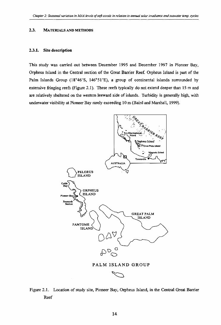
Orpheus Island in the Central section of the Great Barrier Reef. Orpheus Island is part of the
Palm Islands Group (18°46'S, 146°51'E), a group of continental islands surrounded by
extensive fringing reefs (Figure 2.1). These reefs typically do not extend deeper than 15 m and
are relatively sheltered on the western leeward side of islands. Turbidity is generally high, with
underwater visibility at Pioneer Bay rarely exceeding 10 m (Baird and Marshall, 1999).
PALM ISLAND GROUP
Figure 2.1. Location of study site, Pioneer Bay, Orpheus Island, in the Central Great Barrier
Reef
14

Chapter 2: Seasonal variation in MAA levels of soft corals in relation to annual solar irradiance and seawater temp. cycles
2.3.2. Species description
The alcyonacean soft corals Sinularia flexibilis Quoy & Gaimard (Versefeld, 1980) and
Lobophytum compactum Tixier-Durivault, 1956, (Tixier-Durivault, 1958) which are common
reef flat corals on the inner and mid-shelf reefs of the Great Barrier Reef (Dinesen, 1983), were
used for most experiments. Their contrasting growths forms, Lobophytum compactum featuring
a solid leathery growths form, whereas Sinularia flexibilis has a comparatively delicate structure
with fine branchlets), offers scope for comparisons between differential susceptibility towards
thermal and solar stress between solid and more delicate species.
2.3.3. Sampling regime and preparation of experimental corals
To determine if there are annual cycles in MAA tissue concentrations, colonies located on the
reef slope at a depth of 2-3 m were tagged in situ in November 1995 (Figure 2.2). I sampled colonies of the dioecious soft corals Sinularia flexibilis and Lobophytum compactum at monthly
intervals over two subsequent years, from January 1996 through December 1997. Five large
mature male and female colonies of similar colony size were used (stalk diameters of 10-13 cm for Sinularia flexibilis and colony diameters averaging 70-90 cm for Lobophytum compactum). I collected tissue samples of all 20 test corals at each sampling date. To determine the level of
intra-colony variation in tissue concentrations of various biochemical parameters, four random
tips (a-d; 1-2 g wet weight/tip) were collected from each colony. Samples were snapfrozen at -
20° C and kept under dark conditions until analysed biochemically. All samples were
subdivided and analysed for zooxanthellae densities and chlorophyll a + c2, MAA, carotenoid
and protein concentrations, so that there were 4 replicate determinations per colony for each
biochemical parameter (Figure 2.2).
15

Species
Sex
Colony
Lobophytum compactum Sinularia flexibilis
0
Extraction
subsamples homogenised in seawater (s)
subsamples homogenised in Methanol (M)
zooxanthellae counts
zooxanthellae pellet through centrifugation
MAA carotenoid (peridinin) analysis analysis
extracted in acetone remaining pellet hydrolised in NaOH
1 2 3 4 5 6 7 8 9 10
Subsample
A 1 2 3 4
chlorophyll a + c2 protein analysis analysis
Chapter 2: Seasonal variation in MAA levels of soft corals in relation to annual solar irradiance and seawater temp. cycles
Figure 2.2. Overview of experimental design and sample preparation for the different
biochemical analyses, N = 40 determinations (i.e. 10 colonies x 4 subsamples for each
biochemical parameter).
16

Chapter 2: Seasonal variation in MAA levels of soft corals in relation to annual solar irradiance and seawater temp. cycles
2.3.4. Biochemical analysis of zooxanthellae densities, chlorophyll, carotenoid, MAA and
protein concentrations
In order to monitor potential photo-acclimatory responses through time, zooxanthellae,
chlorophyll and carotenoid concentrations were determined as follows.
I. Quantification of zooxanthellae densities. Zooxanthellae population densities were
monitored as indicators of possible bleaching and recovery of host tissues. Tissue samples for
the analysis were fully homogenised in 0.45 gm filtered and autoclaved seawater. An aliquot of
each sample was fixed in formalin and stored at 4° C until analysis. Zooxanthellae densities
were calculated from quadruplicate haemocytometer counts for each of four sub-samples per
colony and normalised to coral wet weight. Coral wet weight was determined prior to freezing
by weighing dry blotted samples.
Chlorophyll analysis. Homogenised tissue samples were centrifuged, the supernatant
decanted and the zooxanthellae containing pellet extracted at 4° C for 24 hours in 100% acetone
under dark conditions. After extraction, samples were centrifuged again and the supernatant
used to quantify chlorophyll a and c2 spectro-photometrically using equations in Jeffrey and
Humphrey (1975).
Carotenoid (peridinin) analysis. Peridinin, the main carotenoid in dinoflagellelates
(Kazlaukas et al. 1976) was quantified in order to examine its contribution as an accessory
pigment and possible photoprotectant. Weighed and defrosted tissue samples were fully
homogenised and sequentially extracted 3 times for 60 minutes in 3 ml of 100% methanol. To
avoid photo-degradation of carotenoids (and also MAAs) samples were kept on ice and
protected from light during extraction. The combined extract was clarified by centrifugation,
and then peridinin immediately quantified spectro-photometrically using equations in Jeffrey
and Haxo (1968).
Analysis of mycosporine-like amino acids (MAAs). Individual MAAs were separated and
quantified by reverse-phase, isocratic high-performance liquid chromatography (hplc).
Separation was accomplished using a Brownlee RP-8 column (spheri-5, 4.6. mm i.d. x
250 mm), protected with an RP-8 guard column (spheri-5, 4.6. mm i.d. x 30 mm). The mobile
phase consisted of 25% methanol and 0.1% acetic acid (v: v) in water and was run at a flow rate
of 0.8 mL/min. Peaks were detected by UV absorbance at 313 mu and 340 nm (dual wave
detection). Prior to hplc analysis, MAA samples were processed in the same fashion as the
17

Chapter 2: Seasonal variation in MAA levels of soft corals in relation to annual solar irradiance and seawater temp. cycles
carotenoid samples and stored at — 20° C. Additional treatment involved filtration of samples
through 0.45 gm millepore filters to ensure removal of particulate matter from the solution.
Identification of MAAs was attained by co-chromatography with verified standards of palythine
and palythinol from the zoanthid Palythoa tuberculosa; porphyra-334 and shinorine from the
red alga Porphyra tenera, and asterina-330 and palythene from the ocular lens tissue of the
coral trout Plectropomus leopardus. Peak areas were integrated on a Hewlett-Packard
integrator and quantification of individual MAAs was achieved by comparing the integrated
hplc peak areas with pre-determined peak areas of chromatographically quantified standards
(standards and calibration courtesy W. Dunlap). All measurements were corrected for the
extraction efficiencies of individual MAAs of each species by the method described in Dunlap
and Chalker (1986) and ranged from 95.5% - 99.6%.
Partitioning of MAAs between zooxanthellae and soft coral host. In order to determine
whether MAAs are stored in the animal or algal part of the coral association, cells were
separated and the MAA concentrations of each part determined. Entire lobes (Lobophytum compactum) or "branchlets" (Sinularia flexibilis) of test corals (3 females, 2 males) were
collected and divided in half. One half was immediately frozen and later used to determine
protein and MAA concentrations of the entire coral association. The second half was
homogenised in artificial seawater to yield the zooxanthellae fraction of the association. This
was achieved by repeated centrifugation, discarding of supernatant and washing until a pellet
free of animal cells was achieved (confirmed by microscopy of extracts). Quantification of
MAAs and protein present in the zooxanthellae fraction and in the entire association was then
carried out as described below. Protein content and MAA concentration of the animal tissue
were calculated as the difference between the whole fraction and the zooxanthellae fraction.
Protein determination. Protein concentrations were determined in order to normalise MAA
concentrations to a representative unit of tissue. After methanol extraction, tissue residues were
kept frozen until their protein content was determined. Hot 1N NaOH was used to digest tissue
samples, which were then brought to room temperature and the mean protein content of
duplicate aliquots measured using the method of Lowry et al. (1951). Bovine albumin was used
as a standard.
Statistical analysis. I used repeated measures analyses to test for differences in biochemical
parameters over time. Two factorial ANOVA was used to test for differences between sexes.
Prior to analysis of variance, data were tested for normality (Shapiro-Wilks statistics) and
homogeneity of variances (Bartlett and Cochran tests) and data transformed where appropriate.
18

Chapter 2: Seasonal variation in MAA levels of soft corals in relation to annual solar irradiance and seawater temp. cycles
After transformation, variances in protein levels in Sinularia flexibilis were still found to be
heterogeneous, however, to a lesser degree. Given that ANOVA is considered robust to all but
major deviations from homogeneity (Underwood, 1981), repeated measures analyses were
carried out with the data. The significance of changes in the composition of MAAs through
time was determined by MANOVA of arc sine transformed percentage compositions of MAAs
(Sokal and Rohlf, 1995). Simple and multiple linear regression analyses were carried out in
order to determine the relationship between levels of a number of biochemical parameters and
solar irradiance or seawater temperature. I used STATVIEW, SUPERANOVA and SPSS
software packages for the above tests.
2.3.5. Relationship between seawater temperature, solar radiation and biochemical parameters
In order to determine the relationship between variations in MAA concentrations and either
annual solar radiation or seawater temperature variation, I acquired the appropriate data from
two sources. Temperature data were obtained from the Great Barrier Reef Marine Park
Authority (GBRMPA) temperature-monitoring program (courtesy R. Berkelmans).
Temperatures were monitored at 2-3 m depth on the reef flat at Cattle Bay, which is within
3-4 km to my study site in Pioneer Bay on Orpheus Island (see Figure 2.1 for location). Means
are based on approximately 1440 readings per month of permanently installed, calibrated
temperature data loggers.
The environmental ambient radiation data were made available by Aurel Moise at the School of
Mathematics, Computer Sciences and Physics at James Cook University and the Australian
Radiation Laboratory (Yallambie, Victoria). All environmental radiation data were collected at
Townsville, approximately 70 km from Orpheus Island.
Monthly means were used for regression analyses between a number of different combinations
of biochemical parameters. Similarly, average monthly doses of total solar downward flux
(300 — 2800 nm) and total UVR (285-400 nm) were used for regression analyses with the same
biochemical parameters.
19

UV-B UV-A
HO OH
OCH3
CO2H R= H asterina max = 330 nm CO2H R= CH3 palythinol X max = 332nm Ry\
4 4 OH IN
'-7 HO OH
OCH3
HO OH ZH CO2H
palythine max = 320 nm
OCH3
ZH
CO2H R= H shinorine max = 334nm R= CH3 porphyra k max = 334nm
.-/-
280 320 360 400
OCH3
ZH
CO2H palythene Amax = 360 nm
'-7 HO OH
Chapter 2: Seasonal variation in MAA levels of soft corals in relation to annual solar irradiance and seawater temp. cycles
2.4. RESULTS
2.4.1. MAA analysis
1. MAA profiles in Lobophytum compactum and Sinularia flexibilis
The typical set of MAAs I identified in both species included shinorine, porphyra-334,
palythine, asterina-330, palythinol and palythene (Figure 2.3).
Xmax peak absorbance (nm)
Figure 2.3. Wavelength and absorbance maxima (X,..) of MAAs present in both the tissues of
Lobophytum compactum and Sinularia flexibilis.
In both coral species palythine made by far the highest contribution to the MAA pool, and on
average comprised 92.79 ± 0.49% and 93.26 ± 0.40% of the pool for Lobophytum compactum and Sinularia flexibilis respectively (Figure 2.4). The second strongest fraction differed
between the two species, with shinorine contributing, on average, 3.25 ± 0.26% for Lobophytum compactum, and palythene contributing 4.32 ± 0.34% in Sinularia flexibilis. The concentrations of the remaining four MAA species were very low and collectively did not contribute more than
4.00 ± 0.20% and 2.43 ± 0.14% to the total pool in L. compactum and S. flexibilis respectively.
20
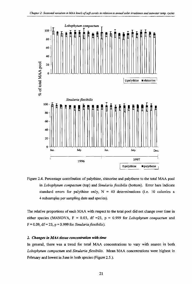
Dec. Jan. July Jan. July
Chapter 2: Seasonal variation in MAA levels of soft corals in relation to annual solar irradiance and seawater temp. cycles
Sinularia flexibilis
~ Ett f f
20 -
0
100
80
60
40
100 -
80 -
60 -
40 -
20 -
0
% o
f to t
al M
AA
poo
l
Lobophytum compactum
I f ttt tt
a palythine ■ shinorine

1 . 1
Li a)
1 , 1 ...; 0 0
Chapter 2: Seasonal variation in MAA levels of soft corals in relation to annual solar irradiance and seawater temp. cycles
small scale bleaching
spawning '96 spawning '97
0 L. compactum female colonies L. compactum male colonies
0
S. flexibilis female colonies S. flexibilis male colonies
600
400 -
nmol
tota
l MA
As/m
g pr
ote i
n
200 -
0
600 -
400 -
200 -
0
I
1 1 , 1.1,1.1,1.1.1 ii,i CD ..j Li .6 o
4 1996
. 1 , 1 . 1 , 1 4.)
04 a P
1997
Figure 2.5. Changes in MAA levels through time in female and male colonies of Lobophytum compactum (top) and Sinularia flexibilis (bottom). Error bars indicate standard errors,
N = 20 determinations (i.e. 5 males and 5 females per species x 4 subsamples, per month).
Two-factorial repeated measures ANOVA demonstrate that MAA concentrations vary
significantly with time in both species (p < 0.0001; Table 2.1), but the significant first order
interaction effect indicates that temporal patterns in MAA concentrations differ among colonies
(p < 0.0001; Table 2.1).
22

Chapter 2: Seasonal variation in MAA levels of soft corals in relation to annual solar irradiance and seawater temp. cycles
Table 2.1. Summary of results for 2-factorial repeated measures ANOVA, testing for differences
in MAA concentrations between colonies and through time. Asterisks denote significant differences (a = 0.05).
Sinularia flexibilis
colony
time
colony x time
MAA tissue concentrations over time
df F-ratio p-value
9 1.935 0.0849 23 18.701 < 0.0001 *
207 1.803 < 0.0001*
9 3.362 0.0058*
23 14.424 < 0.0001* 207 2.456 < 0.0001*
Source of variation
Lobophytum compactum
colony
time
colony x time
The increase in MAA levels from January to February in both species was a function only of
protein losses experienced during the small scale bleaching episode, since no significant
changes occurred with respect to wet weight levels (Scheffe, p = 0.70 and p = 0.33 for Lobophytum compactum and Sinularia flexibilis respectively).
3. Sex-specific differences in MAA tissue concentrations
Differences in the MAA concentrations of male and female colonies were detected, with
females featuring up to 67% and 56% higher concentrations immediately prior to spawning in Lobophytum compactum and Sinularia flexibilis (Figure 2.5). The magnitude of differences in
MAA levels between males and females depended on the time of sampling resulting in a significant interaction term (p < 0.0001 for L. compactum and p < 0.044 for Sinularia flexibilis). Differences between the sexes were greatest in the three months prior to spawning in both species (Figure 2.5).
23

Chapter 2: Seasonal variation in MAA levels of soft corals in relation to annual solar irradiance and seawater temp. cycles
Table 2.2 Summary of results for 2-factorial ANOVA testing for differences in MAA levels
between sexes and through time. Asterisks denote significant differences (a = 0.05).
Sex-specific differences in MAA tissue concentrations
Source of variation
Lobophytum compactum
time
sex
sex x time
Sinularia flexibilis
time
sex
sex x time
df F-ratio p-value
23 16.59 <-- 0.M01* 1 10.78 0.001*
23 3.26 < 0,0001*
23 10.82 < 0.0001* 1 0.44 0.508
23 1.57 0.044*
4. MAA partitioning between animal and plant tissue
Comparison of MAA concentrations between animal and plant tissue in Lobophytum compactum and Sinularia flexibilis revealed that concentrations in the animal tissue are
approximately 9 fold higher then those in the zooxanthellae fraction. This corresponds to
MAAs in host tissues comprising, on average, 92.36 ± 1.44% and 92.82 ± 1.07 % of the total pool for the symbiont — coral association in Lobophytum compactum and Sinularia flexibilis respectively.
in animal 0 plant
300 - 0
200 -
-(1 100 -
93%
92% /
Sinularia flexibilis Lobophytum compactum
Figure 2.6. Partitioning of MAAs between animal and plant tissue in Lobophytum compactum and Sinularia flexibilis (n = 5). Figures within the column represent the percentage of
MAAs of the total pool associated with the animal tissue.
400 -
24

Chapter 2: Seasonal variation in AMA levels of soft corals in relation to annual solar irradiance and seawater temp. cycles
2.4.2. Seasonal responses in biochemical parameters
Zooxanthellae analysis
With the exception of February 1996, when a short-term bleaching event occurred,
zooxanthellae densities remained virtually constant throughout the 24-month monitoring period
in both species (Figure 2.7a). Repeated measures analysis of all 23 monitoring months
(excluding February 1996) confirmed that no significant changes occurred during that period
(F = 0.641, df = 22, p = 08955 for Lobophytum compactum; F--= 1.302, df = 22, p = 0.1605 for Sinularia flexibilis). The bleaching episode in February 1996 corresponded to significant zooxanthellae losses in the range of 22% for Lobophytum compactum and 10 % for Sinularia flexibilis. Both species, however, recovered to background levels within two months (Figure
2.7a). Zooxanthellae concentrations did not differ significantly among colonies independently
of time (Table 2.3). Analysis of all 24 months (i.e. including February 1996; Table 2.3),
revealed that zooxanthellae levels were dependent on the time of sampling in Lobophytum compactum (p < 0.0001; Table 2.3). Zooxanthellae densities did not differ among sampling times in Sinularia flexibilis, and the absence of significant first order interactions indicates that
the overall temporal pattern in zooxanthellae levels did not differ among colonies (p = 0.060 for Lobophytum compactum and p = 0.962 for Sinularia flexibilis; Table 2.3).
Chlorophyll analysis
Levels of chlorophyll were consistently higher in winter than in summer in both Lobophytum compactum and Sinularia flexibilis, which largely explains the significant effect of time on
chlorophyll concentrations (Figure 2.7b; Table 2.3). The significant interaction term (Table 2.3)
suggests that the overall pattern of no variation in chlorophyll among colonies was not consistent
at the level of individual months. Thus, the magnitude of differences in chlorophyll levels among
colonies varied temporally, being significant in some months and inconsequential in others (Table 2.3).
25

b) Chlorophyll a +c2 / zooxanthella
c) Protein tissue concentrations
—4— L. compactum
0 S. fleacibilis
a) Zooxanthellae tissue concentrations
30 -
20 -
10 -
25
8
7
6
5
4
d) Peridinin (carotenoid) tissue concentrations 30 -
20 -
10 -
Chapter 2: Seasonal variation in MAA levels of soft corals in relation to annual solar irradiance and seawater temp. cycles
0 1996 1997
.I,L I , I.1 , I , I , I , I , I.1.1,f,I,L1 —I 1.1.1 1 , 1,1,1 Jan. April July Oct. Jan. April July Oct.
Figure 2.7. Mean concentrations of a range of biochemical parameters in the tissues of the soft coral Lobophytum compactum and Sinularia flexibilis: a) mean densities of zooxanthellae, b) mean chlorophyll a + c2 concentrations, c) mean protein levels and d) mean peridinin concentrations
during a two-year monitoring period. Errors indicate standard errors, N = 20 determinations (i.e. 5 male, 5 female colonies x 4 subsamples per colony, per month).
26
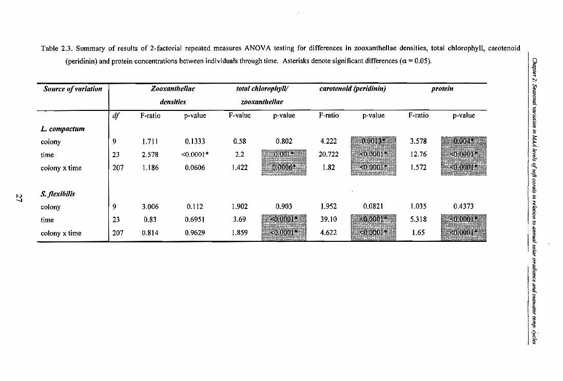
protein
p-value
<011
0.4373
<0
Table 2.3. Summary of results of 2-factorial repeated measures ANOVA testing for differences in zooxanthellae densities, total chlorophyll, carotenoid
(peridinin) and protein concentrations between individuals through time. Asterisks denote significant differences (a = 0.05).
Source of variation Zooxanthellae densities
total chlorophyll/ zooxanthellae
carotenoid (peridinin)
L. compactum df F-ratio p-value F-value p-value F-ratio p-value F-ratio
colony 9 1.711 0.1333 0.58 0.802 4.222 3.578 time 23 2.578 <0.0001* 2.2 0.001* 20.722 <0.0001 * 12.76 colony x time 207 1.186 0.0606 1.422 0.0006* 1.82 <0.0001* 1.572
S. flexibilis colony 9 3.006 0.112 1.902 0.903 1.952 0.0821 1.035 time 23 0.83 0.6951 3.69 <0.0001* 39.10 <0.0001* 5.318 colony x time 207 0.814 0.9629 1.859 1 1* 4.622 <0.0001 * 1.65

Chapter 2: Seasonal variation in MAA levels of soft corals in relation to annual solar irradiance and seawater temp. cycles
Carotenoid analysis
Concentrations of peridinin (the main carotenoid) were consistently higher in summer than in
winter. Averaging winter (July) values over two years, levels were only 47.25% and 49.95% of summer (January) levels for Lobophytum compactum and Sinularia flexibilis respectively (Figure 2.7d). The significant first order interaction term in both species indicates that the
magnitude of differences temporal in peridinin levels differ among colonies varied throughout
the sampling period (p < 0.0001; Table 2.3). In general, peridinin levels differed among
colonies in some months but not in others, however, the overall temporal pattern was clear.
Protein analyst's'
Protein concentrations varied over time for both Lobophytum compactum and Sinularia flexibilis with highest levels in winter (July) and lowest in summer (January) for the latter species (Figure 2.7c). For Lobophytum compactum no clear seasonal pattern could be discerned. Protein concentrations varied significantly between colonies at any given month in Lobophytum compactum, whereas variation in protein levels between colonies of Sinularia flexibilis were not significant (Table 2.3). The presence of a significant interaction term again
suggests that the magnitude of differences in protein concentrations among colonies varied temporally (Table 2.3).
2.4.3. Environmental parameters monitored during the sampling period
I. Solar radiation data for Townsville during the monitoring period
Total solar downflux and total UV radiation followed a pronounced seasonal trend, with peaks in summer and lows in winter (Figure 2.8).
28 .
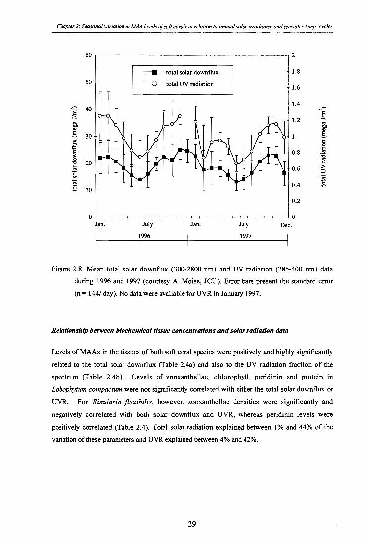
Chapter 2: Seasonal variation in MAA levels of soft corals in relation to annual solar irradiance and seawater temp. cycles
60
2
1.8
1.6
1.4
1.2
—II— total solar downflux
50
total UV radiation
tota
l sol
ar d
ownf
lux
(meg
a J/m
tota
l UV
radi
ation
(meg
a J/m
2)
30 -
0.8
- 0.6
- 0.4
0.2
20 -
10 -
0 --, 1 Jan.
0 Dec. July Jan.
1996
July
1997
Figure 2.8. Mean total solar downflux (300-2800 nm) and UV radiation (285-400 nm) data
during 1996 and 1997 (courtesy A. Moise, JCU). Error bars present the standard error
(n = 144/ day). No data were available for UVR in January 1997.
Relationship between biochemical tissue concentrations and solar radiation data
Levels of MAAs in the tissues of both soft coral species were positively and highly significantly
related to the total solar downflux (Table 2.4a) and also to the UV radiation fraction of the
spectrum (Table 2.4b). Levels of zooxanthellae, chlorophyll, peridinin and protein in
Lobophytum compactum were not significantly correlated with either the total solar downflux or UVR. For Sinularia flexibilis, however, zooxanthellae densities were significantly and
negatively correlated with both solar downflux and UVR, whereas peridinin levels were
positively correlated (Table 2.4). Total solar radiation explained between 1% and 44% of the
variation of these parameters and UVR explained between 4% and 42%.
29
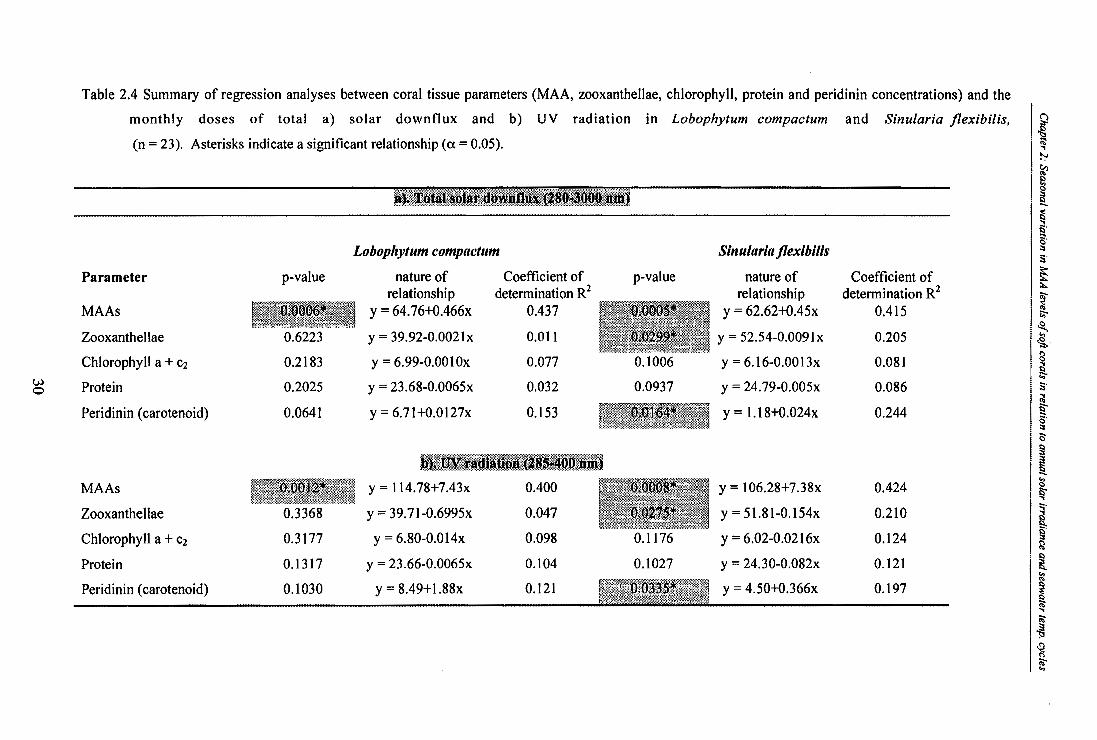
Table 2.4 Summary of regression analyses between coral tissue parameters (MAA, zooxanthellae, chlorophyll, protein and peridinin concentrations) and the monthly doses of total a) solar downflux and b) UV radiation in Lobophytum compactum and Sinularia flexibilis, (n = 23). Asterisks indicate a significant relationship (a = 0.05).
tal solar dowlifillx (284-3000.2
Parameter p-value
Lobophytum compactum
nature of Coefficient of
relationship determination R2 —,„ p-value
Sinularia flexibilis
nature of relationship
Coefficient of determination R2
MAAs y = 64.76+0.466x 0.437 0.0 0 1 = 62.62+0.45x 0.415 Zooxanthellae 0.6223 y = 39.92-0.0021x 0.011 0,0299* y = 52.54-0.0091x 0.205 Chlorophyll a + c2 0.2183 y = 6.99-0.0010x 0.077 0.1006 y = 6.16-0.0013x 0.081
<a 0 Protein 0.2025 y = 23.68-0.0065x 0.032 0.0937 y = 24.79-0.005x 0.086
Peridinin (carotenoid) 0.0641 y = 6.71+0.0127x 0.153 0.0164* y = 1.18+0.024x 0.244
b). UV radi4itioltA2gg-4 MAAs 13m012* y = 114.78+7.43x 0.400 0.0008* y = 106.28+7.38x 0.424 Zooxanthellae 0.3368 y = 39.71-0.6995x 0.047 0.0275 * y = 51.81-0.154x 0.210 Chlorophyll a + e2 0.3177 y = 6.80-0.014x 0.098 0.1176 y = 6.02-0.0216x 0.124 Protein 0.1317 y = 23.66-0.0065x 0.104 0.1027 y = 24.30-0.082x 0.121 Peridinin (carotenoid) 0.1030 y = 8.49+1.88x 0.121 0.0115* y = 4.50+0.366x 0.197

Chapter 2: Seasonal variation in MAA levels of soft corals in relation to annual solar irradiance and seawater temp. cycles
2. Sea surface temperatures
Sea surface temperatures on the reef flat at Cattle Bay followed a clear annual cycle with
minima around 20°C in winter and maxima around 32° C in summer during the two years
(Figure 2.9). While average summer temperatures for the bay reached approximately 29° C,
temperatures over 31° C were recorded in early 1996. This rise in water temperature coincided
with the observed loss of zooxanthellae (Figure 2.7a) and a sharp increase in MAA levels (Figure 2.5) in both species.
35 -
0-average temperature -A-minimum temperature -0- maximum temperature
10 Jan. April July Oct. Jan. April July Oct.
1996 1997
Figure 2.9. Sea temperatures at 2-3 m depth on the reef flat at Cattle Bay (Orpheus Island) during the
monitoring period (courtesy R. Berkelmans, GBRMPA). Full circles represent the average
monthly temperatures, while open triangles and diamonds present the minimum and maximum temperatures respectively.
Relationship between biochemical tissue concentrations and sea-surface temperatures
Seawater temperatures were positively and significantly related with MAA and peridinin concentrations in both L. compactum and S. flexibilis (Table 2.5). Conversely, temperature was negatively and significantly correlated with zooxanthellae, chlorophyll, and protein
concentrations in both species (Table 2.5). Seawater temperature explained between 32% and
88% of the seasonal variation in these parameters. Multiple linear regression analyses revealed that 70.8% of variation in MAA levels in L. compactum and 71% in S. flexibilis could be explained by solar radiation and temperature combination (R 2 = 0.708, F = 24.25, df = 2, p < 0.0001 for L. compactum and R2 = 0.710, F = 24.50, df = 2, p < 0.0001 for S. flexibilis).
tem
p era
ture
(° C
)
30
25 -
20 -
15 -
31

Table 2.5. Summary of regression analyses between monthly average sea temperatures at the study site and MAA, zooxanthellae, chlorophyll, protein and peridinin tissue concentrations in Lobophytum compactum and Sinularia flexibilis (n = 24). Asterisks indicate that a relationship is significant (a= 0.05).
Temperature
Lobophytum compactum Sinularia flexibilis Parameter p-value nature of
relationship
correlation p-value coefficient R2
nature of
relationship correlation
coefficient R2 MAAs 0,0006* y = -197.89+20.35x 0.417 0.006r* y = -198.86+20.04x 0.412
zooxanthellae 0.011* y = 49.33-0.45x 0.541 0.014* y = 58.81-0.444x 0.585
La chlorophyll a + c2 <0.0001* y = 8.55-0.122x 0.477 - 0 0001* y = 9.91-0.137x 0.537
Iv protein <0,000l* y = 42.42-0.867x 0.654 0.0022* y = 31.54 -0.37x 0.321 peridinin <0.0001* y = -21.77+1.38x 0.879 ,:0.0001* y = -35.63+1.97x 0.757
(carotenoid)
Chapter 2: Seasonal variation in MAA levels of soft corals in relation to annual solar irradiance and seawater temp. cycles
2.5. DISCUSSION
The results presented here clearly show that MAA concentrations in shallow water colonies of Lobophytum compactum and Sinularia flexibilis are significantly correlated to annual cycles in
seawater temperature and solar radiation. Furthermore, prior to spawning in both years, female
colonies of both soft coral species featured substantially higher MAA levels than their male
counterparts. Levels in female corals did not differ from those of males immediately after
spawning, presumably because eggs were supplied with high levels of MAAs, providing
corroborative evidence for the importance of MAAs for the survival of juvenile soft corals. In
addition, this study demonstrates that annual variations in levels of carotenoids, chlorophyll and
protein concentrations occur and follow seasonal (temperature) trends, with maxima for
carotenoids, and minima for the other parameters during the Australian summer.
2.5.1. The relationship between solar radiation, temperature and MAA levels
Annual patterns in MAAs suggest they are responding to a seasonal cue, which could be driven
by solar radiation and temperature alike. Multiple linear regression analyses revealed that 70.8% of variation in MAA concentrations in Lobophytum compactum and 71% in Sinularia flexibilis can be explained by solar radiation and temperature together. Patterns of high MAA tissue
concentrations in summer when incident solar radiation is highest and minima in winter when
radiation is lowest are consistent with MAA's functioning as photoprotectants (Jokiel and York,
1982; Shick et al., 1991, Gleason and Wellington, 1993, Shick et al. 1995). Comparable changes
in MAA levels were found to be correlated to annual cycles of solar radiation in the mucus of Fungia repanda (Drollet et al., 1997).
Production of MAAs involves a substantial investment of nitrogen (Figure 2.3), which is
considered to be highly limited in the nutrient-poor waters of coral reefs (Cook, 1983). The
observed increase in biosynthesis of MAAs in summer (approximately twice the winter
production) when environmental conditions are most extreme, is consistent with their induction
constituting a strategy as is commonly reported in terrestrial (reviewed in Greshenzon, 1994) and
marine systems (Paul and Fenical, 1986; Coll et al., 1987). Induction of energetically costly
MAAs only in response to environmental stress minimises metabolic cost until the ecological
benefit of their production can be realised (Hay, 1996). In absolute terms, average
concentrations of 251.29 ± 17.32 nmol per mg protein in Lobophytum compactum and 328.78 ± 16.05 nmol per mg protein in Sinularia flexibilis, were in the range of those reported for
33

Chapter 2: Seasonal variation in MAA levels of soft corals in relation to annual solar irradiance and seawater temp. cycles
Acropora microphthalma (approximately 220 nmol/mg protein) at comparable depths (Shick et
al., 1995), suggesting that soft corals do not invest in the production of MAAs to a higher degree
than their hard coral counterparts.
The positive and significant relationship detected between MAA concentrations and seawater
temperatures (Table 2.5) is intriguing in light of previous evidence that MAA levels are
negatively correlated with temperature in the range of 30- 32° C (Lesser et al., 1990; Glynn et
al., 1992). The experimental studies of Lesser and co-workers (1990) on the effects of temperature on MAA levels in Palythoa sp., in fact suggested that these compounds are
thermolabile. The positive correlation between MAAs and temperature discerned in this study,
however, suggests that degradation of MAAs under typical summer seawater temperatures may
not take place. Thermal degradation accompanied by compensatory biosynthesis, masking any
temperature effects on MAA levels can not be excluded. To date a protective function of MAAs
against thermal stress has not been proposed, yet multiple functions of such compounds cannot
be excluded. In fact, observations that mechanical stress (Scelfo, 1985) and water velocity
(Jokiel et al., 1997) can also trigger an increase in MAA levels, suggest that stimuli other then
high irradiance are involved in the regulation of MAAs.
2.5.2. Composition of MAAs in Lobophytum compactum and Sinularia flexibilis
The presence of a combination of MAAs with slightly different absorption maxima within
marine organisms has been interpreted as a "broad-band" filter against UVR damage (Dunlap et
al., 1986). My findings of a MAA profile composed of six different MAAs in Lobophytum compactum and Sinularia flexibilis is therefore consistent with results from other studies (Shick
et al., 1991; Gleason, 1993; Shick et al., 1995). Shinorine, porphyra-p 334, palythine, asterina,
palythinol and playthene have regularly been described as typical MAA components in other
corals (Shick et al., 1995; reviewed in Dunlap and Shick, 1998). While palythine has been
described as a major component in other corals (Shick et al., 1995; Jokiel et al., 1997; Teai et al.,
1997), a predominance of nearly 95% (Figure 2.4) as observed in both Lobophytum compactum and Sinularia flexibilis, has not been reported before. Palythine has its absorption peak at 320
nm, and thus of all the types of MAAs present in Lobophytum compactum and Sinularia flexibilis, it absorbs radiation most closely in the UV—B region, a region which has been shown
to be the major biocidal component in the solar spectrum (Chalker-Scott et al., 1992; Berghahn
et al., 1993). UV-B radiation is particularly detrimental because its wavelengths of 290 — 320 nm
overlap with the upper absorption range of DNA and proteins. Therefore, lower-end absorption
34

Chapter 2: Seasonal variation in MAA levels of soft corals in relation to annual solar irradiance and seawater temp. cycles
of UV-B by MAAs such as palythine possibly provide some direct protection for DNA and
proteins (Carroll and Shick, 1996). The total radiant flux in the UV-A region is typically at least
an order of magnitude higher than that of UV-B (Peterson, 1995), therefore stress by UV-A must
not be underestimated. In fact, Warner et al. (1996) implicated UV-A and blue light, and not
UV-B as a depressant of photosynthesis in zooxanthellae isolated from Montastrea annularis under increased temperature. Hence the primary function of MAAs, such as shinorine, porphyra-
334, asterina, palythinol and palythene which all absorb in the UV-A region, is most likely to
interact with such wavelengths before they interact with other cellular components to form
reactive oxygen species. Dissipation of energy through vibration is believed to be the mechanism
by which oxidative stress is alleviated by MAAs (M. Shick, pers. comm.).
2.5.3. Fluctuations of MAAs in soft corals— selective bioaccumulation or active regulation?
One of the most important questions arising from my findings of strong seasonal variations in
MAA levels in soft coral tissues, is whether the observed variations merely reflect changes in
phytoplankton availability and accumulation or a true compensatory response to changing
environmental conditions experienced by the individual corals. To date the metabolic costs for
the production, accumulation, and maintenance of MAAs in coral tissues are not known, and the
origin of their production in corals has not been conclusively established. Given the high
diversity of non-autotrophic organisms that contain MAAs in their tissues, dietary origin of the
compounds must be important in organisms such as sea-urchins and holothurians (reviewed in
Dunlap and Shick, 1998). For these groups, dietary accumulation and even modification of
MAAs through the microbial gut flora have been demonstrated (Shick et al., 1992; Caroll and
Shick, 1996). In the symbiotic algal—cnidarian association, however, evidence available to date
suggests that the algal partner is the actual producer of the MAAs (Banaszak and Trench,
1995a,b; M. Shick pers. comm.).
Assuming that MAAs produced in the coral association are of algal origin, the question as to
how much of the coral's MAA pool is derived from dietary sources and how much through their
zooxanthellae partner, remains difficult to establish. Both heterothrophy (Sebens, 1987; Fabricius, 1995a,b,c; Anthony 1999) and autotrophy (Muscatine et al., 1984) are well-established
sources of nutrition in corals. If the observed variations were a mere reflection of a highly
dynamic phytoplankton availability and selective bio-accumulation, I would expect to see
corresponding changes in the MAA profile, as has been observed in sea-urchins (Karentz et al.,
35

Chapter 2: Seasonal variation in MAA levels of soft corals in relation to annual solar irradiance and seawater temp. cycles
1997, Shick pers. comm.). Feeding experiments with sea-urchins on different food sources
produced typical "signatures" of certain MAAs, which remained for a considerable time after the
diet was terminated (M. Shick pers. comm.). In this study, however, the percentage contribution
of the different MAAs to the total pool remained virtually constant throughout the two year
monitoring period in both soft coral species (Figure 2.5). This supports the notion that MAAs
were derived from the same source continually and not (predominantly) from variable exogenous
sources. In conclusion, while partial dietary acquisition of MAAs cannot be excluded, I suggest de novo production in the coral association as a response to environmental conditions is the most
likely scenario for the corals tested in this experiment.
2.5.4. Photoacclimatory responses and MAAs
Zooxanthellae densities did not change significantly in either species throughout the 2-year
monitoring period, with the exception of a small-scale bleaching event in February 1996 (Figure
2.7a). This stability of the coral-algal symbiosis under-non stress situations during which neither
partner outgrows the other, has been described as one of the characteristic features of the
symbiosis (Drew, 1972; Hoegh-Guldberg and Smith, 1989b; Jones, 1995). The results of my
study are in contrast to findings of Stimson (1997) and Fagoonee and co-workers (1999),
however, who proposed seasonally reduced numbers in summer and increased densities in winter in Pocillopora damnicornis and Acropora formosa respectively. However, both those studies
suffer from high variability, unbalanced designs and low replication, placing some doubt on the significance of their findings.
The stability of zooxanthellae levels I found for both species of soft coral suggests considerable
host control over the numbers of symbionts with 38 and 47 million algal cells/ per gram wet
weight, representing ideal mean densities in Lobophytum compactum and Sinularia flexibilis respectively. Nutrient and space limitations on the algal partner (Jones and Yellowlees, 1997)
and the mitotic index of zooxanthellae specifically have been suggested as levels at which
control is exerted by the host (Muscatine and Pool, 1979; McCloskey et al., 1996). McCloskey
and coworkers (1996) found expulsion of algal cells to increase directly in response to elevated
irradiance, yet the overall algal density remained constant. Similarly, Jones (1995) found the
mitotic index of Acropora formosa to be highest, when zooxanthellae density was lowest. Both
studies suggest that compensatory cell divisions may maintain ideal densities of zooxanthellae,
most of which would explain the stability I found in zooxanthellae populations over two years.
36

Chapter 2: Seasonal variation in MAA levels of soft corals in relation to annual solar irradiance and seawater temp. cycles
In contrast to the stability found for zooxanthellae densities, total chlorophyll levels per
individual alga fluctuated strongly with season in both species and were inversely correlated to
the thermal conditions (Table 2.5). In absolute terms, levels of total chlorophyll varied between
4 and 6 pg per zooxanthellae in Lobophytum compactum and 5.5 and 7 pg per zooxanthellae in Sinularia flexibilis throughout the 24 months. This is consistent with the range of 4 -7 pg
chlorophyll a per zooxanthellae described for Acropora formosa (Jones, 1995). The seasonal
fluctuations discerned in this study are in concordance with a number of studies which reported
photoinhibition via decreases in chlorophyll concentrations in response to high irradiance levels
(Hoegh-Guldberg and Smith, 1989a; Kleppel et al., 1989; Lesser, 1996; Ambarsani et al., 1997;
Salih et al., 1998), increased temperature (Iglesias-Prieto et al., 1992; Warner et al., 1996) and a
combination of these factors (Lesser et al., 1990; Salih, 1998) in a wide range of coral — algal
associations. The significant inverse correlation I found between chlorophyll levels and
temperature is particularly intriguing because it is consistent with recent findings that
photoinhibition is primarily a function of temperature, which only secondarily leaves
zooxanthellae more light sensitive (Jones et al., 1998).
Another component of the photo-acclimatory response system of corals is the carotenoid class of
compounds, of which peridinin constitutes as much as 77-84% of the total carotenoid pool,
accompanied by smaller quantities of diatoxanthin, diadinoxanthin and 13-carotene (Jeffrey and
Haxo, 1968; Ambarsani et al., 1997). Peridinin concentrations underwent annual variations
similar to those found in the MAA levels, with significantly higher concentrations in summer
than in winter in both species (Figure 2.7d). Thus, carotenoid levels in Lobophytum compactum and Sinularia flexibilis were positively correlated to thermal conditions and those of the latter
species also with the solar radiation. (Tables 2.4 and 2.5).
Some of the peridinin within zooxanthellae is non-covalently bound and highly ordered in the so-
called peridinin-chlorophyll-protein (PCP) complex (Prezelin, 1976; Iglesias-Prieto, et al. 1992;
Iglesias-Prieto and Trench, 1997). It has been firmly established that peridinin serves as a light-
harvesting pigment, however, it has also been proposed to act as a photoprotectant of the
photosynthetic apparatus (Chang and Trench, 1984; Jeffrey et al., 1997). In evolutionary terms,
it is believed that the light-harvesting function was the primary role of carotenoids, with the latter
function assumed only secondarily (Jeffrey et al., 1997). My findings of highest peridinin levels
when chlorophyll levels were lowest, is possibly a function of changes to compensate for the
degradation of chlorophyll, and is consistent with peridinin taking on a light harvesting function.
In analogy, an inverse relationship between chlorophylls and xanthophylls has recently been
confirmed on a diurnal scale, with higher xanthophyll to chlorophyll ratios after midday when
photoinhibiton in zooxanthellate corals is strongest (Brown et al., 1999). The authors concluded
37

Chapter 2: Seasonal variation in MAA levels of soft corals in relation to annual solar irradiance and seawater temp. cycles
that photoprotection through xanthophylls was the underlying function for this inverse
relationship.
Carotenoids are produced de novo by the algal partner of the association and within the limits of
genetic disposition, high irradiation has been shown to greatly induce their production in a large
variety of algae (Harding and Shropshire, 1980; Paerl 1984; Ambarsani et al., 1997). While the
actual photo-regulation process is largely unknown, the photo-receptors that modulate carotenoid
biosynthesis have been positively identified as chlorophyll, chlorophyllide, cryptochrome and
phytochrome (Harding and Shropshire, 1980). Thus a positive feedback regulation mechanism
possibly controls chlorophyll and peridinin production, providing a strategy to counteract a loss
of efficiency in the photosynthetic apparatus under unfavourable environmental conditions.
While the observation of seasonal maxima for peridinin occurring in summer can be explained
by the potential of peridinin to compensate for reduced chlorophyll levels, my findings are also
consistent with a photoprotectant role for this carotenoid. Higher protection against further
photoinhibition and ultimately degradation of chlorophyll becomes essential during exposure to
high solar irradiance and temperature, a scenario corroborated in this study by the significant and
positive correlation of MAA and peridinin levels with solar radiation and sea temperatures
(Tables 2.4 and 2.5). The function of carotenoids is not based on a UVR absorption capacity
which means they do not function as sunscreens as is the case with most MAAs (Shick et al.,
1996). Their photoprotective functions are connected to their capacity to quench activated
photosensitisers and singlet oxygen (Shimidzu et al., 1996; Jeffrey et al., 1997) and to their
potential to act as chain-breaking antioxidants (Kohen et al., 1995). While typically diatoxanthin
and diadinoxanthin are described as the photo-protectants in the zooxanthellar system
(Ambarsani et al., 1997; Brown et al., 1999), I believe that peridinin has the structural basis to
possibly also act in this capacity (for detailed discussion of the structure-function relationship see
Chapter 7). Indirect corroborative support for this hypothesis that peridinin has a function other
than as a light-harvesting accessory pigment, can be derived from my findings that the eggs of
Lobophytum compactum are devoid of zooxanthellae, yet feature high peridinin levels (Chapter
7). Thus, my results are consistent with conclusions that peridinin may play either a light-
harvesting role, compensating for decrease in chlorophyll concentration, or possibly a
photoprotective role in soft corals, counteracting protein tissue damage and degradation of
chlorophyll.
38

Chapter 2: Seasonal variation in MAA levels of soft corals in relation to annual solar irradiance and seawater temp. cycles
2.5.5. The role of MAAs in the reproductive cycle of soft corals
The increases in MAA concentrations in female colonies of the two soft coral species did not
occur linearly during the 24 months of their gametogenic cycle. During the last 3 months prior to
spawning, concentrations increased rapidly by up to 67% over that of male colonies. While both
species followed this strategy. this trend was statistically significant only for Lobophytum compactum (Scheffe, p < 0.0001). The male-female comparison through time revealed that the
trend was (just) non-significant in Sinularia flexibilis (Scheffe, p = 0.0508). The basis of the
observed increase in maternal tissues is the strong accumulation of MAAs in female tissues
immediately before spawning, presumably to provide eggs with the UV-protectants, since MAA
levels in females dropped dramatically just after spawning. In Lobophytum compactum, eggs
contain a subset of MAAs identical to those of the corresponding "mothers", however, at
approximately 3-times higher levels (Michalek-Wagner and Willis, 2000b, see Chapter 7). My
findings of strong increases in MAA levels in females of both soft coral species are consistent
with similar patterns found for the sponge Dysidea herbacea and the sea-urchin Mastocarpus stellatus, where gonads underwent a similar rapid increase in MAA tissue concentrations prior to
spawning (Bandaranyake et al., 1997; Carroll and Shick, 1996).
While both Lobophytum compactum and Sinularia flexibilis have two year oogenic cycles (Aliflo
and Coll, 1989), the provision of eggs with MAAs appears to occur only at the very final stages
of egg maturation (Figure 2.5). The reason for this late provision could be to avoid potential
metabolic turnover of MAAs in eggs. Information on metabolism of MAAs is very scarce, and
clearance or turnover rates are largely unknown. An approximately six-weekly turnover rate has
been suggested for Zoanthus caribaerum (Chalker pers. comm. to Scelfo, in Scelfo, 1985).
Furthermore, Carreto and co-workers (1990) suggested "a rapid metabolism" for MAAs in the
red-tide dinoflagellate Alexandrium excavatum. Helbling et al. (1996) provide the only other
information available and propose a MAA loss rate 4-5 times less than the rates of synthesis in
the alga Thalassiosira. My findings of delayed supplementation of soft coral eggs with MAAs
are consistent with turnover rates being rapid, so that the investment into MAAs is avoided
before it is needed.
The high metabolic cost involved in providing eggs with MAA levels 3-fold more than those of
adult tissues, appears to be well compensated for by the photoprotective properties that have
been established for MAAs in eggs. Gleason and Wellington (1995) provided evidence that eggs
with higher MAA levels experienced lower mortality when exposed to UV-B than their
conspecifics with lower levels. Photoprotection has also been confirmed for the eggs of the sea-
39

Chapter 2: Seasonal variation in MAA levels of soft corals in relation to annual solar irradiance and seawater temp. cycles
urchin Strongylocentrotus droebachiensis by the inverse relationship between MAA levels and
UVR—induced inhibition of early cell division (Adams and Shick, 1996).
While protection against high irradiances has been conclusively established as an important
function of MAAs in early life stages of benthic invertebrates, MAAs have also been
implicated as development control regulators in terrestrial fungi (Leach, 1965) and some marine
invertebrates (Grant, et al., 1985; Bandaranyake et al., 1997). For example Bandaranyake
and co-workers (1997) found MAA concentration changes were linked to the
reproductive cycle rather than to the solar radiation cycle, they concluded that MAAs probably
act as reproductive regulators. Grant et al. (1985) observed that concentrations
of MAAs decreased in developing cysts of Artemia sp., with motile cysts being devoid
of MAAs. At the same time, however, they observed an increase in an MAA precursor,
gadusol (a potential antioxidant), suggesting that a "retrobiossynthetic" conversion
had taken place (Grant et al. 1995; Dunlap and Shick, 1998). This in turn suggests yet
another function of (some) MAAs and their precursors, i.e. to act as an antioxidant
as has been shown for mycosporine-glycine and a number of gadusol-derivatives
(Dunlap and Yamamoto, 1995; Dunlap and Shick, 1998). Thus, while my study
did not attempted to assess the role of MAAs as reproductive hormones, it is possible that MAAs
fulfil multi-functional properties in corals, extending the adaptive value of MAAs beyond UV protection.
2.5.6. Regulation of MAA synthesis and translocation
Dietary origin and bio-accumulation of MAAs in gonads prior to spawning has
clearly been established in heterotrophic invertebrates (Carroll and Shick, 1996; Carefoot
et al., 1998). Assuming that MAAs are produced by the algal partner in response to
prevailing environmental conditions in soft corals, the significant concentration increases in
female gonads suggest mechanisms of up-regulation other then environmentally
induced changes. A phenomenon termed "host release factor" is recognized as the
mechanism by which the animal regulates translocation of photosynthetic products
from their algal partner (Cook, 1983). While the underlying molecular mechanism by
which the control is exerted has not been conclusively established, it has been demonstrated
experimentally that host extract added to zooxanthellae significantly increases
translocation rates, and that these are correlated to host protein concentrations of the
extract (Sutton and Hoegh-Guldberg, 1990; McDermott and Blanquet, 1991). By analogy,
40

Chapter 2: Seasonal variation in MAA levels of soft corals in relation to annual solar irradiance and seawater temp. cycles
it is possible that MAA production and translocation from the alga to the animal tissue is closely
controlled by the host, thus ensuring increase in the gonads at times when environmental
conditions would not stimulate intensive production of MAAs.
An alternative, yet not mutually exclusive, explanation for the regulation of MAAs
prior to spawning includes the storage of the zooxanthellae-produced MAA precursor at
all times in the animal tissue. It is possible that the algal partner does not produce the final MAA
product, but only its precursor, gadusol, which could be modified by the host after
translocation to build the final MAA product. A similar shared production is believed to occur
with certain sterols, such as gorgosterol, in soft corals (Anderson, 1985). While my study did not
attempt to establish whether shared production of MAAs is carried out within the soft
coral zooxanthellae association, the possibility of a shared production serves as a suitable model
to explain the sharp MAA increase prior to spawning. When partitioning MAA concentrations
in animal and plant tissue I typically found less then 8 % of the MAA pool resident in the
algal tissue (Figure 2.6). This is consistent with findings of Shick et al. (1995) who detected
only about 5% percent of MAAs in the algal fraction of Acropora microphthalma at a comparable depth. The much higher MAA levels in the animal tissue is in accordance
with the theory that the coral host is the "first line of defence" against photodamage
of its symbiotic algae (Dunlap and Shick, 1998). However, it also suggests that
irrespective of origin and the mode of regulation, most of the MAA pool is under host control
and thus may be transferred to the eggs at any desired time.
2.5.7. Summary and conclusions
In conclusion, MAA production so closely linked to environmental conditions suggests
that they are an integral component of the soft corals' biochemical defence system against
high irradiance and/or temperature stress and thus bleaching. This is further corroborated
by the higher production of MAAs in females than males prior to spawning,
presumably to protect the eggs and progeny from high solar radiation. As much as MAAs
may contribute to photoprotection, they cannot provide full protection against bleaching,
rather they constitute one line of a sophisticated biochemical defence system which
includes other components such as anti-oxidants and oxygen detoxifying enzymes.
My data suggest that carotenoids possibly play an important role in the
prevention of photoinhibition, given the highly inverse relationship between peridinin
and chlorophyll throughout the seasons. Findings of extensive seasonal fluctuations
41

Chapter 2: Seasonal variation in MAA levels of soft corals in relation to annual solar irradiance and seawater temp. cycles
in the levels of carotenoids and chlorophyll, combined with virtually constant
zooxanthellae levels, furthermore suggest a considerable degree of plasticity
in photo-acclimatory strategies available to soft corals.
42

Chapter 3 The effects of high irradiance and temperature on tissue concentrations of UV-absorbing MAAs
Bleached Sinularia flexibilis on partially bleached Porites sp. during a natural bleaching event at Orpheus Island, March 1998
43

Chapter 3. The effects of high irradiance and temperature on tissue concentrations of UV-absorbing MAAs
3.1. ABSTRACT
Mycosporine-like amino acids (MAAs) are believed to be an important component of a coral's
biochemical defence system against high irradiance, and thus could play a role in their
protection against irradiance stress during coral bleaching episodes. This study investigates the
role of temperature and UV radiation (UVR) alone and in combination on levels of MAAs,
using manipulative bleaching experiments and data collected during a natural bleaching
episode. UVR and temperature were found to act synergistically in the bleaching process in the soft corals Lobophytum compactum and Sinularia flexibilis, i.e. the combined treatment leading
to greater losses of zooxanthellae and chlorophyll than the sum of single treatments. The long
standing notion that MAAs are thermo-labile, and thus chemically degraded during a thermal
bleaching event, however, could not be confirmed. To the contrary, MAA levels increased in
response to simultaneous exposure to elevated temperature (32° C) and UVR, in experimentally
and naturally bleached colonies, suggesting increased resource allocation towards photo-
protection in corals already experiencing thermal stress. Hence I found no evidence that
thermal degradation of MAAs in corals is the mechanism responsible for the accelerated
bleaching responses observed in corals exposed simultaneously to elevated temperature and
irradiance. Subsequent studies with high and low MAA-acclimatised soft corals, which
examined whether increased investment into MAAs results in reduced bleaching, revealed that
there was no ecological advantage in high MAA acclimatised colonies when bleaching was
solely triggered by increased temperature. When solar radiation alone, or in combination with
temperature, act as stress factors, however, increased MAA levels do translate into higher protection against bleaching in soft corals.
44

Chapter 3. The effects of high irradiance and temperature on tissue concentrations of UV-absorbing MAAs
3.2. INTRODUCTION
While most large-scale bleaching events recorded during the last two decades have been
attributed to thermal stress, there is an increasing understanding that other environmental co-
factors have the potential to greatly increase the bleaching response (reviewed in Brown, 1997a;
Hoegh-Guldberg, 1999). To date, a number of studies have addressed the interactive effects
between temperature and salinity (Coles and Jokiel, 1978; Hoegh-Guldberg and Smith, 1989a),
temperature and nutrients (Mate, 1997), temperature and pathogens (Kushmaro et al., 1996;
1997) and temperature and high irradiance (Coles and Jokiel, 1978; Reaka-Kudla, 1993; Dennis
and Wicklund, 1993; Lesser et al., 1990; Lesser, 1996; Drollet et al., 1994; Salih et al., 1998)
and provided some evidence for greatly increased bleaching response when both factors occur
together. In fact, the severity of the 1998 mass bleaching event on the GBR, during which 87 %
of inshore reefs showed signs of bleaching, is believed to be based on a combination of high
freshwater run-off caused by cyclone Sid, followed by elevated temperatures and increased
irradiance penetration due to very calm conditions (Berkelmans and Oliver, 1999). Among the
(co-) factors causing bleaching, possible synergistic interactions between temperature and light,
both in the visible and UV region have received most attention, since the highest incidence of
coral bleaching usually occurs in shallow water (Berkelmans and Oliver, 1999) and because
bleaching preferentially starts on the upper, light-exposed surfaces of corals (Harriott, 1985; Hoegh-Guldberg, 1999).
Key observations relating bleaching to the general phenomenon of photoinhibition have been
brought forward for a number of years, suggesting that the disruption of photosystem II reaction
centres and the subsequent loss of the oxygen-evolving complex as the initial site of
temperature damage (Renger et al., 1989; Aro et al., 1993; Warner et al., 1996). More recent
evidence suggests that the initial impact of temperature is disruption of the electron flow to the
dark reactions of photosynthesis, leaving zooxanthellae subsequently more sensitive to light,
which means that damage to the PS II is only secondary (Jones et al., 1998). This model
therefore provides an important basis for the understanding of the interaction of light and
temperature, where photodamage is primarily a function of temperature (Jones et al., 1998). It is
based on findings that elevated temperatures in Stylophora pistillata initially results in a
decreased capacity to process the excitation energy coming from the dark reactions of
photosynthesis. Subsequently, zooxanthellae become more sensitive to light, resulting in an
over-reduction of biochemical components of the light reactions, which eventually leads to
destruction of chloroplasts and photoinhibition (Jones et al., 1998). The subsequent discarding
of thermally damaged and dysfunctional zooxanthellae from the coral association (Suharsono et
al., 1993) provides therefore an explanation for bleaching.
45

Chapter 3. The effects of high irradiance and temperature on tissue concentrations of UV-absorbing MAAs .....
If corals under thermal stress are indeed more sensitive to light, investment into protection
against high solar irradiance through biochemical defence systems, such as MAAs, could
represent a crucial strategy to avoid a potentially fatal combination of the two stress factors.
Yet, thermal degradation of MAAs, and hence protection against solar damage, has been
suggested to take place in coral exposed to elevated temperatures (Lesser et al., 1990; Glynn et
al., 1992). If this interpretation is correct, a coral already under thermal stress would be exposed
to additional irradiance, and particularly UV stress (Lesser et al., 1990). The effects of
interaction between temperature and light on MAAs levels could therefore serve as a further
model to explain the observed exacerbated bleaching response when both stress factors occur
simultaneously (Lesser et al. 1990; Coles and Jokiel, 1978; Salih et al., 1998). To date, there
have been no systematic comparisons of MAA levels in bleached corals and compared to those
of their unbleached counterparts, in order to test the hypothesis that lack of bleaching may be
mediated by elevated production of MAAs. The very limited observations reported so far were
by Drollet and co-workers (1997) who found no "noticeable shift" in MAA concentrations
during a bleaching episode in Tahiti (yet did not supply any data). Given the possibility that
MAAs may play a role in the understanding of a possible synergistic interaction between
temperature and light, research into the fate of MAAs during the bleaching process have been
identified as an area that warrants study in a major review of MAAs by Dunlap and Shick (1998).
My general aim in this chapter is to determine the nature of the interaction between temperature
and UVR leading to the bleaching response in Lobophytum compactum and Sinularia flexibilis. If the interaction is synergistic, then I will investigate whether thermal destruction of MAAs is
the key to understanding the interaction using an experimentally simulated bleaching
disturbance. In order to evaluate the role of MAAs in soft corals during a natural bleaching
disturbance, I will compare MAA levels in bleached and unbleached conspecifics. Particular
emphasis will be paid to determining whether thermal destruction or (possibly) active up-
regulation of MAA levels occurs under bleaching conditions. Finally, I will investigate whether
higher resource allocation towards MAAs in Lobophytum compactum also translates into
increased protection against bleaching. This will be done by comparing the bleaching responses
in colonies with experimentally manipulated high and low MAA levels, following exposure to
experimental thermal and irradiance stress.
46

Chapter 3. The effects of high irradiance and temperature on tissue concentrations of UV-absorbing MAAs
3.3. MATERIALS AND METHODS
3.3.1. Effects of elevated temperature and UV radiation alone and in combination on MAA levels and bleaching responses
I. Experimental design: In order to examine the impact of elevated temperature and UVR
separately and in combination on bleaching responses and MAA levels, I subjected colonies of Lobophytum compactum and Sinularia flexibilis to experimentally manipulated temperature and
UVR treatments. Tank facilities at the Orpheus Island Research Station (OIRS) were used
during May and June 1996 for experimental manipulations. Experimental colonies (n = 64)
were randomly collected from the reef flat in Pioneer Bay, tagged, dislodged with some basal
substratum attached, and left in situ (firmly wedged into crevices in the reef) to recover for four
months prior to the start of the experiment. All colonies were similar in size, that is stalk diameters ranged from 5-7 cm for Sinularia flexibilis and colony diameters averaged 13-15 cm for Lobophytum compactum. In May 1996 corals were transported to facilities at OIRS, where
they were mounted in 10 cm high stands and placed in aerated flow-through tanks under a solar
weaveTM roof, to prevent rainwater dilution and rain damage to the UV lights.
Each factor, temperature and UV radiation, was examined at two treatment levels, ambient and
elevated. Thus, there were four treatments in total: a control in which both factors were
maintained at ambient levels, an elevated temperature (ambient UVR) treatment, an elevated
UVR (ambient temperature) treatment, and a treatment in which both factors were elevated
(combined treatment) (Figure 3.1). To account for possible induction of MAA levels due to
mechanical stress (as suggested by Scelfo, 1985), colonies of each species were tagged, but left in situ as handling controls and sampled at the same intervals as experimental corals. Each of
the four treatments was replicated in four tanks, each of which was supplied with four
individuals of both soft coral species (Figure 3.1). In order to simulate a slight, moderate and
strong UVR and temperature stress episode, samples were collected at time 0 (before) and after
one, two and three weeks of exposure. Two tips of each colony were collected, frozen
immediately at -20° C and kept under dark conditions until analysed biochemically for MAA,
zooxanthellae, protein and chlorophyll tissue concentrations. Coral colonies were returned to
their site of origin upon termination of the experiment.
47

Experimental colonies in situ control colonies
increased ambient unhandled dislodged (handling controls) (tank controls)
L.compactum S. flexibilis
1 8
0 1 2 3 weeks
Chapter 3. The effects of high irradiance and temperature on tissue concentrations of UV-absorbing MAAs
Treatment
increased ambient increased ambient
Increased Elevated Elevated Treatment temperature UVR temperature Control and UVR treatment treatment treatment
tank replication
species replication
colony replication
time of sampling
1 2 3 4
Lobophytum compactum Sinularia flerrbilis
1 2 3 4
0 1 2 3 weeks
iii
Figure 3.1. Experimental design and set-up to test the effects of UV radiation and temperature
alone and in combination on biochemical parameters (MAA, zooxanthellae and
chlorophyll tissue concentrations) in L. compactum and S. flexibilis. Colonies were sampled at time 0, 1, 2, and 3 weeks after exposure to the experimental treatments.
2. Technical details of experimental set-up: Elevated temperature treatments were heated to
constant 32 ± 0.5° C and temperature monitored by a computer-controlled heating system
developed by Ray Berkelmans and described in Berkelmans and Willis (1999). Seawater, of
ambient (26 ± 2.0°C) and elevated temperature, was pumped through the 56 litre tanks at 1
litre/min, resulting in a complete turnover of resident water approximately every hour.
In addition, submerged powerhead pumps were used to increase water circulation in the tanks.
The elevated UVR treatment was achieved by using Philips TL'40 W/ 05 actinic lights, which
have a radiation peak at 360 nm, fitted on top of the tanks (Figure 3.2). Spectral irradiance
emitted in the UV-B region equates approximately to 1584 J/m 2/day and that of UV-A to 10132J/m2/day (International light Inc. Radiometer/Photometer, model IL 1400A, courtesy
Malcolm Shick). The UV-A to UV-B ratio was equivalent to natural conditions where UV-A is
48

Chapter 3. The effects of high irradiance and temperature on tissue concentrations of UV-absorbing MAAs
typically one order of magnitude higher then UV-B (Peterson 1996). To put this into biological
perspective, ambient fluxes were 21% and 17% higher for UV-B and UV-A respectively than
fluxes typically experienced on an average cloudless summer day at Orpheus Island
(calculations based on UVR data provided by W. Dunlap, unpublished). UV light was provided
on a 10-hour-on 14-hour-off day/ night cycle (Figure 3.2).
Figure 3.2. Experimental tank set-up under solar weave roof with UV lamps and aeration
tubing at Orpheus Island.
The spectral quality of radiation experienced by corals under the solar weave roof was
equivalent to PAR conditions at 4-5 m water depths in Pioneer Bay (calculations based on data
acquired with a LiCor lightmeter, data courtesy of Ken Anthony). For corals in ambient light
treatments, fluxes for UV-A were approximately 8% lower and those for UV-B were 14%
lower than experienced by corals in their natural environment on a summer day, which was
designated as ambient for the purposes of this experiment (calculations are based on UVR
profiles carried out in Pioneer Bay, data courtesy Walt Dunlap, AIMS, unpublished). Given the
high variability in incident solar radiation (Figure 2.8, Chapter 2), variations in the order of 8%
and 14 % for UV-A and UV-B respectively are probably biologically insignificant. Yet, to
49

Chapter 3. The effects of high irradiance and temperature on tissue concentrations of UV-absorbing MAAs
account for potential masking effects (i.e. falling MAA levels as a consequence of the
experimentally slightly reduced UVR due to the solar weave roof), eight colonies of each
species from the same depth were tagged and dislodged as described above for the experimental
corals, but left in situ as tank controls and sampled at the same intervals.
Biochemical analysis: Zooxanthellae, chlorophyll, protein and MAA tissue concentrations
were analysed as described in Chapter 2, section 2.4.
Coral survivorship: The survival of all test corals was assessed at the end of the 3-week
treatment period and 3 months after their return to the reef of origin. Disintegration of colony
tissue was used as a parameter to differentiate between dead and live corals.
Statistical analysis: Differences in zooxanthellae levels, chlorophyll, protein and MAA
tissue concentrations were tested separately for each species, using 3-factorial nested ANOVA
with time and experimental treatment as fixed and orthogonal factors with tank nested in the
latter two. Considering the polyp to be the sampling unit, which was different for any given
sampling time, "time" was used as a factor in a 3-factorial ANOVA. Post-hoc tests could not be
done on the results of the 3-factor ANOVA to identify which experimental treatments and times
were significantly different because the significant interaction terms indicated that patterns were
not consistent through time. Therefore, to distinguish between the treatment effects at any
given time one-factorial ANOVA, followed by multiple post hoc comparisons were carried out,
using the Scheffe post-hoc test (Underwood, 1997). Prior to analysis of variance, data were
tested for normality (Shapiro-Wilks statistic) and homogeneity of variances (Cochran and
Bartlett's test) and where necessary data were square root transformed. All analyses were
carried out using SPSS 7.5 and SUPERANOVA software.
3.3.2. Comparison of MAA levels in naturally bleached (mass bleaching event 1998) and
unbleached soft corals
1. Sample collection: During a joint field trip and in co-operation with Katherina Fabricius,
AIMS (on board MS "Harry Messel") in April 1998, bleached and unbleached colonies of the
alcyonacean soft corals Lobophytum compactum, Sinularia flexibilis, Sinularia capitalis and
Sarcophyton sp. (Figure 3.3) were collected at three sites at Fantome Island and one site at
Great Palm Island (adjacent to Orpheus Island) (see Chapter 2, Figure 2.1). Collection from
several sites in the Palm Islands was necessary to ensure sufficient replication of colonies from
comparable depths.
50

Chapter 3. The effects of high irradiance and temperature on tissue concentrations of UV-absorbing MAAs
Figure 3.3. Bleached colonies of the four soft coral species: a) Lobophytum compactum,
b) Sinularia flexibilis, c) Sinularia capitalis (on Porites sp.) and d) Sarcophyton sp.
Sampled from the Palm Islands during the natural mass bleaching event in 1998.
51

Chapter 3. The effects of high irradiance and temperature on tissue concentrations of UV-absorbing MAAs
Sampling regime and processing: Tissue samples of naturally bleached individuals were
collected from 1-3 m, whereas unbleached counterparts were sampled at 5-6 m, since no
unbleached colonies occurred above these depths. Determinations of zooxanthellae densities,
plus chlorophyll, peridinin (carotenoid) and MAA concentrations were carried out as described
in Chapter 2. To explore the relationship between depth and MAA concentrations in L. compactum, tissue samples of colonies collected prior to the bleaching event in January 1997
from 1 to 13 m depths were examined for their MAA levels (n = 5 colonies per depth). The
compounds were extracted as described in Chapter 2 and MAA concentrations quantified
spectro-photometrically as absorption at 320 nm.
Statistical analysis: Comparisons of MAA levels were carried out firstly between
unbleached and bleached soft coral colonies affected by the 1998 mass bleaching event.
Secondly, MAA levels were compared between colonies affected by the natural bleaching
episode in 1998 and those of the same species at the same depths in non-bleaching years,
which had been collected for 1996 and 1997 (presented in Chapter 2). The non-parametric
Mann-Whitney test was used for both comparisons, since even after transformation data still
failed to comply with the assumptions of ANOVA for homogeneity of variances. The data set
collected during the natural bleaching event was much smaller than that collected in previous
years, therefore three random equal number data sets were generated from the larger data set
and tested using Mann-Whitney-tests. Regression analysis was carried out to establish whether
the MAA - depth relationship in a non-bleaching year was statistically significant. In order to
compare individuals from different depths in the bleaching year (1998), MAA concentrations
for the deep samples were adjusted to values that would be predicted at the shallow depth for
that sampling date (given no elevated exposure to UVR), using the relationship gained through
regression analysis. The adjusted values were then compared using one-way ANOVA.
3.3.3. Do higher MAA levels protect against bleaching?
To test whether soft corals with higher MAA tissue levels experience less bleaching under
elevated temperature or irradiance conditions, conspecifics with different MAA levels were
submitted to experimental solar and temperature treatments, with a design similar to that
described in section 3.3.1. (for details see below).
1. Translocation to achieve bathymetric MAA acclimation. To obtain colonies with different MAA levels, 32 individuals of Lobophytum compactum (disk diameter approximately 20 cm)
were dislodged from the reef at 4 m in Cattle Bay, Orpheus Island in September 1996. Half of
52

Chapter 3. The effects of high irradiance and temperature on tissue concentrations of UV-absorbing MAAs
them were left at the depth of origin and the remaining 20 were translocated from 4 m to 15 m
within Pioneer Bay so that a bathymetric adaptation would result in colonies with low MAA
levels individuals. Given that Pioneer Bay is a high turbidity environment, this translocation
equated to fluxes in the PAR, UV-A and UV-B region of approximately 6.75%, 2.75% and
0.98% of those experienced at 3-4 m depths (calculations based on PAR and UV profiles
carried out by Walt Dunlap in Pioneer Bay). Corals were maintained at the deep site for five
months and in February 1997, after a decrease of MAA levels in the deep translocated corals
had been confirmed, colonies were returned to their depth of origin (4m) in stages. Colonies
were brought from 15m to 10m, then to 7m and finally to 4 m, remaining at each depth for
3 days to minimise light-related stress associated with a sudden increase in light intensity. This
process almost certainly resulted in an increase in MAA levels (not analysed), yet analysis of
samples immediately prior to the start of the experiment confirmed that levels were still
approximately 50% lower than those of high-MAA acclimatised colonies.
Experimental solar irradiance and temperature treatment. After this step-by-step relocation of deep colonies to 4 m, "low-MAA" acclimatised colonies and those colonies that were
maintained at 4m for the five months ("high-MAA acclimatised") were transferred to aerated
flow-through tanks at OIRS. Colonies of both groups were then submitted to 4 experimental
treatments, similar to those described in section 3.3.1. (control, high solar irradiance, increased
temperature and a combination of the latter two factors) experimental treatment was only
assessed at time 0 (before) and time 1 (after 10 days of exposure). The only difference in the
experimental set-up was that increased irradiance was not supplied in the form of UV lights, but
by placing tanks in the open sun, exposing test corals to increased PAR and UV radiation at the
same time. All treatments were carried out in duplicate with four colonies in each tank.
Zooxanthellae densities, chlorophyll, protein and MAA levels were analysed as described in
Chapter 2, section 2.3.2. Two tissue subsamples per colony were collected before the
experimental treatment and after 10 days (i.e. 4 treatments x 2 times x 8 colonies x
2 subsamples). Upon termination of the experiment, colonies were returned to their site of origin.
Statistical analysis: Three-factorial nested ANOVA, with MAA—levels and experimental
treatment as fixed and orthogonal factors and tank being nested in treatment was used to test for
significant differences in biochemical parameters. To distinguish between the treatment effects
after 10 day exposure, post hoc comparisons were carried out using the Scheffe post-hoc test
(Underwood, 1997). One-way ANOVA was used to compare MAA concentrations between low
and high-light acclimatised colonies. As a conservative measure, MAA levels were re-
normalised to mean protein concentrations of the unbleached colonies.
53

Chapter 3. The effects of high irradiance and temperature on tissue concentrations of UV-absorbing MAAs
3.4. RESULTS
3.4.1. Effects of temperature and UV radiation alone and in combination on MAA levels and bleaching
Analysis of variance confirmed that in both L. compactum and S. flexibilis, experimental
treatments and time of exposure had highly significant effects on zooxanthellae densities, as
well as on chlorophyll, protein and MAA levels (Table 3.1). The presence of significant
interaction terms between time and treatment for all parameters assessed (with the exception of
protein concentration changes in L. compactum), however, suggests that the effect of treatment
was highly dependent on the time of exposure. For all biochemical parameters assessed, tank
had no significant effect, either as single factor, in first order interactions with time or
treatment, or in second order interactions with treatment and time (Table 3.1). Thus
irrespective of the treatment and the time of exposure to the treatment, placing the experimental
colonies in tanks had no impact on levels of any of the biochemical parameters.
54

Source of variation Total MAAs Zooxanthellae Total chlorophyll
Protein
L. compactum
time
treatment
tank
time x treatment time x tank
treatment x tank
time x treatment x tank
S. flexibilis
time
treatment
tank time x treatment
time x tank treatment x tank
time x treatment x tank
df F-ratio p-value F-ratio p-value F-ratio p-value F-ratio p-value 3 16.94 <41900t* 403.16 <0 0001 * 86.81 0 btbi* 7.93 X 3 10.41 <0.0001* 398.03 <-'0 0001* 90.98 <00_01* 9.97 50 .00Q 1I 3 0.68 0.673 0.51 0.562 0.07 0.976 0.39 0.763 9 3.73 ,.:<04000t* 138.91 MOO f* 37.12 * 0.96 0.471 9 0.43 0.966 0.33 0.920 0.21 0.993 0.29 0.993 9 0.72 0.95 0.44 0.694 0.09 0.999 0.21 0.977
27 0.45 0.999 0.24 0.992 0.10 0.999 0.20 0.999
3 19.42 <0. * 702.69 <0.0001* 138.32 < 1* 27.81 41 . 00011* 3 13.34 <0 0001 * 565.03 0.0001* 131.04 0 051* 23.01 4.0061* 3 0.17 0.948 0.12 0.919 0.06 0.981 0.18 0.909 9 3.76 riffe* 177.18 eartl * 38.10 00I* 5.34 ;0 OA * 9 0.29 0.997 0.16 0.977 0.61 0.789 0.25 0.986 9 0.33 0.994 0.2 0.967 0.48 0.891 0.35 0.957
27 0.49 0.999 0.25 0.987 0.53 0.976 0.64 0.919
Table 3.1. Summary of results of 3-factorial ANOVA, testing the effects of time, experimental treatment and tank on levels of various biochemical parameters
(MAAs, zooxanthellae, chlorophyll and protein) in two species of soft corals. Asterisks denote significant differences from controls for an alpha level of 0.05; (N = 32 determinations, i.e. 16 colonies per treatment x 2 subsamples per species).
The
effec
ts of hi
g h ir
radi
ance
and
tem
pera
tur e
on
tissu
e co
ncen
tratio
ns of
UV-
abso
rbin
g M
AAs
Chapter 3. The effects of high irradiance and temperature on tissue concentrations of UV-absorbing MAAs
I. Analysis of individual treatment effects on MAA, zooxanthellae and chlorophyll levels
A clear pattern of increase in MAA levels with ongoing treatment time was found in both Lobophytum compactum and Sinularia flexibilis in all experimentally manipulated colonies but not in the controls
(Figure 3.4a, e). The reverse pattern of a decrease with increasing time of exposure was discerned for
zooxanthellae densities, chlorophyll and protein concentrations in both species (Figure 3.4b-h). All
colonies of both soft coral species submitted to experimental treatments and all field controls survived
the course of the three-week experiment. The group means for all response variables are summarised
in Table 3.2, while all ANOVA results are presented in Table 3.3.
Effects of elevated temperature and UV on MAA concentrations. Increases in MAA concentrations
with time were detected in all but the control treatments in both soft coral species. (Figure 3.4a, e).
Prior to the start of the experimental treatments, colonies randomly assigned to the four groups did not
differ significantly in their MAA tissue levels which averaged 214.99 ± 21.21 SE nmol and
267.3 ± 30.9 SE nmol per mg protein for Lobophytum compactum and Sinularia flexibilis respectively (Table 3.3).
Lobophvtum compactum: By the second week of exposure MAA levels had significantly increased by
41% in the combined treatment (Scheffe, p= 0.0019). MAA levels had also increased in the UVR and
temperature treatments after two weeks of exposure by 12% and 20% respectively, but these increases
were statistically not significant. After 3 weeks, marked and significant increases of 52% and 79% in
comparison to controls were found in the UVR (Scheffe, p = 0.040) and the UVR and temperature
combined treatments (Scheffe, p < 0.0001) (Figure 3.4a). Post hoc comparisons between MAA levels
in the UVR and the combined UVR and temperature treatments after three weeks indicated that while
MAA levels increased most in the combined UVR and temperature treatment, means in these two did
not significantly differ from each other (Scheffe, p = 0.371). While the temperature treatment resulted
in MAA levels increasing by 38 %, this was not significant according to Scheffe post hoc-tests
(p= 0.054), which is the most conservative type of post hoc analysis. Analyses using three other post
hoc tests (Fisher-PLSD, Student-Newman-Keuls, and Tukey-Kramer), however, all suggest that the
increase in MAA concentrations is significant (all, p < 0.05). In order to test whether MAA increases
were merely a reflection of the decrease in protein experienced at the same time, MAA tissue
concentrations (at week 3) of all treatments were re-normalised to control (pre-bleaching) protein
levels. A one-way ANOVA detected significant differences in MAA concentrations normalised to
pre-bleaching protein levels among the 4 experimental and control treatments (one-way ANOVA,
F = 6.547, df = 1, p = 0.0004). All treatments resulted in a significant increase in comparison to
56

Chapter 3. The effects of high irradiance and temperature on tissue concentrations of UV-absorbing MAAs
controls (UVR: Scheffe, p = 0.0028; temperature: Scheffe, p = 0.00277; combination treatment:
Scheffe, p < 0.0001). UVR and temperature were not different in their capacity to increase MAA concentrations (Scheffe, p = 0.4141). The combined treatment resulted in significantly higher MAA
levels than the temperature treatment (Scheffe, p = 0.0403) but not the UVR treatment (Scheffe, p = 0.2126).
Sinularia flexibilis: Following one week of exposure no significant differences in MAA levels could
be discerned among the four treatment groups (Table 3.3). Significant increases from 267.3 ± 30.9 SE
nmol to 333 ± 31.18 SE nmol total MAAs per mg protein were detected in conspecifics exposed to the
combined temperature and UVR treatment after two weeks (Scheffe, p = 0.0225), while the slight
increases in temperature and UVR treated colonies were statistically non significant (Scheffe,
p=0.2654 and p = 0.8902 respectively) (Figure 3.4e). After three weeks, as in L. compactum all treatments resulted in significant increases in MAA levels on a protein basis. Temperature treatment
resulted in the mildest increase (32%, Scheffe, p < 0.0001), followed by UVR (43%, Scheffe,
p < 0.0001) and the strongest impact was caused by the combined treatment (65%, Scheffe, p < 0.0001) (Figure 3.4e).
In order to analyse whether MAA increases were a reflection only of the decrease in protein
experienced at the same time, MAA tissue concentrations (at week 3) of all treatments were re-
normalised to "pre-bleaching" protein levels. The effects differed significantly among the four
treatments (one-way ANOVA, F = 9.491, df = 1, p < 0.0001). Results of a post hoc, confirmed that
significant increases occurred in the UVR, (Scheffe, p < 0.0001), temperature (Scheffe, p = 0.0175) and combined treatments (Scheffe, p < 0.0001). As in L. compactum, UVR and temperature did not differ in their capacity to increase MAA concentrations (Scheffe, p = 0.0603), the combined treatment,
however, resulted significantly greater increases than temperature (Scheffe, p = 0.00182) but not the UVR treatment (Scheffe, p = 0.6199).
57

control -a- temperature
-x- UV UV + temperature
1 3 3 0 2 2 0 1
a) L compactum: MAA tissue concentrations
1.
100
b). L. compactum: zooxanthella densities
c) L compactum: chlorophyll concentrations
d) L compactum: protein concentrations
1) S. flexibilis: zooxanthella densities S
S. flexibilis: chlorophyll concentrations
T1.
S. flexibilis : protein concentrations
Time of exposure to experimental treatment (weeks)
300
25 -
e) S. flexibilis: MAA tissue concentrations
T
J.
20
mg
prot
ein/
g w
et w
eigh
t
15 1
500
1.tg c
hlor
ophy
ll /m
g pr
otei
n
60 -
40
20 -
0 -
1.2
0.8
0.4
0
Chapter 3. The effects of high irradiance and temperature on tissue concentrations of UV-absorbing MAAs
Figure 3.4. Changes in levels of biochemical parameters through time in tissues of Lobophytum
compactum and Sinularia flexibilis submitted to experimentally elevated temperature and
UVR treatments: a) + e) MAAs, b) + zooxanthellae, c) + g) chlorophyll and d) + h) protein.
N = 32 determinations, (i.e. 16 colonies x 2 subsamples per species). Controls represent
experimental treatment controls. Error bars denote standard errors.
58

Table 3.1. Summary of results of 3-factorial ANOVA, testing the effects of time, experimental treatment and tank on levels of various biochemical parameters
(MAAs, zooxanthellae, chlorophyll and protein) in two species of soft corals. Asterisks denote significant differences from controls for an alpha level of 0.05; (N = 32 determinations, i.e. 16 colonies per treatment x 2 subsamples per species).
Cha
pter
3. T
he ef
fect
s of hi
g h ir
radi
ance
and
tem
pera
ture
on
tissu
e co
ncen
trat io
ns of
UV-
abso
rbin
g M
AAs
Total MAAs Zooxanthellae Total chlorophyll Protein Source of variation
df F-ratio
3 16.94
3 10.41
3 0.68 9 3.73
9 0.43
9 0.72
27 0.45
3 19.42
3 13.34
3 0.17 9 3.76
9 0.29
9 0.33
27 0.49
L. compactum time
treatment
tank time x treatment
time x tank
treatment x tank
time x treatment x tank
S. flexibilis
time treatment
tank time x treatment
time x tank
treatment x tank
time x treatment x tank
p-value F-ratio p-value F-ratio p-value F-ratio p-value fo,0001* 403.16 :0.0001 * 86.81 <0.000t* 7.93 <0.00O1* <0. 001* 398.03 0.0001 90.98 '<0.0001'* 9.97 : 0 .(1-94t*
0.673 0.51 0.562 0.07 0.976 0.39 0.763 gmoot* 138.91 37.12 !<0.0001* 0.96 0.471
0.966 0.33 0.920 0.21 0.993 0.29 0.993 0.95 0.44 0.694 0.09 0.999 0.21 0.977
0.999 0.24 0.992 0.10 0.999 0.20 0.999
<0 0001i* 702.69 60.0001:* 138.32 '60 Q * -o,001* 565.03 <0.0001* 131.04 k0:666-t*
0.12 0.919 0.06 0.981 177.18 (0,9001* 38.10 ADD* 0.16 0.977 0.61 0.789
0.2 0.967 0.48 0.891
0.25 0.987 0.53 0.976
0.948
:0Q- 0.997
0.994
0.999
27.81 <0.0001*
23.01 <9,00O4* 0.18 0.909 5.34 <0 000, 0.25 0.986
0.35 0.957
0.64 0.919
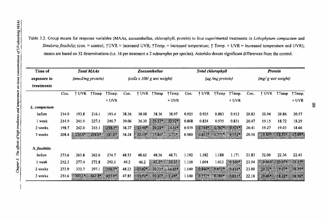
Con. t UVR tTemp ?Temp. Con. UVR ?Temp ?Temp. Con. t UVR ?Temp tTemp.
+ UVR + UVR + UVR
38.36 38.08
39.06 36.30
38.27 33.46*
38.24 30.35*
48.53 48.62
49.2 46.2
48.23 42.02*
47.85 35.59*
38.36 38.97 0.925 0.925 0.883
35.17* 32,92* - 0.868 0.834 0.935
20.28* 14.30* 0.939 V.744* 0,782*
17.86* 1,71* 0.980 0,437* 0,771*
48.36 48.71 1.192 1.182 1.188
43.5* 39.3* 1.150 1.094 1.013
30.71* 14.65* 1.140 0,866* 0.812*
10
I k4 1 1.140 1 0.572* 0,789*
0.912 20.83 20.94 20.86 20.57
0.831 20.47 19.15 18.72 18.29
20.41 19.27 19.03 18.66
20.56 .4 - *
22.00 22.36 22.43 1.171 21.83
0 86 22.04
23.00
22.18
4 1 -Jo .. i 1 ,to:; 1 Table 3.2. Group means for response variables (MAAs, zooxanthellae, chlorophyll, protein) to four experimental treatments in Lobophytum compactum and t 3
•
i Sinularia ilexibilis; (con. = control; t UVR = increased UVR; t Temp. = increased temperature; t Temp. + UVR = increased temperature and UVR);
.:1 ' .": I z I 1 Time of Total MAAs Zooxanthellae Total chlorophyll I exposure to (nmol/mg protein) (cells x 106/ g wet weight) (pg /mg protein) .Y. I treatments
means are based on 32 determinations (i.e. 16 per treatment x 2 subsamples per species). Asterisks denote significant differences from the control.
Protein
(mg/ g wet weight)
L. compactum
Con.
before 214.9
1 week 214.9
2 weeks 198.7
3 weeks 208.4
flexibills
before 273.6
1 week 252.3
2 weeks 273.9
3 weeks 251.6
t UVR tTemp tTemp.
+ UVR
193.8 216.1 193.4
241.9 227.5 246.7
262.0 245.1 ' 298.1*
0* 381.6* ..
263.8 262.6 274.7
277.4 272.8 292.1
332.7 297.1 366.1.'1 ' 1 8''
..z

Chapter 3. The effects of high irradiance and temperature on tissue concentrations of UV-absorbing MAAs
Effects of elevated temperature and UV on zooxanthellae densities. A decrease of
zooxanthellae densities with increasing time of exposure was discerned for both soft coral hosts
(Figure 3.4b, f). Prior to the start of the experiment, individual corals that had been randomly
assigned to the 4 treatment groups did not differ significantly in their zooxanthellae densities in
either soft coral species (average 38.36 (x106)± 1.29 SE and 48.58 (x106) ± 1.32 SE algal cells per gram coral wet weight in L. compactum and S. flexibilis respectively) (Tables .3.2/ 3.3).
Lobophytum compactum: After one week of exposure, significant loss of algal cells was found
in colonies submitted to the temperature treatment (Scheffe, p < 0.0001) and to an even greater
degree in colonies exposed to the combined temperature and UVR treatment (Scheffe,
p < 0.0001) (Figure 3.4b). The combined treatment resulted in significantly higher losses of
zooxanthellae (Scheffe, p = 0.0234) than the sole elevated temperature treatment, while UVR
alone did not result in any significant reduction of algal cells (Scheffe, p = 0.053) after one
week. After two weeks of exposure, all but the control colonies showed significant losses in
zooxanthella densities (Table 3.3.). UVR treatment alone had the mildest effect, sustaining a
reduction in zooxanthellae densities of 13% (Scheffe, p = 0.0084), followed by a 21% reduction
in the elevated temperature treatment (Scheffe, p < 0.0001) and a 62% reduction in the
combined treatment (Scheffe, p < 0.0001). While elevated temperatures appear to have a
greater effect than that of elevated UVR alone, zooxanthellae losses were not statistically
significant between the two treatments (Scheffe, p = 0.1519). After 3 weeks, effects of all
3 experimental treatments were greater, however, the ranking of effects remained the same
(Figure 3.4b). Corals submitted to the elevated UVR treatment alone showed 21% decreases in
zooxanthellae numbers (Scheffe p < 0.0001), temperature-treated colonies sustained losses of
53% (p < 0.0001) and the combined UVR and temperature treatment resulted in a near total loss
(96%) of the algal partner (Scheffe, p < 0.0001) (Tables .3.2/ 3.3). It is important to note that
the losses in the combined treatment were significantly greater (one-way ANOVA, F = 53.27,
df = 1, p < 0.0001) than the sum of losses in the separate elevated temperature and UVR treatments.
Sinularia flexibilis: A significant decrease in zooxanthellae densities was found in both the
temperature (Scheffe, p = 0.0006) and temperature and UVR combined treatment after one
week (Scheffe, p < 0.0001), while UVR alone did not affect zooxanthellae levels significantly
(Scheffe, p = 0.1327) (Figure 3.4f, Table 3.3). The combined manipulation resulted in
significantly greater losses (19 %) than the elevated temperature treatment (10 %) (Scheffe,
p = 0.0179). After 2 weeks all experimental treatments resulted in significant losses of the
algal partner from the association (Table 3.3.), losses averaging 13% in the UV treatment
(Scheffe, p = 0.0006), 37% in the temperature treatment (Scheffe, p < 0.0001) and 70 % in the
61

Chapter 3. The effects of high irradiance and temperature on tissue concentrations of UV-absorbing MAAs
combined treatment (Scheffe, p < 0.0001). As in L. compactum, after 3 weeks the UVR
treatment resulted in a moderate (25%) but significant decrease in zooxanthella levels (Scheffe,
p < 0.0001), while temperature and the combined UVR and temperature treatments resulted in
strong and significant losses of 48% and 79 % respectively (Scheffe, both p < 0.0001). Unlike
in L. compactum the effect of the combined manipulation (98 % loss) was not significantly
different than the sum of the UVR and temperature treatments (one—way ANOVA, F = 0.031,
df = 1; p = 0.8608).
Effects of elevated temperature and UV on chlorophyll concentrations. As with
zooxanthellae densities, chlorophyll levels decreased with ongoing exposure in all but the
control treatments in both L. compactum and S. flexibilis. Prior to the start of the experiment,
chlorophyll tissue concentrations averaged 0.924 ± 0.056 SE and 1.19 ± 0.067 SE tg per mg
protein in L. compactum and S. flexibilis respectively, and did not differ significantly between
individuals assigned to the 4 groups (Figure 3.4c, g; (Tables .3.2/ 3.3).
Lobophytum compactum: After two weeks all colonies in experimental treatments sustained
decreases in chlorophyll levels (Tables .3.2/ 3.3) although decreases were statistically
significant for only the UVR treatment (Scheffe, p = 0.0391) and the combined UV and
temperature treatment (Scheffe, p < 0.0001), with mean losses of 20% and 54% respectively
(Figure 3.4c). After three weeks, however, all but the control corals experienced significant
chlorophyll losses (Table 3.3) with the mildest loss (17%) caused by the temperature treatment
(Scheffe, p = 0.0040), followed by a 53% loss in the UVR treatment (Scheffe, p < 0.0001) and a
97% loss in the combined treatment (Scheffe, p < 0.0001). Reductions in chlorophyll levels
differed significantly among all treatments (Scheffe, p < 0.0001). After two and three weeks of
exposure, the combined UVR and temperature treatment resulted in a greater decrease in
chlorophyll concentration than the two single treatments summed together, the decrease due to
the synergistic interaction being statistically significant at two weeks (one-way ANOVA,
F = 32.61, df = 1, p < 0.0001), but not after in the three weeks (one-way ANOVA, F = 1.092,
df = 1, p = 0.300).
Sinularia flexibilis: Photosynthetic pigment concentrations decreased in all treatments but the
control after 1 week (Table 3.3.), yet, only the 27% loss in the combined UVR and temperature
manipulation was significant (Scheffe, p = 0.0059) (Figure 3.4g). The losses caused by the
temperature and UVR treatments reached 15% (Scheffe, p = 0.3894) and 8% (Scheffe,
p = 0.9175) respectively, but were not significantly different from the controls or from each
other (Scheffe, p = 0.999). A more pronounced reduction in chlorophyll levels was found after
2 weeks (Table 3.3.) with significant losses of 27%, 27% and 64% in comparisons to controls in
62

Chapter 3. The effects of high irradiance and temperature on tissue concentrations of UV-absorbing MAAs -------------
the UVR (Scheffe, p= 0.0021), the temperature (Scheffe, p = 0.0028) and the combined
treatment (Scheffe, p < 0.0001) respectively. While the UVR and the temperature treatments
had similar treatments effects, the combined treatment resulted in greater decreases in
chlorophyll than either of the others (Scheffe, p < 0.0001). After 3 weeks, all three treatments
led to strongly diminished chlorophyll levels (Table 3.3.), with the mildest reductions caused by
the temperature treatment (38%, Scheffe, p < 0.0001), followed by that of UVR treatment
(52%, Scheffe, p < 0.0001) and the strongest reductions experienced in the combined treatment
(97%, Scheffe, p < 0.0001). While UVR effects appear stronger than temperature effects
(Figure 3.4g), statistically, both treatments are not different (Scheffe, p = 0.113). The combined
UVR and temperature treatment caused significantly higher loss of chlorophyll when compared
to UVR (Scheffe, p < 0.0001) and temperature alone (Scheffe, p < 0.0001). Furthermore, as with chlorophyll decrease in L. compactum the combined treatment resulted in significantly
greater losses after 2 weeks than the sum of the single factor treatments together (one-way
ANOVA, F = 22.36, df = 1, p < 0.0001). After 3 weeks this over-additive effect was no longer
statistically significant (one-way ANOVA, F = 0.230, df = 1, p = 0.632).
Effects of elevated temperature and UV on protein concentrations. At the start of the experiment, protein concentration in the 4 treatment groups averaged 20 .85 ± 0.787 SE and
21.83 ± 0.617 SE mg per gram coral wet weight in L. compactum and S. flexibilis respectively and did not differ significantly among the four treatment groups (Tables .3.2/ 3.3; Figure 3.4d, h).
L. compactum: After two weeks of exposure, significant changes were detected (Table 3.3.). in
both the temperature (Scheffe, p = 0.326) and in the combined treatments (Scheffe, p = 0.0068)
with losses of 9% and 11% respectively (Figure 3.4d). Elevated UVR resulted in protein losses
of 8% but these were not significant (Scheffe, p = 0.0766). Three weeks of exposure led to
significantly decreased protein levels in all three treatments (Table 3.3.), with 10% reductions
in the UVR treatment (Scheffe, p = 0.0138), 11% reductions in the temperature treatment
(Scheffe, p = 0.0029) and 15% reductions in the combined treatment (Scheffe, p < 0.0001) (Figure 3.4d).
S. flexibilis: In contrast to L. compactum, exposure of one week brought about significant
decreases in the temperature and combined treatment in S. flexibilis (Scheffe, p = 0.0172,
p < 0.0001 for the former and latter treatment respectively) (Figure 3.4h, Table 3.3.). The 8%
decrease in the UVR treatment was not significant (Scheffe, p = 0.090). After two weeks, all
experimental manipulations but the controls caused significant decreases of 7% (Scheffe,
63

Chapter 3. The effects of high irradiance and temperature on tissue concentrations of UV-absorbing MAAs
p = 0.0094), 13% (Scheffe, p < 0.0001) and 14% (Scheffe, p < 0.0001) in the UVR,
temperature and combined treatment respectively. After three weeks of exposure losses of
11%, 17% and 16% (all p < 0.0001) were sustained in the UVR, temperature and combined
treatment respectively. Temperature and the combined treatment did not differ in their capacity
to decrease protein in week 2 (Scheffe, p = 0.6011) or week 3 (Scheffe, p = 0.999). The effects
of the combined treatments on protein concentrations were not found to be synergistic.
3.4.2. Controls of all parameters (experimental controls)
Lobophytum compactum and Sinularia flexibilis colonies held as experimental controls in
ambient seawater underwent no significant release of zooxanthellae (one-way ANOVA,
F = 0.0118, df = 3, p = 0.998 for L.c.; F = 0.2069, df = 3, p = 0.8914 for [), nor changes in
chlorophyll (F = 1.046, p = 0.374; for L.c; F = 0.8235, p = 0.302 for U..), protein (F = 0.541, p = 0.7204 for L.c - F= 0.910, p = 0.179 for S.f) or MAA concentrations (F = 0.7958, p = 0.341 for L.c- F = 0.7197, p = 0.447 for .S,L) during the 3 week treatment period. Furthermore,
experimental controls showed no significant differences at any time in comparison to handled
(tank controls) and unhandled field controls in any of the parameters assessed (Table 3.4).
Table 3.4. Results of one-way ANOVA (df = 1) comparing experimental controls of each soft
coral species with handled (tank controls) and unhandled field controls with respect to
their zooxanthellae densities and chlorophyll, protein and MAA levels. For field
controls, n = 16 determinations (i.e. 8 colonies x 2 subsamples), whereas for tanks
controls n = 32 determinations (i.e. 16 colonies x 2 subsamples).
Parameter Unhandled field controls Handled field controls L. compactum F-ratio p-value F-ratio p-value Zooxanthellae 0.1639 0.6875 0.0095 0.922 Chlorophyll 0.1863 0.6680 0.0342 0.854 Protein 0.4877 0.4885 0.0033 0.954 MAAs 0.0234 0.8791 0.0499 0.833 S. flexibilis
Zooxanthellae 0.1639 0.6875 0.0095 0.922
Chlorophyll 0.1863 0.6680 0.0342 0.854
Protein 0.4877 0.4885 0.0033 0.954
MAAs 0.0234 08791 0.0499 0.833
64

Chapter 3. The effects of high irradiance and temperature on tissue concentrations of UV-absorbing MAAs
3.43. Relationships between the biochemical parameters
Zooxanthellae levels in both species were found to be significantly and inversely correlated to
MAA levels (Table 3.5). The nature of protein and MAA concentrations was similarly negative
in both L. compactum and S. flexibilis, with increasing MAA concentrations with decreasing
protein levels. With ongoing loss of zooxanthellae, protein levels decreased significantly in
either species, thus resulting in a positive relationship between the two parameters.
Table 3.5. Regression analyses assessing the relationships between: 1) zooxanthella densities
and MAA tissue concentrations, 2) protein concentrations and MAA tissue levels and
3) protein concentrations and zooxanthella levels. Asterisks denote significant
relationships (cc = 0.05) while R2 denotes the coefficient of determination, n = 512
determinations (i.e. 128 colonies x 4 sampling times).
Parameter Lobophytum compactum F-ratio p-value relationship R2
zooxanthellae/MAAs 81.43 < 0.000i* y = 364.13-3.7205x 0.138 protein/MAAs 11.15 0.0008* y = 412.61-9.0766x 0.108 protein/ zooxanthellae 39.33 < 0 0001* y = 11.239+0.2707x 0.370
Sinularia flexibilis
zooxanthellae/MAAs 89.33 < 0.0001* y = 414.49-2.8979x 0.149 protein/MAAs 17.78 < 0.0001* y = 437.30-7.0029x 0.088 protein/ zooxanthellae 85.74 < 0 0001* y = 11.358+0.11936x 0.391
3.4.4. Analysis of MAA levels in naturally bleached and unbleached soft corals
1. Comparison biochemical parameters between unbleached and bleached soft corals
during the 1998 mass bleaching event
The bleaching episode in the summer of 1998 provided an ideal opportunity to test the
relationship between bleaching as a result of a natural thermal disturbance and MAA levels in
soft coral tissues. Analysis of tissue samples collected from naturally bleached soft corals
following the thermal disturbance in the summer of 1998 shows that bleached colonies of
Lobophytum compactum, Sinularia flexibilis, Sinularia capitalis and Sarcophyton sp. were
characterised by increased MAA concentrations when compared to their unbleached
counterparts. Conversely, chlorophyll, carotenoid and protein levels were lower in bleached
than in unbleached colonies (Figure 3.5).
65

>1 CL 0 O C)
O
0. bA
20
0.) 40
60
chlorophyll
0
800 MAAs
protein
0-
600 -
a) 400 - w Sa.
0 tap
75, E 200 -E
0
30 —
d) carotenoid (peridinin)
o bleached unbleached
0 Lobophytum compactum
Sinularia Sinularia Sarcophyton flexibilis capitalis sp.
400 -
Chapter 3. The effects of high irradiance and temperature on tissue concentrations of UV-absorbing MAAs
Figure 3.5. Comparison of biochemical parameters between bleached and unbleached colonies
of four soft coral species collected during a natural thermal bleaching event: a)
chlorophyll; b) MAAs; c) protein and d) carotenoid (peridinin) levels. Error bars denote
standard errors (n = 14 determinations, i.e. 7 colonies x 2 subsamples). The depth-adjusted MAA level in unbleached L. compactum (b) is shown in the dashed column.
Statistical analyses confirmed that bleached and unbleached colonies of the four soft coral
species sampled, differed significantly in their zooxanthellae, chlorophyll, protein and MAA tissue levels (Mann-Whitney tests) (Table 3.6)
66

abso
rptio
n a t
320
nm
y = 3.0617 - 2.4104 x log(x) R Z = 0.753
X X X x
X W
2.5
2.0 -
1.5 -
1.0 -
0.5 -
0.0
Chapter 3. The effects of high irradiance and temperature on tissue concentrations of UV-absorbing MAAs
Table 3.6. Results of Mann-Whitney tests, comparing biochemical parameters between
bleached and unbleached conspecifics of 4 soft coral species sampled during the 1998
natural bleaching event. Asterisks denote significant differences, n = 14 determinations
(i.e. 7 colonies x 2 subsamples per species).
Parameter Species L. compactum S. flexibilis S. capitalis Sarcophyton sp.
chlorophyll <0.0001* <0.0001* 0.00017 <0 .w.) 1 * MAAs 0.0011* <0.0001* 0.0308* 0.0067* protein <0.0001* <0.0001* <0.0001* <0.0001* carotenoid <0.0001* <0.0001* <0.0001* 0.0001*
While the results of the Mann-Whitney tests clearly provide support for significant differences
between bleached and unbleached conspecifics of all four species for each of the parameters
assessed, MAA comparisons are possibly confounded by the fact that they were collected at
different depths and thus can have naturally different levels of MAAs. To correct for depth-related effects on MAA concentration, I calculated the relationship between depth and MAA
concentrations (prior to the bleaching episode) and found a highly significant inverse relationship, for colonies of Lobophytum compactum in Pioneer Bay (regression analysis, F= 117.40, p < 0.0001) (Figure 3.6). The decline in MAAs with depth in unbleached colonies
parallels reductions in light levels experienced at 6m depth, which equate to 42% less fluxes in
the PAR region and 45% and 47% for the UV-A and UV-B regions respectively (calculations based on data provided by Walt Dunlap).
0 3 6 9
12
15 depth (m)
Figure 3.6. Relationship between depth and MAA concentrations in Lobophytum compactum at Pioneer Bay prior to the bleaching episode (January 1998; n = 5 per depth).
67

Chapter 3. The effects of high irradiance and temperature on tissue concentrations of UV-absorbing MAAs
From the regression relationship between MAA level and depth, I calculated that individuals
living at 6 m have MAA tissue concentrations approximately 48% below those of their
conspecifics at 2 m at the study site. In order to compare the naturally bleached Lobophytum compactum colonies (all collected around 2 m) with the unbleached ones (collected around
6 m), the levels of the deeper colonies had to be adjusted by 48% prior to comparison. Also, in
order to test whether increases in MAA concentrations in L. compactum represented true up-regulation, or only a reflection of reduced protein levels following bleaching, concentrations
were then re-normalised to mean protein levels of the unbleached conspecifics. The analysis
(Mann-Whitney tests) revealed that bleached L. compactum had significantly higher MAA tissue concentrations (p = 0.0432) when compared to the tissues of their unbleached
counterparts corrected for both depth and protein loss. No comparisons using depth-adjusted
MAA levels were carried out for the other three soft corals, since their relationships between
depth and MAA levels are not known.
2. Comparison of MAA levels in 1998 with those of previous years
In another approach to establish whether increases in MAA concentrations are an active
response to bleaching, I compared MAA concentrations in tissues collected during the April
1998 natural bleaching event, with concentrations in tissue samples collected at the same depth
close to the study site at the same time of the year in the two previous years (see Chapter 2).
MAA levels were approximately 79.5% and 63.1% higher in bleached samples of Lobophytum compactum and Sinularia flexibilis collected in April 1998, compared to levels in unbleached colonies collected in previous years (Figure 3.7).
68

35 -
30 - a)
1.1.g c
hlor
ophy
ll/ g
wet
we i
ght
25 -
5-
0
600
nmol
MA
As /
mg
p rot
ein
0 Lobophytum compactum Smularia flexibilis
20
15
10 -
400
200
Chapter 3. The effects of high irradiance and temperature on tissue concentrations of UV-absorbing MAAs
J Mar-96
Apr-96
D Mar-97
Apr-97
0 Apr-98
Figure 3.7. Comparison of a) chlorophyll and b) MAA levels in unbleached and bleached conspecifics of L. compactum and S. flexibilis. Data collected during March 1996 to April 1997 represent concentrations under non-bleaching conditions, whereas data
collected in April 1998 represent bleached conditions. Each sampling period in
1996/1997 comprises n = 40 determinations (i.e. 10 colonies x 4 subsamples) whereas
in April 1998, n = 14 determinations (i.e. 7 colonies x 2 subsamples).
In both L. compactum and S. flexibilis the higher concentrations of MAAs were statistically
significant (Mann-Whitney test, p = 0.0168 and p = 0.0021 respectively), while at the same
time chlorophyll levels were significantly decreased (Mann-Whitney test p = 0.0001 and p = 0.0001 respectively). Regression analyses confirmed for both species that the inverse
relationship between MAA and chlorophyll concentrations is statistically significant (F = 9.87,
p = 0.0029 and F = 16.32, p = 0.0001 for L. compactum and S. flexibilis respectively). MAA concentrations re-normalised to pre-bleaching protein levels were still significantly higher in
the bleached than in unbleached colonies of L. compactum (Mann Whitney test, p < 0.05) and S. flexibilis (Mann Whitney test p < 0.05).
69

Chapter 3. The effects of high irradiance and temperature on tissue concentrations of UV-absorbing MAAs
3.4.5. Do higher MAA levels protect against bleaching?
I. Effects of translocation on zooxanthella, MAA, chlorophyll and protein levels Colonies of L. compactum responded to translocation from 4 to 15 m with a 43% decrease in
MAA levels (from 303.93 ± 20.46 to 173.42 ± 11.25 nmol/ mg protein, one-way ANOVA,
df = 1, F = 37.06, p < 0.0001), however, protein and zooxanthellae levels did not change
significantly (one-way ANOVA, df = 1, F = 1.89, p = 0.174, and F = 0.11, p = 0.747
respectively). A significant acclimatory change was detected in chlorophyll concentrations
which increased by approximately 63% from 0.99 ± 0.047 to 1.56 ±0.06 g/ mg protein (one-
way ANOVA, df = 1, F = 52.27, p = 0.0001).
2. Effects of solar and thermal treatment on low and high MAA adjusted conspecifics
MAA level and experimental treatment both had highly significant impacts as single factors
with respect to zooxanthellae and chlorophyll loss in L. compactum (3-factorial ANOVA, Table
3.7). More importantly, a first order interaction between the two factors occurred for the
zooxanthella analysis, which means that the impact of the treatment (i.e. the degree of
bleaching) is dependent on the MAA level. No significant tank effects were discerned, i.e. the
use of tanks had no effects on the impact of any of the experimental treatments or the MAA
level (Table 3.7).
Table 3.7. Summary of results of 3-factorial nested ANOVA, testing the effects of MAA-level
and experimental treatment (fixed and orthogonal factors) and the nested factor tank, on
zooxanthellae and chlorophyll levels. Asterisks denote significant differences from the
control (x= 0.05); (n = 16 determinations, i.e. 8 colonies per treatment x 2 subsamples).
Source of variation
MAA-level
experimental treatment
tank
MAA-level x experimental treatment
Zooxanthellae Total chlorophyll
df F-ratio p-value F-ratio p-value
1 46.59 <0.0001* 7.83 6.007*
3 117.04 .--0 0001* 22.41 *
1 0.08 0.785 0.02 0.857
3 10.52 <0 0001* 1.74 0.172
70

0
c.)
0 0 N
. 6
01)
50 -
40
30
20
10 -
0
a) Zooxanthellae densities
b) Chlorophyll concentrations
a. o 0 0
eto cif 0
lig high MAA acclimatised
0 low MAA acclimatised
control solar temperature temperature +solar
Chapter 3. The effects of high irradiance and temperature on tissue concentrations of UV-absorbing MAAs
Effects of experimental treatments on zooxanthellae levels in low and high MAA acclimatised counterparts.
For comparison of the effects of each of the experimental treatments between the high and
low-MAA acclimatised colonies, one-factorial ANOVA were used. In both high and low-MAA
acclimatised corals, all but the control treatments did have a significantly decreasing effect on
zooxanthellae densities (one—factorial ANOVA, df = 3, F = 35.79, p < 0.0001 and F = 89.02,
p < 0.0001 for the former and latter group respectively; Figure 3.8).
treatment
Figure 3.8. Bleaching response quantified as loss of a) zooxanthellae and b) chlorophyll levels of high and low-MAA acclimatised L. compactum conspecifics after 10-day exposures
to increased temperature, solar radiation or a combination of both factors. Error bars
denote standard error, n = 16 determinations, i.e. 8 colonies x 2 subsamples per
treatment. Asterisks denote a significant difference in bleaching response between the
two groups.
71

Chapter 3. The effects of high irradiance and temperature on tissue concentrations of UV-absorbing MAAs
While low-MAA acclimatised control corals had undergone a limited (6%) loss of their algal
partners by the end of the 10 day treatment, zooxanthellae densities of high-MAA and low-
MAA acclimatised control corals did not differ significantly from each other (Scheffe,
p = 0.3494). Low-MAA-acclimatised conspecifics, however, did undergo a significantly higher
loss of zooxanthellae in the solar treatment (Scheffe, p = 0.0391) in comparison to their high-
MAA adapted counterparts (Figure 3.8a). A similar but more pronounced trend was found in
the combined UV solar and temperature treatments, where high-MAA adapted test corals
showed a significantly lower bleaching response after 10 days than low-MAA adapted corals
(Scheffe, p = 0.0030). Both high and low adapted conspecifics, exhibited statistically similar
bleaching responses when submitted to the increased temperature treatment, with losses of
57.23 ± 4.48 % in the former and 65.16 ± 5.37 % in the latter group (Scheffe, p = 0.5463) (Figure 3.8a).
Effects of experimental treatments on chlorophyll concentrations in low and high MAA acclimatised counterparts. As found for zooxanthellae densities, chlorophyll levels did not
differ significantly between corals maintained as low-MAA and high-MAA acclimatised control (Scheffe, p = 0.999) (Figure 3.8b).
Colonies with lower MAA levels, however, did undergo a significantly higher loss of
chlorophyll in the solar treatment (Scheffe, p = 0.0388) than their high-MAA adapted
counterparts (Figure 3.8b). As with zooxanthellae densities, chlorophyll levels in the combined
UV solar and temperature treatments decreased in a more pronounced trend than in the solar
treatment alone, where high-MAA adapted test corals showed a significantly lower bleaching
response after 10 days than low-MAA adapted corals (Scheffe, p = 0.0109). Both high and low
adapted colonies bleaching responses, statistically not different from each other when submitted
to the increased temperature treatment (Scheffe, p = 0.999) (Figure 3.8b).
72

Chapter 3. The effects of high irradiance and temperature on tissue concentrations of UV-absorbing MAAs
3.4.6. Summary of all results gained from experimentally and naturally bleached soft corals
Question: Are MAAs thermolabilie, i.e. do MAA levels decrease during thermal stress ?
Experimentally induced thermal
bleaching (section 3.4.1)
No
In contrast: increased MAA levels during thermal stress
Comparison between unbleached
and bleached colonies affected by the 1998 mass bleaching event (section 3.4.2/1)
No
In contrast : MAA levels higher in bleached than unbleached colonies
Comparison between 1998 bleached
corals and unbleached corals in previous years (section 3.4.2/3)
No
In contrast : MAA levels higher in bleached than unbleached colonies
Question: Do MAAs protect against thermal stress, i.e. do higher MAA levels translate into less bleaching ?
Experimentally induced thermal bleaching in high
and low-MAA acclimatised
corals (section 3.4.5)
No
Experimentally induced solar
bleaching in high and low-MAA acclimatised
corals (section 3.4.5)
Yes
Experimentally induced
thermal + solar bleaching in high
and low-MAA acclimatised
corals (section 3.4.5)
Yes
The ecological benefit of higher MAA investment is realised when bleaching is induced by solar stress or
combined solar+thermal stress
Figure 3.9. Overall summary of results of Chapter 3.
73

Chapter 3. The effects of high irradiance and temperature on tissue concentrations of UV-absorbing MAAs
3.5. DISCUSSION
This study shows clearly that temperature and UVR have the capacity to act synergistically,
resulting in a greatly increased bleaching response in Lobophytum compactum and Sinularia flexibilis when both factors occur simultaneously. The key to understanding this synergy,
however, is not as previously assumed the degradation of MAAs (Lesser, 1990; Glynn, 1993),
given that the compounds do not appear to be thermo-labile (within the range of temperature
examined here). In fact, significant increases in MAA concentration were found in
experimentally and naturally bleached soft corals that had been exposed to increased
temperature. Moreover, the study with high and low-MAA acclimatised soft corals provides
first evidence that metabolic investment into increased MAA production in soft corals translates
into reduced bleaching, when a coral is exposed to high irradiance and combined high
temperature and irradiance conditions.
3.5.1. The experimentally induced bleaching response in the soft corals Lobophytum compactum and Sinularia flexibilis
Exposure to elevated temperatures (32° C) between 1 and 2° C over summer maxima, (Chapter
2, Figure 2.9) clearly has the potential to cause severe bleaching via loss of zooxanthellae in both Lobophytum compactum and Sinularia flexibilis. Comparable temperature thresholds for
inducing bleaching have been reported for a number of scleractinian corals (Jokiel and Coles,
1978; Glynn and D'Croz, 1990; Hoegh-Guldberg and Salvat, 1995; Berkelmans and Willis,
1999). In this study, the loss of zooxanthellae progressively increased in a time-dependent
fashion in both species. Yet, Sinularia flexibilis is clearly more susceptible to increased temperature losing of 78% of its zooxanthellae population after 3 weeks, whereas Lobophytum compactum lost only 53% of its algal standing stock in this period. Greater thermal susceptibility of Sinularia flexibilis experimentally is consistent with field observations during
the 1998 bleaching event, in which many colonies of Sinularia flexibilis were already "bone-white" when Lobophytum compactum at the same depths were still light brown in colour.
Marked species-specific susceptibility to thermal stress has been observed frequently (Brown
and Suharsono, 1990; Gleason, 1993; Hoegh-Guldberg and Salvat, 1995; Marshall and Baird,
1999), and has been suggested to be based on differential thermal susceptibility of the algal
symbiont (Warner et al., 1996) or the host itself (Edmunds, 1994). The differences in bleaching
susceptibility observed in my study, however, could also be based on the fact that Lobophytum compactum and Sinularia flexibilis feature very different tissue characteristics. While the
former species has a thick leathery coenenchyme, into which the polyps can fully retract (which
74

Chapter 3. The effects of high irradiance and temperature on tissue concentrations of UV-absorbing MAAs
provides shading for thermally stressed zooxanthellae), the latter species has a comparatively
thinner coenenchyme, leaving zooxanthellae more exposed to light. Differences in tissue
thickness and hence shading, have also been suggested to be the underlying reason for the
observed differences in bleaching susceptibility between Acropora spp. and Pocillopora spp. (reviewed in Hoegh-Guldberg, 1999).
Increases of 21% and 17% over summer maxima in UV-B and UV-A also caused time-
dependent decrease in symbionts, yet with losses of only 21% and 25 % of the algal standing stock in Lobophytum compactum and Sinularia flexibilis respectively, UVR treatment effects were much lower than temperature treatment effects. Thus, unlike in their thermal
susceptibility, the two species were approximately equivalent in their susceptibility to UVR
(Figure 3.4). If differences in tissue thickness were also to determine the hosts' susceptibility to
solar stress, one would expect Lobophytum compactum also to be less susceptible to bleaching than Sinularia flexibilis. Other factors, such as the genetic variability of the host (Edmunds,
1994) or that the algal symbiont (Rowan et al., 1997), and thus potential differential
susceptibility to UVR, are possibly responsible the observed differences in the (irradiance-induced) bleaching response of Lobophytum compactum and Sinularia flexibilis.
Elevated temperatures and UVR were equally potent in their capacity to reduce chlorophyll
levels in zooxanthellae after 2 and 3 weeks of exposure in both Lobophytum compactum and Sinularia flexibilis. The degradation of chlorophyll after one week, (with overall losses rising
to 36% and 35% in UVR treated colonies and 21 % and 47% in temperature treated colonies after 3 weeks in L. compactum and S. flexibilis respectively), is in agreement with a number of
other studies. Degradation of algal chlorophyll under conditions of high irradiance (Hoegh-
Guldberg and Smith, 1989a; Lesser, 1990; Kinzie, 1993; Ambarsani et al., 1997; Jones, 1997;
Salih et al., 1998) and increased temperature (Iglesias-Prieto et al. 1992; Warner et al. 1996) has
been widely documented for other dinoflagellate-cnidarian symbiotic associations.
3.5.2. Effects of synergistic interactions between high irradiance and temperature on
zooxanthellae and chlorophyll level
The interactive effects of UVR and temperature have been recognised as early as 1978 (Coles
and Jokiel), and several subsequent studies have shown a greatly enhanced capacity for
bleaching when both factors occur together (Reaka-Kudla, 1993; Denis and Wicklund, 1993;
Lesser et al., 1990; Drollet et al., 1997). While most studies have used the term synergy, none
have compared the combined treatment response to the sum of individual treatment responses,
75
Chapter 3. The effects of high irradiance and temperature on tissue concentrations of UV-absorbing MAAs
which should be significantly higher in a synergistic interaction. Thus, I believe the
significantly greater loss in both zooxanthellae numbers and chlorophyll concentrations
recorded in this study, when elevated temperature and UVR occurred in combination compared
to the sum of losses when these two factors were elevated singly, represent the first records of
truly synergistic with respect to bleaching. The synergistic response was observed for both
Lobophytum compactum and Sinularia flexibilis, both on the level of zooxanthellae and
chlorophyll loss. These findings can be readily explained by the light-temperature interaction
model proposed by Jones et al. (1998), which suggests that thermally stressed zooxanthellae are
more light-sensitive which subsequently leads to increased bleaching. Although synergistic
interactions may not occur under all circumstances they may explain the intensity of some of
the bleaching events for which temperature has been ruled out as a sole causative factor
(Atwood, 1992). For example, the severity of the 1998 mass bleaching event in the GBR, in
which bleaching was evident on 87% of inshore reefs along the coast has been suggested to be
based on a combination of stress factors (hypo-salinity and elevated temperature and irradiance)
(Berkelmans and Oliver, 1999).
3.5.3. Are MAAs the key to understanding the observed synergistic interaction?
Experimental exposure to elevated UVR significantly increased MAA concentrations in both Lobophytum compactum and Sinularia flexibilis in a time-dependent fashion as has been shown
in a number of studies for hard corals (Jokiel and Coles, 1982; Kinzie, 1993; Gleason and
Wellington, 1993). Most noteworthy, however, was that the highest increases occurred in the
combined temperature and UVR treatment in both soft coral species (Figure 3.4.a, e). To date
there is no evidence available to support that increased temperature can trigger the production
of MAAs. In contrast, my initial assumption was that MAAs may be thermo-labile, thus are
possibly chemically degraded during exposure to increased temperatures (Lesser et al., 1990;
Glynn et al., 1992). Under this scenario, a coral experiencing thermal stress would be
additionally exposed to UVR stress via degradation of its UV protection — a possible basis for
the synergistic bleaching response. I found no evidence of degradation of MAAs in colonies
exposed to the temperature treatment or the combined temperature and UVR treatment,
however, suggesting that degradation of MAAs and subsequent increased exposure to solar
stress is not the key to understanding the synergistic interaction between light and elevated
temperature, at least for temperatures (32° C) used in experimental manipulations in this study.
The model proposed by Jones et al. (1998), which suggests that increased light-sensitivity
occurs in thermally stressed zooxanthellae thus provides, to date, the best understanding for the
synergy between light and elevated temperature in the bleaching process.
76

Chapter 3. The effects of high irradiance and temperature on tissue concentrations of UV-absorbing MAAs
3.5.4. Up-regulation of MAAs in response to thermal stress
Three independent investigations of MAA levels in response to thermal stress indicate that
MAAs are not thermo-labile, but are actively up-regulated under thermal stress, and even more
when thermal and irradiance stress occur simultaneously. Increases in MAA concentrations in
soft corals exposed to elevated temperatures are intriguing, since to date MAAs have not been
thought to provide protection against thermal stress. The possibility that the observed increases
in MAA levels were the result of a dietary uptake during the experimental treatment, was
dismissed on the grounds that no changes in MAA concentrations occurred in the field or tank
controls during the 3 week treatment period (Table 3.4). Significant increases in MAA levels
re-normalised to pre-treatment protein concentrations confirmed that increases were not merely
a reflection of loss of protein levels, experienced in all bleached colonies, presumably due to
host cell detachment (Gates et al., 1992). Therefore increases in MAAs in the experimentally
elevated temperature treatment provided first evidence that active up-regulation of MAAs
occurs in response to thermal stress. Secondly, this was further corroborated by my findings of
increased MAA levels in bleached soft corals (in comparison to their unbleached conspecifics;
Figure 3.7. and Table 3.6), which were sampled during the mainly temperature driven GBR
bleaching event in summer of 1998 (Berkelmans and Oliver, 1999). Thirdly, more
confirmatory support for the theory of MAA up-regulation under thermal stress was derived
from comparisons between bleached colonies of both L. compactum and S. flexibilis, which had approximately 70% higher levels than their unbleached conspecifics collected in the appropriate
season and depth in previous years (Figure 2.9.).
While my results, both from the experimentally induced and natural bleaching episodes
demonstrated that bleached corals feature higher MAA levels than their unbleached
counterparts, the question remained whether higher investment into MAA production also
translated into increased protection against bleaching. Given the direct UV protecting function
that has been shown for MAAs (Garcia-Pichel et al., 1993) and the increased light sensitivity of
thermally stressed zooxanthellae (Jones et al., 1998), I expected some ecological advantage
(i.e. less bleaching) to compensate for the investment into MAA production. This was
confirmed by my findings that high MAA-acclimatised Lobophytum compactum experienced
less bleaching when exposed to the elevated solar irradiance or the combined solar irradiance-
temperature treatments than their low MAA-acclimatised counterparts. This strongly suggests
that an ecological benefit is connected to investment into MAA production. The benefit
provided by MAAs appears, however, to be only realised, when (elevated) irradiance is
involved in the bleaching process, since both high and low MAA acclimatised corals responded
equally strongly to thermally induced bleaching. Thus, while MAAs do not protect against
77

Chapter 3. The effects of high irradiance and temperature on tissue concentrations of UV-absorbing MAAs
bleaching, when solely induced by thermal stress, in interaction with high solar irradiance they
reduce loss of zooxanthellae significantly. This suggests some, yet to be established, interaction
of MAAs with other cellular components at the level of light and temperature induced molecular damage.
In summary, in addition to the bulk of indirect evidence supporting the UV protecting role of
MAAs (reviewed in Dunlap and Shick, 1998), this is first direct evidence that MAAs have the
capacity to reduce the bleaching response in corals under high solar irradiance conditions. It is
important to note, however, that irrespective of the experimental treatment, the presence of
MAAs in the coral tissues did not completely prevent bleaching in any group, which is
consistent with findings that MAAs prevent only about 20-30% of the UVR reaching cellular
targets (Garcia-Pichel, 1993).
3.5.5. Possible regulation of MAA induction
Given that bleaching is typically associated with reduced photosynthetic rates (Glynn, 1993,
Kinzie, 1993) and subsequent substantial loss of lipid reserves (Porter et al. 1989; see Chapter
4), it seems paradoxical to invest extra resources towards protection against a stress factor,
other then the one experienced at that particular moment. Typically under thermal stress,
energy is diverted to the expression of another part of the sub-cellular response system, the heat
shock proteins (hsps) (Sharp et al., 1994; Black et al., 1995). Hsps have been implicated in
various processes concerning avoidance and repair of protein damage in cnidarians, suggesting
they play an important role in the thermo-tolerance of different species. Recently, however, the
induction of heat-shock proteins has been found to be also activated by ultraviolet radiation in
cell lines (Ohnishi et al, 1996). Given that incident solar fluxes and high sea surface
temperatures typically occur simultaneously, selection has possibly not discriminated between
the two environmental variables. This non-discrimination has also been suggested to be the
underlying mechanism resulting in MAA induction by UVR, PAR and water flow (Jokiel et al.,
1997), the authors speculating that MAA production could be increased via flow-modulated
increase in photosynthesis and thus carbon availability for the synthesis of the compounds.
Therefore a variety of regulators may be involved in the control MAA synthesis. Alternatively,
resource allocation towards UV protection at a time when strong thermal stress is experienced,
can be interpreted as a preventive strategy, designed to avoid a potentially lethal combination of
stresses. The combination of sublethal UV-A and temperature stress, for example, has been
shown to result in lethal consequences for cichlid fishes when administered simultaneously
(Winckler and Fidhiany, 1996). Given that corals live so precariously close to their upper
78

Chapter 3. The effects of high irradiance and temperature on tissue concentrations of UV-absorbing MAAs
thermal limits (Coles and Jokiel, 1978; Jokiel and Coles, 1977; 1990; Berkelmans and Willis,
1999) a slight increase in solar radiation (as e.g. experienced under doldrum conditions) could
prove similarly fatal, and can possibly be prevented by up-front investment into additional UVR
protection. Finally, it can not be excluded that MAAs are non-specific, multi-functional
compounds, which are produced in response to a variety of environmental stresses. Increases in
MAA tissue concentration in response to mechanical stress and breakage have been reported in Montipora verrucosa (Scelfo, 1986) and Zoanthus pacificus (Scelfo, 1985). Scelfo (1985)
interpreted immediate MAA amplification upon breakage as a feasible adaptation to possible
storm damage, which could transport colony fragments to high-light environments. While all
three hypotheses presented here (i.e. unknown regulators of MAA synthesis; thermally induced
MAA synthesis to avoid combinations of stresses and up-front investment) are speculative, each
is consistent with my findings of highest investment into production of MAAs in response to
combined solar irradiance and temperature treatment.
3.5.6. Summary and conclusions
My results suggest that MAAs are not thermolabile, which means that the degradation of the
compounds is not the key to understanding the synergistic relationship between irradiance and
elevated temperature in the bleaching process. Converse to the notion of depressed MAA levels under thermal stress, both Lobophytum compactum and Sinularia flexibilis actively up-regulated their MAA levels in response to elevated temperatures and solar irradiance alike. While
investment into MAAs does not confer an advantage when bleaching is induced solely by
elevated temperature, when high irradiance and temperature act as stress factors simultaneously,
increased MAA levels translate into higher protection against bleaching. Hence, the cost-to-
benefit-ratio becomes positive for the coral not only under high irradiance situations, but also
when exposed to increased temperature.
79
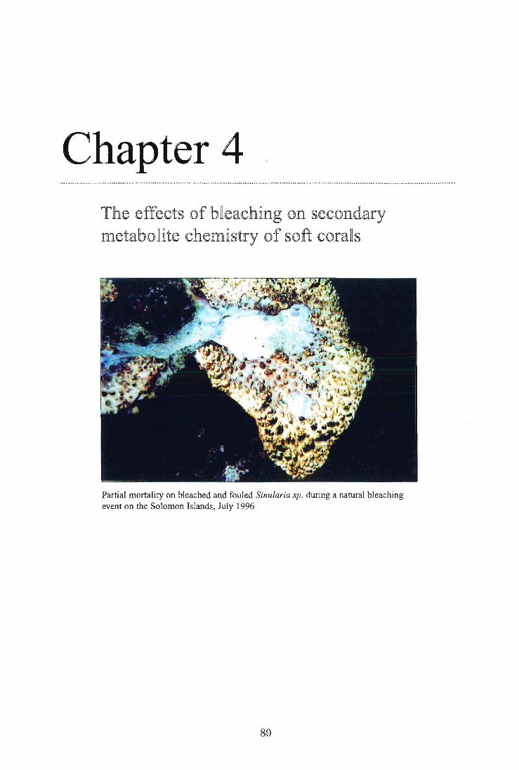
Chapter 4 T of effects of bleac ling on secondary metabolite chemistry of so -t corals
Partial mortality on bleached and fouled Sinularia sp. during a natural bleaching event on the Solomon Islands, July 1996
80

Chapter 4: The effects of bleaching on the secondary metabolite chemistry of soft corals
4.1. ABSTRACT
The loss of zooxanthellae during a short term, experimentally simulated bleaching event resulted in significant changes to the secondary metabolite chemistry of the alcyonacean soft corals Sinularia flexibilis and Lobophytum compactum. The concentration of flexibilide, the most active cytotoxic secondary metabolite (with anti-microbial properties) in the tissues of Sinularia flexibilis increased by 126%, while that of its principal terpenoid algaecide, sinulariolide, simultaneously decreased to just 8% of that found in the controls. The changes were short-lived, however, with concentrations of both compounds returning to control levels one month after bleaching, although zooxanthellae levels of experimentally bleached colonies were still only approximately 20% of controls. Similarly, concentrations of isolobophytolide, the principal terpenoid secondary metabolite of Lobophytum compactum, underwent a significant reduction for one month following bleaching. Like their non-bleached counterparts, bleached individuals of both Sinularia flexibilis and Lobophytum compactum experienced neither significant algal overgrowth nor predation following bleaching. Full recovery of zooxanthellae to baseline levels in all bleached corals occurred within four months. These results suggest that some soft corals are capable of surviving short-term bleaching events and the detrimental algal overgrowth that is often associated with bleaching, possibly by regulating their secondary metabolite chemistry to counteract fouling. Support for the notion that resource allocation towards production of specific algaecides in bleached specimens translates into higher protection, was gained through analysis of naturally bleached soft corals. Bleached and overgrown individuals had significantly lower concentrations of species-specific algaecides than either their unbleached or bleached but unfouled conspecifics.
81

Chapter 4: The effects of bleaching on the secondary metabolite chemistry of soft corals
4.2. INTRODUCTION
Bacterial and algal fouling have been reported to pose some of the greatest threats to corals weakened by bleaching (Nair et al., 1987; Kushmaro et al., 1996, Fabricius, 1999). Algal
fouling (Figure 4.1a - d) has the potential to reduce the coral's feeding capacity and compete with the coral for resources, and hence affect growth and ultimately survival rates
(Coll et al., 1987; Leone et al., 1995).
Figure 4.1. Progression of algal overgrowth on various bleached soft corals in the wake of
the 1998 mass-bleaching event. a). Algae colonising the tips of bleached colonies of
Sinularia sp.; b) Increased algal overgrowth on larger parts of colonies of bleached
Sinularia sp.; c) Algae firmly engulf substantial parts of bleached Sinularia sp.; d)
Tissue disintegration and death of bleached and overgrown Sinularia sp. (photo
courtesy K. Fabricius).
82

Chapter 4: The effects of bleaching on the secondary metabolite chemistry of soft corals
Colonisation of corals by certain bacterial strains has been shown to be a normal part of reef ecology and is partially controlled by regular mucus sloughing (Ducklow, 1979; Rublee
et al., 1980). However, infection of corals with bacterial strains not normally found on their surface has been reported, particularly in connection with thermally-induced bleaching events (Ritchie et al, 1993; Jindal et al., 1995). The connection between increased
temperature and facilitated pathogen entry is believed to be due to changes in the bacterial adhesion function and virulence of certain associated bacterial strains (Kushmaro et al.,
1996; 1997; Toren et al., 1998). Thus, bleaching obviously has the potential to alter the natural defence system of corals, thereby allowing opportunistic bacteria to capitalise on their weakened state.
Terpenoid secondary metabolites are an integral part of the natural biochemical defence system in soft corals and have been suggested to compensate for the absence of an immune system (Williams et al. 1989). Soft corals are amongst the most prolific producers of secondary metabolites which they use for functions such as anti-microbial, anti-fouling and anti-feeding agents (reviewed in Sammarco and Coll, 1990; Maida et al. 1995b; Aceret, 1995; Michalek and Bowden, 1997; Kelman et al., 1998). For example, flexibilide, a diterpene present in the tissues of the common alcyonacean soft coral Sinularia flexibilis, has general cytotoxic and antibacterial functions (Aceret, 1995). When released into the water column it acts as an allelopathic agent capable of disrupting respiration in neighbouring corals in concentrations as low as 5 — 10 ppm (Coll and Webb, 1984). Structurally very similar, but very different in function, are the diterpenes sinulariolide and sarcophytoxide, which act as specific algaecides in Sinularia flexibilis and Sarcophyton sp. respectively (Tursch, 1976; Coll et al., 1986; Michalek and Bowden, 1997). Feeding deterrents and ichthyotoxins were found in the aqueous extracts of 90% and 60% respectively of 160 soft coral species tested on the GBR (La Barre, 1986a). The ichthytoxin isolobophytolide is the principal terpenoid secondary metabolite in Lobophytum compactum and occurs in concentrations of up to 15% of the coral dry weight (Leone, 1995). Due to the presence of anti-feeding compounds in the tissues of soft corals,
only a few specialised predators such as some chaetodontid fishes (Wylie and Paul, 1989) and the egg cowry Ovula ovum (Coll et al., 1983) are known to be able to overcome the coral's defence system. The latter predator enzymatically converts soft coral toxins i.e.
sarcophytoxide to the less toxic compound 7, 8-deoxysarcophytoxide, allowing it to utilise
this food source (Coll et al., 1983). Surveys on natural predation rates experienced by soft corals reveal that on average, only 1 % of individuals have bite marks (Griffith, 1994).
This high level of protection against predation supports the premise that substantial
metabolic investment into production of these compounds is compensated for by the important ecological role secondary metabolites play in the survival of soft corals.
83

Chapter 4: The effects of bleaching on the secondary metabolite chemistry of soft corals
While the exact metabolic costs for the production and turnover rates of terpenoid secondary metabolites are not known, it is believed that most of the carbon necessary for the synthesis is of autotrophic origin. The rationale for this premise is that the algal partner translocates up to 95% of its photosynthates to the coral host, thereby providing up to 143% of the daily energetic costs in the association (Muscatine et al., 1984; Davies, 1991). Furthermore, zooxanthellae are intrinsically involved in the uptake and efficient recycling of nitrogen and phosphorus of the association (Miller and Yellowlees, 1989; Sutton and Hoegh-Guldberg, 1990; Wang and Douglas, 1998) and play a key role in light- enhanced calcification of hard corals (Barnes and Chalker, 1990). Bleaching via loss of zooxanthellae or degradation of photosynthetic pigments, and subsequent reduced photosynthetic output, will therefore translate into nutritional constraints for the coral host. While heterotrophy cannot be discounted as an important factor contributing to nutrition of soft corals (Fabricius, 1995a; Lewis, 1982), heterotrophic feeding is highly reduced under bleaching conditions (Fabricius, 1999). Given that zooxanthellar recovery times in corals affected by strong bleaching events have been reported to last many months (Porter et al., 1989), extreme nutritional deprivation requiring metabolisation of lipids and proteins, can occur in bleached specimens. Energy allocation towards the production of significant concentrations of specific secondary metabolites may thus be compromised. Besides their well-established role as primary producers, there is also (controversial) evidence that zooxanthellae may be involved directly in the production of specific secondary metabolites (Papastephanou and Anderson, 1982). Bleaching could therefore indirectly impair the production of secondary metabolites through nutritional constraints, or directly interfere with metabolite production, if the metabolites are indeed produced by the zooxanthellae. In either case, bleached soft corals could be more susceptible to bacterial and algal fouling or more vulnerable to predation. The general objective of this chapter is to determine if secondary metabolite production changes in soft corals as a result of bleaching and if so, to investigate ecological implications such as increased algal overgrowth and predation. I specifically focus on changes in concentrations of the algaecide, sinulariolide and the cytotoxic antimicrobial agent, flexibilide, which are species-specific to Sinularia flexibilis. For Lobophytum compactum, which has only one major terpenoid secondary metabolite, the ichthyotoxin isolobophytolide, I followed changes in the chemistry of bleached colonies and simultaneously looked for evidence of changes in rates of predation. The natural bleaching event that occurred in 1998 provided an opportunity to test my experimental findings. I compared the nature and extent of changes in the respective species-specific secondary metabolites associated with unbleached, bleached and bleached but not fouled conspecifics of Lobophytum compactum, Sinularia flexibilis and Sarcophyton sp.
84

Chapter 4: The effects of bleaching on the secondary metabolite chemistry of soft corals
4.3. MATERIAL AND METHODS
4.3.1. Site description
This experiment took place in Pioneer Bay and at the Orpheus Island Research Station (OIRS) during October 1996 and July 1997 (see Chapter 2, Figure 2.1 for more details).
4.3.2. Collection and preparation of experimental corals
Twenty random colonies of both Sinularia flexibilis (= S.f) Quoy & Gaimard and Lobophytum compactum (= L.c.) Tixier-Durivault were collected from the reef flat of Pioneer Bay (3-4m depth). Entire colonies were chiselled off at their bases with some substratum attached and left to condition firmly wedged into reef crevices for five months prior to the start of the experiment. Stalk diameters ranged from 7-9 cm for the former species and colony diameters ranged from 20-25 cm for the latter species.
4.3.3. Experimental design
To test the impact of coral bleaching on the production of specific secondary metabolites, algal overgrowth and predation, 10 individuals of each coral species were artificially bleached while the remaining 10 colonies were left unbleached to serve as treatment controls (Figure 4.2). Five randomly chosen colonies of each species at 3-4 m depth in Pioneer Bay were left tagged in situ to serve as unhandled and unchiselled field controls. Colonies were sampled immediately before and after the bleaching treatment, and then at monthly intervals from March 1997 through July 1997.
85

Chapter 4: The effects of bleaching on the secondary metabolite chemistry of soft corals
Experimental colonies in situ control colonies
Bleaching treatment
Caging treatment
Species replication
Bleached Not bleached Bleached Not bleached (treatment controls) (treatment controls)
r 1 r , 'i Not caged Not caged
A A A A L. compaction Sjlexibilis
unhandeled field controls
(handling controls)
A L. compactum Sflexibilis
Colony replication
1 2 3 4 5
Time of sampling 0 1 2 3 4 5 6
Figure 4.2. Experimental design and set-up to test the effects of bleaching on secondary metabolites, algal overgrowth and predation in L. compactum and S. flexibilis Colonies were sampled before the bleaching treatment (time 0), immediately after (1) and then in monthly intervals.
4.3.4. Bleaching treatment
The simulation of a strong bleaching event with zooxanthellae losses of more than 90% was achieved by treating the experimental corals with a combined increased temperature and solar irradiance treatment for 12 days. Temperature in the experimental flow-through tanks at Orpheus Island research station was increased from 29° C ambient to 31.5 ± 0.5° C using a computer controlled heating system (courtesy Ray Berkelmans, Great Barrier Reef Marine Park Authority, Townsville). Photosynthetically active radiation (PAR) and ultraviolet radiation (UVR) were increased through translocation of corals from 3-4 m to 50 cm (in the experimental tanks). This translocation equated to an approximately 50% increase in PAR and 33% increase in UVR respectively (calculations were based on UV/PAR depth profiles carried out at Orpheus Island in November 1996 by W. Dunlap, Australian Institute of Marine Science, Townsville, unpublished data). The timing of the experiment, March (1997), coincided with the most likely timing for a natural bleaching.
86

Chapter 4: The effects of bleaching on the secondary metabolite chemistry of soft corals
4.3.5. Quantification of zooxanthellae densities
Zooxanthellae population densities were quantified as described in Chapter 2, section 2.3.
4.3.6. Chemical analysis of secondary metabolites
Coral tissue was freeze-dried, weighed and the ground tissue was extracted 3 times with
dichloromethane (DCM, 10 ml/ g dry weight of coral tissue) for 1, 8 and 24 hours respectively. Extraction efficiency for both corals was estimated by weighing the crude
extract after each single extraction. Three consecutive extractions proved to be sufficient for both Lobophytum compactum and Sinularia flexibilis with mean extraction efficiencies of 97.5% for the former and 98.1% for the latter after 3 extractions. The 3 consecutive extracts were combined and the remaining solvent was removed in vacuo. A known amount of 2,4 - dinitrobenzene was added as an internal standard and deuterochloroform used as a solvent. Concentrations of selected secondary metabolites (flexibilide and sinulariolide for Sinularia flexibilis; isolobophytolide for Lobophytum compactum) were then determined by 'H NMR, using a Bruker AM 300 NMR spectrometer.
A pulse delay of 4 seconds and a pulse angle of 60° were used to ensure complete relaxation
of the internal standard and 16 scans of each sample were recorded. Spectra were Fourier transformed using zero line broadening and the appropriate signals integrated. Signals recorded for the internal standard 2,4 - dinitrobenzene resonated at 87.81, 88.57 and 89.08
and signals used for flexibilide at 86.46 and 83.97 respectively. Sinulariolide was estimated
on the basis of the methylene proton signal at 86.21 and lipids were quantified by
integration of their signal at 81.25. Isolobophytolide was quantified by integrating the
signal at 85.94. Integration of each spectrum was carried out in triplicate, and mean values
were used to estimate the absolute amount of each compound (in mg).
In order to determine whether secondary metabolites are stored in the animal or algal part of the coral association, cells were separated and concentrations of species-specific
compounds of each partner of the association determined. Entire coral fingers (Lobophytum compactum) and "branchlets" (Sinularia flexibilis) of test corals were collected and divided in half (n = 5). One half was immediately deep frozen and later used
to determine concentrations of the entire coral association. The second half was
homogenised in artificial seawater to yield the zooxanthellae fraction of the association. This was achieved by repeated centrifugation, discarding of supernatant and washing steps
until an animal-cell free pellet was achieved (confirmed by microscopy of extracts).
87

Chapter 4: The effects of bleaching on the secondary metabolite chemistry of soft corals
Quantification of isolobophytolide, sinulariolide and flexibilide present in the zooxanthellae fraction and in the entire association was carried out as described above. Concentrations of the secondary metabolites in the animal tissue were calculated by difference and normalised
to protein concentrations.
4.3.7. Predation and algal fouling experiments
In order to test whether bleaching affected the susceptibility of soft corals to grazing, corals were returned to their site of collection immediately following the experimental bleaching. One half of the bleached and unbleached specimens were placed in cages (Figures 4.2/ 4.3). Cages were designed to prevent macro-grazers from grazing on algae growing on the corals, if indeed, algal overgrowth occurred during the monitoring period. Cages with a mesh size of
1 cm2 were used, which excluded major grazers whilst not interfering with filter feeding of
the corals. Cages were sufficient in size (2m x I m) to avoid contact of colonies to cages or neighbouring test colonies (Figure 4.3). Fouling on the cages was reduced as much as
possible by cleaning cages with wire brushes at each sampling date.
Figure 4.3. Caging set-up with bleached and unbleached specimen of L. compact=
1. Pilot experiment testing the effects of caging: Prior to the start of the experiment, the
effects of caging (with 1 cm 2 mesh size) were tested in a nine-month pilot study for both
Lobophytum compactum and Sinularia flexibilis (n = 5 caged, 5 uncaged colonies). The
presence of extended polyps, algal smothering and tissue necrosis as well as the numbers of
88

Chapter 4: The effects of bleaching on the secondary metabolite chemistry of soft corals
zooxanthellae were assessed on a monthly basis from March 96 to November 96. Previous
short term-tests with mesh sizes of 0.5 cm 2 , which could have excluded some of the possible
micro-grazers, showed algal overgrowth on the cages, leading to shading and smothering.
Although cages with a mesh size of 1 cm 2 ' effectively prevent macro-grazing and macro-
predation of coral tissue; micro-grazers such as amphipods, or smaller gastropods, however,
could not be excluded with this method.
Assessment of algal overgrowth: Algal overgrowth was scored at monthly intervals as
percentage area covered, and documented photographically with a Nikonos 5 underwater
camera.
Assessment of predation: In order to examine whether bleaching affected the
susceptibility of soft corals to predation, I monitored the five bleached and unbleached corals that remained uncaged at monthly intervals from March to July 1997. Bite marks
inflicted by Chaetodon auriga and Ovula ovum (both specialised soft coral feeders) in tank
experiments with L. compactum and S. flexibilis prior to the start of the experiment,
confirmed that bite marks and grazing scars were visible for more than one month. While chaetodontid bite marks (Figure 4.4) were typically visible for 2-3 months, those of Ovula
ovum were visible for over a year. Size and area of bite marks and scars on experimental colonies were scored monthly and documented photographically with a Nikonos 5 underwater camera. Each month, tissue samples (2 lobes of 1-2 g wet weight/ colony) were collected from the centre of each colony and snapfrozen at —20° C pending chemical
analysis.
Figure 4.4. Bite marks inflicted on " fingers" of Lobophytum compactum by Chaetodon
auriga during a pilot experiment, testing for persistence and nature of bite marks.
89

Chapter 4: The effects of bleaching on the secondary metabolite chemistry of soft corals
4. Statistical analysis: The effects of caging on zooxanthellae densities during the pilot study were estimated by 2-factorial ANOVA, with time, and caging as fixed factors.
Variations in dry weight associated with bleaching were assessed by using one-factorial ANOVA. Differences in concentrations of secondary metabolites were assessed by 3-factorial ANOVA, with time, bleaching and caging as fixed and orthogonal factors, followed
by Tukey's HSD post hoc tests for multiple comparisons. Square root and logarithmic transformation were carried out prior to analysis to comply with the assumptions of ANOVA for homogeneity of variances (Cochran's and Bartlett's tests). Considering the polyp to be the sampling unit, which was different for any given sampling time, "time" was
used as a factor in a 3 factorial ANOVA. All analyses were carried out using SPSS 7.5 and SUPERANOVA software.
4.3.8. Analysis of Lobophytum compactum, Sinularia flexibilis and Sarcophyton sp. affected by a natural bleaching event in March/April 1998
In order to validate the findings of the experimentally simulated bleaching event, colonies of the alcyonacean soft corals Lobophytum compactum, Sinularia flexibilis and Sarcophyton sp. affected by the summer 1998 thermal bleaching episode were investigated for their bleaching status and their corresponding secondary metabolite levels.
Sampling regime and processing: Samples were collected during a joint field trip and in co-operation with Katherina Fabricius in April 1998 as described in Chapter 3. Tissue samples of bleached, semi-bleached unbleached, and bleached and overgrown conspecifics of the different species were collected, snapfrozen at — 20° C and later analysed for their chlorophyll, carotenoid, protein and MAA concentrations as described in Chapter 2. Analysis of terpenoid secondary metabolites was carried out as described above. Sarcophytoxide, the principal secondary metabolite in Sarcophyton sp., which acts as an algaecide, was quantified by integrating the 'H-n.m.r. signal at 84.52.
Statistical analysis: Non-parametric Kruskal-Wallis and Mann-Whitney tests were carried out to test for significance of differences in secondary metabolites between bleached
(but unfouled), semi-bleached, unbleached, and bleached and overgrown conspecifics.
90
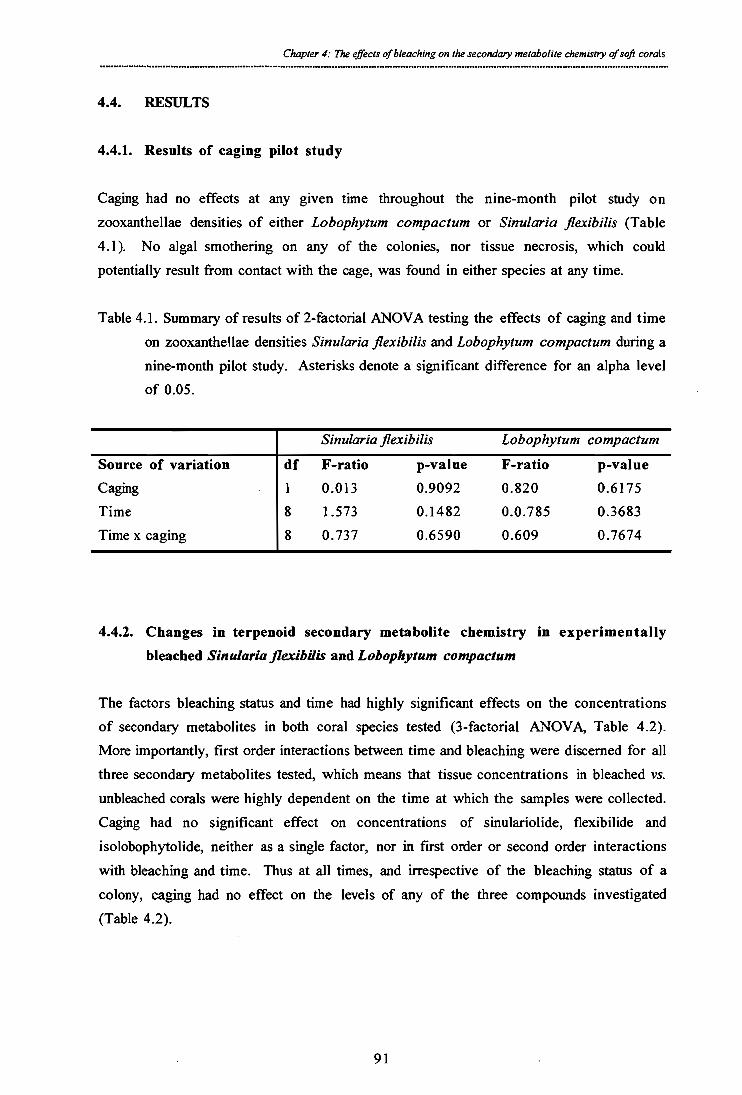
Chapter 4: The effects of bleaching on the secondary metabolite chemistry of soft corals
4.4. RESULTS
4.4.1. Results of caging pilot study
Caging had no effects at any given time throughout the nine-month pilot study on zooxanthellae densities of either Lobophytum compactum or Sinularia flexibilis (Table 4.1). No algal smothering on any of the colonies, nor tissue necrosis, which could potentially result from contact with the cage, was found in either species at any time.
Table 4.1. Summary of results of 2-factorial ANOVA testing the effects of caging and time on zooxanthellae densities Sinularia flexibilis and Lobophytum compactum during a nine-month pilot study. Asterisks denote a significant difference for an alpha level of 0.05.
Sinularia flexibilis Lobophytum compactum Source of variation df F-ratio p-value F-ratio p-value Caging 1 0.013 0.9092 0.820 0.6175 Time 8 1.573 0.1482 0.0.785 0.3683 Time x caging 8 0.737 0.6590 0.609 0.7674
4.4.2. Changes in terpenoid secondary metabolite chemistry in experimentally bleached Sinularia flexibilis and Lobophytum compactum
The factors bleaching status and time had highly significant effects on the concentrations of secondary metabolites in both coral species tested (3-factorial ANOVA, Table 4.2). More importantly, first order interactions between time and bleaching were discerned for all three secondary metabolites tested, which means that tissue concentrations in bleached vs. unbleached corals were highly dependent on the time at which the samples were collected. Caging had no significant effect on concentrations of sinulariolide, flexibilide and isolobophytolide, neither as a single factor, nor in first order or second order interactions with bleaching and time. Thus at all times, and irrespective of the bleaching status of a colony, caging had no effect on the levels of any of the three compounds investigated (Table 4.2).
91

sinulariolide flexibilide isolobophytolide
Source of variation
Time
Bleaching
Caging Time x bleaching
(N) Time x caging
Bleaching x caging
Time x bleaching x caging
df F-ratio 5 14.59
1 13.37
1 1.10
5 15.05
5 0.46
1 0.65
5 0.32
p-value F-ratio p-value F-ratio p-value < 0.6001* 7.59 < 0.0001* 38.32 < 0.0001 * 6.81 0,010* 33.47 < 0.0001 *
0.297 1.00 0.319 0.06 0.809
<0.0001* 7.43 <0.0001* 7.39 <0.0001*
0.803 0.45 0.811 0.82 0.541
0.423 0.03 0.856 0.89 0.348 0.900 0.33 0.895 0.37 0.867
Table 4.2. Summary of results of 3-factorial ANOVA testing the effects of time, bleaching and caging on tissue concentrations of the algaecide sinulariolide, the cytotoxin flexibilide of Sinularia flexibilis and the ichthyotoxin isolobophytolide of Lobophytzum compactum. Asterisks denote a significant
difference (cc = 0.05).

Chapter 4: The effects of bleaching on the secondary metabolite chemistry of soft corals
I. Changes in Sinularia flexibilis: Immediately following the bleaching treatment, bleached specimens of Sinularia flexibilis underwent a significant change in the ratio of their specific secondary metabolites, flexibilide and sinulariolide (Figure 4.5). While concentrations of
sinulariolide decreased by 92%, those for flexibilide increased by 126%. Metabolite levels in the tissues of Sinularia flexibilis were significantly different between February and July (Tukey's HSD test, p < 0.001) for both sinulariolide and flexibilide, although whereas levels
of sinulariolide decreased over the period, levels of flexibilide increased. Multiple
comparisons revealed that concentrations of sinulariolide in bleached corals were significantly different from their non-bleached counterparts and the field controls for a maximum of one month following the bleaching treatment (Tukey's HSD test, p < 0.002).
Similarly, flexibilide levels were significantly different from controls only in March 1997 (Tukey's HSD test, p < 0.0137), and by April significant differences could no longer be
discerned (Tukey's HSD test, p = 0.2696) (Figure 4.5).
93

bleaching treatment
recovery period
flexibilide (antimicrobial agent in Sinularia flexibilis)
8
4=
2
0 mid Feb-97 Mar-97 Apr-97 May- 97 Jun-97 Jul-97
mg
flexi
b ilid
e /g d
ry w
eight
1 0 -
Chapter 4: The effects of bleaching on the secondary metabolite chemistry of soft corals
sinulariolide (algaecide in Sinularia flexibilis)
IIIIIII‘\‘‘\‘‘• ki h. ■' .. -41111141101111111ftilliiiiiiiii.......,_ \ •
/./ •• \\ *
4
•• 0 fi•;.*
% • • - -o- - bleached/not caged --0-- bleached/caged
not bleached/not caged 40— not bleached/caged m— field control
Figure 4.5. Changes in tissue concentrations of the algaecide sinulariolide and the cytotoxic antimicrobial agent flexibilide in Sinularia flexibilis through time. The bleaching treatment was carried out between mid February and the beginning of March 1997. Upon completion of the bleaching treatment corals were returned to the reef of origin, and sampled for 4 months during recovery. Error bars represent standard errors (n = 5 determinations per treatment/ per month).
mg
sinu l
ario
lide/
g dry
wei
ght
94

Jul-97 0
mid Feb-97 Mar-97 Apr-97 May- 97 Jun-97
recovery period bleaching treatment
isolobophytolide (ichthyotoxin Lobophytum compactum) -
-a-- bleached/not caged -o-- bleached/caged
—a— not bleached/not caged 4
- ,
-- not bleached/caged —a— field control
go,
\ ... ."..... ..... ... isolo
boph
ytol
ide
conc
e ntra
tion/
g d
ry w
eigh
t 20 -
18 -
16 -
14 -
12 -
10 -
8 -1-
6 t••• ■•
•;.••••. '... \
\ ■
A/ 1.•
/".1e 4 -
2 - .., =
Chapter 4: The effects of bleaching on the secondary metabolite chemistry of soft corals
2. Changes in Lobophytum compactum: Concentrations of isolobophytolide in L. compactum dropped significantly and stayed depressed for the three months following the bleaching (one-factorial ANOVA, F = 55.43, df = 1, p < 0.0001 for March, F = 5.78, df = 1, p = 0.0246 for April and F = 12.68, df = 1, p = 0.0018 for May) (Figure 4.6). In June levels were no longer different from controls (one-factorial ANOVA, F = 1.039, df = 1, p = 0.319). A significant natural increase in unbleached individuals (Tukey's HSD test, p < 0.001) from February to July was detected, similarly to the trend detected for
flexibilide (Figure 4.6).
Figure 4.6. Changes in tissue concentrations of the ichthyotoxin isolobophytolide in Lobophytum compactum through time. The bleaching treatment was carried out between mid February and the beginning of March 1997. Upon completion of the bleaching treatment corals were returned to the reef of origin, and sampled for 4 months during recovery (n = 5 determinations per treatment). Error bars represent standard errors.
Analysis of 'H.-n.m.r. spectra showed that bleaching in Lobophytum compactum did not result in the production of additional compounds, which might have been viewed as a stress-related induction of secondary metabolites.
95

Chapter 4: The effects of bleaching on the secondary metabolite chemistry of soft corals
4.4.3. Changes in relative lipid concentrations in experimentally bleached Sinularia flexibilis and Lobophytum compactum
Bleaching resulted in significant reductions in lipid levels for both species, however, these were dependent on time, since first order interactions between time and bleaching were
found (3-factorial ANOVA, Table 4.3). Caging of colonies had no effect on lipid levels, not as a single factor, nor first or second order interactions with time and bleaching (Table
4.3). Thus, irrespective of the time and bleaching status of individuals, caging had no effect on the levels of lipids in either species.
Table 4.3. Summary of results of 3-factorial ANOVA testing the effects of time, bleaching and caging on lipid in Sinularia flexibilis and Lobophytum compactum. Asterisks denote a significant difference for an alpha level of 0.05.
Sinularia flexibilis Lobophytum compacturn Source of variation df F-ratio p-value F-ratio p-value Time 5 5.62 < 0.0001* 25.67 < 0.0&01 * Bleaching 1 60.55 < 0.0001* 58.64 < 0.0001* Caging 1 0.05 0.831 2.60 0.110 Time x bleaching 5 6.47 < 0.0001* 10.57 < 0.0001* Time x caging 5 0.36 0.872 0.16 0.976 Bleaching x caging 1 0.15 0.695 0.50 0.483 Time x bleaching x caging 5 0.14 0.982 0.05 0.998
One month after bleaching lipid levels were reduced on average by 82% in Sinularia flexibilis and 78% in Lobophytum compactum (Figure 4.7). Regression analysis of lipid recovery to control levels revealed that lipid concentrations were highly and significantly correlated to concentrations of symbiotic algae in Sinularia flexibilis and Lobophytum compactum respectively (R2= 0.97/ 0.922; p = 0.005/ 0.02) (compare Figures 4.7 and 4.8).
The overall loss of lipids and zooxanthellae due to bleaching was also reflected in variations in the dry weight. In L. compactum, upon termination of the bleaching treatment, the dry weight decreased significantly from 26.59% ± 1.246 SE (in unbleached colonies) to
22.87% ± 1.199 SE (in bleached colonies) of the coral wet weight (one-way ANOVA, F = 5.049, df = 1, p = 0.0345). Similarly, in S. flexibilis, the dry weight in bleached colonies constituted, on average, only 22.42% ± 0.305 SE of the wet weight, whereas
24.85% ± 0.66 SE of the wet weight in unbleached colonies was made up by the dry weight (one-way ANOVA, F = 4.41, df = 1, p = 0.046) (data not shown).
96
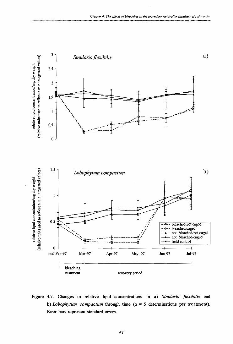
Sinularia flexibilis
------
---------
1.5 - Lobophytum compactum
--0-- bleached/not caged --o-- bleached/caged —k— not bleached/not caged —0— not bleached/caged --0— field control
-4" -------- .... -- g-
/I .// /
//
// //
// /
0
Chapter 4: The effects of bleaching on the secondary metabolite chemistry of soft corals
mid Feb-97 Mar-97 Apr-97 May- 97 Jun-97 Jul-97
bleaching I treatment
recovery period
Figure 4.7. Changes in relative lipid concentrations in a) Sinularia flexibilis and b) Lobophytum compactum through time (n = 5 determinations per treatment). Error bars represent standard errors.
97

60
b) 50
Sinularia flexibilis
a)
a-- bleached/not caged bleached/caged
--a— not bleached/not caged not bleached/caged
field control
50
40
30
20
10
Lobophytum compactum
y = 1.02*10" 63) R2 =0.994
v = 1.96*10 ("9321* R2 =0.993
-----
------
zoox
anth
ella
e de
nsit i
es (
x 10
6)/g
wet
wei
ght
zo
oxan
thel
lae
dens
ities
(x
106)
/g w
e t w
eig
ht
Chapter 4: The effects of bleaching on the secondary metabolite chemistry of soft corals
4.4.4. Partition of secondary metabolites between animal and algal tissue
On average 28.2 ± 3.45% of isolobophytolide, 21.4 ± 4.76% of flexibilide and 23.98 ± 4.36% of sinulariolide were found to be associated with the algal fraction (normalised to protein) of the coral-alga association.
4.4.5. Algal recovery in experimentally bleached S. flexibilis and L. compactum
Bleached individuals of both coral species regained their normal zooxanthellae population densities within four months of experimental bleaching (4.8). No total or partial mortality of test corals occurred during the monitoring period.
.. N. / .. 40 - a
x‘ ___G___ y= 1.96.4 0'2932'4 R2 =0.981 " ..
a , ;r: i ___0•__ Y= 1.98*10 ("M" R2 =0.960 m././ a
30 - \ --',----.2(-
\ ----;;""-- 20 - \ =(6-- \\
N , .1.--';';-3- 10 - \
= '-
0
mid Feb-97
Mar-97 Apr-97
May- 97
Jun-97 Jul-97
bleaching treatment recovery period
Figure 4.8. Changes in zooxanthella tissue densities in bleached and unbleached individuals of a) Lobophytum compactum and b) Sinularia flexibilis through time. Equations set inside the graph describe the exponential nature of recovery in bleached individuals (n = 5 determinations per treatment).
98

Chapter 4: The effects of bleaching on the secondary metabolite chemistry of soft corals
Recovery of algal symbionts followed a near exponential function for both species with correlation coefficients of R2= 0.981/ 0.960 and R 2=0.993/ 0.994 for uncaged/caged individuals of Sinularia flexibilis and Lobophytum compactum respectively (Figure 4.8).
Density of zooxanthellae, i.e. recovery, was highly dependent on the time, however, density was not significantly affected by caging in either Lobophytum compactum or Sinularia flexibilis (Table 4.4). Furthermore, no first order interaction between caging and time was detected in either species (Table 4.4). Thus at no time during the monitoring period, and irrespective of the zooxanthella density of colonies, did caging affect the recovery of zooxanthellae.
Table 4.4. Summary of results of 2-factorial ANOVA, testing for the effects of time and caging on the recovery of zooxanthellae in experimentally bleached colonies of Lobophytum compactum and Sinularia flexibilis. Asterisks denote significant differences for an alpha level of 0.05.
Source of variation df F-ratio p-value Lobophytum compactum Time 5 485.61 --, 0.0001* Caging 1 3.06 0.082 Time x Caging 5 0.76 0.580
Sinularia flexibilis
Time 5 485.61 dboo 4 * Caging 1 3.06 0.119 Time x Caging 5 0.76 0.566
4.4.6. Predation experienced by bleached and unbleached conspecifics of Sinularia flexibilis and Lobophytum compactum
During the monitoring period from February to July 1997, bleached and recovering corals, like their unbleached counterparts, experienced very little predation (Table 4.5). The results equate to 5.3% and 6.7% of all colonies of Sinularia flexibilis and Lobophytum compactum respectively showing bite marks (bleached and unbleached treatments combined). No more than two bite marks were detected on any coral, and 75% of colonies were predation free. Tests for homogeneous distributions of the numbers of bite marks between bleached and unbleached colonies showed that these were normally distributed, both
99

Chapter 4: The effects of bleaching on the secondary metabolite chemistry of soft corals
in Lobophytum compactum (p = 0.9479) and Sinularia flexibilis (p = 0.4685) (Kolmogorov-Smirnov Goodness of Fit test). The diameter of bite marks that had occurred averaged 0.5 cm in both species and was thus not considered to be significant predation. However, it is noteworthy, that bite marks occurred in both uncaged and caged corals, suggesting that either cryptic predators were present within the substrate the corals were attached to, or predators small enough to fit through a 1 cm2 mesh entered the cage (Table 4.5).
Table 4.5. Number of bite marks on test corals during the monitoring period. The figures in brackets denote the number of corals from which the bite marks were scored (n = 5 colonies per treatment). Shaded areas highlight bleached individuals.
Treatment Feb. 97 Mar. 97 April 97 May 97 June 97 July 97 Sinularia flexibilis
blekhedicaged 2 (1 ► bleached/not caged 0 I 0
not bleached/caged 0 0 2 (2) 0 1 0 not bleached/not caged 0 0 0 0 0 0 field controls 0 0 0 1 0 0 Lobophytum compactum
bleached/caged
bleached/riot caged 0 0 2 (1)
not bleached/caged 0 0 0 0 1 0 not bleached/not caged 1 0 0 1 0 1 field controls 1 0 0 1 0 0
4.4.7. Algal overgrowth experienced by bleached and unbleached conspecifics of Sinularia flexibilis and Lobophytum compactum
No algal-overgrowth was detected during the monitoring period on any of the test corals, irrespective of whether bleached or unbleached, caged or uncaged.
100

.4 30 - Lobophytum compactum
isolobophytolide
co 20
10 -
40 Sarcophyton sp.
sarcophytoxide
mg s
arcop
hytox
ide/ g
dry w
eight
20
30
1 0
0
Sinularia flexibilis
sinulariolide flexibilide
bleached + overgrown
mg f
lexibi
lide/
sinula
riolid
e / g
dry w
eight 25
20
10
15
0
5
bleached semi-bleached unbleached
Chapter 4: The effects of bleaching on the secondary metabolite chemistry of soft corals
4.4.8. Analysis of Lobophytum compactum, Sinularia flexibilis and Sarcophyton sp. affected by the 1998 natural bleaching event in the Palm Island group
A comparison of bleached (but unfouled), unbleached, and bleached plus fouled colonies of Sinularia flexibilis and Sarcophyton sp. revealed highest tissue concentrations of their specific secondary metabolites in bleached (unfouled) colonies, followed by semi-bleached and unbleached ones. The lowest concentrations were found in either species in the bleached and fouled colonies (Figure 4.9). For Lobophytum compactum, only a comparison between bleached and unbleached counterparts could be carried out, since no semi-bleached and bleached and fouled colonies could be found. For quantitation of the bleaching status of colonies please see Figure 3.5 in Chapter 3.
Figure 4.9. Tissue concentrations of species-specific secondary metabolites in bleached, semi-bleached, unbleached, and bleached and overgrown individuals of Lobophytum compactum, Sarcophyton sp. and Sinularia flexibilis (n = 14 determinations per category per species). No semi-bleached or bleached and overgrown individuals were available for Lobophytum compactum. Error bars represent the standard error.
101

Chapter 4: The effects of bleaching on the secondary metabolite chemistry of soft corals
For all three species, significant differences were found between bleached, semi-bleached, unbleached, and bleached and overgrown individuals with respect to their relative lipid levels
and to their respective terpenoid secondary metabolites (Kruskal-Wallis tests, Table 4.6).
Table 4.6. Results of Kruskal-Wallis tests, testing for significant differences between
bleached and unbleached conspecifics of soft coral species affected by the 1998 natural bleaching event. Isolobophytolide only occurs in Lobophytum compactum, sinulariolide and flexibilide are found in tissues of Sinularia flexibilis, while sarcophytoxide is specific to Sarcophyton sp. Asterisks denote significant differences (n = 7 determinations per species).
Parameter Species
L. compactum S. flexibilis Sarcophyton sp. Relative lipid concentration 0.0017*
0.0028* 0:140
Isolobophytolide 0.0017*
Sinulariolide C0025* Flexibilide 0 0002* Sarcophytoxide 0.0347*
Isolobophytolide (in Lobophytum compactum), sarcophytoxide (in Sarcophyton sp.), flexibilide and sinulariolide (both in Sinularia flexibilis) concentrations were significantly higher in bleached than in unbleached conspecifics (Mann-Whitney tests, Table 4.7) Similarly, for all compounds assessed, bleached colonies featured significantly higher concentrations than their bleached and fouled counterparts.
Table 4.7. Results of Mann-Whitney tests, comparing levels of secondary metabolites among bleached (unfouled), unbleached and bleached plus fouled conspecifics of soft coral species affected by the 1998 natural bleaching event. Asterisks denote significant differences (n = 7 determinations per species).
L. compactum S. flexibilis Sarcophyton sp. isolobophytolide sinulariolide flexibilide sarcophytoxide
bleached vs. 0.0017* 0.0017* 0.0017* 0.025 unbleached
bleached vs. 0.0181* 0.0017* 0.0017* bleached & fouled
unbleached vs. 0.0040* 0.9491 0.1797 bleached & fouled
102

Chapter 4: The effects of bleaching on the secondary metabolite chemistry of soft corals
While bleached and overgrown Sinularia flexibilis had lower levels of flexibilide and sinulariolide when compared to unbleached colonies, this was statistically significant only for the latter compound. Likewise, mean levels of sarcophytoxide were lower in conspecifics that were bleached and overgrown than in unbleached individuals, however, this was not statistically significant.
103

Chapter 4: The effects of bleaching on the secondary metabolite chemistry of soft corals
4.5. DISCUSSION
The experimentally induced loss of zooxanthellae in the soft corals Sinularia flexibilis and Lobophytum compactum resulted in a significant — if short lived — changes in the composition of species-specific secondary metabolites. In bleached colonies of Sinularia flexibilis, sinulariolide concentrations fell significantly while flexibilide levels increased simultaneously. Similarly to decreases in sinulariolide, isolobophytolide levels dropped in bleached Lobophytum compactum and were accompanied by significant reductions in the lipid levels in both species.
Stress-induced increase in secondary metabolites is a well-established phenomenon in both terrestrial (reviewed in Greshenzon, 1994) and marine systems (Paul and Fenical, 1986;
Coll, 1987; Paul and Van Alstyne, 1988). My findings of highly increased levels of flexibilide in experimentally bleached colonies of S. flexibilis and increased levels in all tested species—specific secondary metabolites in naturally bleached colonies, suggest that soft corals also follow this strategy. Sinulariolide and isolobophytolide, however, did not always follow this trend. Given that environmental stress, and increased temperatures in particular, are known to change the bacterial epifauna on corals, (Jindal et al., 1994 Nair et al., 1987, Ritchie et al., 1993), facilitate bacterial adhesion (Kushmaro et al., 1997) and increase virulence of some bacteria (Toren et al., 1998), increased investment into anti-microbial defence after bleaching may represent an important strategy.
4.5.1. Fouling of experimentally bleached soft corals
Flexibilide, which contains an a-methylene-b-lactone moiety, is considered to be the most biologically active terpene present in Sinularia flexibilis. It acts as a cytotoxic agent (Aceret et al., 1995) and has strong allelopathic effects (including interference with photosynthesis, and eventual necrosis in neighbouring corals) (Webb and Coll, 1983). Its most important biological activities in relation to bleaching are its strong anti-bacterial properties (Aceret et al., 1995). The observed up-regulation of flexibilide may represent a
direct strategy to prevent the adhesion and entry of bacterial pathogens into bleached specimens. Given that marine fouling is a multi-step successional process, where
colonisation by bacteria precedes the establishment of micro-and macroalgal spores (Davis,
1989), the allocation of resources to an increased production of flexibilide in experimentally bleached S. flexibilis could be viewed as an attempt to interrupt the "fouling cascade" at an early stage.
104

Chapter 4: The effects of bleaching on the secondary metabolite chemistry of soft corals
Theoretically, the observed absence of algal fouling from all test corals could have been due to a general lack of algal fouling at the site during the course of the experiment, or perhaps to an insufficient monitoring timeframe. Both explanations, however, can be ruled out since the plastic tags I used to label the experimental colonies were so heavily overgrown by
algae that they had to be renewed every month. Furthermore, the experimental timeframe of six months should have also been sufficient to observe algal overgrowth if it occurred, given that algal clearance experiments at One Tree Island have shown that within 30-60
days most reef sites recover their standing stock of epilithic algae (Hatcher and Larkum, 1983). Moreover, complete algal overgrowth colonies has been observed within four weeks for bleached hard corals (B. Schaffelcke, pers. comm., Marshall, pers. comm.), and five weeks for soft corals (pers. observ.). I therefore interpret the absence of algal fouling on experimentally bleached Sinularia flexibilis to be a function of co-occurring changes in their secondary metabolite chemistry. In particular the increase in the anti-bacterial agent, flexibilide, appears to have occurred at the expense of the algaecide, sinulariolide, possibly to avert the first successional stages of the fouling process.
The notion that regulation of secondary metabolites may aid in the prevention of algal fouling is further corroborated by data gained from the natural 1998 bleaching event. Bleached colonies of Sinularia flexibilis and Sarcophyton sp which remained free of fouling, also featured the highest concentrations of their respective algaecides, whereas those overgrown by algae had the lowest levels. Similarly, Coll and co-workers (1987) correlated the absence of certain secondary metabolites in Lobophytum pauciflorum to the occurrence of algal fouling on those conspecifics which lacked the compounds (Coll et al., 1987). Concentrations of secondary metabolites in the bleached and overgrown colonies were the lowest of all groups tested, suggesting that these colonies exhausted their energy reserves due to nutritional constraints. This hypothesis is consistent with reports that starvation of alcyoniid soft corals 2 - 3 months after the 1998 event, which led to volume reductions of 60 - 80% and were followed by algal overgrowth and death (Fabricius, 1999). It is likely that certain algaecidal thresholds have to be maintained in order to prevent fouling, and if production cannot be maintained due to nutritional constraints, fouling of weakened corals
may occur. A second possibility includes the "shutdown" of secondary metabolite
production, as a result of competitive interactions, which has been suggested to be the underlying mechanism for a failing isolobophytolide production in Lobophytum compactum where tissue necrosis of the coral is caused by contact with the red alga Plocamium hamatum (Leone et al., 1995).
105

Chapter 4: The effects of bleaching on the secondary metabolite chemistry of soft corals
4.5.2. Predation of experimentally bleached soft corals
Levels of isolobophytolide an ichthytoxin and principal terpenoid secondary metabolite in Lobophyturn compactum dropped significantly in experimentally bleached colonies. Energetic constraints, as revealed by a 78% lipid level reduction following the bleaching, may again explain the decreased production of this compound. Unlike in Sinularia flexibilis, however, a change in resource allocation towards the production of another compound could not be detected by 1 H-n.m.r. analysis. The only function that has been experimentally demonstrated for isolobophytolide to date, is its function as an ichthyotoxin. The compound has the capacity to kill Gambusia of finis at concentrations of 10 ppm within 2 hours of exposure (Bowden, unpublished data). Despite levels of the compound decreasing by 83% following bleaching, neither significant predation (nor algal overgrowth) could be detected within the monitoring period. A number of reasons may explain this lack of observed predation. Firstly, isolobophytolide levels of 17% of pre-bleaching levels for a limited period could have been sufficient to avoid significant predation. In light of cost-efficiencies for defence this explanation is doubtful, given that under non-stress situations a much higher level of production is maintained.
Secondly, predation at the site may have been "unusually" low. This explanation, however, does not accord with the fact that bite marks were detected on average 5.3% of colonies of Sinularia flexibilis and 6.7% of colonies of Lobophytum compactum. These findings are slightly higher then results provided by Griffith (1994) who found predation on soft corals in Great Barrier Reef region to be generally very low (around 1%). Therefore some predation obviously took place at the site during the experiment. Given, that bite marks were found not only found on un-caged colonies, but also in the caged corals (mesh size 1 cm 2), predators other than chaetodontid fishes or the egg cowry Ovula ovum, known
specialised soft coral predators, must have been responsible for the observed bite marks. Small species of cowries and nudibranchs, either hidden within the substrate to which the soft corals were attached (pers. observ.), or entering the cages through the mesh, could have been responsible for the observed bite marks on caged specimens.
A third explanation for the observed lack of major predation on bleached colonies includes
the possibility of production of a compound or compounds that protect against predation, such as pukalide (Coll et al., 1989) or prostaglandins which have not been detected by
1 H- n. m .r.
A fourth alternative explanation is that potential predators have learned to avoid certain soft corals (Chaetodon melannotus, eg uses visual cues to select octocorals; Alin°, 1989), and this learned behaviour is maintained in spite of reduced tissue protection. This notion
106

Chapter 4: The effects of bleaching on the secondary metabolite chemistry of soft corals
is corroborated by the fact that bite marks were found to the same degree on bleached and unbleached corals, i.e. predators obviously did not distinguish between the two groups. In
summary, no explicit protective role for isolobophytolide could be discerned, given the lack of major predation on experimentally bleached L. compactum despite reduced levels of this secondary metabolite. It cannot be ruled out, however, that the compound fulfils other, as
yet unknown ecological functions in the coral and thus to be able to monitor the impacts of decreased levels adequately requires additional research.
4.5.3. Experimentally bleached versus naturally bleached soft coral conspecifics
Naturally bleached Lobophytum compactum featured significantly increased levels of isolobophytolide in comparison to their non-bleached counterparts. This is contrary to observations following experimentally induced bleaching and suggests some protective function for the ichthyotoxin isolobophytolide under extreme bleaching conditions. Likewise, I found that the levels of flexibilide in naturally bleached individuals were about twice as high as in the experimentally bleached ones, in addition to a simultaneous increase of sinulariolide in Sinularia
Part of the observed increases may be due to the fact that concentrations of species-specific secondary metabolites are normalised to the dry weight, which decreased slightly in bleached colonies due to losses in storage lipids, zooxanthellae cells and host protein. Based on the observed decrease in dry weight in bleached colonies of 14% and 10% in L. compactum and S. flexibilis respectively, only 10-14% of the increases can be explained with losses in dry weight. Given that e.g. flexibilide levels increased by 126% in experimentally bleached and by 361% in naturally bleached S. flexibilis, I interpret the increase as a true up-regulation response and not as a mere reflection of changes in the dry weight in bleached colonies.
Yet, the increased investment into production of secondary metabolites during the natural bleaching episode appears paradoxically because energy reserves would have almost
certainly been more exhausted in the naturally than the experimentally bleached colonies, given that the magnitude of the latter bleaching was much higher. While the
experimentally induced bleaching resulted in a loss of nearly 95% of the zooxanthellae
standing stock, exposures to increased temperature and solar irradiance levels lasted for 12 days only. In contrast, the massive bleaching event experienced on the GBR in early 1998,
is believed to have been caused by a number of stresses that occurred over several weeks. Preceding high water temperatures of 31-32° C (with occasional peaks of 33° C),
freshwater run-off caused by cyclone Sid had already stressed corals via severely reduced
107

Chapter 4: The effects of bleaching on the secondary metabolite chemistry of soft corals
salinity (Fabricius, 1998; Berkelmans and Oliver, 1999). High irradiance penetration due to extremely calm conditions coincided with the increases in temperature and most likely
greatly exacerbated the stress experienced by corals (Fabricius, 1998; Berkelmans and Oliver, 1999).
Secondary metabolites have been suggested to compensate for the immune system in lower
invertebrates (Williams, 1989). Thus, extensive exposure to stress, such as the 1998 bleaching episode, may require an unusually high level of protection (i.e. high concentrations of algaecides and antimicrobial agents) to counteract fouling with opportunistic bacterial pathogens or algae. High investment into secondary metabolites under conditions of nutritional constraints, however, can only be maintained for a certain time. Thus, if time/ energy thresholds are exceeded, continued high investment into secondary metabolites may ultimately result in a fatal metabolic burden. While this interpretation is speculative, it is consistent with my findings of extremely low algaecide levels in bleached and fouled soft coral colonies, and reports that starvation in bleached alcyoniid soft corals resulted in colony death upon 2 —3 months following the 1998 GBR bleaching episode Fabricius (1999). In summary, I suggest that the differential responses in the experimentally and naturally bleached soft corals may be explained by the magnitude and the nature of the respective bleaching episodes and the metabolic investment connected to it.
Further research into the nature of regulation of secondary metabolites is required to ultimately determine whether the magnitude of a bleaching event and thus the energetic constraints connected to it do indeed determine the biochemical response. The rapid 92% decrease in levels of sinulariolide, the algaecide found in Sinularia flexibilis by the onset of experimental bleaching, for example, could be both the result of a resource re-allocation due to nutritional constraints or the outcome of a chemical conversion. On average only 24% of the compound was found to be associated with the algal partner; thus the loss of
zooxanthellae can only explain up to one quarter of the decrease in sinulariolide concentrations. One explanation for the decrease in levels is that nutritional constraints associated with the loss of zooxanthellae, and mirrored by an 82% reduction in storage
lipids, may have been indirectly responsible for a lower production of the algaecide. If
nutritional constraints indeed required a redistribution of resources, flexibilide was clearly
favoured over sinulariolide. Given that both compounds share a similar structural
relationship, an alternative explanation could involve enzymatic inter-conversion of one
compound into the other, as suggested by Maida and co-workers in 1993 (Figure 4.10).
108

Sinulariolide
11,12- Dehydroflexibilide
0
\ hypothetical bis-epoxide intermediate
Dihydroflexibilide
0-
Flexibilide
Chapter 4: The effects of bleaching on the secondary metabolite chemistry of soft corals
Figure 4.10. Cembranolide diterpene secondary metabolites derived from Sinularia flexibilis and possible biosynthetic pathways linking them, as suggested by Maida et al., 1993.
Seaweeds are known to immediately convert weak deterrents into strong deterrents upon mechanical damage of the alga (Paul and Van Alstyne, 1992). Thus, by analogy, the observed increase of flexibilide in experimentally bleached Sinularia flexibilis may not have been the result of a redirected resource allocation, but the outcome of a conversion of sinulariolide into flexibilide. If increased antibacterial properties of flexibilide were required quickly following bleaching and if normal levels were inaedequate, then biochemical conversion of sinulariolide, rather then de novo synthesis, may present an energetically preferable response to a short-term bleaching event. Rapid turnover of terpenoids has been confirmed for the eggs of Lobophytum compactum, in which terpenoid secondary metabolites can be fully metabolised within days (Heaton, 1988). Unfortunately, the
collection of samples in this study was only carried out at monthly intervals, which
precludes an analysis of the increase and decrease of flexibilide and sinulariolide respectively
on a finer temporal scale. Yet, irrespective of the strategy involved in the increase in flexibilide, the absence of algal overgrowth in bleached colonies of Sinularia flexibilis, suggests that soft corals possibly do have some capacity to avoid bacterial and subsequent algal fouling after bleaching.
109
Chapter 4: The effects of bleaching on the secondary metabolite chemistry of soft corals
4.5.4. Summary and conclusions
This study has provided evidence that the soft corals Sinularia flexibilis and Lobophytum compactum are capable of surviving a severe short-term bleaching event, however, with the metabolic burden of strong reductions in the levels of lipids as a consequence. I suggest that their ability to control the production (or the conversion) of specific secondary metabolites to meet specific ecological requirements may play an important role in the survival of soft corals during short-term disturbances such as bleaching events. This notion is corroborated by the results from the intensive 1998 natural bleaching event, in which bleached but unfouled soft corals featured the highest concentrations of their respective algaecides/antimicrobial agents, whereas bleached and fouled colonies had the lowest tissue concentrations. Thus, investment into species-specific algaecides and antimicrobial agents, as found in naturally bleached soft corals obviously has the potential to prevent fouling in association with bleaching events.
110

Chapter 5 The effects of the zooxanthellae on the secondary metabolite chemistry and development of Lobophytum compactum
Detail of eight week old primary polyp of Lobophytum compactum. Brown spots represent clusters of zooxanthellae
111

Chapter 5: The effects of zooxanthellae on the secondary metabolite chemistry and development of L .compactum
5.1. ABSTRACT
The question of who controls the production of secondary metabolites in soft corals, the algal
symbiont or the animal host has been debated for the last 30 years, but has not been resolved
due to inherent problems with residual and/ or dietary terpenes in adult colonies. In a novel
approach I have assessed the secondary metabolite chemistry of freshly metamorphosed coral
polyps, with and without zooxanthellae using extremely sensitive electro-spray and fourier-
transform mass spectrometry. In this chapter I report the effects different strains of
zooxanthellae have on the terpenoid secondary metabolite chemistry and development of the
alcyonacean soft coral Lobophytum compactum. Coral larvae of the same genetic background
were reared, then inoculated with zooxanthellar strains of different taxonomic and geographic
origin, and their terpenoid chemistry analysed.
Control over production of terpenoid secondary metabolites lies clearly with the animal host,
since all algal-animal associations, irrespective of zooxanthellae origin, were found to produce
isolobophytolide, the principal terpenoid in Lobophytum compactum. Moreover, even though
the control group had no access to zooxanthellae they still expressed isolobophytolide
confirming unequivocally that the algal partner is not essential for the production of terpenoids
in the association. Importantly this implies that soft corals affected by bleaching can still
maintain the production of these ecologically important compounds, if they have sufficient
energy reserves to allocate towards secondary metabolite production.
112

Chapter 5: The effects of zooxanthellae on the secondary metabolite chemistry and development of L .compactum
5.2. INTRODUCTION
The evolutionary success of alcyonacean soft corals, a major component of Indo-Pacific reefs
has been in part attributed to the quantity and diversity of terpenoid secondary metabolites in
their tissues (Tursch, 1976; Sammarco and Coll, 1992). Whilst the production of secondary
metabolites represents a significant metabolic cost, the energy expenditure for the production of
these compounds is well compensated for by the multiple functions they fulfil within the
organism, as discussed in Chapter 4. They enhance the coral's fitness in defending them against
predation (LaBarre et al., 1986a; Wylie and Paul, 1989), increasing their competitive ability
(Sammarco et al., 1983; Maida, 1995b) and enhancing mating opportunities (Coll, et al., 1989;
Coll et al., 1995), as well as being active as anti-microbial (Aceret, 1995; Slattery et al., 1995b)
and anti-fouling compounds (Kiefer and Rinehart, 1986; Michalek and Bowden, 1997).
While it has been established that zooxanthellae are the primary producers in the coral-alga
association (Patton and Burris, 1983; Muscatine et al., 1984), the question of which organism
carries out, and more importantly, controls the production of ecologically important secondary
metabolites is still controversial. Historically, terpenes were viewed as plant products (Scheuer,
1973) and thus "by default" zooxanthellae were believed for a long time to be the sole producers
of terpenoids in the symbiotic association. This premise was supported by results from one of
the first studies on octocoral secondary metabolites carried out by Ciereszko in 1962. The author
concluded that terpenes were of algal origin after he found high concentrations of the diterpene
crassin acetate in zooxanthellae isolated from the gorgonian Pseudoplexaura crassa. While Papastephanou and Anderson (1982) confirmed through in vitro experiments the algal origin of
the compound (using radioactively labelled precursors), the authors suggested that the gorgonian
host controlled synthesis. Apparent evidence for possible host control of terpene production was
provided by Ciereszko (1989) who found that several species of aposymbiotic soft corals
harboured the same or similar types of terpenes in their tissues, but in much lower quantities than
zooxanthellae bearing species. It remained ambiguous, however, whether or not the terpenes in
aposymbiotic soft corals were acquired from dietary sources. Kokke et al. (1984) also concluded
that terpenes were of animal origin, based on comparisons of 12C/13C ratios for products from
different octocorals and their cultured zooxanthellae. In addition, Bandurraga and co-workers
(1982) interpreted the isolation of terpenoids from the azooxanthellate gorgonian Lophogorgia
alba as evidence that the animal itself was responsible for the production. As pointed out above,
an inherent problem associated with all of these studies (with the exception of Papastephanou
and Anderson, 1982) is the difficulty of excluding the possibility that terpenoids were of dietary
origin. Thus, there is currently no conclusive evidence that the animal produces or controls the
production of terpenes in symbiotic soft corals.
113

Chapter 5: The effects of:ooxanthellae on the secondary metabolite chemistry and development of L .compactum
Quantitative and qualitative variations in secondary metabolite chemistry have been documented
among soft coral species, both within and between species (Harvell and Fenical, 1989; Harvell et
al., 1993; Bowden unpublished data). If indeed the algal partner is responsible for the production
of secondary metabolites, then variation in terpenoid products between and within one species
could be due to genetically different strains of zooxanthellae (Rowan and Powers, 1991; Loh et
al., 1998) which may have different biochemical and physiological characteristics (Schoenberg
and Trench, 1980 a-c). Evidence for substantial impact of the algal partner on cnidarian host
physiology has been provided by Kinzie and Chee (1979) and Fitt (1985) who experimentally
demonstrated significant differences in the host's growth rate, depending on the type of
zooxanthella with which a cnidarian host became inoculated. While we are starting to understand
the genetic, biochemical and physiological diversity of various zooxanthellar strains, their impact
on soft coral metabolism remains virtually unresolved and warrants further study.
In this study I tested the effects of zooxanthellae of different geographic and taxonomic origin on
the secondary metabolite chemistry of the alcyonacean coral Lobophytum compactum. To overcome confounding problems with residual terpenoids in adult colonies and possibly dietary
origin of the compounds, I infected freshly metamorphosed polyps of the same genetic
background with different algal symbionts, under controlled conditions that excluded dietary
terpenoids. This soft coral species is particularly suitable for the study since it has only one
major terpenoid secondary metabolite, the cembranoid diterpene, isolobophytolide, (Coll et al.,
1986), an ichthyotoxin (Bowden, unpublished data). By infecting newly metamorphosed polyps
with different strains of zooxanthellae, (while some were left uninfected) I addressed two
questions — firstly, whether the zooxanthellar partner was necessary for the production of
terpenoid secondary metabolites, and secondly, which partner controlled the production of certain metabolites.
114

Chapter 5: The effects of zooxanthellae on the secondary metabolite chemistry and development of L .compactum
5.3. MATERIALS AND METHODS
5.3.1. Gamete collection
Egg and sperm were collected in November 1996 during the annual mass coral spawning event
in Pioneer Bay, adjacent to Orpheus Island Research Station (OIRS), in the Central section of
the Great Barrier Reef. Eggs were collected in situ from one Lobophytum compactum female (to ensure genetic similarity of offspring) from the reef slope at 3-4 m depth by placing egg nets
equipped with collecting jars on top of the colony. Egg nets were assembled approximately
1 hour prior to spawning (17:00) and retrieved 3 hours later (20:00). To collect sperm, three
male colonies that had been dislodged from the reef substratum two months prior to spawning
were arranged in well aerated flow-through aquaria at OIRS 4 days prior to spawning. Males
were placed individually in highly aerated tanks only two hours prior to spawning. Water
containing sperm from the male with the highest sperm count, was decanted immediately after
spawning and diluted to optimal concentrations required for fertilisation success (10 6 sperm/ml, Willis et al., 1997).
5.3.2. Fertilisation
Spawned eggs were washed 3 times in millipore filtered seawater (0.45Rm) and then incubated
in ten 3L screw-top containers with sperm water at a concentration of 1.1 x 10 6 sperm/ml. Sperm from the most prolific male spawner was selected to fertlise all eggs. After 3 hours,
sperm-water was discarded, the fertilised eggs washed in millipore filtered seawater, returned to
the containers and left overnight in high flow-through aquaria at OIRS. Planulae were
transported the next day to the James Cook University Aquarium facilities. To assess
fertilisation rates, after exposure to sperm, an aliquot of each 3L container, containing the
fertilised and washed embryos was fixed in Bouins solution for 24 hours and then stored in vials
containing 70% ethanol. One hundred eggs per vial were examined and those that had
undergone cleavage were scored as fertilised. Percent fertilisation was then calculated for
8 replicate vials per jar (n = 80 measures of percentage fertilisation).
115

Chapter 5: The effects of zooxanthellae on the secondary metabolite chemistry and development of L .compactum
5.3.3. Larval rearing
Sub-samples of 500 larvae were reared in separate 10 litre aquaria, supplied with filtered flow-
through seawater and maintained under temperature controlled (25-29°C) conditions. Metal-
halide lights were set on a 12 hour light: 12 hour dark cycle (light was approximately 800 gEm-2sec -1 ). Clay tiles which were autoclaved to ensure no zooxanthellae were present
served as settlement substrata for the coral larvae. The tiles were left to pre-condition in 11um
filtered seawater for one month prior to the start of the experiment. A filter system which
excluded all particles > 1 gm was used at all times to ensure dietary exclusion of terpenoids in
phytoplankton or zooplankton. Furthermore, the aquarium system was kept free of adult soft
corals to ensure no terpenoids could be taken up from the water column.
53.4. Experimental design and symbiont infection
The experimental set-up comprised 3 replicate tanks per treatment (= strain of zooxanthellae),
each containing a sub-set of 500 larvae (n = 8 treatments x 3 tanks x 500 larvae) (Figure 1).
After larvae had metamorphosed, one of seven mixtures of gymnodinoid swanners and non-
motile coccoid zooxanthellae were added to each of the treatment tanks on a daily basis for two
weeks. The treatment strains included zooxanthellae derived from Lobophytum compactum (maternal strain), and from the soft coral species Lobophytum microlobulatum, Sarcophyton trocheliophorum and Sinularia flexibilis, which were growing in close vicinity to the "mother" coral in Pioneer Bay. Furthermore, zooxanthellae derived from the actinarian coral Zoanthus sociatus (Symbiodinium pilosum, isolated by B. Fitt, Jamaica), the jellyfish Cassiopeia xamachana (isolated by E. Dearne 1983, Florida, available as strain CS-153 from CSIRO
micro-algal culture collection, Tasmania), and the clam Tridacna squamosa (isolated by R. Rowan, GBR central section) were offered for infection (Figure 5.1.). The terminology
"strain" is used senu Kinzie and Chee (1979) and Fitt (1985) to denote that zooxanthellae are of
different geographic and taxonomic origin, and vary in their morphology and swarming
behaviour. No claim is made that these different types ("strains") of zooxanthellae necessarily
represent genetically different clades. All algal strains were kept in monoclonal cultures at
James Cook University (JCU). To check that no parental provision of zooxanthellae had
occurred, some eggs and larvae were sacrificed for microscopical examination (light
microscope and inverted microscope) prior to infection. Positive inoculation of polyps with
zooxanthellae was confirmed in situ by examining polyps from all treatments at regular intervals, using stereo microscopy.
116

Chapter 5: The effects of zooxanthellae on the secondary metabolite chemistry and development of L .compactum
control zooxanthellar strains derived from various invertebrates treatment
no zooxanthellae (A) A 02\ D 02; 0;\ 0/;\ A A
tank 1 2 3 replication
larvae 500 larvae/tank replication
Figure 5.1. Experimental design for infection of coral polyps with zooxanthellae. While control
polyps were not offered any zooxanthellae, all other treatments received zooxanthellae
derived from various invertebrates of similar and different geographic origin: Treatment A: no zooxanthellae, B: Lobophytum compactum (maternal strain), C: Lobophytum microlobulatum, D: Sarcophyton trocheliophorum, E: Sinularia flexibilis, F: Zoanthus sociatus., G: Cassiopeia xamachana, H: Tridacna squamosa. Each treatment was replicated in 3 tanks fitted with 500 planulae at the start of the experiment.
5.3.5. Offspring survivorship and polyp growth
The number of surviving polyps and their growth were assessed on a two-weekly basis for five
months after fertilisation. Growth was recorded as the number of polyps per colony.
5.3.6. Preparation of polyps for terpenoid chemical analysis
After three months, eight single polyps from each treatment were harvested, freeze-dried and
then washed three times with distilled water to remove excess salt. The whole animal was then
extracted with 100% methanol (0.2 ml) under 10 min sonication. Samples were analysed on a
Bruker 47e fourier transform mass spectrometer equipped with an atmospheric pressure
Analytica electrospray ion source. Diluted extract was infused through an electrospray needle
117

Chapter 5: The effects ofzooxanthellae on the secondary metabolite chemistry and development of L .compactum
at the rate of 100 ul per hour. Positive ion mode was used with a scan range from 80 to 3000
daltons and a cycle time of 1.3 sec. Resolution in the region of interest was approximately
10,000. Sixteen scans from each extract were summed and processed. The identity of
compounds was established by accurate mass measurement of the sodium ion complex.
In addition to mass spectrometry, early larval chemistry, parental secondary metabolite
chemistry and that of cultured zooxanthellae was confirmed by 'H NMR, using a Bruker AM
300 NMR spectrometer. All samples were freeze dried, weighed and the ground tissue was
extracted 3 times with dichloromethane (DCM, 10 ml/ g dry weight of coral tissue) for 1, 8 and
24 hours respectively. The 3 consecutive extracts were combined and the remaining solvent was removed in vacuo. Sixteen scans of each sample were recorded, spectra then Fourier
transformed using zero line broadening and the appropriate signals integrated as described in Chapter 4, section 4.3.6.
5.3.7. Statistical analysis
The significance of differences in polyp growth rates was tested by two factorial-ANOVA with
treatment as a fixed factor and tanks nested in treatment. Multiple comparisons (Tukey's HSD
post hoc test) were then carried out to test differences between treatments. The SPSS 7.5 software package was used for the analysis.
118

Chapter 5: The effects of zooxanthellae on the secondary metabolite chemistry and development of L .compactum
5.4. RESULTS
5.4.1. Fertilisation
Fertilisation rates were high with sperm and eggs from the selected male and female colony
under the given experimental conditions averaging 92.8% ± 1.8 standard error (SE).
5.4.2. Polyp settlement, metamorphosis and zooxanthellae infection
Polyps started to settle on the clay tiles 4 days after fertilisation and approximately 80% had
settled after one week. Metamorphosis and development of the primary polyp followed 3 - 4
days after settlement. Post-settlement development began with the opening of the "mouth"
(actinopharynx) and subsequent development of the tentacles and pinnules. Uptake (feeding) of
all zooxanthellae strains offered followed immediately after metamorphosis. The establishment
of a symbiosis (Figure 5.2), however, occurred only for 5 of the 7 strains offered.
Zooxanthellae derived from Zoanthus sociatus and Lobophytum microlobulatum did not result in the establishment of a symbiosis at any stage, and polyps died after 3 months. Thus no
chemical analysis and growth rate data are provided for these two experimental groups. Control
polyps, which were not offered any zooxanthellae, never became inoculated, thus confirming
that no uncontrolled infection occurred during the experiment. These polyps started to
degenerate after 2 months and were dead 3 months after spawning. It was established by
- n.m.r. that none of the cultured strains of zooxanthellae contained isolobophytolide in their tissue prior to infection.
119

Chapter 5: The effects of zooxanthellae on the secondary metabolite chemistry and development of L .compactum
Figure 5.2. Confocal microscopy images of a single three months old Lobophytum compactum
polyp, infected with zooxanthellae derived from Cassiopeia xamachana. This technique
allows a separation of animal and plant tissue, with the green colour denoting animal
tissue (a), while red represents the symbiotic algal cells (b). The superimposed image
(c) clearly shows that zooxanthellae have populated most of the polyp after 3 months,
with highest densities found in the light-exposed tentacles. (Confocal micrograph
courtesy of A. Salih and the Electron Microscopy Unit, University of Sydney).
5.4.3. Polyp growth
Significant differences in growth rates were found between polyps infected with different
strains of zooxanthellae (2-factorial ANOVA, F = 13.65, df = 4, p < 0.0001) (Figure 5.3.).
Tanks (nested in treatment) had no effects on the polyp growth (2-factorial ANOVA,
F= 0.63, df = 2, p = 0.535). Thus all treatment effects were independent from the tanks in which
they were carried out. Post-hoc comparisons of polyps infected with the maternal strain
120

3.60 79 75 82 ZI polyps/colony
Chapter 5: The effects of zooxanthellae on the secondary metabolite chemistry and development of L .compactum
strain (L. compactum) versus zooxanthellae derived from either of the two other soft corals
species (S. flexibilis and S. trocheliophorum), revealed that all colonies grew at similar rates
(Tukey's HSD test, p = 0.992, p = 0.999 for S. flexibilis and S. trocheliophorum
respectively). Those individuals infected with the jellyfish (C. xamachana) or the clam-derived (T. squamosa) strains, however, attained significantly lower growth rates than the control
polyps (Tukey's HSD test, p = 0.05 for the former and p < 0.0001 for the latter species)
(Figure 5.3).
mea
n nu
mbe
r of p
olyp
s/ co
lony
Lobophytum Sinularia flex'bilis Sarcophyton Cassiopeia Tridacna compactum trocheliophorum xamachana squamosa
original host from which zooxanthellae for inoculation were isolated
Figure 5.3. Mean number of polyps per colony in Lobophytum compactum inoculated with
different strains of zooxanthellae five months after settlement. Lobophytum compactum, Sinularia flexibilis and Sarcophyton trocheliophorum strains are derived
from soft coral hosts, whereas Cassiopeia xamachana and Tridacna squamosa strains
are strains derived from a jellyfish and a clam respectively. Figures on top of columns
represent the respective number of surviving colonies at five months. Error bars
represent the standard error.
The differences in growth are further substantiated by the fact that a number of colonies infected
with the maternal strain and with strains derived from other alcyonacean soft corals had reached
the 8-polyp stage after 5 months. Primary polyps infected with the jellyfish and clam-derived
strains, however, never reached this stage during the monitoring period. Polyps infected
with the clam-derived strain of zooxanthellae did not reach the 4-polyp stage during the five
months.
121
Chapter 5: The effects ofzooxanthellae on the secondary metabolite chemistry and development of L .compactum
5.4.4. Secondary metabolite chemistry
NMR analysis of 6 week old polyps (pooled extracts, n = 8) inoculated with each of the five
different strains of zooxanthellae, showed no detectable amounts of isolobophytolide (Figure
5.4, shows example for polyps inoculated with the maternal strain).
122

isolobophytolide
b)
Chapter 5: The effects of zooxanthellae on the secondary metabolite chemistry and development of L .compactum
6 5 4 3 2 6016
Figure 5.4. 11-1 - n.m.r. spectra a of a) six week old inoculated polyps of Lobophytum compactum (pooled sample, n = 8) showing no traces of isolobophytolide or other methylene lactones and that b) of adult polyps with signals for the methylene lactone protons of isolobophytolide (see structure
within) clearly visible at b5.94 and b6.23 (n = 8). Signals observed in the six weeks old polyp
extract were assigned to lipids.
123

Chapter 5: The effects of zooxanthellae on the secondary metabolite chemistry and development of L .compactum
After 3 months, however, all zooxanthellae-host combinations resulted in the expression of the
diterpene isolobophytolide, that is the typical Lobophytum compactum secondary metabolite.
The calculated exact mass of the sodium ion complex of isolobophytolide (C201-128Na03 + ) is
339.19363 and corresponds well with the observed mass, which ranged from 339.1906 to
339.1956 in the polyps. The compound, however, was not expressed in all polyps of each
cross-infection, with higher percentage expression in polyps inoculated with either of the soft
corals strains (Table 5.1).
Table 5.1. Percentage of individuals expressing typical Lobophytum compactum chemistry
(isolobophytolide) after infection with different strains of zooxanthellae (n = 8 polyps
per treatment). (No chemical analysis was carried out with polyps that were not
successfully infected with the Zoanthus sp. and Lobophytum microlobulatum strain).
Treatment % polyps with isolobophytolide
present in tissue (n = 8 polyps)
Lobophytum compactum 87.5 (maternal soft coral strain, Orpheus Island, GBR)
Sinularia flexibilis 100
(soft coral strain, Orpheus Island, GBR)
Sarcophyton trocheliophorum 87.5
(soft coral strain, Orpheus Island, GBR )
Cassiopeia xamachana 75
(jellyfish strain, Florida)
Tridacna squamosa 50
(clam strain, Great Barrier Reef)
Control (no zooxanthellae 75
Note that the group that had no access to zooxanthellae (control) also produced
isolobophytolide. No minor or atypical terpenoid products other than those found in the
"mother" and "father" coral were produced in any of the symbiotic associations infected with
different strains of zooxanthellae.
124

110
100
90
80
70
60
50
40 2.00 2.50 3.00 3.50
Chapter 5: The effects ofzooxanthellae on the secondary metabolite chemistry and development of L .compactum
5.4.5. Correlation between growth and expression of secondary metabolite chemistry
Regression analysis indicated that the growth rate of colonies (number of polyps per treatment after 5 months) was strongly (R 2 = 0.864) and significantly (df = 1, F = 16.54, p = 0.02)
correlated with the number of polyps that expressed the secondary metabolite isolobophytolide (Figure 5.5).
mean number of polyps/ colony ± SE
Figure 5.5. Relationship between growth of colonies inoculated with different strains of
zooxanthellae (as mean number of polyps/colony) and the percentage of polyps
expressing isolobophytolide. Zooxanthellae strains were derived from Tridacna squamosa (= T.s.), Cassiopeia xamachana (= C.s.), Lobophytum compactum (maternal strain, = L.c), Sarcophyton trocheliophorum (= S. t.) and Sinularia flexibilis (= S.f). Error bars denote the standard error.
125

Chapter 5: The effects of zooxanthellae on the secondary metabolite chemistry and development of L .compactum
5.5. DISCUSSION
This study unequivocally demonstrates for the first time, that control over production of
terpenoid secondary metabolites lies with the coral host and that zooxanthellae are not essential
for the production process. However, links between polyp growth and investment into
production of the secondary metabolite suggest that zooxanthellae influence the quantity of the
production via primary metabolism.
5.5.1. Control and production of terpenoid coral secondary metabolites
During the 30 year debate about control and production of terpenoids in corals, a number of
studies have provided evidence for host control and/or production of terpenoids. Evidence has
been either indirect (Kokke et al., 1984; Ciereszko, 1989) or experimental designs have not
accounted for possible dietary sources (Schwartz and Scheuer, 1981; Bandurraga et al., 1982)
and thus the origin of the compounds has never been conclusively established. By excluding
dietary access to terpenoids and testing experimentally inoculated primary polyps that had no
residual isolobophytolide (maternal supplies had been used up) I have provided direct evidence
for host control and the independence of the animal from its algal symbiont for terpenoid production.
Bacteria are known to be prolific producers of secondary metabolites (Fenical, 1993; Pain,
1998) and are commonly associated as epifauna on Lobophytum compactum and on soft corals in general (Ducklow, 1979; Rublee, 1980; K.M-W unpublished). Therefore bacterial origin of
terpenoids has also to be taken into consideration. Bacterial involvement in the production of
the diterpene isolobophytolide, however, is highly unlikely for a number of reasons. Firstly,
bacteria are predominantly associated with coral mucus and are not intracellular symbionts,
therefore translocation of the compound to the host tissue, which harbours about 70% of the
isolobophytolide (K.M-W unpublished) would be extremely difficult. Furthermore, polyps
were washed 3 times with distilled water prior to chemical analysis, which would have removed
most of the mucus and its associated epifaunal bacteria, including the bacterial terpenoids.
Finally, despite intensive research into bacterial secondary metabolites, to date no terpenoid
products have been reported from marine bacteria (reviewed in Fenical, 1993). Thus, I believe
that the animal host is most likely the producer of isolobophytolide in Lobophytum compactum.
Maternal provision of eggs with secondary metabolites is well established for L. compactum with epi-thunbergol, alcyonal-A and isolobophytolide as typical terpenoid egg products
126

Chapter 5: The effects of zooxanthellae on the secondary metabolite chemistry and development of L .compactum
(Heaton, 1988; Almeida de Leone, 1993). Fast metabolisation (within days) of some of these
egg - specific compounds has been reported in freshly metamorphosed L. compactum polyps (Heaton, 1988). By the time of settlement, epi-thunbergol and alcyonal-A, are no longer
detectable and concentrations of isolobophytolide have greatly decreased within one week old
planulae (Heaton, 1988). Therefore, it was not surprising that I did not detect any traces of
isolobophytolide (detectable by 'H-n.m.r) in the six week old polyps. This absence of
terpenoids in the six week old polyps and the lack of isolobophytolide in some of the three
month old polyps is an important confirmation that the products I observed were not just the
remains of parental provision to eggs, but produced by the progeny themselves.
While all cross-infected groups tested in this study expressed the typical Lobophytum compactum chemistry, not every polyp of each treatment contained a detectable amount of
isolobophytolide in its tissue at three months of age. The reason for the absence of
isolobophytolide in the three month old polyps is most likely due to insufficient energy reserves
to divert to the production of secondary metabolites. The animals tested were very young and
thus resource allocation to primary metabolism and growth was obviously favoured in some
individuals as an early life history strategy. It is also possibly that the establishment of high
densities of algal symbionts initially requires host energy reserves and nutrients, and only when a certain density of algal symbionts is reached, does a positive energy budget for the host
follow. If this interpretation was correct, only once zooxanthellae cells reach a certain density
within their juvenile host (primary polyp) sufficient energy can be diverted to primary and
secondary metabolism. While this is speculative, this interpretation is supported by the results
of the regression analysis, which showed that the groups with high growth rates also contained
the highest number of individuals that expressed isolobophytolide (Figure 5.1). Thus, while
these results have demonstrated that different algal symbionts do not influence the type of
terpenoid compound that is produced, they indirectly impact on the production via provision of
energy through primary production. This has important implications for soft coral colonies
affected by bleaching. While the production of e.g. terpenoid algaecides is controlled by the
host, with reduced energetic contribution from the algal symbiont, energy reserves may be
insufficient to maintain the production of threshold concentrations. This is consistent with the
highly reduced levels of isolobophytolide I observed in experimentally bleached Lobophytum compactum (Chapter 4; Figure 4.5.) which only returned to background levels with
zooxanthellar recovery. Moreover, those soft coral colonies affected by the 1998 mass-
bleaching event, which were fouled and overgrown also had the lowest levels of their specific
algaecide (Chapter 4; Figure 4.8) further corroborating the importance of zooxanthellae to the
primary metabolism of the association.
127

Chapter 5: The effects of zooxanthellae on the secondary metabolite chemistry and development of L .compactum
5.5.2. The impact of different strains of zooxanthellae on colony growth
The type of zooxanthellae a host becomes inoculated with, has substantial impact on the
association, given the significant variations in polyp growth rates. Polyps living symbiotically
with strains derived from other alcyonacean soft corals species did not experience significantly
different growth rates from polyps inoculated with the maternal zooxanthellae (Figure 5.5).
Those living symbiotically with the jellyfish or clam strains, however, featured significantly
lower growth rates. My findings are consistent with those of a number of studies, in which the
possibility for cross-infection of coelenterate and mollusc hosts with various zooxanthellar
strains has been demonstrated (Kinzie, 1974; Kinzie and Chee, 1979; Fitt, 1985). Those studies
found significant differences in the growths rates of an anemone (Fitt, 1985) and gorgonian soft
coral hosts (Kinzie, 1974; Kinzie and Chee, 1979) depending on the zooxanthellar strain used for inoculation.
The ranking of growth rates of the coral-alga associations tested in this study showed that
polyps that were symbiotic with zooxanthellae isolated from certain alcyonacean hosts performed best, followed by those of Cassiopeia xamachana (jellyfish). The lowest growth rates were found in individuals inoculated with the mollusc strain derived from Tridacna squamosa. It appears that strains isolated from closely related hosts (L. compactum, S. flexibilis, S. trocheliophorum, phylum: Cnidaria, class: Alcyonaria,) perform better than those from either less closely related (Cassiopeia xamachana, phylum: Cnidaria, class: Scyphozoa) or unrelated strains (Tridacna squamosa, phylum: Mollusca, class: Gastropoda). Given that the strain isolated from the most closely related host, Lobophytum microlobulatum, however, did not result in the establishment of a symbiosis in the first place, suggests that phylogenic
closeness cannot be the only factor involved in regulating host-symbiont interactions. Surface
recognition phenomena, which are known to play an important part in the establishment of the
symbiosis (Trench, 1997), may be one factor responsible for the failure to form symbiotic associations with the Zoanthus sociatus or Lobophytum microlobulatum derived strains. In the case of the latter strain, however, major differences in the secondary metabolite chemistry (Coll
et al., 1986) and subsequent chemical incompatibility may also be responsible. Unlike L. compactum, which produces cembranoid diterpenes, L. microlobulatum produces mainly
lobanes together with germacrene and eudesmane-derived diterpenoids (Coll et al., 1986).
Some soft coral secondary metabolites have been demonstrated to have negative impacts on
zooxanthella motility, with total immobilisation of the alga in some cases (Ciereszko and
Guillard 1989). Thus, a chemical incompatibility between the zooxanthellae of L. microlobulatum and the polyps of L. compactum, could have been the basis for the unsuccessful inoculation.
128

Chapter 5: The effects of zooxanthellae on the secondary metabolite chemistry and development of L .compactum
There is now overwhelming evidence that there are algal strains that are more susceptible to
light and temperature stress than other strains (Dustan, 1982; Masuda et al., 1993) suggesting
that inoculation with a "suitable" strain is crucial for the fitness of the symbiosis. The micro-
environment from which a particular zooxanthellar strain is derived has been suggested to be
the key-factor to determine the susceptibility to e.g. a certain photic regime (Berner et al., 1987;
Rowan and Knowlton, 1995; Rowan et al., 1995; 1997). All soft coral zooxanthellar strains
tested in this study were derived from the same site and depths as the maternal strain at Orpheus
Island and thus the environmental conditions, that is the photic and temperature regime, were
very similar for the three strains. In contrast, the Cassiopeia xamachana strain, for example,
stems from a host that is pelagic for part of the time, and thus has most likely been adapted to
different light, temperature and salinity levels. This could make it a less than ideal symbiotic
partner, which is consistent with the observation of reduced growth rates of the juvenile soft corals.
5.5.3. Summary and conclusions
This study has provided unequivocal evidence that the algal partner is not directly required for
the synthesis of terpenoid secondary metabolites in soft corals. The strong correlation between
polyp growth and investment into production of secondary metabolites suggests, however, that
via primary production zooxanthellae have the capacity to indirectly influence secondary
metabolism. This has important implications for the soft corals affected by bleaching. With or at
reduced energetic contribution from the algal symbiont, energy reserves in bleached colonies
may be insufficient to maintain the production of threshold levels of ecologically important
terpenoids such as algaecides, facilitating the entry of pathogens into already weakened colonies.
129

Chapter 6 The impacts of bleaching on the soft coral Lobophytum compactum. I. Fecundity, fertilisation and offspring viability
A freshly metamorphosed primary polyp of Lobophytum compactum
130

Chapter 6: The impacts of bleaching on the soft coral L. compactum. I. Fecundity. fertilisation and offspring viability
6.1. ABSTRACT
In this study I document long term effects of a simulated bleaching event on the reproductive
output and offspring viability of the soft coral Lobophytum compactum. Corals were subjected to temperature and UV radiation treatments to produce both moderately (48-60%) and heavily
bleached (90-95%) colonies. Although bleached colonies recovered their zooxanthellae within 4
months, impacts on reproductive output were significant for at least two spawning seasons. In
the first year, both polyp fecundity and mean oocyte diameters were inversely correlated with
the degree of bleaching, with complete failure of fertilisation in the group of heavily bleached
colonies. Survival and growth of sexual offspring did not differ significantly between
moderately bleached and unbleached treatments. Although no further reductions in
zooxanthellae densities were recorded throughout the subsequent year, egg size and fecundity of
the heavily bleached soft corals were still significantly reduced 20 months later. Severe
bleaching clearly has long-term sublethal impacts, reducing overall reproductive output for at least two spawning seasons.
131

Chapter 6: The impacts of bleaching on the soft coral L. compactum. 1. Fecundity. fertilisation and offspring viability
6.2. INTRODUCTION
The evolutionary success of shallow tropical coral reefs in a highly oligotrophic system is
generally attributed to the predominance of symbiotic relationships between coral hosts and
their algal symbionts (Odum, 1971). Tight nutrient recycling of crucial plant nutrients
(ammonia and phosphate) from the coral's waste metabolism and the translocation of
photosynthates (i.e. glycerol, lipids, amino acids, small peptides) are the fundamental processes
contributing to the associations' success (Trench, 1979; Miller and Yellowlees, 1989; Sutton
and Hoegh-Guldberg, 1990; Wang and Douglas, 1998; Swanson and Hoegh-Guldberg, 1998).
Under ideal conditions, it is estimated that the autotrophic production of zooxanthellae can
provide as much as 143% of the daily energy budget of the coral host (Davies 1991). Thus,
disruption of the symbiosis due to extensive bleaching, will translate into nutritional constraints
for the coral association. Consequently, extensive bleaching has been shown to result in
decreased lipid and protein concentrations in host tissues (Fitt et al., 1993), reduced calcification
(Clausen and Roth, 1975; Leder et al.,, 1991), reduced growth (Szmant and Gassman, 1990;
Goreau and Macfarlane, 1991) and, in extreme cases, in the coral's death due to starvation (Fabricius 1999).
The loss of zooxanthellae not only results in decreased autotrophic input to the association, but
may also require catabolisation of storage lipids. In extreme cases, catabolisation of structural
lipids and proteins, which has been reported for recovery after (mechanical) injury in corals may
be required (Meesters et al., 1994; Ward, 1995). Furthermore, heterotrophic feeding, which has
been suggested to play an integral role in soft coral diets (more so than in hard corals)
(Fabricius, 1995a) is highly reduced in bleached soft coral colonies (Fabricius, 1999). Devoid of
both principal modes of nutrition, i.e. autotrophic input through zooxanthellae and heterotrophic
feeding, and possibly having to re-allocate remaining resources towards recovery, corals
affected by bleaching may not have sufficient energy for reproduction.
To date, only one pioneering study, carried out by Szmant and Gassman (1990) has investigated
the possible effects of bleaching on gametogenesis and provided the first, if limited evidence,
that bleaching has the potential to disrupt the reproductive cycle. Given the important role that
sexual reproduction plays in the maintenance and replenishment of reefs, studies of the possible
longer-term, sub-lethal impacts of bleaching on reproduction are clearly required.
In this study I simulated moderate (approx. 50% reduction in zooxanthellae densities) and
strong (90-95% reduction) bleaching events and monitored the effects of the disturbance on
132

Chapter 6: The impacts of bleaching on the soft coral L. compactum. L Fecundity, fertilisation and offspring viability
reproductive output in the common, dioecious soft coral Lobophytum compactum for the subsequent two spawning seasons. This chapter documents the impact of the disturbance on adult soft corals (fecundity and egg size), their gametes (fertilisation) and their offspring
(survival and growth of juveniles). Chapter 7 documents the impact of the disturbance on
biochemical parameters associated with fitness and reproduction in the experimental adults and their gametes.
133

Chapter 6: The impacts of bleaching on the soft coral L. compactum. I. Fecundity, fertilisation and offspring viability
6.3. MATERIALS AND METHODS
6.3.1. Site description
This study took place between October 1995 and December 1997 in Pioneer Bay (for location,
see figure 2.1 in Chapter 2). All corals were collected from the reef slope at 3-4m depth and
subsequently returned to the same site during the recovery phase.
63.2. Species description
Lobophytum compactum Tixier-Durivault, 1956, a common reef flat coral on the inner and mid-
shelf reefs of the Great Barrier Reef (Dinesen 1983), was used in experimental manipulations
described below. As found in some soft corals (Achituv and Benayahu, 1990) and typical for this genus, polyps of L. compactum are dimorphic with autozooids functioning as feeding
polyps and bearing gonads, whereas siphononzoids are reduced in size and function solely to
circulate water throughout the colony (Tixier-Durivault, 1958; Versefeldt, 1983). L. compactum is gonochoric and has a two-year oogenic cycle (Alifio and Coll, 1989) so that large and small
eggs, representing two different year cohorts, are present simultaneously within autozooids
(Alirio and Coll 1989). In contrast, spermatogenesis requires only one year (Alifio and Coll
1989; K.M-W, unpublished), i.e. mature oocytes are produced every year in synchrony with the
annual cycle of sperm production in male colonies, but mature females carry two generations of oocytes at any one time, as described for Sarcophyton glaucum (Benayahu and Loya, 1986).
Staging of oocytes was carried out after Yamazato et al. (1981). The species typically spawns on the 4th day after the November full moon at Orpheus Island, with egg release starting around
18:30 and ending around 20:30 (Alin and Coll 1989; K.M-W, unpublished).
63.3. Collection and preparation of experimental corals
Ten randomly-selected female colonies of Lobophytum compactum were divided in situ into 4
approximately equal-sized pieces. Each fragment was randomly assigned to a different group (a,
b, c, or d) and each group subjected to a different experimental treatment. Only female colonies
of a similar initial diameter were used. To avoid reversed puberty associated with reduction in
colony size below a minimum threshold, all females were approximately 1 m 2 in size and
134

Chapter 6: The impacts of bleaching on the soft coral L. compactum. I. Fecundity, fertilisation and offspring viability
fragments were between 36-40 cm in diameter. After chiselling and tagging, all coral pieces
were left to recover at their site of origin for 5 months. Group "a" were chiselled colonies that remained in situ for the duration of the study (handling controls) whereas group "b" corals were
transported to flow-through tanks where they were maintained at ambient temperatures and light
(tank controls). Group "c" and "d" corals were submitted to increased solar radiation and
temperature to simulate a moderate and severe bleaching event respectively. In addition, ten
random colonies (group "e" corals) from the same site and depth as experimental colonies were tagged and left in situ as unchiselled field controls (chiselling controls).
6.3.4. Bleaching treatment
In order to simulate a moderate bleaching event (approx. 50% loss of zooxanthellae), group "c"
corals were submitted to an enhanced solar radiation and temperature treatment for 8 days. The
simulation of a comparatively strong bleaching event (90-95% loss of zooxanthellae) was
accomplished by treating group "d" corals to the same experimental treatment for 12 days.
Temperatures in flow-through experimental tanks at Orpheus Island Research Station were
increased from 29-30° C (ambient) to 31° C ± 0.5° C (see Berkelmans and Willis, 1999, for
details of the experimental set-up). Photosynthetically active radiation (PAR) and ultraviolet
radiation (UVR) were increased through translocation of corals from 3-4 m in situ to 50 cm in the experimental tanks. This translocation was approximately equivalent to an increase of 50%
in PAR and 33% in UVR respectively according to light profiles recorded in Pioneer Bay using
a Li-Cor light sensor and UV sensor (calculations based on data provided by W. Dunlap,
unpublished). The timing of the experiment, March 1996, was set to coincide with the most
likely timing for a natural bleaching event.
6.3.5. Sampling regime and analysis of biochemical and reproductive parameters
At the conclusion of the bleaching treatment, corals were returned to the site of collection and
their recovery monitored initially at 2-weekly and then at monthly intervals between March
1996 and November 1997. Each month, tissue samples (5 lobes in the range of 1-2 g wet
weight / colony) were collected and frozen at —20° C prior to determination of zooxanthellae
densities.
135

Chapter 6: The impacts of bleaching on the soft coral L. compactum. I. Fecundity, fertilisation and offspring viability ----- ......
Quantification of zooxanthellae tissue concentrations: Zooxanthellae densities were monitored as indicators of bleaching and recovery of host tissues and quantified as described in Chapter 2, section 2.3.2).
Gamete capture: Eggs were quantitatively collected by placing egg nets fitted with
collecting jars on top of each experimental and control female colony. Egg nets were deployed
approximately 1 hour prior to spawning (17:00) and retrieved 3 hours later (20:00). To collect
sperm, male colonies were placed in well-aerated flow-through aquaria at Orpheus Island
Research Station 4 days prior to spawning and placed individually in highly aerated containers 2
hours prior to spawning. Four male colonies of L. compactum were collected randomly and thus their bleaching histories were unknown. Sperm water was decanted immediately after spawning
and diluted to concentrations found to optimise fertilisation (10 6 spenn/ml, Willis et al., 1997).
Fecundity and egg size determination: Immediately prior to spawning, tissue samples at least 3 cm long were collected from ridges located at the centre of each female colony in all
experimental treatments. Samples were fixed in the field with 10% seawater formalin for 24
hours, rinsed in freshwater and then stored in 70% ethanol (Jokiel and Coles, 1977; Szmant,
1991). Staging of oocytes was carried out after Yamazato et al. (1981). Ten polyps per colony
were dissected to estimate polyp fecundity. In each polyp, the diameters of ten stage III oocytes
(mature oocytes) were measured using a calibrated ocular micrometer. Only oocytes over 580 1.1m were scored as mature.
Fertilisation rates: Fertilisation experiments were only carried out during the 1996
breeding season. Spawned eggs from all corals (including the minute quantity of eggs spawned
by the heavily bleached group) were collected in egg nets and washed 3 times in millipore
filtered seawater (0.45 Rm). Eggs were combined with sperm from each of the four males, that
is 4 egg-sperm combinations per female. For each combination, eggs were incubated in
quadruplicate vials with sperm at a concentration of 1.2 x 10 6 sperm/ml. After 6 hours, sperm-
water was discarded, the fertilised eggs washed in millipore filtered seawater, fixed in Bouins
solution for 24 hours and then stored in 70% ethanol. One hundred eggs per vial were examined
and those that had undergone cleavage were scored as fertilised.
136

Chapter 6: The impacts of bleaching on the soft coral L. compactum. I Fecundity, fertilisation and offspring viability
6.3.6. Larval rearing
Approximately 500 larvae from the moderately bleached group and each of the control groups
(combined from all female colonies within each group) were reared in separate 10 litre aquaria
supplied with 1 um filtered flow-through seawater and maintained under temperature controlled
(25-29° C) conditions. Metal-halide lights provided light in the PAR region (approximately
800 uEm-2sec-I ) to each aquarium and were set on a 12 hour light: 12 hour dark cycle.
Previously conditioned clay tiles served as settlement substratum for the coral larvae. After
larvae had metamorphosed, gymnodinoid swarmers and non-motile coccoid zooxanthellae derived from Lobophytum compactum (maternal strain) and kept in culture at James Cook
University were added to each of the four treatment tanks on a daily basis for two weeks.
63.7. Juvenile growth
Colony growth was assessed on a two-weekly basis for five months after settlement of larvae and recorded as the number of polyps per colony.
63.8. Statistical analysis
Differences in fertilisation rates, fecundity, egg sizes and growth of polyps were tested for
significance by one way ANOVA followed by multiple comparisons (Tukey's HSD test) using
the SPSS 7.5 software package. Regression analyses were carried out in order to establish the
relationships between polyp fecundity and both the degree of bleaching and time for recovery of
zooxanthellae densities.
137
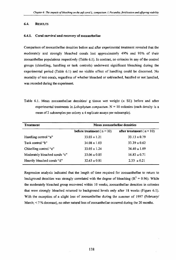
Chapter 6: The impacts of bleaching on the soft coral L. compactum. 1. Fecundity. fertilisation and offspring viability
6.4. RESULTS
6.4.1. Coral survival and recovery of zooxanthellae
Comparison of zooxanthellae densities before and after experimental treatment revealed that the
moderately and strongly bleached corals lost approximately 49% and 93% of their
zooxanthellae populations respectively (Table 6.1). In contrast, no colonies in any of the control
groups (chiselling, handling or tank controls) underwent significant bleaching during the
experimental period (Table 6.1) and no visible effect of handling could be discerned. No
mortality of test corals, regardless of whether bleached or unbleached, handled or not handled,
was recorded during the experiment.
Table 6.1. Mean zooxanthellae densities/ g tissue wet weight SE) before and after experimental treatments in Lobophytum compactum. N = 10 colonies (each density is a mean of 2 subsamples per colony x 4 replicate assays per subsample).
Treatment Mean zooxanthellae densities
before treatment ( n = 10) after treatment ( n = 10) Handling control "a" 33.03 ± 1.21 33.13 ± 0.79 Tank control "b" 34.08 ± 1.03 33.39 ± 0.62 Chiselling control "e" 33.05 ± 1.24 34.40 ± 1.09 Moderately bleached corals "c" 33.06 ± 0.85 16.83 ± 0.71 Heavily bleached corals "d" 32.63 ± 0.81 2.33 ± 0.21
Regression analysis indicated that the length of time required for zooxanthellae to return to
background densities was strongly correlated with the degree of bleaching (R 2 = 0.96). While the moderately bleached group recovered within 10 weeks, zooxanthellae densities in colonies
that were strongly bleached returned to background levels only after 18 weeks (Figure 6.1).
With the exception of a slight loss of zooxanthellae during the summer of 1997 (February/
March; < 7 % decrease), no other natural loss of zooxanthellae occurred during the 20 months.
138

Zoox
anth
ella
e de
nsiti
es (x
106)/
g c
oral
we t
wei
g ht
II II I Dec- Jan- Feb- Mar- Apr- May- Jun- 96 97 97 97 97 97 97
I ill Jul- Aug- Sep- Oct- Nov- 97 97 97 97 97
1997 1996
start / end of bleaching treatment
1 st spawning season
2 nd spawning season
1 0 -
C1'
0 II !III! Nov- Dec- Jan- Feb- Mar- Apr- May- Jun- Jul- Aug- Sep- Oct- Nov- 95 95 96 96 96 96 96 96 96 96 96 96 96
chiselling control "a"
handling control "b"
A— field control "e"
-A- - moderately bleached "c"
- strongly bleached "d"
Figure 6.1. Mean zooxanthellae densities (± SE) in three control and two experimental groups of Lobophytum compactum between November 1995 and
November 1997. N = 10 colonies (i.e. each density is a mean of 2 subsamples per colony x 4 replicate assays per subsample). Vertical bars on the
time scale denote the initiation of the experiment, the beginning (1 March 96) and end (15 March 96) of the bleaching treatment and the spawnings in
November 1996 and 1997 respectively.
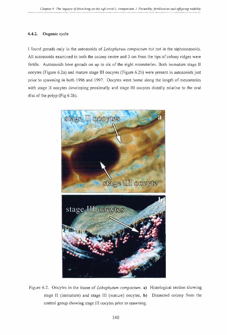
Chapter 6: The impacts of bleaching on the soft coral L. compactum. I. Fecundity, fertilisation and offspring viability
6.4.2. Oogenic cycle
I found gonads only in the autozooids of Lobophytum compactum but not in the siphonozooids.
All autozooids examined in both the colony centre and 3 cm from the tips of colony ridges were
fertile. Autozooids bore gonads on up to six of the eight mesenteries. Both immature stage II
oocytes (Figure 6.2a) and mature stage III oocytes (Figure 6.2b) were present in autozooids just
prior to spawning in both 1996 and 1997. Oocytes were borne along the length of mesenteries
with stage II oocytes developing proximally and stage III oocytes distally relative to the oral
disc of the polyp (Fig 6.2b).
Figure 6.2. Oocytes in the tissue of Lobophytum compactum. a) Histological section showing
stage II (immature) and stage III (mature) oocytes, b) Dissected colony from the
control group showing stage III oocytes prior to spawning.
140

Chapter 6: The impacts of bleaching on the soft coral L. compactum. I. Fecundity, fertilisation and offspring viability
By the 1996 November spawning, gametes of severely bleached females were at a stage
normally reached in May, which means that their development lagged behind controls by five
months. Interestingly, the unspawned eggs did not change in size or resume development until
April of 1997, when they were once more in phase with the normal oogenic cycle (per. obs.).
Maturation of all other oocytes in their second year of development was accompanied by a
gradual colour change from white to bright pink (Figure 6.2b), which is due to a protein bound
carotenoid in the eggs (B. Bowden, pers. corn.). Oocyte maturation was found to occur
synchronously within and between colonies. Mature stage III oocytes (diameter of 600-620 Rm)
were recorded around September, that is 2-3 months prior to spawning and 22-23 months after
first appearing as stage II oocytes with an initial diameter of 50 pm (data not shown). The mean
diameters of the large and small oocytes at spawning were 630 ± 1.45 µm (n = 1000) and
110 ± 1.26 [AM (n = 1000) respectively. No parental provision of zooxanthellae was detected in
eggs of L. compactum. Uptake of zooxanthellae and establishment of a symbiosis occurred only after metamorphosis.
6.4.3. Polyp fecundity
Chiselling and handling of coral colonies had no significant effect on their fecundity (Figure 6.3, cf. handling and tank controls vs undisturbed field (= chiselling controls)).
a b
C d e
Mea
n Po
lyp
Fecu
ndity
± S
E
1996 ❑ 1997 chiselling handling moderate y heavily field control
control control bleached bleached
25 -
20 -
15-
10-
5-
*
* zero
0
Treatment Group
Figure 6.3. Mean polyp fecundity (± SE) in bleached and unbleached colonies of Lobophytum compactum, one (1996) and two (1997) spawning seasons after experimental treatment.
Fecundity is expressed as total number of mature oocytes (> 58012m)/ polyp. N = 100
polyps per group. Asterisks denote significantly different means for comparisons
between treatments and controls in each year.
141

Mea
n Po
lyp
Fecu
n dity
30
25
20
15
10
5
0
Chapter 6: The impacts of bleaching on the soft coral L. compactum. I. Fecundity, fertilisation and offspring viability
In 1996 the mean fecundity of the moderately bleached colonies (13 ± 0.4, n =100) was
approximately half that of the chiselling controls (23 ± 0.6, n =100). The heavily bleached
group failed to produce any mature oocytes at all in the first spawning season (Fig 6.3). In the
second year following experimental treatment fecundity of the moderately bleached group was
equivalent to that of the controls (Tukey's HSD test, p = 0.95). Reproductive output, of the
heavily bleached group was still significantly reduced (Tukey's HSD test, p = 0.003) by approximately 20% (Figure 6.3).
A strong negative relationship was found between the degree of bleaching (in March 1996) and
the number of mature eggs (stage III oocytes) in the gonads of Lobophytum compactum immediately prior to the first spawning season (in November 1996) (Figure 6.4). An analysis of
variance of the regression of fecundity (numbers of stage III oocytes/polyp) on zooxanthellae
density confirmed that the relationship was highly significant (R 2 = 0.92, F = 587.46, p < 0.0001).
0 ate
y = -0.35448+0.7210x R2 =0.924
0 10 20 30 40 50
Zooxanthellae Densities (x 106)/ g wet weight
Figure 6.4. Relationship between polyp fecundity and zooxanthellae densities in Lobophytum compactum. Mean values were used to calculate the correlation
coefficient, N = 10 colonies) for determinations of zooxanthellae densities (i.e.
each density is a mean of 2 subsamples per colony x 4 replicate assays per
subsample); N = 100 polyps for fecundity estimates.
142

Chapter 6: The impacts of bleaching on the soft coral L. compactum. I. Fecundity, fertilisation and offspring viability
6.4.4. Egg size comparisons
The diameters of stage III oocytes immediately prior to spawning followed a normal distribution
in all experimental treatments, with the majority of eggs from females in control groups falling
into the 605-655 [km size class (Figure 6.5). I found a highly significant difference in egg size,
however, between control and experimental groups in the first (1996) spawning season (one-
factorial ANOVA, F = 1101.27, p < 0.0001). Egg sizes of the moderately and severely bleached
groups differed both from the controls (Tukey's HSD test, both p < 0.0001) and from each other
(Tukey's HSD test, p < 0.0001). Chiselling and handling of the colonies did not significantly
affect egg size, as no statistical difference in egg size could be discerned between the
undisturbed field colonies (chiseling controls) and the handling or tank controls in either year
(Tukey's HSD test for 1996 comparisons: p = 0.18, and 0.19 for field vs handling and field vs tank controls respectively; Tukey's HSD test for 1997 comparisons: p = 0.97, and 0.72
respectively). The most striking difference in the size-frequency distribution of eggs from
moderately bleached colonies in comparison to control colonies was the strong decrease in the
number of eggs in the two largest size classes. Despite this loss of large eggs, the modal size class of 605-655 ttM was the same as that found in distributions for all of the control groups. In
contrast, the modal size class for corals that had been heavily bleached decreased to 455-505
pm. Moreover, I found no eggs greater than 555 11111 in diameter (Figure 6.5). Typically, a
diameter of approximately 580 tkm is the minimum size required for eggs to be spawned. With a
mean diameter of 468.1 1.- 1.47 the eggs from heavily bleached corals were approximately
25% smaller than control eggs (mean diameter = 633.4 ± 1.86 tim; Figure 6.5).
I continued to detect a significant difference in mean egg size among control and experimental
groups in the second spawning season following treatment (one-factorial ANOVA, F=60.182,
p< 0.0001). Eggs from heavily bleached colonies were still significantly smaller than those from
control colonies (Tukey's HSD test, p < 0.0001) by approximately 5% (Figure 6.5). Eggs from
moderately bleached corals, however, were not significantly different from those of control
colonies (Tukey's HSD test, p = 0.94) by the second breeding season.
143

Egg size classes (gm) Egg size classes (pm)
handling (tank) controls 632.7 ± 1.7
moderately bleached 600.1 ± 1.6 *
handling (tank) controls 629.24 ± 1.5
moderately bleached 625.03 ± 1.8
heavily bleached 600.1 ± 1.7*
Freq
uenc
y (N
o. s
tage
III o
ocyt
es)
600 400 200
0
600 400 200 -
0-
600 - 400 - 200 -
0-
600 400 200
0
heavily bleached 468.1 ± 1.4 *
600 400 200
0
600 400 200
0
600 400
200
0
600
400
200
0
field controls 634.1 ± 1.9
field controls 629.6 ± 1.5
705 75 455 505 555 605 655 705 755
Chapter 6: The impacts of bleaching on the soft coral L. compactum. I. Fecundity, fertilisation and offspring viability
1996 1997
chiselling controls 629.8 ± 1.4 600
400 200
0 7777S1.,
Figure 6.5. Size - frequency distributions of stage III eggs derived from bleached and unbleached colonies of Lobophytum compactum immediately prior to the 1996 and 1997 spawning seasons. N = 1000 eggs per treatment (i.e. 10 eggs x 10 polyps x 10
colonies). Numbers in the top right corner of each graph denote mean egg diameter (gm) ± SE. Asterisks denote a significant difference from the field control.
144
600 400 200
0
chiselling controls 632.9 ± 2.0

Mea
n %
Fer
t ilisa
tion
Chapter 6: The impacts of bleaching on the soft coral L. compactum. 1. Fecundity, fertilisation and offspring viability .... -------
6.4.5. Spawning in 1996 and 1997
With the exception of the heavily bleached colonies of Lobophytum compactum, colonies in all experimental and control groups released all of their stage III oocytes in November 1996. In the
heavily bleached group, 8 of the 10 colonies released a few eggs in November, but the rest did
not spawn. In November 1997, all but three of the experimental colonies in the heavily
bleached group released eggs (data not shown). Surprisingly, even though gonads of these three
colonies contained mature-sized pink eggs, no spawning was detected in the following three months.
6.4.6. Fertilisation rates
Mean fertilisation success differed significantly among the two experimental and three control
groups (one-factorial ANOVA, F = 21.59, p < 0.0001). This was entirely due to the fact that
eggs collected from the heavily bleached group did not fertilise (Figure 6.6). No statistical
differences in fertilisation rates were found between gametes derived from the moderately
bleached corals (93 ± 2 mean percent fertilisation, n = 40) and those of the three control groups
(92 ± 2, 92 ± 2 and 93 ± 2 mean percent fertilisation for the chiselling, handling and tank
controls respectively, n = 40 for all groups, one-factorial ANOVA, df = 3, F = 0.49, p = 0.69).
chiselling handling moderately heavily field control control bleached bleached control
Treatment Group
Figure 6.6. Mean fertilisation success (2-. SE) for eggs retrieved from bleached and unbleached colonies of Lobophytum compactum in the first spawning season following
experimental treatment. N = 400 eggs per treatment. The asterisk denotes significantly
different fertilisation success.
145

Chapter 6: The impacts of bleaching on the soft coral L. compactum. 1. Fecundity, fertilisation and offspring viability
6.4.7. Survivorship and growth of offspring
No differences in survivorship or growth were detected between juveniles originating from the
moderately bleached group and any of the control groups of corals (Table 6.2). Larvae (Figure
6.7a) started to settle on the clay tiles 4 days after fertilisation and approximately 80% had
settled after one week. Metamorphosis and development of the primary polyp followed within
3-4 days after settlement. Post-settlement development began with the opening of the "mouth"
(actinopharynx) and subsequent development of the tentacles and pinnules (Figure 6.7b).
Juveniles reached the four-polyp stage after approximately 4 months (Figure 6.7c). Uptake of
zooxanthellae followed immediately after metamorphosis and significant densities were visible
within two weeks.
Figure 6.7. Developmental stages of Lobophytum compactum: a) planulae 48 hours after
fertilisation; b) one-month old primary polyp; c) four-month old juvenile (4-polyp
stage).
146

Table 6.2. Juvenile survival and growth measured as number of polyps per colony five months after spawning. A total of 500 larvae were monitored in each
treatment. Multiple polyp stage refers to the number of polyps/ colony. Heavily bleached corals did not produce larvae.
Treatment Number of surviving
juveniles after 5 months
Multiple polyp stage
1 2 3 4
Total number of
polyps / treatment Mean number of
polyps / colony (± SE) Chiselling control "a" 36 2 7 10 17 114 3.16 ± 0.16
Handling control "b" 30 1 4 7 18 102 3.18 ± 0.16
Field control "e" 33 1 4 9 19 112 3.39 ± 0.14
Moderately bleached colonies "c" 34 3 2 8 21 115 3.38 ± 0.16
--.1 Strongly bleached colonies `d" Heavily bleached corals did not produce larvae
Chapter 6: The impacts
of bleaching on the
soft coral L. compactum. I. Fecundity, fertilisation and offs pring
viability

Chapter 6: The impacts of bleaching on the soft coral L. compactum. 1. Fecundity, fertilisation and offspring viability
6.5. DISCUSSION
This study shows clearly, for the first time, that the reproductive output of soft corals may be
affected by bleaching events and that the degree of impact increases with the intensity of the
disturbance. Moderate bleaching reduced both the number and size of mature oocytes in
experimental colonies of the soft coral Lobophytum compactum. Given that lipids comprise up
to 65% of the egg dry weight, smaller mean egg sizes mean that larvae are energetically less
well-equipped for survival once eggs are fertilised (chapter 7; Michalek-Wagner and Willis,
2000b). Severe bleaching reduced mean size of mature oocytes below an apparent viability
threshold so that they were retained for an entire year beyond the normal time of release.
Although mature-sized eggs developed in all severely bleached females in the second breeding
season following experimental treatment, lack of spawning in 30% of these females
demonstrates that the impact of such disturbances can extend beyond 20 months.
I also demonstrate that the rate of zooxanthella recovery depends on the degree of bleaching
experienced by the coral host. Recovery of algal stocks in the moderately and strongly bleached
groups of experimental soft corals took approximately 70 and 126 days, respectively. These
results compare well with the approximate 120-day zooxanthellae recovery period I found for
experimentally (> 90%) bleached soft corals reported in chapter 4. Davies (1991) estimated that
corals could survive periods of decreased photosynthetic productivity for 28 to 114 days
through metabolisation of lipid reserves. Although lipid reserves were sufficient to allow the
recovery of all severely bleached colonies of Lobophytum compactum in this study without any (even partial) mortality, the reduced fecundity of these colonies indicates that recovery occurred
at a significant cost to resources normally allocated to reproduction.
The 45% reduction in fecundity of Lobophytum compactum in the moderately bleached group and complete failure to produce mature eggs in the severely bleached group in the first year
clearly demonstrate that fecundity is a good indicator of sublethal stress in Lobophytum compactum. Similarly, differences in egg sizes between bleaching treatments suggest that egg
size can also be used to gauge impact severity in natural bleaching disturbances. It has been
well-established that reproductive failure can be induced in corals by stresses such as turbidity,
sedimentation (Kojis and Quinn, 1984), high seawater temperatures (Jokiel and Guinther, 1978)
and anthropogenic pollution (Loya and Rinkevich, 1979). Although a number of authors have
suggested that fecundity could be used as an indicator of sublethal stress (Kojis and Quinn,
1984; Harrison and Wallace, 1990), it is generally not easy to separate sublethal impacts on
reproduction from natural variations in fecundity due to factors such as differing size and age of
148

Chapter 6: The impacts of bleaching on the soft coral L. compactum. I. Fecundity, fertilisation and offspring viability
colonies sampled (Wallace, 1985; Szmant, 1991; Sier and Olive, 1994; Slattery et al., 1999). I
reduced potential sources of natural variation in fecundity by standardising the size of
experimental colonies and the location of sampling within a colony. The low variation in
fecundity I found between colonies within a treatment or between the two years, suggesting that
standardisation of sampling may allow fecundity to be used more widely as a sublethal indicator
of stress.
The reduced fecundities of moderately bleached Lobophytum compactum (13 ± 0.44 mature
eggs/polyp) in comparison to control colonies (24 ± 0.67), despite the bleaching event occurring
after the typical complement of oocytes had developed, (i.e. midway through the oogenic cycle),
suggest that some early stage oocytes had been resorbed to allow development of the remaining
ones. Thus, energy allocated to reproduction was apparently directed towards maintaining
fewer eggs than normal to ensure that they attained a mature size. This interpretation is
consistent with observations of Yamazato and co-workers (1981) who noted that some
reduction in the number of oocytes commonly occurs in early oogenesis in L. crassum, even in
the absence of detectable stress, and is presumably linked to re-allocation of energy to support remaining oocytes.
Although resorption of some immature eggs also occurred in strongly bleached Lobophytum compactum (19.6 ± 1.2 mature eggs/polyp), such redirection of reserves was still not sufficient
to allow production of mature eggs in the first year. Eggs that were produced had a mean size
25 % smaller than those of controls and were apparently neither spawned nor resorbed but
retained until the next (1997) spawning season. The almost complete failure of all strongly
bleached females to spawn in 1996, eight months after experimental treatment is consistent with
the interpretation that developmental delays associated with recovery from the disturbance
prevented oocytes from attaining maturity by the time of the breeding season. The fact that a
few under-sized eggs were spawned during the 1996 breeding period demonstrates the
pervasiveness of seasonal proximate cues that trigger spawning in mass spawning soft corals.
The retention of most stage III oocytes for a full year beyond their expected release suggests
that there are inherent benefits in retaining nearly mature eggs until the next mass spawning
period (e.g. increased sperm availability and reduced mortality because of increased potential
for predator satiation (Babcock and Heyward, 1986; Johnson, 1992). Although a number of
studies have demonstrated that mature oocytes may be resorbed following incomplete spawning
in hard corals (Rinkevich and Loya 1979; Szmant and Gassman 1990; Sier and Olive, 1994),
this may not always be the optimal solution. Resorption of a full complement of oocytes,
relatively advanced in their development, followed by de novo gametogenesis is unlikely to be
149

Chapter 6: The impacts of bleaching on the soft coral L. compactum. I. Fecundity, fertilisation and offspring viability
as energetically favourable as retention of late stage oocytes for a full year. Lack of
development of under-sized oocytes in severely bleached corals until oogenesis was in phase
with field populations suggests that energy allocation strategies of soft corals are highly plastic.
Both mature and immature oocytes may be resorbed to provide energy for remaining oocytes, or
the soft coral may suspend oogenesis altogether until it is once more in phase with annual gametogenic cycles.
Differences in fertilisation success of eggs between severely bleached and control colonies in
the first spawning season following experimental treatment were most likely due to differences
in mean sizes of gametes and associated maturity factors. Eggs of severely bleached females
were 26% smaller than those of control females (468.1 ± 1.47 gm in the heavily bleached group
vs. 634.1 ± 1.94 gm in controls) and resulted in zero fertilisation. Sperm-egg histo-
incompatibility can be ruled out as an explanation for the lack of fertilisation because all crosses
were replicated using the same clone combinations in all treatments. Since heavily bleached
females retained the majority of their eggs for a further year, it is unlikely that eggs had
undergone the final maturation steps required to become receptive to sperm. Although eggs
produced by moderately bleached females were 5% smaller than eggs of control females, they
had similar fertilisation success (93% versus 92% fertilisation success, respectively). Despite an
approximate 45% reduction in the number of spawned eggs in moderately bleached colonies,
eggs that were spawned were able to fertilise and develop normally. Survival and growth rates
of offspring did not differ significantly between moderately bleached and control colonies, thus,
the strategy of allocating energy towards a smaller number of eggs to ensure that they attain maturity was successful for moderately bleached Lobophytum compactum. The fewer number of eggs available for fertilisation and development from bleached corals is consistent with
observations of reduced recruitment following natural bleaching events (Glynn et al., 1996) and
disproportionately reduced representation of families in recruitment assemblages that are most
affected by bleaching events (Gleason, 1996).
In conclusion, these results demonstrate that bleaching disturbances have the potential to
decrease reproductive output or suspend gametogenesis entirely in the subsequent breeding
season. They also show that soft corals may recover from bleaching by re-allocating energy
reserves that would normally be directed to reproduction towards recovery. Although
zooxanthella recovery was completed within a few months, reproductive output was negatively
affected for at least another 15 months in cases of severe bleaching. Thus, although
zooxanthellae concentrations track the immediate response and recovery of the coral host
following a bleaching disturbance, they do not give an accurate picture of sublethal impacts. I
believe that zooxanthellae densities should only be used as an indicator of coral health in
150

Chapter 6: The impacts of bleaching on the soft coral L compactum. 1. Fecundity, fertilisation and offspring viability
conjunction with other indicators of sublethal stress, such as fecundity. If bleaching episodes
occur with increasing intensity and at shorter intervals in the future (see Williams and Bunkley-
Williams, 1990; Jones et al., 1997, Hoegh-Guldberg, 1999), then the longer term implications of
this study are that sustained reductions in reproductive output and fertilisation success may
impede replenishment of coral assemblages and, ultimately, natural reproductive rhythms of
corals with long oogenic cycles may be completely disrupted.
151

Chapter 7 The impacts of bleaching on the soft coral Lobophytum compactum. II. Biochemical changes in adults and their gametes
Experimentally bleached colony of Lobophytum compactum after relocation to the reef
152

Chapter 7: The impacts of bleaching on L. compactum. II. Biochemical changes in adults and their gametes
7.1. ABSTRACT
In this chapter I demonstrate that experimental bleaching reduces important biochemical parameters in adult tissues of the soft coral Lobophytum compactum. These losses are subsequently passed on to their gametes, possibly jeopardising their viability. Although
zooxanthellae densities of experimental corals recovered within 4.5 months, protein, lipid,
MAA and carotenoid concentrations were reduced for at least eight months in adult tissues.
Reductions in tissue concentrations were amplified when they were passed on to gametes, with
the greatest reductions occurring in lipids and proteins. Although reductions in MAAs were
relatively smaller when passed on to gametes, even minor proportional reductions have
significant implications for larval survival, given that I found MAA levels to be approximately
three times higher in eggs than in maternal tissues. By the second spawning season (20 months
after experimental bleaching), the biochemical composition of adult tissues and their gametes
was indistinguishable from that of control (unbleached) corals.
153

Chapter 7: The impacts of bleaching on L. compactum. II. Biochemical changes in adults and their gametes ..... --------
7.2. INTRODUCTION
It is established to date that nutritional constraints associated with bleaching have negative
effects on the primary metabolism of affected corals (Porter et al., 1989; Leder et al., 1991; Fitt
et al., 1993). Reductions in levels of important primary metabolites, such as proteins and lipids,
and secondary metabolites, such as carotenoids and MAAs could have dire consequences for the
capacity to recover after a bleaching event. Moreover, reductions in levels of certain
biochemical parameters could be passed on to the next generation, thereby potentially reducing the fitness and survival of the progeny.
In this chapter I focus on the effects of bleaching on important indicators of the biochemical
status of adult soft corals and their gametes, in particular, on tissue concentrations of lipids,
proteins, UV protecting mycosporine-like amino acids and anti-oxidative carotenoids.
Reductions in these parameters help to explain decreases in the reproductive output and gamete viability of the soft coral, Lobophytum compactum that I reported in Chapter 6.
Comparisons of adult and larval tissues show that lipids comprise up to 70% of the dry weight
of coral larvae (Arai et al., 1993; Richmond, 1996), whereas concentrations rarely exceed 30 -
40% in adult tissues (Patton et al., 1977; Stimson, 1987; Ward, 1995). As the majority of both
hard and soft corals spawn eggs that lack zooxanthellae and, with few exceptions, coral larvae
are not known to feed (Tranter et al., 1982; Wright, 1986), parental provision of lipid reserves is
essential. Biochemical changes in adult tissues undoubtedly affect the next generation and
significant changes may compromise the survival and dispersal of larvae of most coral species.
Investment into biochemical defence systems for protection against high irradiance stress of the
highly exposed early planktonic larval stages is a life-history trait not exclusive to corals.
Parental provision of both MAAs (Alitio, 1989; Carefoot et al., 1998; Chiocarra et al., 1986;
Adams and Shick, 1996) and carotenoids (Almeida de Leone, 1993; Dall et al., 1995; Tsushima
et al., 1995) has been documented for a number of invertebrates with pelagic larval stages. The
presence of the former compounds has also been shown experimentally to decrease mortality of
larvae due to UVR (Gleason and Wellington, 1995). The established efficacy of carotenoids to
quench singlet oxygen and scavenge deleterious free radicals and reactive oxygen species
(Goodwin, 1976; Paerl 1984; Ben-Amotz and Avron, 1983; Shick et al., 1996) makes them an
important part of the diverse photo-protective system of corals (Ambarsari et al., 1997). Carotenoids cannot be synthesised de novo by the animal host and available evidence suggests that the zooxanthellae partner is the most likely candidate for the production of MAAs (Dunlap
and Chalker, 1986). Bleaching therefore has the potential to reduce the fitness of gametes
154 •

Chapter 7: The impacts of bleaching on L. compactum. II. Biochemical changes in adults and their gametes
directly through reductions in lipid and protein reserves available for gamete development or
indirectly through reduced production of photo-protective MAAs and carotenoids.
In this Chapter I describe the impact of a bleaching event, occurring approximately midway
through gametogenesis, on the subsequent biochemical composition of adult females and their
gametes for 20 months, a period that spanned two spawning seasons. To determine the longer
term, sublethal impacts of reductions in zooxanthellae densities on the biochemical constitution
of the coral host, I simulated a moderate (approx. 50% loss of zooxanthellae) and severe (90-
95% loss) bleaching event (for details see Chapter 6). I then compared a series of biochemical
parameters between bleached and unbleached colonies of the soft coral, Lobophytum compactum. Here I demonstrate that biochemical data provide important information for
understanding the longer-term, sublethal effects of bleaching and ultimately for understanding
how, and to what extent, bleaching affects the fitness of corals and the viability and dispersal potential of their offspring.
155

Chapter 7: The impacts of bleaching on L compactum. II. Biochemical changes in adults and their gametes
7.3. MATERIAL AND METHODS
7.3.1. Experimental design and sampling regime
The same colonies of Lobophytum compactum that were collected in March 1996 and exposed to elevated seawater temperature and solar radiation treatments to monitor the impacts of
experimental bleaching on reproduction (see Chapter 6) were sampled for biochemical analyses
as described below. After the bleaching treatment, corals were returned to their reef of origin
and recovery of zooxanthellae densities monitored at monthly intervals (see Chapter 6). Tissue
samples (5 lobes of 1-2 g wet weight/ colony) of all 50 test corals (i.e. 10 colonies per
treatment) were collected immediately prior to spawning for two subsequent years (November
1996 and 1997) and frozen at -20° C until analysed biochemically. For each of the four
biochemical parameters outlined below, comparisons were made among colonies in moderately
bleached, heavily bleached and unbleached treatments. Colonies representing the unbleached
treatment included all those used as chiselling, handling and field controls (see Chapter 6).
Gametes were captured with egg nets or dissected from the gonads of test corals.
7.3.2. Biochemical analyses
I. Lipid analysis: Samples of adult females and their eggs were collected for lipid analyses
immediately prior to spawning in November 1996 and 1997 and frozen at -20° C. Samples
were freeze-dried, weighed (to 0.001g), crushed with mortar and pestle, and immersed for 24 h
in a 2:1 (v:v) chloroform:methanol solution to extract the lipids. Extracts were then washed
once with a 0.88% potassium chloride solution, three times with a 1:1 (v:v) methanol : water
solution and dried for 24 hours before being re-weighed. The total lipid content is expressed as
% lipid per gram tissue dry weight (Stimpson, 87; Harland et al., 1992).
Carotenoid analysis: The quantification of the carotenoid peridinin in adult colonies, and
that of a closely related peridinin derivative (altered by only one functional group and
indistinguishable from peridinin by spectro-photometric analysis, B. Bowden pers. corn.) found
in the eggs were carried out as described in Chapter 2, section 2.3.2.
Analysis of MAAs: MAAs were analysed as described in Chapter 2, section 2.3.2.
4. Protein determination: Proteins were analysed as described in Chapter 2, section 2.3.2.
• 156

Chapter 7: The impacts of bleaching on L. compactum. II. Biochemical changes in adults and their gametes
7.3.3. Statistical analysis
The significance of differences in lipid, protein, MAA and carotenoid tissue concentrations
among moderately bleached, severely bleached and control colonies were tested by one-way
ANOVA followed by multiple comparisons using Tukey's HSD test. Significance of correlations between zooxanthellae densities immediately following experimental treatment and
each of the biochemical parameters eight months later were tested by analysis of variance of regression. The SPSS 7.5 software package was used for analyses
157
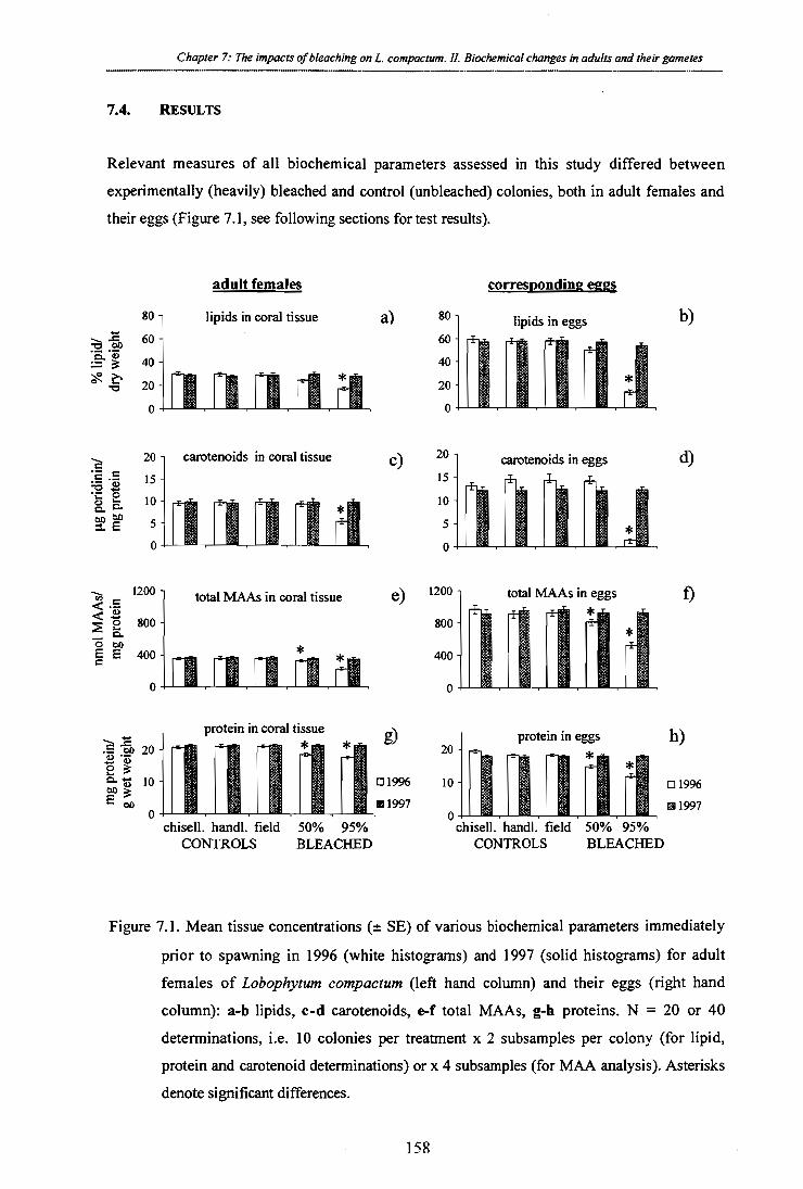
protein in coral tissue 20 -
2 Gt. on 10 -
0 I I I chisell. handl. field 50% 95%
CONTROLS BLEACHED
Chapter 7: The impacts of bleaching on L. compactum. 11. Biochemical changes in adults and their gametes
7.4. RESULTS
Relevant measures of all biochemical parameters assessed in this study differed between
experimentally (heavily) bleached and control (unbleached) colonies, both in adult females and
their eggs (Figure 7.1, see following sections for test results).
adult females corresponding eggs
80
60
40 -
20 -
0
lipids in coral tissue a) 80 - 60-
40 -
20 -
0
lipids in eggs b)
= o 4;
1
20
15
10
5
0
1200 - ¢
800 "a" 00 g E 400 -
0
carotenoids in coral tissue
total MAAs in coral tissue e) 1200 -
800 -
I I I
carotenoids in eggs
111
total MAAs in eggs
c) 20 -
15 -
10 -
5-
0
400 -
d)
f)
0
g) protein in eggs h) 20 -
El 1996 10 - Eli 1997 1
01996
mi 1997 0 chisell. handl. field 50% 95%
CONTROLS BLEACHED
Figure 7.1. Mean tissue concentrations (± SE) of various biochemical parameters immediately
prior to spawning in 1996 (white histograms) and 1997 (solid histograms) for adult females of Lobophytum compactum (left hand column) and their eggs (right hand column): a-b lipids, c-d carotenoids, e-f total MAAs, g-h proteins. N = 20 or 40
determinations, i.e. 10 colonies per treatment x 2 subsamples per colony (for lipid,
protein and carotenoid determinations) or x 4 subsamples (for MAA analysis). Asterisks denote significant differences.
158

Chapter 7: The impacts of bleaching on L. compactum. II. Biochemical changes in adults and their gametes
It is noteworthy that these differences were detected eight months after experimental treatment.
Reductions in lipids, carotenoids, MAAs and proteins were significant for severely bleached
corals but only reductions in proteins and MAAs were significant for moderately bleached corals.
7.4.1. Lipid analysis
Lipid concentrations in tissues of adult females immediately prior to spawning (November
1996) and their spawned eggs were both positively correlated with zooxanthellae densities in
female tissues just after experimental bleaching (March 1996) ( ‘1.2females = 0.41, p < 0.0001; ?eggs = 0.62, p < 0.0001; Figure 7.2). Thus, the lower the density of zooxanthellae after
experimental bleaching, the longer the time required for recovery of zooxanthellae numbers and
the lower the subsequent lipid concentrations in both females and their corresponding eggs (Figure 7.2).
Eggs derived from moderately bleached soft corals in the first spawning season after impact
contained on average 14% less, while eggs of heavily bleached females contained 76% less total
lipid than those of control corals (Figure 7.1b). Reduced concentrations of total lipids were also
found in the maternal tissues of corals in the moderately and severely bleached corals (i.e. 12%
and 41% reduction in lipids respectively) in comparison to control corals (Figure 7.1a). Note
that concentrations of lipids in eggs are typically twice as high as those in maternal tissue.
While the lower lipid concentrations in both the females and their gametes in the heavily
bleached group were significantly lower than controls (one-factorial ANOVA, females: F = 17.19 df = 4, p < 0.0001; gametes: F = 60.71, df = 4, p < 0.0001), the trend for lower lipid
concentrations in moderately bleached females and their eggs was not statistically significant.
159

0
0 mo
eggs: y = 17.756 + 1.2390x R2 = 0.617
o lipid; eggs
lipid; females 0
I • g. • NI
females: y = 17.353 + 0.37186x R2 = 0.407
0 0
8
30 40 0
0 50 10 20
% li
p id/
dry
wei
g ht
100
80
60
40
20
Chapter 7: The impacts of bleaching on L compactum. II. Biochemical changes in adults and their gametes
Zooxanthellae cells (x 10 6)/ g wet weight
Figure 7.2. Relationship between zooxanthellae densities in adult females immediately
following experimental bleaching and tissue concentrations of lipids eight months later (1996 spawning), in both eggs and females of Lobophytum compactum, n = 100.
Lipid concentrations in tissue samples of both moderately and heavily bleached corals and their
eggs taken immediately prior to the 1997 spawning (i.e. 20 months after the bleaching
treatment), were not significantly different from those of control corals (one-factorial ANOVA,
F = 0.30, df = 4, p = 0.88 for females; F = 0.57, df = 4, p = 0.67 for eggs).
7.4.2. Carotenoids
The major carotenoid found in Lobophytum compactum is peridinin, accompanied by small amounts of diadinoxanthin, diatoxanthin and 13-carotene, a complement similar to that found in
dinoflagellate symbionts in general (Jeffrey et al., 1997). Carotenoid levels in tissues of adult
females immediately prior to spawning (November 1996) and their spawned eggs were both
positively correlated with zooxanthellae densities in female tissues just after experimental bleaching (March 1996) (r2remaies = 0.20, p < 0.0001; ?eggs = 0.42, p < 0.0001). Immediately prior to the first spawning season (8 months after experimental bleaching), concentrations of
peridinin in corals from the severely bleached group were reduced by 90% in eggs (one-factorial
ANOVA, F = 31.17, df = 4, p < 0.0001) (Figure 7.1d) and by 43% in adult females (one-
factorial ANOVA, F = 9.82, df = 4, p < 0.0001) (Figure 7.1c) in comparison to concentrations
160
Chapter 7: The impacts of bleaching on L. compactum. 11. Biochemical changes in adults and their gametes
in eggs and females from the control group. I detected no discernible effect of the elevated
temperature and solar radiation treatment on the levels of peridinin in the moderately bleached
group of corals 8 months after experimental bleaching, either in adult female tissues (Tukey's
HSD test, p = 0.99) or their eggs (Tukey's HSD test, p = 0.99). In 1997, the second spawning
season following the disturbance, no significant differences in peridinin levels were detected
between experimental and control corals for either adult females (one-factorial ANOVA,
F = 0.05, df = 4, p = 0.99; Figure 7.1c) or their eggs (one-factorial ANOVA, F = 0.23 df = 4, p = 0.99; Figure 7.1d).
7.4.3. Mycosporine-like amino-acids (MAAs)
The typical MAAs I identified in adult Lobophytum compactum and their eggs were shinorine, porphyra-334, palythine, asterina, palythinol and traces of palythene. Palythine represented by
far the largest fraction, comprising 90-95% of the total MAA content in both eggs and adult
female tissue. Levels of MAAs in eggs were approximately 3 times those in the maternal tissue
(cf. Figs. 7.1e, 7.1f), revealing a high level of parental provision of UV protectants.
MAA levels in tissues of adult females immediately prior to spawning (November 1996) and
their spawned eggs were both positively correlated with zooxanthellae densities in female tissues just after experimental bleaching (March 1996) (r2
females = 0.22, p < 0.0001; ?eggs = 0.39,
p < 0.0001). In the first spawning season following experimental bleaching, elevated seawater
temperature and UV treatments significantly reduced levels of MAAs, by 8% in moderately
bleached adult females (one-factorial ANOVA, F = 8.92, df = 4, p < 0.0001; Figure 7.1e) and by
13% in their eggs (one-factorial ANOVA, F = 19.5, df = 4, p < 0.0001; Figure 7.10 in
comparison to control corals. Reductions in MAAs were even greater in the heavily bleached
group, ranging from 35% (p < 0.001) for females to 44% (p < 0.001) for their corresponding
gametes. By the second spawning season following the event, MAA concentrations of
experimental corals did not differ significantly from controls for either adult females (one-
factorial ANOVA, F = 0.14, df = 4, p = 0.97; Fig 7.1e) or their gametes (one-factorial ANOVA,
F= 0.23, df = 4, p = 0.92; Fig 7.10.
161

Chapter 7: The impacts of bleaching on L. compactum. II. Biochemical changes in adults and their gametes
5 10 15
retention time (min)
Figure 7.3. Typical HPLC spectrum of MAAs extracted from eggs of Lobophytum compactum.
7.4.4. Proteins
As for lipids, I found a positive correlation between zooxanthellae densities after experimental
treatment and protein levels eight months later at the time of spawning (r 2females = 0.34, p < 0.0001; ?eggs = 0.53, p < 0.0001; Figure 7.4). The impact of experimental bleaching was not as
extreme for proteins as it was for lipids, average protein reductions in eggs being 17% for
moderately and 38% for heavily bleached corals (see Figure 7.1h). I found a similar though less
extreme trend in adult female tissues, with reductions of approximately 11% and 17% in
moderately and heavily bleached corals respectively (Figure 7.1g). In samples collected
immediately prior to the second (1997) spawning I found no significant differences in protein
concentrations between bleached and unbleached treatments for either eggs (one-factorial
ANOVA, F = 0.02, df = 4, p = 0.99; Fig 7.1h) or adult female tissues (one-factorial ANOVA, F = 0.10, df = 4, p = 0.98; Fig 7.1g).
abso
rba n
ce u
nits
162

0 0 10 20 30 40 50
females: y = 16.996 + 0.11499x R 2 = 0.335
o protein; eggs
• protein; females
eggs: y = 11.233 + 0.21575x R2 = 0.530
mg
p rot
ein/
g w
e t w
eigh
t
Chapter 7: The impacts of bleaching on L. compactum. II. Biochemical changes in adults and their gametes _... .... .
Zooxanthellae cells (x 10 6)/ g wet weight
Figure 7.4. Relationship between zooxanthellae densities in adult females following
experimental bleaching and protein concentrations eight months later in both eggs and females of Lobophytum compactum, n = 100.
163

Chapter 7: The impacts of bleaching on L. compactum. II. Biochemical changes in adults and their gametes
7.5. DISCUSSION
The results presented here clearly show that both moderate and severe bleaching disturbances reduce the levels of proteins and MAAs in female colonies of Lobophytum compactum and their eggs for up to eight months after a disturbance. In addition, severe bleaching disturbances also
reduce lipids and carotenoids in both adult females and their eggs for a similar period of time.
It is well established that photosynthetic rates of corals are high in shallow water and that the
amount of carbon translocated from symbiotic algae easily meets the host's requirements for
respiration and growth (Muscatine et al., 1984). With the loss of zooxanthellae following
bleaching disturbances, however, this situation can change dramatically. I found that corals
suffered reductions of up to 17% in tissue protein in comparison to unbleached corals, eight
months after bleaching. Reductions in lipids of up to 41% in heavily bleached corals were even
more dramatic. Given that zooxanthellae are estimated to comprise only 5-12 % of the biomass
of corals (Muscatine et al., 1984), the reductions found in this study could not have been due to
the loss of the algal partner, but reflect real changes in the tissue of the coral host. Porter et al.
(1989) estimated that the carbon contribution of zooxanthellae towards the animal's metabolic requirements changed from 112% in unbleached Montastrea annularis to 51% in corals that had lost approximately 86% of their zooxanthellae population in a natural bleaching event. As a
consequence, host levels of tissue protein, lipid and carbohydrates eight weeks after the
disturbance had dropped by 38.8%, 66.3% and 44.2 % respectively (Porter et al., 1989). This
study demonstrates that the impact of such a natural disturbance, which was similar to the
impact experienced by the heavily bleached group of soft corals, may continue for at least eight
months. Even more critical was the finding that both protein and lipid levels were reduced in
mature eggs immediately prior to spawning. Thus nutritional perturbations experienced by the
host were passed on to their gametes and affected their viability eight months later (see Chapter 6).
I found that reductions in tissue levels of protein and lipids eight months after experimental
bleaching were correlated with reductions in zooxanthellae densities immediately after the
disturbance. Thus, not only does the magnitude of the bleaching directly affect the time
required for recovery of zooxanthellae densities (see Chapter 6); it also determines the
biochemical composition of the host and their gametes for an extended period of time.
Reductions in both proteins and lipids in tissues of severely bleached females were
approximately doubled in magnitude when passed on to their offspring (cf. 17% reductions in
tissue protein in adult females versus 38% in their eggs; 41% reductions in lipids of females
versus 76% reductions in their eggs). Protein and lipid reductions of this magnitude, prevented
164

Chapter 7: The impacts of bleaching on L. compactum. II. Biochemical changes in adults and their gametes
eggs from attaining maturity during the ensuing spawning season (see Chapter 6). Although the
11-12% reductions in proteins and lipids in moderately bleached females reduced both their
fecundities and egg sizes, the consequent 17% and 14% reductions in proteins and lipids passed
on to their eggs did not apparently affect their ability to be fertilised (ibid.). By the following
spawning season (20 months after the disturbance), both protein and lipid levels had recovered
to background levels. This is consistent with findings of Fitt et al. (1993) that reduced protein and lipid levels in Montastrea annularis persisted for 10 months after an intense natural bleaching event.
The large size (632vm mean diameter) and high lipid composition (65%) of eggs of
Lobophytum compactum combined with lack of either zooxanthellae or feeding in larvae prior to metamorphosis indicate that planulae of L. compactum are lecithotrophic. The 65% lipid content I found for eggs of L. compactum was similar to the 60 to 70% lipid compositions reported for eggs of acroporids and other broadcast spawning species (Arai et al., 1993). Wax
esters (mainly cetyl palmitate) constitute the largest fraction of the energy reserves of both
corals and their eggs and represent a high-energy source because they are metabolised at a
slower rate than other lipids. Thus wax esters can function as long term energy sources and
support long distance dispersal (Benson and Muscatine, 1974; Benson et al., 1978).
Larvae developing from lecithotrophic eggs are dependent on their mothers for energy reserves
for survival. Studies of the competence and longevity of coral larvae (Richmond, 1987;
Havenhand, 1993) highlight the correlation between stored energy reserves and dispersal
potential. Accordingly, maternal investment has been found to be up to an order of magnitude
greater in azooxanthellate in comparison to zooxanthellate larvae (Ben-David-Zaslow and
Benayahu, 1996; 1998). I found that lipids, the primary energy reserve of coral larvae, suffered
the greatest reductions of the four parameters analysed following experimental bleaching. If
mothers bleach to the extent that their lipid reserves are decreased during gametogenic
development, azooxanthellate larvae are more likely to suffer reduced viability and dispersal
than zooxanthellate larvae, which may derive 13-27% of their requirements from photosynthates
translocated from their algal partner (Richmond, 1987).
In addition to direct impacts of bleaching associated with reductions in lipids and proteins,
bleaching can also indirectly reduce the fitness of both adults and their larvae by interfering
with the production of secondary metabolites. I found significant reductions in the carotenoid
peridinin in both heavily bleached females (by 43%) and their eggs (by 90%). Given that
carotenoids are primarily acquired from zooxanthellae or partially through heterotrophic feeding
in corals (Davies, 1976; Frank and Cogdell, 1996), reduced concentrations following bleaching
165

Chapter 7: The impacts of bleaching on L. compactum. II. Biochemical changes in adults and their gametes
are not surprising. Kleppel and co-workers (1989) found similar reductions in carotenoids
immediately following a natural bleaching event. Such reductions are likely to have far-
reaching effects, because carotenoids function as: accessory pigments in the form of a peridinin-
chlorophyll-protein complex (Prezelin, 1976); photoprotectants due to their capacity to quench
chlorophyll excited states; scavengers of singlet oxygen molecules; and dissipaters of excess
energy (Frank and Cogdell, 1996). Increased concentrations of some carotenoids (i.e.
xanthophylls) found by Ambarsani et al. (1997) following a solar bleaching event, presumably relate to their role as photoprotectants.
Peridinin-chlorophyll-protein complexes constitute more than three-quarters of the carotenoid
pool in dinoflagellates (Jeffrey and Haxo, 1968), thus their presence in corals is generally
assumed to be related to their role as a major component of the light harvesting apparatus of
symbiotic dinoflagellates (Iglesias-Prieto, et al., 1992; Iglesias-Prieto and Trench, 1997). Their
role as accessory photosynthetic pigments in eggs that are devoid of zooxanthellae, however, is
not as clear. I suggest that it is likely that peridinin functions as a photoprotectant in eggs and
subsequently in larvae. Although only diatoxanthin and diadinoxanthin, which are present in
small quantities in dinoflagellates and are known to be light inducible (Ambarsani et al., 1997;
Brown et al., 1999), are typically described as photoprotectants, perididin also has the structural
basis to act in this capacity. The potency of carotenoids as effective quenchers of reactive
oxygen species is correlated to the number of conjugated double bonds in these compounds
(Shimidzu et al., 1996). The eight double bonds of perididin (in comparison to ten and eleven
in diadinoxanthin and diatoxanthin respectively) certainly suggest that it could act as a photo-
protectant. This role would be consistent with my finding that carotenoid levels were
approximately one-third greater in eggs than in their mothers in both bleached and unbleached L. compactum. Many invertebrate larvae are similarly equipped with high levels of carotenoids,
which would counteract increased UV levels encountered during their pelagic stage (Dall et al.,
1995; Tsushima et al., 1995). If this is the primary role of perididin in eggs, then the 90% loss I
found for carotenoids in the eggs of heavily bleached L. compactum would have resulted in severe oxidative stress.
It is well established that MAAs are an important line of defence against damage due to high
solar irradiance and particularly UVR in corals and many other invertebrates and algae
(Chapter 3; reviewed in Dunlap and Shick, 1998). The 8 % and 35 % reductions I measured in
tissues of moderately and strongly bleached females eight months after bleaching implies that
they might have experienced oxidative stress and possibly cell and tissue damage, particularly
as MAA losses were undoubtedly higher during the earlier portions of the experimental period.
While such losses did not result in mortality of adults in this study, reductions in MAA
166

Chapter 7: The impacts of bleaching on L. compactum. II. Biochemical changes in adults and their gametes
concentrations were amplified as they were passed on to the next generation (to 13 and 44%
reductions in eggs derived from moderately and heavily bleached corals respectively). Since
UVR is potentially much higher for coral eggs and larvae, which typically spend their first 24
hours at the sea surface and then continue to develop in the water column (Willis and Oliver,
1988; 1990), substantial reductions in MAAs in eggs are potentially lethal. The occurrence of
reduced MAA levels as a longer-term effect of bleaching is particularly intriguing with respect
to my findings of highly increased MAA levels during and immediately after a bleaching
disturbance (Chapter 3). This suggests that immediate resource allocation towards MAA
production after bleaching translates into a longer-term metabolic burden for soft corals, i.e.
sub-optimal MAA levels months after the disturbance.
The conclusion that the potential for UV damage is greater for pelagic larvae than for adults is
corroborated by my finding that MAA levels were approximately three times higher in eggs of Lobophytum compactum in comparison to maternal tissues. Parental provision of MAAs in
larvae is not exclusive to corals, but has been demonstrated to be a common strategy for
avoiding UV damage in the early life history stages of other taxa with planktonic larvae (e.g. sea
hares (Carefoot et al., 1998) and sea urchins (Chiocarra et al., 1986; Adams and Shick, 1996)).
While I did not compare the effects of UVR on gametes with low versus high MAA levels in
this study, evidence from other studies clearly demonstrate the negative impact of UVR on
invertebrate larvae with reduced MAA levels (Gleason and Wellington, 1995; Adams and
Shick, 1996). Behavioural modifications, vertical mixing of the water column or early cryptic
settlement may mitigate UV-induced mortality in marine larvae (Gleason and Wellington,
1995). However, the surface development and dispersal of many coral larvae (Willis and Oliver,
1988; 1990) preclude such avoidance strategies and expose developing coral larvae to extreme levels of UVR.
While it is clear that carotenoids are either produced by the algal partner (Davies, 1976; Frank
and Cogdell, 1996) or acquired through heterotrophy, the origin of MAAs in the association has
not been established unequivocally. In terrestrial plant and fungi systems, MAAs have been
shown to be produced via the shikimic acid pathway (Favre-Bovin, 1987) but this pathway is
absent in metazoans. Therefore, except for possible acquisition through the diet, zooxanthellae
may be the primary supplier of MAAs in the coral association (Dunlap and Chalker, 1986).
Since zooxanthellae levels had returned to background levels by June and August for the
moderately and heavily bleached group respectively (see chapter 6; Michalek-Wagner and
Willis, 2000a), normal production of MAAs and carotenoids could have been resumed for up to
five months prior to sampling. The reduced levels of both MAAs and carotenoids at the time of
sampling suggest, however, that energy was allocated elsewhere. Since reductions in lipid and
167

...... Chapter 7: The impacts of bleaching on L. compactum. II. Biochemical changes in adults and their gametes
protein levels were not as great as reductions in MAAs and carotenoids in adult females or their
eggs eight months after the experimental bleaching, it is possible that whatever resources were
available were allocated towards recovery of these primary biochemical constituents, at the cost
of secondary metabolites. If this indeed was the case, then it suggests that the coral host exerts
considerable control over zooxanthellae metabolism and/or the products translocated. Such host
control has been established for a number of zooxanthella-host interactions (Cook and D'Elia, 1987).
In summary, this study demonstrates that bleaching events may alter the biochemical
composition of soft corals for an extended period of time. In addition, not only does bleaching
have the potential to reduce reproductive output in Lobophytum compactum (see Chapter 6; Michalek-Wagner and Willis, 2000a), but reductions in key biochemical parameters may also be
passed on to their gametes. As a consequence larvae may be biochemically less well-equipped,
and potentially suffer reduced fitness and survival. Thus, even if corals survive a bleaching
disturbance, their reproductive outputs are likely to be reduced or totally impaired for a
significant period of time and the effects of bleaching are likely to be carried into the next
generation. Given that a number of recent bleaching events have been associated with high
mortality (Glynn et al., 1996; Fabricius, 1999; reviewed in Hoegh-Guldberg, 1999), sexual
reproduction plays an integral part in the replenishment of a reef. An important longer term
implication of this study is that gamete development may be impaired for a significant period of
time after a bleaching event, even when corals recover their zooxanthellae, and thus inhibit
recovery processes dependent on replenishment of coral populations.
168

Chapter 8 Summary and general discussion
Colonies of Sinularia flexibilis during a natural bleaching event at Orpheus Island, March 1998
169

Chapter 8: Summary and general discussion
8. Summary and General Discussion
8.1. Brief summary of major findings
Soft corals employ a number of biochemical defence systems, including photo-protective
MAAs (Chapters 2 & 3) or antimicrobial and antifouling agents (Chapters 4 & 5) to prevent and
counteract damage associated with bleaching. Through nutritional constraints associated with
bleaching, however, production of these compounds can be compromised in adult soft corals,
and changes subsequently passed on to the next generation via reduced numbers of less-well
equipped progeny (Chapters 6 & 7). Before discussing the future implications of results
synthesised from these studies, I will next briefly highlight the major findings of each study.
8.1.1. Seasonal variations in MAA concentrations in male and female soft corals in
relation to annual solar irradiance and temperature cycles (Chapter 2)
My findings of MAA production so closely linked to environmental conditions suggests that the
compounds are an integral component of the biochemical defence system and protect soft corals
against stresses associated with high irradiance and/or temperature stress which induce
bleaching. Further support for this notion comes from the observation of higher MAA
production in females than males prior to spawning (Chapter 2), presumably to protect the eggs and progeny from high solar radiation.
8.1.2. The effects of high irradiance and temperature on tissue concentrations of UV
absorbing MAAs in soft corals (Chapter 3)
High irradiance and elevated temperature have the potential to act synergistically, and thus cause highly acerbated bleaching in Lobophytum compactum and Sinularia flexibilis. The key to understanding this synergy, however, is not a thermal degradation of MAAs as previously
assumed, given that MAAs are not thermolabile at temperatures typically experienced during
bleaching disturbances. Conversely, my findings of active up-regulation of MAA levels in
response to increased temperature, both in experimentally and naturally bleached soft corals,
suggest other functions for MAAs and/or different regulating factors.
Subsequent studies (Chapter 3) have shown that higher MAA levels in Lobophytum compactum do not confer an ecological advantage with respect to bleaching, if bleaching is solely triggered

Chapter 8: Summary and general discussion
by increased temperature. In contrast, when UVR and temperature act as stress factors
simultaneously, increased MAA levels also translate into higher protection against bleaching.
8.1.3. The effects of bleaching on the secondary metabolite chemistry of alcyonacean soft corals (Chapter 4)
This study has provided evidence that the soft corals Lobophytum compactum and Sinularia flexibilis are capable of surviving a severe short-term bleaching event, but lipid stores may be
reduced as a consequence. Most importantly, rapid regulation and/or investment into species-
specific algaecides and antimicrobial agents, as found in experimentally and naturally bleached
soft corals, suggests that secondary metabolites may aid in the prevention of fouling of weakened corals after a bleaching episode.
8.1.4. The impact of different strains of zooxanthellae on the secondary metabolite chemistry and growth of the soft coral host Lobophytum compactum (Chapter 5)
The major finding of this study is that control over production of terpenoid secondary
metabolites lies clearly with the animal host and that the algal partner is not essential for the
production of terpenoids in the association. Substantial differences in colony growth in hosts
inoculated with different algal strains and a positive correlation between growth and investment
into production of secondary metabolites, suggest that via primary production, zooxanthellae
may still exert a strong impact on secondary metabolite chemistry. This means that while the
host controls the production of ecologically important terpenes, when energetic contribution
from the algal symbiont is reduced or absent because of a bleaching event, energy reserves may
be insufficient to maintain the production of significant concentrations.
8.1.5. Impacts of bleaching on the soft coral Lobophytum compactum: I. Fecundity, fertilisation and offspring viability; II. Biochemical changes in adults and their
gametes (Chapters 6 & 7).
The results of these studies have shown that a strong short-term bleaching event has the
capacity to reduce reproductive output in Lobophytum compactum via reduced fecundity and fertilisation success for two consecutive spawning seasons after a disturbance. Furthermore,
bleaching caused reductions in key biochemical parameters (lipids, MAAs, proteins,
171

Chapter 8: Summary and general discussion
carotenoids) in adult tissues, which were subsequently passed on to their gametes, possibly jeopardising their fitness and survival.
8.2. General discussion
Soft corals, like most shallow reef dwelling organisms, have evolved a range of biochemical
strategies to prevent, counteract and repair damage associated with solar stress. The
requirement for phototrophy in the symbiotic coral association has precluded morphological
forms of protection, such as scales, hair or feathers, and thus this challenge has been overcome
to some degree by the production of MAAs (which allow photosynthesis while screening UVR)
(reviewed in Dunlap and Shick, 1998). Other components of a soft coral's complex
biochemical defence system against solar stress also include antioxidants, oxygen detoxifying
enzymes (Lesser et al., 1990), fluorescent pigment granules (Salih et al., 1998) and possibly the
light inducible pocilloporin pigment (Dove et al., 1995; reviewed in Shick et al., 1996). Given
the highly unpredictable nature of stress or disturbances, it would be energetically too costly for
an organism to invest into all avoidance/repair systems at all times. Induction of secondary
metabolites under stress conditions, when benefits of production are realised, is a well-
established phenomenon in both terrestrial (reviewed in Greshenzon, 1994) and marine systems
(Paul and Fenical, 86; Coll et al. 1987; Paul and Van Alstyne, 1988; 1992). My research has
shown that variable investment into MAA production in response to variable environmental
conditions also occurs in soft corals (Chapter 2). The lack of whole colony mortality observed
in either experimentally bleached colonies of Lobophytum compactum or Sinularicz flexibilis suggests that such (host) control over MAA production contributes to a certain degree of
tolerance to strong fluctuations of irradiance and temperature (20-31° C), the temperatures
typically experienced by reef flat populations. Yet exposure to temperatures only 1 to 2° C
above the normal summer maximum over a number of weeks (as experienced in summer 1998)
killed every single colony in shallow depths, highlighting that the potential for tolerance limits
to be exceeded by only small temperature excursions above typical maxima with potentially fatal consequences.
Although MAAs have received substantial attention for their possible role as photoprotectants
in recent years (reviewed in Dunlap and Shick, 1998), until now it was not clear whether higher
metabolic investment into MAAs also translates into increased protection against bleaching.
The results of my research demonstrate that when solar stress is involved in the bleaching
process, increased levels of MAAs do indeed provide some protection against bleaching
(Chapter 3). Yet, it is important to recognise that even high MAA levels cannot protect fully

Chapter 8: Summary and general discussion
against solar bleaching and provide no direct protection against thermally induced bleaching,
although MAAs may compensate for increasing light-sensitivity of thermally damaged zooxanthellae (Chapter 3).
To date all major bleaching events have been linked to thermal anomalies or a combination of
temperature and light (Glynn, 1993; Glynn, 1996; Hoegh-Guldberg and Salvat, 1995;
Berkelmans and Oliver, 1999), with the interaction between elevated temperature and light
thought to be the key to understanding the severity of mass bleaching events (Coles and Jokiel,
1978; Lesser et al., 1990; Reaka-Kudla, 1993; Berkelmans and Oliver, 1999). Prior to my
research it was believed that thermal destruction of MAAs and subsequent high exposure to
UVR represented a possible molecular level at which a light—temperture interaction could take
place (Lesser et al., 1990; Glynn et al., 1992). The conclusion derived from my studies,
however, is that MAAs are not thermolabile, but conversely, MAAs actually increase during
exposure to elevated temperature (i.e. 32 °C), which have been frequently recorded during
natural bleaching events (reviewed in Brown 1997a; reviewed in Hoegh-Guldberg, 1999)
(Chapter 3). Hence, MAAs are not the key to understanding the temperature — light interaction.
Recently, aided by technical development of PAM (pulse-amplitude modulation fluorometry),
an underlying model for the interaction of light and temperature has been provided by Jones et
al. (1998). It appears that primary damage caused by elevated temperatures results in a
decreased capacity of zooxanthellae to process the excitation energy coming from the dark
reactions of photosynthesis, which leads secondarily to an increased sensitivity to light, even
under normal light conditions. Under the scenario of possible global climate change (Williams
and Bunkley-Williams, 1990), minute changes in temperature could thus push corals closer to
not only their upper thermal, but also their irradiance limit, leaving them more exposed to
bleaching despite some defence against solar stress. Thus, the significance of MAAs with
respect to bleaching could be the avoidance of a fatal combination of stresses, by reducing
additional high radiation hitting cellular targets. Alternatively, MAAs could diminish the
probability of enhanced impacts due to synergistic interactions between temperature and light
(Chapter 3) before thermal thresholds are reached and damage to the zooxanthellae
photosynthetic apparatus occurs.
Terpenes have long been recognised as multifunctional vectors in the chemical defence of
terrestrial plants (reviewed in Gershenzon, 1994) and soft corals (reviewed in Coll, 1992), but
this study provides the first evidence that secondary metabolites may also be involved in
overcoming the medium-term secondary impacts associated with bleaching. Coral recovery
after mechanical injury or disturbance is typically accompanied by re-allocation of resources
away from growth (Bak et al., 1977; Chadwick and Loya, 1990; Van Veghel and Bak, 1994;
173

Chapter 8: Summary and general discussion
Ward, 1995) and reproduction (Rinkevich, 1996; Hall, 98) towards regeneration of injured
polyps. Similar trade-offs have also been established for the recovery of corals after bleaching
(Porter et al., 1989; Leder et al., 1991; Szmant and Gassman, 1990). Increased production of
secondary metabolites is one process potentially requiring extra resources in the weeks and
months after bleaching, in order to prevent secondary effects such as fouling by opportunistic
bacteria and algae (Chapter 4). The results of my research suggest that soft corals regulate and
increase investment into specific terpenoid secondary metabolites after bleaching (Chapter 4).
Energy budgets calculated for higher plants suggest that the production of terpenoids requires
higher resources than the biosynthesis of many primary metabolites and other classes of
secondary metabolites, due to their often high degree of reduction and requirement for enzymes
that cannot be shared with other pathways (reviewed in Gershenzon, 1994). In the same way
that the magnitude, type and intensity of injury (Oren et al., 1997) may limit how much energy
remains for investment into the prevention of secondary infection, e.g. through increased mucus
production (Hall, 1998), the severity of bleaching may limit investment into secondary
metabolites. This notion is supported by the fact that algal overgrowths after the intensive
natural bleaching event in summer 1998 (around 3-4 weeks), occurred only in soft corals with
significantly reduced algaecide levels. Given the high degree of uncertainty as to the exact
impact of pathogens on corals following bleaching (Kushmaro et al., 1996, 1997; Toren et al.
1998) and the fact that coral diseases (e.g. black band and white band disease, coral plague,
aspergillosis) involving a variety of pathogens have increased dramatically during the past few
years (Negelkerken et al., 1997; Richardson, 1998) this is an area of research that should
become the focus of much greater research effort.
On a longer time scale, I also found a strong correlation between the strength of bleaching and
the degree to which energy was diverted away from reproduction (Chapter 6). I discerned two
strategies by which energy was diverted from reproduction, firstly by reduced egg numbers and
mean egg size for two spawning seasons after the disturbance (Chapter 6), and secondly by
reductions in key biochemical parameters such as lipids, MAAs, carotenoid and proteins
(Chapter 7). Reductions in these parameters continued for at least 8 months after the
disturbance in adult colonies and were also passed on to progeny, possibly compromising their
fitness and survival (Chapter 7). No coral mortality occurred in any experimental colonies
within two years, lending support to the notion that coral reefs can recover and flourish when
disturbances are intermediate in intensity. If bleaching episodes occur with increasing intensity
and at shorter intervals in the future (see Williams and Bunkley-Williams, 1990; Jones et al.,
1997, Hoegh-Guldberg, 1999), then the longer term implications of this study are that complete
recovery of corals might not be possible and ultimately, natural reproductive rhythms may be
174

Chapter 8: Summary and general discussion
completely disrupted. Paradoxically, sexual reproduction may become increasingly important
for the replenishment of reefs, since a number of recent bleaching events have been associated
with high mortality (Glynn et al., 1996; Fabricius, 1999; Berkelmans and Oliver, 1999).
Moreover, it is important to evaluate the effects of bleaching within the context of the multitude
of other natural disturbances such as injury, storms, crown-of-thorns outbreaks, or
anthropogenic disturbances, such as increased sediment loads, eutrophication, overfishing,
pollution and the possible introduction of pathogens (Hughes, 1994; reviewed in Brown, 1997a;
Connell et al., 1997). While corals may be resilient enough to overcome any one of these
disturbances, in combination it may result in the large-scale degradation of coral reefs.
8.3. Final conclusions
Given that coral bleaching is currently perceived as one of the greatest threats to the health of
coral reefs world-wide (reviewed in Hoegh-Guldberg, 1999), the results of this research
substantially increase our understanding of sub-lethal effects of bleaching in soft corals.
This is the first detailed study on bleaching in soft corals and provides new insights into the
mechanisms that protect soft (and potentially hard) corals against bleaching and on the longer-
term effects of bleaching. Specifically, my research has provided substantial understanding of
the function of photo-protective MAAs both under natural conditions and during the bleaching
process in soft corals. Furthermore it has shown that specific secondary metabolites may be
involved in the prevention of fatal bacterial and/or algal fouling on soft corals after a bleaching
disturbance. These two studies have shown that soft corals have a repertoire of photo-protective
and antifouling defence strategies to counteract damage related to short-term bleaching events.
However, observations of mass mortality of soft corals following the 1998 GBR mass bleaching
event have made it clear that their capacity to withstand major bleaching events is limited.
Thirdly, my findings of substantial effects of both moderate and severe bleaching disturbances
on the reproductive output of soft corals highlight the need to extend research into bleaching
well beyond zooxanthellae recovery. Thus, this is one of the first pieces of evidence to provide
environmental managers with information on the longer-term effects of bleaching. These
findings will enable environmental managers to set ecologically meaningful criteria for
protection of reefs against additional damage, which could prove fatal in the wake of bleaching events.
175

References
176

REFERENCES
Aceret, T. L., Sammarco, P. W. & Coll, J. C. (1995). Toxic effects of alcyonacean diterpenes on
scleractinian corals. J. ap. Mar. Biol. EcoL, 188, 63-78.
Achituv, Y. & Benayahu, Y. (1990). Polyp dimorphism and functional, sequential
hermaphroditism in the soft coral Heteroxenia fuscescens (Octocorallia). Mar. Ecol. Progr. Ser. , 64, 263-269.
Adams, N. L. & Shick, J. M. (1996). Mycosporine-like amino acids provide protection against
ultraviolet radiation in eggs of the green sea urchin Stongylocentrus droebachiensis. Photochem. PhotobioL, 64(1), 149-158.
Alin°, P. M. (1989) Ecological and chemical aspects of interactions of soft corals (Octocorallia:
Coelenterata). PhD thesis, James Cook University.
Alifio, P. M. & J.J. Coll (1989). Observations of the synchronized mass spawning and
postsettlement activity of octocorals on the Great Barrier Reef, Australia: Biological aspects. Bull. Mar. Science, 43(3), 697-707.
Almeida de Leone, P. (1993) Natural products chemistry and chemical ecology of tropical
Australian soft corals. PhD thesis, James Cook University.
Ambarsari, I., Brown, B., Barlow, R., Briton, G. & Cummings, D. (1997). Fluctuations in algal
chlorophyll and carotenoid pigments during solar bleaching in the coral Goniastrea aspera at Phuket, Thailand. Mar. Ecol. Progr. Ser., 159, 303-307.
Anderson, D. G. (1985). Gorgosterol biosynthesis : Localization of squalene formation in the
zooxanthellar component of various components. Comp. Biochem. Physiol., 81B, 423-428. Anthony, K. R. N. (1999). Coral suspension feeding on fine particulate matter. J. Exp. Mar.
Biol. Ecol., 232, 85-106.
Arai, A., Kato, M., Heyward, A., Ikeda, Y., Iuzuka, T. & Maruyama, T. (1993). Lipid
composition of positively buoyant eggs of reef building corals. Coral reefs, 12, 71-75.
Aro, E.-M., Virgin, I. & Andersson, B. (1993). Photoinhibition of photosystem II. Inactivation,
protein damage and turnover. Biochimica and Biophysica Acta, 1143, 113-134.
Atrigenio, M. P. & Alifio, P. M. (1996). Effects of the soft coral Xenia puertogalaerae on the recruitment of scleractinian corals. J. Exp. Mar. Biol. Ecol., 203, 179-189.
Atwood, D. K., Hendee, J. C. & Mendez, A. (1992). An assessment of global warming stress on
carribbean coral reef ecosystems. Bull. Mar. Sci., 51(1), 118-130.
Babcock, R. C. & Heyward, A. J. (1986). Larval development of certain gamete-spawning
scleractinian corals. Coral Reefs, 5, 111-116.
Baird, A. H. & Marshall, P. A. (1998). Mass bleaching of corals in the Great Barrier Reef.
Coral Reefs, 17(4), 376.
177

Bak, R.P.M., Brouns, J.J.W.M. &F.M.L. Heys (1977). Regeneration and aspects of spatial
competition in the scleractinina corals Agaricia agaricites. 3 rd International Coral Reef Congress, Miami, 2, 143-148
Bak, R. P. M. & Borsboom, J. L. A. (1984). Allelopathic interaction between a reef coelenterate
and benthic algae. Oecologia, 63, 194-198.
Banazak, A. T. & Trench, R. K. (1995a). Effects of ultraviolet (UV) radiation on marine
microalgal-invertebrate symbioses. II. The synthesis of mycosporine-like amino acids in
response to exposure to UV in Anthopleura elegantissima and Cassiopeia xamachana. J. Exp. Mar. Biol. Ecol., 194, 233-250.
Banazak, A. T. & Trench, R. K. (1995b). Effects of ultraviolet UV radiation on marine micro-
algal invertebrate symbioses. I. Resonse of the algal symbionts in culture and in hospite. J. Exp. Mar. Biol. Ecol., 194, 213-232.
Bandaranayake, W. M., David, B. & Sim, R. G. (1997). Chemical composition during maturing
and spawning of the sponge Dysidea herbacea (Porifera: Demispongiae). Comp. Biochem. Physiol., 118B(4), 851-859.
Bandurraga, M. M., McKittrick, B. & Fenical, W. (1982). Diketone cembranolides from the pacific gorgonian Lophogorgia alba. Tetrahedron, 38(2), 305-310.
Barnes, D. J. & Chalker, B. E. (1990). Calcification and photosynthesis in reefbuilding corals
and algae. In Z. Dubinsky (Ed.), Ecosystems of the World: Coral Reefs, New York,
Elsevier, pp. 109-131.
Barton, I. J. (1983). The Australian UV-B monitoring network. Commonwealth Scientific and
Industrial Research Organization, Division of Atmospheric Physics, Tech. Rep. 46 Ba77a7z, F. A., Chiariello, N. R., Coley, P. D. & Pitelka, L. F. (1987). Allocating resources to
reproduction and defense. BioScience, 37, 59-67.
Ben-Amotz, A. & Avron, M. (1983). On the factor which determine massive 13-carotene
accumulation in the halotolerant alga Dunaliella bardawil. Plant Physiol., 72, 593-597.
Ben-David-Zaslow, R. & Benayahu, Y. (1996). Longevity, competence and energetic content in
planulae of the soft coral Heteroxenia fuscescens. J Exp Mar Biol Ecol, 206, 55-68.
Ben-David-Zaslow, R. & Benayahu, Y. (1998). Competence and longevity in planulae of
several species of soft corals. Mar. Ecol. Progr. Ser., 163, 235-243.
Benayahu, Y. & Loya, Y. (1977). Space partitioning by stony corals, soft corals and benthic
algae on the coral reefs of the northern Gulf of Eilat (Red Sea). Helgolander wiss. Meeresuntersuch., 30, 362-382.
Benayahu, Y. & Loya, Y. (1986). Sexual reproduction of a soft coral: synchronous and brief
annual spawning of Sarcophyton glaucum (Quoy & Gaimard, 1833). Biol. Bull., 170, 32- 42.
178

Benayahu, Y., Achituv, Y. & Berner, T. (1988). Embryogenesis and aquisition of algal
symbionts by planulae of Xenia umbellata (Octocorallia: Alcyonacea). Mar. Biol., 100, 93-101.
Benayahu, Y., Weil, D. & Malik, Z. (1992). Entry of algal symbionts into oocytes of the coral.
Tissue and Cell, 24(4), 473-482.
Benson, A. A., Patton, J. S., & Abraham, S. (1978). Energy exchange in coral reef ecosystems.
Atoll.Res. Bull., 220, 33-55.
Bently, R. (1990). The shikimate pathway - a metabolic tree with many branches. Crit.Rev. Biochem., 25, 307-384.
Berghahn, R., Bullock, A. M. & Karakiri, M. (1993). Effects of solar radiation on the
population dynamics of juvenile flatfish in the shallows of the Wadden Sea. J. Fish Biol., 42(3), 329-345.
Berkelmans, R. & Oliver, J. K. (1999). Large scale bleaching of corals on the Great Barrier Reef. Coral Reefs, 18(1), 55-60.
Berkelmans, R. & Willis, B. L. (1999). Seasonal and spatial variation in the upper thermal limits of corals. in press
Berner, T., Achituv, Y., Dubinsky, Z. & Benayahu, Y. (1987). Pattern of distribution and
adaptation to different irradiance levels of zooxanthellae in the soft coral Lithophyton arboreum (Octocorallia, Alcyonacea). Symbiosis, 3, 23-40.
Bijlsma, L., Ehler, L. & Klein, C. N. (1995). Coastal zones and small islands. In: R. T. Watson,
M. C. Zinyowera & R. H. Moss (Eds.). Impacts, adaptations and mitigations of climate
change: scientific-technical analyses. The second assessment report of intergovernmental
panel on climate change, New York, Cambridge University Press, 240 —278
Black, N. A., Voellmy, R., & Szmant, A. M. (1995). Heat shock protein induction in Montastrea faveolata and Aiptasia pallida exposed to elevated temperatures. Biol. Bull., 188, 234-240.
Booth, C. R., Morrow, J. H., Coohill, T. P., Cullen, J. J., Federick, J. E., Hader, D. P., Holm-
Hansen, 0., Jeffrey, W. H., Mitchell, D. L., Neale, P. J., Sobolev, I., Leun, J. v. d. &
Worrest, R. C. (1997). Impacts of solar UVR on aquatic microorganisms. Photochem. Photobiol., 65(2), 252-269.
Bowden, B., Coll, J., Tapiolas, D. & Willis, R. (1985). Some chemical aspects of spawning.
5 `International Coral Reef Congress, Tahiti, 4, 325-329.
Brown, B. E. & Suharsono (1990). Damage and recovery of coral reefs affected by El Nifto-
related seawater warming in the Thousand Islands, Indonesia. Coral Reefs, 8, 163-170. Brown, B. E. & Ogden, J. C. (1993). Coral bleaching. Scientific American, Jan., 64-70.
179

Brown, B. E., Tissier, M. D. A. L. & Dunne, R. P. (1994). Tissue retraction in the scleractinian coral Coeloseris mayeri, its effect upon coral pigmentation for heat balance. Mar. Ecol. Progr. Ser., 105, 209-218.
Brown, B. E., Dunne, R. P., Scoffin, T. P. & Tissier, M. D. A. L. (1994). Solar damage in intertidal corals. Mar. Ecol. Progr. Ser., 105, 219-230.
Brown, B. E., Dunne, R. P. & Chansang, H. (1996). Coral bleaching relative to elevated
seawater temperature in the Andaman Sea (Indian Ocean) over the last 50 years. Coral Reefs, 15, 151-152.
Brown, B. E. (1997a). Coral bleaching: causes and consequences. Coral Reefs, 16, 129-138. Brown, B. E. (1997b). Disturbances to reefs in recent times. In: C. Birkeland (Ed.), Life and
death of coral reefs. London, Chapman & Hall, 354-379.
Brown, B. E., Ambarsani, I., Warner, M. E., Fitt, W. K., Dunne, R. P., Gibb, S. W. &
Cummings, D. G. (1999). Diurnal changes in photochemical efficiency and xanthophyll
concentrations in shallow water reef corals: Evidence for photoinhibition and photoprotection. Coral Reefs, 18(2), 99-105.
Carefoot, T. H., Harris, M., Taylor, B. E., Donavan, D. & Karentz, D. (1998). Mycosporine-like amino acids: possible UV protection in eggs of the sea hare Aplysia dactylomela. Mar. Biol., 130, 389-396.
Carreto, J. J., Carignan, M. 0., Daleo, G. & Marco, S. G. D. (1990). Occurrence of
mycosporine-like amino acids in the red-tide dinoflagellate Alexandrium excavatum: UV photoprotective compounds? J. Plankton Res. 12(5), 909-921.
Carroll, A. K. & Shick, J. M. (1996). Dietary accumulation of UV-absorbing mycosporine-like
amino acids (MAAs) in the green sea urchin (Stongylocentrotus droebachiensis). Mar. Biol., 124, 561-569.
Chadwick, N. E. & Loya, Y. (1990). Regeneration after experimental breakage in the solitary reef coral Fungia granulosa, Klunzinger 1879. J. Exp. Mar. Biol. Ecol., 142, 221-234.
Chalker-Scott, L., Scott, J. D., Dunning, C. & Smith, K. (1992). Effect of ultraviolet-B radiation
(280-320nm) on survivorship of zebra mussel larvae (Dreissena polymorpha): a potential control strategy. J. Shellfish Res., 11(1), 221-224.
Chang, S. & Trench, R. K. (1984). The isoelectric forms, quaternary structure and amino acid
composition of peridinin-chlorophyll-a-proteins from the symbiotic dinoflagellate
Symbiodinium microadriaticum Freudenthal. Proc. R. Soc. Lond. B, 222, 259-271.
Chiocara, F., Zeuli, L. & Novellino, E. (1986). Occurence of mycosporine related compounds in
sea urchin eggs. Comp. Biochem. Physiol., 85B(2), 459-461.
Ciereszko, L. S. (1962). Chemistry of coelenterates. III. occurrence of antimicrobial terpenoid
compounds in the zooxanthellae of alcyonarians. Trans. N.Y. Acad. Sciences, Ser. 11(24), 502-505.
180

Ciereszko, L. S. (1988/1989). Sterol diterpenoid production by zooxanthellae in coral reefs: a review. Biol. Oceanogr., 6, 363-374.
Ciereszko, L. S. & Guillard, R. R. L. (1989). The influence of some cembranolides from
gorgonian corals on motility of marine flagellates. J. Exp. Mar. Biol. Ecol., 127, 205-210. Clausen, C. D. & Roth, A. A. (1975). Effect of temperature and temperature adaptation on
calcification rate on hermatypic coral Pocillopora damnicornis. Mar. Biol., 33, 93-100. Coles, S. L., Jokiel, P. L. & Lewis, C. R. (1976). Thermal tolerance in tropical versus
subtropical Pacific reef corals. Pac. Sci., 30, 159-166.
Coles, S. L. & Jokiel, P. L. (1978). Synergistic effects of temperature, salinity and light on the
hermatypic coral Montipora verrucosa. Mar. Biol., 49, 187-195.
Coll, J. C., Barre, S. L., Sammarco, P. W., Williams, W. T. & Bakus, G. J. (1982). Chemical
defenses in soft corals (Coelenterata: Octocorallia) of the Great Barrier Reef: A study of comparative toxicities. Mar. Ecol. Progr. Ser., 8, 271-278.
Coll, J. C., Tapiolas, D. M., Bowden, B. F., Webb, L. & Marsh, H. (1983). Transformation of
soft coral (Coelenterata; Octocorallia) terpenes by Ovula ovum (Mollusca: Prosobranchia). Mar. Biol., 74, 35-40.
Coll, J. C., Bowden, B. F., Konig, G. M. & R.Braslau (1986). Studies of Australian soft corals.
XXXX. The natural products chemistry of alcyonacean soft corals with special reference to the genus Lobophytum. Bull. Soc. Chim. Belg., 95(9-10), 815-834.
Coll, J. C., Price, I. R., Konig, G. M. & Bowden, B. F. (1987). Algal overgrowth of alcyonacean soft corals. Mar. Biol., 96, 129-135.
Coll, J. C., Bowden, B. F., Heaton, A., Scheuer, P. J., Li, M. K. W., Clardy, J., Schulte, G. K. &
Finer-Moore, J. (1989). Structures and possible functions of epoxipukalide and pukalide. J. Chem. EcoL, 15(4), 1177-1191.
Coll, J. C., Bowden, B. F. & Clayton, M. N. (1990). Chemistry and coral reproduction.
Chemistry in Britain, 26(8), 761-763.
Coll, J. C. (1992). The chemistry and chemical ecology of octocorals (Coelenterata, Anthozoa,
Octocorallia). Chem. Rev., 92, 613-631.
Coll, J. C., P.A. Leone, B.F. Bowden, Carroll, A. R., Konig, G. M., Heaton, A., Nys, R. d.,
Maida, M., Alino, P. M., Willis, R. H., Babcock, R. C., Florian, Z., Clayton, M. N., Miller,
R. L. & Aldersalde, P. L. (1995). Chemical aspects of mass spawning in corals II. (-) Epi-thunbergol, the sperm attractant in the eggs of the soft coral Lobophytum crassum. Mar. BioL, 123, 137-143.
Connell, J. H., Hughes, T. P. & Wallace, C. C. (1997). A 30- year study of coral abundance,
recruitment, and disturbance at several scales in space and time. Ecological Monographs, 67(4), 461-488.
181.

Cook, C. B. (1983). Metabolic Interchange in algae-invertebrate symbiosis. Int. Rev. of Cytol., Suppl. 14, 177-210.
Cook, C. B. & D'Elia, C. F. (1987). Are natural populations of zooxanthellae ever nutrient-limited? Symbiosis, 4, 199-212.
Cook, C. B., Logan, A., Ward, J., Luckhurst, B. & Berg, C.J. (1990). Elevated temperatures
and bleaching on a high latitude coral reef: the 1988 Bermuda event. Coral Reefs, 9, 45-49.
Dall, W., Smith, D. M. & Moore, L. E. (1995). Carotenoids in the tiger prawn Paneaus esculentus during ovarian maturation. Mar. BioL, 123(3), 435-441.
Davies, B. H. (1976). Carotenoids. In: T. W. Goodwin (Eds.) Chemistry and Biochemistry of
Plant Pigments. London, Academic press, 38-155.
Davies, P. S. (1991). Effect of daylight variations on the energy budgets of shallow-water corals. Mar. Biol., 108, 137-144.
Davis, A. R., Targett, N. M., McConnell, 0. J. & Young, C. M. (1989). Epibiosis of marine
algae and benthic invertebrates: Natural products chemistry and other mechanisms
inhibiting settlement and overgrowth, In: P.J. Scheuer (Ed.), Bioorganic Marine
Chemistry, Vol. 3. Springer-Verlag, Berlin, 85-114
Dennis, G. D. & Wicklund, R. I. (1993). The relationship between environmental factors and
coral bleaching at Lee Stockland Island. In: Ginsburg R.N. (Ed.) Proceedings of the
colloqium on global aspects of coral reefs: health, hazards and history. Rosenstiel School
of Marine and Atmospheric Science, University of Miami, Miami, 167-173
de Nys, R., Coll, J. C. & Price, I. R. (1991). Chemically mediated interactions between the red alga Plocamium hamatum (Rhodophyta) and the octocoral Sinularia cruciata (Alcyonacea). Mar. BioL, 108, 315-320
Dinesen, Z. D. (1983). Patterns in the distribution of soft corals across the central Great Barrier Reef. Coral Reefs, 1, 229-236.
Dove, S. G., Takabayashi, M. & Hoegh-Guldberg, 0. (1995). Isolation and partial
characterization of the pink and blue pigments of pocilloporid and acroporid corals. Bio. Bull., 189, 288-297.
Drew, E. A. (1972). The biology and physiology of alga-invertebrate symbioses. II. The density
of symbiotic algal cells in a number of hermatypic hard corals and alcyonarians from
various depths../. Exp. Mar. Biol. Ecol., 9, 71-75.
Drollet, J. H., Faucon, M., Maritorena, S. & Martin, P. M. V. (1994). A survey of environmental
physico-chemical parameters during a minor coral mass bleaching event in Tahiti in 1993. Aust. J. Mar. Freshw. Res., 45, 1149-1156.
Drollet, J. H., Teai, T., Faucon, M. & Martin, P. M. V. (1997). Field study of compensatory
changes in UV-absorbing compounds in the mucus of the solitary coral Fungia repanda
182
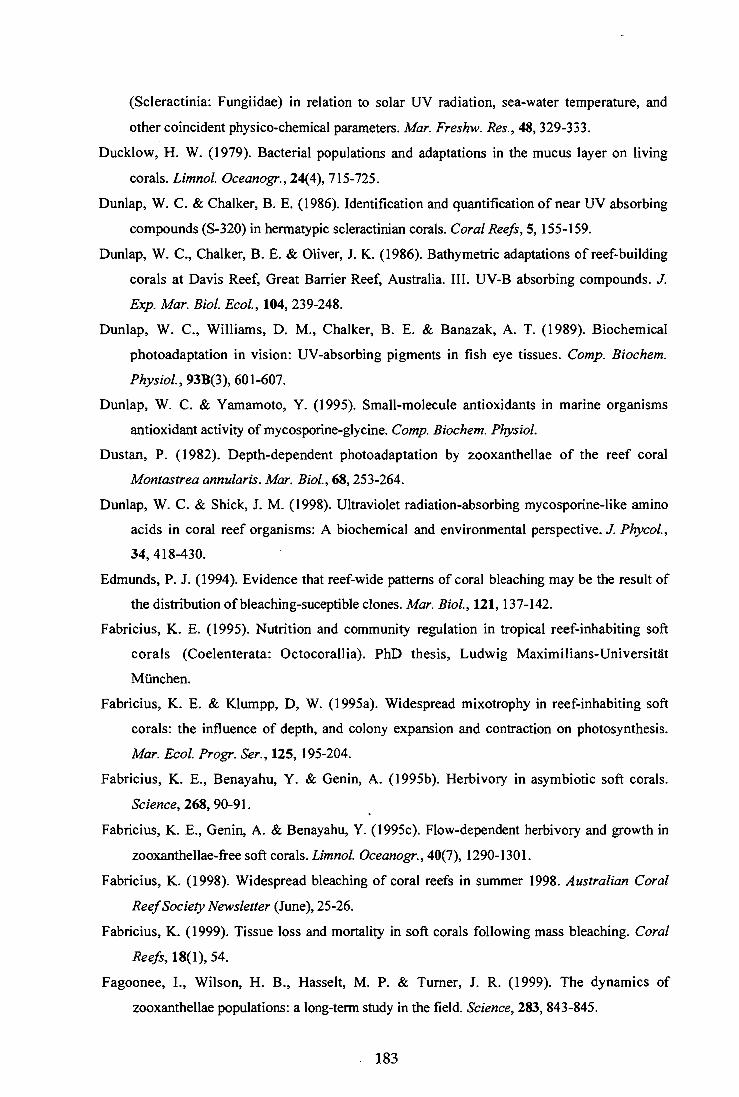
(Scleractinia: Fungiidae) in relation to solar UV radiation, sea-water temperature, and
other coincident physico-chemical parameters. Mar. Freshw. Res., 48, 329-333. Ducklow, H. W. (1979). Bacterial populations and adaptations in the mucus layer on living
corals. Limnol. Oceanogr., 24(4), 715-725.
Dunlap, W. C. & Chalker, B. E. (1986). Identification and quantification of near UV absorbing
compounds (S-320) in hermatypic scleractinian corals. Coral Reefs, 5, 155-159. Dunlap, W. C., Chalker, B. E. & Oliver, J. K. (1986). Bathymetric adaptations of reef-building
corals at Davis Reef, Great Barrier Reef, Australia. III. UV-B absorbing compounds. J. Exp. Mar. Biol. Ecol., 104, 239-248.
Dunlap, W. C., Williams, D. M., Chalker, B. E. & Banazak, A. T. (1989). Biochemical
photoadaptation in vision: UV-absorbing pigments in fish eye tissues. Comp. Biochem. Physiol., 93B(3), 601-607.
Dunlap, W. C. & Yamamoto, Y. (1995). Small-molecule antioxidants in marine organisms antioxidant activity of mycosporine-glycine. Comp. Biochem. Physiol.
Dustan, P. (1982). Depth-dependent photoadaptation by zooxanthellae of the reef coral Montastrea annularis. Mar. Biol., 68, 253-264.
Dunlap, W. C. & Shick, J. M. (1998). Ultraviolet radiation-absorbing mycosporine-like amino
acids in coral reef organisms: A biochemical and environmental perspective. J. Phycol., 34, 418-430.
Edmunds, P. J. (1994). Evidence that reef-wide patterns of coral bleaching may be the result of the distribution of bleaching-suceptible clones. Mar. Biol., 121, 137-142.
Fabricius, K. E. (1995). Nutrition and community regulation in tropical reef-inhabiting soft
corals (Coelenterata: Octocorallia). PhD thesis, Ludwig Maximilians-Universitat Munchen.
Fabricius, K. E. & Klumpp, D, W. (1995a). Widespread mixotrophy in reef-inhabiting soft
corals: the influence of depth, and colony expansion and contraction on photosynthesis. Mar. Ecol. Progr. Ser., 125, 195-204.
Fabricius, K. E., Benayahu, Y. & Genin, A. (1995b). Herbivory in asymbiotic soft corals. Science, 268, 90-91.
Fabricius, K. E., Genin, A. & Benayahu, Y. (1995c). Flow-dependent herbivory and growth in
zooxanthellae-free soft corals. Limnol. Oceanogr., 40(7), 1290-1301.
Fabricius, K. (1998). Widespread bleaching of coral reefs in summer 1998. Australian Coral Reef Society Newsletter (June), 25-26.
Fabricius, K. (1999). Tissue loss and mortality in soft corals following mass bleaching. Coral Reefs, 18(1), 54.
Fagoonee, I., Wilson, H. B., Hasselt, M. P. & Turner, J. R. (1999). The dynamics of
zooxanthellae populations: a long-term study in the field. Science, 283, 843-845.
183

Falkowski, P. G., Jokiel, P. L. & R. A. Kinzie (1990). Irradiance and corals. In: Z. Dubinsky
(Ed.), Ecosystems of the World - Coral Reefs, Amsterdam, Elsevier, 89-107.
Faulkner, D. J. (1992). Marine natural products. Nat. Prod. Chem., 9(4), 323-363.
Favre-Bovin, J., Arpin, N. & Brevard, C. (1976). Structure de la mycosporine (p310). Can. J. Chem., 54, 1105-1113.
Favre-Bovin, J. Bernillon, J. N. Satin & Arpin, N. (1987). Biosynthesis of mycosporines:
mycosporine glutaminol in Trichothecium roseum. Biochemistry, 26(9), 2509-2514. Fenical, W. (1993). Chemical studies of marine bacteria. Chem. Rev., 93, 1673-1684.
Fisk, D. A. & Done, T. J. (1985). Taxonomic and bathymetric patterns of bleaching in corals,
Myrmidon Reef (Queensland). 5 th International Coral Reef Congress, Tahiti, 6, 149-154.
Fitt, W. K. (1985). Effect of different strains of the zooxanthella Symbiodinium microadriadicum on growth and survival of their coelenterate and molluscan hosts. 5 th International Coral Reef Congress, Tahiti, 6, 131-136
Fitt, W. K., Spero, H. J., Halas, J., White, M. W. & Porter, J. W. (1993). Recovery of the coral Montastrea annularis in the Florida Keys after the 1987 Caribbean " bleaching event". Coral Reefs, 12, 57-64.
Fitt, W. K. & Warner, M. E. (1995). Bleaching patterns of four species of Caribbean reef corals. Biol. Bull., 189, 198-307.
Fleischmann, E. M. (1989). The measurement and penetration of ultraviolet radiation into
tropical marine water. Limnol. Oceanogr., 34(8), 1623-1629.
Frank, H. A. & Cogdell, R. J. (1996). Carotenoids in photosynthesis. Photochem. Photobiol., 63(3), 257-264.
Garcia-Pichel, F., Wingard, C. E. & Castenholz, R. W. (1993). Evidence regarding the UV
sunscreen role of a mycosporine-like compound in the cyanobacterium Gloeocapsa sp. App. Environmental Microbiology, 59(1), 170-176.
Gates, R. D. (1990). Seawater temperature and sublethal coral bleaching in Jamaica. Coral Reefs, 8, 193-197.
Gates, R. D., Baghdasarian, G. & Muscatine, L. (1992). Temperature stress causes host cell
detachment in symbiotic cnidarians: implications for coral bleaching. Biol. Bull., 182, 324-332.
Gershenzon, J. (1994). Metabolic costs of terpenoid accumulation in higher plants. J. Chem. Ecol., 20(6), 1281-1328.
Gleason, D. F. (1993). Differential effects of ultraviolet radiation on green and brown morphs of
the Caribbean coral Porites asteriodes. Limol. Oceanogr., 38(7), 1452-1463.
Gleason, D. F. &. Wellington, G.M. (1993). Ultraviolet radiation and coral bleaching. Nature, 365, 836-838.
184

Gleason, D. F. & Wellington, G. M. (1995). Variation in UVB sensitivity of planula larvae of
the coral Agaricia agaricites along a depth gradient. Mar. Biol., 123, 693-703.
Gleason, M. G. (1993). Effects of disturbance on coral communities: bleaching in Moorea,
French Polynesia. Coral Reef 12, 193-201.
Gleason, M. G. (1996). Coral recruitment in Moorea, French Polynesia: the importance of patch
type and temporal variation. J. Experimental Mar. Biol. and Ecol., 207, 79-101.
Glynn, P.W. & Stewart, R.H. (1973). Distribution of coral reefs in the Pearl islands in relation
to thermal conditions. LimnoL Oceanogr. 18, 367-379.
Glynn, P. W. & D'Croz, L. (1990). Experimental evidence for high temperature stress as a
cause of El Nitio-coincident coral mortality. Coral Reefs, 8, 181-191.
Glynn, P. W. & Weerdt, W. H. d. (1991). Elimination of two reef-building hydrocorals
following the 1982-83 El Nitlo Warming event. Science, 253, 69-71.
Glynn, P. W., Imai, R., Sakai, K., Nakano, Y. & Yamazato, K. (1992). Experimental responses
of Okinawan (Ryuku Islands, Japan) reef corals to high sea temperature and UV radiation.
7th International Coral Reef Symposium, Guam, 1, 27-37
Glynn, P. & Feingold, J. S. (1992). Hydrocoral species not extinct. Science, 257, 1845. Glynn, P. W. (1993). Coral reef bleaching: ecological perspectives. Coral Reefs, 12, 1-17. Glynn, P. W. (1996). Coral reef bleaching: Facts, hypotheses and implications. Global Change
Biol., 2, 495-509.
Glynn, P. W., Coley, S. B., Gassman, N. J., Black, K., Cortes, J. & Mate, J. L. (1996). Reef
coral reproduction in the eastern Pacific: Costa Rica, Panama, and Galapagos Islands
(Ecuador). III. Agariciidae (Pavona gigantea and Garineroseris planulata). Mar. Biol., 125(579-601).
Goodwin, T. W. (1976). Distribution of Carotenoids. In: T. W. Goodwin (Ed), Chemistry and
Biochemistry of Plant Pigments, London, Academic Press, 225-257
Goreau, T. F., Goreau, N. I. & Yonge, C. M. (1964). Mass expulsion of zooxanthellae from
Jamaican coral reef communities after hurricane Flora. Science, 145, 383-386. Goreau, T. J. & Macfarlane, A. H. (1990). Reduced growth rate of Montastrea annularis
following the 1987-1988 coral-bleaching event. Coral Reefs, 8, 211-215.
Grant, P. T., Middleton, C., Plack, P. A. & Thomson, R. H. (1985). The isolation of four
aminocyclohexamines (mycosporines) and a structurally related derivative of cyclohexane-
1:3 dione (gadusol) from the brine shrimp, Anemia. Comp. Biochem. Physiol., 80B(4), 755-759.
Griffith, J. K. (1994). Predation on soft corals (Octocorallia: Alcyonacea) on the Great Barrier
Reef, Australia. Aust. J. Mar. Freshw. Res., 45, 1281-1284.
Hall, V. (1998). Injury and regeneration of common reef-crest corals at Lizard Island, Great
Barrier Reef, Australia. PhD thesis, James Cook University.
185

Harding, R. W. & Jr., W. S. (1980). Photocontrol of carotenoid biosynthesis. Ann. Rev. Plant Physiol., 31, 217-38.
Harland, A. D., Davis, P. S. & Fixter, L. M. (1992). Lipid content of some Caribbean corals in relation to depth and light. Mar. Biol., 113, 357-361.
Harriott, V. J. (1985). Mortality rates of scleractinian corals before and during a mass bleaching evnt. Mar. Ecol. Prog. Ser., 21, 81-88.
Harrison, P. L. & Wallace, C. C. (1990). Reproduction, dispersal and recruitment of
scleractinian corals. In Z. Dubinsky (Ed.), Ecosystems of the World - Coral Reefs,
Amsterdam, Elsevier, 133-207.
Harvell, C. D. & Fenical, W. (1989). Chemical structural defenses of Caribbean gorgonians
(Pseudopterogorgia spp.): Intracolony localization of defense. Limnol. Oceanogr., 34(2), 382-389.
Harvell, C. D., Fenical, W., Roussis, V., Ruesink, J. L., Griggs, C. C. & Greene, C. H. (1993).
Local and geographic variation in the defensive chemistry of a West Indian gorgonian coral (Briareum asbestinum). Mar. Ecol. Progr. Ser., 93, 165-173.
Hatcher, B. G. & Larkum, A. W. D. (1983). An experimental analysis of factors controlling the
standing crop of epilithic algal community on a coral reef. J. Exp. Mar. Biol. Ecol., 69, 61-84.
Havenhand, J. N. (1993). Egg to juvenile period, generation time, and the evolution of larval type in marine invertebrates. Mar. Ecol. Progr. Ser., 97, 247-260.
Hay, M. E. (1996). Marine chemical ecology: what's known and what's next? J. Exp. Mar. Biol. Ecol., 200, 103-134.
Heaton (1988) Chemical studies on soft corals. PhD thesis, James Cook University.
Helbling, E. W., Chalker, B. E., Dunlap, W. C., Holm-Hansen, 0. & V. Villafdie (1996). Photoacclimation of Antarctic marine diatoms to solar ultraviolet radiation. J. Exp. Mar. Biol. Ecol., 204, 85-101.
Hoegh-Guldberg, 0. & Smith, G. J. (1989a). The effect of sudden changes in temperature, light
and salinity on the population density and export of zooxanthellae from the reef corals
Stylophora pistillata Esper and Seriatopora hystrix Dana. J. Exp. Mar. Biol. Ecol., 129, 279-303.
Hoegh-Guldberg, 0. & Smith, J. G. (1989b). Influence of the population density of
zooxanthellae and supply of ammonium on the biomass and metabolic characteristics of
the reef corals Seriatopora hystrix and Stylophora pistillata. Mar. Ecol. Progr. Ser., 57, 173-186.
Hoegh-Guldberg, 0. & Salvat, B. (1995). Periodic mass-bleaching and elevated sea
temperatures: bleaching of outer reef slope communities in Moorea, French Polynesia. Mar. Ecol. Progr. Ser., 121, 181-190.
186

Hoegh-Guldberg, 0. (1999). Climate change, coral bleaching and the furture of the world's coral reefs. Aus. Mar. Freshw. Res., in press
Hoegh-Guldberg, 0. & Jones, R. J. (1999). Photoinhibition and photoprotection in symbiotic dinoflagellates from reef-building corals. Mar. Ecol Progr Ser., 183, 63-86.
Hoeksema, B. W. (1991). Control of bleaching in mushroom coral populations (Scleractinia:
Fungiidae) in the Java Sea: stress tolerance and interference by life history strategy. Mar. Ecol. Progr. Ser., 74, 225-237.
Hughes, T. P. (1994). Catastrophes, phase shifts, and large-scale degradation of a Caribbean coral reef. Science, 265, 1547-1551.
Hughes, T. P. & Connell, J. H. (1999). Multiple stressors on coral reefs: A long-term perspective. Limnol. Oceanogr., 44(3), 932-940.
Iglesias-Prieto, R., Matta, J. L., Robins, W. A. & Trench, R. K. (1992). Photosynthetic response
to elevated temperature in the symbiotic dinoflagellate Symbiodinium microadriaticum in culture. Proc. Natl. Acad. Sci., 89, 10302-10305.
Iglesias-Prieto, R. & Trench, R. K. (1997). Acclimation and adaptation to irradiance in
symbiotic dinoflagellates. II. Response of chlorophyll-protein complexes to different photon-flux intensities. Mar. Biol., 130, 23-33.
Ishikura, M., Kato, C., & Maruyama, T. (1997). UV-absorbing substances in zooxanthellate and azooxanthellae clams. Mar. Biol., 128, 649-655.
Jeffrey, S. W. & Haxo, F. T. (1968). Photosynthetic pigments of symbiotic dinoflagellates (zooxanthellae) from corals and clams. Biol. Bull. mar. biol. Lab. Woods Hole, 135, 141-148.
Jeffrey, S. W. & Humphrey, G. H. (1975). New spectrophotometric equations for determining
chlorophylls a, b, c1 and c2 in higher plants, algae and natural phytoplankon. Biochem. Physiol. Pflanzen, 167, 191-194.
Jeffrey, S. W., Mantoura, R. C. F. & Wright, S. W. (Ed.). (1997). Phytoplankton pigments in
oceanography: guidelines to modern methods. United Nations Educational, Scientific and
Cultural Organization, Paris.
Jerlov, N. G. (1950). Ultra-violet radiation in the sea. Nature, 166, 111-112.
Jindal, A., Ritchie, K. B., Hayes, R. L., Goreau, T. J. & Smith, G. W. (1995). Bacterial ecology
of selected corals following the 1994 South Central bleaching event. In: L. Kass (Ed.),
27th Meeting Assoc. Mar. Labs. Caribbean, abstract only
Johnson, K. G. (1992). Synchronous planulation of Manicina areolata (Scleractinia) with lunar periodicity. Mar. Ecol. Progr. Ser., 87, 265-273.
Jokiel, P. L. & Coles, S. L. (1977). Effects of temperature on the mortality and growth of
Hawaiian reef corals. Mar. BioL, 43, 201-208.
187

Jokiel, P. L. & Guinther, E. B. (1978). Effects of temperature on reproduction in the hermatypic coral Pocillopora damnicornis. Bull. Mar. Sci., 28(4), 786-789.
Jokiel, P. L. & York, R. H. (1982). Solar ultraviolet photobiology of the reef coral Pocillopora damnicornis and symbiotic zooxanthellae. Bulletin of Marine Science, 32(1), 301-315.
Jokiel, P. L. & York, R.H. (1984). Importance of ultraviolet radiation in photoinhibition of macroalgal growth. Limnol. Oceanogr., 29(1), 192-199.
Jokiel, P. L. & Coles, S. L. (1990). Response of Hawaiian and other Indo-Pacific reef corals to
elevated temperature. Coral Reefs, 8, 155-162.
Jokiel, P. L., Lesser, M. P. & Ondrusek, M. E. (1995). UV-absorbing compounds in the coral
Pocillopora damnicornis: effects of light, water flow and ultraviolet radiation. In: D. Gulko
& P. L. Jokiel (Eds.), Ultraviolet Radiation and Coral Reefs, Hawaii Inst. Mar. Biol. Tech.
Rep., Honolulu, 87
Jokiel, P. L., Lesser, M. P. & Ondrusek, M. E. (1997). UV-absorbing compounds in the coral
Pocillopora damnicornis: Interactive effects of UV radiation, photosynthetically active radiation, and water flow. Limnol. Oceanogr., 42(6), 1468-1473.
Jones, R. J. (1995) Sublethal stress assessment in scleractinia and regulatory biology of the
coral-algal symbiosis. PhD thesis, James Cook University'.
Jones, R. J. (1997). Changes in zooxanthellar densities and chlorophyll concentrations in corals during and after a bleaching event. Mar. Ecol. Progr. Ser., 158, 51-59.
Jones, R. J., Berkelmans, R. & Oliver, J. K. (1997). Recurrent bleaching of corals at Magnetic Island (Australia) relative to air and seawater temperature. Mar. Ecol. Progr. Ser., 158, 289-292.
Jones, R. J. & Yellowlees, D. (1997). Regulation and control of intracellular algae (= zooxanthellae) in hard corals. Phil. Trans. R. Soc. Lond. B., 352, 457-468.
Jones, R. J., Hoegh-Guldberg, 0., Larkum, A. W. D. & Schreiber, U. (1998). Temperature-
induced bleaching of corals begins with impairment of dark metabolism in zooxanthellae.
Plant, Cell and Environment, 21, 1219-1230.
Karentz, D., McEuen, E. S., Land, M. C. & Dunlap, W. C. (1991). Survey of mycosporine-like
amino acid compounds in Antarctic marine organisms: potential protection from
ultraviolet exposure. Mar. Biol., 108, 157-166.
Karentz, D., Dunlap, W. C. & Bosch, I. (1997). Temporal and spatial occurrence of UV-
absorbing mycosporine-like amino acids in tissues of the Antarctic sea urchin Sterechinus neumayeri during spring-time ozone depletion. Mar. BioL, 129, 343-353.
Karsten, U., Franklin, L. A., Lining, K. & Wiencke, C. (1998). Natural ultraviolet radiation and
photosynthetically active radiation induce formation of mycosporine-like amino acids in
the marine macroaolga Chondrus crispus (Rhodophyta). Planta, 205, 257-261.
188

Kato, M. (1987). Mucus-sheet formation and discoloration in the reef-building coral Porites cylindrica: effects of altered salinity and temperature. Galaxea, 6, 1-16.
Kazlaukas, R., Murphy, P. T., Wells, R. J., Schoenholzer, P. & Coll, J. C. (1978). Cembranoid
constituents from an Australian collection of the soft coral Sinularia flexibilis. Aust. J. Chem., 31, 1817-1824.
Kiefer, P. A., K.L. Rinehart, J. & Hooper, I. R. (1986). Renillafoulins, antifouling diterpenes from the sea pansy Renilla reniformis (Octocorallia). J. Org. Chem., 51, 4450-4454.
Kelman, D., Kushmaro, A., Loya, Y., Kashman, Y. & Benayahu, Y. (1998). Antimicrobial
activity of a Red Sea soft coral, Parerythopodium fulvum fulvum: reproductive and developmental considerations. Mar. Ecol. Progr. Ser., 169, 87-95.
Kinzie, R. A. (1974). Experimental infection of aposymbiotic gorgonian polyps with zooxanthellae. J. Exp. Mar. Biol. Ecol., 15, 335-345.
Kinzie III, R. A. & Chee, G. S. (1979). The effect of different zooxanthellae on the growth of
experimentally re-infected hosts. Biol. Bull., 156, 315-327.
Kinzie, R. A. I. (1993). Effects of ambient levels of solar ultraviolet radiation on zooxanthellae and photosynthesis of the reef coral Montipora verrucosa. Mar. Biol., 116, 319-327.
Kleppel, G. S., Dodge, R. E. & Reese, C. J. (1989). Changes in pigmentation associated with the bleaching of stony corals. Limnol. Oceanogr., 34(7), 1331-1335.
Knowlton, N., Weil, E., Weigt, L. A. & Guzman, H. M. (1992). Sibling species in Montastrea annularis, coral bleaching and the coral climate record. Science, 255, 330-331.
Koh, E. G. L. (1997). Do scleractinian corals engage in chemical warfare against microbes? J. Chem. Ecol., 23(2), 379-398.
Kohen, E., Santos, R., & Hirschberg, J. G. (1995). Photobiology. San Diego, Academic Press.
Kojis, B. L. & Quinn, N. J. (1984). Seasonal and depth variation in fecundity at two reefs in Papua New Guinea. Coral Reefs, 3, 165-172.
Kokke, W. C. M. C., Fenical, W., Bohlin, L. & Dierassi, C. (1981). Sterol synthesis by cultured
zooxanthellae; implications concerning sterol metabolism in the host-symbiont association
in Caribbean gorgonians. Comp. Biochem. Physiol., 68 B, 281-287.
Kokke, W. C. M. C., Epstein, S., Look, S. A., Rau, G. H. & Fenical, W. (1984). On the origin of
terpenes in symbiotic associations between marine invertebrates and algae (zooxanthellae).
J. Biol. Chem., 259(13), 8168-8173.
Krupp, D. A. & Blanck, J. (1995). Preliminary report on the occurrence of mycosporine-like
amino acids in the eggs of the Hawaiin scleractinian corals Montipora verrucosa and Fungia scrutinaria. In D. Gulko & P. L. Jokiel (Eds.), Ultraviolet Radiation and Coral Reefs (pp. 129-134). Honolulu: Hawai'i Inst. Mar. Biol. Tech. Rep..
Kuffner, I. B., Ondrusek, M. E. & Lesser, M. P. (1995). Distribution of mycosporine-like amino
acids in the tissues of Hawaiian Scleractinia: A depth profile. In: D. Gulko & P. L. Jokiel
189

(Eds.), Ultraviolet Radiation and Coral Reefs, Hawaii Inst. Mar. Biol. Tech. Rep..
Honolulu, 77-86
Kiihl, M., Cohen, Y., Dalsgaard, T., Jorgensen, B. B. & Revsbech, N. P. (1995).
Microenvironment and photosynthesis of zooxanthellae in scleractinian corals studied with microsensors for 02, pH and light. Mar. Ecol. Progr. Ser., 117, 159-172.
Kushmaro, A., Loya, Y., Fine, M. & Rosenberg, E. (1996). Bacterial infection and coral bleaching. Science, 380, 396.
Kushmaro, A., Rosenberg, E., Fine, M. & Loya, Y. (1997). Bleaching of the coral Oculina patagonica by Vibrio AK-1. Mar. Ecol. Progr. Ser., 147, 159-165.
La Barre, S., Coll, J. C. & Sammarco, P. W. (1986a). Defensive strategies of soft corals
(Coelenterata: Octocorallia) of the Great Barrier reef. II. The relationship between toxicity and feeding deterrence. Biol. Bull., 171, 565-576.
La Barre, S. C., Coll, J. C. & Sammarco, P. W. (1986b). Competetive strategies of soft corals
(Coelenterata: Octocorallia): III. Spacing and aggressive interactions between alcyonaceans. Mar. Ecol. Progr. Ser., 28, 147-156
Leach, C. M. (1965). Ultraviolet-absorbing substances associated with light-induced sporulation in fungi. Can. J. Bot., 34, 185-200.
Leder, J. L., Szmant, A. M. & Swart, P. K. (1991). The effect of prolonged "bleaching" on
skeletal banding and stable isotopic composition in Montastrea annularis. Coral Reefs, 10, 19-27.
Leone, P. A., Bowden, B. F., Carroll, A. R. & Coll, J. C. (1995). Chemical consequences of
relocation of the soft coral Lobophytum compactum and its placement in contact with the red alga Plocamium hamatum. Mar. Biol., 122, 675-679.
Lesser, M. P., Stochaj, W. R., Tapley, D. W. & Shick, J. M. (1990). Bleaching in coral reef
anthozoans: effects of irradiance, ultraviolet radiation, and temperature on the activities of
protective enzymes against active oxygen. Coral Reefs, 8, 225-232.
Lesser, M. P. (1996). Acclimation of phytoplankton to UV-B radiation: oxidative stress and
photoinhibition of photosynthesis are not prevented by UV-absorbing compounds in the
dinoflagellate Prorocentrum micans. Mar. Ecol. Progr. Ser., 132, 287-297.
Lesser, M. P. (1997). Oxidative stress causes coral bleaching during exposure elevated
temperatures. Coral Reefs, 16, 187-192.
Lewis, J. B. (1982). Feeding bahaviour and feeding ecology of the Octocorallia (Coelenterata:
Anthozoa). J. Zool. Lond., 196, 371-384.
Loh, W., Crater, D. A. & 0. Hoegh-Guldberg, 0. (1998). Zooxanthellae from scleractinian
corals of One Tree Island (Great Barrier Reef). In: J. G. Greenwood & N. J. Halls (Eds.),
Australian Coral Reef Society 75 th Anniversary Conference, Heron Island, 141-150
190 .

Lowry, H. 0., Rosenbrough, N. J., Fan, A. L. & Randall, R. J. (1951). Protein measurement
with the folin phenol reagent. J. Biol. Chem., 193, 265-275
Loya, Y. & Rinkevich, B. (1979). Abortion effects in corals induced by oil pollution. Mar. Ecol. Progr. Ser., 1, 77-80.
Maida, M., Carroll, A. R. & Coll, J. C. (1993). Variability of terpene content in the soft coral
Sinularia flexibilis (Coelenterata: Octocorallia), and its ecological implications. J. Chem. Ecol., 19(10), 2285-2296.
Maida, M., Sammarco, P. W. & Coll, J. C. (1995a). Preliminary evidence for directional
allelopathic effects of the soft coral Sinularia flexibilis (Alcyonacea: Octocorallia) on scleractinian coral recruitment. Bull. Mar. Sci., 56(1), 303-311.
Maida, M., Sammarco, P. W. & Coll, J. C. (1995b). Effects of soft corals on scleractinian coral
recruitment. I. Directional allelopathy and inhibition of settlement. Mar. Ecol. Progr. Ser., 121, 191-202.
Manabe, S., Stouffler, R.J., Spelman, M.J. & K. Bryan (1991). Transient responses of a coupled
ocean -atmosphere model to gradual changes of atmospheric CO2. Part. 1: Annual mean response. J. Climate, 4, 785-818 .
Marshall, P. A. & Baird, A. H. (1999). Variation in assemblage response and taxa
susceptibilities to bleaching of corals in the Central Great Barrier Reef. Mar. Ecol. Progr. Ser., in press.
Masuda, K., Goto, M., Maruyama, T. & Micyachi, S. (1993). Adaptation of solitary corals and
their zooxanthellae to low light and UV radiation. Mar. Biol. 117, 685-691. Mate, J. L. (1997). Experimental responses of Panamanian reef corals to high temperature and
nutrients. 8th International Coral Reef Symposium, Panama, 1, 515-520.
McCloskey, L. R., Cove, T. G. & Verde, A. E. (1996). Symbiont expulsion from the anemone
Anthopleura elegantissima (Brandt) (Cnidaria; Anthozoa). J. Exp. Mar. Biol. EcoL , 195, 173-186.
McDermostt, A. & Blanquet, R. S. (1991). Glucose and glycerol uptake by isolated
zooxanthellae from Cassiopeia xamachana: Transport mechanism and regulation by host
homogenate fractions. Mar. Biol., 108, 129-136.
Meesters, E. H. & Bak, R. P. M. (1993). Effects of coral bleaching on tissue regeneration
potential and colony survival. Mar. Ecol. Progr. Ser., 96, 189-198.
Meesters, E. H., Noordeloos, M. & Bak, R. P. M. (1994). Damage and regeneration: links to
growth in the reef-building coral Montastrea annularis. Mar. Ecol. Progr. Ser., 112, 119-128.
Michalek, K. & Bowden, B. F. (1997). A natural algacide from soft coral Sinularia flexibilis (Coelenterata, Octocorallia, Alcyonacea). J. Chem. Ecol., 23(2), 259-273.
191

Michalek-Wagner, K. and B.L. Willis (2000): Impacts of bleaching on the soft coral
Lobophytum compactum. I. Fecundity, fertilisation and offspring viability. Coral Reefs, accepted pending minor revision
Michalek-Wagner, K. and B.L. Willis (2000): Impacts of bleaching on the soft coral
Lobophytum compactum. II. Biochemical changes in adults and their gametes. Coral Reefs, accepted pending minor revision
Miller, D. J. & Yellowlees, D. Y. (1989). Inorganic nitrogen uptake by symbiotic marine
cnidarians: a critical review. Proc. R. Soc. Lond B. 237, 109-125.
Muscatine, L. and R.R. Pool ( 1979) Regulation of numbers of intracellular algae. Proc. R. Soc. Lond. B, 204, 131-139.
Muscatine, L. (1980). Productivity of zooxanthellae. In: P. G. Falkowski (Ed.), Primary
productivity of the sea, Academic Press, London, 381-402.
Muscatine, L., Falkowski, P. G., Porter, J. W. & Dubinsky, Z. (1984). Fate of photosynthetic
fixed carbon in light-and shade adapted colonies of the symbiotic coral Stylophora pistillata. Proc. R. Soc. Lond. B, 222, 181-202.
Nair, S. & Simidu, U. (1987). Distribution and significance of heterotrophic marine bacteria with antibacterial activity. Appl. Environ. Microbiol., 53(12), 2957-2962.
Negelkerken, I., Buchan, K., Smith, G. W., Bonair, K., Bush, P., Garzon-Ferreira, J., Botero, L.,
Gayle, P., Harvell, C. D., Heberer, C., Kim, K., Petrovic, C., Pors, L. & Yoshioka, P.
(1997). Widespread disease in Caribbean sea fans: II. Patterns of infection and tissue loss. Mar. Ecol. Progr. Ser., 160, 255-263.
Nii, C. M. & Muscatine, L. (1997). Oxidative stress in the symbiotic sea anemone Aiptasia pulchella (Carlgren, 1943): Contribution of the animal to superoxide ion production at elevated temperature. Biol. Bull., 192, 444-456.
Odum, E. P. (1971). Fundamentals of ecology ( 3 rd edition) London, Academic Press.
Ohnishi, K., Matsumoto, H., Takahashi, A., Wang, X. & Ohnishi, T. (1996). Heat shock
transcription factor, HSF, is activated by ultraviolet irradiation. Photochem. Photobiol., 64(6), 949-952.
Oliver, J. (1985). Recurrent seasonal bleaching and mortality of corals on the Great Barrier
Reef. 5 th International Coral Reef Congress, Tahiti, 4, 201-206.
Oren, U., Benyahu, Y. & Loya, Y. (1997). Effect of lesion size and shape on regeneration of the
Red Sea coral Favia favus. Mar. Ecol. Progr. Ser., 146, 101-107.
Paerl, H. W. (1984). Cynobacterial carotenoids: their roles in maintaining optimal
photosynthetic production among aquatic bloom forming genera. Oecologia, 61, 143-149. Pain, S. (1998) Foul medicine. New Scientist, 7 February, 11.
192

Papastephanou, C. & Anderson, D. G. (1982). Crassin acetate biosynthesis in a cell-free
homogenate of zooxanthellae from Pseudoplexaura porosa (Houttyun); implication to the symbiotic process. Comp. Biochem. Physiol., 73B(3), 617-624.
Pass, M. A., Capra, M. F., Carlisle, C. H., Lawn, I. & Coll, J. J. (1989). Stimulation of
concentrations in the polyps of the soft coral Xenia elongata by compounds extracted from other alcyoneacean soft corals. Comp. Biochem. Physiol., 94C(2), 677-681.
Patton, J. S., Abraham, S. & Benson, A. A. (1977). Lipogenesis in the intact coral Pocillopora capitata and its isolated zooxanthellae: Evidence for a light-driven carbon cycle between
symbiont and host. Mar. Biol., 44, 235-247.
Patton, J. S. & Burris, J. E. (1983). Lipid synthesis and extrusion by freshly isolated
zooxanthellae (symbiotic algae). Mar. Biol., 75, 131-136.
Paul, V. J. & Fenical, W. (1986). Chemical defense in tropical green algae, order Caulerpales.
Mar. Ecol. Progr.Ser., 34, 255-264.
Paul, V. J. & Van, K. L. A. (1988). Chemical defense and chemical variation in some tropical Pacific species of Halimeda (Halimedaceae: Chlorophyta). Coral Reefs, 6, 263-270.
Paul, V. J. & Van, K. L. A. (1992). Activation of chemical defenses in tropical green algae
Halimeda spp. J. exp. Mar. Biol. Ecol., 160, 191-203.
Pearson, R. G. (1981). Recovery and recolonization of coral reefs. Mar. Ecol. Progr. Ser., 4, 105-122.
Peterson, P. M. J., Smith, R. C., Patterson, K. W. & Jokiel, P. L. (1995). A biological weighting
function for phytoplankton growth inhibition. In: D. Gulko & P. L. Jokiel (Eds.),
Ultraviolet Radiation and Coral Reefs, Hawaii Inst. Mar. Biol. Tech. Rep.., Honolulu, 51-60.
Porter, J. W., Fitt, W. K., Spero, H. J. & Rogers, C. S. (1989). Bleaching in reef corals:
physiological and stable isotopic responses. Proc. Natl. Acd. Sci., 86, 9342-9346.
Prezelin, B. B. (1976). The role of peridinin-chlorophyll a-proteins in the photosynthetic light
adaptation of the marine dinoflagellate, Glenodinium sp. Planta, 130, 225-233.
Reaka-Kudla, M. L., O'Connell, D. S., Regan, J. D. & Wicklund', R. I. (1993). Effects of
temperature and UV-B on different components of coral reef communities from the
Bahamas. In: Ginsburg R.N. (Ed.) Proceedings of the colloqium on global aspects of coral
reefs: health, hazards and history. Rosenstiel School of Marine and Atmospheric Science,
University of Miami, Miami, 126-131.
Renger, G., Voelker, M., Eckert, H. J., Fromme, R., Holm-Veit, S. & Graeber, P. (1989). On the
mechanism of photosystem II deterioration by UV-B irradiation. Photochem. Photobiol., 49, 97-105.
Richardson, L.L. (1998): Coral diseases: what is really known? TREE, 13(11), 438-4443
193

Richmond, R. H. (1987). Energetic relationships and biogeographical differences among
fecundity, growth and reproduction in the reef coral Pocillopora damnicornis. Bull. Mar. Sci., 41(2), 594-604.
Richmond (1996). Reproduction and recruitment in corals: critical links in the persistence of
reefs. . In: C. Birkeland (Ed.), Life and death of coral reefs. London, Chapman & Hall,
175-197.
Rinkevich, B. & Loya, Y. (1979). The reproduction of the Red sea coral Stylophora pistillata. I. Gonads and planulae. Mar. Ecol. Progr. Ser., 1, 133-144.
Rinkevich, B. & Loya, Y. (1987). Variability in the pattern of sexual reproduction of the coral
Stylophora pistillata at Eilat, Red Sea: A long-term study. Biol. Bull., 173, 335-344.
Rinkevich, B. (1996). Do reproduction and regeneration in damaged colonies compete for
energy allocation? Mar. Ecol. Progr. Ser. 43, 297-302
Ritchie, K. B., Dennis, J. H., McGrawth, T. & Smith, G. W. (1993). Bacteria associated with
bleached and non-bleached areas of Montastrea annularis. In: L. Kass (Ed.), 5 th Symp. Nat. Hist. Bahamas, San Salvador, Bahamas, 75-79
Rittschoff, D., Hooper, I. R., Branscomb, E. S. & Coslow, J. D. (1985). Inhibition of barnacle
settlement and behaviour by natural products from whip corals. J. Chem. Ecol., 11, 551-563.
Rowan, R. & Powers, D. A. (1991). A molecular genetic classification of zooxanthellae and the
evolution of animal-algal symbiosis. Science, 251, 1348-1350.
Rowan, R. & Knowlton, N. (1995). Intraspecific diversity and ecological zonation in coral-algal symbiosis. Proc. Natl. Acad. Sci., 92, 2850-2853.
Rowan, R., Knowlton, N., Baker, A. & Jara, J. (1997). Landscape ecology of algal symbionts
creates variation in episodes of coral bleaching. Nature, 388, 265-228.
Rublee, P. A., Lasker, H. R., Gottfried, M. & Roman, M. R. (1980). Production and bacterial
colonization of mucus from the soft coral Briareum asbestinum. Bull. Mar. Sci., 30(4), 888-893.
Salih, A., Hoegh-Guldberg, 0. & Cox, G. (1998). Bleaching reponses of symbiotic
dinoflagellates in corals: The effects of light and elevated temperature on their morphology
and physiology. In: J. G. Greenwood & N. J. Halls (Eds.), Australian Coral Reef Society
75th Anniversary Conference, Heron Island, The University of Queensland Printery, 199-
215.
Sammarco, P. W., Coll, J. C., Barre, S. L. & Willis, B. (1983). Competitive strategies of soft
corals (Coelenterata: Octocorallia): Allelopathic effects on selected scleractinian corals.
Coral Reefs, 1, 173-178.
194

Sammarco, P. W., Coll, J. C. & La Barre, S. (1985). Competitive strategies of soft corals
(Coelenterata: Octocorallia). II Variable defense responses and susceptibility to scleractinian corals. J. Exp. Mar. Biol. Ecol., 91, 199-215.
Sammarco, P. W., Barre, S. L. & Coll, J. C. (1987). Defensive strategies of soft corals
(Coelenterata: Octocorallia) of the Great Barrier Reef. III. The relationship between
ichthyotoxicity and morphology. Oecologia, 74, 93-101.
Sammarco, P. W. & Coll, J. C. (1990). Lack of predictability in terpenoid function. Multiple
roles and integration with related adaptations in soft corals. J. Chem. Ecol., 16(1), 273-289.
Sammarco, P. W. & Coll, J. C. (1992). Chemical adapatations in the Octocorallia: evolutionary considerations. Mar. Ecol. Progr. Ser., 88, 93-104.
Sammarco, P. W. & Coll, J. C. (1996). Secondary metabolites - or primary? Re-examination of
a concept through a marine example. 8 th International Coral Reef Symposium, Panama, 2, 1245-1249.
Scelfo, G. (1985). The effects of visible and ultraviolet solar radiation on a UV-absorbing
compound and chlorophyll a in a Hawaiian zoanthid. 5th Intenational Coral Reef Congress, Tahiti, 1985, 6(107-112).
Scelfo, G. M. (1986). Relationship between solar radiation and pigmentation of the coral Montipora verrucosa and its zooxanthellae. In: P. L. Jokiel, R. H. Richmond & R. A.
Rogers (Eds.), Coral reef population biology. Hawaii Inst. Mar. Biol. Tech. Rep., Honolulu, 440-451.
Scheuer (1973). Chemistry of Marine Natural Products. New York, Academic Press.
Schoenberg, D. A. & Trench, R. K. (1980a). Genetic variation in Symbiodinium (Gymnodinium) microadriaticum Freudenthal, and specificity in its symbiosis with marine invertebrates. I. Isoenzyme and soluable protein patterns of axenic cultures of Symbiodinium microadriaticum. Proc. R. Soc. Lond., 207B, 405-427.
Schoenberg, D. A. & Trench, R. K. (1980b). Genetic variation in Symbiodinium (Gymnodinium) microadriaticum Freudenthal, and specificity in its symbiosis with marine invertebrates. II.
Morphological variation in Symbiodinium microadriaticum. Proc. R. Soc. Lond., 207B, 429-444.
Schoenberg, D. A. & Trench, R. K. (1980c). Genetic variation in Symbiodinium (Gymnodinium) microadriaticum Freudenthal, and specificity in its symbiosis with marine invertebrates.
III. Specifity and infectivity of Symbiodinium microadriaticum. Proc. Roy. Soc. Lond., 207B, 445-460.
Schwartz, R. E. & Scheuer, P. J. (1981). The coralenoilides, constituents of pink coral,
Corallium sp. Tetrahedron, 37(16), 2725-2733.
Sebens, K. P. (1987). Coelenterate energetics. In: F. J. Vernberg & T. J. Panadian (Eds.),
Animal Energetics, New York, Academic Press, 55-120.
195

Sharp, V. A., Miller, D., Bythell, J. C. & Brown, B. E. (1994). Expression of low molecular
weigth HSP 70 related polypetides from the symbiotic sea anemone Anemonia viridis Forskall in response to heat shock. J. Exp. Mar. Biol. Ecol., 179, 179-193.
Shick, M., Lesser, M. P. & Stochaj, W. R. (1991). Ultraviolet radiation and photoactive stress in
zooxanthellae anthozoa: the sea anemone Phyllodiscus semoni and the octocoral Clavularia sp. Symbiosis, 10, 145-173.
Shick, J. M., Dunlap, W. C., Chalker, B. E., Banazak, A. T. & Rosenzweig, T. K. (1992).
Survey of ultraviolet radiation-absorbing mycosporine-like amino acids in organs of coral reef holothuroids.Mar. Ecol. Progr. Ser. , 90, 139-148.
Shick, J. M., Lesser, M. P., Dunlap, W. C., Stochaj, R., Chalker, B. E. & Won, J. W. (1995).
Depth-dependent responses to solar ultraviolet radiation and oxidative stress in the zooxanthellate coral Acropora microphthalma. Mar. Biol., 122, 41-51.
Shick, M. J., Lesser, M. P. & Jokiel, P. (1996). Effects of ultraviolet radiation on corals and other coral reef organisms. Global Change Biology, 2, 527-545.
Shimidzu, N., Goto, M. & Miki, W. (1996). Carotenoids as singlet oxygen quenchers in marine organisms. Fisheries Science, 62(1), 134-137.
Sier, C. J. S. & Olive, P. J. W. (1994). Reproduction and reproductive variability in the coral Pocillopora verrucosa from the Republic of Maledives. Mar. Biol., 118, 713-722.
Slattery, M. & McClintock, J. B. (1995a). Population structure and feeding deterrence in three
shallow-water Antarctic soft corals. Mar. Biol., 122, 461-470.
Slattery, M., McClintock, J. B. & Heine, J. N. (1995b). Chemical defenses in Antarctic soft corals: evidence for anti-fouling compounds. J. Exp. Mar. Biol. Ecol., 190, 61-77.
Slattery, M., Hamann, M. T., McClintock, J. B., Perry, T. L., Puglisi, M. P. & Yoshida, W. Y.
(1997). Ecological roles for water-borne metabolites from Antarctic soft corals. Mar. Ecol. Progr. Ser., 161, 133-144.
Slattery, M., Hines, G. A., Strainer, J. & Paul, V. J. (1999). Chemical signals in gametogenesis,
spawning, and larval settlement and defense of the soft coral Sinularia polydactyla. Coral Reefs, 18,75-84.
Sokal, R.R. and F.J. Rohlf (1995). Biometry. New York, W.H. Freeman and Company.
Stanley, G. D. j. (1981). Early history of scleractinian corals and geological consequences.
Geology, 9, 507-511.
Stanley, G. D. & Swart, P. K. (1995). Evolution of the coral-zooxanthellae symbiosis during
Triassic: a geochemical approach. Paleobiology, 21(2), 179-199.
Stimson, J. (1997). The annual cycle of density of zooxanthellae in the tissues of field and laboratory Pocillopora damnicornis ( Linnaeus). J. Exp. Mar. Biol. Ecol., 214, 35-48.
Stochaj, W. R., Dunlap, W. C. & Shick, J. M. (1994). Two new UV-absorbing mycosporine-
like amino acids from the sea anemone Anthopleura elegantissima and the effects of
196

zooxanthellae and spectral irridiance on chemical composition and content. Mar. Biol., 118, 149-156.
Suharsono, Pipe, R. K. & Brown, B. E. (1993). Cellular and ultra-structural changes in the
endoderm of the temperate sea anemone Anemonia viridis as a result of increased temperature. Mar. Biol., 116, 311-318.
Sutton, D. C. & Hoegh-Guldberg, 0. (1990). Host-zooxanthellae interactions in four temperate
marine invertebrate symbioses: assessment of effect of host extracts on symbionts. Biol. Bull., 178, 175-186.
Swanson, R. & Hoegh-Guldberg, 0. (1998). Amino acid synthesis in the symbiotic sea anemone Aiptasia pulchella. Mar Biol, 131, 83-93.
Szmant, A. M. & Gassman, N. J. (1990). The effects of prolonged "bleaching" on the tissue
biomass and reproduction of the reef coral Montastrea annularis. Coral Reefs, 8, 217-224. Szmant, A. (1991). Sexual reproduction by the Caribbean reef corals Montastrea annularis and
M cavernosa. Mar. Ecol. Progr. Ser., 74, 13-25.
Targett, N. M., Bishop, S. S., McConnell, 0. J. & Yoder, J. A. (1983). Antifouling agents against the benthic marine diatom, Navicula salinicola Homarine from the gorgonians Leptogorgia virgulata and L. setacea and analogs. J. Chem. Ecol., 9(7), 817-829.
Teai, T., Drollet, J. H., Bianchini, J.-P., Cambon, A. & Martin, P. M. V. (1997). Widespread
occurence of mycosporine-like amino acid compounds in scleractinians from French Polynesia. Coral Reefs, 16, 169-176.
Tixier-Durivault, A. (1958). Revision de la famille des Alcyoniidae: les genres Sacrophytum et Lobophytum. Zoologische Verhandelingen, 36, 1-179.
Toren, A., Landau, L., Kushmaro, A., Loya, Y. & Rosenberg, E. (1998). Effect of temperature on the adhesion of Vibrio AK-1 to Oculina patagonica and coral bleaching. AppL Environ. Microbiol., 64, 1379-1384.
Tranter, P. R. G., Nicholson, D. N. & Kinchington, D. (1982). A description of the spawning
and post-gastrula development of the cool temperate coral Carophyllia smithi (Stokes and Broderip). J Mar Biol Assoc UK, 62, 845-854.
Trench, R. K. (1971). The physiology and biochemistry of zooxanthellae symbiotic with marine
coelenterates: I. The assimilation of photosynthetic products of zooxanthellae by two
marine coelenterates. Proc. Roy. Soc. Lond., 177 B, 225-235.
Trench, R. K. (1979). The cell biology of plant-animal symbiosis. Ann. Rev. Plant Physiol., 30, 485-531.
Trench, R. K. (1993). Microalgal-invertebrate symbioses: A review. Endocytobiosis & Cell Res., 9, 135-175.
Trench, R. K. (1997). Diversity of symbiotic dinoflagellates and the evolution of microalgal-
invertebrate symbioses. 8 th International Coral Reef Symposium, Panama, 2, 1275-1286.
197

Tsushima, M., Byrne, M., Amemiya, S. & Matsuno, T. (1995). Comparative biochemical
studies of carotenoids in sea urchins-III. Relationship between developmental mode and
carotenoids in the Australian echinoids Heliocidaris erythrogramma and H. tuberculata and a comparison with Japanese species. Comparative Biochemistry and Physiology, 110B, 719-723.
Tursch, B. (1976). Some recent developments in the chemistry of alcyonaceans. Pure & Appl. Chem., 48, 1-6.
Uchio, Y., Eguchi, S., Kuramoto, J., Nakayama, H. & Hase, T. (1985). Denticulatolide, an
ichthyotoxin peroxide-containing cembranolide from the soft coral Lobophytum denticulatum. Tetrahedron Letters, 26(37), 4487-4490.
Uehara, T., Sato, M. & Yamazato, K. (1987). General description of developmental stages in a soft coral Lobophytum crassum Marenzeller. Galaxea, 6, 185-193.
Underwood, A. J. (1981). Techniques of analysis of variance in experimental biology and ecology. Oceanogr.Mar. Ann. Rev., 19, 513-605.
Underwood, A. J. (1997). Experiments in Ecology (1st ed.). Cambridge, Cambridge University Press.
Van Alstyne, K. L. & Paul, V. J. (1992). Chemical and structural defenses in the sea fan Gorgonia ventalina: effects against generalist and specialist predators. Coral Reefs, 11, 155-159.
Van Alstyne, K. L., Wylie, C. R. & Paul, V. J. (1994). Antipredator defenses in tropical soft
corals (Coelenterata: Alcyonacea) II. The relative importance of chemical and structural defenses in three species of Sinularia. J. Exp. Mar. Biol. Ecol., 178, 17-34.
Van Veghel, M. L. J. (1994). Reproductive characteristics of the polymorphic Caribbean reef building coral Montastrea annularis. I. Gametogenesis and spawning behavior. Mar. Ecol. Progr. Ser., 109, 209-219.
Van Veghel, M. L. J. & Bak, R. P. M. (1994). Reproductive characteristics of the polymorphic Caribbean reef building coral Montastrea annularis. III. Reproduction in damaged and regenerating colonies. Mar. Ecol. Progr. Ser., 109, 229-233.
Verseveldt (1980). A revision of the genus Sinularia May (Octocorallia, Alcyoneacea). Zoologische Verhandelingen, 179, 1-128.
Verseveldt (1983). A revision of the genus Lobophytum von Marenzeller (Octocorallia, Alcyonacea). Zoologische Verhandelingen, 200, 1-103.
Wallace, C. C. (1985). Seasonal peaks and annual fluctuations in recruitment of juvenile
scleractinian corals. Mar. Ecol. Progr. Ser., 21, 289-298.
Wang, J.-T., & Douglas, A. E. (1998). Nitrogen recycling or nitrogen conservation in an alga-
invertebrate symbiosis? J. Exp. Biol., 201, 2445-2453.
198

Ward, S. (1995). The effect of damage on the growth, reproduction and storage lipids in the
scleractinian coral Pocillopora damicornis (Linnaeus). J. Exp. Mar. Biol. Ecol., 187, 193-206.
Warner, M. E., Fitt, W. K. & Schmidt, G. W. (1996). The effects of elevated temperature on the
photosynthetic efficiency of zooxanthellae in hospite from four different species of reef
coral: a novel approach. Plant, Cell and Environment, 29, 291-299.
Webb, L. & Coll, J. C. (1983). Effects of alcyonarian coral terpenes on scleractinian coral
photosynthesis and respiration. Toxicon, Suppl. 3, 485-488.
Williams, D. H., Stone, M. J., Hauck, P. R. & Rahman, S. K. (1989). Why are secondary
metabolites (natural products) biosynthesized? J. Natural Products, 52(6), 1189-1208. Williams, E. jr. & Bunkley-Williams, L. (1990). The world-wide coral reef bleaching cycle and
related sources of coral mortality. Atoll Res. Bull., 335, 1-67.
Willis, B. F. & Oliver, J. K. (1988). Inter-reef dispersal of coral larvae following the annual
mass spawning on the Great Barrier Reef. 6 th International Coral Reef Symposium, Townsville, 2, 853-859.
Willis, B. L. & Oliver, J. K. (1990). Direct tracking of coral larvae: implications for dispersal
studies of planktonic larvae in topographically complex environments. Ophelia, 32(1-2), 145-162.
Willis, B. L., Babcock, R. C., Harrison, P. L. & Wallace, C. C. (1997). Experimental
hybridization and breeding incompatibilities within the mating systems of mass spawning reef corals. Coral Reefs, Suppl. 16, 55-65.
Winckler, K. & Fidhiany, L. (1996). Combined effects of constant sublethal UVA irridation and
elevated temperature on the survival and general metabolism of the convict-cichlid fish,
Cichlasoma nigrofasciatum. Photochem. PhotobioL, 64(4), 437-491.
Wright, N. (1986). Aspects of reproduction and planula development in the reef coral Cyphastrea ocellina. In: P. L. Jokiel, R. H. Richmond & R. A. Rogers (Eds.), Coral reef
population biology. Hawaii Inst. Mar. Biol. Tech. Rep., Honolulu, 179-182.
Wylie, C. R. & Paul, V. J. (1989). Chemical defenses in three species of Sinularia (Coelenterata, Alcyonacea): effect against generalist predators and the butterflyfish
Chaetodon unimaculatus. J. Exp. Mar. Biol. Ecol., 129, 141-160.
Yamazoto, K., Sato, M. & Yamashiro, H. (1981). Reproductive biology of an alcyonacean coral
Lobophytum crassum Marenzeller. 4 th International Coral Reef Symposium, Manila, 2, 671-678
Yap, H. T., Montebon, A. R. F. & Oertzen, J.-A. v. (1995). Experimental manipulations of a
solitary coral (Fungia, Scleractinia) with emphasis on the effects of light. Bull. Mar. Sci., 56(1), 319-329.
199

Yonge, C. M. & Nichols, A. G. (1931). Studies on the physiology of corals. V. The effects of
starvation in light and darkness on the relationship between corals and zooxanthellae. Sci. Rep. Gr. Barr. Reef Exp. 1928-1929,1, 177-211.
200