the cardiovascular system consists of blood, the heart and blood vessels
TRANSCRIPT


The Cardiovascular The Cardiovascular SystemSystem
Consists of blood, the heart and Consists of blood, the heart and blood vesselsblood vessels

Blood Blood
Blood: A fluid connective tissueBlood: A fluid connective tissue Blood makes up about 8% of body weight Blood makes up about 8% of body weight
( 4-6 liters) (pH 7.4)( 4-6 liters) (pH 7.4) Hematology : The study of bloodHematology : The study of blood Erythropoiesis: RBC formation Erythropoiesis: RBC formation Hematopoiesis: Process of forming any Hematopoiesis: Process of forming any
blood cellsblood cells

Functions:Functions:
1. Transportation1. Transportation:: Of gases to/from cells and lungsOf gases to/from cells and lungs Of nutrients, enzymes, hormones and Of nutrients, enzymes, hormones and
blood cells all around the bodyblood cells all around the body Of wasts from cells to kidneys, lungs and Of wasts from cells to kidneys, lungs and
sweat glandssweat glands

Functions:Functions:
2. Regulation: ( homeostasis)2. Regulation: ( homeostasis) Of pH through buffer systemsOf pH through buffer systems Of Temperature by absorbing and Of Temperature by absorbing and
distribute heatdistribute heat Of water content of cells by diffusion Of water content of cells by diffusion
to/from heart to/from heart

Functions:Functions:
3. Protection: 3. Protection: Against toxins and invaders with WBCsAgainst toxins and invaders with WBCs Against blood loss through clottingAgainst blood loss through clotting

Components of bloodComponents of blood
Plasma: (about 50% total volume)Plasma: (about 50% total volume) The liquid portion (97% water) The liquid portion (97% water) Contains various dissolved substances Contains various dissolved substances
( gases, nutrients, anti-bodies and fibers)( gases, nutrients, anti-bodies and fibers)
Formed Elements: cells and cell Formed Elements: cells and cell fragmentsfragments

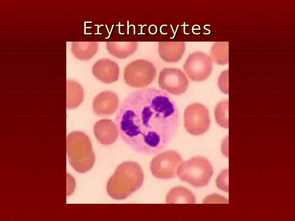
Erythrocytes: Red Blood Cells Erythrocytes: Red Blood Cells (RBC)(RBC)
Lack nucleus and Lack nucleus and mitochondria mitochondria
Circulating lifespan of 120 Circulating lifespan of 120 daysdays
Each erythrocyte contains 280 Each erythrocyte contains 280 million hemoglobin moleculesmillion hemoglobin molecules

Erythrocytes: Red Blood Cells Erythrocytes: Red Blood Cells (RBC)(RBC)
Destroyed by phagocytic cells in Destroyed by phagocytic cells in liver, spleen, and bone marrowliver, spleen, and bone marrow
Cannot reproduce in blood streamCannot reproduce in blood stream Anemia: abnormal low count in Anemia: abnormal low count in
RBCsRBCs Erythropoiesis: rbc formationErythropoiesis: rbc formation

Erythrocytes: Red Blood Cells Erythrocytes: Red Blood Cells (RBC)(RBC)
Woman usually have 4-5 million Woman usually have 4-5 million erythrocytes per cubic millimeter of erythrocytes per cubic millimeter of bloodblood
men have 5-6 million. If this number is men have 5-6 million. If this number is considerably higherconsiderably higher
polycythemia polycythemia may be the cause. If the may be the cause. If the number is considerably less, the number is considerably less, the person has anemiaperson has anemia. .
ErythrocytesErythrocytes
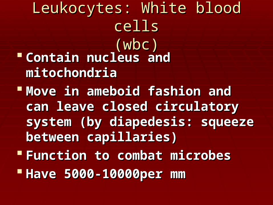
Leukocytes: White blood cellsLeukocytes: White blood cells(wbc)(wbc)
Contain nucleus and mitochondriaContain nucleus and mitochondria Move in ameboid fashion and can Move in ameboid fashion and can
leave closed circulatory system (by leave closed circulatory system (by diapedesis: squeeze between diapedesis: squeeze between capillaries)capillaries)
Function to combat microbesFunction to combat microbes Have 5000-10000per mmHave 5000-10000per mm
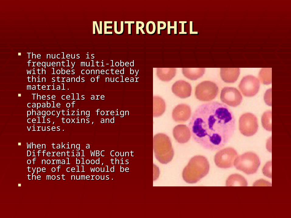
NEUTROPHILNEUTROPHIL The nucleus is frequently The nucleus is frequently
multi-lobed with lobes multi-lobed with lobes connected by thin strands connected by thin strands of nuclear material.of nuclear material.
These cells are capable These cells are capable of phagocytizing foreign of phagocytizing foreign cells, toxins, and viruses. cells, toxins, and viruses.
When taking a Differential When taking a Differential WBC Count of normal WBC Count of normal blood, this type of cell blood, this type of cell would be the most would be the most numerous.numerous.

NEUTROPHILNEUTROPHIL If the count exceeds this If the count exceeds this
amount, the cause is amount, the cause is usually due to an acute usually due to an acute infection such as infection such as appendicitis, smallpox or appendicitis, smallpox or rheumatic fever.rheumatic fever.
If the count is If the count is considerably less, it may considerably less, it may be due to a viral infection be due to a viral infection such as influenza, such as influenza, hepatitis, or rubella. hepatitis, or rubella.
Destroy and eat bacteria Destroy and eat bacteria and dead cells at and dead cells at inflamed siteinflamed site

EOSINOPHILEOSINOPHIL
The nucleus often has The nucleus often has two lobes connected by a two lobes connected by a band of nuclear material. band of nuclear material. (Does it looks like a (Does it looks like a telephone receiver?) telephone receiver?)
The granules contain The granules contain digestive enzymes that digestive enzymes that are particularly effective are particularly effective against parasitic worms in against parasitic worms in their larval form. their larval form.

EOSINOPHILEOSINOPHIL
These cells also These cells also phagocytize antigen - phagocytize antigen - antibody antibody
These cells account for These cells account for less than 5% of the less than 5% of the WBC's complexes. WBC's complexes.
Primarily combat large Primarily combat large invaders, such as worms invaders, such as worms flukes and decrease flukes and decrease allergic reactionsallergic reactions
BASOPHILBASOPHIL
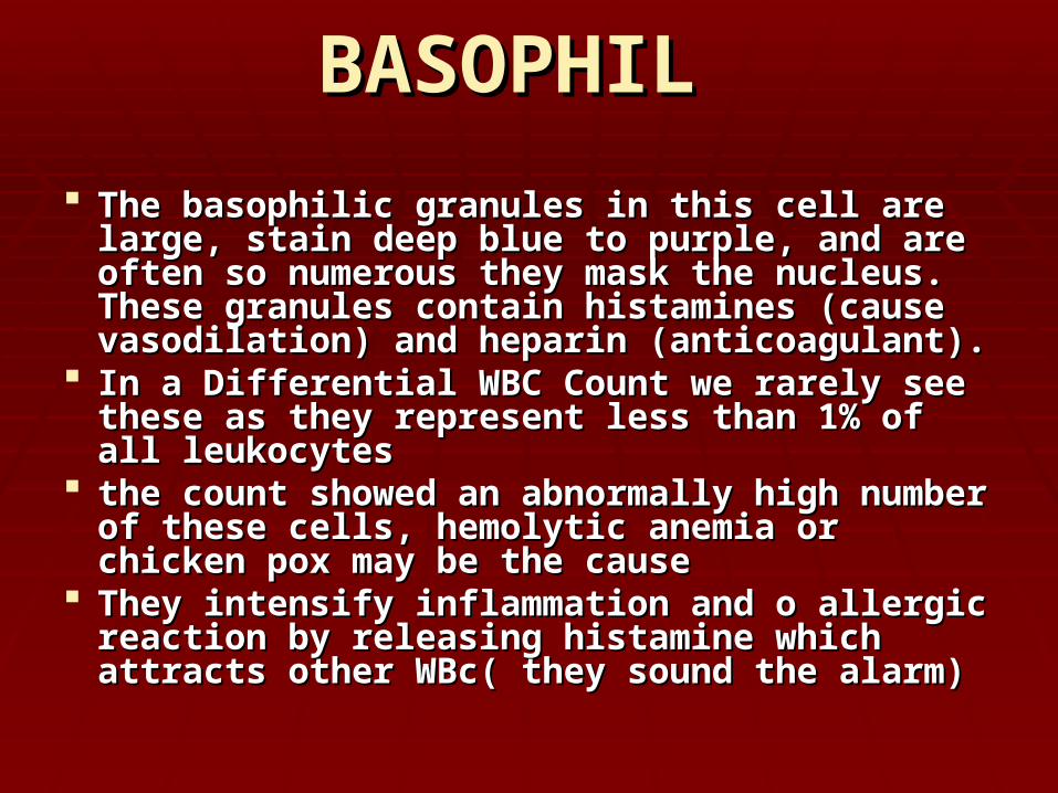
The basophilic granules in this cell are large, The basophilic granules in this cell are large, stain deep blue to purple, and are often so stain deep blue to purple, and are often so numerous they mask the nucleus. These numerous they mask the nucleus. These granules contain histamines (cause granules contain histamines (cause vasodilation) and heparin (anticoagulant). vasodilation) and heparin (anticoagulant).
In a Differential WBC Count we rarely see these In a Differential WBC Count we rarely see these as they represent less than 1% of all leukocytes as they represent less than 1% of all leukocytes
the count showed an abnormally high number the count showed an abnormally high number of these cells, hemolytic anemia or chicken pox of these cells, hemolytic anemia or chicken pox may be the cause may be the cause
They intensify inflammation and o allergic They intensify inflammation and o allergic reaction by releasing histamine which attracts reaction by releasing histamine which attracts other WBc( they sound the alarm)other WBc( they sound the alarm)

BASOPHILBASOPHIL

LYMPHOCYTELYMPHOCYTE
Its nucleus is very large for the size of the cell Its nucleus is very large for the size of the cell and stains dark purple. (Notice that the nucleus and stains dark purple. (Notice that the nucleus almost fills the cell leaving a very thin rim of almost fills the cell leaving a very thin rim of cytoplasm cytoplasm
This is the second most numerous leukocyte, This is the second most numerous leukocyte, accounting for 25-35% of the cells counted in a accounting for 25-35% of the cells counted in a Differential WBC Count. Differential WBC Count.
the number of these cells exceeds the normal the number of these cells exceeds the normal amount, one would suspect infectious amount, one would suspect infectious mononucleosis or a chronic infection mononucleosis or a chronic infection

LYMPHOCYTELYMPHOCYTE

MONOCYTEMONOCYTE
This cell is the largest of the leukocytes This cell is the largest of the leukocytes and is agranular. The nucleus is most and is agranular. The nucleus is most often "U" or kidney bean shaped; These often "U" or kidney bean shaped; These cells account for 3-9% of all leukocytes cells account for 3-9% of all leukocytes
In people with malaria, endocarditis, In people with malaria, endocarditis, typhoid fever, and Rocky Mountain spotted typhoid fever, and Rocky Mountain spotted fever, monocytes increase in number. fever, monocytes increase in number.

MONOCYTEMONOCYTE

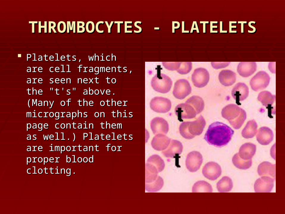
THROMBOCYTES - PLATELETSTHROMBOCYTES - PLATELETS
Each cubic millimeter of blood should Each cubic millimeter of blood should contain 250,000 to 500,000 of these. If the contain 250,000 to 500,000 of these. If the number is too high, spontaneous clotting number is too high, spontaneous clotting may occur. If the number is too low, may occur. If the number is too low, clotting may not occur when necessary clotting may not occur when necessary
THROMBOCYTES - PLATELETSTHROMBOCYTES - PLATELETS
Platelets, which are Platelets, which are cell fragments, are cell fragments, are seen next to the "t's" seen next to the "t's" above. (Many of the above. (Many of the other micrographs on other micrographs on this page contain this page contain them as well.) them as well.) Platelets are Platelets are important for proper important for proper blood clotting.blood clotting.

Do you know what types of cells Do you know what types of cells are these? are these?

Do you know what types of cells Do you know what types of cells are these?are these?
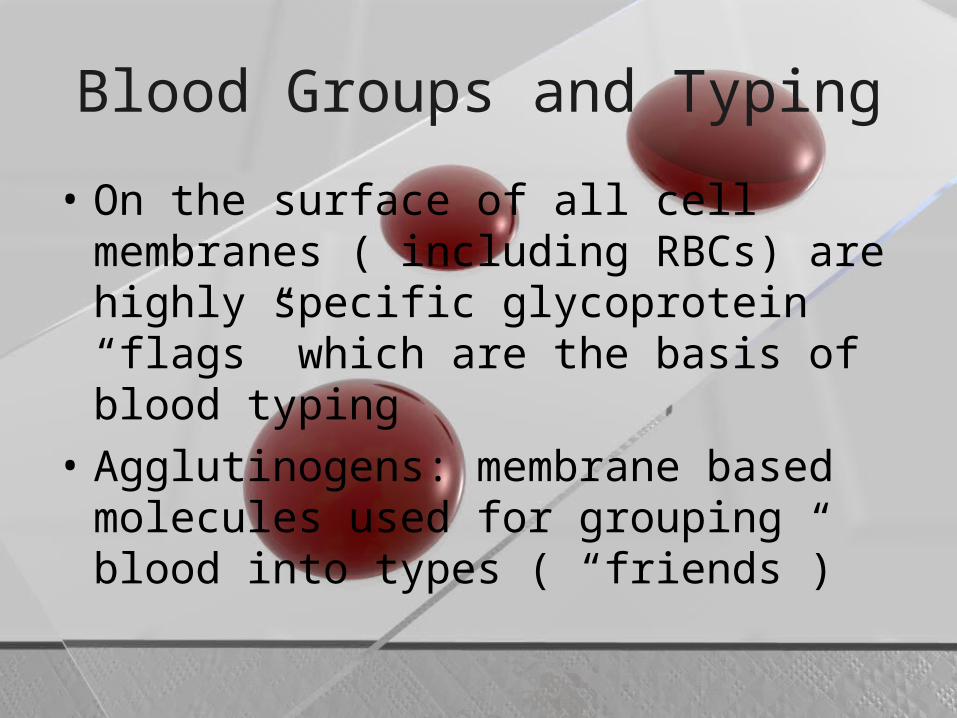
Blood Groups and Typing
• On the surface of all cell membranes ( including RBCs) are highly specific glycoprotein “flags” which are the basis of blood typing
• Agglutinogens: membrane based molecules used for grouping blood into types ( “friends”)

2 types of grouping blood based on type of agglutinogens present
• 1. ABO blood grouping : has 2 agglutinogens (A, B)
• The blood plasma also contains proteins called agglutinins (enemies) that the blood will react against

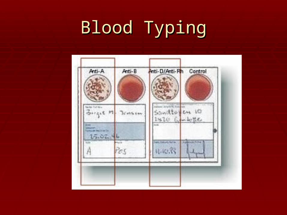
Blood Typing
• % pop Blood type Agglutinogens Agglutinins accepts
• 42% A A B A, O
• 10% B B A B, O
• 4% AB A, B NONE All• universal
acceptor• 45% O NONE A, B Only O• universal donor

Blood Typing
• 2. “Rh” Blood grouping system:• Based on the presences or absence of a single
flag ( agglutiongen)
• If present = Rh + 85% ++, +-• If not = Rh - 15% --• • As a general rule Rh negative group can give
safely to Rh positive group, but Rh positive should not be given to Rh negative

In the ABO system
• The agglutinins are preformed and the first exposure ( transfusion) of the opposing blood type will initiate the clumping reaction
• However in the Rh system, the agglutinins are not formed until the first exposure to the “wrong” agglutinogen ( works like immune system)… therefore clumping reaction wont occur until 2nd transfusion

Rh factor
• If mom is Rh negative and father is heterozygous Rh positive there is a 50% chance the child will be Rh positive
• The Childs Rh positive RBC leak into the placenta and the moms circulatory system when the placental tissue breaks down… causing mom to produce Anti-Rh antibodies
• The Anti-Rh antibodies pass across the placenta and destroy the RBC of the Rh positive child resulting in brain damage, mental retardation or even death
• This is called Erythroblastic fetalis• Solution: Rh negative woman receive an
Rh immunoglobulin injection or midway through 1st pregnancy or no later than 48 hrs after giving birth
Blood TypingBlood Typing

Blood TypingBlood Typing

Blood TypingBlood Typing

BLOOD BLOOD DISORDERDISORDER
SS

Sickle cell anemiaSickle cell anemia An inherited condition An inherited condition
which results in some which results in some erythrocytes being erythrocytes being malformed. malformed.
The gene for this The gene for this condition causes the condition causes the hemoglobin to be hemoglobin to be incorrectly formed, which incorrectly formed, which in turn causes some in turn causes some erythrocytes to take on a erythrocytes to take on a crescent shape.crescent shape.
These cells are not able These cells are not able to carry adequate to carry adequate amounts of oxygen to amounts of oxygen to cells cells

PolycythemiaPolycythemia
condition in which condition in which there is a net increase there is a net increase in the total number of in the total number of red blood cellsred blood cells in the in the body. The body. The overproduction of red overproduction of red blood cells may be due blood cells may be due to a primary process in to a primary process in the the bone marrowbone marrow or it or it may be a reaction to may be a reaction to chronically chronically low oxygen levelslow oxygen levels or, or, rarely, a rarely, a malignancymalignancy..

Thrombocytopenia Purpura Thrombocytopenia Purpura
a blood disorder a blood disorder characterized by an characterized by an abnormal decrease in the abnormal decrease in the number of platelets in the number of platelets in the blood. Platelets are cells blood. Platelets are cells in the blood that help stop in the blood that help stop bleeding. A decrease in bleeding. A decrease in platelets can result in platelets can result in easy bruising, bleeding easy bruising, bleeding gums, and internal gums, and internal bleeding. bleeding.

LEUKEMIALEUKEMIA


Hemostasis: prevention of blood Hemostasis: prevention of blood lossloss
3 mechanisms3 mechanisms:: 1. vascular spasm1. vascular spasm: damage to smooth : damage to smooth
muscle in blood vessels or platelets, muscle in blood vessels or platelets, causing a release of chemicals which causing a release of chemicals which cause cause vasoconstrictionvasoconstriction… reducing blood … reducing blood flow/loss lasing about 30 minflow/loss lasing about 30 min

2. Platelet plug formation2. Platelet plug formation:: If If thrombocytesthrombocytes (platelets) come in (platelets) come in
contact with collagen fibers in a damaged contact with collagen fibers in a damaged vessels wall… a chemical change occursvessels wall… a chemical change occurs
The platelets become sticky and cling to The platelets become sticky and cling to each other and to the damaged area each other and to the damaged area forming a plug ( forming a plug ( thrombusthrombus) or clot ) or clot
If the clot moves it is called an If the clot moves it is called an embolusembolus II

3. Coagulaton:3. Coagulaton: A clot is a mesh of protein fibers in which formed A clot is a mesh of protein fibers in which formed
elements are trappedelements are trapped Over 30 different hormones and enzymes are Over 30 different hormones and enzymes are
involved in coagulationinvolved in coagulation Liquid protein fibers in the blood plasma solidify Liquid protein fibers in the blood plasma solidify
and form a mesh over the damaged areaand form a mesh over the damaged area RBCs and platelets become trapped in the RBCs and platelets become trapped in the
mesh… mean while the fibers shrink, tightening mesh… mean while the fibers shrink, tightening and closing the wound and closing the wound
ThrombosisThrombosis: clotting in an unbroken : clotting in an unbroken vessel… if the clot (vessel… if the clot ( thrombus thrombus) moves it is ) moves it is called an called an embolusembolus … …
if it drifts until it blocks an small vessel, this if it drifts until it blocks an small vessel, this is called an is called an embolism (embolism ( can cause heart can cause heart attacks, strokes, or death)attacks, strokes, or death)
LeukemiaLeukemia: a malignant disease ( cancer) : a malignant disease ( cancer) of blood forming tissue… resulting in of blood forming tissue… resulting in excessive numbers of WBCs which crowd excessive numbers of WBCs which crowd out the RBCsout the RBCs
Mononucleosis: viral infection attacking Mononucleosis: viral infection attacking lymph tissuelymph tissue
Results in lmphocytes which are abnormal Results in lmphocytes which are abnormal , no cure, it just runs its course, no cure, it just runs its course

HeartHeart
A hollow 4 chambered muscular organ A hollow 4 chambered muscular organ about the size of a clenched fist and about the size of a clenched fist and weights about 255 g in females and 310 g weights about 255 g in females and 310 g in malesin males
Located in the thoracic cavity between the Located in the thoracic cavity between the lungslungs
2/3 of the heart is located left of the 2/3 of the heart is located left of the midline with the apex in contact with the midline with the apex in contact with the diaphragmdiaphragm


Enclosed in a loose fitting serous Enclosed in a loose fitting serous sac called pericardial sacsac called pericardial sac
FunctionsFunctions: : Separate the heart from other organsSeparate the heart from other organs Contains pericardial fluid for lubricaitonContains pericardial fluid for lubricaiton Layers of pericardiumLayers of pericardium: outer fibrous , : outer fibrous ,
inter serous layerinter serous layer Pericardial cavityPericardial cavity: between parietal : between parietal
pericardium and visceral pericardiumpericardium and visceral pericardium

Layers of heart wallLayers of heart wall
EpicardiumEpicardium: outermost layer: outermost layer
Myocardium:Myocardium: cardiac muscle, thickest cardiac muscle, thickest portion, it contractsportion, it contracts
Endocardium:Endocardium: thin epithelial lining of the thin epithelial lining of the heart chambersheart chambers

Chambers and valvesChambers and valves
4 chambers4 chambers
The two upper heart chambers are atriaThe two upper heart chambers are atria ( atrium) (( atrium) (auriclesauricles on outside of the heart) on outside of the heart)
The wall between the two atria is called the The wall between the two atria is called the interatrial septuminteratrial septum
The depression in the interatrial septum is called The depression in the interatrial septum is called the the fossa ovalisfossa ovalis, which used to be an opening , which used to be an opening during fetal development and blood could flow during fetal development and blood could flow from right atrium to left atrium (can be seen in from right atrium to left atrium (can be seen in the right atrium) the right atrium)

The two lower chambers are called the The two lower chambers are called the ventriclesventricles
The wall between the 2 ventricles is called The wall between the 2 ventricles is called the the interventricular septuminterventricular septum
The The left ventrical wall is thickerleft ventrical wall is thicker than the than the right, due to the way the blood circulates in right, due to the way the blood circulates in the body the body
Chordae tendinaeChordae tendinae: cords that connect : cords that connect valves to papillary musclesvalves to papillary muscles
Papillary musclePapillary muscle: small raised projections : small raised projections of muscle tissue that connect of muscle tissue that connect chordae chordae tendinaetendinae to endothelium of heart to endothelium of heart
Trabeculae carneaeTrabeculae carneae: distinct ridges in : distinct ridges in walls of ventricleswalls of ventricles
A condition called A condition called prolapseprolapse involves leaky involves leaky valves due to stretched chordae tendineaevalves due to stretched chordae tendineae

ValvesValves
1. Bicuspid (mitral ) valve1. Bicuspid (mitral ) valve: located : located between left atrium and left ventricle between left atrium and left ventricle ( most commonly replaced valve)( most commonly replaced valve)
2. Tricuspid valve2. Tricuspid valve: located between right : located between right atrium and ventricleatrium and ventricle
3. Aortic semilunar valve3. Aortic semilunar valve: opens from : opens from right ventricle to base of aortaright ventricle to base of aorta
4. Pulmonary semilunar valve: opens from 4. Pulmonary semilunar valve: opens from left ventricle to pulmonary arteryleft ventricle to pulmonary artery
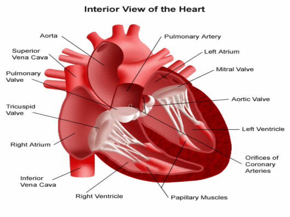

Coronary circulatory systemCoronary circulatory system
The heart has its own circulatory system The heart has its own circulatory system Most heart problems are due to faulty or blocked Most heart problems are due to faulty or blocked
coronary circulation… resulting in poor nutrients coronary circulation… resulting in poor nutrients and oxygen supply to myocardiumand oxygen supply to myocardium
IschemiaIschemia: reduced blood flow, weakens heart: reduced blood flow, weakens heart Angina: chest ( heart) pain due to ischemia ( heart) pain due to ischemia Infaction:Infaction: death of an area of tissue due to an death of an area of tissue due to an
interupted blood supply interupted blood supply
Myocardial infarctionMyocardial infarction: heart attack due to : heart attack due to infarction and weakened heart , usually infarction and weakened heart , usually caused by an caused by an embolusembolus or a or a thrombusthrombus
Atherosclerosis:Atherosclerosis: build up of cholesterol build up of cholesterol or other fatty compounds on the inside of or other fatty compounds on the inside of blood vessel wall, decreases or blocks blood vessel wall, decreases or blocks circulationcirculation

Coronary artery diseaseCoronary artery disease: a condition of : a condition of damaged heart muscle due to one of the damaged heart muscle due to one of the above reductions in blood supply. The above reductions in blood supply. The number 1 cause of death in USAnumber 1 cause of death in USA
Heart murmersHeart murmers: unusual sounds : unusual sounds generally due to valve disorders ( leaky): generally due to valve disorders ( leaky):
1. faulty chordae tendinea1. faulty chordae tendinea 2. disease damaged valves/ cusps2. disease damaged valves/ cusps

Cardiac cycle (blood pressure)Cardiac cycle (blood pressure)
The 2 atria contract, pumping blood into The 2 atria contract, pumping blood into the 2 ventricles which are relaxed while the 2 ventricles which are relaxed while being filled.being filled.
Then the ventricles contract while the Then the ventricles contract while the relaxed atria are refilled from the vena relaxed atria are refilled from the vena cava (pulmonary veins)cava (pulmonary veins)
Systole:Systole: phase of contraction (top #) phase of contraction (top #) DiastoleDiastole: phase of relaxation ( bottom #): phase of relaxation ( bottom #)
So during ventricular diastole, the tri and So during ventricular diastole, the tri and bicuspids are openbicuspids are open
The 2 atria are in systole and the ventricles are The 2 atria are in systole and the ventricles are fillingfilling
Then the atrial diastole and ventriclular systoleThen the atrial diastole and ventriclular systole The cuspids close, similunars open, ventricles The cuspids close, similunars open, ventricles
empty, Atria fillempty, Atria fill Then brief pause and cycle repeatsThen brief pause and cycle repeats 8 sec per cycle with about 72 beats per minute8 sec per cycle with about 72 beats per minute
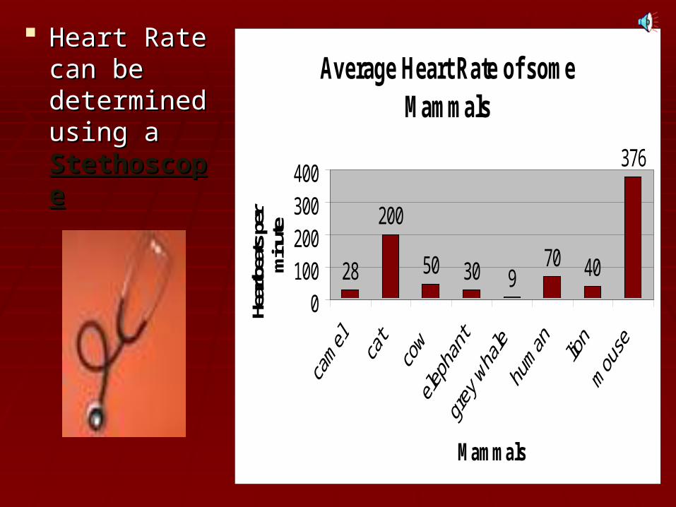
Heart Rate Heart Rate can be can be determined determined using a using a StethoscopeStethoscope
Average Heart Rate of some Mammals
28
200
50 30 970 40
376
0100200300400
Mammals
Hear
tbea
ts p
er
min
ute

LubLub
If you listen to your heartbeat, it makes a lub dub
sound.
The lub is when blood is pushed out of the
heart into the body and the dub is the reloading of the heart with more blood ready to push it
out to the body
Dub

The heart is made of The heart is made of cardiac musclecardiac muscle.. When the cells receive an When the cells receive an electricalelectrical
impulseimpulse they contract - causing a they contract - causing a heartbeat.heartbeat.
Cardiac muscle is Cardiac muscle is myogenicmyogenic - it can - it can contract on its own, without needing contract on its own, without needing nervenerve impulsesimpulses..
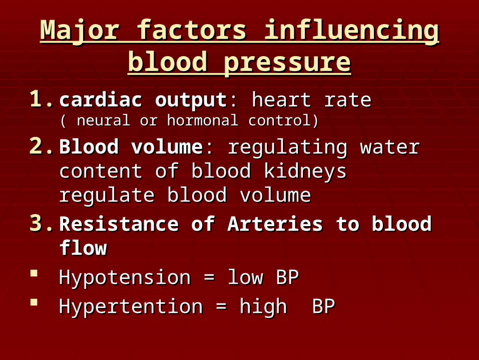
Major factors influencing blood Major factors influencing blood pressurepressure
1.1. cardiac outputcardiac output: heart rate : heart rate ( neural or ( neural or hormonal control)hormonal control)
2.2. Blood volumeBlood volume: regulating water content : regulating water content of blood kidneys regulate blood volumeof blood kidneys regulate blood volume
3.3. Resistance of Arteries to blood flowResistance of Arteries to blood flow Hypotension = low BPHypotension = low BP Hypertention = high BPHypertention = high BP

The heart is actually a double pump with 2 The heart is actually a double pump with 2 separate circulatory systemsseparate circulatory systems
1.1. Pulmonary circulationPulmonary circulation: The short loop : The short loop pumped by the right ventricle from the pumped by the right ventricle from the heart to the lungs and back to the heartheart to the lungs and back to the heart
Serves only to bring blood in contact with Serves only to bring blood in contact with alveoli resulting in gas exchangealveoli resulting in gas exchange
It does not meet the needs ( metabolic) It does not meet the needs ( metabolic) of the lung tissueof the lung tissue
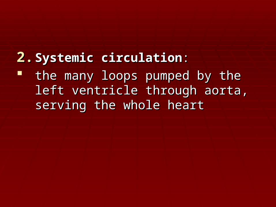
2.2. Systemic circulationSystemic circulation: : the many loops pumped by the left the many loops pumped by the left
ventricle through aorta, serving the whole ventricle through aorta, serving the whole heartheart
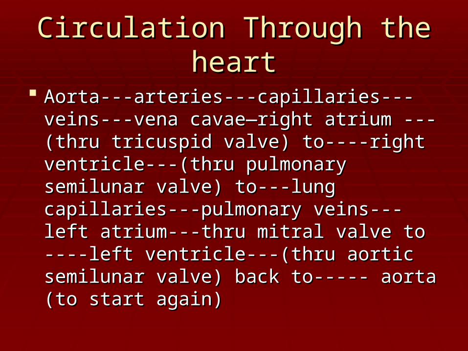
Circulation Through the heartCirculation Through the heart
Aorta---arteries---capillaries---veins---vena Aorta---arteries---capillaries---veins---vena cavae—right atrium ---(thru tricuspid valve) cavae—right atrium ---(thru tricuspid valve) to----right ventricle---(thru pulmonary to----right ventricle---(thru pulmonary semilunar valve) to---lung capillaries---semilunar valve) to---lung capillaries---pulmonary veins---left atrium---thru mitral pulmonary veins---left atrium---thru mitral valve to ----left ventricle---(thru aortic valve to ----left ventricle---(thru aortic semilunar valve) back to----- aorta (to start semilunar valve) back to----- aorta (to start again)again)

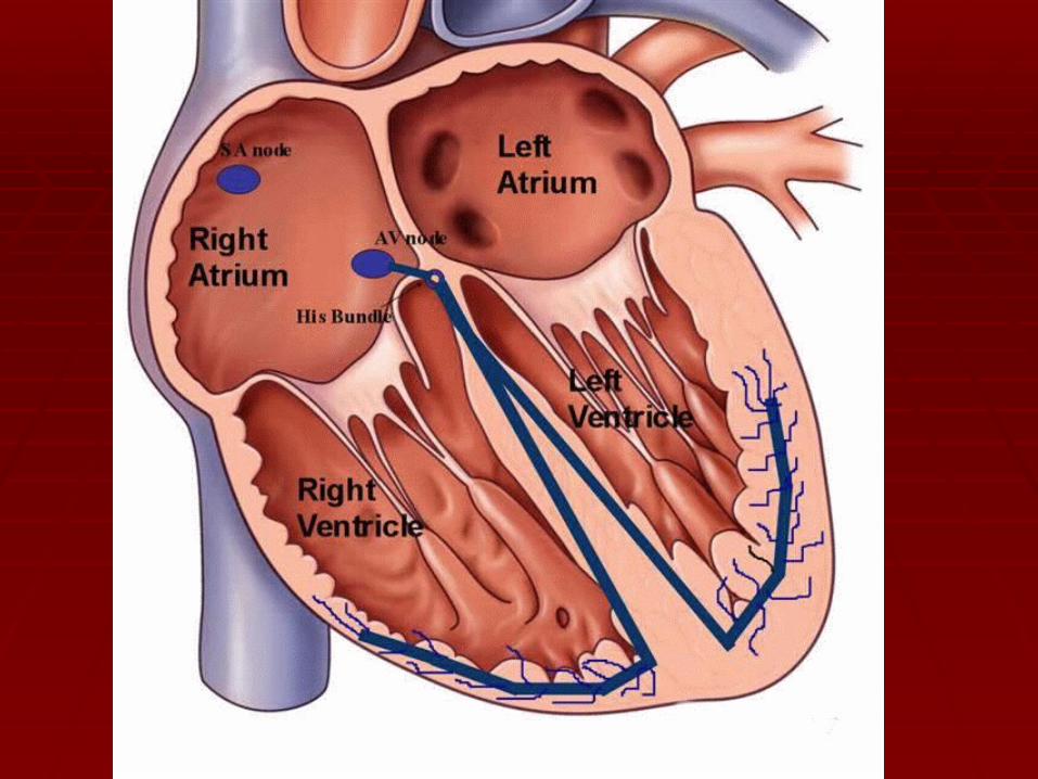
Conduction systemConduction system
Cells in the heart beat as a unit due to the Cells in the heart beat as a unit due to the intercalated disksintercalated disks which weaves the cell which weaves the cell endings togetherendings together
The orderly sequence of atrial and The orderly sequence of atrial and ventricular contraction is maintained by an ventricular contraction is maintained by an intrinsic (internal) regulating system called intrinsic (internal) regulating system called a a conduction systemconduction system

Conduction systemConduction system
This conduction system consists of This conduction system consists of specialized muscle tissue which generates specialized muscle tissue which generates and conducts electrical impulses to and conducts electrical impulses to stimulate the cardiac muscle to contract in stimulate the cardiac muscle to contract in a cycle as a unita cycle as a unit
Consists of the S.A node and the A.V. Consists of the S.A node and the A.V. nodenode

Sinoatrial node (SA node)Sinoatrial node (SA node)
This specialized node is found on the This specialized node is found on the upperupper inside wallinside wall of the of the right atriumright atrium..
The SA node is known as the The SA node is known as the pacemakerpacemaker of the of the heart and initiates a heartbeat every 0.85 heart and initiates a heartbeat every 0.85 seconds. seconds.
This signal travels across the This signal travels across the atriaatria causing them causing them to to contract contract and load the ventricles with blood. and load the ventricles with blood.
Ventricles are electrically insulated from atria - Ventricles are electrically insulated from atria - so they don’t contract yet.so they don’t contract yet.

Sinoatrial node ( S.A, node or Sinoatrial node ( S.A, node or pacemaker)pacemaker)
Located in the right atrial wallLocated in the right atrial wall
Initiate cardiac cycle ( Initiate cardiac cycle ( sets the pacesets the pace) by ) by sending impulses to both atria causing sending impulses to both atria causing them to contract…them to contract…
This impulse also travels to the This impulse also travels to the atrioventricular node atrioventricular node

Atrio-ventricular node (AV node)Atrio-ventricular node (AV node) The The AV nodeAV node is located on the is located on the bottombottom surface of surface of
the right atriathe right atria and is responsible for initiating the and is responsible for initiating the contraction of the ventricles.contraction of the ventricles.
Electrical impulse passes to ventricles via Electrical impulse passes to ventricles via AV nodeAV node and the and the Bundle of HisBundle of His. They pass the impulse to . They pass the impulse to the base of the ventricles (~ 0.1 s delay).the base of the ventricles (~ 0.1 s delay).
The bundle of His is a group of fibres that conduct The bundle of His is a group of fibres that conduct impulses to impulses to Purkyne fibresPurkyne fibres which carry impulses to which carry impulses to left & right ventricles.left & right ventricles.
Ventricles then contract from the bottom upwards. Ventricles then contract from the bottom upwards.

Atrioventricular node (A.V.)Atrioventricular node (A.V.) Found on the septum (wall) between the Found on the septum (wall) between the
right atrium and right ventricleright atrium and right ventricle Sends impulses to the ventricles over a Sends impulses to the ventricles over a
bundle of branching conducting fibers bundle of branching conducting fibers called the called the A.V. bundle ( bundle of his)A.V. bundle ( bundle of his)
These fibers keep branching until the These fibers keep branching until the smallest make contact with cells in the smallest make contact with cells in the myocardiummyocardium
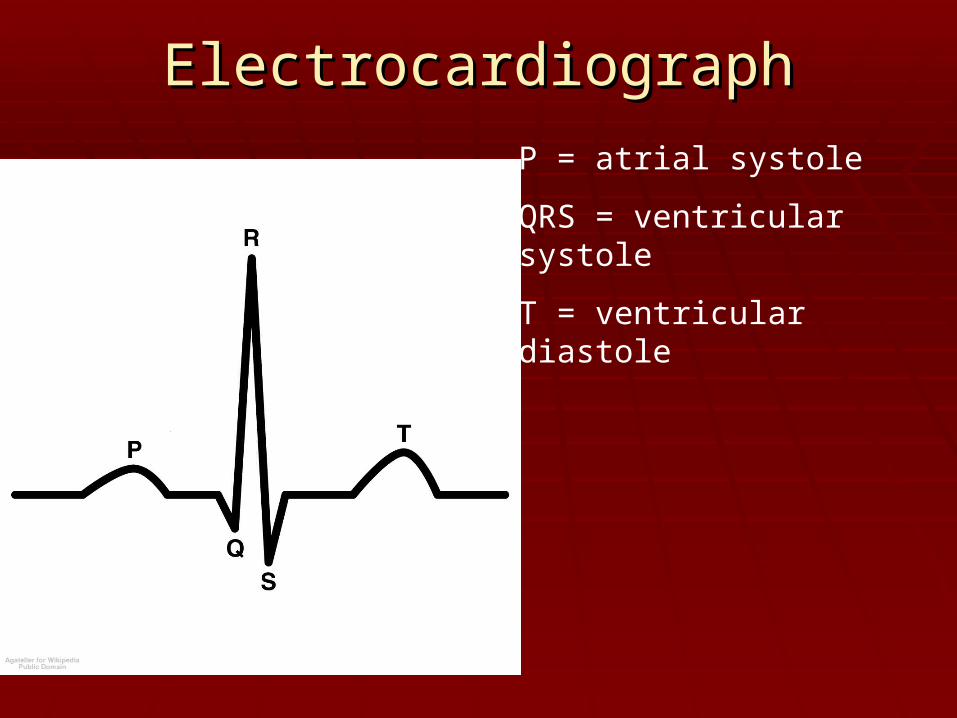
ElectrocardiographElectrocardiograph
P = atrial systole
QRS = ventricular systole
T = ventricular diastole
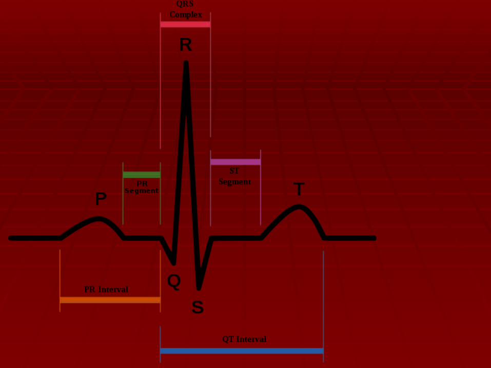

Q wave: absent on EKG. Necrosis (death Q wave: absent on EKG. Necrosis (death of an area of the heart muscle produces a of an area of the heart muscle produces a Q wave on EKG) used to diagnose heart Q wave on EKG) used to diagnose heart attackattack
Myocardial infarction is usually related to Myocardial infarction is usually related to the left ventriclethe left ventricle
PR interval: problem in conduction system PR interval: problem in conduction system ( S.A node A.V node)( S.A node A.V node)

HOW TO READ EKGHOW TO READ EKG
Determine the heart rate from EKGDetermine the heart rate from EKG
1.1. First find a specific R wave that peaks on First find a specific R wave that peaks on a heavy linea heavy line
2.2. Next count off 300, 150, 100, 75, 60, 50 Next count off 300, 150, 100, 75, 60, 50 for every thick line. Know these numbers for every thick line. Know these numbers , you will use them, you will use them
3.3. Where the next R wave falls determines Where the next R wave falls determines the ratethe rate


Ventricular fibrillation is an abnormal heart Ventricular fibrillation is an abnormal heart
rhythm that is disorganized and irregular.rhythm that is disorganized and irregular.

Ventricular tachycardia is a rapid, regular Ventricular tachycardia is a rapid, regular heart rhythm that originates in the lower heart rhythm that originates in the lower
chambers of the heart.chambers of the heart.

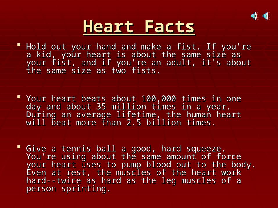
Heart FactsHeart Facts Hold out your hand and make a fist. If you're a kid, your Hold out your hand and make a fist. If you're a kid, your
heart is about the same size as your fist, and if you're an heart is about the same size as your fist, and if you're an adult, it's about the same size as two fists. adult, it's about the same size as two fists.
Your heart beats about 100,000 times in one day and Your heart beats about 100,000 times in one day and about 35 million times in a year. During an average about 35 million times in a year. During an average lifetime, the human heart will beat more than 2.5 billion lifetime, the human heart will beat more than 2.5 billion times. times.
Give a tennis ball a good, hard squeeze. You're using Give a tennis ball a good, hard squeeze. You're using about the same amount of force your heart uses to about the same amount of force your heart uses to pump blood out to the body. Even at rest, the muscles pump blood out to the body. Even at rest, the muscles of the heart work hard--twice as hard as the leg muscles of the heart work hard--twice as hard as the leg muscles of a person sprinting. of a person sprinting.


circulationcirculation


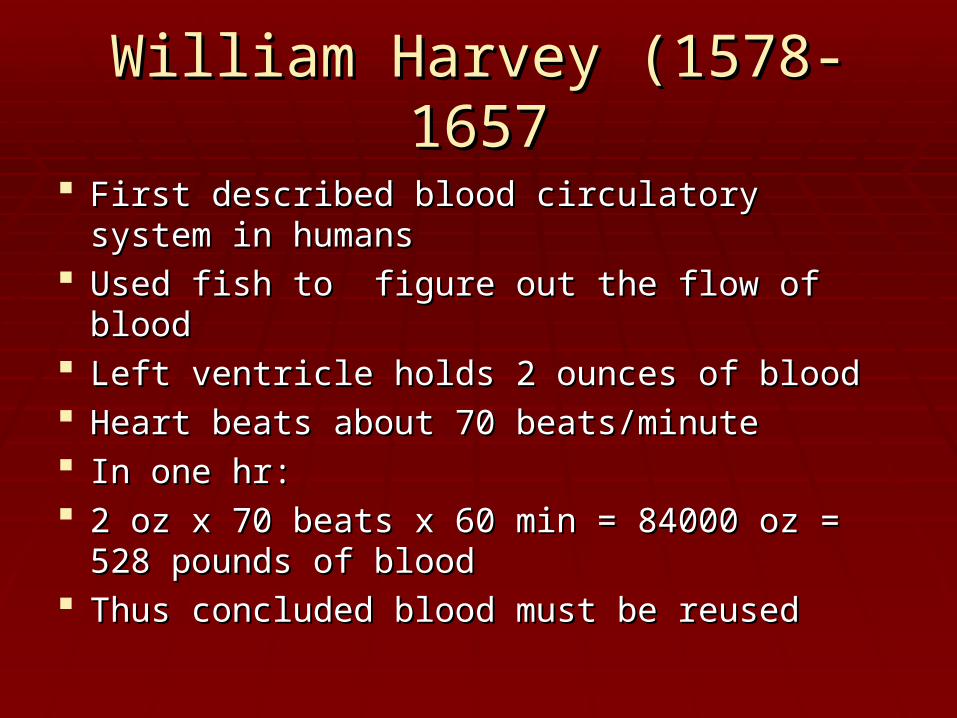
William Harvey (1578-1657William Harvey (1578-1657
First described blood circulatory system in First described blood circulatory system in humanshumans
Used fish to figure out the flow of bloodUsed fish to figure out the flow of blood Left ventricle holds 2 ounces of bloodLeft ventricle holds 2 ounces of blood Heart beats about 70 beats/minuteHeart beats about 70 beats/minute In one hr:In one hr: 2 oz x 70 beats x 60 min = 84000 oz = 528 2 oz x 70 beats x 60 min = 84000 oz = 528
pounds of bloodpounds of blood Thus concluded blood must be reusedThus concluded blood must be reused
Vessels and routesVessels and routes
Arteries:Arteries: blood vessels which carry blood vessels which carry blood away from the heartblood away from the heart
Arterial walls consist of 3 layers Arterial walls consist of 3 layers ( tunicas)( tunicas) around a hollow (lumen) tube around a hollow (lumen) tube
1.1. Tunica intimaTunica intima: internal lining: internal lining2.2. Tunica mediaTunica media: middle thickest : middle thickest
tunica( made of smooth muscle)tunica( made of smooth muscle)3.3. Tunica externaTunica externa: tough outer coat that : tough outer coat that
acts as prtectionacts as prtection

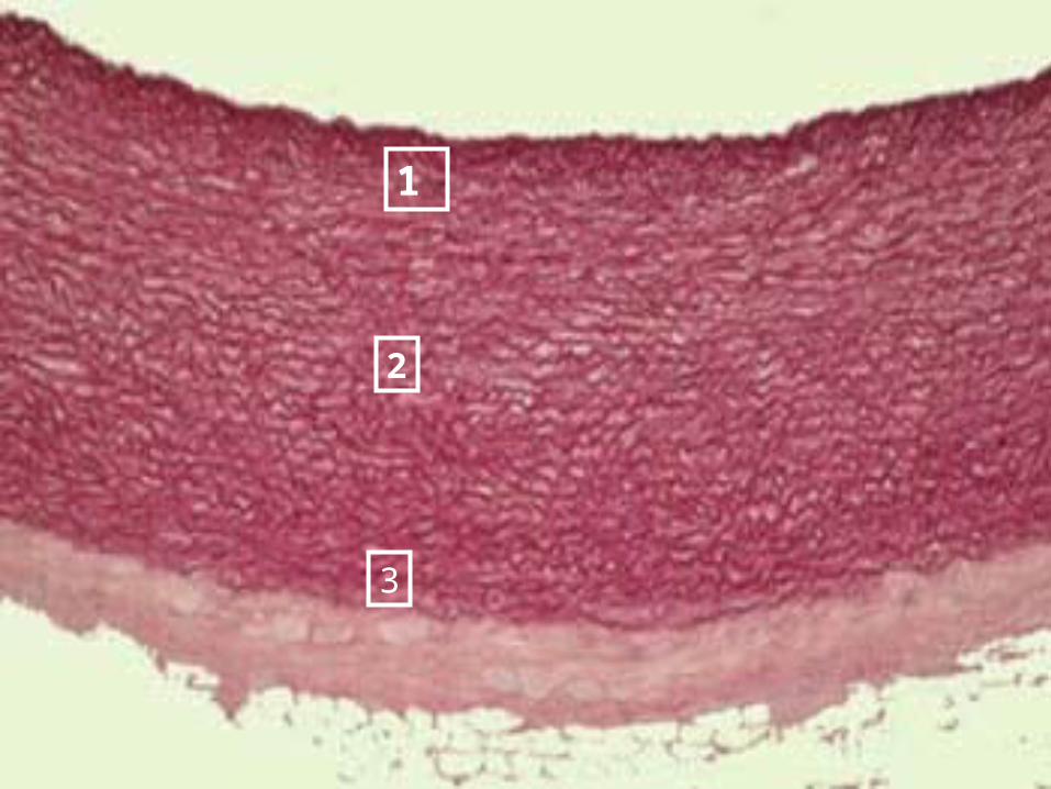
1
2
3

Arterioles:Arterioles: small arteries small arteries Capillaries:Capillaries: microscopic blood vessels, microscopic blood vessels,
the walls of which consist of only a thin the walls of which consist of only a thin layer of endothelium.layer of endothelium.
CapillarieCapillaries allow for the exchange of s allow for the exchange of nutrients, gases and waste to take place nutrients, gases and waste to take place by diffusion between blood plasma and by diffusion between blood plasma and various tissue cells.various tissue cells.

Venules:Venules: small vessels that receive blood small vessels that receive blood from capillaries and merge into veinsfrom capillaries and merge into veins
Veins:Veins: carry blood to the heart, have very carry blood to the heart, have very low pressure and valveslow pressure and valves

2 factors that assist venous blood 2 factors that assist venous blood movementmovement
1.1. Valves:Valves: veins have backflow preventing veins have backflow preventing valves valves
If faulty or leaky results in varicose If faulty or leaky results in varicose veinsveins
2.2. Muscle contractions:Muscle contractions: When muscles which surround the When muscles which surround the
veins ( especially arms and legs) veins ( especially arms and legs) contract they squeeze the veins contract they squeeze the veins pushing blood from valve to valvepushing blood from valve to valve

varicose veinsvaricose veins


Pulse:Pulse: the alternating expansion and the alternating expansion and elastic recoil of the walls of an artery ( due elastic recoil of the walls of an artery ( due to ventricular systole and diastole)to ventricular systole and diastole)
Pulse ratePulse rate: heart rate = 70 to 80 per min: heart rate = 70 to 80 per min Blood pressure: pressure (measured in Blood pressure: pressure (measured in
arteries) exerted by left ventricular systole arteries) exerted by left ventricular systole and remaining during diastoleand remaining during diastole
Average B.P. 120/80Average B.P. 120/80
The difference between the 2 numbers is The difference between the 2 numbers is a measure of the health of the arterya measure of the health of the artery
Hepatic Portal CirculationHepatic Portal Circulation
Blood enters liver from 2 sources:Blood enters liver from 2 sources:1.1. The liver is supplied with oxygen rich blood by The liver is supplied with oxygen rich blood by
hepatic artery ( not part of hepatic portal hepatic artery ( not part of hepatic portal system)system)
2.2. Veins drain the stomach, intestines, pancreas Veins drain the stomach, intestines, pancreas etc… and merge into the hepatic portal vein etc… and merge into the hepatic portal vein which brings this nutrient rich blood to the liver which brings this nutrient rich blood to the liver for processingfor processing
Blood leaves the liver by way of the hepatic Blood leaves the liver by way of the hepatic vein and enters the inferior vena cavavein and enters the inferior vena cava

A prtal system is:A prtal system is:
Arteries—capillaries—veins—capillaries—Arteries—capillaries—veins—capillaries—veinsveins
As opposed to normal:As opposed to normal:
Artery—capillary --veinsArtery—capillary --veins

Fetal circulationFetal circulation
Differs from adults because lungs, kidneys Differs from adults because lungs, kidneys and GI tract are not functionaland GI tract are not functional
Fetal and maternal blood does not mixFetal and maternal blood does not mix
Key Differences in a fetusKey Differences in a fetus
1.1. Foramen ovaleForamen ovale: hole between 2 atria, so : hole between 2 atria, so blood can bypass the right ventricle and blood can bypass the right ventricle and pulmonary circuit ( later closes leaving a pulmonary circuit ( later closes leaving a scar called the scar called the fossa ovalisfossa ovalis))
2.2. Ductus arteriosusDuctus arteriosus: vessel running from : vessel running from pulmonary trunk to aorta, skipping pulmonary trunk to aorta, skipping pulmonary circuit.pulmonary circuit.
3.3. Umbilical arteriesUmbilical arteries: 2 arteries which : 2 arteries which branch off the internal iliacs carry branch off the internal iliacs carry deoxygenated blood and waste to the deoxygenated blood and waste to the placentaplacenta
4.4. Umbilical veinUmbilical vein: leaves placenta with : leaves placenta with oxygenated blood heading toward fetal oxygenated blood heading toward fetal heart, it bypasses the liver by way of the heart, it bypasses the liver by way of the ductus venosusductus venosus ( which leads straight ( which leads straight into the inferior vena cava)into the inferior vena cava)

Another modification involves fetal Another modification involves fetal hemoglobinhemoglobin
It is slightly more attracted to oxygen than It is slightly more attracted to oxygen than is normal hemoglobin so fetus cant “steal” is normal hemoglobin so fetus cant “steal” moms oxygenmoms oxygen