the cardiovascular system: blood vessels dawn a. drooger, rn, bsn
TRANSCRIPT
The Cardiovascular System: Blood Vessels
Dawn A. Drooger, RN, BSN
Blood Vessels
Blood is carried in a closed system of vessels that begins and ends at the heart
The three major types of vessels are arteries, capillaries, and veins
Arteries carry blood away from the heart, veins carry blood toward the heart
Capillaries contact tissue cells and directly serve cellular needs
Generalized Structure of Blood Vessels
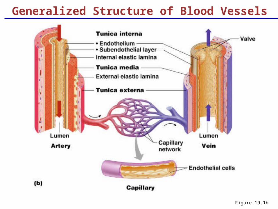
Arteries and veins are composed of three tunics – tunica interna, tunica media, and tunica externa
Lumen – central blood-containing space surrounded by tunics
Capillaries are composed of endothelium with sparse basal lamina
Generalized Structure of Blood Vessels
Figure 19.1b
Tunics
Tunica interna (tunica intima)
Endothelial layer that lines the lumen of all vessels
In vessels larger than 1 mm, a subendothelial connective tissue basement membrane is present
Tunica media
Smooth muscle and elastic fiber layer, regulated by sympathetic nervous system
Controls vasoconstriction/vasodilation of vessels
Tunics
Tunica externa (tunica adventitia)
Collagen fibers that protect and reinforce vessels
Elastic (Conducting) Arteries
Thick-walled arteries near the heart; the aorta and its major branches
Large lumen allow low-resistance conduction of blood
Contain elastin in all three tunics
Withstand and smooth out large blood pressure fluctuations
Capillaries
Capillaries are the smallest blood vessels
Walls consisting of a thin tunica interna, one cell thick
Allow only a single RBC to pass at a time
Gas exchange occurs only here
Continuous capillaries of the brain:
The blood-brain barrier

Sinusoids
Highly modified, leaky, fenestrated capillaries with large lumens
Found in the liver, bone marrow, lymphoid tissue, and in some endocrine organs
Allow large molecules (proteins and blood cells) to pass between the blood and surrounding tissues
Blood flows sluggishly, allowing for modification in various ways
Capillary Beds
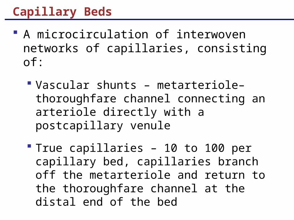
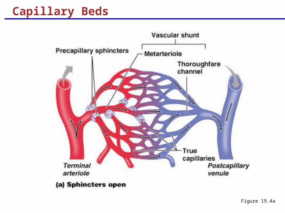
A microcirculation of interwoven networks of capillaries, consisting of:
Vascular shunts – metarteriole–thoroughfare channel connecting an arteriole directly with a postcapillary venule
True capillaries – 10 to 100 per capillary bed, capillaries branch off the metarteriole and return to the thoroughfare channel at the distal end of the bed
Capillary Beds
Figure 19.4a

Venous System: Venules
Are formed when capillary beds unite
Allow fluids and WBCs to pass from the bloodstream to tissues
Postcapillary venules – smallest venules, composed of endothelium and a few pericytes
Large venules have one or two layers of smooth muscle (tunica media)
Venous System: Veins
Veins are:
Formed when venules converge
Composed of three tunics, with a thin tunica media and a thick tunica externa consisting of collagen fibers and elastic networks
Capacitance vessels (blood reservoirs) that contain 65% of the blood supply

Venous System: Veins
Veins have much lower blood pressure and thinner walls than arteries
To return blood to the heart, veins have special adaptations
Valves (resembling semilunar heart valves), which prevent backflow of blood
Venous sinuses – specialized, flattened veins with extremely thin walls (e.g., coronary sinus of the heart and dural sinuses of the brain)
Blood Flow
Actual volume of blood flowing through a vessel, an organ, or the entire circulation in a given period:
Is measured in ml per min.
Is equivalent to cardiac output (CO), considering the entire vascular system
Is relatively constant when at rest
Varies widely through individual organs, according to immediate needs
Blood Pressure (BP)
Force per unit area exerted on the wall of a blood vessel by its contained blood
Expressed in millimeters of mercury (mm Hg)
Measured in reference to systemic arterial BP in large arteries near the heart
The differences in BP within the vascular system provide the driving force that keeps blood moving from higher to lower pressure areas

Resistance
Resistance – opposition to flow
Measure of the amount of friction blood encounters as it passes through vessels
Generally encountered in the systemic circulation
Referred to as peripheral resistance (PR)
The three important sources of resistance are blood viscosity, total blood vessel length, and blood vessel diameter

Resistance factors that remain relatively constant are:
Blood viscosity – thickness or “stickiness” of the blood
Blood vessel length – the longer the vessel, the greater the resistance encountered
Changes in vessel diameter are frequent and significantly alter peripheral resistance
Resistance Factors: Viscosity and Vessel Length
Resistance Factors: Blood Vessel Diameter
Small-diameter arterioles are the major determinants of peripheral resistance
Fatty plaques from atherosclerosis:
Cause turbulent blood flow
Dramatically increase resistance due to turbulence

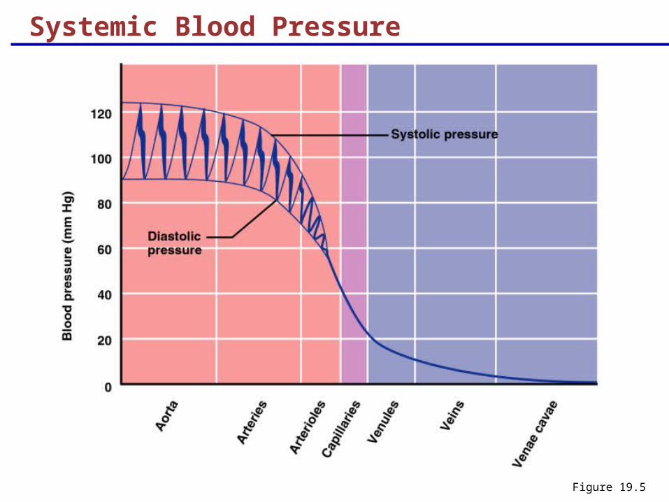
Systemic Blood Pressure
The pumping action of the heart generates blood flow through the vessels along a pressure gradient, always moving from higher- to lower-pressure areas
Pressure results when flow is opposed by resistance
Systemic pressure:
Is highest in the aorta
Declines throughout the length of the pathway
Is 0-8 mm Hg in the right atrium
The steepest change in blood pressure occurs in the arterioles
Systemic Blood Pressure
Figure 19.5
Arterial Blood Pressure
Arterial BP reflects two factors of the arteries close to the heart
Their elasticity (compliance or distensibility)
The amount of blood forced into them at any given time
Blood pressure in elastic arteries near the heart is pulsatile (BP rises and falls)
Arterial Blood Pressure
Systolic pressure – pressure exerted on arterial walls during ventricular contraction
Diastolic pressure – lowest level of arterial pressure during a ventricular cycle
Pulse pressure – the difference between systolic and diastolic pressure
Mean arterial pressure (MAP) – pressure that propels the blood to the tissues
MAP = diastolic pressure + 1/3 pulse pressure
Capillary Blood Pressure
Capillary BP ranges from 20 to 40 mm Hg
Low capillary pressure is desirable because high BP would rupture fragile, thin-walled capillaries
Low BP is sufficient to force filtrate out into interstitial space and distribute nutrients, gases, and hormones between blood and tissues
Venous Blood Pressure
Venous BP is steady and changes little during the cardiac cycle
The pressure gradient in the venous system is only about 20 mm Hg
A cut vein has even blood flow; a lacerated artery flows in spurts
Factors Aiding Venous Return
Venous BP alone is too low to promote adequate blood return and is aided by the:
Respiratory “pump” – pressure changes created during breathing suck blood toward the heart by squeezing local veins
Muscular “pump” – contraction of skeletal muscles “milk” blood toward the heart
Valves prevent backflow during venous return
InterActive Physiology®: Cardiovascular System: Anatomy Review: Blood Vessel Structure and FunctionPLAYPLAY
Maintaining Blood Pressure
Maintaining blood pressure requires:
Cooperation of the heart, blood vessels, and kidneys
Supervision of the brain
Cardiac Output (CO)
Cardiac output is determined by venous return and neural and hormonal controls
Resting heart rate is controlled by the cardioinhibitory center via the vagus nerves
Stroke volume is controlled by venous return (end diastolic volume, or EDV)
Under stress, the cardioacceleratory center increases heart rate and stroke volume
The end systolic volume (ESV) decreases and MAP increases
Controls of Blood Pressure
Short-term controls:
Are mediated by the nervous system and bloodborne chemicals
Counteract moment-to-moment fluctuations in blood pressure by altering peripheral resistance
Long-term controls regulate blood volume

Short-Term Mechanisms: Neural Controls
Neural controls of peripheral resistance:
Alter blood distribution to respond to specific demands
Maintain MAP by altering blood vessel diameter
Neural controls operate via reflex arcs involving:
Baroreceptors
Vasomotor centers of the medulla and vasomotor fibers
Vascular smooth muscle

Short-Term Mechanisms: Vasomotor Center
Vasomotor center – a cluster of sympathetic neurons in the medulla that oversees changes in blood vessel diameter
Maintains blood vessel tone by innervating smooth muscles of blood vessels, especially arterioles
Cardiovascular center – vasomotor center plus the cardiac centers that integrate blood pressure control by altering cardiac output and blood vessel diameter
Short-Term Mechanisms: Vasomotor Activity
Sympathetic activity causes:
Vasoconstriction and a rise in blood pressure if increased
Blood pressure to decline to basal levels if decreased
Vasomotor activity is modified by:
Baroreceptors (pressure-sensitive), chemoreceptors (O2, CO2, and H+ sensitive), higher brain centers, bloodborne chemicals, and hormones
Increased blood pressure stimulates the cardioinhibitory center to:
Increase vessel diameter
Decrease heart rate, cardiac output, peripheral resistance, and blood pressure
Short-Term Mechanisms: Baroreceptor-Initiated Reflexes

Declining blood pressure stimulates the cardioacceleratory center to:
Increase cardiac output and peripheral resistance
Low blood pressure also stimulates the vasomotor center to constrict blood vessels
Short-Term Mechanisms: Baroreceptor-Initiated Reflexes
Short-Term Mechanisms: Chemical Controls
Blood pressure is regulated by chemoreceptor reflexes sensitive to oxygen and carbon dioxide
Prominent chemoreceptors are the carotid and aortic bodies
Reflexes that regulate blood pressure are integrated in the medulla
Higher brain centers (cortex and hypothalamus) can modify BP via relays to medullary centers
Chemicals that Increase Blood Pressure
Adrenal medulla hormones – norepinephrine and epinephrine increase blood pressure
Antidiuretic hormone (ADH) – causes intense vasoconstriction in cases of extremely low BP
Angiotensin II – kidney release of renin generates angiotensin II, which causes intense vasoconstriction
Endothelium-derived factors – endothelin and prostaglandin-derived growth factor (PDGF) are both vasoconstrictors
Chemicals that Decrease Blood Pressure
Atrial natriuretic peptide (ANP) – causes blood volume and pressure to decline
Inflammatory chemicals – histamine, prostacyclin, and kinins are potent vasodilators
Alcohol – causes BP to drop by inhibiting ADH
Long-Term Mechanisms: Renal Regulation
Long-term mechanisms control BP by altering blood volume
Baroreceptors adapt to chronic high or low blood pressure
Increased BP stimulates the kidneys to eliminate water, thus reducing BP
Decreased BP stimulates the kidneys to increase blood volume and BP

Kidney Action and Blood Pressure
Kidneys act directly and indirectly to maintain long-term blood pressure
Direct renal mechanism alters blood volume
Indirect renal mechanism involves the renin-angiotensin mechanism

Kidney Action and Blood Pressure
Declining BP causes the release of renin, which triggers the release of angiotensin II
Angiotensin II is a potent vasoconstrictor that stimulates aldosterone secretion
Aldosterone enhances renal reabsorption and stimulates ADH release
InterActive Physiology®: Cardiovascular System: Blood Pressure RegulationPLAYPLAY

Monitoring Circulatory Efficiency
Efficiency of the circulation can be assessed by taking pulse and blood pressure measurements
Vital signs – pulse and blood pressure, along with respiratory rate and body temperature
Pulse – pressure wave caused by the expansion and recoil of elastic arteries
Radial pulse (taken on the radial artery at the wrist) is routinely used
Varies with health, body position, and activity

Measuring Blood Pressure
Systemic arterial BP is measured indirectly with the auscultatory method
A sphygmomanometer is placed on the arm superior to the elbow
Pressure is increased in the cuff until it is greater than systolic pressure in the brachial artery
Pressure is released slowly and the examiner listens with a stethoscope

Measuring Blood Pressure
The first sound heard is recorded as the systolic pressure
The pressure when sound disappears is recorded as the diastolic pressure
InterActive Physiology®: Cardiovascular System: Measuring Blood Pressure
PLAYPLAY
Variations in Blood Pressure
Blood pressure cycles over a 24-hour period
BP peaks in the morning due to waxing and waning levels of retinoic acid
Extrinsic factors such as age, sex, weight, race, mood, posture, socioeconomic status, and physical activity may also cause BP to vary

Alterations in Blood Pressure
Hypotension – low BP in which systolic pressure is below 100 mm Hg
Hypertension – condition of sustained elevated arterial pressure of 140/90 or higher
Transient elevations are normal and can be caused by fever, physical exertion, and emotional upset
Chronic elevation is a major cause of heart failure, vascular disease, renal failure, and stroke
Hypotension
Orthostatic hypotension – temporary low BP and dizziness when suddenly rising from a sitting or reclining position
Chronic hypotension – hint of poor nutrition and warning sign for Addison’s disease
Acute hypotension – important sign of circulatory shock
Threat to patients undergoing surgery and those in intensive care units

Hypertension
Hypertension maybe transient or persistent
Primary or essential hypertension – risk factors in primary hypertension include diet, obesity, age, race, heredity, stress, and smoking
Secondary hypertension – due to identifiable disorders, including excessive renin secretion, arteriosclerosis, and endocrine disorders