the cardiac cycle, cardiac output, cardiac · pdf filelearning objectives • describe the...
TRANSCRIPT

Chapter 19 (3)
The Cardiac Cycle

Learning Objectives
• Describe the Cardiac Cycle
• Describe events that occur during the systolic and diastolic phases of the cardiac cycle
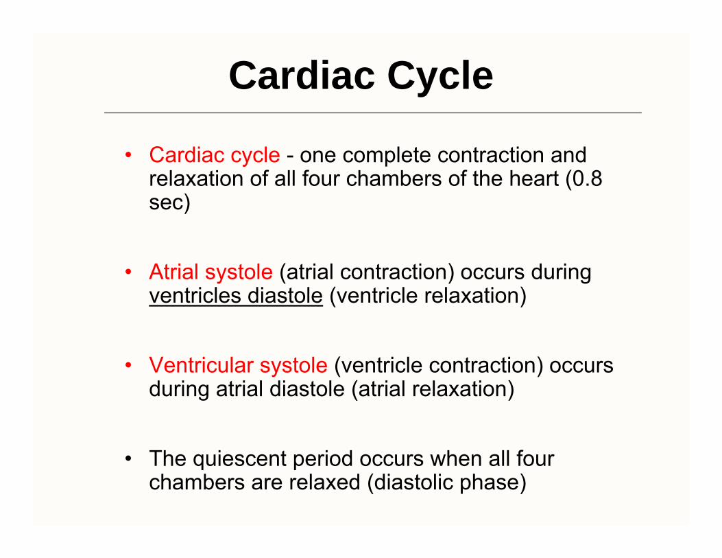
Cardiac Cycle
• Cardiac cycle - one complete contraction and relaxation of all four chambers of the heart (0.8 sec)
• Atrial systole (atrial contraction) occurs during ventricles diastole (ventricle relaxation)
• Ventricular systole (ventricle contraction) occurs during atrial diastole (atrial relaxation)
• The quiescent period occurs when all four chambers are relaxed (diastolic phase)
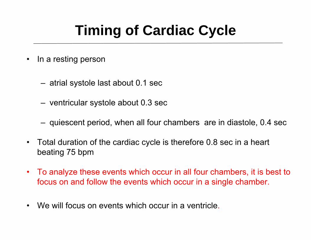
Timing of Cardiac Cycle
• In a resting person
– atrial systole last about 0.1 sec
– ventricular systole about 0.3 sec
– quiescent period, when all four chambers are in diastole, 0.4 sec
• Total duration of the cardiac cycle is therefore 0.8 sec in a heart beating 75 bpm
• To analyze these events which occur in all four chambers, it is best to focus on and follow the events which occur in a single chamber.
• We will focus on events which occur in a ventricle.
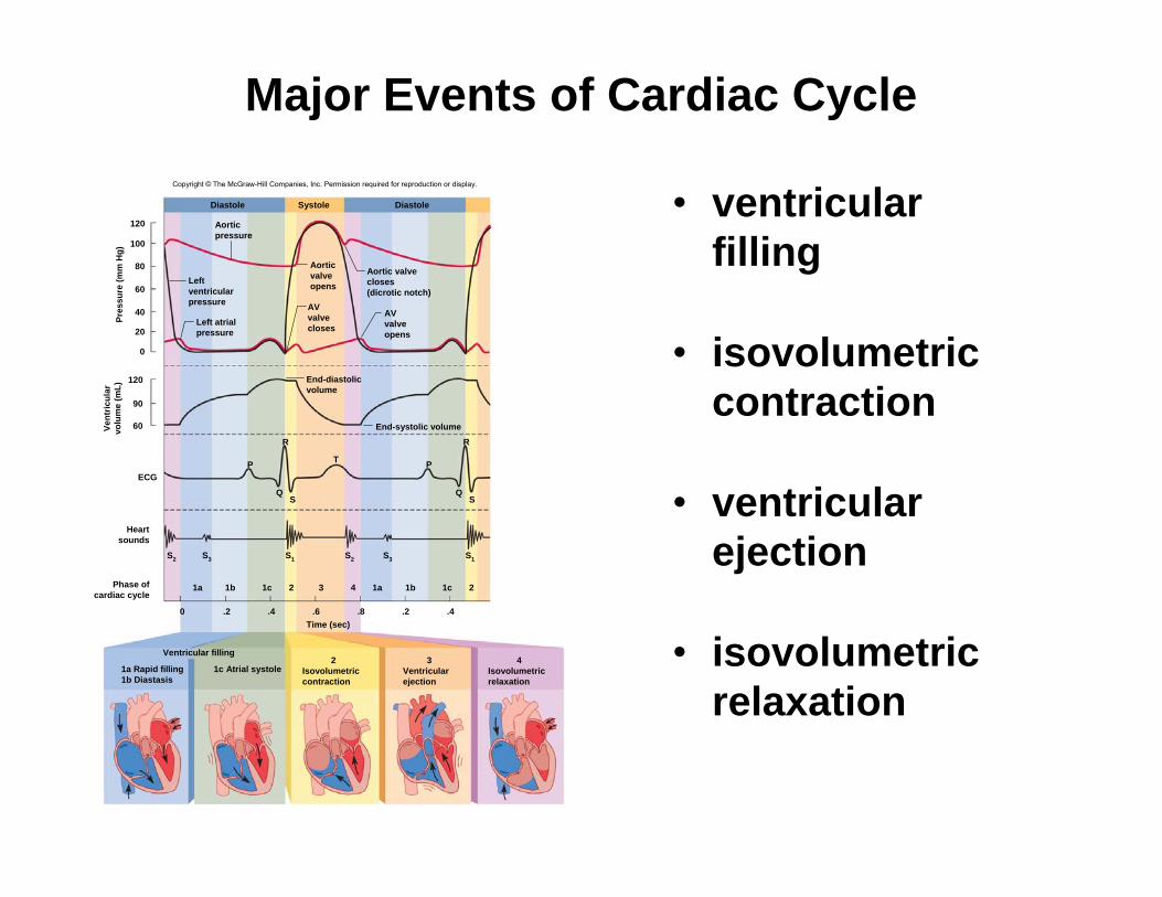
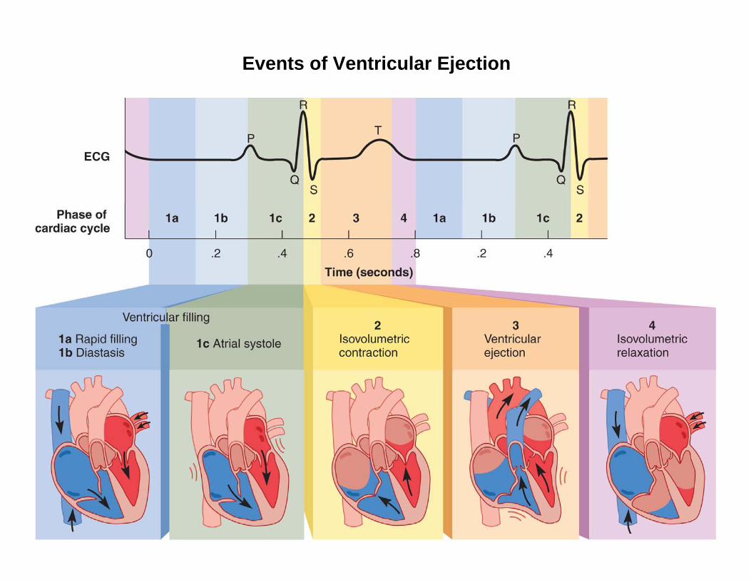
Major Events of Cardiac Cycle
• ventricular filling
• isovolumetric contraction
• ventricular ejection
• isovolumetric relaxation
Copyright © The McGraw-Hill Companies, Inc. Permission required for reproduction or display.
ECG
Pres
sure
(mm
Hg)
120
60
90
120
100
80
60
40
20
0
SystoleDiastole Diastole
0 .2 .4 .6 .8 .2 .4
End-systolic volume
Q
R
S
P
S1
T P
Q
R
S
S2 S3 S3
1a 1b 1c 3
S1
2 1a 1b 1c 24
S2
Ventricular filling
Vent
ricul
arvo
lum
e (m
L)
Heartsounds
Phase ofcardiac cycle
Aorticpressure
Leftventricularpressure
Left atrialpressure
Aorticvalveopens
Aortic valvecloses(dicrotic notch)
AVvalvecloses
AVvalveopens
End-diastolicvolume
Time (sec)
1a Rapid filling1b Diastasis
1c Atrial systole2
Isovolumetriccontraction
3Ventricularejection
4Isovolumetricrelaxation

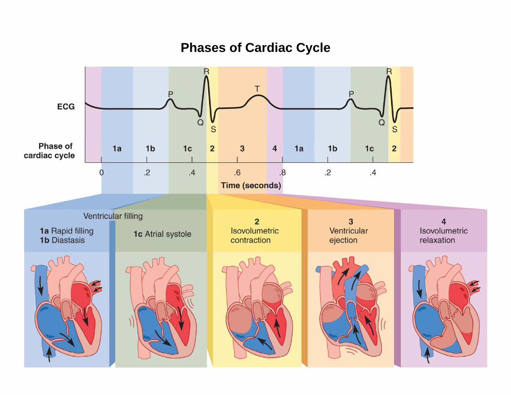
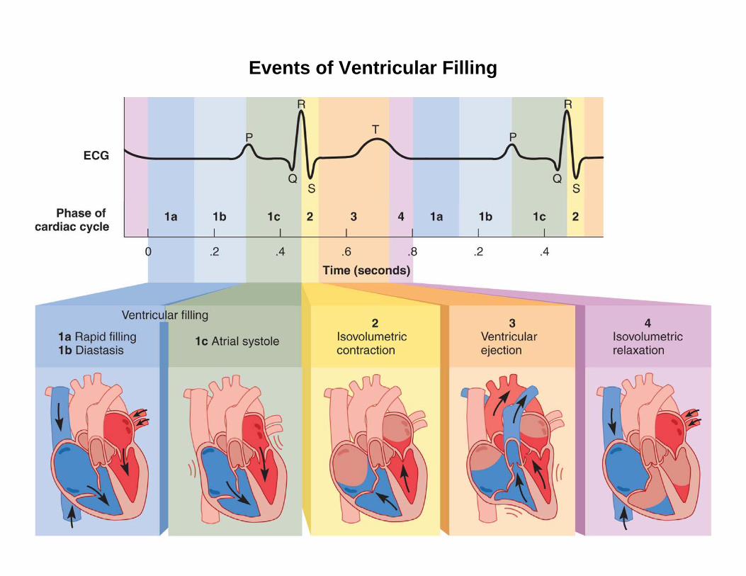
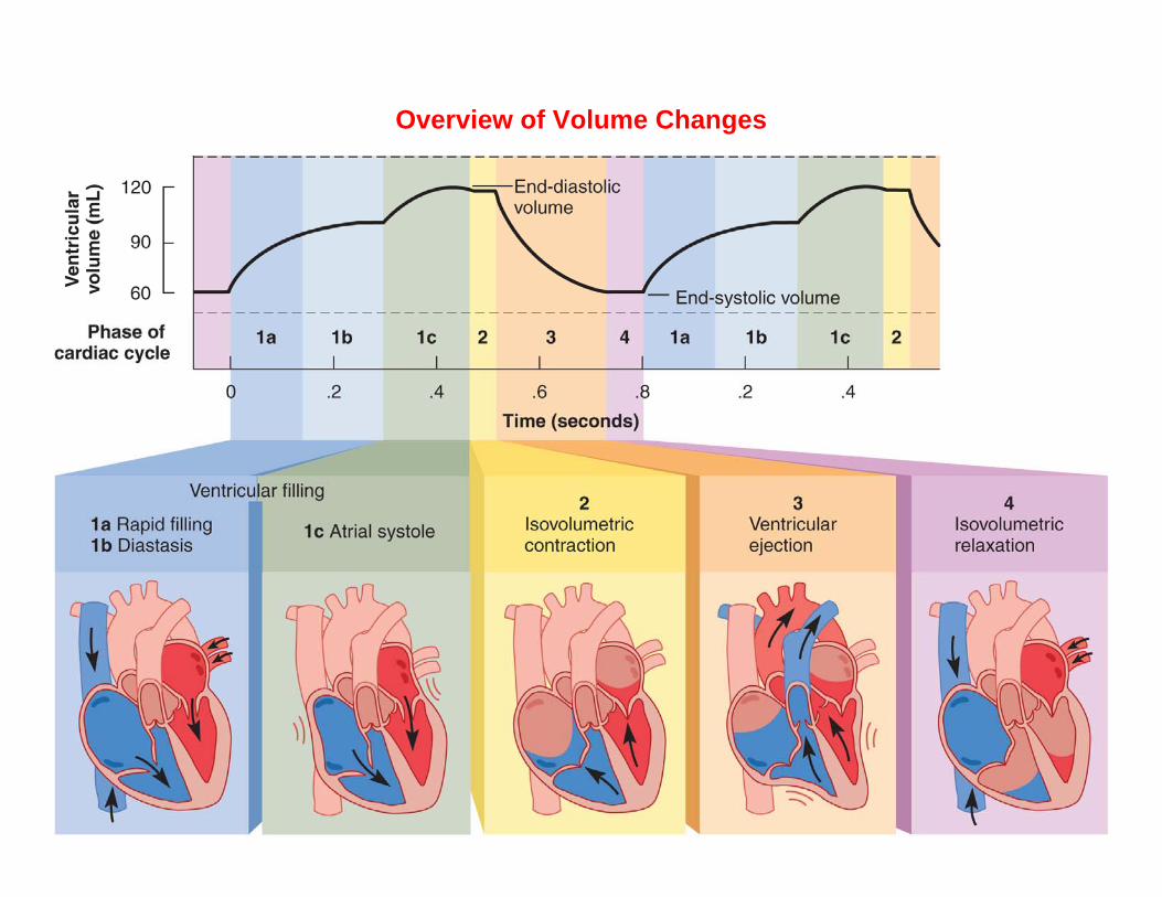
Phases of Cardiac Cycle
• ventricular filling / associated in part with atrial systole
• isovolumetric contraction of ventricles
• ventricular ejection
• isovolumetric relaxation
• all events in one cardiac cycle are completed in 0.8 second
Phases of Cardiac Cycle

Events of Ventricular Filling (1 of 3)
• Events associated with ventricular diastole = Ventricular Filling
– Ventricles expand
– Ventricular pressure drops below pressure in atria
– AV valves open and blood flows into the ventricles
Events of Ventricular Filling (2 of 3)
• Ventricular filling occurs in three phases:
– rapid ventricular filling - first one-third • blood enters very quickly / before atrial
systole begins
– diastole continues in atria - second one-third• marked by slower filling• P wave occurs at the end of diastasis
– atrial systole - final one-third • atria contract
Events of Ventricular Filling (3 of 3)
• Reaching the “end-diastolic volume” of ventricles (EDV)
– amount of blood contained in ventricles at the end of ventricular filling
– same volume in right and left ventricle // must never be different
– 130 mL of blood in each ventricle at end of ventricular diastole
Events of Ventricular Filling
Events of Iso-volumetric Contraction (1 of 2)
• Atria repolarize and relax // remain in diastole for the rest of the cardiac cycle
• Ventricles depolarize– this initiates the QRS complex– Depolarization followed by the contraction
• AV valves close as ventricular blood pressure increases // forcing blood to surge back against the AV cusps
– heart sound S1 occurs at the beginning of this phase // closing of AV valves
Events of Iso-volumetric Contraction (1 of 2)
• Now entering the ‘isovolumetric’ contractionphase
– ventricles contracting but they do not eject blood // why?
– Note: both AV and semilunar values are STILL CLOSED
– because pressure in the aorta (80 mm Hg) and in pulmonary trunk (10 mm Hg) is still greater than in the pressure in the two ventricles
• Cardiocytes exert force, but with all four valves closed, the blood cannot go anywhere // rapid increase in pressure

Events of Iso-volumetric Contraction
Events of Ventricular Ejection (1 of 2)
• ejection of blood begins when the ventricular pressure exceeds arterial pressure and forces semilunar valves open
– pressure peaks in left ventricle at about 120 mm Hg and 25 mm Hg in the right
• blood spurts out of each ventricle rapidly at first – rapid ejection
• then more slowly under reduced pressure – reduced ejection
• ventricular ejections last about 200 – 250 msec
– corresponds to the plateau phase of the cardiocyteaction potential
Events of Ventricular Ejection (2 of 2)
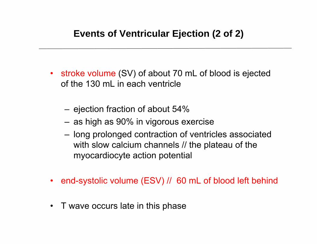
• stroke volume (SV) of about 70 mL of blood is ejected of the 130 mL in each ventricle
– ejection fraction of about 54%– as high as 90% in vigorous exercise– long prolonged contraction of ventricles associated
with slow calcium channels // the plateau of the myocardiocyte action potential
• end-systolic volume (ESV) // 60 mL of blood left behind
• T wave occurs late in this phase

Events of Ventricular Ejection
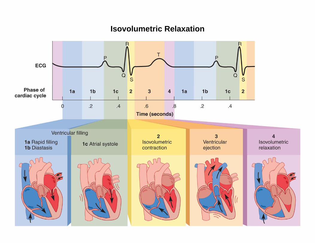
Isovolumetric Relaxation
• Early ventricular diastole /// when T wave ends and the ventricles begin to expand
• Elastic recoil and expansion would cause pressure to drop rapidly and suck blood into the ventricles
– blood from the aorta and pulmonary briefly flows backwards
– filling the semilunar valves and closing the cusps
– creates a slight pressure rebound that appears as the dicrotic notch of the aortic pressure curve

Isovolumetric Relaxation
– heart sound S2 occurs as blood rebounds from the closed semilunar valves and the ventricle expands
– ‘isovolumetric’ because semilunar valves are closed and AV valves have not yet opened // ventricles are therefore taking in no blood
– when AV valves open, ventricular filling begins again
Isovolumetric Relaxation
Overview of Volume Changes
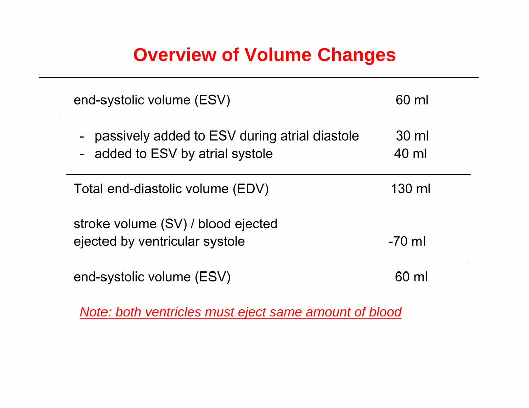
end-systolic volume (ESV) 60 ml
- passively added to ESV during atrial diastole 30 ml- added to ESV by atrial systole 40 ml
Total end-diastolic volume (EDV) 130 ml
stroke volume (SV) / blood ejectedejected by ventricular systole -70 ml
end-systolic volume (ESV) 60 ml
Note: both ventricles must eject same amount of blood
Overview of Volume Changes

Heart Sounds
• auscultation - listening to sounds made by body
• first heart sound (S1), louder and longer “lubb”, occurs with closure of AV valves, turbulence in the bloodstream, and movements of the heart wall
• second heart sound (S2), softer and sharper “dupp” occurs with closure of semilunar valves, turbulence in the bloodstream, and movements of the heart wall
• S3 - rarely heard in people over 30
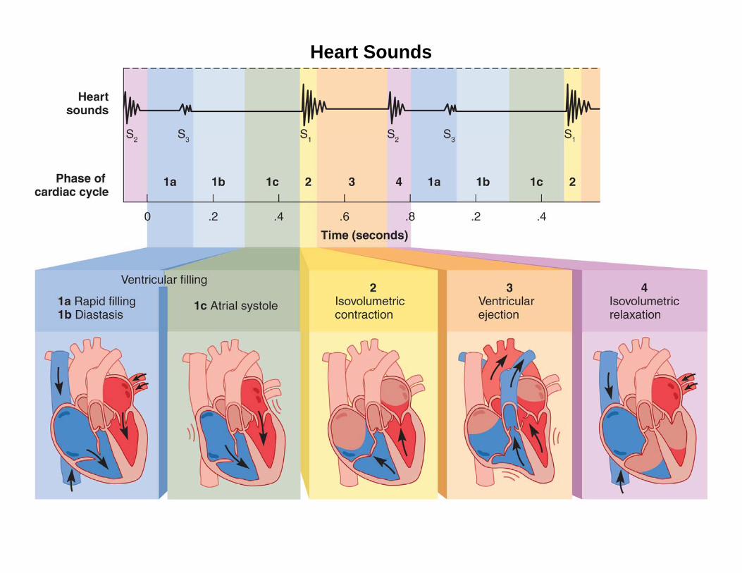
Heart Sounds
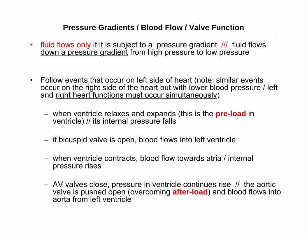
Pressure Gradients / Blood Flow / Valve Function
• fluid flows only if it is subject to a pressure gradient /// fluid flows down a pressure gradient from high pressure to low pressure
• Follow events that occur on left side of heart (note: similar events occur on the right side of the heart but with lower blood pressure / left and right heart functions must occur simultaneously)
– when ventricle relaxes and expands (this is the pre-load in ventricle) // its internal pressure falls
– if bicuspid valve is open, blood flows into left ventricle
– when ventricle contracts, blood flow towards atria / internal pressure rises
– AV valves close, pressure in ventricle continues rise // the aortic valve is pushed open (overcoming after-load) and blood flows into aorta from left ventricle
Pressure Gradients / Blood Flow / Valve Function
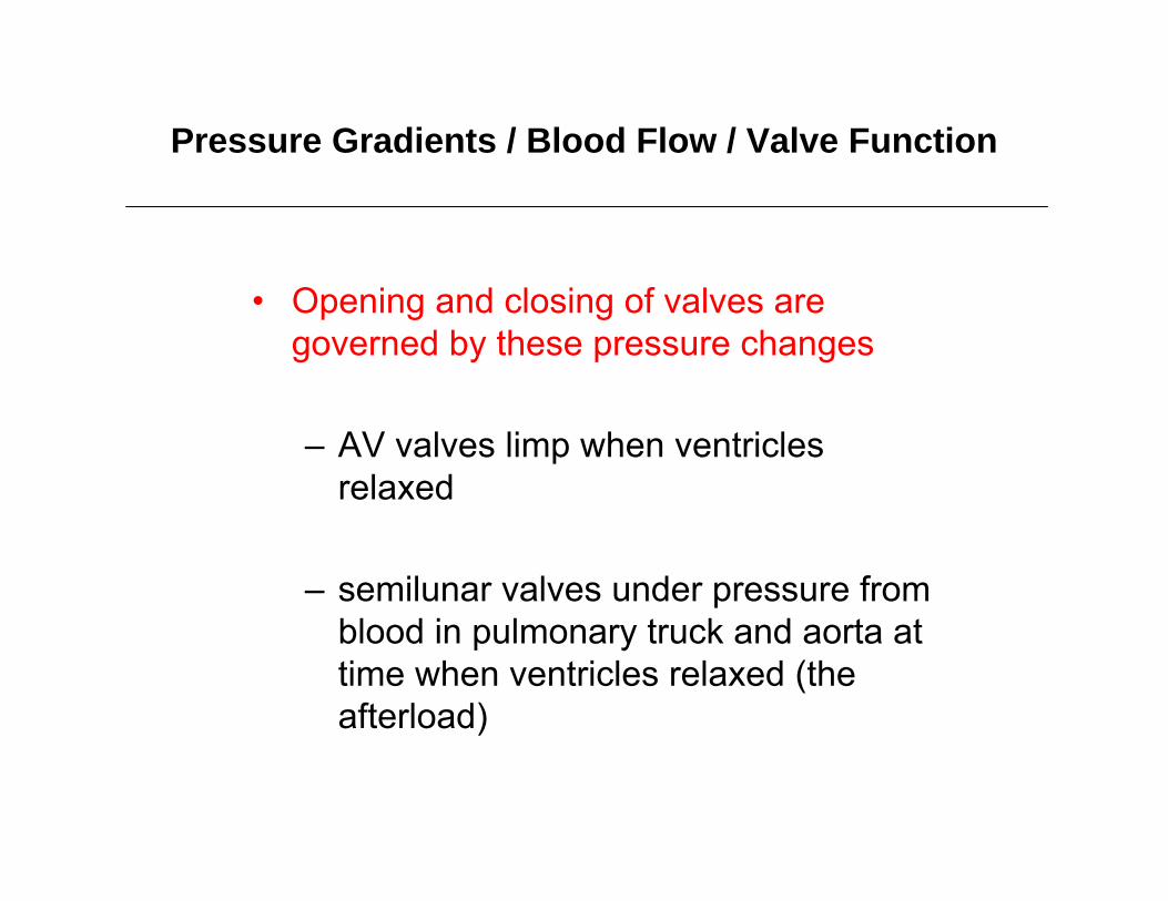
• Opening and closing of valves are governed by these pressure changes
– AV valves limp when ventricles relaxed
– semilunar valves under pressure from blood in pulmonary truck and aorta at time when ventricles relaxed (the afterload)

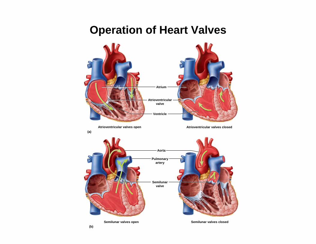
Aorta
Semilunar valves open
Atrioventricular valves closed
Semilunar valves closed
Atrium
(b)
Atrioventricular valves open(a)
Pulmonaryartery
Semilunarvalve
Atrioventricularvalve
Ventricle
Operation of Heart Valves
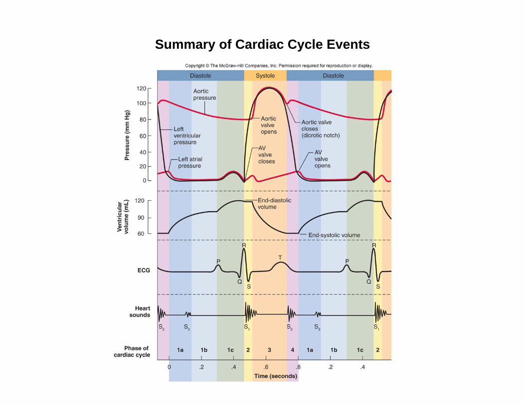
Summary of Cardiac Cycle Events

Cardiac Output (CO)
cardiac output = stroke volume x heart rate
Volume of blood ejected by ventricle in 1 minute
CO = 70 ml / Beat x 75 Beat / Minutes = 5.25 L / Min
Cardiac Output May Be Changed By
chronotropic effects (time // related to the heart rate)
inotropic effects (related to the force of contraction)
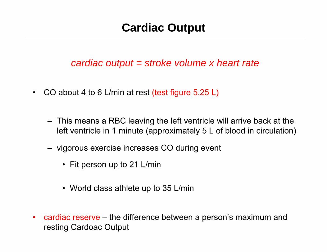
Cardiac Output
cardiac output = stroke volume x heart rate
• CO about 4 to 6 L/min at rest (test figure 5.25 L)
– This means a RBC leaving the left ventricle will arrive back at the left ventricle in 1 minute (approximately 5 L of blood in circulation)
– vigorous exercise increases CO during event
• Fit person up to 21 L/min
• World class athlete up to 35 L/min
• cardiac reserve – the difference between a person’s maximum and resting Cardoac Output
Cardiac Output
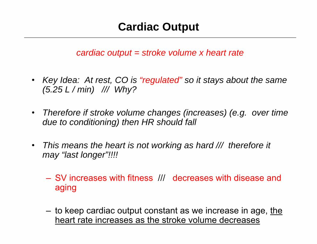
cardiac output = stroke volume x heart rate
• Key Idea: At rest, CO is “regulated” so it stays about the same (5.25 L / min) /// Why?
• Therefore if stroke volume changes (increases) (e.g. over time due to conditioning) then HR should fall
• This means the heart is not working as hard /// therefore it may “last longer”!!!!
– SV increases with fitness /// decreases with disease and aging
– to keep cardiac output constant as we increase in age, the heart rate increases as the stroke volume decreases
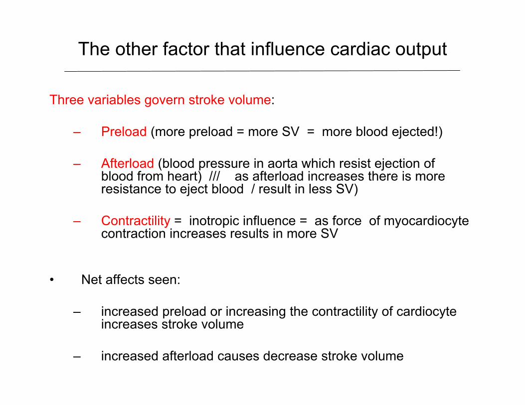
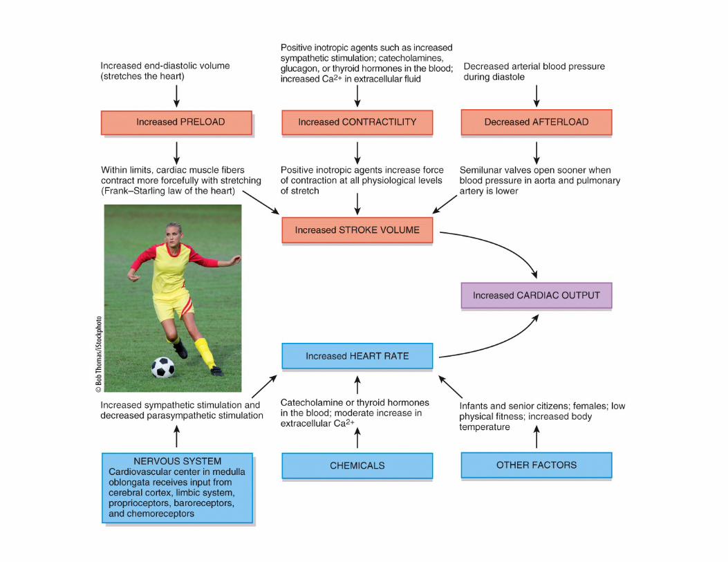
The other factor that influence cardiac output
Three variables govern stroke volume:
– Preload (more preload = more SV = more blood ejected!)
– Afterload (blood pressure in aorta which resist ejection of blood from heart) /// as afterload increases there is more resistance to eject blood / result in less SV)
– Contractility = inotropic influence = as force of myocardiocytecontraction increases results in more SV
• Net affects seen:
– increased preload or increasing the contractility of cardiocyteincreases stroke volume
– increased afterload causes decrease stroke volume
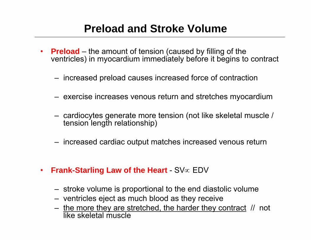
Preload and Stroke Volume
• Preload – the amount of tension (caused by filling of the ventricles) in myocardium immediately before it begins to contract
– increased preload causes increased force of contraction
– exercise increases venous return and stretches myocardium
– cardiocytes generate more tension (not like skeletal muscle / tension length relationship)
– increased cardiac output matches increased venous return
• Frank-Starling Law of the Heart - SV∝ EDV
– stroke volume is proportional to the end diastolic volume– ventricles eject as much blood as they receive– the more they are stretched, the harder they contract // not
like skeletal muscle
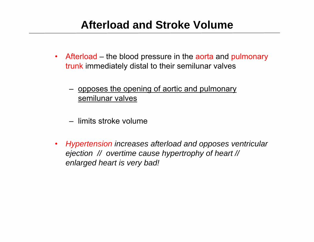
Afterload and Stroke Volume
• Afterload – the blood pressure in the aorta and pulmonary trunk immediately distal to their semilunar valves
– opposes the opening of aortic and pulmonary semilunar valves
– limits stroke volume
• Hypertension increases afterload and opposes ventricular ejection // overtime cause hypertrophy of heart // enlarged heart is very bad!
Role of Circulation, Afterload and Ventricular Failure
• Anything that impedes arterial circulation in either the systemic or pulmonary circuit may also increase afterload
• E.g. // lung diseases will restrict blood flow into pulmonary circulation // blood “backs up”or “builds up above the semilunar valve
• Cor pulmonale – right ventricular failure due to obstructed pulmonary circulation
– These diseases restrict blood flow through lungs: emphysema, chronic bronchitis, and black lung disease
Contractility and Stroke Volume
• contractility refers to how hard the myocardium contracts for any given preload
• positive inotropic agents that increase contractility
– hypercalcemia can cause strong, prolonged contractions and even cardiac arrest in systole
– catecholamines increase calcium levels
– glucagon stimulates cAMP production
– digitalis raises intracellular calcium levels and contraction strength

Contractility and Stroke Volume (cont.)
• negative inotropic agents reduce contractility
– hypocalcemia can cause weak, irregular heartbeat and cardiac arrest in diastole
– hyperkalemia reduces strength of myocardial action potentials and the release of Ca2+ into the sarcoplasm
– vagus nerve has an effect on atria (the nodes) which reduces heart rate
– However…..few vagus nerves innervate myocardiocytes in ventricles /// therefore vagus has no significant negative inotropic effect
Heart Rate
• Heart rate varies throughout life
– infants have HR of 120 bpm or more
– young adult females avg. 72 - 80 bpm
– young adult males avg. 64 to 72 bpm
– heart rate rises again in the elderly
• Positive chronotropic agents – factors that raise the heart rate
• Negative chronotropic agents – factors that lower heart rate
Chronotropic Effects of the Autonomic Nervous System
• autonomic nervous system
– does not initiate the heartbeat, – but ANS modulates the rhythm and force
• cardiostimulatory effect
– some neurons of the cardiac center transmit signals to the heart by way of sympathetic pathways
• cardioinhibitory effect
– others transmit parasympathetic signals by way of the vagus nerve
Chronotropic Effects of the Autonomic Nervous System (cont.)
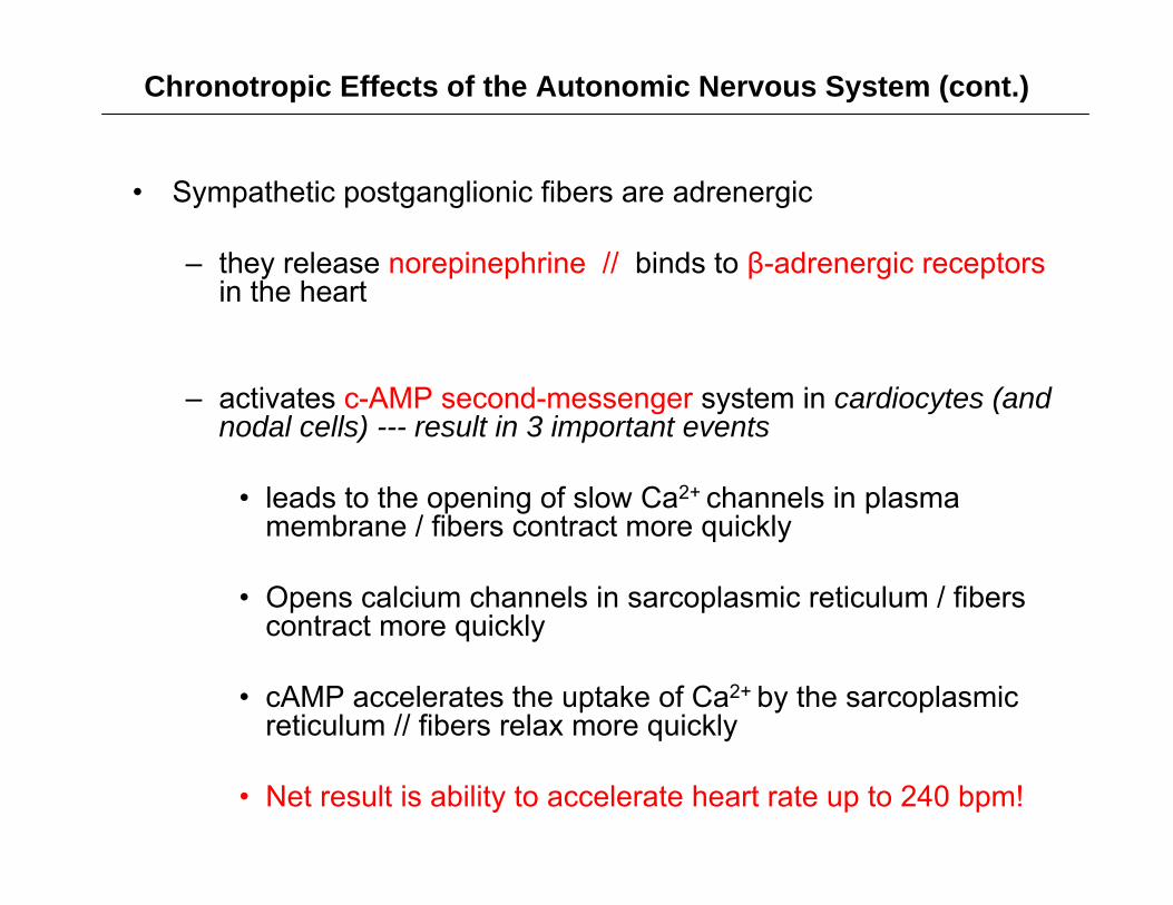
• Sympathetic postganglionic fibers are adrenergic
– they release norepinephrine // binds to β-adrenergic receptorsin the heart
– activates c-AMP second-messenger system in cardiocytes (and nodal cells) --- result in 3 important events
• leads to the opening of slow Ca2+ channels in plasma membrane / fibers contract more quickly
• Opens calcium channels in sarcoplasmic reticulum / fibers contract more quickly
• cAMP accelerates the uptake of Ca2+ by the sarcoplasmic reticulum // fibers relax more quickly
• Net result is ability to accelerate heart rate up to 240 bpm!
Chronotropic Effects May Reduce Stroke Volume
By accelerating the rate of contraction (how fast calcium is added to sarcoplasm) and then accelerating the reuptake of calcium into sarcoplasmic reticulum to increase rate of relaxation --- heart rate is increased!
• Sympathetic NS (norepinephrine) able to increase the heart rate as high as 240 bpm
– Note: at these high heart rates / diastole becomes too brief for complete filling of the ventricles!!!!
– So at 240 bpm both stroke volume and cardiac output are reduced

Chronotropic Effects of the Autonomic Nervous System
• parasympathetic (vagus nerves) are cholinergic fibers // inhibitory effects on the SA and AV nodes
– acetylcholine (ACh) binds to muscarinic receptors (cAMP mediated)
– opens K+ gates in the nodal cells
– as K+ leaves the cells, they become hyperpolarized and fire less frequently
– heart slows down
– parasympathetics effect on the heart is faster than sympathetics
Chronotropic Effects of the Autonomic Nervous System (cont.)
• Vagal Tone (parasympathetic tone)
– the heart has a intrinsic “natural” firing rate of 100 bpm
– vagal tone – holds down this natural heart rate to 70 – 80 bpm at rest
– Caused by steady background firing rate of the vagus nerves
Chronotropic Chemicals
• chemicals affect heart rate // in addition to the neurotransmitters from cardiac nerves
– blood born adrenal catecholamines (NE and epinephrine) are potent cardiac stimulants
• drugs may also stimulate heart
– nicotine stimulates catecholamine secretion
– thyroid hormone increases number adrenergic receptors on heart so more responsive to sympathetic stimulation
– caffeine inhibits cAMP breakdown /// therefore can prolong the adrenergic effect
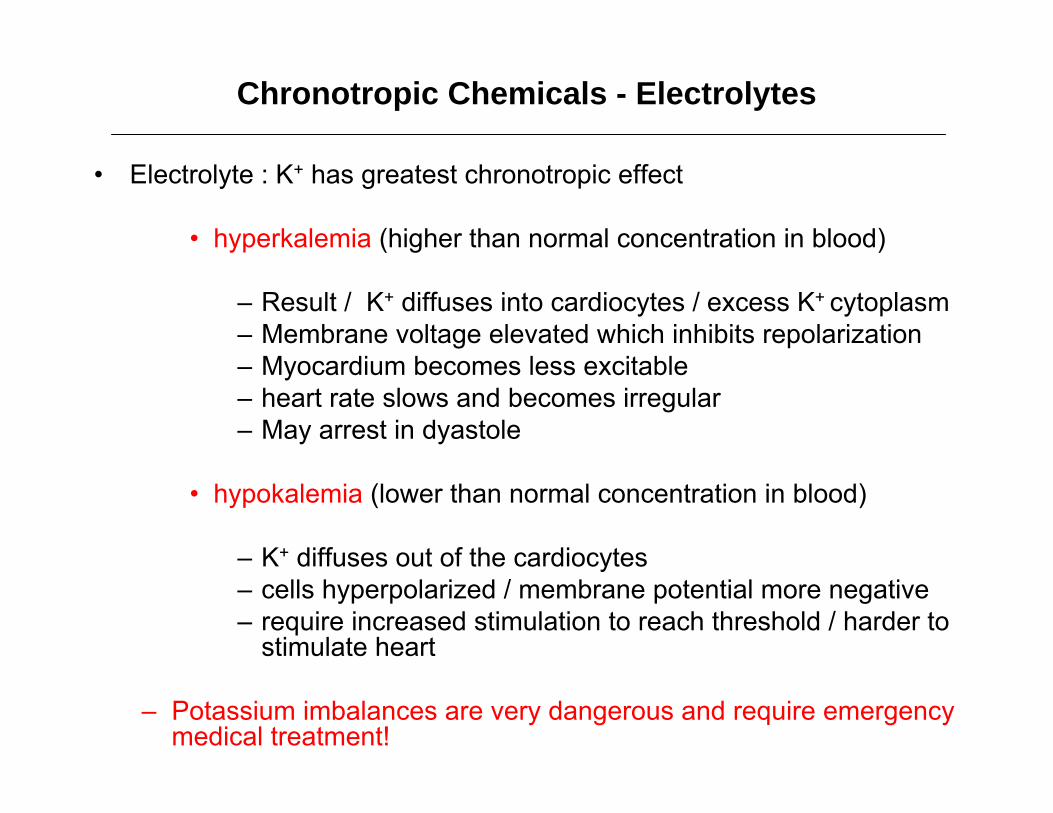
• Electrolyte : K+ has greatest chronotropic effect
• hyperkalemia (higher than normal concentration in blood)
– Result / K+ diffuses into cardiocytes / excess K+ cytoplasm– Membrane voltage elevated which inhibits repolarization– Myocardium becomes less excitable– heart rate slows and becomes irregular– May arrest in dyastole
• hypokalemia (lower than normal concentration in blood)
– K+ diffuses out of the cardiocytes– cells hyperpolarized / membrane potential more negative– require increased stimulation to reach threshold / harder to
stimulate heart
– Potassium imbalances are very dangerous and require emergency medical treatment!
Chronotropic Chemicals - Electrolytes

• Electrolyte : Ca2+ also affect heart rate
– hypercalcemia – excess of Ca2+
– decreases heart rate and contraction strength– Slow heart rate
– hypocalcemia – deficiency of Ca2+
– increases heart rate– More of an affect on contraction strength– Rare condition
– Greater effect is on nerve fibers causing action potential in somatic nerve fibers going to skeletal muscles (like diaphragm) / death from respiratory arrest!
Chronotropic Chemicals - Electrolytes

Heart Function Terms
• Pulse pressure – surge of pressure produced by each heart beat that can be felt by palpating a superficial artery with thefingertips
• Tachycardia - resting adult heart rate above 100 bpm
– stress, anxiety, drugs, heart disease, or fever
– loss of blood or damage to myocardium
• Bradycardia - resting adult heart rate of less than 60 bpm
– in sleep, low body temperature, and endurance trained athletes

Exercise and Cardiac Output• exercise makes the heart work harder // stroke volume
increases // heart rate can be slower and still reach target cardiac output // increase cardiac reserve
• Exercise stimulate proprioceptors in skeletal muscles that sendsignal to cardiac center
– at beginning of exercise, signals from joints and muscles reach the cardiac center
• sympathetic output from cardiac center increases cardiac output
– increased muscular activity /// increases venous return
• increases preload /// results in an increase cardiac output
– increase in both heart rate and stroke volume can both cause increases in cardiac output
Exercise and Cardiac Output
• exercise produces ventricular hypertrophy
– increased stroke volume allows heart to beat more slowly at rest
– athletes with increased cardiac reserve can tolerate more exertion than a sedentary person
Valvular Insufficiencies
• valvular insufficiency (incompetence) // any failure of a valve to prevent reflux (regurgitation) the backward flow of blood
• valvular stenosis – cusps are stiffened and opening is constricted by scar tissue
• result of rheumatic fever autoimmune attack on the mitral and aortic valves
• heart overworks and may become enlarged
Valvular Insufficiencies
• heart murmur – abnormal heart sound produced by regurgitation of blood through incompetent valves
• mitral valve prolapse – insufficiency in which one or both mitral valve cusps bulge into atria during ventricular contraction
• hereditary in 1 out of 40 people
• may cause chest pain and shortness of breath

Congestive Heart Failure
– results from the failure of either ventricle to eject blood effectively
– One ventricle ejects proper amount of blood while the other ventricle ejects less blood
– The ventricle which ejects less blood is the failing ventricle
– usually due to a heart weakened by
• myocardial infarction• chronic hypertension• valvular insufficiency• congenital defects in heart structure.
Congestive Heart Failure // Left Ventricular Failure
– Left ventricle ejects less blood (e.g. Rt V ejects 70 ml and Lt V ejects 50 ml)
– Rt. Ventricle is ejecting 20 ml more blood than Lt. ventricle during each cardiac cycle
– extra “20 ml” most go somewhere // it accumulates in the lung interstitial space // pulmonary edema
– shortness of breath or sense of suffocation

Unbalanced Ventricular Output
Left ventricular failure results in
pulmonary edemaPressure backs up.
1
2
3
(a) Pulmonary edema
32
1
Right ventricularoutput exceeds leftventricular output.
Fluid accumulates inpulmonary tissue.
Note:
Cor pulmonale will also results in pulmonary edema
Due to lung emphysema and other disease states which cause restriction (fibrosis) in lung tissue
Enlarged right heart // these condition will contribute to right heart failure
Congestive Heart Failure // Rt. Ventricular Failure
– Right ventricle ejects less blood (e.g. Lt V ejects 70 ml and Rt V ejects 50 ml)
– Left ventricle ejects extra 20 ml of blood per cardiac cycle
– Rt ventricle can not receive the total volume so extra 20 ml filters into the systemic interstitial space // systemic edema -seen primarily in the legs
– enlargement of the liver, ascites (pooling of fluid in abdominalcavity), distension of jugular veins, swelling of the fingers, ankles, and feet
• Note: Either condition will lead eventually to total heart failure
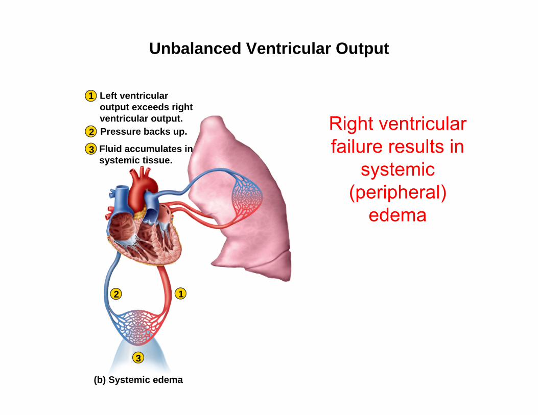
Unbalanced Ventricular Output
Right ventricular failure results in
systemic (peripheral)
edema
Pressure backs up.
1
2
3
(b) Systemic edema
12
3
Left ventricularoutput exceeds rightventricular output.
Fluid accumulates insystemic tissue.
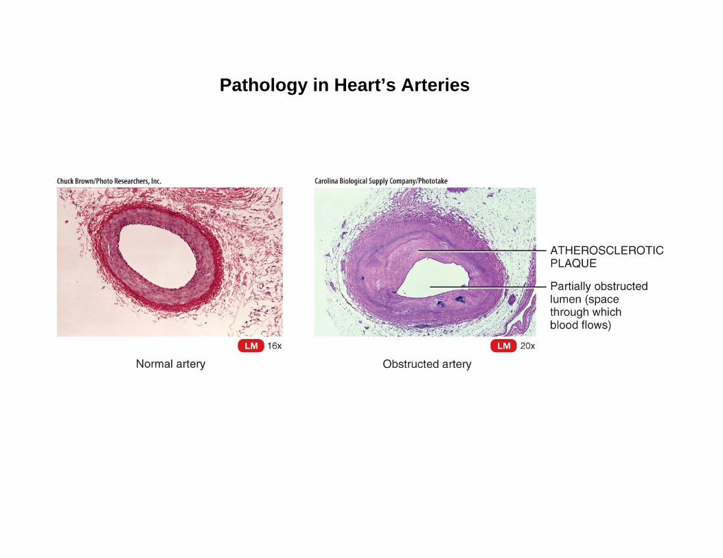
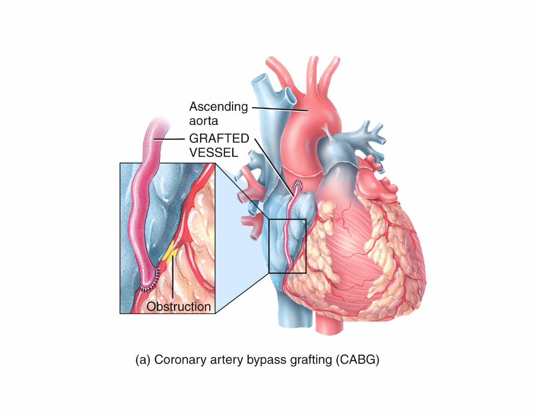
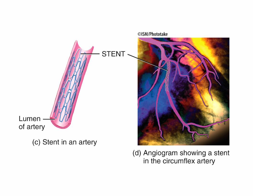
Pathology in Heart’s Arteries