the capsid protein of semliki forest virus has clusters of basic amino acids and prolines in its
TRANSCRIPT

Proc. Natl. Acad. Sci. USAVol. 77, No. 11, pp. 6376-6380, November 1980Biochemistry
The capsid protein of Semliki Forest virus has clusters of basicamino acids and prolines in its amino-terminal region
(nucleotide sequencing/cDNA/molecular cloning/protein-RNA interactions)
H. GAROFF, A.-M. FRISCHAUF, K. SIMONS, H. LEHRACH, AND H. DELIUSEuropean Molecular Biology Laboratory, Meyerhofstrasse 1, Postfach 10.2209, 69 Heidelberg, Federal Republic of Germany
Communicated by John C. Kendrew, July 21, 1980
ABSTRACT The amino acid sequence of the capsid (C)protein was deduced from the nucleotide sequence of the Cgene. This part of the viral 42S RNA genome was transcribedinto double-stranded cDNA. The cDNA was cloned in theEscherichia coil X1776-pBR322 host-vector system and thenthe base sequence was determined with the technique describedby Maxam and Gilbert. The amino acid sequence of the C pro-tein shows a clustering of basic amino acids and prolines withinthe first 110 amino acids.
from the virus. This specificity can probably be explained bythe formation of bonds between the C protein in the nucleo-capsid and the spanning segments of the spike glycoproteins(9).A more detailed understanding of SFV structure and as-
sembly at the molecular level is difficult without the knowledgeof the amino acid sequences of the structural proteins. We re-port here the primary structure of the C protein.
Semliki Forest virus (SFV) is a simple membrane virus of thealphavirus group. It has been used extensively as a model systemto study the structure and assembly of cellular membranes (1).The virus particle consists of an icosahedral nucleocapsid sur-rounded by a membrane. The nucleocapsid is a complex ofabout 240 capsid proteins (C protein, Mr = 30,000) (2, 3) anda RNA molecule (42S), the viral genome (4). The membraneconsists of a lipid bilayer with about 240 external glycoproteinspikes (5). Each spike contains three different glycopolypep-tides: El (Mr = 49,000), E2 (Mr = 52,000), and E3 (Mr =10,000) (6, 7). The E2 polypeptide spans the membrane; thereare about 30 amino acid residues present on the internal sideof the viral membrane (8, 9).The virus enters the host cell by absorptive endocytosis (10).
Inside the lysosomes of the cell, the low pH probably triggersa fusion of the viral membrane with the lysosomal membrane(10, 11). This allows the nucleocapsid to enter the cell cyto-plasm, where the viral genome is uncoated so that it can act asa mRNA for synthesis of polymerase molecules. The viral RNApolymerase synthesizes new 42S RNA molecules and smaller26S RNA molecules. The latter molecule is homologous to the3' end of the viral genome (12) and functions as a mRNA forthe SFV structural proteins, which are translated from a singleinitiation site (13). The C protein is made first. As soon as it iscompleted it is cleaved from the growing polypeptide chain,and the ribosomes continue to read off the membrane proteinsin the order E3, E2, and El (8, 14). The membrane proteins arecotranslationally translocated across the membrane of the en-doplasmic reticulum and transported to the plasma membrane(15, 16).The assembly of the nucleocapsid in the cell cytoplasm is not
understood. Newly synthesized capsid proteins are known tobe associated with the large subunit of the ribosome before theycomplex with the 42S RNA into nucleocapsids (17). The finalstep in SFV assembly, budding, takes place at the cell surface(18). The nucleocapsid binds to the cytoplasmic aspect of theplasma membrane which folds around the nucleocapsid. In thebudding process the virus-spike glycoproteins are included intothe viral membrane, whereas the host proteins are excluded
MATERIALS AND METHODSMaterials. Oligo(dT)-cellulose (T3) was from Collaborative
Research (Waltham, MA). All isotopes were purchased fromRadiochemical Centre (Amersham, England). Restriction en-donuclease Xho I and Ava I were from P-L Biochemicals. ClaI, HindIII, HindII, Alu I, Pst I, and BamHI were fromBoehringer. Hpa II, Hha I, HinfI, Hae III, Sau I, Taq I, andEcoRI were gifts from V. Pirrotta. Reverse transcriptase wasgenerously provided by J. Beard. DNA polymerase was a giftfrom W. McClure. S1 nuclease and T4 polynucleotide kinasewere prepared as described (see ref. 19).
Isolation of RNA. The 42S RNA was isolated from purifiedvirus particles. SFV (5 mg of protein) was dissociated in 5 mlof 10 mM Tris, pH 7.4/10mM NaCl/1.5 mM MgCl2/1% Na-DodS04. The clear solution was extracted twice with redistilledphenol/chloroform/isoamyl alcohol, 2.4:2.4:0.1 (vol/vol). TheRNA in the waterphase was precipitated with ethanol anddissolved in H20 (1 mg/ml).The 26S RNA was isolated from infected BHK-21 cells.
Thirty Falcon bottles (75 cm2) with a monolayer of BHK-21cells were infected with SFV at about 100 plaque-forming unitsper cell (20). The cells were cooled on ice 6 hr after infection,and a cytoplasmic extract was prepared as described (21). Thisextract (--25 ml) was made 2% in NaDodSO4 and then dilutedto 100 ml with 10 mM Tris, pH 7.4/50 mM NaCl. After ex-tracting twice with phenol/chloroform/isoamyl alcohol, 48:48:2(vol/vol), the nucleic acids in the water phase were precipitatedand taken up in 10 mM Tris, pH 7.4/1 mM EDTA/0.1% Na-DodSO4. From this preparation poly(A)-containing RNAmolecules were isolated by chromatography on oligo(dT)-cel-lulose (22) and fractionated further on a 10-30% (wt/vol) su-crose gradient (23). The RNA in the 26S fraction was collected,precipitated, and dissolved in H20 (n1 mg/ml). All RNAsamples were stored at -700C.
Preparation of Double-Stranded cDNA. About 10 Mg of 42SRNA was used as template for cDNA synthesis. The procedureto prepare double-stranded (ds) cDNA has been described (19).For colony hybridizations (see below) single-stranded (ss) cDNAthat contained one 32P-labeled nucleotide was used.
Molecular Cloning. The ds cDNA was inserted into the
Abbreviations: SFV, Semliki Forest virus; C, capsid; ds, doublestranded; ss, single-stranded; kb, kilobase; bp, base pair.
6376
The publication costs of this article were defrayed in part by pagecharge payment. This article must therefore be hereby marked "ad-vertisement" in accordance with 18 U. S. C. §1734 solely to indicatethis fact.

Proc. Natl. Acad. Sci. USA 77 (1980) 6377
Pst I site of the plasmid pBR322 by using the oligo(dG-dC)-tailing procedure described by R6wekamp and Firtel (24).Escherichia coli X1776 was transformed with the hybridplasmid (25) in a P3 physical containment laboratory at theEuropean Molecular Biology Laboratory. After growth thecolonies were hybridized against 32P-labeled ss cDNA from 428RNA to detect chimeric plasmids (26). Several strongly hybri-dizing colonies were found. These were screened for length ofthe hybrid plasmid by using the miniscreening technique ofBarnes (27). The clones containing the longest hybrid plasmidswere then grown in 1-liter cultures. Hybrid plasmids werepurified from these clones either by chromatography on hy-droxyapatite (28) or by equilibrium centrifugation in dye/CsClgradients. E. coli tRNA, which still contaminated the plasmidfraction, was removed by gel filtration. All plasmid preparationswere stored at -300C in H20 (1 mg/Ml).
This work was done in accordance with the German guide-lines for recombinant DNA research.
Characterization of Hybrid Plasmids. A preliminarycharacterization of the insert in the hybrid plasmid was madewith electron microscopy. The insert was cleaved from thevector with Pst I and isolated by electrophoresis on an agarosegel (see below). Purified inserts were then denatured and hy-bridized to 428 or 26S RNA in a buffer containing 80% (vol/vol)formamide (29). The nucleic acids were treated with gene 32protein of the T4 phage and adsorbed to mica sheets (30). Thelength of the insert molecule and its location (that is, its distancefrom the 3' end of the RNA molecule) were measured. Valuesin base pairs were obtained by comparison to the DNA of phagePM 2 as an internal standard [10 kilobases (kb)] or to in vitrotranscripts of phage T7. A molecular weight of 4.2 X 10⁶ wasfound for the 42S RNA. This corresponds to 12.7 kb.Mapping with restriction endonucleases was done as de-
scribed by Smith and Birnstiel (32). The digestion conditionsfor the various restriction endonucleases were as follows: 10mMTris, pH 7.5/10 mM MgCl2/10 mM dithiothreitol was usedwith Xho I, Ava I, Hinfl, Hpa II, Hae III, Hha I, Sau I, andBamHI. The MgCl2 concentration was lowered to 2 mM fordigestion with Taq L. NaCI (50 mM) was included in the bufferfor digestions with HindUl, Hindll, Pst L Alu L and Cla I; 100mM NaCI, was included in digestions with EcoRI. The incu-bation temperature was 70'C for Tag I, 30'C for Pst I, and370C for the rest. Partial digestion products of the end-labeledDNA molecule were obtained by transferring one fourth (2.51A) of the reaction volume at 5, 15, 30, and 60 min to 10 ,ul of40mM Tris, pH 7.8/5mM sodium acetate/0.1 M EDTA/2.5%glycerol/ 0.025% bromphenol blue. The amount of enzymeadded was calculated to give a nearly complete digestion in 60min.
Nucleotide Sequence Determinations. DNA fragmentswere end-labeled with 32P and the sequence was determinedby using base-specific chemical cleavages as described byMaxam and Gilbert (33). The polynucleotide kinase reactionwas used to label 5' termini with 32P. Recessive 3' ends of DNAfragments were labeled in reactions with DNA polymerase and[32P]deoxynucleoside triphosphates (34). End-labeled fragmentswere isolated by agarose gel electrophoresis and subsequentelectroelution into hydroxyapatite as described by Tabak andFlavell (35). The two strands of the labeled DNA fragmentswere dissociated in 0.3 M NaOH (10 min at 370C) and sepa-rated by electrophoresis in 6% (wt/vol) polyacrylamide gels (20X 40 cm) (36). The front plate was cooled to 20C. After elec-trophoresis at 300 V for 10 hr, the bands were located by au-toradiography, appropriate zones were cut from the gel, andthe DNA was eluted by shaking the gel pieces at 370C for 12hr in 0.5 ml of 40 mM Tris, pH 7.8/5 mM sodium acetate/imM EDTA.
. . ~0.5 kimhybrid O a,
4S RNA
FIG. 1. Electron micrograph of 42S RNA hybridized to the SFVcDNA insert ofpBR SFV 3 isolated after cleavage with Pst I. The dshybrid region appears as a thin thread in comparison with the ss re-gion complexed with gene 32 protein.
End-labeled DNA samples that had gone through fivebase-specific cleavage reactions (G, G+A, C+T, C, and C+A)were analyzed on sequencing gels essentially as described (37).The thickness of the gel was reduced to 0.2 mm. In some runsthe temperature of the gel was kept at 700C by using a plateheated with water instead of one of the normal glass plates.Before autoradiography the gel was fixed in 10% acetic acid for10 min and dried in an oven at 70'C. In order to make handlingof the thin gel easy, it was covalently bound to one of the glassplates by treating this with a silane mixture before casting thegel (unpublished data).The nucleotide sequences read from the gels were overlapped
by using a computer program that will be described else-where.
RESULTSCharacterization of a Clone Containing the C Gene.
Electron microscopic measurements on hybrids between therecombinant plasmid DNA and SFV RNA suggested that oneof the plasmids, pBR SFV 3 contained the entire C protein gene.Length measurements of the insert molecule when hybridizedto 42S RNA (Fig. 1) indicated that it contained about 1600 basesand that its 5' end was located some 2800 bases from the 3' endof the 42S RNA molecule. Hybridization against 26S RNA (Fig.
ANA
i/' <- hybrid
* ** ~2565 RNA
;
0 /:m
Gbp-ul|m
FIG. 2. Electron micrograph of 26S RNA hycDNA insert of pBRSFV 3.
?,
, ¢ e
$, _.¢r
0J * .. He *e., @ j_ @ !
* ^ -? na g
a
A._ 4 v _ _
tI _
s * * be
R R
. _ o
teems t
:>12<)o as A, Serif..
ebridized to the SFV
Biochemistry: Garoff et al.
v . 'r,L.
i
'a, ..
eq , -a'
. II
S... 61
.1
Proc. Nati. Acad. Sci. USA 77 (1980)
r
a It
laI.I
I
7 f ~
a
I I
)UiII
* r
I
\-;( I
'I'lbU Ii11(1f IN
.s<.
X s)s'I
I/uS ,l~l)
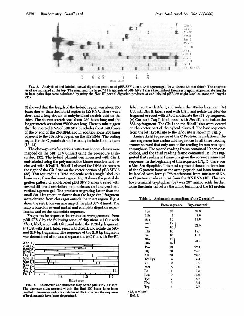
FIG. 3. Analysis of end-labeled partial digestion products of pBR SFV 3 on a 1.4% agarose gel (20 X 40 cm; 1.5 mm thick). The enzymes
used are indicated at the top. The small and the large Pst I fragments of pBR SFV 3 mark the limits of the insert region. Approximate lengthsin base pairs (bp) were calculated by using the Hae III partial digestion products of end-labeled pBR322 (right lane) as standard lengths(31).
2) showed that the length of the hybrid region was about 250bases shorter than the hybrid region in 42S RNA. There was a
short and a long stretch of unhybridized nucleic acid on thesides. The shorter stretch was about 250 bases long and thelonger stretch was about 2800 bases long. These results suggestthat the inserted DNA of pBR SFV 3 includes about 1400 basesof the 5' end of the 26S RNA and in addition some 250 basesadjacent to the 26S RNA region on the 42S RNA. The codingregion for the C protein should be totally included in this insert(13, 14).The cleavage sites for various restriction endonucleases were
mapped on the pBR SFV 3 insert using the procedure as de-scribed (32). The hybrid plasmid was linearized with Cla I,
end-labeled using the polynucleotide kinase reaction, and re-
cleaved with HindIII. HindIII cleaved the DNA six bases tothe right-of the Cla I site on the vector portion of pBR SFV 3(38).-This resulted in a DNA molecule with a single label 750bases away from the insert region. Fig. 3 shows the partial di-gestion pattern of end-labeled pBR SFV 3 when treated withseveral different restriction endonucleases and analyzed on a
vertical agarose gel. The products migrating faster than thesmall Pst I fragment or slower than the large Pst I fragmentwere derived from cleavages outside the insert region. Fig. 4shows the restriction enzyme map of the pBR SFV 3 insert. Themap is based on several partial and complete digestion exper-
iments and on the nucleotide sequence.Fragments for sequence determination were generated from
pBR SFV 3 by the following series of digestions. (i) Cut withXho I, label, recut with Cla I, and isolate the 1920-bp fragment.(ii) Cut with Ava I, label, recut with EcoRl, and isolate the 596-and 216-bp fragments. The sequence of the 216-bp fragmentwas determined after strand separation. (iii) Cut with EcoRI,Xho IAva IEcoRiHinfl_
Taq I.....
Hae III I LI
HhaI-4Sau I'
Pst I I I
0.5 1.0 1.5Kilobases
FIG. 4. Restriction endonuclease map of the pBR SFV 3 insert.The cleavage sites present within the first 200 bases have beenomitted. The arrows indicate stretches ofDNA in which the sequenceof both strands have been determined.
label, recut with Xho I, and isolate the 947-bp fragment. (iv)Cut with Hinfl, label, recut with Cla I, and isolate the 1447-bpfragment or recut with Xho I and isolate the 473-bp fragment.(v) Cut with Taq I, label, recut with HindII, and isolate the881-bp fragment. The Cla I and the HindII sites were locatedon the vector part of the hybrid plasmid. The base sequencefrom the left EcoRI site to the XhoI site is shown in Fig. 5.Amino Acid Sequence of the C Protein. Translation of the
base sequence into amino acid sequences in all three readingframes showed that only one of the reading frames was openthroughout. The second reading frame contained 10 nonsensecodons, and the third reading frame contained 12. This sug-gested that reading in frame one gives the correct amino acidsequence. In the beginning of this sequence (Fig. 5) there wasa Met-Asn dipeptide. This must represent the amino terminusof the C protein because the same peptide had been found tobe labeled with formyl [a5S]methionine from initiator tRNAin C protein made in vitro from the 26S RNA (13). The car-
boxy-terminal tryptophan (39) was 267 amino acids furtheralong the chain just before the amino terminus of the E3 protein
Table 1. Amino acid composition of the C protein*
LysHisArgAspAsnThrSerGluGinProGlyAla1/2 CysValMetIleLeuTyrPheTrp
* Mr = 29,828.t Ref. 2.
From sequence
367
15121016101115J2320234
198
119765
Experimentalt
33.97.0
13.8
21.5
15.711.1
28.7
22.124.523.54.4
17.27.0
10.510.26.76.42.7
Ii
PU-0078 Biochemistry: Garoff et al.
0

Proc. Natl. Acad. Sci. USA 77 (1980) 6379
AGA ATT CTC ATT ATA GCC CAC TAT TAT ACC ACC ATG AAT TAC ATC CCT ACG CAA ACC mTF TAC CGC CGC CCG TCGMET ASN TYR ILE PRO THR GL!R THR PHE TYR GLY ARC ARC TRP (14)
CWC CCG CGC CCG GCG GCC CGT CCT TMG CCG TTG CAG GCC ACT CCG GTC CCT CCC GTC GTC CCC GAC TlC CAG GCCARC PRO ARC PRO ALA ALA ARC PRO TRP PRO LEU GLN ALA THE PRO VAL ALA PRO VAL VAL PRO ASP PHE GLN ALA (39)
CAG CAC ATC CAG CAA CTiC ATC ACC CCC GTA AAT GCCG CMT ACA ATE AGA CAG AAC CCA ATT GCT CCT CCT AGG CCTGLN GLW MET CL! GLN LEU ILE SER ALA VAL ASN ALA LEU TEA MET ARC GL!! ASN ALA ILE ALA PRO ALA ARC PRO (64)
CCC AAA CCA AAG AAG AAG AAG ACA ACC AAA CCA AAG CCG AAA ACC CAG CCC AAG AAG ATE AAC GGA AAA ACG CAGPRO LYS PRO LYS LYS LYV LYS TEA THR LYS PRO LYS PRO LYS THR GLN PRO LVS LYS ILE ASN GLY LYS THE GLN (89)
CAG CM MC AAC AAA GAC AAG CM CCC GAC MG AAG MC AAG AAA CCC GSA AAA AGA GM AGA ATG TC AT AAGGLN CLN LYS LYS LYV ASP LYS CL!N ALA ASP LYS LYS LYV LYS LYS PRO CLY LYS ARC GLU ARC NET CYS PET LYS (114)
ATT GM AAT GAC TCT ATC TTC GAA GTC AAA CAC GAA GCA MG GTE ACT GCC TAC CCC TCC CTG GT GGC GAC AMILE GLU ASN ASP CY8 ILE PRE CLU VAL LYS HIS CLU CLY LYV VAL TEE GLY TYR ALA CYV LEU VAL CLY ASP LV! (139)
GTC ATC AAA CCT GCC CAC GTE AA CA GTC ATC CAC MC GCC GAC CTC CCA AAG CTA CCT TC AAG AAA TCG ACCVAL MET LYS PRO ALA HIS VAL LYV GLY VAL ILE ASP ASN ALA ASP LEU ALA LYV LEU ALA PEE LV! LVS SER SER (164)
AAG TAT GAC CTT GAG TGT CCC CAC ATA CCA GTT CAC ATE AGC TCC CAT CCC TCA MG TAC ACG CAT GAG AAG CCCLYS TYR ASP LEU GLU CYV ALA GLI ILE PRO VAL HIS NET ARC SEA ASP ALA SER LYV TYR TEE HIS GLU LVS PRO (189)
CAG GGA CAC TAT AAC TCC CAC .CAC GGC CCT GT CAG TAC ACC GCA CGT ACG TrC ACT ATA CCG ACA GGA CCC GGCGLU GLY HIS TYR ASN TRP HIS HIS GLY ALA VAL GLN TYR SER GLY GLY ARC PHE TEE ILE PRO TEE GLY ALA GLY (214)
AAA CCG GGA GAC ACT GCC CCG CCC ATC mTF GAC MC MG GCG AGG GTA GTE GCT ATC GTC CTE GGC GC GCC AACLY8 PRO GLY ASP SER GLY ARC PRO ILE PEE ASP AS! LYV GLY ARG VAL VAL ALA ILE VAL LEU GLY GLY ALA AS! (239)
GAG GGC TCA CGC ACA GCA CTE TCG GTG GTC ACC TCG MC AAA CAT ATE GTG ACT AGA GTE ACC CCC GAG GGG TCCGLU GLY SER ARC TE ALA LEU SER VAL VAL TEE TRP ASN LV! ASP MET VAL TEE ARC VAL TEE PRO GLU GLY SEA (264)
GAA GAG TGG TCC GCC CCC CTG ATT ACT GCC ATG TGT GTC CTT GCC MT GCT ACC TC CCG TGC TC CAG CCC CCGGLU GLU TRP
TGT GTA CCT TGC TGC TAT CAA AAC MC CCA GAG GCC ACA CTA CCC ATE C
FIG. 5. The nucleotide sequence from the left EcoRI site to the Xho I site (see Fig. 4) on the pBR SFV 3 insert, with the deduced aminoacid sequence for the C protein beneath.
(unpublished data). Table 1 shows the amino acid compositionand Mr of the C protein as calculated from the amino acid se-
quence. The values are very close to those found experimentally(2). After this work was completed, Boege et al. (40) reportedthe amino acid sequence and composition data of several trypticpeptides derived from the SFV C protein. Their data are ac-
commodated within our nucleotide sequence.
The sequence of the 267 amino acids in the C protein showsa striking clustering of basic amino acids and proline within thefirst 110 residues of the protein (Fig. 5). This segment includes21 lysine residues, 9 arginine, and 16 proline. Only three as-
partic acids and one glutamic acid are present within this re-
gion. The first basic cluster is an arginine pair at positions 12and 13. This is followed shortly by three Arg-Pro sequences.
There is a Arg-Pro-Pro-Lys-Pro-Lys-Lys-Lys-Lys sequence
starting at position 63. Close to this sequence there is a Lys-Pro-Lys-Pro-Lys sequence starting at position 74 and a Pro-Lys-Lys sequence starting at position 81. Three lysines are
found at positions 92-94 and five lysines and one proline atpositions 100-105. The carboxy terminus is acidic. Three glu-tamic acid residues are found within the six last residues. Therest of the polypeptide chain shows a relatively even distributionof basic and acidic amino acids.
DISCUSSIONThe C protein plays a fundamental role in the assembly of theSFV particle. At least three interactions are of importance: in-teractions between C protein subunits, between C proteins andthe 42S RNA, and between C proteins and the virus spike gly-coproteins. Newly made C proteins bind rapidly to 42S RNAmolecules in the cytoplasm of infected cells. A complex of one
RNA molecule with its complement of C proteins folds to formthe icosahedral nucleocapsid. Empty capsids have not beendetected in the infected cell. Apparently the C proteins cannotself-associate to form the capsid shell. Binding to the RNA seemsnecessary for nucleocapsid assembly. After the nucleocapsidhas been assembled, it diffuses to the plasma membrane, whereit probably becomes bound to the cytoplasmic domains of thevirus spike glycoproteins that are spanning the plasma mem-brane (9). The surface-bound nucleocapsid then acts as a tem-plate for the budding process, ensuring incorporation of thevirus spike glycoproteins into the budding segment of theplasma membrane. Each C protein is postulated to have onebinding site for the virus spike glycoproteins, and when all thebinding sites on the C proteins of the nucleocapsid are filled,the virus particle is released into the extracellular medium.
Before a virus can infect a host cell and start a new round ofvirus replication, most of the C protein interactions establishedduring assembly of the virus particle have to be disrupted. Thispresumably takes place after the virus has entered the lysosomesby adsorptive endocytosis (10, 11). It is possible that the acidpH of the lysosomes not only causes fusion of the SFV mem-brane with the lysosomal membrane but also destabilizes theprotein-protein and the protein-RNA interactions of the Cprotein. The SFV nucleocapsid undergoes a conformationalchange upon treatment with slightly acidic buffer (pH 6.0) (41).Whether this structural change induces uncoating is notknown.The striking feature of the primary structure of the SFV C
protein is the clustering of basic amino acid residues (lysine andarginine) with proline within the first 110 amino acid residuesfrom the amino terminus of the protein. We suggest that these
Biochemistry: Garoff et al.

Proc. Natl. Acad. Sci. USA 77 (1980)
clusters are involved in RNA binding in the SFV nucleocapsids.The abundant basic side chains offer charges for salt bridgeswith phosphate groups in the RNA. Clusters of basic amino acidresidues and proline have also been found in histone HI fromsea urchin (42) and to a lesser extent in other histone proteins(43). It is interesting in this context that histone-DNA complexesdissociate at low pH, the HI histone being the first removed (50).A Pro-Lys-Arg-Lys sequence and a Pro-Arg-Pro sequence arealso present in the amino-terminal region of the polyoma virusVP1 protein (44). A Pro-Lys-Lys-Pro-Lys sequence is found inthe corresponding region of the SV40 virus VP1 protein (45).VP1 is the major protein component in the icosahedral nucle-ocapsid of these viruses. In the carboxy-terminal region of thecore antigen from hepatitis B virus, the sequence Ser-Pro-(Arg)3occurs three times, and there is a double repeat of the sequenceSer-Pro-(Arg)4-Ser-Gln (46).
There is evidence that suggests that RNA-protein interactionsare of major importance in stabilizing the SFV nucleocapsid.The RNA in the nucleocapsid is sensitive to low concentrationsof RNase. Such treatment leads to considerable shrinkage of thenucleocapsid and to disruption of the nucleocapsid structureat higher RNase concentrations (47); The nucleocapsid is alsosensitive to concentrations of NaDodSO4 that are considerablylower than those normally needed to disrupt protein-proteininteractions (48). A number of plant viruses, of which cucumbermosaic virus is the prototype, show similar sensitivity to RNaseand NaDodSO4 (49). It has been suggested that these virusesare stabilized mainly by RNA-protein interactions. It will beinteresting to see whether the primary structures of the coatproteins from these viruses also possess extensive clusters of basicamino acids and prolines.We would like to thank Evelyn Kiko and Hilkka Virta for excellent
technical assistance, Vince Pirrotta for stimulating discussions, KenMurray for a critical reading of the manuscript, and Annie Biais andWendy Moses for typing the manuscript.
1. Simons, K., Garoff, H., Helenius, A. & Ziemiecki, A. (1978) inFrontiers in Physicochemical Biology, ed. Pullman, B. (Aca-demic, New York), pp. 387407.
2. Simons, K. & K.Uriainen, L. (1970) Biochem. Biophys. Res.Commun. 5, 981-988.
3. Laine, R., Soderlund, H. & Renkonen, 0. (1973) Intervirology1, 110-118.
4. Simmons, D. T. & Strauss, J. H. (1972) J. Mol. Biol. 71, 599-613.
5. Bonsdorff, C.-H. & Harrison, S. C. (1975) J. Virol. 16, 141-145.
6. Garoff, H., Simons, K. & Renkonen, 0. (1974) Virology 61,493-504.
7. Ziemiecki, A. & Garoff, H. (1978) J. Mol. Biol. 122,259-269.8. Garoff, H. & S6derlund, H. (1978) J. Mol. Biol. 124,535-549.9. Garoff, H. & Simons, K. (1974) Proc. Natl. Acad. Sci. USA 71,
3988-3992.10. Helenius, A., Kartenbeck, J., Simons, K. & Fries, E. (1980) J. Cell
Biol. 84, 404-420.11. White J. & Helenius, A. (1980) Proc. Natl. Acad. Sci. USA 77,
3273-3277.12. Kennedy, S. I. T. (1976) J. Mol. Biol. 108, 491-511.13. Glanville, N., Ranki, M., Morser, J., Kairiainen, L. & Smith, A.
E. (1976) Proc. Natl. Aced. Sci. USA 73,3059-3063.14. Clegg, J. C. S. (1975) Nature (London) New Biol. 254, 454-
455.15. Garoff, H., Simons, K. & Dobberstein, B. (1978) J. Mol. Biol. 124,
587-600.
-16. Ziemiecki, A., Garoff, H. & Simons, K. (1980) J. Gen. Virol., inpress.
17. Ulmanen, I., S6derlund, H. & Kiiriiinen, L. (1976) J. Virol. 20,203-210.
18. Acheson, N. H. & Tamm, I. (1967) Virology 32, 123-143.19. Lehrach, H., Frischauf, A.-M., Hanahan, D., Wozney, J., Fuller,
F. & Boedtker, H. (1979) Biochemistry 18,3146-3152.20. Kaariainen, L., Simons, K. & von Bonsdorff, C.-H. (1969) Ann.
Med. Exp. Biol. Fenn. 47,235-248.21. Glanville, N., Morser, J., Uomala, P. & Kiiriiinen, L. (1976) Eur.
J. Biochem. 64, 167-175.22. Aviv, H. & Leder, P. (1972) Proc. Natl. Acad. Sci. USA 69,
1408-1412.23. Dobberstein, B., Garoff, H., Warren, G. & Robinson, P. J. (1979)
Cell 17, 759-769.24. R6wekamp, W. & Firtel, R. (1980) Dev. Biol., in press.25. Villa-Komaroff, L., Efstratiadis, A., Broome, S., Lomedico, P.,
Tizard, E., Naber, S. P., Chick, W. L. & Gilbert, W. (1978) Proc.Natl. Acad. Sci. USA 75,3727-3731.
26. Grunstein, M. & Hogness, D. S. (1975) Proc. Natl. Acad. Sd. USA72,3961-3965.
27. Barnes, W. M. (1977) Science 195,393-394.28. Colman, A., Byers, M. J., Primrose, S. B. & Lyons, A. (1978) Eur.
J. Biochem. 91,303-310.29. Chow, L. T., Roberts, J. M., Lewis, J. B. & Broker, T. R. (1977)
Cell 11, 819-836.30. Priess, H., Koller, B., Hess, B. & Delius, H. (1980) Mol. Gen.
Genet. 178,27-34.31. Sutcliffe, J. G. (1978) Nucleic Acids Res. 5, 2721.32. Smith, H. 0. & Birnstiel, M. L. (1976) Nucleic Acids Res. 3,
2387-2398.33. Maxam, A. M. & Gilbert, W. (1980) Methods Enzymol. 65,
499-560.34. Soeda, E., Kimura, G. & Miura, K.-L. (1978) Proc. Nat!. Acad.
Sci. USA 75, 162-166.35. Tabak, H. F. & Flavell, R. A. (1978) Nucleic Acids Res. 5,
2321-2332.36. Hayward, G. S. (1972) Virology 49,342-344.37. Sanger, F. & Coulson, A. R. (1978) FEBS Lett. 87, 107-110.38. Sutcliffe, J. G. (1978) Proc. Natl. Acad. Sci. USA 75, 3737-
3741.39. Kalkkinen, N. (1980) FEBS Lett. 115, 163-166.40. Boege, U., Wengler, G., Wengler, G. & Wittmann-Liebold, B.
(1980) Virology 103, 178-190.41. Soderlund, H., Kariainen, L., Bonsdorff, C.-H. & Weckstrom,
P. (1972) Virology 47,753-760.42. Strickland, W. N., Strickland, M., Brandt, W. F., von Holt, C.,
Lehmann, A. & Wittmann-Liebold, B. (1980) Eur. J. Biochem.104,567-578.
43. Dayhoff, M. (1978) Atlas of Protein Sequence and Structure,Vol. 5, Suppl. 3.
44. Soeda, E., Arrand, J. R. & Griffin, B. E. (1980) J. Virol. 33,619-630.
45. Fiers, W., Contreras, R., Haegeman, G., Rogiers, R., Van deVoorde, A., Van Heuverswyn, H., Van Hereweghe, J., Volckaert,G. & Ysebaert, M. (1978) Nature (London) 273, 113-120.
46. Pasek, M., Goto, T., Gilbert, W., Zink, B., Schaller, H., Mackay,P., Leadbetter, G. & Murray, K. (1979) Nature (London) 282,575-579.
47. S6derlund, H., von Bonsdorff, C.-H. & Ulmanen, I. (1979) J. Gen.Virol. 45, 15-26.
48. Becker, R., Helenius, A. & Simons, K. (1975) Biochemistry 14,1835-1841.
49. Kaper, J. M. (1973) Virology 55, 299-304.50. Murray, K. (1969) J. Mol. Biol. 39, 125-144.
0-080UO Biochemistry: Garoff et al.