the canadian veterinary journal la revue …...sherisse sakals 747volution of e in vitro...
TRANSCRIPT

The cost of a case of subclinical ketosis in Canadian dairy herds
Economic value of ionophores and propylene glycol to prevent disease and treat ketosis in Canada
Comparison of intraoperative and postoperative pain during canine ovariohysterectomy and ovariectomy
Evolution of in vitro antimicrobial resistance in an equine hospital over 3 decades
Presumed masitinib-induced nephrotic syndrome and azotemia in a dog
Hypoadrenocorticism mimicking protein-losing enteropathy in 4 dogs
Total laryngectomy for management of chronic aspiration pneumonia in a myopathic dog
Citrobacter freundii induced endocarditis in a yearling colt
Equine motor neuron disease in 2 horses from Saskatchewan
Diagnostic performance of an indirect enzyme-linked immunosorbent assay (ELISA) to detect bovine leukemia virus antibodies in bulk-tank milk samples
Congenital nutritional myodegeneration in a neonatal foal
2015 CVMA ANNUAL REPORT RAPPORT ANNUEL 2015 DE L’ACMV
The Canadian Veterinary Journal La Revue vétérinaire canadienneThe Canadian Veterinary Journal La Revue vétérinaire canadienne
July/Juillet 2016 Vol. 57, No. 07
July/Juillet 2016 Volume 57, No. 07

Dual Validation.For your peace of mind.
When you’re evaluating a diet, science matters. It matters to us, too. That’s why our NEW UR Urinary® Ox/St™ Canine Formula, along with our entire urinary therapeutic diet portfolio, is backed by a comprehensive dual-validation process, measuring both the concentration and activity of the minerals that produce sterile struvite and calcium oxalate crystals. It’s the nutrition your clients need, backed by science and expertise you can trust. Learn more about our complete line at ProPlanVeterinaryDiets.ca
2xAPR
RSS
Purin
a tra
dem
arks
are
ow
ned
by S
ocié
té d
es P
rodu
its N
estlé
S.A
.
Standard Testing
Purina Testing
NEW!
FOR PERSONAL USE ONLY

INSU
RA
NC
E P
RO
GR
AM
CV
MA
INSURANCE PROGRAM
CVMA
Members save an average of 10% when joining either the Commercial Insurance or Employee Benefits programs!
866-860-CVMA (2862)www.cvmainsurance.com
Available exclusively to members
of the Canadian Veterinary Medical
Association, the specialized CVMA
Insurance Program offers the most
comprehensive and cost-effective insurance
protection for you, your practice and your
employees.
Protect what matters most
Professional Liability
Life & Disability
Business Insurance
FOR PERSONAL USE ONLY

Visit IDEXX.ca/preventivecare to learn more
You can be the di�erence between “I wish we could have done something” and “I’m so glad we caught this soon enough...”
An early diagnosis could save my life.
KNOWINGMAKES ALL THEDIFFERENCE
IN-HOUSE DIAGNOSTICS DIGITAL IMAGING AND TELEMEDICINE REFERENCE LABORATORIES CLIENT AND PRACTICE MANAGEMENT
© 2016 IDEXX Laboratories, Inc. All rights reserved. • 108600-00All ®/TM marks are owned by IDEXX Laboratories, Inc. or its a�liates in the United States and/or other countries. The IDEXX Privacy Policy is available at idexx.ca.
FOR PERSONAL USE ONLY

CVJ / VOL 57 / JULY 2016 675
SCIENTIFIC RUBRIQUE SCIENTIFIQUE
ARTICLES
728 The cost of a case of subclinical ketosis in Canadian dairy herdsKhaled Gohary, Michael W. Overton, Michael Von Massow, Stephen J. LeBlanc, Kerry D. Lissemore, Todd F. Duffield
733 Economic value of ionophores and propylene glycol to prevent disease and treat ketosis in CanadaKhaled Gohary, Michael W. Overton, Michael Von Massow, Stephen J. LeBlanc, Kerry D. Lissemore, Todd F. Duffield
741 Comparison of intraoperative and postoperative pain during canine ovariohysterectomy and ovariectomyAmanda Tallant, Barbara Ambros, Carol Freire, Sherisse Sakals
747 Evolution of in vitro antimicrobial resistance in an equine hospital over 3 decadesAnnie Malo, Caroline Cluzel, Olivia Labrecque, Guy Beauchamp, Jean-Pierre Lavoie, Mathilde Leclere
CASE REPORTS RAPPORTS DE CAS
752 Presumed masitinib-induced nephrotic syndrome and azotemia in a dogLauren Devine, David J. Polzin
757 Hypoadrenocorticism mimicking protein-losing enteropathy in 4 dogsJanne G. Lyngby, Rance K. Sellon
761 Total laryngectomy for management of chronic aspiration pneumonia in a myopathic dogKaren M. Vernau, Stanley L. Marks, Maggie A. Kuhn, William T.N. Culp, Tammy J. Owens, G. Diane Shelton, Tausif Siddiqui, Rachel Pollard, Peter C. Belafsky
767 Citrobacter freundii induced endocarditis in a yearling coltEleonora E.A. Guidi, Aurélie Thomas, Jean-Luc Cadoré, Agnès Benamou Smith
771 Equine motor neuron disease in 2 horses from SaskatchewanMichelle L. Husulak, Katharina L. Lohmann, Kamal Gabadage, Chris Wojnarowicz, Fernando J. Marqués
BRIEF COMMUNICATION COMMUNICATION BRÈVE
778 Diagnostic performance of an indirect enzyme-linked immunosorbent assay (ELISA) to detect bovine leukemia virus antibodies in bulk-tank milk samplesOmid Nekouei, Jean Durocher, Greg Keefe
STUDENT PAPER COMMUNICATION ÉTUDIANTE
781 Congenital nutritional myodegeneration in a neonatal foalJessie MacQuarrie
687 QUIZ CORNER TEST ÉCLAIR
Contents Table des matières
JULY/JUILLET 2016
FOR PERSONAL USE ONLY
REDUCES BODY WEIGHT BY 13% IN 60 DAYS1
IMPROVES MOBILITY IN AS LITTLE AS 21 DAYS2
1Data on file. Hill’s Pet Nutrition, Inc.2Data on file. Hill’s Pet Nutrition, Inc.©2016 Hill’s Pet Nutrition Canada, Inc. ®/™ Trademarks owned by Hill’s Pet Nutrition, Inc.
+OBESITY ARTHRITIS
The only way to treat both is to work together
Meet the world’s only proven single solution for both. Together we can help all of your patients at risk.
For more information, talk to your Hill’s Veterinary Account Manager.
Metabolic+MobilityPRESCRIPTION DIET®
HillsVet.caHillsVet.ca
FOR PERSONAL USE ONLY

CVJ / VOL 57 / JULY 2016 677
Contents Table des matièresFEATURES RUBRIQUES SPÉCIALES
JULY/JUILLET 2016
PRESIDENT’S MESSAGE LE MOT DE LA PRÉSIDENTE
681 Collaboration/CollaborationNicole Gallant
683 VETERINARY MEDICAL ETHICS DÉONTOLOGIE VÉTÉRINAIRE
703 2015 CVMA ANNUAL REPORT RAPPORT ANNUEL 2015 DE L’ACMV
VETERINARY PRACTICE MANAGEMENT GESTION D’UNE CLINIQUE VÉTÉRINAIRE
785 Veterinary diet pricing: Competing with the pet food store/Prix des aliments vétérinaires : comment faire concurrence aux animaleriesChris Doherty
789 DIAGNOSTIC OPHTHALMOLOGY OPHTALMOLOGIE DIAGNOSTIQUEBianca S. Bauer, Bruce H. Grahn, Lynne S. Sandmeyer
BOOK REVIEW COMPTE RENDU DE LIVRE
746 Small Animal Soft Tissue Surgery, 2nd editionMelissa Knowles
Contributors
“Instructions for authors” are available online (www.canadianveterinarians.net).
Les «Directives à l’intention des auteurs» sont disponibles en ligne (www.veterinairesaucanada.net).
770 New Product Nouveaux produit
756 Index of Advertisers Index des annonceurs
791 Classifieds Petites annonces
NOTICES ANNONCES
689 NEWS NOUVELLESHeather Broughton, Isabelle Vallières
N E W S | N O U V E L L E S
FOR PERSONAL USE ONLY

678 CVJ / VOL 57 / JULY 2016
The Canadian Veterinary Journal La Revue vétérinaire canadienne
339 rue Booth Street Ottawa, Ontario K1R 7K1 Telephone: (613) 236-1162 Fax: (613) 236-9681 E-mail: [email protected]/Site Web: www.canadianveterinarians.net
www.veterinairesaucanada.net
© Canadian Veterinary Medical Association 2016 L’Association canadienne des médecins vétérinaires 2016
The Canadian Veterinary Journal is indexed or abstracted in:La Revue vétérinaire canadienne est indexée ou ses articles sont résumés dans :AGRICOL, Biological Abstracts, Capsule Report, Current Contents — Agriculture, Derwent Veterinary Drug File, EMBASE/Excerpta Medica, Index Veterinarius, Index Medicus, Quarterly Index, Science Citation Index, Small Animal Practice, Veterinary Bulletin, Veterinary Reference Service, Veterinary Update.
Photo by/Photo de : Shutterstock
Typesetting/Typographie AN Design CommunicationsPrinted by/Imprimé par The Lowe-Martin Group Ottawa, OntarioISSN 0008-5286
Return undeliverable Canadian addresses to: 339 rue Booth Street Ottawa, Ontario K1R 7K1 e-mail: [email protected]
Subscriptions (2016). Annual: Canada $200 + applicable GST or HST; foreign $215 US; institutional $250. Express subscriptions available. Single issue/back issue: $25 each + GST or HST, if applic able. (All prices subject to change.) Missing issues will be replaced if the Subscriptions Office is notified within 6 months (for requests within Canada) and 1 year (for requests from abroad) of the issue date. The pub lisher expects to supply missing issues only when losses have been sustained in transit and when the reserve stock will permit. Telephone (613-236-1162) or (1-800-567-2862) and fax (613-236-9681) orders accepted with a valid Visa or MasterCard number. Please advise the publisher of address changes promptly.Abonnements (2016). Annuel : Canada 200 $ + TPS ou TVH en vigueur; pays étranger 215 $ É-U; prix d’une institution 250 $. Abonnement express disponible. Anciens numéros (chacun) : 25 $ + TPS ou TVH en vigueur. Les prix sont sujets à changement sans préavis. Les numéros qui ne sont pas reçus seront remplacés si l’éditeur en est informée dans les 6 mois (pour les demandes venant du Canada) et 1 an (pour les demandes venant de l’étranger) suivant la date de parution. L’éditeur s’engage à remplacer les numéros manquants seule ment lorsque les pertes ont été subies en transit et lorsque ses réserves le permettent. On peut payer son abonnement par téléphone (613-236-1162) ou (1-800-567-2862), par télé copieur (613-236-9681) ou par carte de crédit (Visa ou MasterCard). Veuillez aviser le bureau de l’éditeur de tout changement d’adresse.
Editorial policy: All published articles including editorials and letters reflect the opinions of the authors and do not necessarily reflect the opinion of the publisher.Publication of an advertisement does not necessarily imply that the publisher agrees with or supports the claims therein.Politique de la Rédaction : Tous les articles publiés, y compris les éditoriaux et les lettres, représentent l’opinion de l’auteur et non pas nécessairement la position de l’éditeur.La publication d’une annonce ne signifie pas nécessairement que l’éditeur est d’accord avec son contenu ou qu’il l’appuie.
Editor-in-Chief/Rédacteur en chef Carlton Gyles, Guelph, OntarioAssociate Editors/Rédacteurs associés Bruce Grahn, Saskatoon, Saskatchewan Wayne McDonell, Guelph, OntarioFeature Editors/Rédacteurs des chroniques Stephen Raverty, Abbotsford, British ColumbiaTim Blackwell, Fergus, OntarioAndrew Allen, Saskatoon, SaskatchewanAssistant Editors/Rédacteurs adjoints Robert Friendship, Guelph, Ontario Greg Harasen, Regina, Saskatchewan Jacob Thundathil, Calgary, Alberta Ron Johnson, Guelph, Ontario Richard Kennedy, Pincher Creek, Alberta Shawn McKenna, Charlottetown, P.E.I.Managing Editor/Directrice de la rédaction Heather Broughton, Ottawa, OntarioAssistant Managing Editor/Directrice adjointe de la rédaction Stella Wheatley, Ottawa, OntarioEditorial Coordinator/Coordonnatrice de la rédaction Linda Chow, Ottawa, OntarioAdvertising Manager/Gérante de la publicité Laima Laffitte, Wendover, Ontario
Published monthly by/ Publication mensuelle deCanadian Veterinary Medical Association
FOR PERSONAL USE ONLY

Flea and tick protection that goeson and on and on… all month long.
Introducing Simparica™ (sarolaner) Chewables
NOT ALL FLEA AND TICK CHEWS ARE CREATED EQUAL
Safe, monthly chewables for dogs that offer persistent protection from fleasand ticks. Simparica acts fast — it starts killing fleas within 3 hours1 and ticks
within 8 hours* — and keeps going strong for 35 days2,3 without losingeffectiveness at the end of the month.
*Studies show Simparica starts killing ticks in 8 hours and is ≥96.9% effective for 35 days against weeklyreinfestations of Amblyomma americanum, Amblyomma maculatum, Dermacentor variabilis andRhipicephalus sanguineus.2,3
References: 1. Simparica™ (sarolaner) Chewables package insert. Zoetis Canada Inc., Kirkland, QC. 2. Company Data. Study #’s A166C-US-12-128, A166C-US-12-129, A166C-US-12-130, A166C-US-12-131, A166C-US-12-132, A166C-US-12-133, A166C-US-12-135, A166C-US-13-303, A166C-IE-13-160 and A166C-AU-14-419. 3. Company Data. Study # A166C-US-13-318.Zoetis is a registered trademark and Simparica is a trademark of Zoetis or its licensors, used under license by Zoetis Canada Inc. ©2016 Zoetis Inc. All rights reserved. SIM-002
SIM_002 JA_E_CVJ_Layout 1 2016-06-06 3:16 PM Page 1
FOR PERSONAL USE ONLY

DON’T LET TICKS AND FLEAS KEEP THEM APART.
Provide your canine patients with the tick and flea protection they need, with the extraordinary 12-week efficacy of BRAVECTO® Chewable Tablets.
BECAUSE THE BOND THEY SHARE IS EXTRAORDINARY...
TASTY CHEWS ARE EASY TO ADMINISTER... AND HUG-FRIENDLY, TOO!
BRAVECTO® is a registered trademark of Intervet International B.V. Used under license. EXPECT THE EXTRAORDINARYTM is a trademark of Intervet International B.V. Used under license. MERCK® is a registered trademark of Merck Canada Inc. © 2016 Intervet Canada Corp. All rights reserved. CA/BRV/1215/0031
BRAVECTO Ad - Chew Dogs EN CVJ.indd 1 2016-03-31 10:39 AM
FOR PERSONAL USE ONLY

CVJ / VOL 57 / JULY 2016 681
President’s Message Le mot de la présidente
L e monde rétrécit de plus en plus et les changements continuent de se produire. À titre de vétérinaires, nous
devons être prêts à épouser ces changements afin de ne pas tirer de l’arrière. À l’avenir, la collaboration au sein de notre profession et avec d’autres intervenants sera absolument nécessaire. Je crois que le travail qui a été réalisé au cours des dernières années et qui a donné lieu à des changements imminents pour l’antibiogouvernance des antibiotiques témoigne du fait que les intervenants doivent collaborer et communiquer afin d’atteindre leur buts futurs et de veiller à ce que la voix de la profession soit entendue.
En tant que vétérinaires, nous avons tendance à être indépendants et cela peut fonctionner à petite échelle. Cependant, à grande échelle, j’ai appris que nous devons travailler ensemble pour le bien de notre profession et, j’oserais même dire, pour le bien des humains dans l’optique du concept d’«Une santé».
En août dernier, j’ai assisté à la Journée vétérinaire nationale dans la ville de Mexico. C’était un événement fascinant à observer. Le pays rassemble les vétérinaires et les personnes avec qui ils travaillent, dont des collègues du ministère de l’Agriculture et d’autres représentants gouvernementaux. Ce grand groupe célèbre ensuite tous les aspects de la médecine vétérinaire ensemble. Nous devons travailler pour parvenir à ce niveau d’interaction au Canada, où la situation est toujours un peu fragmentée entre les différents groupes vétérinaires et autres regroupements connexes. Nous devons continuer de travailler avec les autres intervenants, comme nous l’avons fait avec tant de succès lors de la mise en œuvre de changements pour l’utilisation des antibiotiques. Il y aura beaucoup d’autres enjeux sur lesquels nous pourrons collaborer, comme le problème des groupes de secours qui amènent des chiens provenant de diverses régions du monde. Les maladies exotiques que ces chiens peuvent potentiellement introduire dans notre pays, ainsi que les préoccupations de bien-être relatives à leur transport, exigent notre attention.
Comparativement à de nombreux autres pays, nous sommes vraiment fortunés de posséder le niveau de médecine organisée
T he world is becoming so much smaller and change con-tinues to happen. As veterinarians, we must be ready to
be part of these changes and not be left behind. Collaboration within our profession and with other stakeholders is absolutely necessary going forward. As I think of all the work over the last few years that resulted in the imminent changes in antibiotic stewardship, it is obvious that collaboration and communication among stakeholders must happen to achieve our future goals and ensure the voice of our profession is heard.
As veterinarians, we tend to be independent and that can work on a small scale. However, on the larger scale, I have learned that we must work together for the good of our profes-sion and, dare I say, the good of humans also as we think of the “One Health” concept.
Last August I attended National Veterinarian Day in Mexico City. This was fascinating to watch. The country brings together veterinarians and those with whom they work, such as col-leagues in the Department of Agriculture and other government officials. This large group then proceeds to celebrate all aspects of veterinary medicine together. We have work to do to get to this level of interaction in Canada, where things are still a bit fragmented with different veterinary and related groups. We must continue to work with other stakeholders, as we have so successfully done in bringing forward the changes in antibiotic usage. There will be many other issues to collaborate on, such as the evolving problem of rescue groups bringing in dogs from different parts of the world. Foreign diseases that these dogs can potentially introduce to our country, as well as the welfare concern for their transport are issues that require attention.
When compared to many countries, we are so fortunate to have the level of organized medicine that we have in Canada, but we still fall a bit short in some ways. Strong provincial associations are important as is a national voice for the profes-sion; one doesn’t negate the other. Collaboration and mutual understanding are crucial going forward as we become more involved in the international veterinary community. We can’t
Collaboration
Collaboration
Use of this article is limited to a single copy for personal study. Anyone interested in obtaining reprints should contact the CVMA office ([email protected]) for additional copies or permission to use this material elsewhere.
L’usage du présent article se limite à un seul exemplaire pour étude personnelle. Les personnes intéressées à se procurer des réimpressions devraient communiquer avec le bureau de l’ACMV ([email protected]) pour obtenir des exemplaires additionnels ou la permission d’utiliser cet article ailleurs.
FOR PERSONAL USE ONLY

682 CVJ / VOL 57 / JULY 2016
LE
MO
T D
E L
A P
RÉ
SID
EN
TE
continue to compete among ourselves. We all need members to exist but we must reach an understanding and vision of what is going on within and outside of Canada. The fact that Canada is well regarded internationally has become apparent to me in this past year and I thank you for the opportunity to see this international face of Canadian veterinary medicine. We are getting there; let’s work together to make organized veterinary medicine in Canada strong at all levels. ■
Nicole Gallant
que nous avons ici au Canada, mais certains secteurs présentent toujours des lacunes. Il est important de posséder des associations provinciales fortes et une voix nationale pour la profession et ces deux positions ne sont pas forcément contradictoires. La collaboration et la compréhension mutuelle représenteront des éléments cruciaux tandis que nous élargirons notre participation au sein de la collectivité vétérinaire internationale. Nous ne pouvons pas continuer à compétitionner entre nous. Nous avons besoin de la présence de tous les membres, mais nous devons aussi parvenir à une compréhension et à une vision de ce qui se passe à l’intérieur et à l’extérieur du Canada. Au cours de la dernière année, j’ai pu prendre connaissance du fait que le Canada possède une excellente réputation internationale et je vous remercie de l’occasion que vous m’avez accordée de voir ce visage international de la médecine vétérinaire canadienne. Nous nous approchons du but. Travaillons ensemble pour assurer la force de la médecine vétérinaire à tous les niveaux. ■
Nicole Gallant
ADVERSE REACTIONS TO FOOD
Taking purity to the next levelNEW & IMPROVEDPrescription Diet® z/d®
HillsVet.ca©2016 Hill’s Pet Nutrition Canada, Inc. ®/™ Trademarks owned by Hill’s Pet Nutrition, Inc.
FOR PERSONAL USE ONLY

CVJ / VOL 57 / JULY 2016 683
Veterinary Medical Ethics Déontologie vétérinaire
Ethical question of the month — July 2016A nearby successful veterinary practice provides state-of-the-art veterinary medicine and surgery to a large and dedicated clientele. This clinic also offers alternative therapies for conditions that do not respond to conven-tional treatments. In the past, these therapies were primarily vitamin and herbal products. Owners signed a consent form recognizing that the treatments were unconventional. There is no evidence that these therapies are effective but the clinic has not charged exorbitantly for these services and some of your best clients have gone to this clinic when you have had nothing left to offer but palliative care.
A board-certified surgeon at this clinic who performs complex orthopedic surgeries has recently begun to surgically implant special magnets and other “positive energy” devices in patients in which conventional therapies were ineffective. The implants are accompanied by vague claims that they “could help.” The prac-tice charges several thousand dollars for these surgeries, but this deters fewer clients than you would have predicted. You believe this clinician is using the trust created through the success of his conventional treat-ments to convince clients to try expensive and unproven “alternative” therapies when in desperate straits. You tolerated the selling of false hope when it was low cost and non-invasive, but this new high cost and invasive approach troubles you. Are you justified in your change in attitude towards the unconventional treatments offered by this practice?
Question de déontologie du mois — Juillet 2016Une pratique vétérinaire prospère avoisinante offre de la médecine et des chirurgies à la fine pointe technologique à une clientèle vaste et dévouée. Cette clinique offre aussi des thérapies parallèles pour des affections qui ne répondent pas à des traitements conventionnels. Par le passé, ces thérapies étaient principalement des vitamines et des produits à base de plantes. Les propriétaires signaient un formulaire de consentement pour reconnaître que les traitements n’étaient pas conventionnels. Il n’y avait aucune preuve que ces traitements étaient efficaces, mais la clinique ne facturait pas des tarifs exorbitants pour ces services et certains de vos meilleurs clients s’y rendaient lorsque vous n’aviez rien d’autre à leur offrir que des soins palliatifs.
Un chirurgien spécialiste à cette clinique qui effectue les chirurgies orthopédiques complexes a récemment commencé à implanter des aimants spéciaux et d’autres dispositifs à «énergie positive» chez les patients qui ne
Responses to the case presented are welcome. Please limit your reply to approximately 50 words and forward along with your name and address to: Ethical Choices, c/o Dr. Tim Blackwell, 6486 E. Garafraxa, Townline, Belwood, Ontario N0B 1J0; telephone: (519) 846-3413; fax: (519) 846-8178; e-mail: [email protected] ethical questions of the month are also welcome! All ethical questions or scenarios in the ethics column are based on actual events, which are changed, including names, loca-tions, species, etc., to protect the confidentiality of the parties involved.
Les réponses au cas présenté sont les bienvenues. Veuillez limiter votre réponse à environ 50 mots et nous la faire parvenir par la poste avec vos nom et adresse à l’adresse suivante : Choix déontologiques, a/s du Dr Tim Blackwell, 6486, E. Garafraxa, Townline, Belwood (Ontario) N0B 1J0; téléphone : (519) 846-3413; télécopieur : (519) 846-8178; courriel : [email protected] propositions de questions déontologiques sont toujours bienvenues! Toutes les questions et situations présentées dans cette chronique s’inspirent d’événements réels dont nous modifions certains éléments, comme les noms, les endroits ou les espèces, pour protéger l’anonymat des personnes en cause.
Use of this article is limited to a single copy for personal study. Anyone interested in obtaining reprints should contact the CVMA office ([email protected]) for additional copies or permission to use this material elsewhere.
L’usage du présent article se limite à un seul exemplaire pour étude personnelle. Les personnes intéressées à se procurer des réimpressions devraient communiquer avec le bureau de l’ACMV ([email protected]) pour obtenir des exemplaires additionnels ou la permission d’utiliser cet article ailleurs.
FOR PERSONAL USE ONLY

684 CVJ / VOL 57 / JULY 2016
DÉ
ON
TO
LOG
IE V
ÉT
ÉR
INA
IRE
An ethicist’s commentary on a dog requiring emergency care after-hoursAs we have so often done in the past, it is helpful to hark back to some of the wisdom that may be found in the writings of Plato. Two related Platonic points are relevant here. When discussing the wise ruler, Plato makes the point that the job of a ruler, or for that matter, a shepherd, or a craftsman, com-prises two distinct aspects: first and foremost one is obliged to improve and protect whatever one exercises one’s professional authority over — secondarily, one is functioning as a wage earner. As we pointed out in a recent column, a medical profes-sional functions first and foremost to treat, fix, heal, and cure,
and only, conceptually, secondarily and derivatively to make money.
I am reminded of the story told to me by my dear friend, veterinarian Brian Forsgren. Early on in his practice, an impov-erished street person showed up at his clinic with a dog who had been run over by a truck and sustained a crushed pelvis. Dr. Forsgren asked when this occurred. The owner replied a few hours ago. Dr. Forsgren then asked why he had waited so long to seek help. The client responded that this had occurred miles away, and he had indeed approached the nearest veterinarian,
répondent pas aux traitements conventionnels. Les implants sont accompagnés d’allégations vagues stipulant que les traitements «peuvent aider». La pratique facture plusieurs milliers de dollars pour ces chirurgies et ce fait ne décourage pas autant de clients que vous auriez pu le croire. Vous estimez que ce clinicien utilise une réputation bâtie sur le succès de ses traitements conventionnels pour convaincre les clients de tenter des thérapies «parallèles» dispendieuses et non éprouvées lorsque les clients se trouvent dans des situations désespérées. Vous étiez tolérant de la vente de ces faux-espoirs lorsqu’ils étaient peu dispendieux et non invasifs, mais vous êtes perturbé par cette nouvelle approche coûteuse et invasive. Votre changement d’attitude envers les traitements non conventionnels offerts par cette pratique est-il justifié?
Comments/Commentaires :
Name/Nom :
Address/Adresse :
Ethical question of the month — April 2016You receive a call as clinic hours are ending from someone who is visiting in your area. Their dog has been hit by a car and they are 8 hours from home. The person describes a dog that appears to be seriously injured. You agree to see the dog but since this person is from out of town, you explain your fees and out-of-hours charges and that some form of payment is required at the time of admission. You can hear on the phone that the person is put off by this and says, “Well, I’ll think about it and call you back.” They do not call back and you call them back after an hour and they are still debating what to do and the dog is described as breathing with difficulty. They still say they will call you when they make a decision. You seriously doubt you will hear from them. Are you in any way responsible for this animal’s outcome?
Question de déontologie du mois — Avril 2016Un peu avant la fermeture de la clinique, vous recevez un appel peu d’une personne en visite dans votre région. Son chien a été heurté par une automobile et elle se trouve à huit heures de trajet de la maison. La personne décrit un chien qui semble être gravement blessé. Vous acceptez d’examiner le chien mais, vu que cette personne provient de l’extérieur de la ville, vous expliquez vos tarifs, les frais après les heures d’ouverture et le besoin d’un dépôt au moment de l’admission. Vous pouvez constater au téléphone que la personne est décontenancée par vos propos et elle dit : «Eh bien, je vais y réfléchir et je vous rappellerai.» La personne ne rappelle pas et vous décidez de la contacter après une heure. Elle se demande toujours quoi faire et elle décrit le chien comme ayant une respiration difficile. Elle dit qu’elle vous rappellera quand elle aura pris une décision. Vous doutez sérieusement que vous aurez de nouveau de ses nouvelles. Êtes-vous responsable du sort de cet animal?
FOR PERSONAL USE ONLY

CVJ / VOL 57 / JULY 2016 685
VE
TE
RIN
AR
Y M
ED
ICA
L E
TH
ICS
who had replied that it would cost many thousand dollars for diagnostics and surgical treatment, and that nothing could be done for less. Brian pondered the absurdity of the previous cli-nician’s narrative. He pointed out that he could cast the animal for a few hundred dollars. He did so, and the animal recovered well. He then resolved to help financially challenged people treat their animals as his life’s work. “After all, who could be myopic enough to think that a poor person’s animal meant less to them than a rich person’s?” Gratifyingly, he built a very successful practice. He is currently in semi-retirement, and continues to stress to his peers the need to place care and empathy before financial limitations.
The lesson here should be obvious. As I have said ad nauseam in this column, the veterinarian’s primary obligation is to the patient. And it is dogma in veterinary circles to affirm that a veterinarian is obliged to administer stabilizing care even to an unowned animal brought in by a good Samaritan not willing to assume financial responsibility. For the veterinarian in this case to start talking money even before looking at the animal is at the very least sleazy and borders on obscene.
First and foremost he or she should insist that the animal be brought to the clinic immediately. There is plenty of time to talk
money after the animal is looked at. And there are also many ways of establishing payment protocols as the animal is being worked on. As a worst-case, the veterinarian can suggest that the owner surrender the animal, and then he or she can treat the animal and adopt it out. As Dr. James Wilson has informed me, he can then provide the newspapers or other media with a touching human interest story that will buy him far more favor-able publicity than he could acquire in any other way. Members of the public will be clamoring to adopt the animal!
Giving priority to the payment even as one is told that the animal is “seriously injured” is hardly the mark of a compas-sionate and committed veterinarian. While I am certainly aware that veterinarians cannot work for nothing, and have repeatedly made this point in my writings, one must be wise enough to recognize exceptional situations and the opportunities they present. To act otherwise is not only unprofessional in the sense identified by Plato, but is also in the end contrary to the veterinarian’s interest and is thus imprudent.
I am grateful to Drs. Brian Forsgren and James Wilson for teach-ing me their ways of, as Plato says, “making a virtue of necessity.”
Bernard E. Rollin, PhD
Resources for Veterinary Excellence
Offers a quick reference to recognizing and treating common cardiac arrhythmias and emergent cardiac conditions in canine and feline patients, designed for fast access during an emergency. This book is a useful patient-side resource for general practitioners, emergency vets, and veterinary students.
The best-selling, practical guide to all areas of equine radiography and radiology written by an experienced group of clinicians with a broad range of backgrounds, including hundreds of normal and clinical images. The fourth edition fully reflects the move to digital imaging, and a companion website offers a wealth of additional images.
Visit wiley.ca/go/veterinary to browse our complete library of veterinary medicine journals, books, and more.
Available wherever books and eBooks are sold. e
9781118912287August 2016 | $290.99
9781119042075July 2016 | $59.99
238811
FOR PERSONAL USE ONLY

GLOBAL DIAGNOSTICS
800.822.2947 www.abaxis.com [email protected]
Why Wait? Your Clients Don’t Want To.In House Blood Diagnostics, While You Wait
Abaxis_CVJ _0716.indd 1 6/6/16 3:14 PM
FOR PERSONAL USE ONLY

CVJ / VOL 57 / JULY 2016 687
1. A 6-year-old boxer dog presents with a skin mass over the lateral thorax. The owner notices that it increases and decreases in size regularly and the dog seems to scratch at the mass. On fine-needle aspiration of the mass, a homog-enous population of round cells containing granules is noted. The most likely diagnosis is which of the following?A. Cutaneous mast cell tumorB. FibrosarcomaC. LymphomaD. Mammary gland tumorE. Cutaneous hemangiosarcoma
2. The most common cause of urinary incontinence in a 3-month-old female dog is which of the following?A. Ectopic ureter(s)B. Bladder neoplasiaC. Urinary tract infection (UTI)D. Primary sphincter mechanism incompetence (PSMI)E. Cystic urolithiasis
3. Which of the following is a simple technique to determine whether a stray cat has intra-abdominal testicles?A. Measure the length of the canine teeth.B. Palpate the abdomen deeply.C. Examine the base of the penis for androgen-responsive
spines.D. Examine the footpads for evidence of hyperkeratosis.E. Measure scrotal thickness.
1. Un chien Boxer âgé de 6 ans présente une masse cutanée dans la partie latérale du thorax. Le propriétaire mentionne que la masse augmente et diminue de grosseur de façon régulière et que le chien la gratte. À l’aspiration à l’aiguille fine de la masse, on observe une population homogène de cellules rondes renfermant des granules. Lequel des diagnostics suivants est le plus probable?A. mastocytome cutané;B. fibrosarcome;C. lymphome;D. tumeur des glandes mammaires;E. hémangiosarcome cutané.
2. Lequel des problèmes suivants constitue la cause la plus commune d’incontinence urinaire chez une chienne âgée de 3 mois?A. uretère(s) ectopique(s);B. néoplasie de la vessie;C. infection du tractus urinaire (ITU);D. insuffisance du mécanisme du sphincter primaire (IMSP);E. urolithiase vésicale.
3. Laquelle des procédures suivantes est une façon simple de déterminer si un chat errant possède des testicules intra-abdominaux? A. mesure de la longueur d’une dent canine;B. palpation en profondeur de l’abdomen;C. examen de la base du pénis pour déceler la présence
d’épines due aux androgènes;D. examen des coussinets plantaires pour déceler la présence
d’hyperkératose;E. mesure de l’épaisseur du scrotum.
Quiz Corner Test éclair
BRAVECTO CVJ - Quiz Corner Question ad.indd 1 2016-03-31 10:45 AM
FOR PERSONAL USE ONLY

688 CVJ / VOL 57 / JULY 2016
TE
ST
ÉC
LA
IR
4. After unilateral ovariectomy for a granulosa cell theca tumor in a mare, which of the following is correct?A. Surgery should be scheduled to remove the contralateral
ovary because these tumors tend to be bilateral.B. The mare will be unable to conceive and maintain a preg-
nancy to full term.C. The mare should be carefully observed for any clini-
cal abnormalities because tumor metastasis will have occurred.
D. The mare can be expected to resume her normal estrus cycle, although it may take up to 12 months after surgery.
E. The mare can be bred immediately after suture removal, 2 weeks after surgery.
5. Which of the following methods would be most useful for determining the etiology of an outbreak of BRD in a group of 550-lb feedlot calves?A. Nasal swab submitted for bacterial cultureB. Thoracic ultrasoundC. Serum samples from affected calves submitted for viral
serologyD. Necropsy of calves that died of chronic pneumonia or
were euthanized because of itE. Transtracheal lavage
4. À la suite d’une ovariectomie unilatérale à cause d’une tumeur des cellules thécales de l’ovaire chez la jument, lequel des énoncés suivants est correct?A. On doit planifier une chirurgie pour enlever l’ovaire
controlatéral parce que cette tumeur a tendance à être bilatérale.
B. La jument sera incapable de concevoir et de maintenir une gestation jusqu’à terme.
C. On doit observer attentivement la jument pour détecter des signes d’anomalies à cause des métastases qui peuvent se développer.
D. On peut penser que la jument reprendra son cycle œstral normal bien que cela puisse prendre jusqu’à 12 mois après la chirurgie.
E. La jument peut être accouplée immédiatement après le retrait des sutures, 2 semaines après la chirurgie.
5. Laquelle des méthodes suivantes serait la plus utile pour déterminer l’étiologie d’une flambée de maladie respiratoire bovine dans un groupe de veaux à l’engraissement pesant chacun 550 lb?A. écouvillon nasal soumis pour culture bactérienne;B. échographie thoracique;C. échantillons de sérum provenant de veaux malades soumis
pour sérologie virale;D. nécropsie de veaux morts de pneumonie chronique ou
euthanasiés à cause de cette affection;E. lavage transtrachéal.
Questions and answers were derived from Review Questions and Answers for Veterinary Boards 2nd ed., a 5-volume series including Basic Sciences, Clinical Sciences, Small Animal Medicine and Surgery, Large Animal Medicine and Surgery, and Ancillary Topics, by kind permission of the publisher, Mosby–Year Book, Inc., St. Louis, Missouri.
Les questions et les réponses sont extraites de Review Questions and Answers for Veterinary Boards 2nd ed., une série de cinq volu mes qui comprend Basic Sciences, Clinical Sciences, Small Animal Medicine and Surgery, Large Animal Medicine and Surgery, et Ancillary Topics, avec l’aimable permission de l’éditeur, Mosby–Year Book, Inc. de St. Louis (Missouri).
(See p. 788 for answers./Voir les réponses à la page 788.)
Have you been checking your e-mail inbox ?
The Canadian Veterinary Medical Association (CVMA) communicates time-sensitive and relevant information and news to its members by e-mail based on the addresses we have on record in our database. If you are not receiving e-mail communication from us, it may be that we do not have a valid e-mail address for you.
Review/update your contact information and stay connected! Also, ensure that you add us ([email protected]) to your safe sender’s list so that our messages do not get blocked.
OnlineLog on at www.canadianveterinarians.net and view your contact information. You can make changes directly online.
Contact CVMABy e-mail at [email protected] or by telephone at 1.800.567.2862. We will confirm the e-mail address we currently have for you and make any necessary changes.
FOR PERSONAL USE ONLY

CVJ / VOL 57 / JULY 2016 689
N E W S | N O U V E L L E S
Promotion spéciale pour votre escapade estivale!
Découvrez le programme de rabais hôteliers de l ’ACMV. De plus, réservez votre
hébergement entre le 1er mai et le 30 septembre et vous serez inscrit à un tirage pour gagner le remboursement de votre réservation. Pour débuter la planification de votre escapade, allez sur le site Web de l’ACMV (veterinairesaucanada.net) et cliquez le lien «Rabais hôtelier» sous l’onglet «Avantages et services aux membres» (les membres devront ouvrir une session).
Special promotion for your summer getaway!
Check out the CVMA hotel discount program! In addition, when you book
your accommodations between May 1st and September 30th, you’ll be entered into a draw to win your booking for free. To start planning your getaway, go to the CVMA website (www.canadianveterinar ians.net) and click on the Hotel Discounts link under the tab Value of Membership (member log-in required).
I am a 4th year veterinary student at the Faculté de médecine vétérinaire, in Saint-Hyacinthe, Quebec, and, last year, I par-
ticipated in one of the veterinary profession’s most enriching and informative workshops on leadership, communications and teamwork: CVMA’s Emerging Leaders Program (ELP).
This workshop took place on July 16, 2015 at the Fairmont Palliser Hotel in Calgary, Alberta, during the CVMA’s Annual Convention. This workshop would not have been possible without the generous support of sponsors Virox, The Personal Insurance Company, Western Financial Group Insurance Solutions, and Zoetis.
Je suis étudiante de quatrième année en médecine vétérinaire à la Faculté de médecine vétérinaire de l’Université de Montréal à
Saint-Hyacinthe et, l’année dernière, j’ai participé à l’un des ateliers les plus enrichissants et les plus informatifs sur le leadership, la communication et le travail d’équipe, le Programme des futurs leaders (PFL) de l’Association canadienne des médecins vétérinaires (ACMV).
Cet atelier s’est déroulé le 16 juillet 2015 à l’hôtel Fairmont Palliser à Calgary, en Alberta, à l’occasion du congrès annuel de l’ACMV. Cet atelier n’aurait pas été possible sans le généreux soutien des commanditaires : Virox, laPersonnelle, Western Financial Group Insurance Solutions et Zoetis.
Dans le cadre de discussions, de jeux interactifs et de vidéos, le Dr Rick DeBowes a aidé les vétérinaires et les technologues vétérinaires agréés à retrouver le plaisir à travailler et à faire face aux divers défis de la profession. Le Dr Rick DeBowes, professeur de chirurgie et directeur du programme «Personal Life Skills» à l’Université d’État de Washington, est très souvent porte-parole et présentateur de différents programmes de leadership. Il est aussi cocréateur de l’Expérience de leadership vétérinaire de l’American Veterinary Medical Association et de plusieurs autres ateliers de leadership interactifs et pratiques présentés dans de nombreux pays, et ce, sur quatre continents.
Hélène Rembeaux
CVMA’s Emerging Leaders ProgramProgramme des futurs leaders de l’ACMV
FOR PERSONAL USE ONLY

690 CVJ / VOL 57 / JULY 2016
N
Pour les vétérinaires récemment diplômés et les vétérinaires chevronnés, le PFL a pour objectif de donner les outils essentiels pour maintenir un équilibre sain entre la vie professionnelle et la vie personnelle.
Cet atelier nous a permis de nous arrêter un instant et de prendre le recul nécessaire pour évaluer sous un autre angle la pratique vétérinaire et nos interactions avec les collègues, les employés, les clients et les patients. Le Dr DeBowes encourage les participants à réfléchir et à travailler sur eux-mêmes pour développer les qualités qui les aideront à devenir de meilleurs leaders.
Au cours des dernières années, les participants au PFL de l’ACMV ont décrit cette expérience comme étant «revigorante» ou «rafraîchissante» qui leur permet d’attaquer leur journée en clinique de façon plus positive et optimiste.
Pour les étudiants en médecine vétérinaire, la précision et la délicatesse en chirurgie sont des aptitudes bien développées dans le cursus de médecine vétérinaire et elles sont importantes pour être un bon vétérinaire. Cependant, l’intelligence émotionnelle, l’empathie et la communication sont toutes aussi importantes mais souvent reléguées en second plan. Le PFL représente un atout considérable pour les participants et il leur permet d’être plus efficaces et attentifs lors des travaux d’équipe, des laboratoires et des exercices chirurgicaux… et bien sûr en stage clinique!
Le prochain atelier aura lieu lors du congrès 2016 de l’ACMV qui se déroulera à Niagara Falls, les 7 et 8 juillet prochains. Cet atelier incomparable est ouvert à tous les membres de l’ACMV et de TTVAC. Pour plus de renseignements et pour vous inscrire, visitez le site Web de l’ACMV : (veterinairesaucanada.net/science-knowledge/emerging-leaders-program)
(par Hélène Rembeaux, représentante sénior sortante et étudiante à la Faculté de médecine vétérinaire de l’Université de Montréal)
Through discussions, interactive games and videos, Dr. Rick DeBowes helped veterinarians and registered veterinary technol-ogists rediscover the joy in working and dealing with the various challenges of the profession. Dr. DeBowes, a professor of surgery and director of the Personal Life Skills Program at Washington State University, is a highly sought-after moderator and pre-senter for various leadership programs. He also co-founded the American Veterinary Medical Association’s Veterinary Leadership Experience and several other interactive and hands-on leadership workshops presented in several countries and on 4 continents.
The objective of the ELP is to give recent graduates, as well as experienced veterinarians and technologists, essential tools that will help them maintain a healthy balance between their professional and personal life.
This workshop gave us a moment to step back and see veteri-nary practice and interactions with colleagues, employees, clients and patients from a different perspective. Dr. DeBowes encour-ages participants to think and work for themselves to develop qualities that will help them become better leaders.
In recent years, CVMA’s ELP participants have described this experience as “energizing” and “refreshing,” allowing them to look at their clinic workday more positively and optimistically.
For veterinary students, surgical precision and dexterity are skills that are well developed in the veterinary medicine cur-riculum and are important in becoming good veterinarians. Emotional intelligence, empathy and communications skills are just as important, but they often take a back seat. The ELP significantly benefits participants and enables them to be more effective and attentive in team projects, labs and surgical exer-cises… and of course during clinical internships!
The next workshop will be held at CVMA’s 2016 Convention in Niagara Falls, Ontario, July 7 and 8. This unparalleled workshop is open to all CVMA and RVTTC members. For more information and to register, visit CVMA’s website (www.canadianveterinarians.net/science-knowledge/emerging-leaders-program).
(by Hélène Rembeaux, Past SCVMA senior representative and student at the Faculté de médecine vétérinaire de l’Université de Montréal)
FOR PERSONAL USE ONLY
CVJ / VOL 57 / JULY 2016 691
NAdieux du président des ÉACMV
J’aimerais entamer mes adieux en remerciant toutes les personnes qui m’ont permis d’occuper ce poste au cours de
la dernière année : tous les étudiants en médecine vétérinaire canadiens pour m’avoir permis d’être votre président des Étudiants de l’ACMV (ÉACMV), mes collègues membres du Comité des ÉACMV de votre travail ardu et de votre dévouement ainsi que le Conseil, l’exécutif et le personnel de l’ACMV qui m’ont offert un soutien incroyable ainsi que tous les renseignements nécessaires.
Pendant mes deux années passées aux Étudiants de l’ACMV, j’ai découvert les rouages internes de notre association nationale de médecins vétérinaires, l’ACMV. Le soutien fourni par l’ACMV aux étudiants canadiens est inégalé : elle nous procure de l’assistance pour l’organisation de notre Symposium étudiant annuel, elle offre des services en ligne à tous les étudiants et elle appuie tous les finissants tandis qu’ils effectuent la transition à l’étape suivante de leur carrière vétérinaire. J’encourage tous les étudiants à en apprendre davantage à propos des avantages offerts par l’ACMV. Pour en savoir davantage à propos de ce que les ÉACMV et l’ACMV peuvent faire pour vous et votre carrière, consultez la section étudiante du site Web de l’ACMV, lisez l’édition étudiante du cyberbulletin de l’ACMV «En direct du 339» ou contactez les représentants des ÉACMV de votre école.
Le travail avec notre Comité des ÉACMV me manquera car j’admire leur dévouement et leur travail infatigable en vue d’améliorer l’expérience de tous les étudiants en médecine vétérinaire canadiens. La planification du Symposium, la rédaction d’articles pour La RVC, la coordination du Sondage auprès des finissants, la publication de notre bulletin VetRap et la cueillette de fonds pour notre nouvel Atelier de leadership étudiant (dont la première édition aura lieu cet automne à la Faculté de médecine vétérinaire à Saint-Hyacinthe, au Québec) figurent parmi les réalisations exceptionnelles auxquelles notre Comité consacre d’innombrables heures tous les ans.
Même si mon mandat auprès des ÉACMV prend fin, je suis excité de quitter mes fonctions en sachant que je laisse mon poste entre les très bonnes mains de notre nouvelle présidente des ÉACMV, Elizabeth Hartnett, de l’Ontario Veterinary College. Elizabeth dirigera un Comité capable et motivé qui se composera de représentants des cinq collèges de médecine vétérinaire canadiens.
Félicitations à tous les diplômés 2016 et, à tous les étudiants, bon succès dans vos études!
(par Justin R. Kristjansson, Western College of Veterinary Medicine, promotion 2017, président 2015–2016 des Étudiants de l’ACMV)
SCVMA President Farewell
I would like to start my farewell by thanking everybody who has allowed me to hold this position for the last year; all Canadian
veterinary students for allowing me to be your Students of the CVMA (SCVMA) president, my fellow SCVMA Committee members for your hard work and dedication, and to the CVMA Council, Executive, and staff for being incredibly supportive, helpful, and informative.
In my 2 years with the Students of the CVMA I have gained great insight into the inner workings of our national veterinary medical association, the CVMA. The support that the CVMA provides to Canadian students is unparalleled; assisting us in hosting our annual student Symposium, providing services to all students online, and supporting our new graduates as they transition into the next stage of their veterinary career. I encourage all students to learn more about what the CVMA has to offer. Browse the student section of the CVMA website, read the student edition of the CVMA’s e-newsletter “Online from 339,” contact your college’s SCVMA representatives to learn more about what the SCVMA, and the CVMA, can do for you and your career.
I will miss working with our SCVMA Committee members and I admire their dedication and tireless work to better every Canadian veterinary student’s experience. The endless hours planning Symposium, writing articles for The CVJ, coordinating the New Graduate Survey, publishing our VetRap newsletter, and fundraising for our new Student Leadership Workshop (which will debut this fall at the Faculté de Médecine Vétérinaire in Saint-Hyacinthe, Quebec) are just some of the proud accom-plishments tackled by our Committee every year.
While my time with the SCVMA is ending, I am excited to leave my position in the very capable hands of our incom-ing SCVMA president, Elizabeth Hartnett from the Ontario Veterinary College. Elizabeth will lead a capable and motivated Committee, with representatives from all 5 Canadian colleges.
Congratulations to all 2016 graduates, and best of luck to all students in your current studies!
(by Justin R. Kristjansson, Western College of Veterinary Medicine, Class of 2017, 2015–2016 Students of the CVMA President)
Justin R. Kristjansson
FOR PERSONAL USE ONLY

692 CVJ / VOL 57 / JULY 2016
N Rencontrez vos représentants 2016–2017 du Comité des ÉACMV!
Le Comité des Étudiants de l’Association canadienne des médecins vétérinaires (ÉACMV) représente l’ACMV et les cinq
collèges de médecine vétérinaire canadiens afin de renforcer les liens entre l’Association et ses membres étudiants.
Sarifa Lakhdhir, représentante des ÉACMV à la Faculté de médecine vétérinaire de l’Université de Calgary (UCVM), est née et a grandi à Calgary, en Alberta. Son amour pour les animaux s’est manifesté à un très jeune âge et, dans sa jeunesse, son film favori était Le Roi lion. Pendant qu’elle était à l’école élémentaire, Sarifa a beaucoup voyagé en Amérique du Nord et elle a visité plusieurs zoos et parcs d’animaux célèbres, comme le Zoo de San Diego et le Parc Safari. Durant ses visites, elle s’est rendue dans les coulisses et s’est renseignée sur le rôle des vétérinaires afin d’assurer la santé et le bien-être des animaux tout en s’amusant à nourrir des animaux comme des okapis, des rhinocéros et des lions. Durant les deux premières années de programme de premier cycle, Sarifa s’est rendue au Costa Rica et en Namibie avec ucalgarycares. Dans le cadre de ces expériences multiculturelles, elle a eu l’occasion de se renseigner à propos du développement international éthique à une échelle locale et internationale. De plus, elle a fait du bénévolat au Zoo de Calgary et dans une clinique pour petits animaux à Calgary. L’été dernier, Sarifa a passé dix semaines au Kenya lors d’un internat avec Vétérinaires sans frontières Canada afin d’appuyer sa mission en vue d’améliorer la vie des Kényans, de leurs collectivités et de leurs animaux. Pendant ce séjour, elle a travaillé avec de petits producteurs laitiers afin d’évaluer et d’améliorer le bien-être, le confort, la nutrition et la production des vaches laitières. À titre de représentante sénior des ÉACMV à l’UCVM pour l’année à venir, Sarifa se réjouit à la pensée de présider le Symposium des ÉACMV 2017 dont le thème sera «Prenez le taureau par les cornes. Soyez responsable de vos actes». Elle espère que le Symposium encouragera les étudiants à assumer la responsabilité de leurs actes, particulièrement en ce qui concerne la manipulation des animaux, le bien-être animal et la gestion responsable de l’environnement.
Elizabeth Hartnett, représentante des ÉACMV à l’Ontario Veterinary College (OVC), a décidé qu’elle désirait être vétérinaire à l’âge de cinq ans et elle a toujours été fascinée par le monde naturel. Elle a grandi dans une famille militaire et a vécu dans plusieurs endroits différents — allant de l’Allemagne au Manitoba, en passant par le Québec — avant que sa famille ne s’installe finalement à Kingston, en Ontario. Ses études de premier cycle à l’Université Queen’s ont porté sur la biologie environnementale et elle a ensuite obtenu une maîtrise en études environnementales à l’Université York, où elle s’est concentrée sur l’éducation humanitaire et l’éthique environnementale.
Pendant plusieurs années, Elizabeth a travaillé dans le domaine des recherches et des politiques environnementales, tout en se portant bénévole au Toronto Wildlife Centre les fins de semaine. Elle s’est rendue compte que ses intérêts dans l’environnement, les politiques publiques et la santé faunique étaient compatibles avec une carrière en médecine vétérinaire et elle s’est décidée à finalement présenter une demande à l’école de médecine
Meet your 2016–17 SCVMA Committee Representatives!
The Students of the Canadian Veterinary Medical Association (SCVMA) Committee represents the CVMA at all
5 Canadian veterinary colleges, strengthening the links between the Association and its student members.
Sarifa Lakhdhir, SCVMA rep-resentative at the University of Calgary — Faculty of Veterinary Medicine (UCVM), was born and raised in Calgary, Alberta. Her love of animals became apparent at a very young age, as her favorite movie growing up was The Lion King. While in grade school, Sarifa trav-elled extensively throughout North America where she visited several world-famous zoos and animal parks
such as the San Diego Zoo and Safari Park. During her visits, she went behind-the-scenes and learned about the role of the veterinarian in ensuring the health and well-being of the animals, all the while enjoying feeding animals such as okapis, rhinos, and lions. During the first 2 years of her undergraduate program, Sarifa travelled to Costa Rica and Namibia with ucalgarycares. Through these cross-cultural experiences, she had the opportu-nity to learn about ethical international development on a local and global scale. In addition, she has volunteered at the Calgary Zoo and at a small animal clinic in Calgary. Last summer, Sarifa spent 10 weeks in Kenya interning with Veterinarians without Borders Canada to build on their mission of helping to improve the lives of Kenyans, their communities, and their animals. While there, she worked with small-holder dairy farmers to assess and improve the welfare, comfort, nutrition, and production of dairy cows. Being the senior SCVMA representative for UCVM for the upcoming academic year, Sarifa is looking forward to chairing the 2017 SCVMA Symposium, themed “Take the bull by the horns. Take charge of your actions.” She hopes the sym-posium will encourage students to take responsibility for their actions especially when it comes to animal handling, animal welfare, and environmental stewardship.
Elizabeth Hartnett, SCVMA representative for the Ontario Veterinary College (OVC), decided she wanted to be a veterinarian at the age of 5 and has always been fascinated by the natural world. Growing up in a military family, she lived in several different places — from Germany to Manitoba to Québec — before her family settled in Kingston, Ontario. She com-pleted her undergraduate studies in
Environmental Biology at Queen’s University and received a Master in Environmental Studies degree from York University, where she focused on humane education and environmental ethics.
Sarifa Lakhdhir
Elizabeth Hartnett
FOR PERSONAL USE ONLY

CVJ / VOL 57 / JULY 2016 693
Nvétérinaire. L’admission à la promotion 2018 de l’OVC était la réalisation d’un rêve.
Elizabeth aime participer aux activités de l’OVC et de la collectivité en générale et elle adore exercer ses compétences cliniques en faisant du bénévolat avec Community Veterinary Outreach. Un fait saillant de la dernière année scolaire a été sa contribution à la planification du Symposium 2016 des ÉACMV «L’éléphant dans la pièce» qui a été organisé par l’OVC et qui lui a donné l’occasion de rencontrer des collègues étudiants provenant de toutes les régions du Canada. Elizabeth est honorée de représenter les étudiants en médecine vétérinaire canadiens à titre de présidente 2016–2017 des ÉACMV et elle s’est engagée à favoriser la collaboration et l’engagement des étudiants à un niveau national.
Mélissa Gohier, représentante de la Faculté de médecine vétérinaire (FMV) de l’Université de Montréal, a déménagé à Saint-Hyacinthe après avoir vécu à Montréal pendant toute sa vie et c’était un grand changement qui en valait la peine, car elle avait toujours rêvé d’étudier à la FMV. Comme plusieurs étudiants dans ce domaine, elle envisageait une carrière de vétérinaire depuis un très jeune âge. Toutefois, cela ne l’a pas empêché de s’épanouir dans des sphères autres que la science et la santé et elle a notamment été membre d’un groupe de danse hip-hop à son Cégep et a organisé le prestigieux spectacle de talent «Cégep en spectacle». Mélissa a voyagé dans de nombreux pays comme l’Australie et le Costa Rica, qui débordaient de biodiversité et de ressources culturelles. Ces visites ont consolidé son rêve de devenir médecin vétérinaire. Une fois acceptée à la FMV, Mélissa souhaitait continuer de s’impliquer et c’est ainsi qu’elle a été choisie pour être la représentante étudiante des ÉACMV, un rôle qui lui tient grandement à cœur. Malgré les cinq années d’études requises, Mélissa trouve que le temps passe très vite et elle est heureuse de travailler fort afin de pouvoir avoir un impact auprès des étudiants durant son passage à la FMV. Elle considère qu’il s’agit des meilleures années pour développer des amitiés sincères, découvrir sa propre voie et définir ses futurs rêves et ambitions.
Traci Henderson, représentante des ÉACMV à le Western College of Veterinary Medicine (WCVM), a grandi dans une ferme d’élevage de bovins dans le sud-est de la Saskatchewan, et c’est de là que provient son intérêt envers la médecine du bétail. Pendant son enfance, Traci participait intensivement au travail auprès du troupeau et elle savait qu’elle désirait une carrière dans le domaine de l’élevage du bétail. Après avoir constaté l’impact que son vétérinaire avait sur le système de production, elle a développé un intérêt envers la santé du troupeau et elle a décidé de poursuivre une carrière en médecine vétérinaire. Après l’école secondaire, Traci a fréquenté le College of Agriculture and Bio Resources de l’Université de la Saskatchewan et elle a obtenu un diplôme en sciences animales. Après avoir terminé son cours de trois ans, elle a été acceptée au WVCM et elle entamera sa troisième année à l’automne. Au cours des deux dernières années, elle s’est fixée l’objectif de participer aux affaires étudiantes du WCVM et elle s’est portée bénévole pour des postes au sein de l’exécutif de la promotion, de l’association étudiante ainsi que divers autres postes bénévoles dans les clubs. Traci a vécu une expérience incroyable l’année dernière en tant que représentante
For several years, Elizabeth worked in environmental research and policy, while volunteering at the Toronto Wildlife Centre on weekends. Realizing that her interests in the environment, public policy and wildlife health were compatible with a career in veterinary medicine, she decided to finally apply to veterinary school. Being accepted into the OVC Class of 2018 was truly a dream come true.
Elizabeth enjoys being involved at the OVC and in the wider community, and loves to practice her clinical skills by volunteering with Community Veterinary Outreach. A highlight of this past school year was helping to plan the 2016 SCVMA Symposium “The Elephant in the Room,” hosted by the OVC, and having the opportunity to meet fellow students from across Canada. Elizabeth is honored to be representing Canadian vet-erinary students as the 2016–2017 SCVMA president, and is committed to fostering student collaboration and engagement at a national level.
Mélissa Gohier, SCVMA representat ive from the Faculté de médecine vété-rinaire (FMV), moved to Sainte-Hyacinthe after living in Montréal her whole life, and found this to be a big but worthwhile change since she had always dreamed of studying at FMV. Like many students in this field, being a veterinarian was something
she wanted from a young age. However, this did not stop her from enjoying fields other than science and health, which include being a member of a hip-hop dance group at her Cégep and by hosting and organizing the annual prestigious national talent show “Cégep en Spectacle.” Mélissa has travelled to several countries such as Australia and Costa Rica, which were full of biodiversity and cultural resources. These visits helped cement her dream of having a career in veterinary medicine. After she was accepted to FMV, Mélissa wanted to continue being involved in student affairs so she applied and was chosen to be the SCVMA student representative, a role which she holds very dear. Despite the required 5 years of studies, Mélissa is finding that time is flying by and she is happy to work hard in order to have an impact on students during her time at the FMV. She considers that these are the best years to develop true friend-ships, discover her own path, and define her future dreams and ambitions.
Traci Henderson, SCVMA representative for the Western College of Veterinary Medicine (WCVM), was raised on a beef farm in southeastern Saskatchewan, from which her interest in livestock medicine stemmed. Growing up, Traci was heavily involved with the herd, and knew that she wanted a career involving livestock production. After realizing the impact that her veterinarian had on their production system, she developed an interest in herd health, and decided to pursue a career in veterinary medicine. After high school, Traci attended the University of Saskatchewan’s College of Agriculture and Bio Resources, pursuing a degree in Animal Science. After
Mélissa Gohier
FOR PERSONAL USE ONLY

694 CVJ / VOL 57 / JULY 2016
N
En 2015, le personnel de La RVC a organisé deux groupes de discussion pour sonder les vétérinaires canadiens à
propos des deux revues scientifiques évaluées par les pairs, La Revue vétérinaire canadienne (La RVC) et la Revue canadienne de recherche vétérinaire (RCRV), qui sont toutes deux publiées par l’Association canadienne des médecins vétérinaires. Une discussion s’est déroulée à Halifax, en Nouvelle-Écosse, durant la Conférence vétérinaire des provinces de l’Atlantique, et l’autre s’est tenue à Calgary, en Alberta, lors du congrès de l’ACMV; dix et neuf participants ont assisté aux réunions, respectivement.
Lors de la fin de semaine des comités de mars, le Comité de la rédaction a examiné les commentaires faits lors des groupes de
In 2015, The CVJ staff held 2 focus groups to tap into Canadian veterinarians’ thoughts about the 2 peer-reviewed science
journals, The Canadian Veterinary Journal (The CVJ) and the Canadian Journal of Veterinary Research (CJVR), published by the Canadian Veterinary Medical Association. One discussion was held in Halifax, Nova Scotia during the Atlantic Provinces Veterinary Conference and the other was held in Calgary, Alberta at the CVMA Convention; there were 10 and 9 participants, respectively.
At the March Committee weekend, the Editorial Committee reviewed the comments made at the focus groups and decided to take action on certain recommendations.
junior des ÉACMV au WCVM et elle a hâte d’assumer les fonctions de représentante sénior au cours de la prochaine année!
Meredith Versteeg est née et a grandi dans une ferme laitière dans une région rurale de la Nouvelle-Écosse et elle a développé un amour pour les vaches et tous les animaux de ferme à un très jeune âge. Après avoir terminé son cours secondaire, Meredith a fréquenté l’Université Dalhousie en tant qu’étudiante au Programme de sciences intégrées avant de s’inscrire à l’Université St. Francis Xavier à Antigonish, en Nouvelle-Écosse, où elle a terminé son cours avec une spécialisation en biologie. Après l’obtention de son diplôme et tout en faisant du bénévolat auprès de la SPCA, Meredith a décidé de s’inscrire au programme de médecine vétérinaire de l’Atlantic Veterinary College (AVC). Elle a été entourée de mentors et de vétérinaires extraordinaires toute sa vie et, vu qu’elle s’intéressait vivement à l’industrie laitière et à la santé des animaux destinés à l’alimentation, elle savait qu’il s’agissait du bon cheminement de carrière pour elle et elle a été ravie d’être acceptée à la promotion 2019. En raison de l’esprit communautaire et des expériences extraordinaires qu’elle a pu vivre lors de sa première année à l’AVC, elle a maintenant vraiment hâte de voir ce qui l’attend en tant qu’étudiante et de nouvelle représentante sénior des ÉACMV. Meredith transmet ses meilleurs vœux de succès à tous ses camarades de classe, à tous les étudiants en médecine vétérinaire ainsi qu’à la nouvelle promotion de 2020 pour une année qui s’avérera assurément excitante.
completing her 3-year degree, she was accepted at the WVCM, and will be entering her 3rd year in the fall. Over the past 2 years, she has made it a goal to become involved in WCVM’s student affairs, volunteer-ing for positions on the class execu-tive, the student association, and var-ious other volunteer club positions. Traci had an amazing experience last year as the junior SCVMA represen-tative for WCVM, and is looking
forward to stepping into the senior position this coming year!Born and raised on a dairy farm
in rural Nova Scotia, Meredith Versteeg developed a love for cows and all farm animals at a very young age. After graduating from high school, Meredith attended Dalhousie University as a student in the Integrated Science Program before moving to St. Francis Xavier University in Antigonish, Nova Scotia where she graduated with an advanced major in Biology.
Following graduation and while volunteering at the SPCA, Meredith decided to pursue the Veterinary Medicine program at the Atlantic Veterinary College (AVC). Having been sur-rounded by great mentors and veterinarians her whole life, and with a strong interest in dairy and food animal health, she knew this was the career path for her and was thrilled to be accepted into the class of 2019. The amazing sense of community and the experiences she was able to gain in her 1st year at AVC have only made her more eager to see what lies ahead as both a student and the new AVC senior representative for the SCVMA. Meredith wishes all the best to her classmates, current veterinary students, and the incoming class of 2020 in what she knows will be an exciting year ahead.
Traci Henderson
Meredith Versteeg
Journals Focus Group OutcomeRésultats du Groupe de discussion sur les revues
FOR PERSONAL USE ONLY

CVJ / VOL 57 / JULY 2016 695
Ndiscussion et il a décidé d’agir afin de mettre en œuvre certaines des recommandations.
Même si les participants aimaient lire les articles de rétrospective ils sont difficiles à obtenir, car ils doivent être sollicités et leur sujet doit être souhaitable, car ils occupent plusieurs pages de la publication, ce qui nuira à l’arriéré de La RVC. Le Dr Carlton Gyles, le rédacteur en chef de La RVC écrira aux responsables cliniques des universités afin d’inviter les personnes à rédiger des articles de rétrospective après avoir identifié des sujets appropriés.
Certaines personnes ont mentionné l’utilité d’une «appli» de la revue, mais cela semble inutile car le format pdf actuel peut facilement être téléchargé sur un téléphone ou un ordinateur et être acheminé à une tablette.
Il existe une demande pour des articles portant sur la pratique, mais il a été signalé que nous pouvons seulement publier les articles que nous recevons. Il faut encourager plus de praticiens à écrire des articles, mais beaucoup d’entre eux considèrent que c’est un travail pénible, particulièrement compte tenu de l’horaire chargé des praticiens. Est-ce que l’ACMV devrait exercer des pressions sur le curriculum afin d’inclure des notions sur la rédaction d’un article scientifique? Une étudiante a dit que sa classe utilise La RVC comme modèle pour la rédaction d’un article. Les participants aux groupes de discussion ont suggéré que les revues fournissent des mentors de rédaction afin de rendre le processus moins intimidant. Même s’il s’agit d’une bonne idée, il sera difficile de trouver de telles personnes. Nous compilerons une liste informelle de personnes à la retraite qui pourraient avoir le temps et les qualifications requises.
On a aussi signalé le besoin d’articles sur la surveillance des maladies comme la rubrique actuelle, Rapport des maladies diagnostiquées au Canada, ainsi que d’une chronique et des articles sur la santé mentale. Une nouvelle rubrique intitulée Bien-être vétérinaire a été inaugurée il y a quelques numéros et nous créerons de courtes annonces qui contiendront des rapports de laboratoires provinciaux et nationaux ainsi que divers bulletins d’information sur les maladies des espèces. Ces annonces pourront être utilisées dans les numéros lorsque l’espace le permet. Cela garantira que les renseignements existants ne sont pas reproduits et permettra aux articles scientifiques d’être publiés plus rapidement, car l’arriéré actuel n’augmentera pas.
Le recours à des annonces a aussi été suggéré pour attirer des évaluateurs, encourager des praticiens à écrire des articles, fournir des conseils sur la façon d’écrire et obtenir de la rétroaction sur une idée d’article afin de commencer la rédaction.
Il serait intéressant d’inclure des renseignements sur le mentorat dans La RVC, particulièrement pour les finissants; cette information pourrait être tirée du Programme de mentorat de l’ACMV après l’établissement du programme.
On a mentionné la suggestion d’inclure des éléments de formation continue dans les revues, mais il a été signalé qu’il y a déjà beaucoup de formation continue dans les revues et que les vétérinaires obtiennent déjà des crédits de formation auprès des organismes de réglementation en lisant des revues comme la nôtre.
On a réitéré lors des deux réunions que les revues sont disponibles en ligne sur le site Web de l’ACMV et qu’elles sont archivées sur PubMedCentral.
Les participants aimaient l’utilisation de la couleur, la reliure sans couture, les nouvelles (particulièrement la nouvelle
While participants enjoyed review articles they are difficult to get; they need to be solicited and focus on a desirable subject as they take up many published pages, which is not good for the backlog of The CVJ. Dr. Carlton Gyles, editor-in-chief of The CVJ will write to university clinical chairs to invite people to write review articles after identifying appropriate subjects.
Some people mentioned the usefulness of a journal “app” but this doesn’t seem necessary as the current pdf can easily be downloaded to a phone or computer and forwarded to a tablet.
There is demand for practice-based articles but it was noted that we can only publish the articles we receive. More practi-tioners need to be encouraged to write, but many find it to be an onerous job, particularly with the demands on practitioners’ time; should CVMA put pressure on educational curriculum to include how to write a scientific article? One current student said her class uses The CVJ as an example of how to write an article. Focus group attendees suggested that the journals provide writ-ing mentors to make the process less intimidating. While this is a good idea it would be difficult to find such people. We will informally put together a list of retired individuals who might have time and the talent.
The need for more disease surveillance articles such as the current feature, Cross-Canada Disease Report was also noted, as well as ask an expert-type feature, and articles on mental health. A new feature titled Veterinary Wellness was started a few issues ago and we will develop a filler ad that will contain provincial/national lab reports and various species-specific disease information/newsletters. This ad could be used in issues when space allows. This would ensure existing information is not duplicated and would allow science articles to be more quickly published as the current backlog would not be increased.
The use of filler ads was also suggested to attract reviewers, encourage practitioners to write articles, and provide tips on how to write and get feedback on an article idea before starting to write.
Mentorship information would be interesting to include in The CVJ, particularly for new graduates; such material could be taken from the CVMA Mentorship Program after the program gets established.
The suggestion of including continuing education (CE) components within the journals was mentioned but it was noted that there is considerable CE in the journals and veterinarians
FOR PERSONAL USE ONLY
696 CVJ / VOL 57 / JULY 2016
N
L’Association canadienne des médecins vétérinaires organise la campagne de la Semaine de la vie animale depuis plus de
30 ans et, cette année, nous désirons insister sur l’importance d’Une santé. La santé animale est inextricablement liée à la santé des humains et à celle de l’environnement et, cette année, nous désirons mettre en lumière l’importance de travailler tous ensemble afin de protéger complètement la santé des animaux, des personnes et de la planète à l’échelle mondiale.
Durant la Semaine de la vie animale, qui se déroulera du 2 au 8 octobre 2016, nous rappellerons aux propriétaires d’animaux qu’en préservant la santé de leurs animaux, ils protègent non seulement leurs animaux mais aussi la santé des humains et de l’environnement. Tous les gestes que vous posez pour protéger les animaux confiés à vos soins contribuent à la santé mondiale de la population et de la planète. Santé animale 1 Santé humaine 1 Santé de la planète = Une santé.
Nous aimerions rappeler aux propriétaires d’animaux que :• Le concept d’UNE SANTÉ nécessite la participation des groupes
de professionnels, notamment les vétérinaires, les médecins et
The Canadian Veterinary Medical Association has been run-ning the Animal Health Week campaign for over 30 years
and this year we want to emphasize the importance of One Health. Animal health is intrinsically tied to the health of humans and that of the environment. This year we want to showcase how important it is that we all work together to protect the health of animals, people and the planet wholly and globally.
During Animal Health Week, from October 2 to 8, 2016, we are reminding animal owners that ensuring the health of their animals not only protects their animals, but ensures the health of humans and the environment as well. Every step you take to protect the animals in your care contributes to the global health of the population and the planet: Animal Health 1 Human Health 1 Planet Health = One Health.
We’d like to remind animal owners that:• The concept of ONE HEALTH involves groups of profes-
sionals, including veterinarians, physicians, and scientists, working together to attain optimal health for animals, people, and the environment.
présentation), la question de déontologie, la présentation des articles de la RCRV dans La RVC, le test éclair et les sujets traités dans La RVC (la plupart des participants étaient des praticiens). Ils aimaient la publicité, qui est parfois utilisée pour attirer de la publicité additionnelle. La valeur et la perception des revues a augmenté et il a été mentionné que l’arriéré n’est pas une mauvaise chose s’il est toujours possible de le gérer, ce que nous tentons toujours de faire pour La RVC. Ce but a déjà été atteint pour la RCRV.
La possibilité d’avoir plus de rubriques commanditées, comme c’est le cas actuellement avec le test éclair, a été recommandée. La directrice de la publicité étudiera la possibilité d’obtenir de nouveaux commanditaires pour d’autres rubriques.
Des groupes de discussion et des sondages auprès des lecteurs additionnels seront réalisés pour obtenir la rétroaction des lecteurs ainsi que de connaître les attentes et les recommandations des auteurs pour l’orientation future des deux revues. Le personnel de la revue et le Comité de la rédaction apprécient le temps et les efforts consacrés à ces réunions et aux sondages et nous aimerions remercier tous les participants de leurs commentaires.
already get CE credit from licensing bodies by reading journals such as ours.
It was reinforced at both meetings that the journals are available online via the CVMA website and are archived on PubMedCentral.
Participants liked the use of color, the perfect binding, the news (especially the redesign), ethics, highlighting CJVR articles within The CVJ, the quiz, and topics covered within The CVJ (most participants were practitioners). They like the advertising, which is something that can be used to attract additional adver-tising. The value and perception of the journals have increased, and it was mentioned that the backlog is not a bad thing if it can be kept under control, which we are still striving to do for The CVJ and have achieved for CJVR.
The possibility of having more sponsored features, such as in the current case with the quiz, was recommended. The advertis-ing manager will look into the possibility of getting additional sponsors for other features.
Additional focus groups and readership surveys will be forthcoming to get input on reader/author expectations and recommendations for the future direction of both journals. The journal staff and the Editorial Committee appreciate the time and effort these meetings and surveys take and would like to thank each participant for their input.
Animal Health 1 Human Health 1 Planet Health = One HealthWorking together for the health of all
Animal Health Week — October 2 to 8, 2016
Santé animale 1 Santé humaine 1 Santé de la planète = Une santé «Travaillons ensemble pour Une seule santé»
Semaine de la vie animale — Du 2 au 8 octobre 2016
FOR PERSONAL USE ONLY

CVJ / VOL 57 / JULY 2016 697
Nles scientifiques, qui travaillent ensemble afin d’assurer la santé optimale des animaux, des humains et de l’environnement.
• La santé des humains, des animaux et des écosystèmes est interconnectée. La santé d’un groupe exige la santé de tous les groupes.
• On peut tous contribuer à UNE SANTÉ pour améliorer la santé des personnes, des animaux et de la planète.
• La santé de votre animal peut avoir un impact important sur votre santé et sur la santé mondiale.
• Les vétérinaires jouent un rôle crucial au sein d’UNE SANTÉ, car ils gèrent le lien entre la santé animale, la santé humaine et l’état de l’environnement.
• Une possession responsable d’animaux de compagnie, qui comprend des visites régulières chez le vétérinaire, la vaccination, la prévention de parasites, l’exercice et une nutrition optimale, protège la santé des humains et de l’environnement de notre planète pour UNE SANTÉ.
Célébrez la Semaine de la vie animaleNous invitons les équipes vétérinaires à célébrer la Semaine de la vie animale avec nous. Chaque année, les équipes considèrent que les expositions dans la salle d’attente sont la façon la plus populaire de célébrer la Semaine de la vie animale, suivies de près par les concours à l’intention de la clientèle. En plus de l’affiche officielle de la campagne, les articles suivants inviteront toute l’équipe vétérinaire à célébrer la Semaine de la vie animale :• Ballons biodégradables• Tatouages temporaires• Décalques de secours pour les fenêtres• Album d’activités de l’ACMV «Petits et grands, ce sont nos
patients!»• Sac à dos sport (chiot) qui peut être utilisé pour toutes sortes
d’activités comme le transport d’équipement de sport ou d’accessoires de plage
• Chapeau de papier pour enfants — Vache• T-shirts (disponibles en tailles pour hommes et femmes)• Blouses chirurgicales avec encolure en V (offertes dans
diverses tailles)Placez votre commande avant la date limite hâtive du 22 juillet
2016 pour courir la chance de gagner une carte-cadeau de 100 $ chez Subway (assez pour sortir toute l’équipe à manger le midi!) et d’autres prix épatants. Lorsque vous placez votre commande en ligne (au lieu d’envoyer le bon de commande par télécopieur ou par la poste) au veterinairesaucanada.net, vous pourrez aussi gagner une carte-cadeau de 50 $ chez Tim Hortons (gâtez les membres de votre équipe travaillante en leur servant une boisson chaude le matin)! La dernière journée pour placer votre commande d’articles de la Semaine de la vie animale est le 5 août 2016.
Nos généreux commanditairesUn généreux soutien de la campagne de la Semaine de la vie animale 2016 est offert par notre commanditaire principal Petsecure assurance maladie pour animaux et par les commanditaires de programmes iFinance Canada (Petcard) et Merial. Ce mois-ci, nous vous invitons à en apprendre davantage à propos de notre commanditaire principal, Petsecure assurance maladie pour animaux.
• The health of humans, animals and ecosystems is intercon-nected. Keeping one healthy requires that all are healthy.
• Everyone can contribute to ONE HEALTH for the better-ment of health in people, animals and the planet.
• The health of your animal can have an important influence on your health and global health.
• Veterinarians play a critical role in ONE HEALTH as they manage the connection between animal health, human health and the state of the environment.
• Responsible animal ownership includes regular veterinary visits, vaccinations, parasite prevention, exercise and optimal nutrition, protecting the health of people and our global environment for ONE HEALTH.
Celebrate Animal Health WeekWe invite veterinary health teams to celebrate Animal Health Week with us. Each year veterinary teams rank waiting room displays as the most popular way of celebrating Animal Health Week, followed closely by client contests. In addition to the official campaign poster, the following items will engage your entire healthcare team and help celebrate Animal Health Week:• Biodegradable Balloons• Temporary tattoos• Pet rescue window decals• CVMA Activity Book “Big or Small, We Help Them All!”• Paws N’ Claws Sports Pack (puppy), which can be used for a
variety of activities like carrying sports gear or beach apparel• Kid-Friendly Cow-Themed Paper Hat• T-shirts (available in men’s and women’s sizes)• V-neck pullover scrub shirts (available in a range of sizes)
Place your order before the early bird deadline on July 22, 2016 for a chance to win a $100 Subway gift card (enough to treat the whole team to lunch!) and other fun prizes. Placing your order online (instead of faxing or mailing it) at (www.canadianveterinarians.net) also gives you a chance to win a $50 Tim Horton’s gift card (treat your hardworking team to some morning steam)! The last day to place your order for Animal Health Week materials is August 5, 2016.
Our generous supportersGenerous support of the 2016 Animal Health Week campaign is provided by Principal Sponsor Petsecure Pet Health Insurance, and Program sponsors iFinance Canada (Petcard), and Merial. This month, we invite you to learn more about our Principal sponsor, Petsecure Pet Health Insurance.
Petsecure Pet Health Insurance has embraced the concept of One Health since its inception over 25 years ago. Petsecure recognizes the strong bond that exists between owners and pets and the impact animals have on humans’ daily lives and well-being. The concept of One Health recognizes that animals affect
FOR PERSONAL USE ONLY

698 CVJ / VOL 57 / JULY 2016
N Petsecure assurance maladie pour animaux épouse le concept d’Une santé depuis sa création il y a 25 ans. Chez Petsecure, nous reconnaissons le solide lien qui existe entre les propriétaires et leurs animaux ainsi que l’impact des animaux sur notre vie quotidienne et notre bien-être. Le concept d’Une santé reconnaît que les animaux exercent sur nous une influence mentale, émotionnelle et physique et que leur santé et la santé de notre planète sont intimement liées et ont un impact direct sur notre propre santé.
Afin d’atteindre le but d’Une Santé, Petsecure estime que nous avons tous l’obligation morale d’assurer la santé et le bien-être de tous les animaux confiés à nos soins. En fournissant de l’assurance pour les animaux de compagnie, Petsecure habilite les propriétaires à contribuer à Une santé en assurant un diagnostic précoce ainsi que le traitement des zoonoses. L’assurance maladie pour animaux de compagnie peut aider à prévenir la propagation des maladies en permettant aux propriétaires d’animaux de solliciter l’attention des vétérinaires dès qu’elle devient nécessaire. En tant que partenaires dans la gestion de la santé d’un animal de compagnie, un propriétaire d’animal et un vétérinaire peuvent prévenir certaines maladies et contribuer à la santé de toutes les personnes qui partagent cette planète. L’assurance maladie pour animaux permet aux propriétaires d’animaux de solliciter des soins médicaux pour leurs animaux en cas de besoin et elle permet aux vétérinaires de choisir le traitement médical optimal pour répondre aux besoins d’un animal de compagnie.
Les animaux de compagnie jouent aussi un rôle crucial en ce qui concerne notre propre santé, particulièrement dans le cas des animaux de thérapie et d’assistance. En offrant un soutien physique, mental et émotionnel à ceux qui en ont besoin, ils contribuent à Une santé.
Des études ont confirmé les nombreux bienfaits positifs que nous dérivons des animaux. Le simple geste de flatter un chien ou un chat crée un lien qui permet d’améliorer l’hypertension, la santé cardiaque et le bien-être général. Les personnes avec des animaux de compagnie ont habituellement plus d’interactions physiques, elles sont plus actives, elles marchent plus et elles socialisent avec d’autres propriétaires.
Petsecure s’implique activement au sein de la collectivité et dans le cadre d’activités de bienfaisance. Petsecure soutient des refuges, des éleveurs et des cliniques vétérinaires par l’entremise d’activités de financement, de salons d’exposition et d’initiatives d’éducation à l’échelle du Canada. Ce soutien est aussi offert à des organisations comme la Croix-Rouge canadienne et à des programmes de bien-être animal qui portent assistance aux animaux de compagnie et à leurs propriétaires. L’appui de ces organisations et de ces programmes contribue à Une santé parce que ces derniers profitent à l’ensemble des personnes et des animaux concernés, ce qui crée ensuite un environnement sain.
On peut accéder à l’assurance maladie pour animaux Petsecure de plusieurs façons :• Appelez-nous au 1-800-268-1169• Envoyez-nous un courriel à [email protected]• Clavardez avec nous en direct au petsecure.com
Petsecure offre un service unique car la compagnie dispose de douze gestionnaires de territoire qui offrent un soutien à la clientèle
us mentally, emotionally and physically, and that their health and the health of our planet are intimately intertwined and directly impacts our own health.
In order to meet the goal of One Health, Petsecure feels we all have a moral obligation to ensure the health and well-being of all animals under our care. By insuring pets, Petsecure empowers owners to contribute to One Health by ensuring early diagnosis and treatment of zoonotic diseases. Pet health insurance can help prevent the spread of disease by allowing pet owners to seek early veterinary attention when needed. As partners in the management of a pet’s health, a pet owner and veterinarian can prevent certain diseases from occurring and contribute to the health of all of us who share this planet. Pet insurance enables pet owners to seek medical attention for their pets when they need it and enables veterinarians to choose the optimal medical treatment a pet needs.
Pets also play a critical role in our own health, especially when it comes to therapy and service animals. By providing physical, mental or emotional support to those who need it, they contribute to One Health.
Studies have shown the many positive benefits we derive from animals. The simple act of petting a dog or cat creates a connection that improves blood pressure, heart health and overall well-being. Individuals with pets are typically more physically engaged, more active, walk more, and socialize with other owners.
Petsecure is heavily involved in community and charitable events. Petsecure supports shelters, breeders, and veterinary clin-ics through fundraisers, trade shows and educational initiatives across Canada. This support also extends to organizations such as the Canadian Red Cross and animal welfare programs that help pets and their owners. Supporting these organizations and programs contributes to One Health, as they benefit all people and animals involved, in turn creating a healthy environment.
Petsecure Pet Health Insurance can be accessed a number of ways:• Call 1-800-268-1169• Email ([email protected])• Chat live at (www.petsecure.com)
Petsecure is unique in that there are 12 territory managers who provide customer support and assistance to veterinary clinics and owners across Canada who carry Petsecure coverage for their pets.
With an award-winning Customer Care Centre, licensed insurance advisors provide quality support to customers and partners. Certified animal health technologists in the claims and underwriting department adjudicate claims and assist veterinarians and veterinary staff through an exclusive Vet Line phone number.
Petsecure offers 4 plans to suit the needs of owners and their pets. The advantages of having Petsecure coverage include: up to 80% of veterinary bills covered for any accident or illness; one annual deductible; and coverage of exam fees and taxes, in addition to prescriptions, hospitalization and much more.
FOR PERSONAL USE ONLY
CVJ / VOL 57 / JULY 2016 699
Net de l’assistance aux cliniques vétérinaires et aux propriétaires du Canada possédant une couverture Petsecure pour leurs animaux de compagnie.
Grâce à un centre de service à la clientèle primé, nos conseillers agréés en assurance offrent un soutien de qualité aux clients et aux partenaires. Les technologues vétérinaires agréés du service des réclamations et de la souscription règlent les réclamations et portent assistance aux vétérinaires et au personnel vétérinaire sur une ligne téléphonique qui leur est réservée.
Petsecure offre quatre régimes pour répondre aux besoins des propriétaires et de leurs animaux de compagnie. Les avantages d’une couverture Petsecure incluent notamment : le remboursement jusqu’à concurrence de 80 % des factures du vétérinaire pour un accident ou une maladie; une seule franchise annuelle; et la couverture des frais d’examen et des taxes, en plus des prescriptions, de l’hospitalisation et plus encore.
Programme de gestion commerciale de l’ACMV
L’un des grands objectifs de l’ACMV consiste à aider les vétérinaires à obtenir «une carrière prospère et une vie
équilibrée». Même si l’atteinte de cet équilibre peut être considérée comme un luxe par beaucoup de praticiens, il est plus facile à atteindre dans les pratiques rentables. Le Programme de gestion commerciale de l’ACMV peut aider tous les membres, sans égard à leur type d’emploi, à obtenir une carrière prospère et une vie équilibrée. Voici comment :• Les données recueillies dans les sondages économiques
provinciaux auprès des pratiques vétérinaires aident les pratiques à évaluer leur rendement financier et leur compétitivité, à établir des tarifs et à déterminer le niveau de la rémunération et des avantages sociaux des vétérinaires salariés et du personnel non-vétérinaire.
• Des articles périodiques sur la gestion commerciale et des rapports sur l’évaluation comparative du rendement à l’échelle nationale sont publiés dans La Revue vétérinaire canadienne.
• Le succès financier des pratiques privées se traduit par des salaires supérieurs, ce qui permet de rehausser la barre pour les vétérinaires travaillant au gouvernement, dans les universités et dans le secteur privé.
• Le succès financier se traduit aussi par une médecine de qualité qui permet aux pratiques d’offrir un personnel qualifié et un service optimal, de l’équipement et des installations améliorées ainsi qu’un meilleur niveau général de soins pour les patients.
Le Programme de gestion commerciale aide les propriétaires et les associés des pratiques à l’aide des services suivants :• la communication de normes comparatives de rendement
pour les revenus, la production, la dotation en personnel, les dépenses et les tarifs.
• la présentation d’un rapport personnalisé, après avoir répondu au Sondage auprès des propriétaires de pratique, qui fournit des recommandations générales afin d’aider à améliorer la rentabilité.
• la présentation d’une estimation de la valeur de la pratique, après avoir répondu au Sondage, qui fournit une estimation de la valeur de la pratique basée sur la rentabilité.
• l’élaboration des guides tarifaires suggérés provinciaux pour les interventions pour les animaux de compagnie et les grands
CVMA Business Management Program
One of the CVMA’s prime objectives is to help veterinarians achieve “a successful career and a balanced life.” Although
achieving this state of balance may be considered a luxury by many practitioners, it is more easily attainable in profitable practices. The CVMA’s Business Management Program can help all members, regardless of employment type, achieve a successful career and a balanced life. Here’s how:• Data collected from the provincial veterinary economic sur-
veys help practices benchmark their financial performance and competitiveness, establish fees, and determine com-pensation and benefits levels for associate veterinarians and non-DVM staff.
• Periodic practice management articles and national bench-marking reports are published in The Canadian Veterinary Journal.
• Economic success in private practice translates into higher salaries, which raises the bar for veterinarians employed in government, academic, or corporate sectors.
• Economic success also translates into quality medicine by allowing practices to provide optimal staffing and service, bet-ter equipment and facilities, and overall higher level patient care.
FOR PERSONAL USE ONLY

700 CVJ / VOL 57 / JULY 2016
N
Votre adhésion à l’ACMV vous offre encore plus. Profitez-en pleinement.
L’Association canadienne des médecins vétérinaires (ACMV) travaille avec diligence pour offrir aux membres de nouveaux
avantages, rabais et services qui sont pertinents et exclusifs. Dans beaucoup de cas, les économies réalisées dépassent largement le coût de la cotisation annuelle.
Clinician’s Brief et Plumb’s Veterinary DrugsL’ACMV offre deux nouveaux avantages aux membres exclusifs : Clinician’s Brief et Plumb’s Veterinary Drugs. La mission de Clinician’s Brief et Plumb’s Veterinary Drugs, qui correspond à celle de l’ACMV, consiste à fournir aux équipes vétérinaires pour petits animaux les outils dont elles ont besoin pour gérer des pratiques prospères, établir des relations de longue date avec les clients et offrir des soins de calibre supérieur aux patients.
Your CVMA Membership Means More. Take Full Advantage.
The Canadian Veterinary Medical Association (CVMA) works hard to bring members new, relevant and exclusive benefits,
discounts and services. In many instances, the savings enjoyed more than cover the cost of your annual membership fee.
Clinician’s Brief and Plumb’s Veterinary DrugsThe CVMA has 2 new, exclusive member benefits: Clinician’s Brief and Plumb’s Veterinary Drugs. Aligned with CVMA’s values, the mission of Clinician’s Brief and Plumb’s Veterinary Drugs is to provide small animal veterinary teams the tools they need to develop successful practices, build lasting client relation-ships, and deliver gold-standard patient care.
animaux ainsi que des rapports sur les salaires du personnel non-vétérinaire et des vétérinaires salariés.
Le Programme de gestion commerciale aide les vétérinaires salariés en leur offrant les services suivants :• une étude de tous les aspects de la rémunération et des
avantages sociaux dans le cadre du Sondage auprès des vétérinaires salariés, qui évalue aussi l’impact d’éléments comme le type de pratique, les années d’expérience, l’emplacement et les types de rémunération.
• la production du Rapport annuel sur la rémunération et les avantages sociaux des vétérinaires salariés qui se fonde sur les résultats du sondage et représente un outil important pour l’évaluation comparative des heures travaillées, des bénéfices et des avantages sociaux dans les provinces et à l’échelle du Canada.
Où pouvez-vous avoir accès à ce service?Pour accéder aux plus récents rapports économiques vétérinaires et aux articles qui ont déjà été publiés sur la gestion commerciale :• Allez au site Web (www.veterinairesaucanada.net);• Ouvrez une session sur le site Web à l’aide de votre prénom,
nom de famille et mot de passe (si vous ne connaissez pas votre mot de passe, vous pouvez le demander à l’ACMV);
• Cliquez sur Pratique et finances et ensuite sur Programme de gestion commerciale.Les services du Programme de gestion commerciale sont
fournis aux membres à l’échelle du pays en collaboration avec les associations provinciales de médecins vétérinaires et les commanditaires.
The Business Management Program helps practice owners/partners by:• providing practice benchmarks for revenue, production, staff-
ing, expenses and fees.• giving a detailed personalized report, upon completion of the
Practice Owners Survey, that provides general recommenda-tions to help improve practice profitability.
• providing a practice value estimate, upon completion of the Survey, that provides an estimate of their practice worth based on profitability.
• developing Provincial Suggested Fee Guides for Companion and Large Animal Procedures, and Wage Reports for Non-DVM Staff and for Associate Veterinarians.
The Business Management Program helps associate veterinarians by:• investigating all aspects of compensation and benefits through
the Associate Survey, which also evaluates the impact of ele-ments such as type of practice, years in practice, location, and types of compensation.
• producing the resulting Annual Report on Compensation and Benefits for Associate Veterinarians is an important benchmarking tool to compare hours worked, incomes and benefits across the province and across Canada.
Where can you access this service?To access the most recent veterinary economic reports and previ-ously published practice management articles:• Go to the website (www.canadianveterinarians.net);• Open a session by logging in using your first name, last
name and password (if you do not know your password, you can request it from the CVMA);
• Click on Practice & Economics and then Business Management Program.The CVMA Business Management Program services are
provided to members across the country with the collaboration of the provincial veterinary medical associations and corporate sponsors.
FOR PERSONAL USE ONLY

CVJ / VOL 57 / JULY 2016 701
NDans le cadre de vos avantages de membre de l’ACMV, vous avez droit gratuitement à une édition mensuelle numérique mondiale de Clinician’s Brief ou à un tarif réduit sur l’abonnement en version imprimée. Voici quelques-uns des avantages de Clinician’s Brief :• Des renseignements pratiques et pertinents sur les actualités
en médecine vétérinaire.• Des renseignements présentés dans un format concis pour les
praticiens à l’horaire chargé.• La publication classée no 1 et comme étant la plus essentielle
par les vétérinaires pour petits animaux pendant neuf ans *. (2007–2015 PERQ et Essential Media Studies)
• Vous pouvez accéder à Clinician’s Brief en tout temps et n’importe où — sur votre ordinateur, votre tablette ou votre appareil mobile.Pour demander votre abonnement numérique gratuit, allez à la
page Web des Avantages et services aux membres de l’ACMV et cliquez sur Clinician’s Brief (il faudra ouvrir une session).
De plus, dans le cadre de vos avantages de membre de l’ACMV, vous recevez un rabais de 30 % sur l’abonnement en ligne individuel ou de la pratique ou sur la version imprimée de Plumb’s Veterinary DrugsMD. Voici quelques-uns des avantages de Plumb’s Veterinary Drugs :• Une ressource numérique conviviale pour accéder à des
renseignements sur les médicaments vétérinaires.• Fournit aux vétérinaires à l’horaire chargé des renseignements
posologiques structurés qui reflètent les dernières recommandations fondées sur des données probantes ainsi qu’une expertise clinique.
• L’appli mobile de Plumb’s vous fournit l’accès instantané et hors ligne aux renseignements posologiques des médicaments, en tout temps et n’importe où.Contactez l’ACMV au 1-800-567-2862 ou par courriel (admin@
cvma-acmv.org) pour obtenir votre code de rabais de membre de l’ACMV dont vous aurez besoin pour profiter du rabais.
Plumb’s Veterinary Drugs et Clinician’s Brief représentent deux autres avantages de l’ACMV qui peuvent aider à positionner votre pratique à l’avant-garde de la médecine vétérinaire.
As part of your CVMA member benefits, you are entitled to a free monthly Global Digital Edition of Clinician’s Brief or a special discounted print subscription. Some features and benefits of Clinician’s Brief include:• Practical, relevant information on the latest topics in veteri-
nary medicine.• Information presented in a brief format for busy practitioners.• Ranked #1 most essential publication by small animal veteri-
narians for 9 years*. (2007–2015 PERQ and Essential Media Studies)
• Access Clinician’s Brief anytime, anywhere — on your desk-top, tablet, or mobile device.To request your free digital subscription, go to the CVMA
Member Benefits and Services web page and click on Clinician’s Brief (member log-in required).
Also, as part of your CVMA member benefits, you receive a 30% discount on the individual or practice online subscription or print edition of Plumb’s Veterinary DrugsTM. Some features and benefits of Plumb’s Veterinary Drugs include:• Easy-to-use, digital resource for accessing veterinary drug
information.• Provides busy veterinarians with streamlined dosing informa-
tion that reflects the latest evidence-based recommendations combined with clinical expertise.
• Plumb’s mobile app provides you with instant, offline access to the drug dosing information, any time, from any location.Contact the CVMA at 1-800-567-2862 or by e-mail
([email protected]) to obtain your CVMA member dis-count code that you will need to provide in order to benefit from the discount.
Plumb’s Veterinary Drugs and Clinician’s Brief are 2 more of the CVMA benefits that can help position your practice on the forefront of veterinary medicine.
FOR PERSONAL USE ONLY

MORESAVINGS
CVMA’s national purchase power delivers more
value to increase your profitability
Hotel discounts worldwide
National and Enterprise Rent-a-Car discounts
The Personal home and auto group savings
Scotiabank® business banking and lending solutions
The CVJ classified ads discount
Staples Advantage™ business products
Adtel® telephone hold service and digital signage
Petro-Canada SuperPass™ fuel/diesel/car wash discount
WSAVA World Congress discounted registration fee
Plumb’s Veterinary DrugsTM subscription discount
MOREKNOWLEDGELeading-edge research,
education and information to enhance your career and lifelong
learning.
The Canadian Veterinary Journal
Canadian Journal of Veterinary Research
Clinician’s Brief™ free global digital edition
CVMA national Convention
CVMA Veterinary Summit
CVMA Emerging Leaders Program
CVMA Canadian Veterinary Reserve
Member e-newsletter ‘Online from 339’
CVMA continuing education portal
VetFolio® Educational Resourcesubscription discount
MORERESOURCES
Practice tools and resources to support you and your practice team.
Early career DVM resource hub
CVMA web store MyVetStore.ca™
Practice owner’s economic survey
Practice diagnostic and valuation report
Provincial suggested fee guide
Associate compensation and benefits report
Compensation report for non-DVM staff
Compensation report for DVMs outside private practice
Practice management articles
CVMA group insurance program
CVMA mentoring program
VetLaw Online™ legal advice column
CVMA Green Veterinary Practice and self-audit tool
Antimicrobial SmartVet mobile app
Veterinarians health and wellness resources
Guidelines for the successful employment of new veterinary
graduates
Sedative, anaesthetic and pain management protocols posters
Guidelines for the legitimate use of compounded drugs in
veterinary practice
Antimicrobial prudent use guidelines for beef cattle, dairy
cattle, poultry and swine
Therapeutic decision cascade poster
Animal abuse resources for practitioners faced with
this issue
Preventive healthcare, nutritional assessment and client education
tools and resources
Animal health week annual awareness campaign
MOREINFLUENCEAdvancing your issues, your concerns and your professional interest.
Government relations to influence policy decisions
International relations to provide the Canadian veterinary
perspective
Media and public relations to provide balanced and trustworthy
information and promote veterinary professionals
Formulation of position statements on animal welfare and
national veterinary issues
Codes of practice for Canadian kennel and cattery operations
Member consultations and online discussions on key veterinary
issues
YOUR CVMA MEMBERSHIP MEANS MORE…MORE…
Learn MORE about your benefits and privileges as a member of the Canadian Veterinary Medical Association. Visit canadianveterinarians.net, or contact the CVMA at 1.800.567.2862, or at [email protected].
03/2016
FOR PERSONAL USE ONLY

CVJ / VOL 57 / JULY 2016 703
2015 Annual Reportof the Canadian Veterinary Medical Association Rapport annuel 2015de l’Association canadienne des médecins vétérinaires
T he Canadian Veterinary Medical Association (CVMA) is the national and international voice for Canada’s veterinar-
ians, providing leadership and advocacy for veterinary medicine. In 2015, the CVMA was comprised of close to 7000 members plus 6350 affiliate veterinary technicians and technologists who are members of the Registered Veterinary Technologists and Technicians of Canada (RVTTC).
To remain relevant to its members, the CVMA continuously reviews its value proposition. A framework document entitled “The Future of the CVMA” identified the CVMA’s strengths and considerations for the future, in light of the environment and feedback obtained from members and non-members.
Some of the CVMA’s strengths include:• Canada’s only national veterinary multi-species organization.• Well-recognized national and internationally.• A national facilitator.• The only national multi-species veterinary organization
engaged in national and international issues, and animal welfare.
• Publishing the only national, general or multi-species, peer-reviewed veterinary journals in Canada.
• The only national, multi-species Convention offering continu-ing education (CE).
• The only national student Symposium.• Offering national exams to Canadian and international vet-
erinary graduates.• Accrediting veterinary technician & technologists programs
across Canada.• Large number (close to 500) of dedicated volunteers from
across Canada.The Future of the CVMA identified the CVMA’s Core
Competencies: animal welfare, and national and international issues, while the journals, the CVMA Convention, the SCVMA Symposium and the programs on career & life balance enhance these core competencies each in their respective way. The national exams, college accreditation and accreditation of veterinary technician programs ensure professional standards.
CVMA’s Core CompetenciesSome of the many considerations for the CVMA’s future are: Continue developing useful practice tools to help veterinarians with decision-making, e.g. Smart Vet app for prudent use of antimicrobials in companion and food animals; continue tar-geted advocacy according to member needs; increase work with national species associations on key advocacy issues; increase
L’Association canadienne des médecins vétérinaires (ACMV) est la voix nationale et internationale des vétérinaires du
Canada et elle assure le leadership et la défense des intérêts de la médecine vétérinaire. En 2015, l’ACMV comptait près de 7000 membres ainsi que 6350 techniciens et technologues vétérinaires affiliés qui sont membres de Technologues et techniciens vétérinaires agréés du Canada (TTVAC).
Pour demeurer pertinente pour ses membres, l’ACMV doit continuellement évaluer sa proposition de valeur. Un document de travail intitulé «L’avenir de l’ACMV» a identifié les forces et les considérations d’avenir de l’ACMV, à la lumière de l’environnement et de la rétroaction obtenue auprès des membres et des non-membres.
Les forces de l’ACMV incluent notamment :• La seule organisation vétérinaire nationale multi-espèces du
Canada.• Une réputation à l’échelle nationale et internationale.• Un modérateur national.• La seule organisation vétérinaire nationale multi-espèces
participant aux enjeux nationaux et internationaux ainsi qu’au bien-être animal.
• La publication des seules revues vétérinaires nationales, générales ou multi-espèces, au Canada.
• Le seul congrès national et multi-espèces offrant de la formation continue.
• Le seul Symposium national des étudiants.• L’administration des examens nationaux aux diplômés en
médecine vétérinaire canadiens et internationaux.• L’agrément des programmes de techniques vétérinaires et de
technologie en santé animale au Canada.• Un nombre important (près de 500) de bénévoles dévoués
provenant de toutes les régions du Canada.L’avenir de l’ACMV a identifié les compétences fondamentales
de l’ACMV : le bien-être animal ainsi que les enjeux nationaux et internationaux, tandis que les revues, le congrès de l’ACMV, le Symposium de ÉACMV et les programmes sur la carrière et une vie équilibrée rehaussent ces compétences fondamentales. Les examens nationaux, l’agrément des collèges et l’agrément des programmes de techniques vétérinaires garantissent le respect des normes professionnelles.
Compétences fondamentales de l’ACMVParmi les nombreuses considérations concernant l’avenir de l’ACMV, citons : La poursuite de la conception d’outils utiles pour la pratique afin de soutenir les vétérinaires dans
FOR PERSONAL USE ONLY

704 CVJ / VOL 57 / JULY 2016
RA
PP
OR
T A
NN
UE
L
la prise de décisions, p. ex., l’appli Intelli-Vet pour l’administration judicieuse des antimicrobiens aux animaux de compagnie et aux animaux destinés à l’alimentation; la poursuite de la défense des intérêts ciblée en fonction des besoins des membres; l’accroissement du travail avec les associations nationales sur les espèces en lien avec les principaux enjeux de défense des intérêts; l’augmentation des activités de défense des intérêts sur les enjeux liés aux animaux de compagnie; l’élargissement de l’engagement des membres sur les enjeux nationaux et l’établissement d’un lien entre les enjeux de défense des intérêts et
la pratique vétérinaire actuelle et future, p. ex., l’impact de l’antibiogouvernance sur la pratique vétérinaire; l’intensification de l’engagement avec les médias; et l’établissement d’un lien entre les intervenants nationaux et leurs efforts, p. ex., relativement aux enjeux liés à la violence envers les animaux.
Représentation et partenariats de l’ACMV : Afin d’accomplir sa mission à titre de voix nationale et internationale des vétérinaires, l’ACMV participe officiellement à 27 représentations auprès d’organismes gouvernementaux et de groupes d’intérêts. L’ACMV représente aussi les vétérinaires canadiens dans plusieurs projets, notamment en vue de fournir de la rétroaction en lien avec les politiques gouvernementales et de travailler avec les autres intervenants. Les partenariats officiels, comme celui avec le Conseil canadien des registraires vétérinaires (CCRV), les Technologues et techniciens vétérinaires agréés du Canada (TTVEC), la Fédération des vétérinaires d’Europe (FVE) et l’American Veterinary Medical Association (AVMA) ainsi que Vétérinaires sans frontières (VSF) améliorent la force de l’Association en vue d’atteindre les buts communs.
Antibiorésistance : Pendant la majeure partie des vingt dernières années, la défense des intérêts de l’ACMV s’est concentrée entre autres sur l’administration judicieuse des antimicrobiens. En 2015, une percée a d’ailleurs été enregistrée lors de l’annonce des modifications réglementaires de Santé Canada qui exigeront une surveillance vétérinaire accrue, particulièrement pour l’utilisation des antimicrobiens dans les aliments et l’eau, ainsi que de nouveaux règlements pour l’importation des médicaments et des ingrédients pharmaceutiques actifs pour utilisation personnelle. Au nom de ses membres, l’ACMV a joué un rôle important dans ce projet. Afin de préparer la profession aux modifications réglementaires, l’ACMV, en collaboration avec le CCRV, a rédigé le document intitulé : «Surveillance vétérinaire de l’utilisation des antimicrobiens — Un cadre de travail pancanadien pour les normes professionnelles régissant les vétérinaires». Durant le Sommet 2016 de l’ACMV qui aura lieu à Niagara Falls, un vaste groupe d’intervenants, y compris les membres de l’ACMV, se réunira afin de fournir de la rétroaction sur ce document.
Le rapport suivant présente un sommaire des principales activités entreprises par l’ACMV en 2015.
advocating for companion animal issues; increase engagement of members on national issues and relate advocacy issues to current and future veterinary practice, e.g. the impact of antimicrobial stewardship on veterinary practice; increase media engagement; and connect national stakeholders and their efforts, e.g. on issues of animal abuse.
CVMA representation and partnerships: In order to fulfill its mission as the national and international voice of veterinar-ians, the CVMA is formally engaged in 27 representations with government agencies and interest groups. The CVMA also represents Canadian veterinarians in numerous undertakings whether this is to provide input into government policy or working with other stakeholders. Formal partnerships such as with the Canadian Council of Veterinary Registrars (CCVR), Registered Veterinary Technologists and Technicians of Canada (RVTTC), the Federation of Veterinarians of Europe (FVE) and the American Veterinary Medical Association (AVMA), and Veterinarians Without Borders (VWB) enhance the Association’s strength in achieving common goals.
Antimicrobial resistance: For the better part of the last 20 years, one of the CVMA’s focal points has been advocacy for the prudent use of antimicrobials. The year 2015 was a break-through year with Health Canada’s announcement on regulatory changes resulting in more veterinary oversight, particularly for the use of antimicrobials in feed and water along with regula-tions for own-use importation of drugs and active pharmaceuti-cal ingredients. On behalf of its members, the CVMA has had a significant stake in this development. In order to prepare the profession for the regulatory changes, the CVMA, in con-junction with the CCVR, has drafted the document entitled: “Veterinary Oversight of Antimicrobial Use — A Pan-Canadian Framework of Professional Standards for Veterinarians.” During the 2016 CVMA Summit in Niagara Falls, a broad range of stakeholders, including CVMA members, will provide input to this document.
The following report provides a summary of the main activi-ties undertaken by the CVMA in 2015.
NATIONAL FACILITATORMODÉRATEUR NATIONAL
NEBBNE
COLLEGE ACCREDITATIONAGRÉMENT DES COLLÈGES
VET TECH PROGRAM ACCREDITATION
AGRÉMENT DES PROGRAMMES DE TECHNIQUES VÉTÉRINAIRES
COMMUNICATION FOCUSSING ON– Policy & Advocacy– Science & Knowledge– Practices & EconomicsCOMMUNICATION PORTANT SUR :– Politiques et défense des intérêts– Science et connaissances– Pratique et finances
NAT. & INT’L ISSUESANIMAL WELFARE
ENJEUX NATIONAUX ET INTERNATIONAUX
BIEN-ÊTRE ANIMAL
JOURNALSREVUES
CONVENTION & SYMPOSIUMCONGRÈS ET SYMPOSIUM
CAREER & LIFE BALANCEÉQUILIBRE CARRIÈRE-VIE
FOR PERSONAL USE ONLY

CVJ / VOL 57 / JULY 2016 705
AN
NU
AL
RE
PO
RT
The CVMA Corporate Partnership Program (CPP) is a compre-hensive sponsorship program that was implemented in 2007 to better recognize a company’s overall financial contribution to the Association. The CPP evolves each year and takes into account the various programs and events that are corporately sponsored, allowing companies that support the CVMA in various aspects to get better recognition for their overall contributions. The 3 levels of sponsorship in 2015 were Platinum (. $80 000), Gold ($48 000–$79 999) and Silver ($27 000–$47 999).
The CVMA would like to extend recognition to the follow-ing sponsors for their overall contribution to the Association for 2015:Gold: ScotiabankSilver: IDEXX Laboratories Merck Animal Health Merial Canada Inc. Petsecure Pet Health Insurance
Le Programme de commandite par des entreprises (PCE) de l’ACMV est un programme de commandite complet qui a été mis en œuvre en 2007 afin de mieux reconnaître la contribution financière globale d’une entreprise à l’Association. Le PCE tient compte des divers programmes et activités qui sont commandités par les sociétés et il permet à ces dernières d’appuyer l’ACMV de diverses façons afin d’obtenir une meilleure reconnaissance de leurs contributions globales. Les trois niveaux de commandite en 2015 étaient Platine (. 80 000 $), Or (de 48 000 $ à 79 999 $) et Argent (de 27 000 $ à 47 999 $).
L’ACMV aimerait reconnaître les commanditaires suivants pour leur contribution globale à l’Association en 2015 :Or : Banque ScotiaArgent : IDEXX Laboratories Merck Santé animale Merial Canada Inc. Petsecure assurance maladie pour animaux
Corporate Partnership Program (CPP)
Programme de commandite par des entreprises (PCE)
Policy & AdvocacyNational and international leadership efforts that advance the concerns and professional interests of members.
Politiques et défense des intérêtsDes efforts de leadership à l’échelle nationale et internationale qui font valoir les préoccupations
et avancent les intérêts professionnels des membres.
The 15 members of the Animal Welfare Committee (AWC) deal with a wide range of issues of concern to veterinarians and the Canadian public in respect to the humane treatment of animals. Position statements are developed by the AWC through diligently accessing current science and carefully considering the ethical dimensions to topics under consideration. The AWC rec-ommended a number of positions to CVMA Council that subse-quently received approval including those on Humane Slaughter of Animals; Humane Training Methods for Dogs; Humane Mass Depopulation; Seal Hunt in Atlantic Canada; and Use of Animals for Scientific Purposes. Under review by the AWC are position statement statements on Electro-immobilization; Induced Molting of Poultry; Castration of Piglets; Cutting Canine Teeth in Dogs; Disbudding and Dehorning of Cattle; Firing of Horses; Tail Docking of Dairy Cattle; Transportation of Pets; Use of Animals in Entertainment and Recreation; and
Les 15 membres du Comité sur le bien-être animal (CBA) gèrent un vaste éventail d’enjeux qui préoccupent les vétérinaires et le public canadien relativement au traitement sans cruauté des animaux. Des énoncés de position sont élaborés par le CBA en accédant avec diligence à la science actuelle et en considérant soigneusement les dimensions éthiques des sujets à l’étude. Le CBA a recommandé plusieurs positions au Conseil de l’ACMV qui ont subséquemment été approuvées, notamment : Abattage sans cruauté des animaux; Méthodes de dressage sans violence pour les chiens; Dépopulation de masse d’animaux de manière non cruelle; Chasse aux phoques dans la région atlantique du Canada; et Utilisation des animaux à des fins scientifiques. Par ailleurs, les énoncés de position suivants font l’objet d’un examen par le CBA : Électro-immobilisation; Mue forcée de la volaille; Castration des porcelets; Coupe des canines chez les chiens; Enlèvement des bourgeons et écornage du bétail; Application de
Animal Welfare
Bien-être animal
FOR PERSONAL USE ONLY

706 CVJ / VOL 57 / JULY 2016
RA
PP
OR
T A
NN
UE
L
pointes de feu aux chevaux; Amputation de la queue des bovins laitiers; Transport des animaux de compagnie; Utilisation des animaux dans le cadre de spectacles et des loisirs; et Onychectomie (dégriffage) des félidés domestiques. L’ACMV sollicite aussi la rétroaction des membres concernant un énoncé de position sur la Dévocalisation des chiens et le CBA procède à la finalisation de la révision du Code de pratiques à l’intention des chenils du Canada en consultation avec des intervenants externes.
Quant aux initiatives concernant la législation gouvernementale, l’ACMV continue de surveiller les divers projets de loi d’initiative parlementaire qui sont déposés afin d’aborder le besoin de modernisation de la législation fédérale sur la cruauté envers les animaux. Nous prévoyons de nouvelles initiatives compte tenu du changement de gouvernement qui a découlé de l’élection fédérale d’octobre 2016. L’ACMV a écrit une lettre au ministre de l’Agriculture pour exprimer ses préoccupations face à la lenteur des progrès relativement au projet de modification de la réglementation sur les exigences relatives au transport sans cruauté des animaux en vertu du Règlement sur la santé des animaux. De plus, une lettre a été écrite à Santé Canada afin d’exprimer des préoccupations concernant les répercussions sur le bien-être animal de l’utilisation des médicaments bêta-adrénergiques agonistes chez les animaux destinés à l’alimentation. À l’échelle internationale, l’ACMV a rédigé une lettre à l’Association mondiale vétérinaire et à la World Small Animal Veterinary Association pour appuyer son opposition de la mise à l’annexe de la kétamine par les Nations Unies, qui pourrait se traduire par une disponibilité réduite de ce médicament important pour les vétérinaires.
L’ACMV a été représentée au sein du Conseil d’administration et de l’exécutif du Conseil national sur les soins aux animaux d’élevage (CNSAE) et elle a continué d’assurer une représentation au sein des comités des codes et de la science ainsi qu’auprès des Comités du programme d’évaluation des soins aux animaux. À l’échelle internationale, l’ACMV a été représentée à l’assemblée annuelle du Comité sur le bien-être animal de l’American Veterinary Medical Association (AVMA).
Plusieurs ressources sur le bien-être animal continuent d’être disponibles sur le site Web de l’ACMV, dont des affiches sur la gestion de la douleur, des codes pour les chenils et les chatteries, des renseignements sur la violence envers les animaux ainsi que des nouvelles et des articles sur le bien-être animal. L’ACMV a aussi formé un groupe de travail national chargé de la présentation de matériel pédagogique sur le transport sans cruauté du bétail fragilisé par l’entremise du site Web de l’ACMV.
Onychectomy (Declawing) of the Domestic Feline. The CVMA is seeking input from members on a position statement on Devocalization of Dogs. The AWC is finalizing the revision of Code of Practice for Canadian Kennel Operations in consulta-tion with external stakeholders.
Regarding actions concerning government legislation, the CVMA continues to monitor various private members bills that are being tabled to address the need for updated federal animal cruelty legislation. New initiatives are expected given the change in government as a result of the October 2016 federal election. The CVMA wrote to the Minister of Agriculture expressing concern regarding the slow progress on the proposed regulatory amendment of the humane transport requirements of the Health of Animals Regulations. In addition a letter was written to Health Canada expressing concern regarding the animal welfare implications of the use of beta-adrenergic agonist drugs in food animals. Internationally, the CVMA wrote a letter to the World Veterinary Association and the World Small Animal Veterinary Association in support of their opposition to the re-scheduling of ketamine by the United Nations, which might have meant reduced availability of this important drug to veterinarians.
The CVMA was represented on the National Farmed Animal Care Council (NFACC) Executive and Board and continues to provide representation on the code and scientific committees as well as the Animal Care Assessment Program Committees. Internationally, the CVMA was represented at the annual meet-ing of the American Veterinary Medical Association (AVMA) Animal Welfare Committee.
A number of animal welfare resources continue to be avail-able on the CVMA website including pain management post-ers; Kennel and Cattery Codes; animal abuse information; and animal welfare news items and articles. The CVMA initiated a national working group focused on providing educational material on humane transport of compromised cattle through the CVMA website.
FOR PERSONAL USE ONLY

CVJ / VOL 57 / JULY 2016 707
AN
NU
AL
RE
PO
RT
The 5 members of the National Issues Committee (NIC) con-sider a wide range of issues of concern to the veterinary profes-sion in Canada. The NIC revised position statements on issues including Legislation Concerning Dangerous Dogs; Extra-Label Drug Use (ELDU); and Aquatic Animal Veterinary Medicine, all of which were approved by CVMA Council. A new posi-tion that is being developed by the NIC on the Transboundary Movement of Dogs will be the focus of the NIC Forum at the 2016 Convention.
A major focus of NIC deliberations continues to be anti-microbial stewardship of veterinary drugs given anticipated changes to federal regulations during 2016. The NIC drafted a response to the Minister of Health regarding the 2014 Canada Federal Framework on Anti-Microbial Resistance (AMR) and is continuing efforts to urge Health Canada to act on regulatory modernization to address regulatory voids related to active phar-maceutical ingredients (API) and own use importation (OUI). The CVMA has released a public statement on the findings of the Office of the Auditor General on Anti-Microbial Resistance (AMR). Furthermore, the CVMA represented veterinarians at a roundtable hosted by the Federal Minister of Health that brought together 30 human and animal stakeholders to discuss antimicrobial stewardship.
The CVMA’s Veterinary Pharmaceutical Stewardship Advisory Group (VPSAG) developed a draft paper entitled “Antimicrobial stewardship — A call to action” focusing on devel-oping key questions around stewardship of antimicrobials by veterinarians in Canada. This led to a paper entitled “Veterinary oversight of antimicrobial use — A pan-Canadian framework of professional standards for veterinarians” that was presented to stakeholders including Health Canada, Public Health Agency and the Canadian Food Inspection Agency. The VPSAG pro-vided input into the development of communication tools to highlight the key role veterinarians play in pharmaceutical stewardship. The CVMA continues to promote the Prudent Use Guidelines for anti-microbial use for food animals and the
Les cinq membres du Comité sur les enjeux nationaux (CEN) se penchent sur un vaste éventail d’enjeux qui préoccupent la profession vétérinaire au Canada. Le CEN a révisé les énoncés de position suivants sur des enjeux qui incluaient notamment : Lois concernant les chiens dangereux; Utilisation des médicaments en dérogation des directives de l’étiquette (UMDDE); et Médecine vétérinaire des animaux aquatiques, qui ont tous été approuvés par le Conseil de l’ACMV. Une nouvelle position qui est en voie d’élaboration par le CEN sur le Mouvement transfrontalier des chiens sera étudiée lors du Forum du CEN qui aura lieu au congrès 2016.
Les délibérations du CEN ont continué de se concentrer sur l’antibiogouvernance des médicaments vétérinaires compte tenu des changements prévus aux règlements fédéraux qui seront mis en œuvre en 2016. Le CEN a rédigé une réponse à la ministre de la Santé concernant le Cadre d’action fédéral 2014 sur la résistance aux antimicrobiens au Canada et il poursuit ses efforts afin d’exhorter Santé Canada à agir pour moderniser la réglementation et combler les lacunes réglementaires concernant les ingrédients pharmaceutiques actifs (IPA) et l’importation pour utilisation personnelle. L’ACMV a publié une déclaration publique sur les constatations du Bureau du vérificateur général concernant l’antibiorésistance. De plus, l’ACMV a représenté les vétérinaires lors d’une table ronde organisée par la ministre fédérale de la Santé qui a réuni 30 intervenants provenant de la médecine humaine et vétérinaire afin de discuter de l’antibiogouvernance.
Le Groupe consultatif sur l’antibiogouvernance des produits pharmaceutiques vétérinaires (GCAPPV) de l’ACMV a rédigé un document provisoire intitulé «Antibiogouvernance — Un appel à l’action» qui a porté sur la détermination des principaux enjeux entourant l’antibiogouvernance des vétérinaires du Canada. Cette initiative a donné lieu à un document intitulé «Surveillance vétérinaire de l’utilisation des antimicrobiens — Un cadre de travail pancanadien pour les normes professionnelles régissant les vétérinaires» qui a été présenté aux intervenants, dont Santé Canada, l’Agence de la santé publique du Canada et l’Agence canadienne d’inspection des aliments. Le GCAPPV a fourni de la rétroaction relativement à la mise au point d’outils de communication afin de souligner le rôle crucial des vétérinaires dans la gouvernance des produits pharmaceutiques. L’ACMV continue de promouvoir les Lignes directrices pour l’administration judicieuse des antimicrobiens aux animaux destinés à l’alimentation ainsi que l’appli Intelli-Vet pour les animaux de compagnie et elle prévoit élaborer de nouveaux outils et applications afin d’appuyer l’antibiogouvernance.
L’ACMV a assuré une représentation lors des réunions avec l’Institut canadien de la santé animale (ICSA); le Comité consultatif canadien sur la réglementation des produits de santé animale (CCCRPSA); le Conseil canadien des registraires vétérinaires (CCRV); le Comité ad hoc sur la gestion responsable des antimicrobiens vétérinaires; le Conseil national sur la santé et le bien-être des animaux d’élevage (CNSBEAE); le Groupe de travail sur l’importation des chiens du Conseil canadien
National Issues
Enjeux nationaux
FOR PERSONAL USE ONLY

708 CVJ / VOL 57 / JULY 2016
RA
PP
OR
T A
NN
UE
L
des médecins vétérinaires en chef (CCMVC); et l’American Veterinary Medical Association (AVMA). L’ACMV a continué d’appuyer la Canadian Global Food Animal Residue Avoidance Databank (CgFARAD) en participant au sous-comité chargé de réorganiser la structure du CgFARAD et en siégeant au nouveau conseil consultatif du CgFARAD. L’ACMV a communiqué les activités de l’Association qui appuient la Stratégie nationale sur la santé animale aux participants à l’assemblée du CNSBEAE. Par ailleurs, le CEN continue de surveiller les activités du Conseil du CNSBEAE et il sollicite des occasions de fournir de la rétroaction sur des enjeux d’importance pour la collectivité vétérinaire (p. ex., surveillance des maladies, intégration des Codes de pratiques dans les règlements provinciaux ainsi qu’antibiogouvernance et antibiorésistance).
L’ACMV a soumis des questions importantes pour les vétérinaires du Canada aux candidats à l’élection fédérale d’octobre et elle a ensuite acheminé une lettre aux ministres du Commerce international et des Affaires étrangères afin d’exprimer la position selon laquelle l’importation des produits en vertu des accords commerciaux internationaux doit se produire en respectant la pleine protection de la santé et du bien-être des animaux, de la santé publique et de la salubrité des aliments à la source et au Canada.
L’ACMV travaille à la préparation d’un plan d’action pour le Groupe consultatif environnemental afin de miser sur l’Initiative d’une pratique vétérinaire écoresponsable de 2014 et d’examiner de nouveaux domaines, y compris la gestion des déchets dans les pratiques vétérinaires.
Antimicrobial Smartvet app for companion animals aiming to develop new tools and applications to support antimicrobial stewardship.
The CVMA provided representation at meetings with the Canadian Animal Health Institute (CAHI); the Canadian Animal Health Products Advisory Committee (CAHPRAC); Canadian Council of Veterinary Registrars (CCVR); Ad Hoc Committee on Veterinary Antimicrobial Stewardship; National Farmed Animal Health and Welfare Council (NFAHWC); Council of Chief Veterinary Officers (CCVO) Task Force on Dog Importation; and the American Veterinary Medical Association (AVMA). The CVMA continued supporting the Canadian Global Food Animal Residue Avoidance Databank (CgFARAD) by participating in the sub-committee tasked to reorganize the CgFARAD structure and by serving on the new CgFARAD Advisory Board. The CVMA communicated to attendees at the NFAHWC meeting the Association’s activities that support the National Animal Health Strategy. The NIC continues to monitor the activity of the NFAHW Council and seeks opportunities to provide input on issues that are important to the veterinary community (e.g. disease surveillance, Codes of Practice incorporation into provincial regulations, and antimi-crobial stewardship/resistance).
The CVMA submitted questions of importance to veterinar-ians in Canada to political candidates in the October federal election following which a letter was forwarded to the Minsters of International Trade and Foreign Affairs expressing the posi-tion that importation of products under international trade agreements must occur under conditions where animal health and welfare, public health and food-safety are fully protected both at source and within Canada.
The CVMA is developing a forward action plan for the Environmental Advisory Group aiming to build on the Green Practice Initiative from 2014 and examining new areas of focus including waste disposal in veterinary practices.
Veterinary medicine does not have any borders. For example importation or exportation of dogs or livestock comes with the potential risks of disease transmission. Antimicrobial resistance must be addressed globally and by every jurisdiction and every veterinary practitioner. The CVMA represents its members by participating in international networks and organizations with the goal of sharing expertise and tools, learning from each-other, and developing joint strategies, positions and actions where applicable.
Here are the groups with which the CVMA has formal ties and examples of the strategies and actions undertaken: the World Organization for Animal Health (OIE): The CVMA is part of the Canadian Government’s delegation to the OIE’s General Assembly and, last year, participated in the OIE annual World Assembly of the Organization for Animal Health Delegates, as well as the roundtable on OIE’s coun-
La médecine vétérinaire n’a pas de frontières. Par exemple, l’importation ou l’exportation de chiens ou du bétail comporte des risques potentiels de transmission de maladie. L’antibiorésistance doit être abordée à l’échelle internationale ainsi que par tous les territoires et les praticiens vétérinaires. L’ACMV représente ses membres en participant à des organisations et à des réseaux internationaux dans le but de partager de l’expertise et des outils, d’apprendre l’un de l’autre et d’élaborer des stratégies, des positions et des actions conjointes au besoin.
Voici les groupes avec lesquels l’ACMV possède des liens officiels ainsi que des exemples des stratégies et des initiatives qui ont été entreprises : l’Organisation mondiale de la santé animale (OIE) : L’ACMV fait partie de la délégation du gouvernement canadien à l’Assemblée générale annuelle de l’OIE et, l’an dernier, elle a participé à l’Assemblée mondiale annuelle de l’OIE pour les délégués de la santé animale ainsi qu’à la table ronde de l’OIE
International Relations
Relations internationales
FOR PERSONAL USE ONLY

CVJ / VOL 57 / JULY 2016 709
AN
NU
AL
RE
PO
RT
sur l’évaluation des services vétérinaires nationaux. L’Association mondiale vétérinaire (AMV) : L’ACMV est représentée au sein du Conseil de l’AMV et elle a participé à l’élaboration d’une position mondiale sur l’antibiorésistance. De plus, l’ACMV a participé à l’initiative mondiale de l’AMV sur l’éradication de la rage. La World Small Animal Veterinary Association (WSAVA) : L’ACMV est représentée au sein du Conseil de la WSAVA. Notre adhésion à la WSAVA a fourni des avantages importants lors de la négociation de conditions exclusives pour les nouveaux privilèges aux membres de l’ACMV, dont VetFolio et Clinician’s Brief. La Fédération des vétérinaires d’Europe/American Veterinary Medical Association (FVE/AVMA) : L’ACMV, la FVE et l’AVMA ont conclu des ententes de collaboration officielles. Ces associations ont élaboré des énoncés de position conjoints dans les domaines suivants : Le contrôle mondial de la rage canine; Le rôle essentiel des vétérinaires dans la protection de la santé animale, humaine, publique et environnementale; L’utilisation responsable et judicieuse des antimicrobiens et L’enseignement de la médecine vétérinaire.
Depuis des décennies, l’ACMV entretient une relation de travail spéciale mutuellement bénéfique avec l’AVMA. L’AVMA et l’ACMV se partagent l’agrément des collèges de médecine vétérinaire et les examens nationaux. Nous profitons d’un accord de réciprocité pour la reconnaissance mutuelle de l’agrément des programmes de techniciens et de technologues vétérinaires. De plus, nous maintenons une représentation au sein du Comité sur le bien-être animal de l’autre association. L’ACMV siège aussi au sein de PANVET. L’ACMV est membre fondateur de l’International Veterinary Officers Coalition (IVOC), qui se compose des présidents et des présidents-directeurs généraux des associations de médecins vétérinaires nationaux des États-Unis, du Canada, de la Grande-Bretagne, de la Nouvelle-Zélande, de l’Australie et de l’Afrique du Sud. Ce groupe nous permet de partager notre expérience, notre position, nos programmes et d’effectuer une évaluation comparative de nos organisations. Le nouveau Guide de mentorat de l’ACMV se base sur l’outil de l’AMV de l’Australie.
try assessment of veterinary services. The World Veterinary Association (WVA): The CVMA is represented on the WVA Council and has participated in the development of a global AMR position. Furthermore, the CVMA participated in the WVA’s global initiative on the eradication of rabies. The World Small Animal Veterinary Association (WSAVA): The CVMA is represented on the WSAVA Council. Our WSAVA member-ship provided significant advantages in negotiating exclusive terms for the new CVMA member benefits such as VetFolio and Clinician’s Brief. The Federation of Veterinarians of Europe/American Veterinary Medical Association (FVE/AVMA): The CVMA, FVE and AVMA maintain a formal agreement of collaboration. These associations developed joint position statements on: Global Control of Canine Rabies; The Essential Role of Veterinarians in Protecting Animal, Human, Public & Environmental Health; Responsible and Judicious Use of Antimicrobials; and Veterinary Education.
For decades, the CVMA has had a special, mutually ben-eficial working relationship with the AVMA. The AVMA and the CVMA share the veterinary college accreditation and the national exams. We enjoy a reciprocity agreement in terms of recognition of each other’s veterinary technician and technolo-gist program accreditation. We maintain representation on each other’s Animal Welfare Committees. The CVMA also holds a seat on the PANVET. The CVMA is a founding member of the International Veterinary Officers Coalition (IVOC), comprising the presidents and CEOs of the national veterinary associations of the United States, Canada, Britain, New Zealand, Australia and South Africa. Within this group, we share our experience, position, programs and benchmark our organiza-tions. The CVMA’s new Mentorship Guide is based on an Australian VMA tool.
The CVMA received 84 media inquiries during 2015, result-ing in the Association being highlighted in such stories rang-ing from Flight Tips for Furry Friends to Antibiotic Resistance: A Growing Problem for Pets. The 2015 CVMA Convention in Calgary saw successful media coverage resulting in a print and online article about antibiotic resistance in pets, radio interviews with 2 Convention speakers, as well as 2 TV interviews with Dr. Nicole Gallant, 2015–16 CMVA president.
The CVMA continues to increase social media channel activ-ity. By the end of 2016, the CVMA’s Facebook page is expected to hit 5000 likes and its combined English and French Twitter followers to be over 8500. Its YouTube Channel featured a number of informative animal health videos, thanks to Dr. Bob
Durant 2015, l’ACMV a reçu 84 demandes de renseignements de la part des médias, ce qui a permis à l’Association d’être mentionnée dans des reportages portant sur divers sujets, dont des conseils sur les voyages en avion avec les animaux de compagnie et la croissance du problème de l’antibiorésistance pour les animaux de compagnie. Le congrès 2015 de l’ACMV à Calgary a permis d’obtenir une excellente couverture médiatique, dont un article imprimé et un article en ligne sur la résistance aux antibiotiques chez les animaux de compagnie, des entrevues à la radio avec deux conférenciers du congrès ainsi que deux entrevues télévisées avec la Dre Nicole Gallant, la présidente 2015–2016 de l’ACMV.
L’ACMV continue d’accroître ses activités dans les médias sociaux. D’ailleurs, on prévoit que, d’ici la fin de 2016, la page
Communications & Public Relations
Communications et relations publiques
FOR PERSONAL USE ONLY

710 CVJ / VOL 57 / JULY 2016
RA
PP
OR
T A
NN
UE
L
Facebook de l’ACMV atteindra 5000 J’aime et que le nombre combiné de suiveurs de Twitter, en français et en anglais, de l’ACMV dépassera 8500 personnes. Son canal YouTube a présenté plusieurs vidéos informatives sur la santé animale, grâce au Dr Bob Bellamy qui a créé les vidéos avec l’aide de Wow Factor Media.
En 2015, l’ACMV a mené une campagne de sensibilisation qui a ciblé des publics sur Facebook et Twitter afin de promouvoir la valeur des soins vétérinaires et de rehausser la compréhension quant aux nombreux rôles des vétérinaires. À l’aide de #VétérinairesPartenaires ou #VetCareEverywhere, une série de déclarations sur les soins vétérinaires a attiré l’attention sur divers sujets, dont la santé des animaux de compagnie et des grands animaux, la santé publique, l’antibiogouvernance, le lien humain-animal, les options de soins préventifs et plus encore. La campagne de sensibilisation se poursuit en 2016.
La Semaine de la vie animale s’est déroulée du 4 au 10 octobre 2015 et a marqué le 30e anniversaire de la campagne. Le thème de 2015, «Des partenaires hors pair en matière de soins vétérinaires préventifs», a insisté sur le fait que, malgré les progrès de la technologie médicale et des soins vétérinaires, le lien entre l’équipe vétérinaire et le client demeure l’une des plus importantes facettes des soins vétérinaires. L’utilisation du mot-clic #célébronslaSVA a facilité la promotion et le partage des célébrations des participants durant cette semaine. Un généreux soutien a été fourni pour la campagne de la Semaine de la vie animale 2015 par les commanditaires principaux Petsecure assurance maladie pour animaux, le commanditaire de programme plus IDEXX et les commanditaires de programme iFinance Petcard et Merial.
Des bulletins de masse ont été envoyés par courriel et par télécopieur et des nouvelles dans les médias sociaux, des articles dans les publications provinciales, des renseignements sur le site Web de l’ACMV et le cyberbulletin mensuel de l’ACMV ont fourni aux membres des mises à jour sur les programmes et l’actualité vétérinaire en général. Des outils pour la pratique, comme ceux qui sont disponibles dans le cadre de la collaboration de l’ACMV avec la Pet Nutrition Alliance, Partners for Healthy Pets et Soins des chats, ont aussi été mis en lumière.
Prix 2015 de l’ACMVDepuis plus de 20 ans, l’ACMV reconnaît fièrement ses membres pour leurs contributions exceptionnelles à la profession vétérinaire ainsi qu’à la santé et au bien-être des animaux.
Bellamy who created the videos with the help of Wow Factor Media.
The CVMA ran an awareness campaign in 2015 targeting public audiences on Facebook and Twitter to promote the value of veterinary healthcare and increase the understanding of the many roles of veterinarians. Using #VetCareEverywhere or #VétérinairesPartenaires, a series of veterinary healthcare state-ments drew attention to a variety of topics such as companion and large animal health, public health, antimicrobial steward-ship, the human-animal bond, preventive healthcare options and more. The awareness campaign continues in 2016.
Animal Health Week ran from October 4 to 10, 2015 and marked the 30th anniversary of the campaign. The 2015 theme, “The Perfect Pair: Partners in preventive veterinary care,” empha-sized that while medical technology and veterinary care have advanced, the connection between the veterinary team and the client remains one of the most important facets of veterinary care. Use of the hashtag #celebrateAHW helped promote and share participants’ celebrations of the week. Generous support of the 2015 Animal Health Week campaign was provided by Principal Sponsor Petsecure Pet Health Insurance, Program Plus Sponsor IDEXX, and Program Sponsors iFinance Petcard, and Merial.
Mass e-mail and fax bulletins, social media updates, articles in provincial publications, information on the CVMA’s website and the CVMA’s monthly eNewsletter provided members with CVMA program updates and general veterinary news. Practice tools, such as those available through the CVMA’s involvement with the Pet Nutrition Alliance, Partners for Healthy Pets and Cat Healthy, were also highlighted.
FOR PERSONAL USE ONLY

CVJ / VOL 57 / JULY 2016 711
AN
NU
AL
RE
PO
RT
En 2015, les personnes suivantes ont été honorées lors de la Cérémonie de remise des prix de l’ACMV qui a eu lieu à Calgary, en Alberta, durant le congrès 2015 de l’ACMV.Dre Susan Kilborn — Prix du praticien des petits animauxDr Yves Tarte — Prix de l’industrie de l’ACMVDr Timothy Blackwell — Prix humanitaire de l’ACMVDr Doug MacDougald — Prix vétérinaire MerckVeterinary Agri-Health Services Ltd. — Prix de la pratique de
l’année de l’ACMVDre Michelle Lem — Prix du président de l’ACMV 2015Dr Timothy H. Ogilvie — Titre de membre à vie de l’ACMV
On a aussi reconnu la présidente des Étudiants de l’ACMV, Mme Guylène Kheirkhah. Les Technologues et techniciens vétérinaires agréés du Canada (TTVAC) se sont aussi joints à la cérémonie de remise des prix pour décerner le Prix du technologue en santé animale/technicien vétérinaire de l’année 2015 à l’une de leurs membres, Mme Nadine Schueller.
2015 CVMA AwardsThe CVMA has proudly recognized its members for their excep-tional contributions to the veterinary profession and to animal health and welfare for over 20 years. In 2015, the following individuals were honored at the CVMA Awards Ceremony in Calgary, Alberta, during the 2015 CVMA Convention.Dr. Susan Kilborn — Small Animal Practitioner AwardDr. Yves Tarte — CVMA Industry AwardDr. Timothy Blackwell — CVMA Humane AwardDr. Doug MacDougald — Merck Veterinary AwardVeterinary Agri-Health Services Ltd. — CVMA Practice of
the Year AwardDr. Michelle Lem — 2015 CVMA President’s AwardDr. Timothy H. Ogilvie — CVMA Life Membership
Recognition was also given to the president of the Students of the CVMA, Ms. Guylene Kheirkhah. The Registered Veterinary Technologists and Technicians of Canada (RVTTC) joined the Awards Ceremony to present one of its mem-bers, Ms. Nadine Schueller, with its 2015 Animal Health Technologist/Veterinary Technician of the Year Award.
Science and KnowledgeLeading-edge research, education, news and information that enhances the lifelong learning
and career development of members.
Science et connaissancesDe la recherche, de la formation, des nouvelles et de l’information de pointe qui améliorent
l’apprentissage continu et le développement de la carrière des membres.
The Canadian Veterinary Journal (The CVJ) and Canadian Journal of Veterinary Research (CJVR) are the only national, general or multi-species, peer-reviewed veterinary journals in Canada. The number of manuscripts submitted to The CVJ in 2015 was 311; the number submitted to the CJVR in 2015 was 124. Efforts are ongoing to reduce the backlog of CVJ manuscripts. The CJVR backlog was addressed successfully in 2015 with the installment of $15 000 approved by CVMA Council; the time from submission to publication decreased from 16.3 months at the beginning of 2015 to 8.6 months at the beginning of 2016. The extra funds were used to prepare and publish additional manuscripts for the online issues of the CJVR; policy adjustments are in place to prevent future back-logs. Both journals will continue to promote the relevance of journal articles to practice, and build demand for the knowledge delivered.
Journal staff conducted 2 focus group discussions in 2015 with the intention of involving veterinarians and getting quali-
La Revue vétérinaire canadienne (La RVC) et la Revue canadienne de recherche vétérinaire (RCRV) sont les seules revues vétérinaires nationales, à contenu général ou multi-espèces, évaluées par les pairs au Canada. Le nombre de manuscrits soumis à La RVC en 2015 a été de 311; le nombre de manuscrits soumis à la RCRV en 2015 a été de 124. Les efforts se sont poursuivis afin de réduire l’arriéré des manuscrits de La RVC. L’arriéré de la RCRV a été corrigé avec succès en 2015 grâce à une subvention de 15 000 $ approuvée par le Conseil de l’ACMV. Le délai entre le moment de la soumission et la publication a été réduit de 16,3 mois au début de 2015 à 8,6 mois au début de 2016. Les fonds supplémentaires ont été utilisés pour préparer et publier des manuscrits additionnels pour les numéros en ligne de la RCRV; des ajustements ont été apportés aux politiques afin de prévenir de nouveaux arriérés. Les deux revues continueront de promouvoir la pertinence des articles de la revue pour la pratique et de miser sur la demande pour les connaissances publiées.
Journals
Revues
FOR PERSONAL USE ONLY

712 CVJ / VOL 57 / JULY 2016
RA
PP
OR
T A
NN
UE
L
En 2015, le personnel des revues a tenu deux groupes de discussion dans le but de faire participer les vétérinaires et d’obtenir des données qualitatives afin d’améliorer les deux revues et de faire en sorte qu’elles répondent aux besoins des vétérinaires canadiens.
Le Comité de la rédaction est heureux d’annoncer que la Dre Eva Nagy a accepté de rester en poste en tant que rédactrice de la RCRV. La Dre Debbie Haines a aussi accepté de continuer en tant que rédactrice associée pour la RCRV et un autre vétérinaire s’est aussi porté bénévole pour travailler en tant que rédacteur associé et une motion sera présentée sous peu au Conseil de l’ACMV aux fins d’approbation.
Chaque année, le service des revues envoie une lettre aux doyens des écoles de médecine vétérinaire canadienne, de la part de l’ACMV, au nom des rédacteurs. Le travail des rédacteurs auprès des revues est reconnu par les écoles afin de déterminer l’avancement et la permanence dans les universités.
En tant qu’avantage aux membres de l’ACMV, les auteurs de l’ACMV profitent de tarifs de publication inférieurs pour La RVC ainsi que de frais inférieurs par page pour la RCRV. Les deux revues continueront de travailler en vue d’équilibrer le budget.
Nous rappelons aux lecteurs et aux membres de l’ACMV que tous les numéros, sauf pour les six derniers mois, de La RVC et de la RCRV sont disponibles au public dans l’archive de PubMed Central (www.pubmedcentral.com); un lien est aussi disponible sur le site Web de l’ACMV (www.veterinairesaucanada.net). Les membres de l’ACMV peuvent visualiser les six plus récents mois des revues dans la section réservée aux membres du site Web de l’ACMV.
tative data to aid in improving both journals and ensuring that they meet the needs of Canadian veterinarians.
The Editorial Committee is pleased to announce that Dr. Eva Nagy has agreed to stay on as editor of the CJVR. Dr. Debbie Haines has also agreed to continue as an associate editor for the CJVR; another veterinarian has also stepped forward to be an associate editor and a motion will be going to the CVMA Council for approval shortly.
The journals department sends an annual letter from the CVMA to the deans of Canadian veterinary schools on behalf of the editors. Editors are recognized by the schools for their work with the journals in determining academic advancement/tenure.
As a benefit to CVMA members, CVMA authors have lower publication fees for The CVJ and lower page charges for the CJVR. Both journals will continue to strive toward a break-even budget.
Readers and CVMA members are reminded that all issues, except the most recent 6 months, of both The CVJ and the CJVR are available to the public on the PubMed Central archive (www.pubmedcentral.com); a link is also available through the CVMA website (www.canadianveterinarians.net). CVMA members can view the most recent 6 months of the journals on the member-only section of the CVMA website.
The 2015 CVMA Annual Convention was held from July 16–19 in Calgary, Alberta attracting 809 attendees. The Convention was organized in cooperation with the Alberta Veterinary Medical Association (ABVMA) and with the Registered Veterinary Technicians and Technologists of Canada (RVTTC).
With over 40 speakers from Canada, the United States and France, over 100 sessions were offered in the following streams: companion animal, equine, bovine, ruminant, animal welfare, exotic pets and wildlife, mindfulness and resilience in veteri-nary medicine, and practice management. Topics ranged from
Le congrès 2015 de l’ACMV s’est déroulé du 16 au 19 juillet à Calgary, en Alberta, et a attiré 809 délégués. Le congrès a été organisé en collaboration avec l’Alberta Veterinary Medical Association (ABVMA) et Technologues et techniciens vétérinaires agréés du Canada (TTVAC).
Grâce aux plus de 40 conférenciers provenant du Canada, des États-Unis et de la France, plus de 100 ateliers ont été offerts dans les volets suivants : animaux de compagnie, équidés, bovins, ruminants, bien-être animal, animaux exotiques et faune, pleine conscience et résilience en médecine vétérinaire ainsi que
Conventions
Congrès
FOR PERSONAL USE ONLY

CVJ / VOL 57 / JULY 2016 713
AN
NU
AL
RE
PO
RT
dentistry to ophthalmology, from endocrine and metabolic diseases to nutrition and clinical pharmacology.
The Convention provided an opportunity for many ancillary groups and organizations to hold meetings onsite including the RVTTC Board, and the Alberta Association of Animal Health Technologists (AAAHT), Canadian Council of Veterinary Registrars, Registered Veterinary Technologists and Technicians of Canada (RVTTC), deans of the veterinary medicine facul-ties, as well as the Christian Veterinary Missions of Canada. The AAAHT hosted a luncheon for almost 100 RVTS with the “Wardens” as entertainment. The Atlantic Veterinary College and Western College of Veterinary Medicine held alumni receptions. A tour of the Faculty of Veterinary Medicine at the University of Calgary was organized as an evening activity and Vets Without Borders — Canada held a fundraising golf tournament and dinner.
For the first time, the CVMA applied to be a RACE (Registry of Approved Continuing Education) provider and submitted the Convention program for RACE Approval. As a result, DVMs were able to earn up to 28 hours of CE from a potential of 136 RACE-approved hours.
New in 2015, a one-hour showcase featuring 5 faculty members from the University of Calgary, Faculty of Veterinary Medicine was introduced. This session addressed 5 specific issues allowing for small group, in-depth discussion.
la gestion d’une pratique. Les sujets incluaient la dentisterie, l’ophtalmologie, les maladies endocriniennes et métaboliques ainsi que la nutrition et la pharmacologie clinique.
Le congrès a fourni l’occasion à de nombreux groupes et organisations de tenir des réunions sur place, dont le Conseil de TTVAC et de l’Alberta Association of Animal Health Technologists (AAAHT), le Conseil canadien des registraires vétérinaires, les Technologues et techniciens vétérinaires agréés du Canada (TTVACE), les doyens des facultés de médecine vétérinaire ainsi que Christian Veterinary Missions of Canada. L’AAAHT a organisé un repas du midi pour près de 100 TVA et le groupe «Wardens» a diverti les participants. L’Atlantic Veterinary College et le Western College of Veterinary Medicine ont tenu des réceptions pour les anciens, tandis qu’une visite de la Faculté de médecine vétérinaire de l’Université de Calgary a été organisée en soirée et Vétérinaires sans frontières — Canada a organisé un tournoi de golf et un dîner pour recueillir des fonds.
Pour la première fois, l’ACMV a présenté une demande pour devenir un fournisseur de formation approuvé par RACE (Registry of Approved Continuing Education) et elle a soumis le programme du congrès aux fins d’approbation par RACE. Par conséquent, les vétérinaires ont pu accumuler jusqu’à 28 heures de formation continue parmi 136 heures potentielles de formation approuvée par RACE.
FOR PERSONAL USE ONLY

714 CVJ / VOL 57 / JULY 2016
RA
PP
OR
T A
NN
UE
L
Pour la première fois en 2015, une présentation d’une heure présentant cinq professeurs de la Faculté de médecine vétérinaire de l’Université de Calgary a été organisée. Cet atelier a abordé cinq enjeux particuliers et a permis une discussion approfondie en petits groupes.
Pour la deuxième année, le jeu de la Quête de l’ACMV faisait partie de l’appli mobile. En 2015, 40 % des délégués ont participé au défi et ont accumulé de 1 à 799 points en s’adonnant aux activités et en gagnant des prix. L’appli «Double Dutch», une application native (qui se trouve sur votre téléphone), a suscité une meilleure participation de la part des délégués et offrait aussi d’autres fonctions administratives que nous voulions utiliser. Les ambassadeurs d’applis ont fourni des démonstrations individuelles et des autocollants d’animaux cocasses étaient donnés aux délégués qui manifestaient de l’APPlitude!
Le Comité de perfectionnement professionnel de l’ACMV a organisé une activité de financement pour recueillir des fonds pour l’organisme de bienfaisance local Tails of Help et des fonds totalisant 406,70 $ ont été amassés.
Le samedi 18 juillet, la course folle a réuni 25 coureurs et la session de yoga a rassemblé 16 participants. Quant à elle, la soirée «Western Hoedown» a attiré 175 participants qui portaient le chapeau blanc et ont été assermentés en tant que
For the 2nd year, the CVMA Quest game was part of the mobile app. In 2015, 40% of the total delegates participated in the challenge and earned from one to 799 points for completing activities and winning prizes. Double Dutch, a native (housed on your phone) app, had better engagement of attendees as well as other administrative functions that we wanted to use. App ambassadors provided one-on-one demonstrations and funky animal stickers were given to those who have APPtitude!
The CVMA Professional Development Committee ran a fundraiser onsite to raise money for a local charity Tails of Help; a total of $406.70 was raised.
On Saturday, July 18, the fun run had 25 runners and the yoga session had 16 participants. The “Western Hoedown” attracted 175 attendees who were white-hatted and sworn in as honorary Calgarians. The group walked to the casino grounds where a western-style dinner was served in the Melrose restau-
FOR PERSONAL USE ONLY

CVJ / VOL 57 / JULY 2016 715
AN
NU
AL
RE
PO
RT
Calgariens honoraires. Le groupe s’est rendu à pied jusqu’au casino pour déguster un dîner à la mode de l’Ouest qui a été servi dans le restaurant Melrose. Le chanteur Cort Delano et le violoneux Paul Brooks ont aussi diverti le groupe. Ensuite, tous les invités se sont dirigés vers la salle Cowboys Dance Hall par une entrée VIP pour une soirée de danse au son de la musique du groupe local High River.
Une vidéo présentant les membres du Comité de perfectionnement professionnel et d’autres personnes a fait la promotion du Sommet de l’ACMV, du Salon des exposants, des ateliers de formation continue et de la destination. La vidéo, qui avait été inaugurée en avril, a reçu 363 vues pour la version anglaise et 45 vues pour la version française.
Le Sommet des leaders vétérinaires de l’ACMV était intitulé «Compétences enseignées, compétences nécessaires — l’ABC des titres de compétence vétérinaires» et plus de 140 délégués y ont participé. Le Sommet a été présidé par la Dre Nicole Gallant et quatre conférenciers invités et plusieurs invités internationaux ont discuté l’importance de l’agrément des collèges de médecine vétérinaire, particulièrement dans le but d’assurer des compétences de niveau d’entrée pour la pratique à la fin des études.
rant. Singer Cort Delano and fiddler Paul Brooks entertained the group. Afterwards, everyone was led into Cowboys Dance Hall via a VIP entrance for a night of dancing to the music of local band High River.
A video featuring members of the Professional Development Committee and others promoted the CVMA Summit, Exhibit Hall, CE sessions and the destination. The video launched in April received 363 views for the English version and 45 views for the French version.
The CVMA Summit of Veterinary Leaders entitled “Skills Taught, Skills Needed — the ABC’s of Veterinary Credentials” was attended by over 140 delegates. Chaired by Dr. Nicole Gallant, 4 guest speakers and several international guests dis-cussed the importance of veterinary college accreditation espe-cially to ensure entry level practice skills upon graduation.
The purpose of the CVR is to provide veterinary surge capacity resources to first-responders in case of large-scale emergencies involving animals, in order to address animal health and welfare.
In 2015, there were 474 CVR members in the CVMA data-base of which 245 have been formally trained.
Over the past 12 months, some reservists were given the opportunity to participate in a provincial exercise and a work-shop: Exercise Angus was organized by Alberta’s provincial gov-ernment. The aim of this exercise was to practice the response to a serious outbreak of a livestock disease in cattle.
The Ontario Livestock & Poultry Council, in conjunction with the Ontario Equestrian Federation, the Ontario Ministry of Agriculture, Food and Rural Affairs, the CFIA and the Animal Health Laboratory at the University of Guelph, held an Equine Foreign Animal Disease Workshop. The objective was to improve the preparedness of the equine sector for an animal disease outbreak.
Over the past year, the CVR has liaised with the US Department of Health and Human Services, Office of Emergency Management with the goal of enabling CVR mem-bers to access this department’s 16 online emergency response training modules. This access is planned for early 2016.
A broad call-up drill is planned for March 2016. This drill is designed to test the responsiveness of CVR members to a call up and to test the CVR management and administrative process and procedures.
La RVC a pour mandat de fournir des ressources vétérinaires d’appoint aux premiers intervenants dans l’éventualité de situations d’urgence à grande échelle touchant les animaux et afin de gérer les enjeux liés à la santé et au bien-être des animaux.
En 2015, il y avait 474 membres de la RVC dans la base de données de l’ACMV et 245 d’entre eux avaient reçu une formation officielle.
Au cours des douze derniers mois, certains réservistes ont eu l’occasion de participer à un exercice provincial et à un atelier. En effet, l’exercice Angus a été organisé par le gouvernement provincial de l’Alberta. Cet exercice avait pour but de pratiquer une intervention lors d’une éclosion grave d’une maladie du bétail.
L’Ontario Livestock & Poultry Council, de concert avec la Fédération équestre de l’Ontario, le ministère de l’Agriculture, des Aliments et des Affaires rurales de l’Ontario, l’ACIA et le Laboratoire de santé animale de l’Université de Guelph, a organisé un atelier sur les maladies animales exotiques des équidés. L’objectif consistait à améliorer l’état de préparation du secteur équin lors de l’éclosion d’une maladie animale.
Au cours de la dernière année, la RVC a effectué la liaison avec le Bureau de la gestion des urgences du ministère de la Santé et des Services humains des États-Unis dans le but de permettre aux membres de la RVC d’accéder aux seize modules de formation en ligne sur les interventions d’urgence de ce ministère. Cet accès est prévu pour le début de 2016.
Un vaste exercice de mobilisation est prévu pour mars 2016. Cet exercice est conçu afin de tester la réaction des membres de la RVC lors d’un appel de mobilisation et de mettre à l’épreuve les processus et les procédures de gestion et d’administration de la RVC.
Canadian Veterinary Reserve (CVR)
Réserve vétérinaire canadienne (RVC)
FOR PERSONAL USE ONLY

716 CVJ / VOL 57 / JULY 2016
RA
PP
OR
T A
NN
UE
L
Practice and EconomicsVeterinary practice tools and resources that have been developed to support members and their healthcare teams.
Pratique et financesDes outils et des ressources pour la pratique vétérinaire qui ont été conçus pour appuyer
les membres et leurs équipes de soins vétérinaires.
In 2015, the CVMA continued its unique benchmarking pro-gram that included valuable economic and practice management tools and resources such as provincial suggested fee guides, compensation and benefits reports for associate veterinarians, non-DVM wage reports, provincial economic reports, and indi-vidual practice reports. The data for these reports come from the annual economic surveys that the CVMA conducts in partner-ship with the provincial veterinary medical associations, Idexx Laboratories, Petsecure, Merck Animal Health and Scotiabank who co-sponsor this program together with the CVMA.
The CVMA and the Association des médecins vétérinaires du Québec en pratique des petits animaux (AMVQ) collaborated to deliver the 2015 economic survey of small animal practitio-ners in Québec, which provided added value to our respective members.
In 2015, the CVMA published 6 veterinary practice manage-ment articles in The CVJ that can also be found in the Business Management Program section of the CVMA website.
The Business Management track at the 2015 CVMA Convention in Calgary, Alberta, “A Successful Career: A Balanced Life,” was comprised of 6 50-minute sessions. Kelly Lynn Cronin tackled practice economics showing how to price shop competitors and how to get employees on board with pricing, and Dr. Jason Coe explored communication skills that can help you better understand your client’s perspective.
Provincial veterinary medical association (VMA) represen-tatives attended the yearly Economic Forum, hosted by the CVMA at the Annual Convention, and were given an overview of the scheduling and delivery of annual surveys and reports to all members. Mr. Darren Osborne, CVMA’s economic consul-tant, presented national data comparisons and discussed ways to improve the program and its delivery.
En 2015, l’ACMV a continué d’offrir son programme d’évaluation comparative du rendement unique en son genre qui comprenait des outils et des ressources utiles pour la gestion des finances et de la pratique, comme les guides tarifaires suggérés provinciaux, les rapports sur la rémunération et les avantages sociaux des vétérinaires salariés, les rapports sur les salaires des employés non-vétérinaires, les rapports économiques provinciaux et les rapports individuels des pratiques. Les données utilisées pour ces rapports proviennent des sondages économiques annuels réalisés par l’ACMV en partenariat avec les associations provinciales de médecins vétérinaires, Idexx Laboratories, Petsecure, Merck Santé animale et la Banque Scotia qui cocommanditent ce programme de concert avec l’ACMV.
L’ACMV et l’Association des médecins vétérinaires du Québec en pratique des petits animaux (AMVQ) ont collaboré pour la livraison du sondage économique 2015 auprès des praticiens pour petits animaux du Québec, ce qui a fourni une valeur ajoutée à nos membres respectifs.
En 2015, l’ACMV a publié six articles sur la gestion commerciale d’une pratique vétérinaire dans La RVC qui peuvent aussi être consultés dans la section du Programme de la gestion commerciale du site Web de l’ACMV.
Le volet sur la gestion commerciale du congrès 2015 de l’ACMV à Calgary, en Alberta, «Une carrière prospère et une vie équilibrée» comportait six ateliers de 50 minutes. Kelly Lynn Cronin s’est penchée sur les finances de la pratique en montrant comment déterminer le prix des concurrents et rallier les employés aux prix établis et le Dr Jason Coe a exploré les aptitudes de communication qui peuvent vous aider à mieux comprendre le point de vue de votre client.
Les représentants des associations provinciales de médecins vétérinaires (AMV) ont assisté aux Forum économique annuel, qui a été organisé par l’ACMV lors du congrès annuel, et où l’on a présenté un aperçu sur le calendrier et la livraison des sondages et des rapports annuels à tous les membres. M. Darren Osborne, le consultant économique de l’ACMV, a présenté des comparaisons des données nationales et a discuté les façons d’améliorer le programme et sa mise en œuvre.
Business Management
Gestion commerciale
FOR PERSONAL USE ONLY

CVJ / VOL 57 / JULY 2016 717
AN
NU
AL
RE
PO
RT
In May, the CVMA Wellness Advisory Group distributed a national survey to the provincial VMAs and veterinary colleges to identify the wellness services and programs currently being offered to veterinarians and veterinary students in Canada, measure their utilization, identify gaps, and consider how the CVMA could fill some of those gaps based on the information gathered.
The Advisory Group recommended the creation of a wellness microsite, part of the CVMA website, to provide open access to a wide range of personal and professional health and wellness resources. This microsite will be created in 2016. The CVMA continues to offer a track in veterinary wellbeing at the CVMA Convention, maintain an online listing of relevant help lines and support services, and support the wellness of recent graduates through its Mentoring Program.
En mai, le Groupe consultatif sur le bien-être de l’ACMV a distribué un sondage national aux AMV provinciales et aux collèges de médecine vétérinaire afin d’identifier les services et les programmes de bien-être qui sont actuellement offerts aux vétérinaires et aux étudiants en médecine vétérinaire du Canada, de mesurer leur utilisation, d’identifier les lacunes et de considérer comment l’ACMV pourrait combler certaines de ces lacunes en se fondant sur les renseignements recueillis.
Le Groupe consultatif a recommandé la création d’un microsite sur le bien-être, qui fera partie du site Web de l’ACMV, afin d’offrir le libre accès à un vaste éventail de ressources personnelles et professionnelles en matière de santé et de bien-être. Ce microsite sera créé en 2016. L’ACMV continue d’offrir un volet sur le bien-être vétérinaire au congrès de l’ACMV, elle met à jour une liste pertinente des lignes d’assistance et des services de soutien et appuie le bien-être des diplômés récents dans le cadre de son Programme de mentorat.
Veterinarians Wellness
Bien-être des vétérinaires
In May, the CVMA introduced its new Web Store Program, a powerful tool that enables veterinary practices to offer the ultimate in customer service. With their own branded web store, practices can offer their clients the convenience of 24/7 online shopping, enhance the product offering without the cost of carrying inventory, reduce their staff ’s administration and obtain payment tracking reports for clients who may not be able to pay for the entire amount at the time of its occurrence. The CVMA Web Store Program is available to practices owned in whole or in part by CVMA members for whom CVMA has negotiated the most favorable financial terms. The service is provided by Acumenex, a Canadian web store provider to the professional market.
En mai, l’ACMV a introduit son nouveau programme de boutique Web, un puissant outil qui permet aux pratiques vétérinaires d’offrir un service à la clientèle optimal. Grâce à une boutique Web à leur image de marque, les pratiques permettent ainsi à leurs clients d’effectuer des achats en ligne 24 heures sur 24 et 7 jours sur 7 et elles améliorent aussi l’éventail de produits offerts sans assumer le coût d’un inventaire ainsi qu’en réduisant l’administration du personnel et en obtenant des rapports de suivi des paiements pour les clients qui ne peuvent payer au moment de l’émission de la facture. Le Programme Ma Vitrine Vétérinaire de l’ACMV est offert aux pratiques appartenant partiellement ou entièrement à des membres de l’ACMV et pour lesquels l’ACMV a négocié les conditions financières les plus favorables. Le service est fourni par Acumenex, un fournisseur de boutiques Web canadien s’adressant au marché professionnel.
CVMA Web Store Program — ‘MyVetStore.ca’
Le programme de boutique Web de l’ACMV — «MaVitrineVeterinaire.ca»
FOR PERSONAL USE ONLY

718 CVJ / VOL 57 / JULY 2016
RA
PP
OR
T A
NN
UE
L
Professional Liability and Commercial Protected Self-Insurance Program (PSIP)In 2015, 1470 practices were insured. The PSIP model is the greatest selling feature for new participants. The average annual rate increased 2% across all practices; this was only the 2nd increase in the 9-year history of the program. The proven rate stability in comparison to competing products differentiates the CVMA program from competitors.
Employee Group Benefits Program (EGBP)In 2015, participation increased by 6%.The EGBP offers flexible options for health and dental coverage, life insurance, accidental death and dismemberment, and disability. Optional wellness benefits such as professional referral and counseling services for insured members and family members, and business assistance and coaching for the practice are also available. Insured clinics benefit from the rate stability generated by spreading claims over the entire TotalGUARDTM /CVMA pool.
Other insurance solutions available include: Affinity program, Veterinary student & graduate program, Personal automobile, home, and travel insurance. The CVMA Insurance Program is available exclusively to CVMA members.
Programme de responsabilité professionnelle et d’assurance auto-protégée commercialeEn 2015, 1470 pratiques étaient assurées. Le modèle de l’assurance auto-protégée est le principal avantage pour les nouveaux participants. Le tarif annuel moyen a augmenté de 2 % pour l’ensemble des pratiques et il s’agissait de la deuxième hausse depuis la création du programme il y a neuf ans. C’est la stabilité éprouvée des tarifs qui distingue le programme de l’ACMV de ceux de ses concurrents.
Programme collectif d’avantages sociaux pour les employésEn 2015, la participation a augmenté de 6 %. Ce programme offre des options souples en matière de protection d’assurance maladie et dentaire, d’assurance vie, d’assurance en cas de décès et de mutilation par accident et d’assurance invalidité. Des avantages facultatifs relatifs au bien-être, comme les services d’aiguillage professionnel et le counseling pour les membres assurés et les membres de la famille admissibles, ainsi qu’une assistance et un encadrement d’affaires pour l’organisation de l’employeur sont également offerts. Les cliniques assurées profitent de la stabilité des tarifs assurée par l’étalement des réclamations sur l’ensemble du bassin TotalGUARDMD/ACMV.
Les autres solutions d’assurance disponibles incluent : le programme Affinité, le programme d’assurance pour étudiants et diplômés et l’assurance automobile, habitation et voyage. Le Programme d’assurance de l’ACMV est offert exclusivement aux membres de l’ACMV.
Group Insurance Program
Programme d’assurance de groupe
Starting a new career and adjusting to life in private practice can be a very exciting, yet challenging time. The CVMA has devel-oped a number of new initiatives to support new veterinarians and help meet their personal and professional needs.
CVMA Mentoring ProgramSince the launch of this new program in February 2015, 33 mentors and 8 mentees have registered to participate in the program. This program is open to CVMA members. Mentor profiles are posted in the mentor roster on the CVMA website. Mentees can self-search a potential mentor and initiate commu-nication directly whenever they are ready to do so. A mentoring guide and FAQ document are also available online.
In July, the CVMA participated in a meeting to learn about the Western College of Veterinary Medicine’s (WCVM) new Mentee Skills Building Program. The CVMA and WCVM are collaborating to further leverage these 2 programs and strengthen the impact of our mentorship efforts to benefit the profession.
Le début d’une nouvelle carrière et l’ajustement au travail en pratique privée peut être une expérience très stimulante qui peut aussi comporter des moments difficiles. L’ACMV a élaboré plusieurs nouvelles initiatives afin d’appuyer les nouveaux vétérinaires et de répondre à leurs besoins personnels et professionnels.
Programme de mentorat de l’ACMV Depuis le lancement de ce programme en février 2015, 33 mentors et 8 mentorés se sont inscrits pour participer au programme. Ce programme est ouvert aux membres de l’ACMV. Les profils des mentors sont affichés dans la liste des mentors sur le site Web de l’ACMV. Les mentorés peuvent effectuer eux-mêmes la recherche pour un mentor potentiel et initier la communication directement lorsqu’ils sont prêts à le faire. Un guide de mentorat et un document de FAQ sont aussi disponibles en ligne.
En juillet, l’ACMV a participé à une réunion pour se renseigner à propos du nouveau programme de création de compétences des
Initiatives for Early Career DVMs
Initiatives pour les vétérinaires en début de carrière
FOR PERSONAL USE ONLY

CVJ / VOL 57 / JULY 2016 719
AN
NU
AL
RE
PO
RT
CVMA Emerging Leaders Program (ELP)In its 6th year, the 2015 CVMA Emerging Leaders Program welcomed 31 participants from across Canada. The participants included 15 selected ELP candidates sponsored by CVMA/VMAs; 5 students sponsored by the CVMA, as well as the program chair, Dr. Melodie Chan, and 10 paid participants. Similar to the 2014 program, facilitator Dr. Rick DeBowes used didactic and experiential learning techniques to help participants gain insight into how they lead, communicate with others, and can become better leaders. To learn more about the CVMA, ELP participants sat with a CVMA Council member during the CVMA Annual General Meeting and Awards Luncheon. At the end of the day, a 30-minute interactive “Mingle Mingle” session was planned with the CVMA Council. This format allowed CVMA Council members to receive direct and personal feedback from the ELP participants to the questions posed. This was well received and provided an opportunity to network in a relaxed and casual atmosphere.
Early Career DVM Resource HubIn 2015, when the CVMA conducted its annual survey of recent graduates, the new veterinarians identified the following top 3 challenges encountered in their first year in practice: 1) lack of mentoring and lack of confidence in their clinical skills, 2) difficult client communications, and 3) financial issues and debt repayment. To support early career DVMs and help set them on the path to a successful career, the CVMA created a dedicated web section on its website that contains useful infor-mation, tools and resources. This web section includes 3 main categories: financial planning and budgeting, communications, and career development. Some of the tools and resources include student loan repayment estimators, a budgeting app for mobile devices, guidelines for successful employment, and instructional communications videos. Over time, more resources will be added to the hub.
mentorés du Western College of Veterinary Medicine’s (WCVM). L’ACMV et le WCVM collaborent afin de mettre ces deux programmes à contribution ainsi que d’accroître l’impact de notre travail de mentorat pour renforcer la profession.
Programme des futurs leaders (PFL) de l’ACMVLe Programme des futurs leaders 2015 de l’ACMV, qui en était à sa sixième année, a accueilli 31 participants provenant de toutes les régions du Canada. Les participants incluaient 15 candidats du PFL sélectionnés pour une commandite par l’ACMV et les AMV; cinq étudiants commandités par l’ACMV ainsi que la présidente du programme, la Dre Melodie Chan, et dix participants payants. Comme lors du programme 2014, l’animateur Dr Rick DeBowes a puisé dans un répertoire d’approches d’apprentissage didactiques et expérientielles pour aider les participants à découvrir comment ils dirigent et communiquent avec les autres et la façon dont ils peuvent devenir de meilleurs leaders. Pour en apprendre davantage à propos de l’ACMV, les participants au PFL se sont assis avec un membre du Conseil de l’ACMV durant l’Assemblée générale annuelle et déjeuner de remise des prix de l’ACMV. À la fin de la journée, une session interactive de 30 minutes appelée «Mingle» a été organisée avec le Conseil de l’ACMV. Ce format a permis aux membres du Conseil de l’ACMV de recevoir une rétroaction directe et personnelle présentée par les participants au PFL sur les questions qui avaient été posées. Cette rencontre a été fort appréciée et elle a fourni l’occasion de réseauter dans une ambiance détendue.
Carrefour des ressources pour les vétérinaires en début de carrière En 2015, lorsque l’ACMV a réalisé son sondage annuel auprès des diplômés récents, les nouveaux vétérinaires ont identifié les trois défis suivants qu’ils avaient vécus pendant la première année de pratique : 1) absence de mentorat et manque de confiance envers leurs compétences cliniques, 2) difficulté à communiquer avec les clients et 3) problèmes financiers et remboursement de la dette. Afin d’appuyer les vétérinaires en début de carrière et de faciliter le lancement d’une carrière prospère, l’ACMV a créé une section sur son site Web qui contient des renseignements, des outils et des ressources utiles. Cette section du site Web comprend trois catégories principales : gestion du budget et finances personnelles, communications et évolution de carrière. Parmi les outils et les ressources offerts dans cette section, citons des calculateurs de remboursement des prêts étudiants, une appli de gestion du budget pour les appareils mobiles, des directives sur l’emploi des finissants et des vidéos de formation sur la communication. De nouvelles ressources seront ajoutées au carrefour à l’avenir.
Rick DeBowes, Facilitator of the ELP.
Rick DeBowes, animateur du PFL.
FOR PERSONAL USE ONLY

720 CVJ / VOL 57 / JULY 2016
RA
PP
OR
T A
NN
UE
L
As the national professional Association, the CVMA is a power-ful, unified voice for Canada’s veterinarians. Our strength is in sharing veterinary science and knowledge, influencing policy decisions that matter most to the profession, and providing services and resources to help meet the needs of veterinarians. In 2015, 6799 veterinarians and student members supported CVMA’s work and leadership by joining as members.
The CVMA is always looking to deliver more value to mem-bers to increase their profitability and professional success. Members can take advantage of the broadest suite of benefits available, including practice tools and resources, position state-ments and research, continuing education, powerful advocacy, and a wide array of discounts and savings on important products and services to help support veterinarians and the practice team.
In 2015, the CVMA introduced a number of new member benefits and services including:
Clinician’s Brief: Members receive a complimentary sub-scription to the monthly Global Digital Edition of Clinician’s Brief, which provides practical clinical information to compan-ion animal practitioners. Members who prefer the print edition are entitled to a discounted subscription price.
Plumb’s Veterinary DrugsTM: Members receive a 30% discount on the individual or practice online subscription. In addition, the Plumb’s mobile app is available exclusively with this subscription.
VetFolio®: As a benefit of our association membership with the WSAVA, CVMA members receive a 20% discount on an individual or practice subscription. In addition, Students of the CVMA are eligible for a free subscription.
À titre d’association professionnelle nationale, l’ACMV est une voix puissante et unie pour les vétérinaires du Canada. Notre force repose sur le partage de la science et des connaissances vétérinaires, la capacité d’influencer les décisions politiques les plus importantes pour la profession ainsi que la prestation de services et de ressources afin d’aider à répondre aux besoins des vétérinaires. En 2015, 6799 vétérinaires et membres étudiants ont appuyé le travail et le leadership de l’ACMV en adhérant en tant que membres.
L’ACMV recherche toujours de nouvelles façons d’offrir plus de valeur aux membres afin d’accroître leur rentabilité et leur succès professionnel. Les membres peuvent profiter du plus vaste éventail d’avantages offerts, dont des outils et des ressources pour la pratique, des énoncés de position et de la recherche, de la formation continue, une puissante défense des intérêts ainsi qu’une vaste gamme de rabais et de réductions sur des produits et des services importants afin d’aider à appuyer les vétérinaires et l’équipe de la pratique.
En 2015, l’ACMV a introduit plusieurs nouveaux avantages et services aux membres qui incluent entre autres :
Clinician’s Brief : Les membres reçoivent un abonnement gratuit à l’édition mensuelle numérique mondiale de Clinician’s Brief, qui offre des renseignements cliniques pratiques aux praticiens pour animaux de compagnie. Les membres qui préfèrent l’édition imprimée peuvent se prévaloir d’un tarif d’abonnement réduit.
Plumb’s Veterinary DrugsMD : Les membres reçoivent un rabais de 30 % sur l’abonnement en ligne individuel ou de la pratique. De plus, l’appli mobile de Plumb’s est disponible exclusivement avec cet abonnement.
VetFolioMD : Dans le cadre des avantages de l’adhésion de notre association à la WSAVA, les membres de l’ACMV reçoivent un rabais de 20 % sur l’abonnement individuel ou de la pratique. De plus, les Étudiants de l’ACMV sont admissibles à un abonnement gratuit.
VALUE OF MEMBERSHIPMembership
VALEUR DE L’ADHÉSION Effectif
The Students of the Canadian Veterinary Medical Association (SCVMA) represent over 1600 student veterinarians from the 5 Canadian veterinary colleges.
The student-led 2015 SCVMA Symposium, held January 15 and 16 in Guelph at the Ontario Veterinary College, was a huge success with over 250 students attending.
The SCVMA conducted its annual New Graduate Survey to gather useful information for future veterinary graduates and the profession. Results were published in the March 2016 edi-tion of The CVJ and can be found in the SCVMA section of the CVMA website.
Les Étudiants de l’Association canadienne des médecins vétérinaires (ÉACMV) représentent plus de 1600 étudiants en médecine vétérinaire dans les cinq collèges de médecine vétérinaire du Canada.
Le Symposium 2015 des ÉACMV, qui est organisé par les étudiants et s’est déroulé les 15 et 16 janvier à Guelph à l’Ontario Veterinary College, a remporté un énorme succès et plus de 250 étudiants y ont participé.
Les ÉACMV ont réalisé leur sondage annuel auprès des finissants afin de recueillir des renseignements utiles pour les futurs diplômés en médecine vétérinaire et la profession. Les
Students of the Canadian Veterinary Medical Association (SCVMA)
Étudiants de l’Association canadienne des médecins vétérinaires (ÉACMV)
FOR PERSONAL USE ONLY

CVJ / VOL 57 / JULY 2016 721
AN
NU
AL
RE
PO
RT
SCVMA members received the annual VetRap student news-letter that showcases articles from each college, as well as 2 student e-newsletters. Throughout the year, the SCVMA sec-tion of the website and the SCVMA Facebook group provide updated information.
First-year students received CVMA-branded lab coats and name badges during each college’s welcome ceremony and the CVMA Awards and the Teacher of the Year Awards were presented.
In the fall, each college hosted the CVMA One Voice Presentation to introduce students to the CVMA and discuss a current animal welfare or national veterinary issue. A CVMA Council member and the SCVMA representative delivered the presentation and facilitated the discussions.
CVMA’s group insurance partner, Western Financial Group Insurance Solutions, hosted an Insurance presentation at each college providing an overview of insurance risks that students may face in their career and explaining the various insurance products and coverages available.
In 2015, the CVMA piloted an international student program and welcomed 42 veterinary student affiliate members from St. George’s University in Grenada.
Student Liaison Advisory GroupMembers of the CVMA Student Liaison Advisory Group rep-resent the CVMA at each of the 5 Canadian veterinary colleges and strengthen the link between the CVMA and its student members. The Advisory Group, comprised of 1 faculty member from each college, provides guidance to SCVMA representatives at their respective colleges. Members also participate in annual CVMA initiatives including the One Voice presentation, lab coat ceremony, and the SCVMA Symposium.
résultats ont été publiés dans l’édition de mars 2016 de La RVC et on peut aussi les consulter dans la section des ÉACMV du site Web de l’ACMV.
Les membres des ÉACMV ont reçu le bulletin étudiant annuel VetRap qui contient des articles provenant de chacun des cinq collèges ainsi que deux cyberbulletins étudiants. Pendant l’année, la section des ÉACMV du site Web et le groupe Facebook des ÉACMV présentent aussi des mises à jour.
Les étudiants de première année ont reçu leurs sarraus et des insignes d’identité affichant le logo de l’ACMV. Les Prix de l’ACMV et les Prix de l’enseignant de l’année ont aussi été décernés à chaque collège.
À l’automne, chaque collège a organisé la présentation «Une voix» de l’ACMV afin d’introduire les étudiants à l’ACMV et de discuter une question de l’heure portant sur le bien-être animal ou les enjeux vétérinaires nationaux. Un membre du Conseil de l’ACMV et le représentant des ÉACMV ont donné la présentation et animé les discussions.
Par ailleurs, le partenaire d’assurance de groupe de l’ACMV, Western Financial Group Insurance Solutions, a organisé une présentation sur l’assurance à chaque collège afin de donner aux étudiants un aperçu des risques d’assurance auxquels ils pourront faire face durant leur carrière et d’expliquer les divers produits et couvertures d’assurance offerts.
En 2015, l’ACMV a organisé un projet pilote avec des étudiants internationaux et a accueilli 42 membres étudiants affiliés de l’Université St. George’s à la Grenade.
Groupe consultatif de liaison avec les étudiantsLes membres du Groupe consultatif de liaison avec les étudiants de l’ACMV représentent l’ACMV à chacun des cinq collèges de médecine vétérinaire canadiens afin de renforcer le lien entre l’ACMV et ses membres étudiants. Le groupe consultatif, qui se compose d’un professeur provenant de chacun des collèges, fournit des conseils aux représentants des ÉACMV à leur collège respectif. Les membres participent aussi à des initiatives annuelles de l’ACMV dans les collèges vétérinaires incluant la présentation «Une voix», la cérémonie des sarraus et le Symposium des ÉACMV.
The 2015/16 SCVMA Committee reps at the SCVMA Symposium closing banquet.
Les représentants du Comité des ÉACMV 2015–2016 au banquet de clôture du Symposium des ÉACMV.
FOR PERSONAL USE ONLY

722 CVJ / VOL 57 / JULY 2016
RA
PP
OR
T A
NN
UE
L
The CVMA-SBCV Chapter continues to grow its profile among British Columbia veterinarians and throughout its stakeholder base, including media, government, pet owners, and the public. We do this by focusing on providing service to our Chapter members, promoting the veterinary profession, and recogniz-ing and offering education on human/animal welfare. Despite this, we continue to encounter lower than desired membership numbers, and we are constantly vigilant about protecting (and not diluting) our benefits of membership.
The Chapter continually renews and strengthens its beneficial relationships with decision-makers and influencers. In addition to being responsive to media requests, the Chapter consulted on a pet travelling initiative with BC Ferries and was credited in its release of improved pet policies just recently. The Chapter is growing its relationship with the College of Veterinarians of BC through an enhanced sharing program, especially with its Council. Additionally, the province’s Chief Veterinarian and University of BC’s Animal Welfare Program chair both have columns in our magazine so our members can be alerted and educated on trends, cautions, and new research.
Last year’s Fall Conference and Trade Show was held at a dif-ferent venue and had an intensive 2-day program; all of which was well-received and lauded. We heard from Conference del-egates that they want to implement what they learn immediately at their practices. We have asked 2016 presenters (in animal behavior, neurology, pet nutrition, and emergency and critical care) to provide our delegates with new information and ways to immediately implement it.
Our flagship product, West Coast Veterinarian magazine, offers both education for our members and economic benefits for the Chapter. To enhance our 3rd-largest source of revenue, we developed a “Prescription for a Subscription” to offer paid subscriptions to the public and to industry. Our Fall Conference and Trade Show included a practice management session, and all sessions for 2016 will include a focus on tips to incorporate new knowledge immediately into practice. We continue to be pleased at the support our Chapter receives from industry at our Fall Conference, in terms of sponsorships, and trade show tables which, again this year, have already sold out.
La Section de l’ACMV-SBCV continue de rehausser son profil parmi les vétérinaires de la Colombie-Britannique et parmi les groupes d’intervenants, dont les médias, le gouvernement, les propriétaires d’animaux et le public. Nous nous acquittons de cette tâche en travaillant afin d’offrir des services à nos membres, en faisant la promotion de la profession vétérinaire ainsi qu’en identifiant et en offrant de l’éducation sur le bien-être des humains et des animaux. Malgré cela, nous continuons d’afficher un effectif inférieur aux attentes et nous sommes constamment vigilants afin de protéger (et non de diluer) les avantages de l’adhésion.
La Section renouvelle et renforce constamment ses relations bénéfiques avec les décideurs et les leaders d’opinion. En plus de répondre aux demandes d’information des médias, la Section a agi comme consultant lors d’une initiative sur le transport des animaux de compagnie auprès de BC Ferries et on a reconnu sa contribution lors de la publication récente de politiques améliorées sur les animaux de compagnie. La Section travaille à l’amélioration de sa relation avec le Collège des vétérinaires de la Colombie-Britannique dans le cadre d’un programme de partage amélioré, particulièrement avec son Conseil. De plus, le médecin vétérinaire en chef de la province et le président du programme de bien-être animal de l’Université de la Colombie-Britannique ont tous deux des rubriques dans notre magazine afin que nos membres puissent être informés et sensibilisés à l’égard des tendances, des mises en garde et de la nouvelle recherche.
La Conférence d’automne et Salon des exposants de l’an dernier ont eu lieu dans un endroit différent et offraient un programme intensif de deux jours. L’événement a été bien accueilli et apprécié. Les délégués à la conférence nous ont dit qu’ils désiraient mettre immédiatement en œuvre les nouveaux apprentissages dans leurs pratiques. Nous avons demandé aux présentateurs 2016 (sur le comportement animal, la neurologie, la nutrition des animaux de compagnie ainsi que les soins d’urgence et critiques) de fournir de nouveaux renseignements et méthodes à nos délégués afin qu’ils puissent mettre les apprentissages immédiatement en œuvre.
Notre produit phare, le magazine West Coast Veterinarian, offre des ressources éducatives et des avantages économiques aux membres de la Section. Afin d’améliorer notre troisième source de revenu, nous avons créé une «Prescription d’abonnement» afin d’offrir des abonnements payants au public et à l’industrie. Notre Conférence d’automne et Salon des exposants incluaient un atelier sur la gestion d’une pratique et tous les ateliers de 2016 incluront des conseils sur la façon d’intégrer immédiatement les connaissances dans la pratique. Nous continuons d’être heureux du soutien offerts par l’industrie à notre Section lors de la Conférence d’automne, car toutes les commandites et les tables du Salon des exposants ont été vendues de nouveau cette année.
CVMA-SBCV Chapter Annual Report
Rapport annuel de la Section de l’ACMV-SBCV
FOR PERSONAL USE ONLY

CVJ / VOL 57 / JULY 2016 723
AN
NU
AL
RE
PO
RT
The CVMA’s Animal Health Technologist/Veterinary Technician Program Accreditation Committee (AHTVTPAC) is responsible for accrediting veterinary technician training programs that meet the minimum standards for educating and training quali-fied personnel who may join the veterinary health care team. Graduates of such accredited programs benefit by having their competence recognized and prospects for employment and mobility enhanced and the veterinary team benefits from well trained technicians and technologists.
The CVMA maintains a reciprocity agreement with the American Veterinary Medical Association that allows for the mutual recognition of our respective accreditation processes.
In 2015, the CVMA Council approved accreditation for one new program, the Maritime Business College in Dartmouth, Nova Scotia. This brings the total number of accredited institu-tions in Canada to 19:• Algonquin College, Ottawa, Ontario• Dalhousie University (formerly Nova Scotia Agricultural
College), Truro, Nova Scotia• Douglas College, New Westminster, British Columbia• Georgian College, Orillia, Ontario• Grand Prairie Regional College, Fairview Campus,
Fairview, Alberta• Lakeland College, Vermilion, Alberta• Maritime Business College, Dartmouth, Nova Scotia• Northern Alberta Institute of Technology, Edmonton,
Alberta• Northern College, Haileyburg, Ontario• Olds College (on-campus and distance learning), Olds,
Alberta• Oulton College, Moncton, New Brunswick• Red River College, Winnipeg, Manitoba• St. Clair College, Windsor, Ontario• St. Lawrence College, Kingston, Ontario• Saskatchewan Institute of Science and Technology,
Saskatoon, Saskatchewan• Seneca College, King City, Ontario• Thompson Rivers University (on-campus and distance
learning), Kamloops, British Columbia• University of Guelph, Ridgetown Campus (on-campus and
distance learning), Ridgetown, Ontario• Vanier College, St-Laurent, Quebec
Le Comité d’agrément des programmes de technologie en santé animale et de techniques vétérinaires (CAPTSATV) est responsable de l’agrément des programmes de formation des techniciens vétérinaires qui satisfont aux normes minimales pour l’éducation et la formation d’un personnel qualifié qui pourra faire partie de l’équipe de soins vétérinaires. Les diplômés de ces programmes agréés profitent du fait que leur compétence est reconnue et que leurs perspectives d’emploi et de mobilité sont améliorées et l’équipe vétérinaire profite de techniciens et de technologues bien formés.
L’ACMV maintient une entente de réciprocité avec l’American Veterinary Medical Association qui permet la reconnaissance mutuelle de nos processus d’agrément respectifs.
En 2015, le Conseil de l’ACMV a approuvé l’agrément d’un nouveau programme, celui du Maritime Business College à Dartmouth, en Nouvelle-Écosse, ce qui fait passer le total des établissements agréés au Canada à 19 :• Algonquin College, Ottawa (Ontario)• Douglas College, New Westminster (Colombie-Britannique)• Georgian College, Orillia (Ontario)• Grand Prairie Regional College, Fairview Campus, Fairview
(Alberta)• Lakeland College, Vermilion (Alberta)• Maritime Business College, Dartmouth (Nouvelle-Écosse)• Northern Alberta Institute of Technology, Edmonton
(Alberta)• Northern College, Haileyburg (Ontario)• Olds College (sur le campus et téléapprentissage), Olds
(Alberta)• Oulton College, Moncton (Nouveau-Brunswick)• Red River College, Winnipeg (Manitoba)• St. Clair College, Windsor (Ontario)• St. Lawrence College, Kingston (Ontario)• Saskatchewan Institute of Science and Technology, Saskatoon
(Saskatchewan)• Seneca College, King City (Ontario)• Université Dalhousie (anciennement Nova Scotia Agricultural
College), Truro (Nouvelle-Écosse)• Université de Guelph, Campus Ridgetown (sur le campus et
téléapprentissage), Ridgetown (Ontario)• Université Thompson Rivers (sur le campus et
téléapprentissage), Kamloops (Colombie-Britannique)• Vanier College, Saint-Laurent (Québec)
SETTING STANDARDSAHT/VT Program Accreditation
ÉTABLISSEMENT DE NORMES Agrément des programmes de TSA/TV
FOR PERSONAL USE ONLY

724 CVJ / VOL 57 / JULY 2016
RA
PP
OR
T A
NN
UE
L
The National Examining Board (NEB) is the first point of contact for foreign trained veterinarians wishing to practice veterinary medicine in Canada. Passing the NEB exams and obtaining a Certificate of Qualification (CQ) allows them to apply for a license in any Canadian province.
The NEB works closely with its counterparts in the Unites States, namely the Educational Commission for Foreign Veterinary Graduates (ECFVG) and National Board of Veterinary Medical Examiners (NBVME), to ensure that the examination process is fair and relevant.
NEB examination processThe NEB approved a new step in the examination process that will become a mandatory step for all candidates taking the Clinical Proficiency Examination (CPE) after January 1, 2017. The Preliminary Surgical Assessment (PSA) has been developed to ensure candidates can demonstrate basic surgical techniques as a prerequisite for CPE registration. A pilot session for the PSA was successfully run for 12 candidates at AVC in Charlottetown, Prince Edward Island on October 3, 2015.
In 2015 the NEB accepted applications from 279 new candi-dates and issued 494 CQs (338 Canadian students, 18 graduates from AVMA-accredited veterinary schools, 138 graduates of non-accredited veterinary schools).
Veterinary college accreditationThe CVMA, in collaboration with the American Veterinary Medical Association — Council on Education (AVMA-COE), is accrediting university programs designed to educate students to become veterinarians. All Canadian veterinary colleges are AVMA/CVMA-COE accredited.
In 2015 AVMA/CVMA-COE site visits were conducted at:• University of Wisconsin• University of Tennessee• Montana State University Cooperative• Colorado State University• University of Copenhagen• Midwestern University• University of Florida• University of Guelph• University of Edinburgh• Texas A&M University• Lincoln Memorial University
Le Bureau national des examinateurs (BNE) est le premier point de contact pour les vétérinaires formés à l’étranger qui désirent exercer la médecine vétérinaire au Canada. La réussite des examens du BNE et l’obtention du Certificat de compétence (CC) leur permet de présenter une demande de permis dans une province canadienne.
Le BNE travaille en étroite collaboration avec ses homologues aux États-Unis, notamment l’Educational Commission for Foreign Veterinary Graduates (ECFVG) et le National Board of Veterinary Medical Examiners (NBVME), afin de veiller à ce que le processus d’examen soit juste et pertinent.
Processus d’examen du BNELe BNE a approuvé une nouvelle étape dans le processus d’examen qui deviendra une étape obligatoire pour tous les candidats qui subissent l’Examen de compétences cliniques (ECC) après le 1er janvier 2017. L’Évaluation chirurgicale préliminaire (ÉCP) a été conçue afin d’assurer que les candidats puissent démontrer des techniques chirurgicales de base comme préalable à l’inscription à l’ECC. Une séance d’examen pilote pour l’ÉCP a été tenue avec succès le 3 octobre 2015 pour 12 candidats de l’AVC à Charlottetown, à l’Île-du-Prince-Édouard.
En 2015, le BNE a accepté des demandes provenant de 279 nouveaux candidats et émis 494 CC (338 à des étudiants canadiens, 18 à des diplômés d’écoles de médecine vétérinaire agréées par l’AVMA, 138 à des diplômés d’écoles de médecine vétérinaire non agréées).
Agrément des collèges de médecine vétérinaireL’ACMV, en collaboration avec l’American Veterinary Medical Association — Council on Education (AVMA-COE), procède à l’agrément des programmes universitaires conçus pour enseigner la médecine vétérinaire aux étudiants. Tous les collèges de médecine vétérinaire canadiens sont agréés par l’AVMA/ACMV-COE.
En 2015, des inspections de l’AVMA/ACMV-COE ont été réalisées dans les établissements suivants :• Université du Wisconsin• Université du Tennessee• Montana State University Cooperative• Université d’État du Colorado• Université de Copenhague• Université Midwestern• Université de Floride• Université de Guelph• Université d’Édimbourg• Université Texas A&M• Université Lincoln Memorial
National Exams
Examens nationaux
FOR PERSONAL USE ONLY

CVJ / VOL 57 / JULY 2016 725
AN
NU
AL
RE
PO
RT
205 Catherine Street, Suite 200, Ottawa, ON K2P 1C3 — T 613 562 2010 — F 613 562 2012 — www.ohcd.ca
INDEPENDENT AUDITORS’ REPORT
To the Members,Canadian Veterinary Medical Association:
We have audited the accompanying financial statements of the Canadian
Veterinary Medical Association, which comprise the statement of financial
position as at December 31, 2015, and the statement of changes in net
assets, operations and cash flows for the year then ended, and a summary
of significant accounting policies and other explanatory information.
Management’s responsibility for the financial statements
Management is responsible for the preparation and fair presentation of these
financial statements in accordance with Canadian accounting standards for
not-for-profit organizations, and for such internal control as management
determines is necessary to enable the preparation of financial statements
that are free from material misstatement, whether due to fraud or error.
Auditors’ responsibility
Our responsibility is to express an opinion on these financial statements
based on our audit. We conducted our audit in accordance with Canadian
generally accepted auditing standards. Those standards require that we
comply with ethical requirements and plan and perform the audit to obtain
reasonable assurance about whether the financial statements are free from
material misstatement.
An audit involves performing procedures to obtain audit evidence about
the amounts and disclosures in the financial statements. The procedures
selected depend on the auditor’s judgment, including the assessment of
the risks of material misstatement of the financial statements, whether due
to fraud or error. In making those risk assessments, the auditor considers
internal control relevant to the entity’s preparation and fair presentation
of the financial statements in order to design audit procedures that are
appropriate in the circumstances, but not for the purpose of expressing an
opinion on the effectiveness of the entity’s internal control. An audit also
includes evaluating the appropriateness of accounting policies used and the
reasonableness of accounting estimates made by management, as well as
evaluating the overall presentation of the financial statements. We believe
that the audit evidence we have obtained is sufficient and appropriate to
provide a basis for our audit opinion.
OpinionIn our opinion, the consolidated financial statements present fairly, in all
material respects, the financial position of the Canadian Veterinary Medical
Association as at December 31, 2015, and the results of its operations
and its cash flows for the year then ended in accordance with Canadian
accounting standards for not-for-profit organizations.
OUSELEY HANVEY CLIPSHAM DEEP LLP
Licensed Public Accountants
Ottawa, Ontario
March 29, 2016
RAPPORT DES AUDITEURS INDÉPENDANTS
Aux membres deL’Association canadienne des médecins vétérinaires
Nous avons effectué l’audit des états financiers ci-joints de L’Association
canadienne des médecins vétérinaires, qui comprennent l’état de la situation
financière au 31 décembre 2015, les états des revenus et dépenses,
l’évolution de l’actif net et des flux de trésorerie pour l’exercice terminé à
cette date ainsi qu’un résumé des principales méthodes comptables et
d’autres informations explicatives.
Responsabilité de la direction pour les états financiers
La direction est responsable de la préparation et de la présentation fidèle
de ces états financiers conformément aux normes comptables canadiennes
pour les organismes sans but lucratif, ainsi que du contrôle interne qu’elle
considère comme nécessaire pour permettre la préparation d’états financiers
exempts d’anomalies significatives, que celles-ci résultent de fraudes ou
d’erreurs.
Responsabilité de l’auditeur
Notre responsabilité consiste à exprimer une opinion sur les états financiers,
sur la base de notre audit. Nous avons effectué notre audit selon les normes
d’audit généralement reconnues du Canada. Ces normes requièrent que
nous nous conformions aux règles de déontologie et que nous planifiions
et réalisions l’audit de façon à obtenir l’assurance raisonnable que les états
financiers ne comportent pas d’anomalies significatives.
Un audit implique la mise en œuvre de procédures en vue de recueillir des
éléments probants concernant les montants et les informations fournis
dans les états financiers. Le choix des procédures relève du jugement
de l’auditeur, et notamment de son évaluation des risques que les états
financiers comportent des anomalies significatives, que celles-ci résultent
de fraudes ou d’erreurs. Dans l’évaluation de ces risques, l’auditeur prend
en considération le contrôle interne de l’entité portant sur la préparation et
la présentation fidèle des états financiers afin de concevoir des procédures
d’audit appropriées aux circonstances, et non dans le but d’exprimer une
opinion sur l’efficacité du contrôle interne de l’entité. Un audit comporte
également l’appréciation du caractère approprié des méthodes comptables
retenues et du caractère raisonnable des estimations comptables faites par
la direction, de même que l’appréciation de la présentation d’ensemble des
états financiers. Nous estimons que les éléments probants que nous avons
obtenus sont suffisants et appropriés pour fonder notre opinion d’audit.
OpinionÀ notre avis, les états financiers donnent, dans tous leurs aspects significatifs,
une image fidèle de la situation financière de L’Association canadienne des
médecins vétérinaires au 31 décembre 2015, ainsi que de sa performance
financière et de ses flux de trésorerie pour l’exercice terminé à cette date,
conformément aux normes comptables canadiennes pour les organismes
sans but lucratif.
OUSELEY HANVEY CLIPSHAM DEEP LLP
Experts — comptables autorisés
FOR PERSONAL USE ONLY

726 CVJ / VOL 57 / JULY 2016
RA
PP
OR
T A
NN
UE
L
CANADIAN VETERINARY MEDICALASSOCIATION
STATEMENT OF FINANCIAL POSITION AS AT DECEMBER 31, 2015
2015
CURRENT ASSETSCash $ 378 682Accounts receivable 157 293Interest receivable 52 829Prepaid expenses 85 955
674 759
INVESTMENTS 2 353 969CAPITAL ASSETS 643 000
$ 3 671 728
CURRENT LIABILITIESAccounts payable and accrued liabilities $ 117 739Government remittances payable 18 497Deferred revenue 768 805
905 041
NET ASSETSInvested in capital assets 643 000Internally restricted net assets: Operational contingency 1 281 713 Program contingency 841 974Unrestricted —
2 766 687
$ 3 671 728
APPROVED ON BEHALF OF THE BOARD AU NOM DU CONSEIL D’ADMINISTRATION
President Président
L’ASSOCIATION CANADIENNE DESMÉDECINS VÉTÉRINAIRES
ÉTAT DE LA SITUATION FINANCIÈRE AU 31 DÉCEMBRE 2015
2014
ACTIF À COURT TERME$ 471 921 Encaisse 158 986 Débiteurs 60 621 Intérêts à recevoir 73 215 Frais payés d’avance
764 743
2 221 091 PLACEMENTS 665 105 IMMOBILISATIONS
$ 3 650 939
PASSIF À COURT TERME$ 98 506 Créditeurs et charges à payer 7 629 Remises gouvernementales à payer 708 291 Revenus reportés
814 426
ACTIF NET 665 105 Investi en immobilisations corporelles Actif net affecté d’origine interne : 1 233 280 Éventualité opérationnelle 938 128 Éventualité de programme — Non affecté
2 836 513
$ 3 650 939
FOR PERSONAL USE ONLY

CVJ / VOL 57 / JULY 2016 727
AN
NU
AL
RE
PO
RT
CANADIAN VETERINARY MEDICALASSOCIATION
STATEMENT OF OPERATIONS FOR THE YEAR ENDED DECEMBER 31, 2015
2015
REVENUECommunications Program $ 58 962Professional development 458 130Journal — CJVR 85 603Journal — CVJ 646 685Membership services 1 280 151National Examination Board 1 383 455Canadian Veterinary Reserve 64 051AHTVTP Accreditation 40 500Secretariat 407 210Students Program 71 328Special projects 111 135Interest 43 130
4 650 340
Inter-departmental revenues and administration allocations (172 684)
4 477 656
EXPENSESCommunications Program 192 444Professional development 449 739Council and committees 328 376Journal — CJVR 82 660Journal — CVJ 715 227Membership services 414 313National Examination Board 1 346 916Canadian Veterinary Reserve 64 051AHTVTP Accreditation 29 354Secretariat 806 238Students Program 125 977Special projects 164 871
4 720 166
Inter-departmental expenses and administration allocations (172 684)
4 547 482
NET EXPENSE FOR THE YEAR $ (69 826)
L’ASSOCIATION CANADIENNE DESMÉDECINS VÉTÉRINAIRES
ÉTAT DES RÉSULTATS POUR L’EXERCICE TERMINÉ LE 31 DÉCEMBRE 2015
2014
REVENUS$ 64 408 Programme de communication 437 322 Perfectionnement professionnel 73 457 Revue — RCRV 598 921 Revue — RVC 1 299 209 Services aux membres 1 255 138 Bureau national des examinateurs 204 587 Réserve canadienne vétérinaire 46 500 Agrément TSA 424 243 Secrétariat 7 855 Programme d’étudiants 106 375 Projets spéciaux 46 317 Intérêts
4 564 332
Revenus entre services (217 311) et frais administratifs
4 347 021
DÉPENSES 187 013 Programme de communication 405 514 Perfectionnement professionnel 351 774 Conseil et comités 70 876 Revue — RCRV 702 124 Revue — RVC 447 239 Services aux membres 1 283 078 Bureau national des examinateurs 204 587 Réserve canadienne vétérinaire 36 622 Agrément TSA 739 219 Secrétariat 73 380 Programme d’étudiants 153 404 Projets spéciaux
4 654 830
Dépenses entre services (217 311) et frais administratifs
4 437 519
$ (90 498) DÉPENSES NETTES POUR L’EXERCICE
FOR PERSONAL USE ONLY

728 CVJ / VOL 57 / JULY 2016
Article
The cost of a case of subclinical ketosis in Canadian dairy herds
Khaled Gohary, Michael W. Overton, Michael Von Massow, Stephen J. LeBlanc, Kerry D. Lissemore, Todd F. Duffield
Abstract — The objective of this study was to develop a model to estimate the cost of a case of subclinical ketosis (SCK) in Canadian dairy herds. Costs were derived from the default inputs, and included increased clinical disease incidence attributable to SCK, $76; longer time to pregnancy, $57; culling and death in early lactation attributable to SCK, $26; milk production loss, $44. Given these figures, the cost of 1 case of SCK was estimated to be $203. Sensitivity analysis showed that the estimated cost of a case of SCK was most sensitive to the herd-level incidence of SCK and the cost of 1 day open. In conclusion, SCK negatively impacts dairy herds and losses are dependent on the herd-level incidence and factors included in the calculation.
Résumé — Coût d’un cas d’acétonémie subclinique dans les troupeaux laitiers canadiens. L’objectif de cette étude consistait à développer un modèle pour estimer le coût d’un cas d’acétonémie subclinique (ASC) dans les troupeaux laitiers canadiens. Les coûts ont été dérivés des valeurs par défaut et comprenaient l’incidence accrue de maladie clinique attribuable à l’ASC, 76 $; un délai plus long avant la gestation, 57 $; la réforme et la mort au début de la lactation attribuable à l’ASC, 26 $; la perte de production laitière, 44 $. Compte tenu de ces chiffres, le coût de 1 cas d’ASC a été estimé à 203 $. Une analyse de sensibilité a montré que le coût estimé d’un cas d’ASC était le plus sensible à l’incidence de l’ASC au niveau du troupeau et au coût d’une journée ouvrable. En conclusion, l’ASC a un impact négatif sur les troupeaux laitiers et les pertes dépendent de l’incidence au niveau du troupeau et des facteurs inclus dans le calcul.
(Traduit par Isabelle Vallières)
Can Vet J 2016;57:728–732
Introduction
A fter calving, all lactating dairy cows go through an inevi-table phase of negative energy balance (1) that results from
the lag of dry matter intake behind milk production, rendering cows susceptible to metabolic diseases (2). Ketosis (clinical and subclinical), a widespread condition in dairy herds across North America, is one such metabolic disease (3). Subclinical ketosis (SCK) results in reduced milk production (4–6) and decreased reproductive performance (4,7) relative to similar cows not diag-nosed with the condition. In addition, affected cows are more likely to develop other diseases including displaced abomasum (DA), clinical ketosis (CK), and metritis (5,8,9), and are more likely to be culled in early lactation (10). As a consequence, the negative effects of SCK will impact herd performance as a whole
(11,12). Therefore, the impacts of SCK on health, reproductive performance, and production can be costly for each affected cow, and can affect profitability of a dairy enterprise.
There have been previous attempts to quantify economic losses associated with SCK. One Canadian study estimated a cost of $78 for a case of SCK (13). This figure might be an underestimate of the actual cost of SCK, not only because the input costs have increased over time, but also because the authors: i) accounted for only 2 diseases on which SCK can have an impact (DA and CK); ii) did not incorporate the increased risk of culling into their calculations; and iii) considered milk loss due to SCK for only 2 wk. Another study estimated that a case of SCK can cost €735 ($1031.00 CDN) (14). This figure might be an overestimation, as some overlapping effects such
Department of Population Medicine, Ontario Veterinary College (Gohary, LeBlanc, Lissemore, Duffield), School of Hospitality, Food and Tourism Management, College of Business and Economics (Von Massow), University of Guelph, Guelph, Ontario N1G 2W1; Department of Population Medicine, University of Georgia College of Veterinary Medicine, Athens, Georgia 30602, USA (Overton).Address all correspondence to Dr. Todd F. Duffield; e-mail: [email protected]. Overton’s current affiliation is Elanco Animal Health, Greenfield, Indiana 46140, USA.This study was funded by Elanco Animal Health (Greenfield, Indiana, USA).Use of this article is limited to a single copy for personal study. Anyone interested in obtaining reprints should contact the CVMA office ([email protected]) for additional copies or permission to use this material elsewhere.
FOR PERSONAL USE ONLY

CVJ / VOL 57 / JULY 2016 729
AR
TIC
LE
as production loss were double-counted. A recent comprehen-sive model estimated the average cost of a case of ketosis to be $289 US (15); however, it was based on conditions within the US dairy industry.
Therefore, there is a need for a model that estimates the cost per case of SCK in Canada as comprehensively as possible, based on currently available information. The objective was to estimate the losses that result from an individual case of SCK in Canadian dairy herds, including a sensitivity analysis to identify influencing input variables.
Materials and methodsA spreadsheet in Excel (Microsoft Office 2010; Microsoft, Redmond, Washington, USA) was developed to estimate the cost of an individual case of SCK. Subclinical ketosis was defined as elevated serum b-hydroxybutyrate (BHBA) $ 1400 mmol/L in either of the first 2 weeks following calving and in a cow not showing clinical signs. Economic losses due to SCK were calculated by summing the losses resulting from reduction in milk production, increased risk of clinical diseases (DA, CK, and metritis), increased risk of culling and death in early lactation, and reduced reproductive performance. The price and cost of different variables in addition to herd or cow data that were used as inputs in the current model are listed in Table 1.
Losses due to reduction in milk production attributable to subclinical ketosisThe estimate of milk loss due to SCK used in the current study was based on the first Dairy Herd Improvement (DHI) test, and was 2.15 kg/ketotic cow (4,5). This amounts to a loss of 65 kg/30 d for a case of SCK when assuming that, on average,
the first test date is at 30 days in milk (DIM). The marginal loss of milk production ($) was calculated by subtracting the feed cost (dry matter basis) saved due to the reduction in milk production caused by SCK from the total loss ($) incurred due to the reduction in milk production associated with SCK.
Losses due to increased disease incidence attributable to subclinical ketosisThe overall loss due to increased clinical disease (CK, DA, and metritis) in cows with SCK is a function of the cost of a clinical case of each disease and the attributable risk (the increment in clinical disease incidence attributed to SCK) for the associated diseases.
The cost of CK was calculated using inputs listed in Table 2, and was $233. The costs of both DA ($707) and metritis ($396) were obtained from McArt et al (15). All disease costs except CK were in US dollars and converted to Canadian dollars using an exchange rate of 1.28. To calculate the attributable risk of increased disease incidence due to SCK, the lactational incidence risk (LIR; the number of cows affected with a certain disease during a lactation divided by the number of cows that calved in the same time period) of CK (3), DA, and metritis (8), and
Table 1. Inputs used to calculate the cost of 1 case of subclinical ketosis (SCK; defined as elevated serum BHBA $ 1400 mmol/L in either of the first 2 weeks following calving in a cow not showing clinical signs)
Item Value Reference
Price/cost Average milk price in Canada ($/L) 0.81 (22) Cost ($) of 1 kg of TMR (dry matter basis) 0.3 (20) to produce 1 L of milk Average value of a cow in a herd ($) 2100 (20) Cost of pregnant replacement heifer ($)a 2500 (23) Cull cow value ($)b 1680 (23) Cost of 1 day open beyond 100 days ($) 3 (21)
Herd/cow data Herd annual turnover rate (%) 35 Median time to pregnancy (d) in cows without 108 (7) SCK Median time to pregnancy (d) in cows with SCK 124 (7) in 1st or 2nd wk postpartum Median time to pregnancy (d) in cows with SCK 130 (7) in 1st and 2nd wk postpartum Dairy efficiencyc 2.3 Milk reduction (kg) for 30 days due to SCK 65 (4,5)a Pregnant replacement heifer price is based on average weight of 454 kg and
$250/cwt.b Cull cow value is based on average weight of 635 kg and $120/cwt.c Milk yield (kg) produced as a result of a cow consuming 1 additional kg of dry
matter above maintenance requirements (24); this calculation accounts only for the energy required to produce marginal milk after considering that NEL concentration of 0.75 Mcal/kg of 4% milk and 1.73 Mcal/kg of feed on a dry matter basis (25).
Table 2. Inputs used to calculate the cost of 1 case of clinical ketosis (defined as a case of ketosis manifested with clinical signs regardless of serum BHBA concentrations)
Item Value Reference
Incidence of clinical ketosis (%) 9.6 (3)300 g of propylene glycol for 4 days ($) 12 (26)Labour wages ($)/hour 15 (27)Number of cows drenched/hour 15Death due to clinical ketosis (%) 1.3 (28)Culling due to clinical ketosis (%) 5 (28)Hourly veterinary fee ($) 160Time (hours) to diagnose and treat 0.3 clinical ketosis by a veterinarianCases of clinical ketosis examined and 10 (30) treated by veterinarians (%)Milk production loss (kg) due to clinical 255 (31) ketosisAverage milk price in Canada ($/L) 0.81 (22)Dairy efficiencya 2.3Cost ($) of 1 kg of TMR (dry matter 0.3 (20) basis) to produce 1 L of milkCost of pregnant replacement heifer ($) 2500 (23)Cull cow price ($) 1680 (23)Average value of a cow in a herd ($) 2100 (20)Cost of 1 day open beyond 100 days ($) 3.0 (21)Increase in days open for cows with 10 (30) clinical ketosisRisk of aspiration pneumonia (%) with 0.003 J.P. Goff, drenching Iowa State University, Ames, Iowa, personal communication, 2015Risk of immediate death after aspiration 0.0024 (32) pneumonia (%)Risk of survival after aspiration 0.0009 (32) pneumonia (%)Risk of culling after survival and 0.0003 (32) treatment (%)a Milk yield (kg) produced as a result of a cow consuming one additional kg of dry
matter above maintenance requirements (24); this calculation accounts only for the energy required to produce marginal milk after considering that NEL concentration of 0.75 Mcal/kg of 4% milk and 1.73 Mcal/kg of feed in dry matter basis (25).
FOR PERSONAL USE ONLY

730 CVJ / VOL 57 / JULY 2016
AR
TIC
LE
the increased risk of developing CK (5,16), DA (5,16,17), and metritis (5) due to SCK were obtained from previous studies. The Basal risk (clinical disease incidence for cows not affected with SCK) and the attributable risk (clinical disease incidence for cows affected with SCK) were then calculated (Table 3).
Losses due to culling and death attributable to subclinical ketosisTo calculate the loss resulting from culling due to SCK, the following steps were performed. First, the cost of premature culling was calculated based on a culling depreciation model (J. Fetrow, University of Minnesota, St. Paul, Minnesota, USA, personal communication, 2014) to estimate current predicted value of a cow in lactation based on the herd’s parity-specific culling risk and current lactation among other input variables. Second, the turnover rate in the first 60 DIM (LIR of culling in the first 60 DIM) was calculated by multiplying the average culling percent until 60 DIM (18,19) by an assumed annual herd turnover rate of 35%. Third, the risk of culling in early lactation for cows with SCK was obtained (10), and the basal and attributable risk of culling due to SCK were calculated. Finally, loss resulting from culling due to SCK is a function of the cost of premature culling and the attributable risk of culling due to SCK (Table 3).
Considering death associated with SCK, calculations were based on an attributable risk of death due to SCK of 2.2% (J.A. McArt, Cornell University, Ithaca, New York, USA, personal communication, 2015). Assuming an average value of $2100 for a lactating cow within a herd (20), loss from death due to SCK is a function of attributable risk of death due to SCK, average value of a lactating cow, and the incidence of SCK in a herd.
Losses due to reduced reproductive performance attributable to subclinical ketosis.The cost of 1 d open beyond 100 DIM was assumed to be $3 (21). All information on the difference in time to pregnancy
between cows diagnosed with SCK and cows without SCK was retrieved from a previous report (7). From this, an average cost of days open for cows with SCK was estimated.
Sensitivity analysisHolding all other inputs constant, by changing the value of 1 input from its baseline (default) level, the change in the cost of a case of SCK was calculated. In an attempt to standardize the change in inputs for sensitivity analysis, the following inputs were altered by adding or subtracting 50% of the baseline: cost of 1 d open, cost of 1 kg of feed, labor costs, average value of a cow in a herd, cost of a replacement heifer, price of a cull cow, and the turnover rate. The range used for the incidence of SCK was based on Duffield (29), and the range in incidence of CK,
Table 3. Inputs and calculations of epidemiological measures of the contribution of subclinical ketosis (SCK) to subsequent clinical diseases, used to estimate the cost of 1 case of SCK (Average odds ratios references in brackets)
Event
Measure DA CK Metritis SCK Culling Death
Increased odds due to SCK 6.42 5.48 2.51 — 1.8 — (5,16,17) (15,16) (5) (10)
LIR (%) 4.8 9.6 16.7 40.0 7.53a — (8) (3) (8)
Basal risk (%)b 1.52 3.44 10.41 — 5.7 —
Attributable risk (%)c 3.3 6.2 6.3 — 1.82 2.2
Cost of disease/eventd ($) 29.73 14.36 31.88 — 7.48 18.48
DA — Displaced abomasum; CK — Clinical ketosis; LIR — Lactational incidence risk.a Percent of culling by 60 DIM (21.5%) multiplied by annual herd turnover rate (35%).b Event occurrence for cows not affected with SCK. Calculated by solving for x in this formula: LIR of a clinical disease/culling =
(percent of cows with SCK 3 increased odds to develop that clinical disease/culling 3 x) 1 (percent of cows without SCK 3 x).c Incremental increase of event attributed to SCK. Calculated using this formula: Attributable risk = (percent of cows with
SCK 3 increased odds to develop that clinical disease/culling 3 x) — (percent of cows with SCK 3 x).d Cost of increased disease incidence due to SCK is a function of the cost of a disease and the attributable risk. Cost of increased
culling due to SCK is a function of cost of premature culling and the attributable risk. Cost of death due to SCK is a function of average value of a cow in a herd, LIR of SCK and attributable risk.
Figure 1. A pie chart showing the breakdown of the cost of 1 case of subclinical ketosis.
Reproduction28%
Milk Production22%
Disease37%
Culling and Death 13%
FOR PERSONAL USE ONLY

CVJ / VOL 57 / JULY 2016 731
AR
TIC
LE
metritis, and displaced abomasum was based on Guard (30). Finally, the range used for milk price was based on adding or subtracting $0.10/L from the baseline used in the analysis.
ResultsLoss resulting from SCK in the current model was $203 per case. The breakdown of the cost of 1 case of SCK is shown in Figure 1. The calculated estimate is lower than the $289.00 US ($370.00 CDN) average cost of a case of ketosis reported by McArt et al (15). Although milk price in Canada is higher than in the US, several differences beyond milk price affected the estimate. For example, reproductive loss due to SCK was based on extended time to pregnancy for subclinically ketotic cows, while in the US study it was based on an increased risk of ovar-ian dysfunction and pregnancy to first insemination (15). In addition, our calculations modelled SCK separately from clini-cal ketosis. While McArt et al (15) did not include the cost of clinical ketosis separately, they still factored treatment costs and other downstream impacts in the total cost of ketosis. However, if the cost of CK and SCK calculated in this study are summed together they will yield a comparable cost of $430.00 CDN to that obtained by McArt et al (15).
DiscussionWhile there is an association between clinical disease incidence and SCK (5,8,9), it is not necessarily true in all herds that clinical disease incidence will increase if the incidence of SCK increases. That being the case, in contrast to McArt et al (15) we modelled clinical disease incidence (DA, CK, and metritis) separately from SCK (i.e., we did not tie the incidence of SCK to the incidence of the associated clinical diseases). Yet, and due to the method of calculation used, an increase in herd incidence
of SCK was associated with the increase of the cost of a case of SCK due to its direct impact on the basal and attributable risk of other clinical diseases and culling. However, sensitivity analysis did not show the variation in clinical disease incidences to be influential (Figure 2).
The estimate obtained in the current study was lower than the loss of €735 ($1031.00 CDN) for a case of SCK reported in another recent study (14). The current study attempted to limit the overestimation and double counting in cost estimate calculations by accounting only for the attributable risk of clini-cal disease incidence due to SCK, but there might still have been some overlap of costs that were impossible to disentangle from interrelated diseases in the fresh period.
Loss calculated in the current study, however, is higher than what was calculated in an earlier Canadian study (13) that estimated the cost of a case of SCK to be $78. The authors of that study only estimated a loss of milk for 2 wk, while in the current study milk loss was estimated to be over 30 d. The authors also used a lower risk for developing disease than used herein; for example, a 3-fold increase in the risk of DA and CK was used, compared to a 6.5 and a 5.5-fold increase in the odds of developing each disease, respectively, in the current study. In addition, culling was not accounted for and milk price in Canada now is 2.7 times higher. It is worth mentioning that the authors of the older study also overestimated the losses due to developing other diseases, as they included basal disease inci-dence in their calculations. This means that losses due to SCK were actually lower than reported.
Losses in the current model were based on using a cut-point for blood BHBA concentrations of 1400 mmol/L. The cut-point was chosen based on the negative impact SCK can have on health and production of lactating dairy cattle (5). Losses will
Figure 2. A Tornado plot to depict sensitivity analysis for estimation of the cost of a case of subclinical ketosis. The plot depicts the change in the cost of a case of SCK due to an increase or decrease of the value of an input. Values in parentheses are baseline values used in building the model while values on the sides of the bars are the minimum and maximum values used for sensitivity analysis. The line in the middle of the plot separating minimum and maximum bars represents loss due to a case of SCK using the baseline (default) values of inputs ($203).
Subclinical ketosis incidence (40%)
1 day open ($3)
Cost of replacement heifer ($2500)
Displaced abomasum incidence (4.8%)
Average value of cow ($2100)
Cull cow price ($1680)
Clinical ketosis incidence (9.6%)
Metritis incidence (16.7%)
Milk price/L ($0.81)
1 kg feed ($0.3)
Turnover rate (35%)
Minimum
Maximum
$130 $150 $170 $190 $210 $230 $250
8% 80%
$1.5 $4.5
$1250 $5000
2% 7%
$1050 $4200
$3360 $840
7% 18%
11% 18%
$0.71 $0.91
$0.45 $0.15
17.5% 52.5%
FOR PERSONAL USE ONLY

732 CVJ / VOL 57 / JULY 2016
AR
TIC
LE
vary by changing the cut-point value, whether SCK is diagnosed in the first or second week after calving, or both. Modeling such thresholds and patterns would have added value to the model; however, this was not done because such a model is prohibitively complex, and data to support the differential impacts of SCK at that level of detail are sparse. Sensitivity analysis showed that the incidence of SCK was the input to which the estimates were most sensitive (Figure 2). The sensitivity was a result of the wide variation in the incidence of SCK among Canadian dairy herds.
In conclusion, the average cost of a case of SCK generated by our model was $203, and varied depending on the herd-level incidence of SCK. Given previous studies testing cows at least weekly over the first 2 wk postpartum has established a typical cumulative incidence of SCK to be 40%; herd-level costs of SCK are substantial. This analysis supports the importance of implementing prevention, monitoring, and treatment programs during the transition period. CVJ
References 1. Herdt TH. Ruminant adaptation to negative energy balance. Influences
on the etiology of ketosis and fatty liver. Vet Clin North Am Food Anim Pract 2000;16:215–230.
2. Gerloff BJ. Dry cow management for the prevention of ketosis and fatty liver in dairy cows. Vet Clin North Am Food Anim Pract 2000; 16:283–292.
3. Carson ME. The association of selected metabolites in peripartum dairy cattle with health and production [MSc Thesis]. Guelph, Ontario: University of Guelph, 2008.
4. Chapinal N, Carson ME, LeBlanc SJ, et al. The association of serum metabolites in the transition period with milk production and early-lactation reproductive performance. J Dairy Sci 2012;95:1301–1309.
5. Duffield TF, Lissemore KD, McBride BW, Leslie KE. Impact of hyperke-tonemia in early lactation dairy cows on health and production. J Dairy Sci 2009;92:571–580.
6. Ospina PA, Nydam DV, Stokol T, Overton TR. Associations of elevated nonesterified fatty acids and beta-hydroxybutyrate concentrations with early lactation reproductive performance and milk production in transi-tion dairy cattle in the northeastern United States. J Dairy Sci 2010; 93:1596–1603.
7. Walsh RB, Walton JS, Kelton DF, LeBlanc SJ, Leslie KE, Duffield TF. The effect of subclinical ketosis in early lactation on reproductive per-formance of postpartum dairy cows. J Dairy Sci 2007;90:2788–2796.
8. Chapinal N, Carson M, Duffield TF, et al. The association of serum metabolites with clinical disease during the transition period. J Dairy Sci 2011;94:4897–4903.
9. Ospina PA, Nydam DV, Stokol T, Overton TR. Evaluation of nonesteri-fied fatty acids and beta-hydroxybutyrate in transition dairy cattle in the northeastern United States: Critical thresholds for prediction of clinical diseases. J Dairy Sci 2010;93:546–554.
10. Roberts T, Chapinal N, Leblanc SJ, Kelton DF, Dubuc J, Duffield TF. Metabolic parameters in transition cows as indicators for early-lactation culling risk. J Dairy Sci 2012;95:3057–3063.
11. Chapinal N, Leblanc SJ, Carson ME, et al. Herd-level association of serum metabolites in the transition period with disease, milk produc-tion, and early lactation reproductive performance. J Dairy Sci 2012; 95:5676–5682.
12. Ospina PA, Nydam DV, Stokol T, Overton TR. Association between the proportion of sampled transition cows with increased nonesterified fatty acids and beta-hydroxybutyrate and disease incidence, pregnancy rate, and milk production at the herd level. J Dairy Sci 2010;93:3595–3601.
13. Geishauser T, Leslie KE, Kelton DF, Duffield TF. Monitoring for sub-clinical ketosis in dairy herds. Compend Contin Educ Proc Vet 2001; 23:s65–s71.
14. Esslemont JR. The costs of ketosis in dairy cows. Proc 27th World Buiatrics Congress, Lisbon, Portugal, 2012:156.
15. McArt JA, Nydam DV, Overton MW. Hyperketonemia in early lactation dairy cattle: A deterministic estimate of component and total cost per case. J Dairy Sci 2015;98:2043–2054.
16. Seifi HA, LeBlanc SJ, Leslie KE, Duffield TF. Metabolic predictors of post-partum disease and culling risk in dairy cattle. Vet J 2011;188: 216–220.
17. LeBlanc SJ, Leslie KE, Duffield TF. Metabolic predictors of displaced abomasum in dairy cattle. J Dairy Sci 2005;88:159–170.
18. Dechow CD, Goodling RC. Mortality, culling by sixty days in milk, and production profiles in high- and low-survival Pennsylvania herds. J Dairy Sci 2008;91:4630–4639.
19. Godden SM, Stewart SC, Fetrow JF, et al. The relationship between herd rBST-supplementation and other factors with risk for removal for cows in Minnesota Holstein dairy herds. Proc Four-State Dairy Nutrition and Management, Dubuque, Iowa, 2003:55–64.
20. Guideline for estimating dairy cow production costs in Manitoba. 2015. Available from: http://www.gov.mb.ca/agriculture/business-and-economics/financial-management/pubs/cop_dairy_cow.pdf Last accessed May 3, 2016.
21. Groenendaal H, Galligan DT, Mulder HA. An economic spreadsheet model to determine optimal breeding and replacement decisions for dairy cattle. J Dairy Sci 2004;87:2146–2157.
22. Dairy Farmers of Ontario. The Milk Producer magazine. Markets. Available from: https://www.milk.org/Corporate/view.aspx?content= aboutus/MilkProducerMagazine Last accessed May 3, 2016.
23. Ontario Stockyards Inc. 2015. Available from: http://www.ontariostock yards.on.ca/pages/reports Last accessed May 3, 2016.
24. Britt JS, Thomas RC, Speer NC, Hall MB. Efficiency of converting nutrient dry matter to milk in Holstein herds. J Dairy Sci 2003;86: 3796–3801.
25. National Research Council. Nutrient Requirements of Dairy Cattle. 7th revised ed. Washington, DC: Natl Acad Press, 2001.
26. McArt JA, Nydam DV, Ospina PA, Oetzel GR. A field trial on the effect of propylene glycol on milk yield and resolution of ketosis in fresh cows diagnosed with subclinical ketosis. J Dairy Sci 2011;94:6011–6020.
27. OMAFRA. 2015. Dairy Farm Wage Rate. Available from: http://www.omafra.gov.on.ca/english/livestock/dairy/facts/wagerate.htm Last accessed May 3, 2016.
28. Gardner IA, Hird DW, Utterback WW, et al. Mortality, morbidity, case-fatality, and culling rates for California dairy cattle as evaluated by the national animal health monitoring system, 1986–87. Prev Vet Med 1990;8:157–170.
29. Duffield TF. Metabolic disorders of ruminants: Subclinical ketosis in lactating dairy cattle. Vet Clin North Am Food Anim Pract. 2000;16: 231–253.
30. Guard CL. The costs of common diseases in dairy cattle. Proc Central Veterinary Conference (CVC) San Diego, California, 2008.
31. Gröhn YT, McDermott JJ, Schukken YH, Hertl JA, Eicker SW. Analysis of correlated continuous repeated observations: Modelling the effect of ketosis on milk yield in dairy cows. Prev Vet Med 1999;39:137–153.
32. Braun U, Schweizer G, Feller B, Pospischil A. Aspriation pneumonia in 40 cows after oral treatment. Schweiz Arch Tierheilkd 2007;149: 363–365.
FOR PERSONAL USE ONLY

CVJ / VOL 57 / JULY 2016 733
Article
Economic value of ionophores and propylene glycol to prevent disease and treat ketosis in Canada
Khaled Gohary, Michael W. Overton, Michael Von Massow, Stephen J. LeBlanc, Kerry D. Lissemore, Todd F. Duffield
Abstract — A partial budget model was developed to evaluate the economic value of Rumensin Controlled Release Capsule (CRC) boluses when administered before calving to reduce disease and increase milk production. After accounting for disease incidences in a herd and the percentage by which Rumensin CRC can reduce them, and the increase in milk production attributable to administration of Rumensin CRC, the return on investment (ROI) per lactation was 4:1. Another partial budget model was developed to estimate the economic value of propylene glycol (PG) to treat ketosis when diagnosed by 3 different cow-side tests or when administered to all cows without using any cow-side testing. After accounting for the sensitivity and specificity of each test, ROI per lactation ranged from 2:1 to 4:1. The ROI was 2:1 when no cow-side testing was used. In conclusion, prevention of diseases that occur in the postpartum period and treatment of ketosis after calving yielded a positive ROI that varies based on disease incidence and method of diagnosis.
Résumé — Valeur économique des ionophores et du propylèneglycol pour prévenir la maladie et traiter l’acétonémie au Canada. Un modèle de budget partiel a été développé pour évaluer la valeur économique des bolus de capsules à libération contrôlée (CLC) de Rumensin lors de l’administration avant le vêlage afin de réduire les maladies et d’accroître la production de lait. Après avoir tenu compte de l’incidence des maladies dans un troupeau et du pourcentage par lequel la CLC de Rumensin peut les réduire et de l’augmentation de la production de lait attribuable à l’administration de la CLC de Rumensin, le rendement du capital investi (RCI) par lactation était de 4:1. Un autre modèle de budget partiel a été développé pour estimer la valeur économique du propylèneglycol (PG) afin de traiter l’acétonémie lors du diagnostic par 3 tests différents pour les vaches ou lors de l’administration à toutes les vaches sans le recours à des tests auprès des vaches. Après avoir tenu compte de la sensibilité et de la spécificité de chaque test, le RCI par lactation s’échelonnait de 2:1 à 4:1. Le RCI était de 2:1 lorsqu’aucun test auprès des vaches n’était utilisé. En conclusion, la prévention des maladies qui se produit dans la période postpartum et le traitement de l’acétonémie après le vêlage a donné un RCI positif qui varie selon l’incidence de maladies et la méthode de diagnostic.
(Traduit par Isabelle Vallières)
Can Vet J 2016;57:733–740
Introduction
T he “classic” transition period has been defined as the 3 wk prior to and following calving (1) and is a critical period in
the lactation cycle of a dairy cow. During this phase cows start to experience a decrease in dry matter intake (DMI) (2) that reaches its nadir at calving and then gradually increases until
a peak at 10 to 12 wk after calving (3). The peak in DMI is preceded by the peak in milk production at 7 to 9 wk following calving, and as a consequence cows will go through a period of negative energy balance (NEB). Cows respond to NEB by mobi-lizing their body fat to meet the energy requirements, resulting in an increase in blood ketones (4). The period of NEB is also associated with depression in immune function (5). As a result,
Department of Population Medicine, Ontario Veterinary College (Gohary, LeBlanc, Lissemore, Duffield), School of Hospitality, Food and Tourism Management, College of Business and Economics (Von Massow), University of Guelph, Guelph, Ontario N1G 2W1, Canada; Department of Population Medicine, University of Georgia College of Veterinary Medicine, Athens, Georgia 30602, USA (Overton).Address all correspondence to Dr. Todd F. Duffield; e-mail: [email protected]. Overton’s current affiliation is Elanco Animal Health, Greenfield, Indiana 46140, USA.This study was funded by Elanco Animal Health, Greenfield, Indiana, USA.Use of this article is limited to a single copy for personal study. Anyone interested in obtaining reprints should contact the CVMA office ([email protected]) for additional copies or permission to use this material elsewhere.
FOR PERSONAL USE ONLY

734 CVJ / VOL 57 / JULY 2016
AR
TIC
LE
almost half of the cows in this phase are affected by infectious or metabolic diseases (6), which in turn will influence their well-being and the profitability of the dairy enterprise.
One of the management approaches commonly used during the entire non-lactating period and extending into lactation is the administration of ionophores such as monensin to help reduce health problems and increase milk production (7). Administration of ionophores to dairy cattle resulted in an improvement in energy metabolism (8), milk production (9), and health, including a reduction in the risk of ketosis and displaced abomasum (DA), but not reproductive performance.
On the treatment side, one of the effective protocols used to treat ketosis is oral administration of propylene glycol (PG) (10,11). However, the number of cows that will or will not receive treatment depends on the underlying risk of hyperketo-nemia and the sensitivity and specificity of the cow-side test used to diagnose ketosis (6), which in turn can affect the outcome of a treatment program and impact its economic value.
The objective of this study was to model the economic value of using Rumensin Controlled Release Capsule (CRC) (Elanco Animal Health, Eli Lilly and Co., Greenfield, Indiana, USA) to help prevent post-parturient diseases in Canada, and to estimate the economic value of using PG in treating cows when diagnosed with ketosis using different cow-side tests, and when administered to all cows without using cow-side testing.
Materials and methodsThe economic value of Rumensin CRC to reduce disease incidence and increase milk production was estimated using a partial budget model in which the increased revenues, decreased
revenues, and increased expenses associated with its administra-tion before calving were compared to when not administered. Following the same approach, a different partial budget model was developed to evaluate the economic value of treating ketotic cows with PG when diagnosed using 3 different cow-side tests and when given to all cows without diagnosis with cow-side testing. For both models, increased revenue was a result of an increase in milk production, decreased revenue was a result of an increase in disease incidence, whereas increased expenses were
Table 1. Values of disease costs used in a partial budget model to evaluate the economic benefit of using Rumensin Controlled Release Capsule (CRC) to reduce disease incidence and increase milk production and to evaluate the economic benefit of using propylene glycol (PG) to treat cows with ketosis when diagnosed by different cow-side tests. All values listed are in Canadian dollars
Disease
Cost estimate DA MET MAST RP CK KET
Total loss 905 507 572 403 233 203Losses excluding milk lossc 833a,b — — — — —Losses excluding milk loss, — 271a 303a 255a 53a,b — veterinary fees, and drugsLosses after excluding the — — — — — 127a cost of clinical disease risk (DA, MET, CK) attributable to ketosisLosses after excluding the — — — — — 152b cost of clinical disease risk (DA, CK) and culling attributable to ketosis
DA — displaced abomasum; MET — metritis; MAST — mastitis; RP — retained placenta; CK — clinical ketosis; cases of ketosis identified by clinical signs regardless of serum BHBA concentrations; KET — ketosis; defined as elevated serum BHBA $ 1400 mmol/L in either of the first 2 wk following calving and without showing clinical signs. a Values used in the partial budget model to estimate the economic value of
Rumensin CRC to reduce disease incidence and increase milk production. b Values used in the partial budget model to estimate the economic value of PG
to treat cows with ketosis. c The cost of milk loss only was excluded from the total cost of a DA because
veterinary fees and drug costs are unique to the surgery.
Table 2. Assumptions and inputs used in the partial budget model to evaluate the economic benefit of using Rumensin Controlled Release Capsule (CRC) to reduce disease incidence and increase milk production. Value listed for each clinical disease is the percentage by which CRC can reduce it whereas values in parentheses are the median incidences used as a default input for each disease in the model
Item Value Reference
Effect of using CRC on disease reduction Metritis 16% (16.7%) 17 Ketosis 50% (40%) 20 Mastitis 9% (40%) 17 Clinical ketosis 25% (9.6%) 17 Retained placenta 8% (16.7%) 17 Displaced abomasum 25% (4.8%) 17Effect of using CRC on milk production Increase production in BCS = 3.25 to 3.75a 0.85 kg/d 15 Increase production in BCS $ 4b 1.2 kg/d 15Time period milk is increased 90 days 15Price of 1 bolus of CRC $18Milk price $0.81 21Dairy efficiencyc 2.3Cost of 1 kg of TMR (dry matter basis) $0.3 22Labor wages/h $15 23CRC boluses administered/h 15a Cows with BCS between 3.25 and 3.75 are assumed to be 65% of the lactating
animals in the default inputs used to build the partial budget model.b Cows with BCS $ 4 are assumed to be 10% of the lactating animals in the default
inputs used to build the partial budget model.c Milk yield (kg) produced as a result of a cow consuming 1 additional kg of dry
matter above maintenance requirements (24); this calculation accounts only for the energy required to produce marginal milk after considering a NEL concentration of 0.75 Mcal/kg of 4% milk and 1.73 Mcal/kg of feed in dry matter basis (25).
Table 3. Assumptions and inputs used for each cow-side test to evaluate the economic benefit of using propylene glycol to treat cows with ketosis, and sensitivity and specificity of each cow-side test used to diagnose ketosis and the number of true positives, false positives, and false negatives within each testing method, assuming a herd size of 1 cow and a cumulative incidence of 40% for ketosis
Precision XTRA Keto-Test Ketostixa
Sensitivityb 0.90 0.8 0.79Specificityb 0.965 0.94 0.96True positive 0.360 0.328 0.190False positive 0.021 0.036 0.254False negative 0.040 0.072 0.210Cost of test $3.0 $2.0 $0.25Cows providing urine — — 60%Cost of 4 doses of PG $12 $12 $12Total cost of PG $4.57 $4.37 $5.33Total cost of labor $0.76 $0.73 $0.89a Sensitivity and specificity of Ketostix is only applied to cows from which a urine
sample can be obtained. The baseline used in this model was 60% of cows will provide a urine sample resulting in a sensitivity and specificity of 47.4% and 57.6% respectively, for Ketostix.
b From reference 6.
FOR PERSONAL USE ONLY

CVJ / VOL 57 / JULY 2016 735
AR
TIC
LE
associated with extra labor used to administer treatments, cost of treatments, and cow-side testing in case of treatment with PG.
Cost of clinical diseasesCosts of DA and metritis were obtained from a recent study (12). The costs of mastitis and retained placenta were obtained from 2 other studies, respectively (13,14), whereas the costs of ketosis and clinical ketosis (CK) were estimated previously (28). Costs of clinical diseases retrieved from the literature were used after excluding milk losses, drug costs, and veterinary fees from total disease costs to avoid double counting in current calculations. Disease costs used in the current analysis are listed in Table 1.
Model for estimating the economic value of Rumensin CRC for disease reductionA partial budget was developed in Excel (Microsoft office 2010; Microsoft, Redmond, Washington, USA) to estimate the eco-nomic value of Rumensin CRC boluses administered in the close-up period to decrease the incidence of disease after calving and to increase milk production. Inputs and assumptions used in the model are listed in Table 2.
The economic benefits of using Rumensin CRC are a func-tion of both the reduction in economic impact of peri-parturient disease and an increase in revenue due to more efficient milk production during lactation. Reduction in economic impact of peri-parturient disease is achieved by a reduction in disease incidence after administration of Rumensin CRC. The percent-
age by which disease incidence is reduced is listed in Table 2. The reduced disease incidence was multiplied by the respective cost of each disease and then compared to costs when Rumensin CRC was not administered. Increased revenue due to increased milk production after administration of Rumensin CRC for cows within each body condition score (BCS) (range: 3.25 to 3.75 and $ 4 in a 5-point scale) was calculated based on a mar-ginal increase in milk production for 90 d (15) by deducting the cost of extra feed consumed after administration of Rumensin CRC from the extra revenue obtained from selling more milk after using Rumensin CRC. Total cost of labor to administer Rumensin CRC was calculated based on hourly wages and the number of Rumensin CRC boluses administered/hour. The net revenue of using Rumensin CRC was compared to not using it and then the return on investment (ROI) was calculated using the following formula:
ROI = (net revenue after not using Rumensin CRC 2 net revenue after using Rumensin CRC)/increased expenses due to using Rumensin CRC
Because it is difficult to disentangle how much of the increase in milk production was due to the direct effect of Rumensin CRC or to the reduction in ketosis, a sensitivity analysis spe-cific to milk production was performed. Hypothetical scenarios starting with an assumption that 100% of the milk increase (i.e., 0.85 kg/d and 1.2 kg/d for cows with a BCS ranging from 3.25 to 3.75 and with a BCS $ 4, respectively) (15) is attributed to the direct effect of Rumensin CRC, and then allowing for
Table 4. Assumptions and inputs used in the partial budget model to evaluate the economic benefit of using propylene glycol (PG) to treat cows with ketosis when diagnosed by different cow-side tests
Item Value Reference
Effect of PG on disease/culling reduction when administered to ketotic cows Clinical ketosis 46% 10 Ketosis 50% 10 Displaced abomasum 37.5% 11 Culling 52.4% 11LIRa of each event affecting cows with ketosis that PG can impact Clinical ketosis 6.2% 28 Ketosis 40% 26 Displaced abomasum 3.3% 28 Culling 1.8% 27Effect of PG on milk yield when administered to ketotic cows Increase in milk production 0.69 kg/d 10 Time period milk is increased 30 d 10Loss due to premature culling $500 28Milk price $0.81 21Dairy efficiencyb 2.3Cost of 1 kg of TMR (dry matter basis) $0.3 22Labor wages/h $15 23Number of cows administered with PG/h 15Number of times PG will be administered 4 10a Lactational incidence risk = the number of cows affected with a certain event during a lactation divided by
the number of cows that calved in the same time period.b Milk yield (kg) produced as a result of a cow consuming 1 additional kg of dry matter above maintenance
requirements (24); this calculation accounts only for the energy required to produce marginal milk after considering a NEL concentration of 0.75 Mcal/kg of 4% milk and 1.73 Mcal/kg of feed in dry matter basis (25).
FOR PERSONAL USE ONLY

736 CVJ / VOL 57 / JULY 2016
AR
TIC
LE
10% of the increase in milk production to be due to reduction of ketosis (i.e., 90% of milk increase is due to the direct effect of administering Rumensin CRC) until assuming that 100% of the milk increase was attributed to reduction of ketosis. Another sensitivity analysis was performed to evaluate the effect of a change of 1 unit of an input at a time on the default ROI obtained from the base model. The following inputs were varied by adding/subtracting 50% of the default value used: price of Rumensin CRC, cost of 1 kg of feed, proportion of cows with BCS ranging from 3.25 to 3.75 and with a BCS $ 4, hourly labor wage, and the number of cows treated/hour. The ranges used for the following inputs were based on ranges reported previously by 1 study (16) for ketosis and by another study (14) for CK, metritis, retained placenta, mastitis, and DA. Finally, milk price was varied by adding/subtracting 10 cents to/from the default value used in the model.
Model for estimating the economic value of propylene glycol for treatment of ketosisAnother partial budget model was built to estimate the eco-nomic value of using oral PG to treat ketosis after being diag-nosed using 1 of 3 different cow-side tests, and after treating all cows without cow-side testing. The 3 tests used were: Precision XTRA (Abbott Laboratories, Abbott Park, Illinois, USA) using a blood sample, Keto-Test (Elanco Animal Health) using a milk sample, or Ketostix (Bayer Animal Health, Shawnee Mission, Kansas, USA) using a urine sample (Table 3). The sensitivity and specificity of each method of diagnosis of ketosis was incorpo-rated into calculations. Other inputs, assumptions, and calcula-tions used in building the model are listed in Tables 3 and 4.
Two studies were referenced to evaluate the impact of PG on disease and culling reduction (10,11). One study (11) reported risk ratios for culling and developing DA in control
cows. According to this study, control cows were 2.1 times more likely to be culled than cows treated with PG. Therefore, treated cows will be 0.476 (1/2.1) times less likely to be culled than control cows, which means that PG decreases culling by 52.4% (1 2 0.476). Using the same approach, the percentage of DA will decrease by 37.5% after administration of PG to ketotic cows. The other study (10) reported hazard ratios for developing CK and resolving ketosis for cows treated with PG. Following the same approach as above it can be calculated that CK will decrease by 46% (1 2 0.54), and ketosis will be resolved by 50% in cows treated with PG.
Sensitivity and specificity, in addition to the cost of each test used to diagnose ketosis, were obtained from a review by LeBlanc (6). Sensitivity and specificity of Ketostix were multi-plied by the assumed percent of cows from which a urine sample can be easily obtained (60%). After assuming a cumulative incidence of ketosis, true positive, true negative, false negative, and false positive numbers for each test were calculated. It was assumed that cows will be treated orally with 300 g of PG for 4 d (11). Total cost of PG was calculated by summing true posi-tives and false positives and then multiplying them by the cost of 1.2 kg of PG. Labor costs were calculated based on hourly wages and the number of cows that can be treated/hour.
The economic value of clinical disease/culling reduction as a consequence of using PG to treat ketosis was calculated for each cow-side test by multiplying disease reduction/culling in ketotic cows treated with PG and the respective cost of each clinical disease/culling. In addition, the cost of increased disease/culling risk in cows diagnosed as false negatives using each cow-side test was calculated by multiplying the number of false negatives resulting from each cow-side test, the attributable risk of clini-cal disease/culling due to ketosis, and the cost of each clinical disease/culling (28).
Figure 1. A Tornado plot depicting the change in the return on investment after using Rumensin controlled release capsule (CRC) in Canada. Values in parentheses are baseline values used in building the model, while values on the sides of the bars are the minimum and maximum values used for sensitivity analysis. The line in the middle of the plot separating minimum and maximum bars represents the return on investment (indicated by $3.95 of return for $1 invested) resulting from using the baseline values of different inputs.
$0 $2 $4 $6 $8 $10
Minimum
Maximum
Rumensin CRC price ($18)
Ketosis incidence (40%)
Cows with BCS 3.25 to 3.75 (65%)
Milk price/liter ($0.81)
Displaced abomasum incidence (4.8%)
1 kg feed ($0.30)
Cows with BCS $ (10%)
Cows administered/hour (15 cows)
Mastitis incidence (40%)
Hourly wage ($15)
Metritis incidence (16.7%)
Retained placenta incidence (16.7%)
Clinical ketosis incidence (9.6%)
$27
8%
33%
$0.71
2%
$0.45
5%
8 cows
31%
$22.5
11%
11%
7%
$9
80%
97%
$0.91
7%
$0.15
10%
23 cows
51%
$7.5
18%
18%
18%
FOR PERSONAL USE ONLY

CVJ / VOL 57 / JULY 2016 737
AR
TIC
LE
For 1 cow with ketosis both marginal increased (if treated) and decreased (if not treated) milk production after administer-ing (if treated) or not administering (if not treated) PG were calculated. These values were multiplied by the number of true positives and false negatives of each cow-side test, respectively, to calculate the marginal revenue for increased milk production when treated and the opportunity cost for milk not produced if not treated.
The net revenue of using PG after diagnosing ketosis with each cow-side test was compared to that of not treating cows
with ketosis and then ROI was calculated as described for Rumensin CRC. Finally, a sensitivity analysis was done to study the impact of the change of an input on the ROI of each diagnostic test.
ResultsModel for estimating the economic value of Rumensin CRC for disease reductionThe ROI after using Rumensin CRC was 4:1. Sensitivity analy-sis varying how much of the increase in milk production could
Figure 2. A Tornado plot depicting the change in the return on investment after using propylene glycol (PG) to treat ketosis diagnosed by Precision XTRA due to the change of 1 input at a time from the minimum to the maximum value. Values in parentheses are baseline values used in building the model while values on the sides of the bars are the minimum and maximum values used for sensitivity analysis. The line in the middle of the plot separating minimum and maximum bars represents the return on investment (indicated by $ of return per $1 of investment) resulting from using the baseline values of different inputs.
$0 $1 $2 $3 $4 $5 $6 $7
Minimum
Maximum
Ketosis incidence (40%)
Cost of PG for 4 days ($12)
Cost of test ($3)
Cows administered/hour (15 cows)
Labor wage/hour ($15)
Milk price/liter ($0.81)
Displaced abomasum incidence (4.8%)
1 kg feed ($0.30)
Turnover rate (35%)
Clinical ketosis incidence (9.6%)
8%
$18
$4.5
8 cows
$22.5
$0.71
2%
$0.45
17.5%
7%
80%
$6
$1.5
23 cows
$7.5
$0.91
7%
$0.15
52.5%
18%
Figure 3. A Tornado plot depicting the change in the return on investment after using propylene glycol (PG) to treat ketosis diagnosed by Keto-Test due to the change of 1 input at a time from the minimum to the maximum value. Values in parentheses are baseline values used in building the model while values on the sides of the bars are the minimum and maximum values used for sensitivity analysis. The line in the middle of the plot separating minimum and maximum bars represents the return on investment (indicated by $ of return per $1 of investment) resulting from using the baseline values of different inputs.
$0 $1 $2 $3 $4 $5 $6 $7
Minimum
Maximum
Ketosis incidence (40%)
Cost of PG for 4 days ($12)
Cost of test ($3)
Cows administered/hour (15 cows)
Labor wage/hour ($15)
Milk price/liter ($0.81)
Displaced abomasum incidence (4.8%)
1 kg feed ($0.30)
Turnover rate (35%)
Clinical ketosis incidence (9.6%)
8%
$18
$3
8 cows
$22.5
$0.71
2%
$0.45
17.5%
7%
80%
$6
$1
23 cows
$7.5
$0.91
7%
$0.15
52.5%
18%
FOR PERSONAL USE ONLY

738 CVJ / VOL 57 / JULY 2016
AR
TIC
LE
be due to the direct effect of Rumensin CRC or the reduction in the incidence of ketosis showed an ROI ranging from 4:1 when 100% of the increase in production is attributed to the administration of Rumensin CRC to 2:1 when 100% of the increase in production is attributed to the reduction of disease. Regarding the general sensitivity analysis, change in the price of Rumensin CRC and the incidence of ketosis had the greatest impact on ROI, followed by the percent of cows with a BCS of 3.25 to 3.75 (Figure 1).
Model for estimating the economic value of propylene glycol for treatment of ketosisReturns on investment for treatment based on testing using Precision XTRA, Keto-Test, or Ketostix, and for treatment with-out testing were 4:1, 4:1, 2:1, and 2:1, respectively. Figures 2 to 4 show Tornado plots representing the sensitivity analysis for each of the cow-side tests. The incidence of ketosis and the cost of treatment were the most sensitive inputs for the 3 tests. In addition, the percent of cows providing a urine sample was among the sensitive inputs for the Ketostix test. Figure 5 shows the results of hypothetical scenarios of different incidences of ketosis where all cows were treated with PG without being tested.
DiscussionThe current model for estimating the economic value of Rumensin CRC for disease reduction showed an economic ben-efit due to helping prevent diseases and increase milk production in a dairy herd. The higher the incidence of ketosis in a herd, the higher the value of using Rumensin CRC is. Reproductive performance and culling were not included as parameters in the model because a meta-analysis showed no impact of monensin on either parameter (17). There are no previous reports of eco-
nomic analysis of using Rumensin CRC to help reduce disease and increase milk production. However, 1 study reported an increase of $0.39/day per cow in return over feed (milk income minus feed cost) when monensin was added to lactating cow rations (18).
Return on investment resulting from the sensitivity analysis specific to milk production is positive for all scenarios (rang-ing from 4:1 to 2:1). However, ROI will be lower if all of the increase in milk production was due to the reduction in inci-dence of ketosis. In the overall sensitivity analysis (Figure 1), the price of Rumensin CRC was the most sensitive input because the capsule is administered to all cows in a herd and there is a wide range of ketosis incidence (8% to 80%) among herds (16). The sensitivity of the proportion of cows with a BCS between 3.25 and 3.75 can be explained by the larger number of cows within this BCS range in a dairy herd than cows with a BCS $ 4.
The model used to evaluate the economic benefits of using PG to treat ketosis diagnosed with different cow-side tests showed positive values when different scenarios were imple-mented. Diseases included in the model (DA and CK) were limited by the literature available that studied the effect of using PG to treat ketosis and the impact of this treatment on other diseases. McArt et al (11) found no difference in time to pregnancy between treated and control cows, hence reproductive performance was not included in the current model. The price of the Precision XTRA meter was not included in calculations because of its relatively low price ($40) that will be distributed over hundreds or thousands of cows.
Although the sensitivity of Ketostix is comparable to that of Keto-Test (79% and 82%, respectively), the low number of cows that are expected to provide a urine sample resulted in reduction of both the sensitivity and specificity of Ketostix, resulting in
Figure 4. A Tornado plot depicting the change in the return on investment after using propylene glycol (PG) to treat ketosis diagnosed by Ketostix due to the change of 1 input at a time from the minimum to the maximum value. Values in parentheses are baseline values used in building the model while values on the sides of the bars are the minimum and maximum values used for sensitivity analysis. The line in the middle of the plot separating minimum and maximum bars represents the return on investment (indicated by $ of return per $1 of investment) resulting from using the baseline values of different inputs.
2$1 $0 $1 $2 $3 $4 $5 $6 $7
Minimum
Maximum
Ketosis incidence (40%)
Cows providing urine (60%)
Cost of PG for 4 days ($12)
Cows administered/hour (15 cows)
Labor wage/hour ($15)
Milk price/liter ($0.81)
Displaced abomasum incidence (4.8%)
1 kg feed ($0.30)
Turnover rate (35%)
Cost of test ($0.25)
Clinical ketosis incidence (9.6%)
8%
30%
$18
8 cows
$22.5
$0.71
2%
$0.45
17.5%
$0.38
7%
80%
90%
$6
23 cows
$7.5
$0.91
7%
$0.15
52.5%
$0.13
18%
FOR PERSONAL USE ONLY

CVJ / VOL 57 / JULY 2016 739
AR
TIC
LE
the lowest ROI among the 3 cow-side tests evaluated. However, when a sensitivity analysis was performed, the percent of cows providing urine was among the sensitive inputs, indicating that if a urine sample was obtained from a high percentage of cows the ROI will be comparable to that of Precision XTRA and Keto-Test (Figure 4). The lower sensitivity of Ketostix meant that the lowest number of true positive cows would be diagnosed among the 3 cow-side tests and therefore losses from diseases and culling after using PG will be the least. However, these values were offset by the opportunity cost of cows diagnosed as false negatives and therefore will not benefit from the PG treat-ments due to the low test sensitivity resulting in the lowest ROI.
Return on investment for both Precision XTRA and Keto-Test were approximately the same. Although the opportunity cost of cows diagnosed as false negatives after using Precision XTRA was lower than that of Keto-Test (because of the higher sensitivity of Precision XTRA), this difference was not large enough to result in a higher ROI for Precision XTRA. In addi-tion, comparable prices of test strips for Precision XTRA and Keto-Test ($3 and $2, respectively) (6) contributed to both tests having comparable results.
Sensitivity analysis showed that the incidence of ketosis was a common sensitive input among the 3 evaluated tests because of the wide range of incidences used in the current model (8% to 80%) to represent North American dairy herds (16). Cost of treatment was sensitive for the 3 evaluated tests; for both Keto-Test and Ketostix the reason was the higher number of false positives diagnosed by both tests compared to Precision XTRA, while for Precision XTRA the reason was the increase in the number of true positive cows that were treated due to the high test sensitivity. One should consider that the sensitivities and specificities of different cow-side tests evaluated in the cur-rent model were relative to a laboratory measurement of serum b-hydroxybutyrate $ 1400 mmol/L (6) which means that they could vary at a different cutoff.
Hypothetical scenarios depicted in Figure 5 (no treatment) show that the ROI when herd incidence of ketosis is 40% and all cows are treated without being tested will be comparable to the ROI after testing with Ketostix and then treating. As the incidence of ketosis increases in a herd the ROI of treating without testing will increase. Such a result agrees with a recent study (19) in which authors found that the strategy of treating all cows without testing is the most cost-effective approach if the incidence of ketosis is . 50%. One can therefore conclude that herd incidence and diagnostic procedures should be con-sidered before implementing any monitoring or prevention programs.
In conclusion, return on investment for Rumensin CRC was positive and its magnitude of impact depends primarily on the herd incidence of ketosis. The impact of propylene gly-col depends on ketosis incidence, and on the method used to diagnose ketosis. However, there was no economic difference between treating cows orally with PG after diagnosis of ketosis using either Precision XTRA or Keto-Test. Using PG to treat all cows without testing for ketosis is comparable to testing with Ketostix and then treating if herd incidence of ketosis is 40%. Treating without testing should be practiced cautiously as there are no reports that studied the impact of PG on health when not needed. The choice of a diagnostic cow-side test should be based on the herd incidence of ketosis, farm settings that can facilitate the use of one test of over another, and the farmer’s goal (e.g., presence of headlocks will facilitate the use of Precision XTRA, whereas testing in the milking parlour allows the efficient use of Keto-Test). CVJ
References 1. Grummer RR. Impact of changes in organic nutrient metabolism on
feeding the transition dairy cow. J Anim Sci 1995;73:2820–2833. 2. Bertics SJ, Grummer RR, Cadorniga-Valino C, Stoddard EE. Effect
of prepartum dry matter intake on liver triglyceride concentration and early lactation. J Dairy Sci 1992;75:1914–1922.
Figure 5. Variation in return on investment (ROI) after treating all cows with propylene glycol (PG) without testing them at different herd incidence levels of ketosis. At an average herd incidence of 40% the return on investment is 2:1 which is similar to the ROI of Ketostix when used to diagnose and treat cows with ketosis using PG.
Ret
urn
on in
vest
men
t
Cumulative incidence of ketosis (%)
$8
$7
$6
$5
$4
$3
$2
$1
$0
2$110 20 30 40 50 60 70 80 90
FOR PERSONAL USE ONLY

740 CVJ / VOL 57 / JULY 2016
AR
TIC
LE
3. Bauman DE, Currie WB. Partitioning of nutrients during pregnancy and lactation: A review of mechanisms involving homeostasis and homeorhesis. J Dairy Sci 1980;63:1514–1529.
4. Herdt TH. Ruminant adaptation to negative energy balance. Influences on the etiology of ketosis and fatty liver. Vet Clin North Am Food Anim Pract 2000;16:215–230.
5. Goff JP. Major advances in our understanding of nutritional influences on bovine health. J Dairy Sci 2006;89:1292–1301.
6. LeBlanc S. Monitoring metabolic health of dairy cattle in the transition period. J Reprod Dev 2010;56 Suppl:S29–35.
7. Duffield TF, Bagg RN. Use of ionophores in lactating dairy cattle: A review. Can Vet J 2000;41:388–394.
8. Duffield TF, Rabiee AR, Lean IJ. A meta-analysis of the impact of monensin in lactating dairy cattle. Part 1. Metabolic effects. J Dairy Sci 2008;91:1334–1346.
9. Duffield TF, Rabiee AR, Lean IJ. A meta-analysis of the impact of monensin in lactating dairy cattle. Part 2. Production effects. J Dairy Sci 2008;91:1347–1360.
10. McArt JA, Nydam DV, Ospina PA, Oetzel GR. A field trial on the effect of propylene glycol on milk yield and resolution of ketosis in fresh cows diagnosed with subclinical ketosis. J Dairy Sci 2011;94:6011–6020.
11. McArt JA, Nydam DV, Oetzel GR. A field trial on the effect of pro-pylene glycol on displaced abomasum, removal from herd, and repro-duction in fresh cows diagnosed with subclinical ketosis. J Dairy Sci 2012;95:2505–2512.
12. McArt JA, Nydam DV, Overton MW. Hyperketonemia in early lactation dairy cattle: A deterministic estimate of component and total cost per case. J Dairy Sci 2015;98:2043–2054.
13. Overton M, Rollin E. Mastitis in the Vital 90TM Days...What’s the Real Cost? Proceedings of The High Plains Dairy Conference, Lubbock, Texas, USA 2014:41–49.
14. Guard CL. The costs of common diseases in dairy cattle. Proc CVC Continuous Education Convention, San Diego, California, USA 2008.
15. Duffield TF. Effects of a monensin controlled release capsule on energy metabolism, health and production in lactating dairy cattle [DVSc Thesis]. Guelph, Ontario: University of Guelph, 1997.
16. Duffield TF. Subclinical ketosis in lactating dairy cattle. Vet Clin North Am Food Anim Pract 2000;16:231–253.
17. Duffield TF, Rabiee AR, Lean IJ. A meta-analysis of the impact of monensin in lactating dairy cattle. Part 3. Health and reproduction. J Dairy Sci 2008;91:2328–2341.
18. McLaren CJ, Lissemore KD, Duffield TF, Leslie KE, Kelton DF, Grexton B. The association of herd milk production and management with a return-over-feed index in Ontario dairy herds. J Dairy Sci 2005; 88:419–425.
19. McArt JA, Nydam DV, Oetzel GR, Guard CL. An economic analysis of hyperketonemia testing and propylene glycol treatment strategies in early lactation dairy cattle. Prev Vet Med 2014;117:170–179.
20. Duffield TF, Sandals D, Leslie KE, et al. Efficacy of monensin for the prevention of subclinical ketosis in lactating dairy cows. J Dairy Sci 1998;81:2866–2873.
21. Dairy Farmers of Ontario. The Milk Producer Magazine. Markets. Available from: https://www.milk.org/Corporate/view.aspx?content= aboutus/MilkProducerMagazine Last accessed May 16, 2016.
22. Guideline for estimating dairy cow production costs in Manitoba. 2015. Available from: http://www.gov.mb.ca/agriculture/business-and-economics/financial-management/pubs/cop_dairy_cow.pdf Last accessed May 16, 2016.
23. OMAFRA. 2015. Dairy Farm Wage Rate. Available from: http://www.omafra.gov.on.ca/english/livestock/dairy/facts/wagerate.htm Last accessed May 16, 2016.
24. Britt JS, Thomas RC, Speer NC, Hall MB. Efficiency of converting nutrient dry matter to milk in Holstein herds. J Dairy Sci 2003;86: 3796–3801.
25. National Research Council. Nutrient Requirements of Dairy Cattle. 7th revised ed. Washington, DC: Natl Acad Press, 2001.
26. McArt JA, Nydam DV, Oetzel GR. Epidemiology of subclinical ketosis in early lactation dairy cattle. J Dairy Sci 2012;95:5056–5066.
27. Roberts T, Chapinal N, Leblanc SJ, Kelton DF, Dubuc J, Duffield TF. Metabolic parameters in transition cows as indicators for early-lactation culling risk. J Dairy Sci 2012;95:3057–3063.
28. Gohary K, Overton MW, von Massow M, LeBlanc SJ, Lissemore KD, Duffield TF. The cost of a case of subclinical ketosis in Canadian dairy herds. Can Vet J 2016;57:728–732.
FOR PERSONAL USE ONLY

CVJ / VOL 57 / JULY 2016 741
Article
Comparison of intraoperative and postoperative pain during canine ovariohysterectomy and ovariectomy
Amanda Tallant, Barbara Ambros, Carol Freire, Sherisse Sakals
Abstract — This study compared physiologic parameters indicating nociception during surgery and pain scores after surgery among dogs undergoing ovariohysterectomy (OHE) and ovariectomy (OVE). Twenty healthy adult female dogs were randomly assigned to either the OHE or the OVE group. Physiologic data collected during surgery included heart rate, respiratory rate, temperature, blood pressure, hemoglobin oxygen saturation, end-tidal CO2 and isoflurane, and vaporizer settings. Postoperative pain was measured using the short form Glasgow Composite Pain Scale, an interactive visual analog scale, and algometry. There were no clinically relevant differences in intraoperative nociception indices between groups. Duration of surgery for OVE was significantly shorter than for OHE (OVE 15.4 minutes, OHE 17.5 minutes, P = 0.04). There was no significant difference between groups in the use of rescue analgesia after surgery, in the average interactive visual analog scale score over the 24-hour postoperative period (P = 0.12), and in algometer readings (P = 0.34).
Résumé — Comparaison de la douleur peropératoire et postopératoire durant l’ovariohystérectomie et l’ovariectomie canines. Cette étude a comparé les paramètres physiologiques indiquant la nociception durant la chirurgie et la cotation des douleurs après la chirurgie parmi les chiennes subissant une ovariohystérectomie (OHE) et une ovariectomie (OVE). Vingt chiennes adultes en santé ont été réparties au hasard soit au groupe OHE ou au groupe OVE. Les données physiologiques recueillies durant la chirurgie incluaient la fréquence cardiaque, la fréquence respiratoire, la température, la tension artérielle, la saturation en oxygène de l’hémoglobine le PCO2 et l’isoflurane de fin d’expiration ainsi que les réglages du nébuliseur. La douleur postopératoire a été mesurée à l’aide de la forme abrégée de l’échelle de douleur composée de Glasgow, d’une échelle analogue visuelle interactive et de l’algométrie. Il n’y avait pas de différences pertinentes sur le plan clinique dans les indices de nociception peropératoire entre les groupes. La durée de la chirurgie d’OVE était significativement plus courte que celle d’OHE (OVE 15,4 minutes, OHE 17,5 minutes, P = 0,04). Il n’y avait aucune différence significative entre les groupes pour le recours à un analgésique de secours après la chirurgie, dans la note d’échelle visuelle interactive moyenne pendant la période postopératoire de 24 heures (P = 0,12) et dans les lectures de l’algésimètre (P = 0,34).
(Traduit par Isabelle Vallières)
Can Vet J 2016;57:741–746
Introduction
O variohysterectomy (OHE) and ovariectomy (OVE) are common procedures for sterilization of female dogs. In a
retrospective study on the long-term effects of OHE and OVE in dogs, no differences were detected in complication rates between the 2 procedures (1). Similar incidences of vaginal discharge (2 of 69 in the ovariectomy group and 2 of 66 in the
ovariohysterectomy group) and urinary incontinence (6 of the ovariectomy group and 9 of the ovariohysterectomy group) were noted between these 2 groups. The authors concluded that there was no indication for removing the uterus (1). A review of articles published between 1967 and 2004 revealed no sig-nificant differences between the 2 techniques in the incidence of long-term postoperative urogenital complications, including endometritis, pyometra, and urinary incontinence (2). The potential advantages of OVE noted were that it is less techni-cally complicated, less time consuming, and associated with less morbidity due to a smaller incision and less intraoperative trauma (2). A 2011 study stated “Ovariectomy has replaced OHE as the preferred procedure for neutering healthy female dogs in many European countries” based on the potential advan-tages described above (3).
The purpose of our study was to investigate and compare intraoperative physiologic changes and postoperative pain scores in dogs undergoing OHE and OVE. We hypothesized
Department of Small Animal Clinical Sciences, Western College of Veterinary Medicine, University of Saskatchewan, Saskatoon, Saskatchewan S7N 5B4.Address all correspondence to Dr. Sherisse Sakals; e-mail: [email protected] of this article is limited to a single copy for personal study. Anyone interested in obtaining reprints should contact the CVMA office ([email protected]) for additional copies or permission to use this material elsewhere.
FOR PERSONAL USE ONLY

742 CVJ / VOL 57 / JULY 2016
AR
TIC
LE
that there would be a significant difference in intraoperative physiologic parameters indicating nociception (elevated heart rate and blood pressure) in dogs undergoing OHE compared to dogs undergoing OVE. We also hypothesized that there would not be a significant difference in postoperative pain scores or algometry between these 2 procedures.
Materials and methodsThe study was approved by the institutional animal care com-mittee. Twenty healthy adult female dogs were obtained from 2 local humane society shelters. Informed consent was obtained from the shelter veterinarians of each shelter. Exclusion criteria included signs of illness or cardiovascular abnormalities, evi-dence of estrus, and pregnancy as noted on physical examina-tion. Body weight ranged from 3.3 to 30.1 kg. The dogs were individually housed in an isolated ward prior to surgery, during recovery, and during pain assessment. The dogs were acclima-tized to this ward for a minimum of 24 h before surgery. The dogs were fed a maintenance diet within 2 h of extubation and walked outside 4 times daily. After recovery from anesthesia and data collection, the dogs were returned to the humane societies for adoption.
Randomization software (http://www.graphpad.com/ quickcalcs/ randomize1.cfm) was used to assign each dog to 1 of 2 treatment groups: OHE or OVE. Prior to surgery a full physical examina-tion was performed. Body temperature, heart rate (HR), respira-tion rate (RR), capillary refill time, mucous membrane color and body weight for each dog were measured and recorded. Body condition scores and American Society of Anesthesiologists physical status classifications were assigned and recorded. Blood was collected from each dog for measurement of packed cell volume, total protein, blood urea nitrogen (Azostix Siemens Diagnostics; Siemens Canada, Mississauga, Ontario) and blood glucose levels (AlphaTrack2; Abbott, Ottawa, Ontario) prior to fasting.
AnesthesiaEach dog was administered carprofen (Rimadyl; Pfizer, Kirkland, Quebec), 4 mg/kg body weight (BW), SC, 30 min prior to induction of anesthesia. An IV catheter was placed in the cephalic vein and anesthesia was induced with propo-fol (Diprivan; AstraZeneca, Mississauga, Ontario), 6 mg/kg BW, IV. If insufficient relaxation for intubation was achieved with this dose, additional propofol was administered IV to effect. Anesthesia was maintained with isoflurane (Forane; Pharmaceutical Partners of Canada, Richmond Hill, Ontario) in 100% oxygen delivered via an endotracheal tube. Heart rate, RR, systolic blood pressure (SAP), mean blood pressure (MAP), diastolic blood pressure (DAP), hemoglobin oxygen saturation, end-tidal carbon dioxide (ETCO2), and end-tidal isoflurane (ETIso), were measured using a multi-parameter anesthetic monitoring device (Datex-Ohmeda; General Electric, Little Chalfont, UK), which was routinely calibrated. Isoflurane vaporizer setting, oxygen flow rate, palpebral reflex, eye position, jaw tone, and body temperature were recorded.
The isoflurane vaporizer setting was adjusted to provide a plane of anesthesia that maintained a relaxed jaw tone, a ven-
tral eye position and absence of a palpebral reflex. Evaluation of anesthetic depth and adjustment of the vaporizer were per-formed by a single board certified anesthesiologist (BA) who was blinded to the surgical procedure being performed. Physiologic and instrument setting recordings were performed every 2 min from the time of induction until extubation.
SurgeryOvariohysterectomy was performed through a ventral midline celiotomy, centered approximately in the cranial third of the distance between the umbilicus and pubis. Each ovary was removed together with the uterus to a level just cranial to the cervix. The suspensory ligament was torn using digital pressure during isolation and exteriorization of the ovary. Hemostasis of the ovarian and uterine vasculature, and sealing and division of the broad ligaments were achieved using a bipolar vessel seal-ing device (LigaSure5 mm blunt tip 20 mm sealer and divider; Covidien, Minneapolis, Minnesota, USA). The uterine body was sealed and divided using the vessel sealing device when the uterine body was # 9 mm (4). When the uterine body exceeded 9 mm, as confirmed by blade handle measurements, a single circumferential ligature of 2-0 PDSII (Ethicon; Johnson & Johnson, Somerville, New Jersey, USA) was placed prior to transection of the uterus. The linea alba was closed in a simple continuous pattern using PDSII with suture sized appropriately for each dog. The subcutaneous tissue was closed using 3-0 Monocryl (Ethicon; Johnson & Johnson) in a simple continu-ous pattern, and the skin was closed with the same suture in an intradermal pattern.
Ovariectomy was performed through a ventral midline celi-otomy centered over the umbilicus. The ovaries were removed by sealing and dividing the proper ligament of the ovary, suspensory ligament and the ovarian pedicle using the vessel sealing device. The suspensory ligament was not torn during OVE. Closure was identical to that described for OHE.
All surgeries were performed by a single board-certified surgeon (SS). The incision length was recorded for each dog. The duration of each phase of surgery and of the entire surgery was recorded.
Anesthesia and surgery were divided into phases. For a base-line measurement, and to achieve a constant plane of anesthesia before surgery, phase 0 began with induction and ended with initiation of the skin incision. Phase 1 began with initiation of the skin incision and ended when one of the ovaries was grasped. Phase 2 began with manipulation of the first ovary and ended at initiation of body wall closure. Phase 3 represented abdominal closure and suturing of subcutaneous tissue and skin.
Pain assessmentEach dog was assessed at 1, 2, 4, 6, 8, 12, 18, and 24 h after surgery by 1 blinded observer (AT). Temperature, HR, and RR for each dog was measured at each evaluation. A short form Glasgow Composite Pain Scale (GCPS) assessment was performed at each time point (5). The GCPS includes 30 descriptors in 6 behavioral categories. The descriptors are ranked numerically according to their associated pain severity. The observer assigns the appropriate descriptor in each category
FOR PERSONAL USE ONLY

CVJ / VOL 57 / JULY 2016 743
AR
TIC
LE
and totals the scores. Dogs with a score of 5/24 or greater were administered rescue pain medication in the form of buprenor-phine (Vetergesic; Reckitt Benckiser Healthcare, Hull, UK), 15 mg/kg BW, SC (5). Following this assessment, an interactive visual analog scale (IVAS) 10 cm long was also used at each evaluation. Interaction involved removing the dog from the kennel, petting the dogs, offering treats, talking to the dogs and noting their responses to the observers. The right side of the line represented unbearable pain and the left represented no pain. A mark was made on the line to correspond to the observer’s perception of the pain felt by each dog at each evaluation. The distance of the mark from the left side of the line was measured and recorded.
An algometer (Topcat Metrology Pressure Rate Onset Device; Topcat Metrology, Little Downham, UK) was used to measure wound sensitivity. To test the mechanical wound threshold a rounded plastic tip was used (diameter of the rounded head = 8 mm) to apply steadily increasing pressure until the animal showed a response. Any sudden movement of the dog away from the device, attempting to stand, turning the head towards the device, vocalization, or attempts to bite were considered a response. Pressure was then instantly released and the applied force (Newton, N) was read from the display. The algometer provides guidance to the user in the form of red and green lights for the application of a constant rate of pressure increase. The algometer’s technical specification of force range of 0.5 to 20 N is accurate to 1/2 1 N; all readings above 20 were recorded as 20 N. Dogs were placed in left lateral recumbency and 3 algom-eter readings were obtained 1 cm cranial to the cranial edge of the incision on each dog prior to premedication and at each observation time point after surgery. The average of the values at each time point was used for statistical analysis.
Statistical evaluationA prospective power analysis was performed based on a clini-cally significant difference of 15 mm for the IVAS, an alpha of 0.05, and a beta of 0.2. Significance for all tests was set at P , 0.05. A sample size of 20 animals was sufficient to deter-mine significant difference between groups. Statistical software (STATA 12; StataCorp LP, College Station, Texas, USA) was used for analysis.
Average SAP, MAP, DAP, and HR in each phase and blood pressure and HR changes between phases of surgery were com-pared using a 2-sample t-test with equal variances between the 2 treatment groups. Following visual inspection the data were pooled over the 24-hour period for subsequent analysis.
End-tidal averages in each phase and changes between phases of surgery, total surgery duration and duration of each phase of surgery, and incision lengths were compared with a 2-sample t-test with equal variance between the 2 treatment groups.
The use of rescue pain medication based on short form GCPS score was compared with a Pearson’s Chi-squared test. Each patient’s average IVAS score over 24 h and peri-incisional algom-eter readings over 24 h were compared between the 2 treatment groups with a two-sample t-test with equal variance. Patients that received rescue pain medication continued to be assessed and included in the analysis.
ResultsThere were no significant differences in average values of SAP, MAP, DAP in each phase between procedures (Table 1). All blood pressure measurements increased from baseline mea-surements in phase 1 and again between phases 1 and 2, but declined between phases 2 and 3 (Table 1). The only statistically significant differences were in the changes of blood pressure between phases. There was a greater increase in MAP between phase 1 and 2 in the OHE group (P = 0.02). The average increase in MAP in the OHE group was 25 mmHg 1/2 14, while the average increase of MAP of the OVE group was 9 mmHg 1/2 15. There was also a greater increase in the DAP between phase 1 and 2 in the OHE group (P , 0.01). The aver-age increase of DAP in the OHE group was 27 mmHg 1/2 13, while the average increase of DAP in the OVE group was 6 mmHg 1/2 14. The MAP and DAP changes between phases 2 and 3 were not significant (P = 0.93 and P = 0.23 respectively).
There were no significant differences between groups in HR in phases 1 and 2 or HR changes between phases of surgery. The HR of the OVE group was greater than that of the OHE group during phase 0 and during phase 3 (Table 1).
The ETIso for the OHE group was lower during phase 1 and phase 2 of surgery than for the OVE group. There was no differ-ence during phase 3 or between the phases (Table 1).
The OHE procedure was of significantly greater duration than the OVE procedure (P = 0.04). The average OHE proce-dure was 17.5 1/2 2.4 min and the average OVE procedure was 15.4 1/2 1.7 min. There were no significant differences between groups in duration of phase 0, phase 1 or phase 2; however, the duration of phase 3 was greater in the OHE group compared to the OVE group (Table 1).
The OHE skin incision lengths were significantly greater than the OVE incisions (P = 0.02). The average skin incision length of the OHE was 6.4 1/2 0.7 cm, while the average skin incision length of the OVE was 5.3 1/2 1.1 cm.
There was no significant difference between groups in the use of rescue analgesia after surgery (1 in each group) as determined by GCPS results (P = 0.37), in the average IVAS score over the 24-hour postoperative period (P = 0.12) or in algometer read-ings (P = 0.34).
There were no significant differences between groups in body weight [OHE mean 19.25 kg SD 1/2 7.0, OVE mean 17.53 kg SD 1/2 6.5 (P = 0.58)].
There were no complications in any of the dogs during surgery or before discharge at 24 h following the conclusion of the study.
DiscussionThe 4 phases for the OHE and OVE procedures were estab-lished by Höglund et al (6) in a comparison of laparoscopic OVE and open OHE. Division of the procedures into phases allowed for identification of changes during portions of the procedures that were expected to be most relevant in reveal-ing differences. It also allowed for steady state anesthesia to be reached in phase 0 prior to any surgical stimulation. The study showed that the differences between the 2 techniques could be
FOR PERSONAL USE ONLY

744 CVJ / VOL 57 / JULY 2016
AR
TIC
LE
isolated to phase 2, representing organ removal (6). This study also showed that repeated non-invasive blood pressure measure-ments could be used as an indicator of intraoperative trauma (6).
Our first hypothesis was that there would be a difference between OHE and OVE in intraoperative physiologic param-eters. Our data showed only slight differences. The significantly greater increase in MAP and DAP in the OHE group between phases 1 and 2 may be because the OHE was more stimulat-ing. However, the average ETIso during phases 1 and 2 was also significantly lower in the OHE group, and could also be responsible for the greater increase in MAP and DAP in the OHE group.
We suspect that the significantly greater increase in MAP and DAP in the OHE group between phases 1 and 2 was due to increased stimulation during handling of the suspensory liga-ment. For the OHE, the suspensory ligament was torn using digital pressure, whereas for the OVE, a vessel sealing device was used to divide the ligament. In the OVE procedure the incision is more cranially placed, centering the activity directly over the ovaries. This location requires less caudal retraction of the ovaries and usually does not require tearing of the sus-pensory ligaments to ligate the ovarian pedicle. We anticipated that the manual tearing of the suspensory ligament would be more stimulating than use of the vessel sealing device. Manual rupture of the ligament involves tearing the ligament from its cranial attachment sites using tension to the point of failure. When sealing and dividing the ligament with the vessel sealing device, there was little to no tension placed on the ligament. It is possible, however, that despite the lack of the tension on the ligament, division of the ligament with the vessel sealing device was more stimulating than we expected, thereby diminishing a difference between groups in the response to treatment of the ligament.
Manipulation of the suspensory ligament and removal of the ovary has been recognized as the most stimulating portion of the spay and surgical trauma has been noted to result in blood pressure changes during OHE and OVE procedures (6). We expected that we would have observed an increase in SAP, as was noted between phases 1 and 2 in the study by Höglund et al (6). The increase in SAP between phases 1 and 2 was also observed by the same researchers in a follow-up study in which a pause of 15 min was introduced after the removal of each ovary in order to ensure a steady state of anesthesia before removal of the second ovary (7). While a statistically significant increase in MAP and DAP was noted between phases 1 and 2 in our study, there was no statistically significant increase in SAP.
The increase in SAP between phases 1 and 2 in the OHE group was 22.8 mmHg and in the OVE group was 12 mmHg (P = 0.09). This approaches but does not reach statistical significance. Transient differences in blood pressure during the procedures in our study may not have been recognized with the 2-minute monitoring interval we used. Continuous monitoring may have allowed us to recognize transient blood pressure changes that occurred within a 2-minute period. Also, we used non-invasive blood pressure (NIBP) monitoring. While NIBP was used in both studies by Höglund et al (6,7), direct blood pressure monitoring via an arterial catheter may have Ta
ble
1.
Com
paris
on o
f par
amet
ers
(mea
n 6
sta
ndar
d de
viat
ion)
mea
sure
d du
ring
the
4 ph
ases
of t
he s
urge
ry
C
hang
e fr
om
Ph
ase
0 Ph
ase
1 Ph
ase
1 to
Pha
se 2
Ph
ase
2 Ph
ase
3
Para
met
er
OV
E O
HE
P-va
lue
OV
E O
HE
P-va
lue
OV
E O
HE
P-va
lue
OV
E O
HE
P-va
lue
OV
E O
HE
P-va
lue
HR
(bpm
) 12
7 6
17
114
6 1
7 0.
04
128
6 1
3 11
6 6
20
0.15
126
6 1
4 11
4 6
12
0.07
11
6 6
14
103
6 1
2 0.
03SA
P (m
mH
g)
108
6 1
7 10
3 6
9
0.22
12
1 6
21
111
6 1
4 0.
23
13
3 6
18
134
6 1
8 0.
92
126
6 1
5 12
3 6
15
0.67
MAP
(mm
Hg)
69
6 1
3 63
6 1
0 0.
13
87 6
17
74 6
16
0.08
9
6 1
5 25
6 1
4 0.
02
97 6
15
98 6
18
0.84
86
6 1
3 87
6 1
4 0.
86D
AP (m
mH
g)
46 6
10
43 6
10
0.23
63
6 1
5 51
6 1
6 0.
10
6 6
14
27 6
13
, 0
.01
69 6
16
78 6
20
0.30
64
6 1
2 65
6 1
7 0.
91ET
Iso
(%)
2.1
6 0
.5
1.9
6 0
.3
0.15
2.
3 6
0.4
1.
9 6
0.2
0.
02
2.
3 6
0.4
1.
9 6
0.3
0.
03
1.8
6 0
.4
1.5
6 0
.3
0.09
Tim
e (m
in)
12.3
6 3
.0
10.4
6 4
.7
0.15
28
6 1
.3
1.9
6 0
.9
0.09
4.5
6 1
.4
6.0
6 2
.0
0.07
7.
9 6
1.1
9.
6 6
1.2
0.
01
Phas
e 0
was
from
indu
ctio
n to
skin
inci
sion.
Pha
se 1
was
from
skin
inci
sion
to m
anip
ulat
ion
of th
e fir
st o
vary
. Pha
se 2
was
from
man
ipul
atio
n of
the
first
ova
ry to
bod
y w
all c
losu
re. P
hase
3 w
as fr
om b
ody
wal
l clo
sure
unt
il co
mpl
etio
n of
skin
cl
osur
e. T
here
wer
e 10
dog
s in
each
of t
he 2
gro
ups.
A 2-
sam
ple
t-tes
t was
per
form
ed o
n ea
ch p
hase
bet
wee
n th
e O
VE
and
OH
E gr
oups
and
to c
ompa
re c
hang
es b
etw
een
phas
es. S
igni
fican
t diff
eren
ces (
t-tes
t) a
re in
dica
ted
in b
old.
OV
E —
ov
arie
ctom
y gr
oup;
OH
E —
ova
riohy
ster
ecto
my
grou
p; H
R —
hea
rt ra
te; S
AP —
syst
olic
pre
ssur
e; M
AP b
lood
pre
ssur
e; D
AP —
dia
stol
ic p
ress
ure;
ET
Iso
— e
nd-t
idal
isof
lura
ne.
FOR PERSONAL USE ONLY

CVJ / VOL 57 / JULY 2016 745
AR
TIC
LE
revealed changes that our monitoring equipment did not detect. However, since changes in MAP and DAP were found despite intermittent monitoring, we are uncertain as to why SAP did not show a similar pattern.
The significantly greater increase in MAP and DAP in the OHE group between phases 1 and 2 may correspond to this group’s significantly lower average ETIso during phase 1 and phase 2. Higher ETIso is associated with vasodilation, which could account for the comparatively lower pressures of the OVE group. Differences in duration of the phases between groups could potentially contribute as well; however, the dura-tions of each phase were not significantly different; therefore, the effects of phase duration was likely minimal. These are unexpected findings as OVE procedures require less abdominal organ manipulation; therefore, in theory, the OVE procedure should require less isoflurane to maintain an adequate plane of anesthesia.
There was a significantly higher average HR during phase 3 of the surgery in the OVE group. This difference of 14 beats/min may not be clinically relevant. Heart rate in itself cannot be correlated to intraoperative nociception. This elevation occurred at a time when differences between the procedures are unappreciable, as this phase represents incision closure, which was performed in identical fashion in both groups.
Incision lengths were statistically significantly shorter by 1.05 cm in the OVE group, and surgery duration was 2.1 min less for the OVE group. While these results were statistically significant, the clinical relevance is questionable. Achieving an incision length as short as possible was not an objective of our study.
Our second hypothesis was supported by the data as neither GCPS nor IVAS scores revealed any significant differences in post-operative pain between the groups. All dogs were awake and eating within 2 h of the procedures. Peeters et al (3) also found no significant difference in postoperative pain between OHE and OVE in dogs. We added algometry to further quantify differences after surgery but this did not identify a statistical difference between groups. This suggests that either the dif-ference in pain is too subtle for us to detect with current pain measurement methods or that there truly are no differences in postoperative pain between the 2 procedures. The GCPS is a behavior-based assessment tool to evaluate acute pain in dogs (5). The GCPS is a standardized evaluation system that is intended to decrease variation among observers. Wagner et al (8) used GCPS as an assessment tool and showed pain-related behaviors were mitigated by the administration of analgesics. All dogs received a preoperative dose of carprofen; therefore, potential differences between the groups may have been dimin-ished due to the analgesic. In a study evaluating carprofen and buprenorphine in premedication protocols, either one used independently provided adequate analgesia (9). Holton et al (10) found that while visual analog scales are commonly used in human medicine, they show observer variability when assessing dogs. These assessments have shown greater agreement when patients are in more severe pain (11). It is possible in our study that the pain experienced was sufficiently mild to lack a notable significant difference between groups.
Algometry was used to increase the objectivity of our post-operative pain scoring and to quantify superficial pain around the incision. In this study wound lengths were 1.05 cm dif-ferent in average length. Pain threshold assessments of these wounds were unlikely to be significantly different. Although intra-abdominal pain assessment may have been a more accurate measure for our study, we are unaware of an effective method of quantitatively measuring pain originating from within the peritoneal cavity in dogs. It is possible that the use of the 8-mm tip diameter may have also obscured differences herein. An inconsistency in patient reactions and higher mechanical thresh-olds were noted with larger diameter probes in a recent study (12). Also, all dogs quickly became acclimated to the routine of assessments followed by positive reinforcement and were very compliant. A high degree of tolerance to both positioning and use of the algometer was noted in the dogs. This is in agreement with a recent study that found that effect of the individual dog is the predominant factor in mechanical threshold testing (12). We found immediate acceptance of application of the algometer. This may be due to the temperament of the dogs, which were obtained from a humane society, similar to another study in which client-owned pets quickly showed avoidance behaviors (13).
The change in MAP and DAP between phase 1 and phase 2 of OHE was significantly greater than for OVE, which may correspond to a greater level of nociception experienced during OHE or may be secondary to the higher levels of isoflurane in phases 1 and 2 in the OVE group. Follow-up studies will include continuous monitoring of physiologic parameters, especially during manipulation of the suspensory ligament and organ removal. There were no differences in postoperative mea-sures of pain between groups. This may be due to an inability of our pain detection methods to ascertain subtle differences, or it may be that there is no difference in postoperative pain experienced between the two procedures. Although we did not find a dramatic difference in our indices of nociception experienced during these procedures, the frequency with which they are performed warrants further investigation of the pain induced.
AcknowledgmentThe authors thank Dr. John Campbell for his direction in our statistical analysis. CVJ
References1. Okkens AC, Kooistra HS, Nickel RF. Comparison of long-term effects
of ovariectomy versus ovariohysterectomy in bitches. J Reprod Fertil Suppl 1997;51:227–231.
2. Van Goethem B, Schaefers-Okkens A, Kirpensteijn J. Making a rational choice between ovariectomy and ovariohysterectomy in the dog: A dis-cussion of the benefits of either technique. Vet Surg 2006;35:136–143.
3. Peeters ME, Kirpensteijn J. Comparison of surgical variables and short-term postoperative complications in healthy dogs undergoing ovario-hysterectomy or ovariectomy. J Am Vet Med Assoc 2011;238:189–194.
4. Barrera JS, Monnet E. Effectiveness of a bipolar vessel sealant device for sealing uterine horns and bodies from dogs. Am J Vet Res 2012; 73:302–305.
5. Reid J, Nolan AM, Hughes JML, Lascelles D, Pawson P, Scott EM. Development of the short-form Glasgow Composite Measure Pain Scale (CMPS-SF) and derivation of an analgesic intervention score. Animal Welfare 2007;16:97–104.
FOR PERSONAL USE ONLY

746 CVJ / VOL 57 / JULY 2016
AR
TIC
LE
6. Höglund OV, Olsson K, Hagman R, et al. Comparison of haemody-namic changes during two surgical methods for neutering female dogs. Res Vet Sci 2011;91:159–163.
7. Höglund OV, Hagman R, Olsson K, Olsson U, Lagerstedt AS. Intraoperative changes in blood pressure, heart rate, plasma vasopres-sin, and urinary noradrenaline during elective ovariohysterectomy in dogs: Repeatability at removal of the 1st and 2nd ovary. Vet Surg 2014;43:852–859.
8. Wagner A, Worland G, Glawe JC, Hellyer P. Multicenter, randomized controlled trial of pain-related behaviors following routine neutering in dogs. J Am Vet Med Assoc 2008;233:109–115.
9. Shih A, Robertson S, Isaza N, Pablo L, Davies W. Comparison between analgesic effects of buprenorphine, carprofen, and buprenorphine with carprofen for canine ovariohysterectomy. Vet Anaesth Analg 2008; 35:69–79.
10. Holton LL, Scott EM, Nolan AM, Reid J, Welsh E, Flaherty D. Comparison of three methods used for assessment of pain in dogs. J Am Vet Med Assoc 1998;212:61–66.
11. Quinn M, Keuler N, Lu Y, Faria M, Muir P, Markel M. Evaluation of agreement between numerical rating scales, visual analogue scoring scales, and force plate gait analysis in dogs. Vet Surg 2007;36:360–367.
12. Harris LK, Murrell JC, van Klink E, Whay H. Influence of experimental protocol on response rate and repeatability of mechanical threshold testing in dogs. Vet J 2015;204:82–87.
13. Coleman K, Schmiedt C, Kirkby K, et al. Learning confounds algo-metric assessment of mechanical thresholds in normal dogs. Vet Surg 2014;43:361–367.
Small Animal Soft Tissue Surgery, 2nd edition
Thieman Mankin KM, ed. 2015. CRC Press, Boca Raton, Florida, USA. 224 pp. ISBN: 9781-4822-2538-9. $37.36 US.
T his book contains 212 scenarios which are the basis for over 425 quiz-type questions. The front of every page
has 2 scenarios, each case is followed by 2 to 4 questions that integrate diagnostic, anesthetic, surgical, and post-operative considerations. The answers, including thorough explanations, are on the back of the page. The front and back page format of this book is convenient, no holding your place while flipping to the back of the book. The photos used are of exceptional quality and add considerable depth and detail to the cases presented. The writing is clear, easy, and interesting to follow. The scenarios and solutions used are current, describing the latest treatment
modalities and using up-to-date standards of care. This book is part of a series of 22 other question/answer review style books, and is available in a compact soft cover or hard copy, as well as various electronic formats including a Kindle and pdf format from the publisher, making it extremely portable.
This book has many strengths, and is perhaps a “must have” for the earnest intern or the surgical resident approaching his or her Board Exams. I would not, however, recommend this book to general practitioners, except perhaps to use as a diversion, similar to the CVJ’s Quiz Corner, during a slow point in the day. The description on the cover, “Self-Assessment Color Review,” sums this book up well.
Reviewed by Melissa Knowles, BSc, DVM, Associate Veterinarian — Northwest Veterinary Services, PO Box 328, Turtleford, Saskatchewan S0M 2Y0.
Book Review Compte rendu de livre
FOR PERSONAL USE ONLY

CVJ / VOL 57 / JULY 2016 747
Article
Evolution of in vitro antimicrobial resistance in an equine hospital over 3 decades
Annie Malo, Caroline Cluzel, Olivia Labrecque, Guy Beauchamp, Jean-Pierre Lavoie, Mathilde Leclere
Abstract — This study identified antimicrobial resistance patterns of commonly isolated bacteria at the Equine Hospital of the Université de Montréal between 2007 and 2013, and compared the results with the resistance patterns observed in tests performed in previous decades in the same hospital. A total of 396 antimicrobial susceptibility tests were analyzed by the Kirby-Bauer method during the period 2007 to 2013 and compared to 233 and 255 tests completed in 1986 to 1988 and 1996 to 1998, respectively. The most common bacteria were Streptococcus equi subsp. zooepidemicus (S. zooepidemicus) and Escherichia coli. Except for resistance of coagulase-positive staphylococci to trimethoprim-sulfamethoxazole, there was no overall increase in resistance observed between 1986 to 1988 and 2007 to 2013 for antimicrobials reported for all 3 periods. However, between 1996 to 1998 and 2007 to 2013, there was an increase in in vitro resistance to enrofloxacin for E. coli and Enterobacter spp., and to ceftiofur for Enterobacter spp. and coagulase-positive staphylococci. No increase in resistance was observed for S. zooepidemicus and no isolate was resistant to penicillin.
Résumé — Évolution de l’antibiorésistance in vitro dans un hôpital équin pendant 3 décennies. L’objectif était d’identifier les patrons de résistance aux antimicrobiens des bactéries fréquemment isolées à l’Hôpital Équin de l’Université de Montréal de 2007 à 2013, pour ensuite les comparer aux données observées au cours des dernières décennies dans le même hôpital. Trois cent quatre-vingt-seize antibiogrammes faits à l’aide de la méthode Kirby-Bauer ont été analysés et comparés aux 233 et 255 ayant été effectués en 1986–1988 et 1996–1998, respectivement. Les bactéries les plus fréquentes étaient Streptococcus equi subsp. zooepidemicus (S. zooepidemicus) et Escherichia coli. Pour les antibiotiques testés pendant les 3 périodes de l’étude, il n’y pas eu d’augmentation de la résistance observée entre 1986–1988 et 2007–2013, à exception de celle des staphylocoques à coagulase positive au triméthoprime-sulfaméthoxazole. Cependant, entre 1996–1998 et 2007–2013, une augmentation de la résistance à l’enrofloxacin a été observée pour E. coli et Enterobacter spp., ainsi qu’une augmentation de la résistance au ceftiofur pour Enterobacter spp. et les staphylocoques à coagulase positive. Aucune augmentation de résistance n’a été observée pour S. zooepidemicus et aucun isolat n’était résistant à la pénicilline.
(Traduit par les auteurs)
Can Vet J 2016;57:747–751
Introduction
A ntimicrobial administration early in the course of severe bacterial infections can increase the likelihood of survival.
Selection of the appropriate antimicrobial is based on multiple factors including identification of pathogens, in vitro suscepti-bility, expected in vivo susceptibility, drug disposition, toxicity, ease of administration, and cost. The risk of increasing resistance among bacteria should also be taken into account during antimi-crobial selection, especially when using those critically important
to human health (1). These include third and fourth generation cephalosporins, aminoglycosides, fluoroquinolones, and mac-rolides, including antimicrobials highly relevant to the practice of equine medicine such as ceftiofur, gentamicin, amikacin, enrofloxacin, and erythromycin. The development of resistance against these and other critically important antimicrobials is particularly worrisome and has been documented in human and equine populations (2–4). As recommended by the American College of Veterinary Internal Medicine (ACVIM), decisions on
Department of Clinical Sciences (Malo, Lavoie, Leclere), Department of Pathology and Microbiology (Cluzel), Veterinary Biomedicine Department (Beauchamp), Faculty of Veterinary Medicine, Université de Montréal, 3200 Sicotte, St. Hyacinthe, Quebec J2S 7C6; Laboratoire d’épidémiosurveillance animale, Ministère de l’Agriculture des Pêcheries et de l’Alimentation du Québec, St. Hyacinthe, Quebec (Labrecque).Address all correspondence to Dr. Mathilde Leclere; e-mail: [email protected] of this article is limited to a single copy for personal study. Anyone interested in obtaining reprints should contact the CVMA office ([email protected]) for additional copies or permission to use this material elsewhere.
FOR PERSONAL USE ONLY

748 CVJ / VOL 57 / JULY 2016
AR
TIC
LE
how to treat bacterial infections should preferably be based on the antimicrobial susceptibility of isolated bacteria. When this is not possible, an empirical antimicrobial selection should be based on predictable susceptibility patterns and local resistance profiles (5). In order to provide guidance for these empirical choices, the goal of this study was to determine the resistance patterns of frequently isolated bacteria from the equine popula-tion of a referral hospital from 2007 to 2013. Its secondary goal was to identify trends in the variation of resistance by comparing data with previous studies performed in the same hospital over 2 previous decades (6,7).
Materials and methodsSelection of bacterial isolatesThe Clinical Bacteriology Laboratory of the Faculty of Veterinary Medicine of the Université de Montréal computer system was searched to identify antimicrobial susceptibility tests performed on aerobic bacterial isolates from patients of the Equine Hospital of the Centre Hospitalier Universitaire Vétérinaire between March 1, 2007 and September 1, 2013. In the event that an isolate was cultured more than once (i.e., the same bacterial species was recovered from two sites in the same patient, or twice from the same site during a hospitalization), only the first isolate was used for analysis. Bacteria, or groups of bacteria with a minimum of 5 isolates were retained for analysis. Actinobacillus spp. included bacteria identified as A. equuli, A. equuli subsp. haemolyticus, and A. suis. Pseudomonas spp. included P. aeruginosa and other Pseudomonas spp. Two groups of Enterobacteriaceae (Escherichia coli and Enterobacter spp.) were analyzed, whereas in the Streptococcus genus, only Streptococcus equi subsp. zooepidemicus (S. zooepidemicus) was analyzed. All coagulase positive staphylococci (including Staphylococcus
aureus and Staphylococcus intermedius) were retained for analysis. Similar criteria were used in 2 previous studies by our group, except for one bacterium-antimicrobial pair from 1986 to 1988 that was originally reported with less than 5 isolates (6).
Antimicrobial resistance testingFollowing the Clinical and Laboratory Standards Institute (CLSI) guidelines (8), isolates were tested for antimicrobial resistance using the Kirby-Bauer disk diffusion method on Mueller-Hinton medium (Mueller-Hinton with 5% sheep blood for Streptococcus spp.). While the Kirby-Bauer method does not determine minimal inhibitory concentrations, qualitative results of bacterial resistance are obtained based on the inhibition zone diameter. Based on CLSI breakpoints, isolates were deemed sus-ceptible, intermediate, or resistant. Veterinary-specific resistance breakpoints were used when available. For this study, and to be consistent with previous reports, intermediate susceptibility was considered resistant. Quality control antimicrobial resistance of E. coli ATCC 25922, Staphylococcus aureus ATCC 25923, and Streptococcus pneumoniae ATCC 49619 were performed monthly. Resistance was compared to the previously reported profiles by the same laboratory using the same technique (6,7). Ceftiofur and enrofloxacin were added after 1986 to 1988, and tetracycline stopped being included in standard Kirby-Bauer for equine isolates in the 2000s. Additional recorded data included the age of the patients and the sample site.
Statistical analysisDifferences among proportions of resistant isolates across 3 time periods were compared with the exact Chi-square test using commercial software (SAS 9.4; SAS Institute, Cary, North Carolina, USA). Pair-wise comparisons between time periods
Table 1. Resistance of frequently isolated Gram-negative bacteria from equine patients in 2007 to 2013, compared to previously reported resistance in 1986 to 1988 (6) and 1996 to 1998 (7): Pasteurellaceae and non-lactose fermenters
Pasteurellaceae Actinobacillus spp.e Non-lactose fermenters (including Actinobacillus suis) Pseudomonas spp.
1986–1988 1996–1998 2007–2013 1986–1988 1996–1998 2007–2013
% (n) % (n) % (n) % (n) % (n) % (n)
Amikacin na 3a (36) 22a (36) 0 (5) 4 (26) 13 (15)
Ampicillin 17 (29) 17 (46) 6 (36) 92 (24) 85 (27) 100 (15)
Ceftiofur na 0 (47) 0 (36) na 88 (26) 100 (15)
Chloramphenicol 25 (4) 0 (19) 0 (36) 87 (23) 57 (14) 79 (14)
Enrofloxacin na 0 (47) 0 (36) na 15 (27) 40 (15)
Erythromycin 13 (23) 33 (46) 31 (26) Intrinsic resistanced
Gentamicin 7 (29) 2 (47) 6 (33) 16 (25) 42 (26) 33 (15)
Penicillin 33a,c (30) 73a,b (26) 3b,c (34) Intrinsic resistanced
Tetracycline 10 (29) 6 (48) na 83 (24) 63 (27) na
TMS 0a (30) 17a (46) 8 (36) 96 (25) 82 (22) 80 (15)
spp. — species; % — percentage of isolates resistant; n — number of isolates; na — data not available; TMS — trimethoprim-sulfamethoxazole.a,b,c For a given bacterium, percentages with the same superscript are significantly different between time periods (P , 0.05).d Intrinsic resistance — in vitro susceptibility cannot be used to predict treatment outcome (15).e Note — changes in guidelines for Pasteurella did not affect Actinobacillus spp.
FOR PERSONAL USE ONLY

CVJ / VOL 57 / JULY 2016 749
AR
TIC
LE
were only performed if there was an overall difference across time periods. The statistical significance level was set to 0.05.
ResultsPatients and sites of samplingThere were 299 samples with bacterial growth on aerobic cul-ture that had a Kirby-Bauer test performed during the period 2007 to 2013. Seventy-nine samples had multiple isolates, for a total of 396 Kirby-Bauer tests performed. Most (35%) of the samples originated from the respiratory system and included tracheal aspirates, nasopharyngeal swabs, and thoracocentesis. Streptococcus zooepidemicus was isolated from 37% of these samples. Other frequent sites included wounds and tissue swabs (13%), blood culture (11%), abscesses (7%), and joint fluid (6%). Patients (n = 272) from which these bacteria were isolated were between 0 and 29 years of age (median: 3 years old). Eighty-five (31%) of them were foals 8 mo old or less.
Resistance over timeThe 396 resistance patterns were analyzed and compared to those obtained in 1986 to 1988 (233) and 1996 to 1998 (255). Tables 1, 2, and 3 summarize the resistance of frequently iso-lated bacteria to commonly used antimicrobials during these 3 periods, and the significant differences between them. For antimicrobials with data available for all 3 periods, there was no overall increase in resistance between 1986 to 1988 and 2007 to 2013, except for resistance of coagulase-positive staphylococci to trimethoprim-sulfamethoxazole (P = 0.04). However, there was a significant increase in resistance of E. coli (P = 0.002) and Enterobacter spp. (P = 0.003) to enrofloxacin, and of Enterobacter spp. (P = 0.03) and coagulase-positive staphylo-cocci (P = 0.02) to ceftiofur between the years 1996 to 1998
and 2007 to 2013. An increase in resistance of Actinobacillus spp. (P = 0.03) and E. coli (P = 0.02) to amikacin was observed between 1996 to 1998 and 2007 to 2013, but resistance from 2007 to 2013 did not differ from that in 1986 to 1988 for E. coli and could not be compared to data from 1986 to 1988 for Actinobacillus spp. The previously observed increase in resistance of coagulase-positive staphylococci to trimethoprim-sulfamethoxazole (TMS) was confirmed, while the increase observed for Actinobacillus was no longer significant (TMS) or had again decreased since the years 1996 to 1998 (penicillin). No increase in resistance was observed for S. zooepidemicus and no isolate was resistant to penicillin. Additional observations were that, among the 7 E. coli isolates resistant to amikacin in the 2007 to 2013 period, 5 were isolated between the end of 2012 and 2013, and 3 were also resistant to fluoroquinolones (enrofloxacin and marbofloxacin). Furthermore, 15% (4 of 26 isolates) of coagulase-positive staphylococci were resistant to cefoxitin, a cephalosporin used to detect methicillin resistance among S. aureus. Among the 4 cefoxitin-resistant isolates, 3 were confirmed to be S. aureus and were isolated from a lymphangitis, an infected castration site and an infected biopsy wound.
DiscussionIn this study, when comparing the results from the years 2007 to 2013 with the ones from 1986 to 1988, we did not observe an overall increase in the antimicrobial resistance of the most common bacteria isolated from our hospital, except for coagulase-positive staphylococci’s resistance to trimethoprim-sulfamethoxazole. However, not all antimicrobials had data for all 3 periods, and we observed a significant increase in resistance between the years 1996 to 1998 and 2007 to 2013 for 3 antimicrobials considered to be critically important to
Table 2. Resistance of frequently isolated Gram-negative bacteria from equine patients in 2007 to 2013, compared to previously reported resistance in 1986 to 1988 (6) and 1996 to 1998 (7): Enterobacteriaceae
Enterobacteriaceae
Escherichia coli Enterobacter spp.
1986–1988 1996–1998 2007–2013 1986–1988 1996–1998 2007–2013
% (n) % (n) % (n) % (n) % (n) % (n)
Amikacin 11 (9) 0a (58) 12a (57) 0 (5) 9 (22) 34e (29)
Ampicillin 63a (46) 34a (58) 53 (57) 90 (20) 91 (22) 97 (29)
Ceftiofur na 32 (56) 18 (57) na 21a (24) 52a (29)
Chloramphenicol 36 (42) 26 (34) 19 (57) 61 (18) 50 (10) 62 (29)
Enrofloxacin na 0a (58) 14a (57) na 5a (22) 41a (29)
Erythromycin Intrinsic resistanced Intrinsic resistanced
Gentamicin 13 (47) 19 (58) 25 (56) 53 (19) 55 (22) 55 (29)
Penicillin Intrinsic resistanced Intrinsic resistanced
Tetracycline 62 (48) 45 (58) na 70 (20) 57 (21) na
TMS 52 (48) 39 (57) 46 (57) 60 (20) 62 (21) 55 (29)
spp. — species; % — percentage of isolates resistant; n — number of isolates; na — data not available; TMS — trimethoprim-sulfamethoxazole.a,b,c For a given bacterium, percentages with the same superscript are significantly different between time periods (P , 0.05).d Intrinsic in vivo resistance, in vitro susceptibility cannot be used to predict treatment outcome (15).e P = 0.05.
FOR PERSONAL USE ONLY

750 CVJ / VOL 57 / JULY 2016
AR
TIC
LE
human health by the World Health Organization (WHO) (1). In addition to the increase in resistance to ceftiofur (Enterobacter spp.), enrofloxacin (Enterobacter spp. and E. coli), and ami-kacin (E. coli), there was a trend (P = 0.05) for an increase of Enterobacter spp.’s resistance to amikacin. A similar increase in resistance of Enterobacteriaceae isolated from equine patients to ceftiofur and other antimicrobials was also recently described by others (2,4), and at least 1 other study observed this change for enrofloxacin (9).
The pressure of selection due to the widespread use of antimi-crobials in veterinary and human patients is an important cause of acquired bacterial resistance and could have played a role in the observed development of resistance to ceftiofur, enrofloxacin, and amikacin. The former (ceftiofur, enrofloxacin) became avail-able in the late 1980s and 1990s, respectively, and have since become widely used by equine practitioners. Amikacin has been on the market for longer, but its use in the equine population (mainly in neonates) has increased in North America since the 1990s (2). However, since Enterobacteriaceae are not typically species-specific and numerous mobile genetic elements confer-ring resistance (plasmids, bacteriophages, transposons) can be transferred between bacteria and across species (10), an increase in resistance is not necessarily only a consequence of increased use of antimicrobials in the species of interest. Furthermore, even if resistance appears to be most common to the older antimicrobials such as tetracycline, streptomycin, penicillin, and sulfonamides (5), there are many exceptions and one can-not assume that increased resistance is always correlated with increased use.
This study also confirmed the continuing susceptibility of S. zooepidemicus to penicillin, also reported by others (11). The lack of resistance to penicillin and the few variations observed for other antimicrobials over time is notable because S. zooepidemicus is considered a mucosal commensal of the upper airways of horses and is therefore widely exposed to all anti-
microbials used in the equine population. Previously observed increase in resistance between 1986 to 1988 and 1996–1998 for Actinobacillus (penicillin and TMS) was not observed again and there was even a decrease in the resistance to penicillin (7). It can be speculated that this is in part due to a decrease in the use of penicillin in our area in the past decade, but this is certainly not the case for TMS, since its ease of administration and observed rarity of side effects make it a widely used antimi-crobial in our area. These fluctuations highlight the importance of long-term studies to evaluate trends over multiple decades.
Drugs designated as critically important antimicrobials by the WHO should be reserved for use after appropriate susceptibil-ity testing or when no alternative is available (1). The ACVIM guidelines recommend that practitioners sort antimicrobials used in their practice into Primary, Secondary, and Tertiary categories, and to favor Primary Use drugs (such as penicil-lin, tetracycline, potentiated sulfonamides) over Secondary or Tertiary Use drugs when choosing an antimicrobial for first line treatment (5). A similar classification is recommended by the British Equine Veterinary Association Protect ME program (12). Some clinical implications can be drawn from the results of this study, particularly regarding respiratory infections. As previously reported (6), S. zooepidemicus was the most commonly isolated bacterium from respiratory samples. None of these isolates were resistant to penicillin, which confirms that this antimi-crobial could be the Primary Use drug of choice for horses with non-life-threatening respiratory tract infections, unless culture and sensitivity testing suggests otherwise. It should be noted that, in addition to the relatively low frequency of in vitro activ-ity of TMS against S. zooepidemicus (65% in 2007 to 2013), this antibiotic is unlikely to have appropriate in vivo activity against S. zooepidemicus, even in the face of apparent suscep-tibility in vitro, making it a poor choice of Primary Use drug for respiratory infections. Indeed, it was suggested that TMS efficacy decreases in purulent exudates due to low pH and high
Table 3. Resistance of frequently isolated Gram-positive bacteria from equine patients in 2007 to 2013, compared to previously reported resistance in 1986 to 1988 (6) and 1996 to 1998 (7): Streptococcus equi subsp. zooepidemicus and coagulase-positive staphylococci
Streptococcus equi subsp. zooepidemicus Coagulase-positive staphylococci
1986–1988 1996–1998 2007–2013 1986–1988 1996–1998 2007–2013
% (n) % (n) % (n) % (n) % (n) % (n)
Amikacin Low-level intrinsic resistanced na 4 (24) 0 (42)
Ampicillin 8 (12) 0 (6) 8 (37) na 25 (20) 43 (42)
Ceftiofur na 0 (9) 2 (86) na 11a (46) 40a (42)
Chloramphenicol 0 (5) 0 (5) 10 (10) 0 (19) 6 (16) 0 (42)
Enrofloxacin na 33 (9) na na 2 (45) 2 (42)
Erythromycin 0 (7) 22a (9) 0a (60) 0 (23) 11 (46) 9 (34)
Gentamicin Low-level intrinsic resistanced 4 (23) 15 (46) 19 (42)
Penicillin 0 (18) 0 (12) 0 (86) 70 (23) 41 (46) 41 (41)
Tetracycline 53 (17) 44 (9) 86 (7) 0a (22) 28a (46) na
TMS 18 (17) 25 (12) 35 (84) 0a,b (24) 33a (46) 17b (42)
subsp. — subspecies; % — percentage of isolates resistant; n — number of isolates; na — data not available; TMS — trimethoprim-sulfamethoxazole.a,b,c For a given bacteria, percentages with the same superscript are significantly different between time periods (P , 0.05).d Low-level resistance of streptococci to aminoglycosides (17).
FOR PERSONAL USE ONLY

CVJ / VOL 57 / JULY 2016 751
AR
TIC
LE
concentrations of p-aminobenzoic acid (13,14). When treating non-respiratory infections, in the presence of life-threatening infections (respiratory or not), or in cases of poorly responding respiratory infections, it becomes even more important to base therapeutic decisions on culture and sensitivity testing, because the second most commonly isolated bacteria (Enterobacteriaceae) have unpredictable resistance patterns.
Resistance should be interpreted with caution when spe-cies and organ-specific breakpoints are not available. Such breakpoints are designed to predict the therapeutic outcome of a bacterial infection based on the expected antimicrobial concentrations that can be achieved in a patient, and ideally in a specific organ, depending on pharmacodynamics and recom-mended dosages (15). Only a limited number of breakpoints had been determined for equine pathogens at the time of the study and these include the following combinations: gentamicin for Enterobacteriaceae, Pseudomonas aeruginosa, Actinobacillus spp. (no organ specified), and ampicillin and ceftiofur for S. zooepidemicus (respiratory disease) (8). Because of this, while a therapeutic failure can most likely be predicted by the use of an antimicrobial in a patient infected by an organism deemed “resistant,” its “susceptibility” should be interpreted with caution in clinical settings. Another point to consider when interpret-ing the results of this study is that bacterial isolates obtained in referral centers might not represent infections treated in the field, but rather cases seen for a second opinion, which could have received prior antimicrobial treatment. Referral center cases could also include nosocomial infections, which could falsely increase resistance compared to community-acquired infections or first line cases. In fact, it has been shown that prior administration of antimicrobial drugs and sampling of hospital-ized patients after their admission can influence susceptibility patterns (16). Unfortunately, it was not possible to extract from the available data the percentage of cases that had received prior antimicrobial treatment. Finally, tetracycline was removed from our routine susceptibility testing of equine bacterial isolates in the 2000s and data are therefore missing for 2007 to 2013.
In conclusion, the results of this study show little overall increase in resistance of common pathogens encountered in the equine species between the periods of 1986 to 1988 and 2007 to 2013 for antimicrobials with data available for all 3 periods. However, a significant and worrisome increase in resistance was observed for some bacteria to aminoglycosides, fluoroquino-lones, and third generation cephalosporins between the periods 1996 to 1998 and 2007 to 2013. Since S. zooepidemicus is the most commonly isolated pathogen from the respiratory tract and it appears to be consistently susceptible to penicillin, this should be the drug of choice for non-life-threatening respiratory infections, unless culture and sensitivity testing suggests other-wise. Since Enterobacteriaceae, Pseudomonas spp., and coagulase-positive staphylococci have unpredictable susceptibility patterns
in individual patients, routine culture and susceptibility testing should always be attempted for non-respiratory infections, or respiratory infections responding poorly to penicillin alone. CVJ
References 1. World Health Organization. Critically Important Antimicrobials for
Human Medicine. 3rd Revision 2011. WHO Document Production Services, Geneva, Switzerland, 2011. Available from: http://apps.who.int/iris/bitstream/10665/77376/1/9789241504485_eng.pdf Last accessed May 10, 2016.
2. Theelen MJ, Wilson WD, Edman JM, Magdesian KG, Kass PH. Temporal trends in prevalence of bacteria isolated from foals with sepsis: 1979–2010. Equine Vet J 2014;46:169–173.
3. European Food Safety Authority and European Centre for Disease Prevention and Control. The European Union Summary Report on antimicrobial resistance in zoonotic and indicator bacteria from humans, animals and food in 2012. Available from: http://www.efsa.europa.eu/en/efsajournal/pub/3590 Last accessed October 29, 2015.
4. Johns IC, Adams EL. Trends in antimicrobial resistance in equine bacte-rial isolates: 1999–2012. Vet Rec 2015;176:334.
5. Morley PS, Apley MD, Besser TE, et al. Antimicrobial drug use in veterinary medicine. J Vet Intern Med 2005;19:617–629.
6. Lavoie JP, Couture L, Higgins R, Laverty S. Aerobic bacterial isolates in horses in a university hospital, 1986–1988. Can Vet J 1991;32:292–294.
7. Peyrou M, Higgins R, Lavoie JP. Evolution of bacterial resistance to certain antibacterial agents in horses in a veterinary hospital. Can Vet J 2003;44:978–981.
8. Clinical and Laboratory Standards Institute. Performance Standards for Antimicrobial Disk and Dilution Susceptibility Tests for Bacteria Isolated From Animals; Approved Standard — 4th ed. CLSI document VET01-A4: Clinical and Laboratory Standards Institute 2013. Available from: http://shop.clsi.org/c.1253739/site/Sample_pdf/VET01A4_sam ple.pdf Last accessed May 10, 2016.
9. Sanchez LC, Giguère S, Lester GD. Factors associated with survival of neonatal foals with bacteremia and racing performance of surviving Thoroughbreds: 423 cases (1982–2007). J Am Vet Med Assoc 2008; 233:1446–1452.
10. Dahmen S, Haenni M, Chatre P, Madec JY. Characterization of blaCTX-M IncFII plasmids and clones of Escherichia coli from pets in France. J Antimicrob Chemother 2013;68:2797–2801.
11. Erol E, Locke SJ, Donahoe JK, Mackin MA, Carter CN. Beta-hemolytic Streptococcus spp. from horses: A retrospective study (2000–2010). J Vet Diagn Invest 2012;24:142–147.
12. British Equine Veterinarians Association. PROTECT ME — Respon-sible antimicrobial use policy. Available from: http://www.beva.org.uk/_uploads/documents/beva-antimicrobial-policy-template-2012.1.pdf Last accessed May 10, 2016.
13. Ensink JM, Bosch G, van Duijkeren E. Clinical efficacy of prophylactic administration of trimethoprim/sulfadiazine in a Streptococcus equi subsp. zooepidemicus infection model in ponies. J Vet Pharmacol Ther 2005;28:45–49.
14. Ensink JM, Smit JA, van Duijkeren E. Clinical efficacy of trimethoprim/sulfadiazine and procaine penicillin G in a Streptococcus equi subsp. zooepidemicus infection model in ponies. J Vet Pharmacol Ther 2003; 26:247–252.
15. Rubin JE. Antimicrobial susceptibility testing methods and interpreta-tion of results. In: Giguère S, Prescott JF, Dowling T, eds. Antimicrobial Therapy in Veterinary Medicine. 5th ed. Ames, Iowa: John Wiley & Sons, 2013.
16. Dunowska M, Morley PS, Traub-Dargatz JL, Hyatt DR, Dargatz DA. Impact of hospitalization and antimicrobial drug administration on anti-microbial susceptibility patterns of commensal Escherichia coli isolated from the feces of horses. J Am Vet Med Assoc 2006;228:1909–1917.
17. Leclercq R, Canton R, Brown DF, et al. Clin Microbiol Infect 2013;19: 141–160.
FOR PERSONAL USE ONLY

752 CVJ / VOL 57 / JULY 2016
Case Report Rapport de cas
Presumed masitinib-induced nephrotic syndrome and azotemia in a dog
Lauren Devine, David J. Polzin
Abstract — Masitinib mesylate is a tyrosine-kinase inhibitor approved for the treatment of nonresectable or recurrent, Grade 2 or 3 mast cell tumors in dogs. This report describes nephrotic syndrome and acute kidney injury attributed to masitinib and illustrates the need for regular monitoring of serum creatinine concentration, urinalysis, and urine protein:creatinine ratio during its use.
Résumé — Présomption de syndrome néphrotique et d’azotémie induits par le masitinib chez un chien. Le mésylate de masitinib est un inhibiteur de la tyrosine-kinase homologué pour le traitement des mastocytes non résécables ou récurrents de grade 2 ou 3 chez les chiens. Ce rapport décrit le syndrome néphrotique et une blessure aiguë au rein attribués au masitinib et illustre le besoin d’une surveillance régulière de la concentration sérique de créatinine, des analyses d’urine et du ratio protéine:créatinine urinaire durant son utilisation.
(Traduit par Isabelle Vallières)
Can Vet J 2016;57:752–756
M asitinib mesylate (Kinavet, AB Science, Short Hill, New Jersey, USA) is a tyrosine-kinase inhibitor that has
been approved for the treatment of nonresectable or recurrent, Grade 2 or Grade 3 mast cell tumors in dogs (1–10). The primary effect of masitinib is selective and potent inhibition of c-KIT-dependent cell proliferation. Masitinib plays a role in the inhibition of platelet-derived growth factor receptor (PDGFR)-dependent cell proliferation and Src family kinases such as LYN, a contributor to IgE-induced mast cell degranula-tion in vitro (2,5,7,11). The effect of masitinib on renal func-tion is theorized to be a direct inhibition of c-KIT on the renal tubules or on the glomerular cells that express PDGF receptors (1,7). Reported renal-related side effects of masitinib include proteinuria, nephrotic syndrome, and renal failure (2). To the authors’ knowledge, this is the first report describing a severe case of Masitinib-related nephrotic syndrome (NS) and acute kidney injury (AKI) with complete recovery of renal function based on biochemistry and urinalysis parameters.
Case descriptionA 3-year-old, 29.8 kg, intact female Weimaraner dog was pre-sented to the University of Minnesota Veterinary Medical Center
(VMC) for evaluation of acute onset of vomiting and abdominal distension of 1 wk duration. A mass had been noted on the left cheek 217 d prior to presentation. A Grade 1 mast cell tumor was diagnosed through incomplete surgical debulking with histopathology on day 292. Several weeks later (Day 277), treatment was initiated with masitinib at a dose of 4.4 mg/kg body weight (BW), PO, q12h.
A complete blood (cell) count (CBC) and serum chemistry performed by the referring veterinarian for an unrelated sur-gery prior to beginning the masitinib therapy (Day 2337), revealed abnormalities that were consistent with mild dehydra-tion (Table 1). A CBC and serum chemistry performed 1 mo (Day 230) after initiation of the medication revealed no sig-nificant abnormalities. The day before presentation (Day 21) to the VMC, the dog had been examined by her referring vet-erinarian for lethargy, inappetance, polydipsia, and abdominal distension of 1 wk duration. At that time, the serum chemistry results indicated panhypoproteinemia (Table 1) on an in-house chemistry analyzer and the referring veterinarian discontinued masitinib and recommended a high protein diet.
The following day (Day 0), the dog was presented to the University of Minnesota emergency service for vomiting and lethargy of 6 h duration. Physical examination revealed moder-ate submandibular edema and profound abdominal distension. As a consequence, packed cell volume, total plasma protein, point-of-care chemistry analysis (Chem81 iStat; Abaxis, Union City, California, USA), 4DX SNAP test (IDEXX, Westbrook, Maine, USA), abdominal radiographs, abdominal FAST scan (Focused Assesment with Sonography for Trauma Scan) and abdominocentesis were performed. The total plasma protein was low at 48 g/L; however, the limited chemistry panel was unremarkable. Serological tests for Diroflaria immitis, Anaplasma phagocytophilum, Ehrlichia canis, and Borrelia burgdorferi were negative. Abdominal radiographs revealed poor serosal detail due
North Carolina State Veterinary Hospital — Internal Medicine, 1060 William Moore Drive, Raleigh, North Carolina 27607, USA (Devine); University of Minnesota — Internal Medicine, St. Paul, Minnesota, 55108, USA (Polzin).Address all correspondence to Dr. Lauren Devine; email: [email protected] of this article is limited to a single copy for personal study. Anyone interested in obtaining reprints should contact the CVMA office ([email protected]) for additional copies or permission to use this material elsewhere.
FOR PERSONAL USE ONLY

CVJ / VOL 57 / JULY 2016 753
CA
SE
RE
PO
RT
to abdominal effusion, and the FAST scan confirmed a moder-ate to severe abdominal effusion. Cytology of the abdominal fluid revealed a transudate with protein (, 25 g/L), and a total nucleated cell count of 680 cells/mL. Systolic blood pressure was 120 mmHg. Upon admission into the Intensive Care Unit, a colloid osmotic pressure was determined to be decreased at 6.5 mmHg [reference interval (RI): 19.95 to 22.05 mmHg], and a metoclopramide (Reglan; Hospira, Lake Forrest, Illinois, USA) constant rate infusion (CRI) and a subcutaneous injection of Maropitant (Cerenia; Zoetis, Madison, New Jersey, USA) were administered to address the vomiting.
The next morning (Day 1), physical examination revealed an approximately 2 cm diameter movable, soft, subcutaneous mass on the left cheek, a mild amount of dependent subcuta-neous edema on the ventral thorax, and abdominal distension. The subcutaneous mass on the left cheek was consistent with recurrence of the previously diagnosed mast cell tumor. A CBC, serum chemistry, abdominal ultrasound, and thoracic radiographs were performed. A serum chemisty panel revealed hypoalbuminemia, hypobicarbonemia, and hypercholesterol-emia (Table 1). The urine sample obtained on admission was not evaluated and reported until Day 3. This urinalysis revealed isothenuria, hematuria, proteinuria, pyuria, and bacteruria (Table 2). The urine protein:creatinine ratio (UPC) was sig-nificantly elevated at 36.3 (normally , 0.2). Abdominal ultra-sound revealed marked abdominal effusion, edema of the gall bladder wall, and subcutaneous edema. Thoracic radiographs and a CBC were unremarkable. Colloid osmotic pressure on Day 2 remained low at 7.6 mmHg (RI: 19.95 to 22.05 mmHg). Treatment for Days 1 and 2 included Hetastarch (Hydroxyethyl starch; Teva Parenteral Medicines, Irvine, California, USA) and a proton pump inhibitor (Pantoprazole; Protonix, Pfizer, Madison, New Jersey, USA). Dexamethasone-SP (Dexamethasone Sodium Phosphate; Vedco, St. Joseph, Missouri, USA) and diphen-hydramine (Benadryl; Parkedale Pharmaceuticals, Rochester, Michigan, USA) were administered once intravenously fol-lowed by oral diphenhydramine. Diphenhydramine and
dexamethasone-SP were administered to minimize increased vascular permeability secondary to histamine from the mast cell tumor. Maropitant was continued throughout the course of hospitalization.
On the third day, a moderate amount of a subcutaneous edema of the vulva, perianal region, and hind limbs was pres-ent. Pyrexia of 40.6°C and a mild catheter site inflammation were discovered that afternoon. Purulent debris exuded from the catheter site during removal and the body temperature returned to normal (37.9°C) several hours after removal of the catheter and addition of antibiotics. Cytologic examination of the purulent debris revealed a marked septic neutrophilic inflammation with cocci bacteria. A serum chemistry, urine sodium, and Rocky Mountain spotted fever titer revealed pan-hypoproteinemia, progressive hypobicarbonemia, and azotemia (Table 1). Urine sodium concentration determined on a urine sample obtained on Day 3 was elevated at 59 mmol/L (prer-enal: , 20 mmol/L) possibly indicating active renal tubular dysfunction or a physiologic response post hetastarch therapy (14). The Rocky Mountain spotted fever titer was negative. Ampicillin sublactam (Pfizer, New York, New York, USA), 22 mg/kg body weight (BW), q8h, and enrofloxacin (Bayer, Shawnee Mission, Kansas, USA), 10 mg/kg BW, q24h, were initiated and Prednisone (Prednisone; Teva Parenteral Medicines, Irvine, California, USA), 0.5 mg/kg BW, q24h was continued to prevent mast cell degranulation. Hetastarch was discontinued due to the progression of the subcutaneous edema and poor colloid osmotic pressure response suggesting possible overfill. Ondansetron (Zofran; Wockhart, Mumbai, India) and famoti-dine (Pepcid; Westward Pharmaceutical, Eatontown, New Jersey, USA) were administered for the vomiting prior to presentation.
The catheter site became severely inflamed and painful on Day 4. An ultrasound of the limb revealed 3 separate pock-ets of fluid; culture and sensitivity testing of the fluid grew Staphylococcus intermedius group resistant to ampicillin and penicillin, but susceptible to enrofloxacin. Three separate blood cultures revealed no bacterial growth after 5 d. A CBC revealed
Table 1. Changes in biochemistry values over time
CREA BUN TP Albumin Sodium Chloride Phosphorus Bicarbonate Cholesterol 53 to 3.2 to 50 to 27 to Globulin 145 to 109 to 1.07 to 15 to 3.7 toDay 141 mmol/L 11.0 mmol/L 69 g/L 37 g/L 17 to 35 g/L 153 mmol/L 118 mmol/L 2.2 mmol/L 25 mmol/L 9.66 mmol/L
2337 61.88 3.93 78 45 32 138 NA 1.26 NA NA230a 61.88 4.28 68 42 26 142 NA 1.42 NA NA21b 97.24 6.78 34 21 13 130 NA 2.45 NA NA1c 114.92 7.14 35 9 26 144 120 1.65 11.8 11.993 150.28 17.14 25 , 15 NDd 140 126 2.07 7.6 9.144e 256.36 25.35 38 10 27 141 122 3.13 6 10.085 344.76 32.84 36 11 24 140 120 2.71 9.8 8.626 362.44 32.48 36 11 24 142 122 2.68 10.8 8.819 316.24 26.76 37 12 25 141 122 2.61 9.5 10.7716 97.24 9.28 44 16 28 143 111 1.65 22.9 12.7130 70.72 4.64 58 27 31 145 112 1.20 24 9.1944 70.72 5.71 60 30 30 145 111 1.20 22.7 6.99196 70.72 6.07 63 35 28 145 111 1.03 22.4 5.96
Masitinib was initiated on day 277. a A CBC and serum chemistry were performed within 1 mo of initiation of therapy (day 230). b Masitinib was discontinued on day 21. c The subcutaneous mass on the left check was noted in the same location as the previously incompletely excised mast cell tumor. d The globulin value was not detectable on the chemistry analyzer.e The catheter site infection was observed. CREA — creatine; BUN — blood urea nitrogen; TP — total protein; NA — not available.
FOR PERSONAL USE ONLY

754 CVJ / VOL 57 / JULY 2016
RA
PP
OR
T D
E C
AS
an inflammatory leukogram and a mild nonregenerative anemia: leukocytosis 22 470/mL (RI: 3880 to 14 570/mL), neutrophilia 20 220/mL (RI: 2100 to 11 200/mL), with a regenerative left shift 220/mL (RI: 0 to 130/mL), lymphopenia 450/mL (RI: 780 to 3360/mL), and a monocytosis 1570/mL (RI: 0 to 1200/mL). A nonrengenerative anemia was present with a hematocrit of 34% (37.5% to 60.3%), red blood cell concentration of 4.96 3 106/mL (RI: 5.44 to 8.79 3 106/mL), and a hemoglobin concentration of 118 g/L (RI: 133 to 207 g/L) were noted. The serum chemistry revealed progression of the renal dysfunction with progressive elevations in blood urea nitrogen (BUN), cre-atinine, and phosphate, worsened hypobicarbonemia, and static serum albumin concentration (Table 1). Urinalysis revealed a persistent proteinuria with occasional white blood cells, but no bacteria (Table 2). A urine culture was sterile. The prothrom-bin time (PT) was within normal limits, but the activated partial thromboplastin time (aPTT) was slightly prolonged at 21.3 s (RI: 9.8 to 14.6 s). Venous blood gas (EC71 iStat; Abaxis) revealed a metabolic acidosis with respiratory compensa-tion: pH of 7.095 (RI: pH 7.3 to 7.47), hypobicarbonatemia 10 mmol/L (RI: 17.8 to 27.2 mmol/L), pCO, 32.5 mmHg (RI: 28.9 to 44.4 mmHg), and total CO2 11 mmol/L (18.6 to 28.4 mmol/L). Metabolic acidosis was treated with sodium bicarbonate (Hospira). Massage and warm compresses were initi-ated to improve blood and lymphatic circulation to the affected limb. A fentanyl patch (Duragesic Patch; Noven Pharmaceutical, Miami, Florida, USA) was applied for the pain, and clinicare liquid diet (CliniCare; Abbott, Abbott Park, Illinois, USA) was administration through a nasoesophageal feeding tube.
Clinical improvement of the catheter site infection was noted on Day 5. A serum chemistry profile revealed further progression of the azotemia, and a relatively static hyper-phosphatemia, hypoalbuminemia, and hypobicarbonatemia (Table 1). The point-of-care venous blood gas analysis revealed improvement in the metabolic acidosis with a pH 7.137, hypo-bicarbonatemia 10.2 mmol/L (RI: 17.8 to 27.2 mmol/L), and pCO2 37.1 mmHg (RI: 28.9 to 44.4 mmHg). Pantoprazole and Trimethoprim sulfamethoxazole (SMX-TMP; Amneal Pharmaceutical, Hauppauge, New York, USA) were added to the treatment protocol, and ampicillin sulbactam was discontinued based on the culture and sensitivity results of the catheter site infection.
On Day 6, the serum creatinine concentration continued to rise; however, the other blood values remained relatively stable
(Table 1). The UPC ratio remained elevated (Table 2). The dog’s clinical signs appeared to be improving, and she was discharged from the hospital the same afternoon.
The dog was monitored closely on an outpatient basis and renal profiles were repeated on days 9, 16, 30, and 44. The UPC ratio values were repeated on days 16, 30, and 44 and a urinalysis was performed on day 16. Three days after dis-charge (Day 9), a renal profile revealed mild improvement in serum creatinine, urea nitrogen, phosphorus, and bicarbonate concentrations while serum albumin concentration remained static. Hypercholesterolemia worsened, most likely attribut-able to recent feeding (Table 1). One week later (Day 16), the azotemia had completely resolved and both hypoproteinemia, and hypoalbuminemia had improved. The UPC ratio was mark-edly improved but proteinuria persisted (Table 2). By day 30, the renal and electrolyte values had returned to pre-masitinib levels and the UPC ratio was normal (Table 1). These values were similar on days 44 and 196.
DiscussionThe Weimaraner dog in this case report had laboratory and physical examination findings consistent with nephrotic syn-drome (NS) and concurrent acute kidney injury (AKI) presumed secondary to masitinib administration. Masitinib mesylate is labeled for use in dogs for Grades 2 or 3 nonresectable mast cell tumors at a dose of 12 mg/kg BW per day (2,4). In this case, masitinib was used at a dose lower than recommended, and in an extralabel fashion in that the mast cell tumor was Grade 1. The noteworthy and not previously reported aspect of this case is the recovery of normal kidney function with no apparent long-term sequellae despite masitinib-associated severe nephrotic syndrome and concurrent IRIS AKI Grade II. This case demonstrates that dogs affected by severe masitinib-associated nephrotic syndrome with concurrent AKI can recover and return to normal levels of kidney function without long-term complications when treated by withdrawal of masitinib and supporting therapy.
Nephrotic syndrome is defined as the presence of hypo-albuminemia, proteinuria, extravasulcar fluid accumulation, and hypercholesterolemia. Nephrotic syndrome is seemingly uncommon in dogs, with the median number of dogs diag-nosed with NS at tertiary institutions reported to be 0.5 every 12 mo (12). Hypoalbuminemia and proteinuria occur due to the loss of selective permeability of the glomerulus. The hypo-albuminemia may cause secondary hypercholesterolemia due to
Table 2. Urinalysis and urine protein:creatinine values over time
OccultDay USG pH blood Protein WBC Bacteria UPC
0a 1.013 7.5 21 31 5 to 20/HPF Many rods/HPF 36.34 1.021 6.5 11 31 occasional none NA6 1.017 6.5 21 31 5 to 20/HPF none 23.716 1.013 6.5 11 21 occasional none 8.330 0.144 0.1196 1.025 7.5 Trace Negative Negative Negative 0.1a The day 0 sample was not reported until day 3. USG — urine specific gravity; WBC — white blood cells; UPC — urine protein:creatinine ratio, which is normally , 0.2, urine samples were collected by cystocentesis on days 4 to 196; HPF — high-powered field; NA — not available.
FOR PERSONAL USE ONLY

CVJ / VOL 57 / JULY 2016 755
CA
SE
RE
PO
RT
hepatic biosynthesis upregulation, changes in plasma viscosity, or as a compensatory mechanism to low plasma oncotic pres-sure (12,13). Ascites occurs in approximately 75% of canine NS cases. Pitting edema and subcutaneous fluid, the most common finding in people with NS, is noted in 60% of dogs. The ascites fluid is characterized as a pure transudate with a nucleated cell count of less than 1500 cells/mL and a protein concentration , 25 g/L (12,13). It is possible that masitinib has an undescribed direct effect on peripheral blood vessels. Edema, primarily periorbital, was noted in 26% of humans during a masitinib trial for rheumatoid arthritis. In a sizable proportion of patients, the edema persisted and was thought to be secondary to PDGFR inhibition in the periorbital region (11). Periorbital edema has been described as a possible complication of masitinib in humans and it may be a contributing factor to the subcutane-ous edema in the dog in the present case.
The progressive azotemia in this case is consistent with AKI, which may be attributable to the drug’s effect on the glomerulus, renal tubules, or both. Other causes of AKI such as pyelone-phritis and hypoperfusion are less likely, given the continued progressive azotemia despite adequate volume support and broad spectrum antibiotics. In the safety and efficacy study published by Hahn et al (4), renal disorders were reported in 12 out of 161 dogs (7.5%) following treatment with masitinib, compared to 2 dogs (4.9%) in the placebo group. Renal dysfunction in this population of dogs was characterized as glomerulonephritis, renal failure, nephrotic syndrome, and/or proteinuria. Renal biopsy with electron microscopy was not performed in any of the dogs with noted renal disorders; therefore, the type of glomerular or tubular lesion was not definitively diagnosed in this study. All of the dogs affected with renal dysfunction had baseline BUN and creatinine values that were high/normal to mildly elevated (1,2). Three of the 12 dogs were euthanized and 6 dogs recovered without sequelae (1). In contrast with the findings in the safety and efficacy study, the dog in the current case had a normal creatinine prior to the development of AKI. A safety trial of masitinib mesylate in cats documented significant elevations in serum creatinine concentrations and a corresponding drop in serum albumin concentrations from baseline levels (15). Although changes in creatinine and serum albumin concentrations were noted from baseline, neither value appeared outside reference ranges in the population of study cats. Furthermore, 10% of the cats in this study experienced reversible proteinuria (15).
Renal complications associated with masitinib have been reported in the veterinary literature. Minimal change nephropa-thy secondary to masitinib administration was described in a giant schnauzer (7) and in a Labrador retriever (1). Clinical find-ings in the schnauzer included progressive panhypoproteinemia, and proteinuria without azotemia. The Labrador presented with vomiting, and abdominal pain, which progressed to severe sub-cutaneous edema and oligouric renal failure. Light microscopy on both dogs’ kidneys postmortem revealed podocyte enlarge-ment, mild thickening or splitting of the basement membrane in the Bowman’s capsule, and slight swelling of the proximal tubular epithelium. Lesions observed by electron microscopy in both dogs confirmed podocyte effacement, swelling, and
villous transformation. Patchy interstitial inflammation and multiple microthrombi were noted upon cross section of the Labrador’s kidney. While the Schnauzer’s nephrotic syndrome clinically resolved, the Labrador was ultimately euthanized due to increasing renal dysfunction (1,7).
Interpretation of the UPC ratio in this case is complicated by the bacteriuria and pyuria detected in the first urine sample. The urinary tract infection (UTI) is a possible cause for proteinuria. In our case, the method of the first urine collection was unclear and, while it was collected on day 1, it was not analyzed for 3 d. A meta-analysis of 80 human studies examining associations between proteinuria and asymptomatic UTI found that in only 10 studies did UTI have a significant effect on proteinuria (16). Although significantly elevated UPC ratio values have been reported in several dogs with experimentally induced cystitis (17), a prospective study of the effect of pyuria and bacteriuria in dogs revealed no correlation between the degree of pyuria and the magnitude of increase in UPC ratio. For 39 dogs with 6 to 20 white blood cells/high-powered field (HPF), similar to this dog, the UPC ratio ranged from 0 to 0.55 (median: 0.16). The UPC ratio values in 26 dogs with observed bacteriuria ranged from 0 to 1.4 (median: 0.2) (18). Similar to our case, the Labrador reported to have masitinib-associated minimal change nephropathy had 5 to 10 WBC/HPF, 41 protein, 31 blood, and a UPC ratio of 12 prior to euthanasia (1). Therefore, it is likely that the markedly elevated UPC ratio observed in the current case was not a consequence of bacterial UTI. It is possible that this dog’s AKI may be a consequence of pyelonephritis; how-ever, pyelonephritis does not adequately explain the nephrotic syndrome in this dog. Further, the association between AKI and masitinib is well-established in the efficacy and safety study and previous case reports. In addition, there was no evidence of pyelonephritis in this dog.
The catheter site infection observed in this case is likely attributed to immunosuppression secondary to kidney dysfunc-tion and/or administration of corticosteroid therapy to prevent mast cell degranulation. However, secondary infections have been well-documented in humans with NS. Humans have an up to 13-fold increase in risk for the development of bacterial infections and sepsis (19,20). However, this complication has yet to be clearly associated with NS in the veterinary literature.
Most dogs in the study by Hahn et al (4) and the dog in the minimal change nephropathy case report (7) recovered fully with no long-term sequelae. Despite the severity of the azotemia and NS, the dog in this case report recovered rapidly, and with-out relapse or long-term sequelae after discontinuation of the medication. Based on the number of dogs noted to have eleva-tions in renal values and/or proteinuria in the study by Hahn et al (4), it seems prudent to recommend serial monitoring of serum chemistry, urinalysis, and UPC ratio for all dogs receiv-ing masitinib mesylate. Significant elevations in renal values and/or proteinurina dictate discontinuation of the medication and subsequent monitoring of renal function. Serial UPC ratio may be an early marker for renal dysfunction in dogs treated with masitinib (6). When this dog was initially presented to the referring veterinarian, she had a serum creatinine above her previously observed values but not greater than the reference
FOR PERSONAL USE ONLY

756 CVJ / VOL 57 / JULY 2016
RA
PP
OR
T D
E C
AS
range. Elevations in serum creatinine above baseline may war-rant discontinuation of masitinib, or at the very least increased monitoring, even if the serum creatinine concentration remains within reference range. CVJ
References 1. Brown MR, Cianciolo RE, Nabity MB, Brown CA, Clubb FJ,
Lees GE. Masitinib-associated minimal change disease with acute tubular necrosis resulting in acute kidney injury in a dog. J Vet Intern Med 2013;27:1622–1626.
2. Gentilini F. Letter to the editor: “Masitinib is safe and effective for the treatment of canine mast cell tumors.” J Vet Intern Med 2010;24:6; author reply 7.
3. Daigle J, Moussy A, Mansfield CD, Hermine O. Masitinib for the treatment of canine atopic dermatitis: A pilot study. Vet Res Commun 2010;34:51–63.
4. Hahn KA, Legendre AM, Shaw NG, et al. Evaluation of 12- and 24-month survival rates after treatment with Masitinib in dogs with nonresectable mast cell tumors. Am J Vet Res 2010;71:1354–1361.
5. London CA. Tyrosine kinase inhibitors in veterinary medicine. Top Companion Anim Med 2009;24:106–112.
6. Smrkovski OA, Essick L, Rohrbach BW, Legendre AM. Masitinib mesylate for metastatic and non-resectable canine cutaneous mast cell tumours. Vet Comp Oncol 2015;13:314–321.
7. Sum SO, Hensel P, Rios L, et al. Drug-induced minimal change nephropathy in a dog. J Vet Intern Med 2010;24:431–435.
8. Takeuchi Y, Fujino Y, Fukushima K, et al. Biological effect of tyrosine kinase inhibitors on three canine mast cell tumor cell lines with various KIT statuses. J Vet Pharmacol Ther 2012;35:97–104.
9. Thamm DH, Rose B, Kow K, et al. Masitinib as a chemosensitizer of canine tumor cell lines: A proof of concept study. Vet J 2012;191: 131–134.
10. Procoli F. Clinical trial on the efficacy of masitinib in canine IBD. Vet Rec 2010;167:760.
11. Walker UA. More about masitinib. Arthritis Res Ther 2009;11:120.12. Klosterman ES, Pressler BM. Nephrotic syndrome in dogs: Clinical
features and evidence-based treatment considerations. Top Companion Anim Med 2011;26:135–142.
13. Klosterman ES, Moore GE, de Brito Galvao JF, et al. Comparison of signalment, clinicopathologic findings, histologic diagnosis, and prognosis in dogs with glomerular disease with or without nephrotic syndrome. J Vet Intern Med 2011;25:206–214.
14. Waldrop JE. Urinary electrolytes, solutes, and osmolality. Vet Clin North Am Small Anim Pract 2008;38:503–512, ix.
15. Daly M, Sheppard S, Cohen N, et al. Safety of masitinib mesylate in healthy cats. J Vet Intern Med 2011;25:297–302.
16. Nicolle LE. Asymptomatic urinary tract infection is unlikely to cause proteinuria or microalbuminuria. Nat Clin Pract Nephrol 2007;3:242–243.
17. Bagley RS, Center SA, Lewis RM, et al. The effect of experimental cys-titis and iatrogenic blood contamination on the urine protein/creatine ratio in the dog. J Vet Intern Med 1991;5:66–70.
18. Vaden SL, Pressler BM, Lappin MR, Jensen WA. Effects of urinary tract inflammation and sample blood contamination on urine albumin and total protein concentrations in canine urine samples. Vet Clin Pathol 2004;33:14–19.
19. Wei CC, Yu IW, Lin HW, Tsai AC. Occurrence of infection among children with nephrotic syndrome during hospitalizations. Nephrology (Carlton) 2012;17:681–688.
20. Wu HM, Tang JL, Cao L, Sha ZH, Li Y. Interventions for prevent-ing infection in nephrotic syndrome. Cochrane Database Syst Rev 2012;4:CD003964.
The participation of advertisers in the CVJ is an indication of their com-mitment to the advancement of veterinary medicine in Canada. We encourage our readers to give their products and services appropriate consideration. — Ed.
Le support des annonceurs démontre leur engagement pour l’avancement de la médecine vétérinaire au Canada. Nous vous encourageons à prendre connaissance de leurs services et produits. — NDLR
Index of AdvertisersIndex des annonceurs
Abaxis ................................................................................................686
Animal Health Laboratory, Laboratory Services Division ........800
Borden Ladner Gervais LLP ............................................................800
Canadian Veterinary Medical Association .........................702, 777
Chiron Compounding Pharmacy ...................................................800
Elanco ...............................................................................................OBC
FMS Medical Systems Ltd. ............................................................799
Gallant Custom Laboratories, Inc. ..............................................800
Hill’s Pet Nutrition Canada, Inc. .........................................676, 682
IDEXX Laboratories .........................................................................674
Jackson & Associates......................................................................800
Merck Canada ..................................................................................680
Muskwa-Kechika Adventures ........................................................800
Nestle Purina Canada, Inc. ............................................................. IFC
Practice One Consulting ................................................................799
University of Montreal ...................................................................798
UXR, Inc. ..........................................................................................800
Western Financial Group Insurance Solutions ...........................673
Wiley Canada ...................................................................................685
Zoetis ........................................................................................679, IBC
FOR PERSONAL USE ONLY

CVJ / VOL 57 / JULY 2016 757
Case Report Rapport de cas
Hypoadrenocorticism mimicking protein-losing enteropathy in 4 dogs
Janne G. Lyngby, Rance K. Sellon
Abstract — Four dogs referred for suspected protein-losing enteropathy based on clinical signs, severe hypoalbuminemia, and hypocholesterolemia, and in 2 dogs, abdominal effusion or peripheral edema, were diagnosed with hypoadrenocorticism. Dogs with hypoadrenocorticism may have features of protein-losing enteropathy, including ascites or peripheral edema, which have not been described in dogs with hypoadrenocorticism.
Résumé — Hypoadrénocorticisme imitant l’entéropathie avec perte de protéines chez 4 chiens. Quatre chiens recommandés pour une entéropathie suspectée avec perte de protéines fondée sur les signes cliniques, accompagnée d’une hypoalbuméniémie grave et de l’hypocholestérolémie et, chez 2 chiens, d’une effusion abdominale ou d’un œdème périphérique, ont reçu un diagnostic d’hypoadrénocorticisme. Les chiens atteints d’hypoadrénocorticisme peuvent présenter des signes d’entéropathie avec perte de protéines, y compris de l’ascite ou de l’œdème périphérique, qui n’ont pas été décrits chez les chiens atteints d’hypoadrénocorticisme.
(Traduit par Isabelle Vallières)
Can Vet J 2016;57:757–760
S ome clinicopathologic abnormalities of hypoadrenocorti-cism, such as hypoalbuminemia, hypoglycemia, and hypo-
cholesterolemia (1) could be misinterpreted as liver disease or gastrointestinal disease from protein-losing enteropathy (PLE), potentially leading to inappropriate diagnostic and treatment interventions. To the authors’ knowledge, there are no published reports in the veterinary literature of hypoadrenocorticism resulting in hypoalbuminemia severe enough to cause abdomi-nal effusion or edema. The purpose of this report is to describe 4 dogs with PLE-like features, including abdominal effusion and peripheral edema, that were attributed to hypoadrenocorticism.
Case descriptionsCase 1A 5-year-old spayed female Labrador retriever (dog 1) weighing 35.1 kg was referred with a 6- to 8-week history of worsen-ing lethargy, decreased appetite, chronic anemia, ascites, and panhypoproteinemia. There was no history of vomiting or diarrhea, but there were 3 episodes of regurgitation. The dog had developed melena at the time of presentation. The dog was up-to-date on vaccinations had not travelled outside the Pacific Northwest, and was not on any medications prior to referral.
On hematologic evaluation performed 4 d before referral, there was a mild hypochromic anemia. Mean corpuscular volume (MCV), reticulocyte count, and blood smear exami-nation were not performed. There was no evidence of a stress leukogram. Serum biochemical abnormalities included hypopro-teinemia, hypoalbuminemia, and hypoglobulinemia (Table 1). A post-prandial bile acid concentration was normal. No abnor-malities were noted on urinalysis.
On examination at the referral hospital 4 days later, the dog was quiet, alert, and responsive. The dog was underweight with a body condition score (BCS) of 2/5. The rectal temperature was 38.7°C. There was mild to moderate generalized muscle atrophy. Abdominal effusion made abdominal palpation chal-lenging. Heart and lung sounds were normal. Peripheral lymph nodes appeared normal. There was no information regarding rectal examination noted in the medical record. The remainder of the physical examination was normal.
Abdominal ultrasound showed marked ascites and mild enlargement of medial iliac (1.5 cm) and mesenteric lymph nodes (1.4 cm). No abnormalities were noted in the stomach, or small or large intestine. The adrenal glands were not visual-ized. Results of peritoneal fluid analysis were consistent with a pure transudate (protein: 15 g/L, nucleated cell count/mL: 1430 with 46% non-degenerate neutrophils, 38% small lym-phocytes, 12% macrophages, and 4% eosinophils) attributed to hypoalbuminemia.
Serum biochemical abnormalities included hypocholesterol-emia, hypoalbuminemia, hypoglobulinemia, and hypocalcemia (Table 1); ionized calcium was not determined. The diagnosis of hypoadrenocorticism was confirmed on adrenocorticotropic hormone (ACTH) stimulation test (Table 1).
The dog was treated with prednisone (PredniSONE Tablets USP; Boehringer Ingelheim Roxane Laboratories, Columbus,
Department of Veterinary Clinical Sciences, College of Veterinary Medicine, Washington State University, 205 Ott Road #1002, Pullman, Washington 99164, USA.Address all correspondence to Dr. Janne Lyngby; e-mail: [email protected] of this article is limited to a single copy for personal study. Anyone interested in obtaining reprints should contact the CVMA office ([email protected]) for additional copies or permission to use this material elsewhere.
FOR PERSONAL USE ONLY
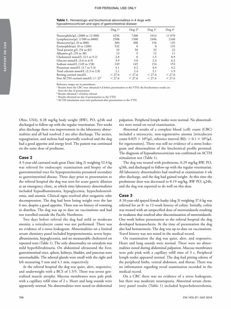
758 CVJ / VOL 57 / JULY 2016
RA
PP
OR
T D
E C
AS
Ohio, USA), 0.28 mg/kg body weight (BW), PO, q24h and discharged to follow-up with the regular veterinarian. Two weeks after discharge there was improvement in the laboratory abnor-malities and all had resolved 2 mo after discharge. The ascites, regurgitation, and melena had reportedly resolved and the dog had a good appetite and energy level. The patient was continued on the same dose of predisone.
Case 2A 5-year-old castrated male great Dane (dog 2) weighing 52.4 kg was referred for endoscopic examination and biopsy of the gastrointestinal tract for hypoproteinemia presumed secondary to gastrointestinal disease. Three days prior to presentation to the referral hospital the dog was seen for acute gastric dilation at an emergency clinic, at which time laboratory abnormalities included hypoalbuminemia, hypoglycemia, hypocholesterol-emia, and anemia. Clinical signs resolved after orogastric tube decompression. The dog had been losing weight over the last 6 mo, despite a good appetite. There was no history of vomiting or diarrhea. The dog was up to date on vaccinations and had not travelled outside the Pacific Northwest.
Two days before referral the dog had mild to moderate anemia; a reticulocyte count was not performed. There was no evidence of a stress leukogram. Abnormalities on a limited serum chemistry panel included hypoproteinemia, severe hypo-albuminemia, hypoglycemia, and no measureable cholesterol on repeated tests (Table 1). The only abnormality on urinalysis was mild hyperbilirubinuria. On abdominal ultrasound the liver, gastrointestinal tract, spleen, kidneys, bladder, and pancreas were unremarkable. The adrenal glands were small with the right and left measuring 5 mm and 4.1 mm, respectively.
At the referral hospital the dog was quiet, alert, responsive, and underweight with a BCS of 1.5/5. There was severe gen-eralized muscle atrophy. Mucous membranes were pale pink with a capillary refill time of 2 s. Heart and lung sounds were apparently normal. No abnormalities were noted on abdominal
palpation. Peripheral lymph nodes were normal. No abnormali-ties were noted on rectal examination.
Abnormal results of a complete blood (cell) count (CBC) included a microcytic, non-regenerative anemia [reticulocyte count 0.025 3 106/mL, reference interval (RI): . 0.1 3 106/mL for regeneration]. There was still no evidence of a stress leuko-gram and abnormalities of the biochemical profile persisted. The diagnosis of hypoadrenocorticism was confirmed on ACTH stimulation test (Table 1).
The dog was treated with prednisone, 0.29 mg/kg BW, PO, q24h, and discharged to follow-up with the regular veterinarian. All laboratory abnormalities had resolved at examination 4 wk after discharge, and the dog had gained weight. At this time the prednisone dose was decreased to 0.19 mg/kg, BW PO, q24h, and the dog was reported to do well on this dose.
Case 3A 10-year-old spayed female husky (dog 3) weighing 37.6 kg was referred for an 8- to 12-week history of colitis. Initially, colitis was treated with an unspecified dose of metronidazole resulting in weakness that resolved after discontinuation of metronidazole. One week before presentation to the referral hospital the dog developed hematochezia. At the time of presentation the dog also had hematemesis. The dog was up-to-date on vaccinations. Travel history was not noted in the medical record.
On examination the dog was quiet, alert, and responsive. Heart and lung sounds were normal. There were no abnor-malities noted during abdominal palpation. Mucous membranes were pale pink with a capillary refill time of 3 s. Peripheral lymph nodes appeared normal. The dog had pitting edema of the peripheral limbs, ventral abdomen, and thorax. There was no information regarding rectal examination recorded in the medical record.
On a CBC there was no evidence of a stress leukogram, but there was moderate neutropenia. Abnormal serum chem-istry panel results (Table 1) included hypocholesterolemia,
Table 1. Hematologic and biochemical abnormalities in 4 dogs with hypoadrenocorticism and signs of gastrointestinal disease
Dog 1a Dog 2b Dog 3c Dog 4c
Neutrophils/mL (2000 to 12 000) 4256 7300 1014 11 070Lymphocytes/mL (1500 to 6000) 2508 1500 2496 2160Monocytes/mL (0 to 800) 304 300 156 135Eosinophils/mL (0 to 1200) 532 0 0 135Total protein g/L (54 to 82) 33 50 32 22Albumin g/L (29 to 38) 13 9 12 11Cholesterol mmol/L (3.5 to 9.2) 2.8 0 1.5 0.9Glucose mmol/L (3.6 to 6.9) 3.9 3.8 2.4 6.2Sodium mmol/L (149 to 158) 149 145 154 153Potassium mmol/L (3.7 to 5.3) 4.1 4.2 4.4 4.2Total calcium mmol/L (2.3 to 2.8) 2.1 2.4 2.1 1.9Resting cortisol nmol/L , 27.6 , 27.6 , 27.6 , 27.6Post ACTH cortisol nmol/L (. 2)d , 27.6 , 27.6 , 27.6 , 27.6
Reference ranges are in parentheses.a Results from the CBC were obtained 4 d before presentation to the VTH; the biochemistry results are
from the day of presentation.b Results obtained 1 d before referral.c Results obtained on day of presentation to the VTH.d ACTH stimulation tests were performed after presentation to the VTH.
FOR PERSONAL USE ONLY

CVJ / VOL 57 / JULY 2016 759
CA
SE
RE
PO
RT
hypoproteinemia, hypoalbuminemia, hypoglycemia, and hypo-calcemia attributed to hypoalbuminemia; ionized calcium was not determined. The diagnosis of hypoadrenocorticism was confirmed on ACTH stimulation test (Table 1). The CBC was repeated the following day and the neutrophil count had normalized.
An abdominal ultrasound showed fluid-filled and hypomotile small and large bowels and a scant amount of free peritoneal fluid. The remaining abdomen was normal; however, there was no specific mention of the adrenal glands in the ultrasound report. The thoracic radiographs were unremarkable except for evidence of hypovolemia.
The patient was administered prednisolone sodium succinate (Zoetis, Kalamazoo, Michigan, USA), 0.5 mg/kg BW, IV, q12h, for 1 d until she regained her appetite. With resumption of food consumption the dog was discharged to follow-up with the regular veterinarian with prednisone, at 0.53 mg/kg BW, PO, q24h for 1 wk and then decreased to 0.13 mg/kg BW, PO, q24h. Four weeks after discharge all previously documented clinical and biochemical abnormalities had normalized.
Case 4A 5-year-old neutered male Labrador retriever (dog 4) weighing 28 kg was evaluated for a 2-day history of lethargy, vomiting, diarrhea, and hematochezia. Previous history included an avul-sion of the right front dew claw 2 wk earlier, and an episode of vomiting and diarrhea 8 mo prior.
On examination, the dog was quiet, alert, and responsive, but shortly became obtunded and laterally recumbent. The dog had a BCS of 3/5. Mucous membranes were pink and capillary refill time was , 1 s. The rectal temperature was 36.8°C. Heart rate was 156 beats/min and respiratory rate was 16 breaths/min. Femoral pulses were hypokinetic but synchronous. There were normal breath sounds and no arrhythmia, but there was a left-sided grade I/VI systolic murmur loudest over the left apex. The dog seemed uncomfortable with abdominal palpation, but there were no other abnormalities. Peripheral lymph nodes apeared normal. Hematochezia was noted on rectal examination. The remainder of the physical examination was normal.
Abdominal radiographs were taken at the time of presenta-tion to the referral hospital. There was good serosal detail, but the small and large bowels were fluid-filled with no evidence of obstruction. There was no stress leukogram on a CBC. Abnormal results of a serum chemistry panel (Table 1) included hypocholesterolemia, hypoproteinemia with both hypoalbu-minemia and hypoglobulinemia, and hypocalcemia attributed to hypoalbuminemia. An ionized calcium test performed the following day (day 2) was normal (1.32 mmol/L, RI: 1.12 to 1.40 mmol/L). The diagnosis of hypoadrenocorticism was con-firmed on ACTH stimulation test (Table 1).
After establishing a diagnosis of hypoadrenocorticism, pred-nisone, 0.27 mg/kg BW, PO, q12h, was given for 1 wk and then decreased to 0.27 mg/kg BW, PO, q24h. The dog was clinically normal at the time of discharge on day 3, with no lethargy, vomiting, or diarrhea. At a 4-week follow-up the dog was clinically normal except for mild polyuria/polydipsia. All biochemical abnormalities had resolved.
DiscussionAll 4 dogs in this report were referred for evaluation of what was believed to be primary gastrointestinal (GI) disease; all were diagnosed with hypoadrenocorticism. Although the presence of a primary enteropathy was not definitively excluded in any of these dogs, resolution of both clinical signs and laboratory abnormalities of hypoproteinemia, hypoalbuminemia, and hypocholesterolemia following therapy with physiologic replace-ment doses of prednisone supports the likelihood that all of the clinical features of these dogs were a consequence of untreated hypoadrenocorticism and not a concurrent enteropathy.
The dogs in this report were suspected of having PLE, a spectrum of GI diseases characterized by loss of serum proteins into the GI tract. Dogs with PLE are often seen for chronic or relapsing diarrhea and/or vomiting, and commonly exhibit weight loss. They may have ascites, pleural effusion, or periph-eral edema, usually as a consequence of hypoalbuminemia and low oncotic pressure (2). Common laboratory findings include hypoalbuminemia, hypocholesterolemia, and potentially hypo-globulinemia, lymphopenia, hypocalcemia, hypomagnesemia, and hypocobalaminemia (1–2). The diagnostic approach to a dog suspected of having PLE commonly involves exclusion of other causes of hypoalbuminemia including protein-losing nephropathy, liver failure, and third space losses, but hypoadre-nocorticism is not routinely mentioned as a differential diagnosis (2–3). The 4 cases described here highlight the importance of considering hypoadrenocorticism as a differential diagnosis for a PLE-like presentation in dogs with hypoalbuminemia and/or hypocholesterolemia, even when such dogs exhibit clinical consequences of enteric protein loss, such as peripheral edema or ascites.
The pathophysiologic mechanism driving hypoalbuminemia in hypoadrenocorticism is unknown. Loss of albumin into the GI tract or decreased intake/assimilation of proteins, is the main suspected cause (4). The human literature suggests that glucocorticoids enhance barrier function of epithelial cells due to suspect epithelial-specific properties, but more research is needed in this area (5).
Two of the 4 dogs had fluid accumulation (abdominal effu-sion, dog 2; peripheral edema, dog 3) attributed to hypoalbu-minemia. It is well-known that hypoadrenocorticism can mimic gastrointestinal disease; however, to our knowledge there is no literature describing ascites or peripheral edema secondary to hypoadrenocorticism-induced hypoalbuminemia (4,6). Effusions and edema are, however, relatively common in dogs with PLE, potentially increasing the likelihood that a clinician erroneously suspects PLE as the cause of the clinical features in dogs affected by hypoadrenocorticism.
Hypocholesterolemia is common in dogs with what has been historically referred to as atypical, or glucocorticoid-deficient, hypoadrenocorticism. A recent paper (7) found that dogs with “atypical” hypoadrenocorticism had mineralocorticoid deficien-cies despite having normal electrolyte concentrations, but for the sake of this discussion, we will retain the atypical hypoad-renocorticism designation. In one study, 13/17 (76.5%) dogs with atypical hypoadrenocorticism had hypocholesterolemia
FOR PERSONAL USE ONLY

760 CVJ / VOL 57 / JULY 2016
RA
PP
OR
T D
E C
AS
(6). In a study by Thompson et al (8), dogs with glucocorticoid-deficient hypoadrenocorticism had significantly lower albumin and cholesterol concentrations than dogs with typical hypo-adrenocorticism. The diagnostic challenge posed by atypical hypoadrenocorticism, and often the more prolonged course of disease before diagnosis, could contribute to the increased frequency of hypocholesterolemia in affected dogs (8,9). All the dogs of our report had hypocholesterolemia, and in dog 2, the hypocholesterolemia was severe. The mechanism for hypocho-lesterolemia in hypoadrenocorticism is not known. It has been suggested that glucocorticoids play a role in fat absorption from the GI tract; decreased mobilization or increased utilization of fatty acids secondary to high ACTH concentrations has also been proposed (1,6,9).
Dogs with hypoadrenocorticism often have no stress leuko-gram because of cortisol deficiency. In the study by Thompson et al (8), all dogs had normal lymphocyte, monocyte, and eosinophil counts, and Scott-Moncrief (9) reported that 97% of dogs with hypoadrenocorticism lacked a stress leukogram. Since hyperkalemia and/or hyponatremia often prompt suspicion of hypoadrenocorticism and subsequent testing for the disease, rec-ognition that a patient lacks a stress leukogram becomes critical to generating suspicion of atypical hypoadrenocorticism even when none of the classic electrolyte abnormalities are present (8–9). None of the dogs in this report had a stress leukogram.
In a retrospective study, pre- and post-ACTH serum corti-sol concentrations were below 27.6 nmol/L in 37/42 of dogs with hypoadrenocorticism, whereas the remaining 5/42 had pre- and post-ACTH serum cortisol levels between 27.6 and 55.2 nmol/L (8). Thus, a resting cortisol can be a useful and relatively inexpensive tool in ruling out hypoadrenocorticism. It could be argued that an ill patient without a stress leukogram should have a basal cortisol concentration measured before more invasive and expensive diagnostics are performed. If the basal cortisol is less than 55.2 nmol/L the sensitivity and specificity for detecting hypoadrenocorticism are 100% and 63% to 78%, respectively, and obtaining post-ACTH cortisol concentration is recommended to confirm the diagnosis (10–11).
Three dogs in this report had small or non-detectable adrenal glands during abdominal ultrasonography. It has previously been described that dogs with hypoadrenocorticism had significantly thinner adrenal glands than those of healthy dogs, but dogs with atypical hypoadrenocorticism were not well-represented in these reports (12,13). All patients in the paper by Wenger et al (13)
had unspecified electrolyte abnormalities, and Hoerauf and Reusch (12) did not describe laboratory results/abnormalities for their patients. Adrenal ultrasonography may be of value in dogs with clinical signs otherwise suggestive of PLE as small adrenal glands could enhance the suspicion of hypoadrenocorticism.
In conclusion, the challenge of diagnosing atypical hypoadre-nocorticism resides in generating clinical suspicion. The absence of a stress leukogram in dogs with hypoalbuminemia and/or hypocholesterolemia, even with ascites or peripheral edema, should warrant a resting cortisol, and ACTH stimulation testing if indicated, before pursuing causes of primary GI disease. CVJ
References1. Stockham SL, Scott MA. Lipids. In: Stockham SL, Scott MA, eds.
Fundamentals of Veterinary Clinical Pathology. 2nd ed. Ames, Iowa: Blackwell Publishing, 2008:770–771.
2. Dossin O, Lavoué R. Protein-losing enteropathies in dogs. Vet Clin North Am Small Anim Pract 2011;41:399–418.
3. Willard MD. Small intestine — Malabsorption. In: Washabau RJ, Day MJ, eds. Canine & Feline Gastroenterology. 1st ed. St. Louis, Missouri: Saunders, 2013:678–683.
4. Langlais-Burgess L, Lumsden JH, Mackin A. Concurrent hypoadreno-corticism and hypoalbuminemia in dogs: A retrospective study. J Am Anim Hosp Assoc 1995;31:307–311.
5. Fischer A, Gluth M, Weege F, et al. Glucocorticoids regulate barrier function and claudin expression in intestinal epithelial cells via MKP-1. Am J Physiol Gastrointest Liver Physiol 2014;306:G218–2128.
6. Lifton SJ, King LG, Zerbe CA. Glucocorticoid deficient hypoadrenocor-ticism in dogs: 18 cases (1986–1995). J Am Vet Med Assoc 1996;209: 2076–2081.
7. Baumstark ME, Sieber-Ruckstuhl NS, Müller C, Wenger M, Boretti FS, Reusch CE. Evaluation of aldosterone concentrations in dogs with hypoadrenocorticism. J Vet Intern Med 2014;28:154–159.
8. Thompson AL, Scott-Moncrieff JC, Anderson JD. Comparison of classic hypoadrenocorticism with glucocorticoid-deficient hypoadreno-corticism in dogs: 46 cases (1985–2005). J Am Vet Med Assoc 2007; 230:1190–1194.
9. Scott-Moncrief JC. Hypoadrenocorticism. In: Ettinger EC, Nelson RW, Reusch CE, Scott-Moncrieff JC, Behrend EN, eds. Canine and Feline Endocrinology. 4th ed. St. Louis, Missouri: Saunders, 2015:485–520.
10. Lennon EM, Boyle TE, Hutchins RG, et al. Use of basal serum or plasma cortisol concentrations to rule out a diagnosis of hypoadreno-corticism in dogs: 123 cases (2000–2005). J Am Vet Med Assoc 2007; 231:413–416.
11. Bovens C, Tennant K, Reeve J, Murphy KF. Serum cortisol concentra-tion as a screening test for hypoadrenocorticism in dogs. J Vet Intern Med 2014;28:1541–1545.
12. Hoerauf A, Reusch CE. Ultrasonographic evaluation of the adrenal glands in six dogs with hypoadrenocorticism. J Am Anim Hosp Assoc 1999;35:214–218.
13. Wenger M, Mueller C, Kook PH, Reusch CE. Ultrasonographic evalu-ation of adrenal glands in dogs with primary hypoadrenocorticism or mimicking diseases. Vet Rec 2010;167:207–210.
FOR PERSONAL USE ONLY

CVJ / VOL 57 / JULY 2016 761
Case Report Rapport de cas
Total laryngectomy for management of chronic aspiration pneumonia in a myopathic dog
Karen M. Vernau, Stanley L. Marks, Maggie A. Kuhn, William T.N. Culp, Tammy J. Owens, G. Diane Shelton, Tausif Siddiqui, Rachel Pollard, Peter C. Belafsky
Abstract — A 5-month-old female pit bull terrier dog evaluated for ataxia, progressive regurgitation, and recurrent aspiration pneumonia had markedly elevated creatine kinase activity, non-inflammatory generalized myopathy, and severe esophageal dysmotility. A narrow-field total laryngectomy was performed. The dog is doing well 30 months after surgery, and no longer has episodes of aspiration pneumonia, despite intermittent regurgitation. This case represents the first application of total laryngectomy for the prevention of chronic recurrent aspiration pneumonia in the dog.
Résumé — Laryngectomie totale pour la gestion d’une pneumonie par aspiration chronique chez un chien myopathique. Une chienne Pit Bull Terrier âgée de 5 mois évaluée pour de l’ataxie, de la régurgitation progressive et une pneumonie par aspiration récurrente présentait une activité de la créatine kinase particulièrement élevée, une myopathie généralisée non inflammatoire et un trouble de motilité de l’œsophage grave. Une laryngectomie totale à champ étroit a été réalisée. La chienne se porte bien 30 mois après la chirurgie et n’a plus d’épisodes de pneumonie par aspiration, malgré une régurgitation intermittente. Ce cas représente la première application d’une laryngectomie totale pour la prévention d’une pneumonie par aspiration chronique récurrente chez un chien.
(Traduit par Isabelle Vallières)
Can Vet J 2016;57:761–766
S ince Theodor Billroth performed the first human laryngec-tomy for laryngeal carcinoma in 1873, the procedure has
been modified and improved many times (1–6). Total laryngec-tomy has been used to prevent chronic aspiration in 4 human patients (7); however, application of the procedure for treatment of chronic aspiration in humans was limited due to complete loss of phonation and naso-laryngeal breathing, hyposmia, and atrophy of nasal mucosa (7,8). Various medical and surgical alternatives to laryngectomy have been used for chronic aspira-tion in humans, including dietary and behavioral modification, oral care and sitting posture, levodopa to improve swallow and cough reflex, tracheostomy with gas bag, laryngeal diversion, tracheoesophageal diversion, laryngeal diversion with a speech fistula, dynamic laryngotracheal closure, and a paced glottis
closure (9–13). Conservative measures are generally preferred for concerns of voice preservation in humans. However, when medi-cal management fails, total laryngectomy is the gold standard treatment of chronic and life-threatening aspiration pneumonia in humans (7). Experimental laryngectomy was first performed in the dog in the early 19th century (6,14), and was exclusively used for the surgical management of laryngeal cancer in dogs (15–17). To the best of our knowledge, a total laryngectomy has not been performed for the treatment and prevention of chronic recurrent aspiration pneumonia in the dog.
This report documents the successful management via total laryngectomy of a 5-month-old female pit bull terrier with progressive regurgitation and recurrent aspiration pneumonia secondary to a severe non-inflammatory congenital myopathy.
Department of Surgical and Radiological Sciences (Vernau, Culp, Pollard), Department of Medicine and Epidemiology (Marks), William R. Pritchard Veterinary Medicine Teaching Hospital (Owens), University of California, Davis, School of Veterinary Medicine, Davis, California, USA; Department of Otolaryngology (Belafsky, Siddiqui, Kuhn), School of Medicine, Sacramento, California, USA, and Department of Pathology, School of Medicine (Shelton), University of California, San Diego, La Jolla, California, USA.Address all correspondence to Dr. Stanley L. Marks; e-mail: [email protected] work was completed at the William R. Pritchard Veterinary Medical Teaching Hospital (VMTH), University of California, Davis and the University of California, San Diego. The study was not supported by any grants or other funding.Use of this article is limited to a single copy for personal study. Anyone interested in obtaining reprints should contact the CVMA office ([email protected]) for additional copies or permission to use this material elsewhere.
FOR PERSONAL USE ONLY

762 CVJ / VOL 57 / JULY 2016
RA
PP
OR
T D
E C
AS
Case descriptionA 5-month-old female pit bull terrier dog was referred to the William R. Pritchard Veterinary Medical Teaching Hospital (VMTH), University of California, Davis, for evaluation of an abnormal gait. The dog was rescued 1 mo previously, and the previous medical history was unknown. At presentation, the dog weighed 16 kg with a body condition score (BCS) of 4/9. The pelvic limbs were held in an adducted position, and the pelvic limb musculature was palpably firm, with limited range of motion and apparent pain on gentle manipulation of the coxofemoral (CF) joints. The thoracic limbs had a wide-based stance with elbow abduction, but normal range of motion in all joints. On neurological examination, abnormalities were restricted to a short-strided gait in all 4 limbs, reduced flexor reflexes and delayed proprioception in the pelvic limbs. Bilateral CF luxation was noted on pelvic radiographs. A serum chemistry profile revealed hyperphosphatemia [2.2 mmol/L, reference range (RR): 0.84 to 1.68 mmol/L], hypercalcemia (3 mmol/L, RR: 2.4 to 2.8 mmol/L), and marked elevation of creatine kinase (CK) activity (74 272 IU/L, RR: 55 to 257 IU/L) with elevated alanine aminotransferase (ALT) (295 IU/L, RR: 21 to 72 IU/L) and aspartate aminotransferase (AST) (1339 IU/L, RR: 20 to 49 IU/L). The alkaline phosphatase (ALP) activity was mildly
increased (97 IU/L, RR: 14 to 91 IU/L). The elevated CK and AST were consistent with a myopathy, and the elevated ALP activity and increased phosphorus were associated with osteo-blastic activity during growth. A complete blood (cell) count (CBC) and urinalysis were unremarkable. Infectious disease titers for Neospora (direct fluorescent antibody) and Toxoplasma (latex agglutination test) were negative.
The dog was placed under general anesthesia for an electro-diagnostic evaluation by electromyography (EMG) and biopsies were collected from the cranial tibial, vastus lateralis, triceps, and adductor muscles. In addition, the dog underwent a bilateral femoral head ostectomy (FHO) and tenotomies of the pectineus muscle to improve the CF pain and ability to ambulate. The pectineus muscle was also biopsied during the FHO. The EMG was abnormal and characterized by diffuse complex repetitive discharges in the distal and proximal thoracic and pelvic limb, epaxial, head, and tongue muscles. All muscle biopsies revealed a non-inflammatory generalized myopathy with dystrophic features, most severe in the adductor (Figure 1) and pectineus muscles.
Following adoption, the dog was noted to have progressive dysphagia and regurgitation. Thoracic radiographs revealed a fluid-filled caudal thoracic esophagus and a videofluoroscopic
Figure 1. Cryosections from the adductor, diaphragm, cranial tibial, and pharyngeal muscles stained with hematoxylin and eosin (H&E). Excessive variability in myofiber size is evident in the adductor, diaphragm, and pharyngeal muscles with marked endomysial fibrosis separating individual muscle fibers in the adductor and diaphragm muscles consistent with a severe degenerative myopathy. The cranial tibial muscle is relatively spared. Bar = 50 mm for all images.
FOR PERSONAL USE ONLY

CVJ / VOL 57 / JULY 2016 763
CA
SE
RE
PO
RT
swallow study revealed evidence of severe esophageal dysmotil-ity in the cranial esophagus characterized by retention of large volumes of liquid barium and retrograde motility of the bolus into the pharynx (Figure 2A–C). In addition, the dog had multiple episodes of gastroesophageal reflux and a sliding (Type I) hiatal hernia (Figure 2D). Persistent regurgitation was managed with famotidine (Famotidine; Major Pharmaceuticals, Livonia, Michigan, USA), 1 mg/kg body weight (BW), PO, q24h, sucralfate (Carafate; Nostrum Laboratories, Kansas City, Missouri, USA), 1 g PO, q8h, and omeprazole (Omeprazole delayed release tablets; Dexcel Pharma Technologies, Yokneam, Israel), 1 mg/kg BW, PO, q24h. Feedings were changed to a commercial complete and balanced puppy food fed in small volumes 3 times daily. Although the dog was fed in a dog sitting position, regurgitation persisted. The dog underwent correc-tive surgery for the hiatal hernia characterized by a left-sided gastropexy, esophagopexy, and esophageal hiatal plication 2 mo later. The dog’s diaphragm was found to be markedly thickened secondary to the severe myopathy and the diaphragmatic defect was reduced. Two biopsies were obtained from the left crura of the diaphragm and the muscle was found to be severely affected with a dystrophic process similar to that observed in the other muscles (Figure 1). The frequency of regurgitation decreased for approximately 3 mo; however, clinical signs progressed shortly thereafter and the dog developed aspiration pneumonia that was treated with ampicillin/sulbactam (Unasyn; Pfizer, New York, New York, USA), 50 mg/kg BW, IV, q8h. A repeat videofluoroscopic swallow study revealed severe segmental esophageal dysmotility characterized by the absence of primary peristaltic waves and pooling of barium in the proximal esopha-
gus. Liquid barium was subsequently refluxed through the upper esophageal sphincter into the pharynx and nasopharynx, and a large amount of barium was subsequently aspirated into the trachea. Persistence of gastroesophageal reflux and hiatal hernia-tion was noted. Progression of the underlying myopathy with concurrent esophagitis secondary to reflux was considered the likely etiology. Esophagoscopy revealed that the proximal half of the esophagus was abnormally dilated and the gastroesophageal junction had rugal folds protruding above the diaphragmatic pinch. Significant reflux was observed with the application of gastric pressure. A 24-French percutaneous endoscopic gas-trostomy (PEG) tube (EndoVive PEG tube; Boston Scientific, Marlborough, Massachusetts, USA) was placed to facilitate feeding, and this was replaced with a 24-Fr low-profile button (Bard Button; Bard Access Systems, Salt Lake City, Utah, USA) after 2 mo.
Progressive regurgitation and recurrent aspiration pneumonia necessitated frequent visits and hospitalizations at the VMTH, with antimicrobial therapy consisting of ampicillin/sulbactam (Unasyn; Pfizer), 50 mg/kg BW, IV, q8h and enrofloxacin (Baytril; Bayer Healthcare, Shawnee Mission, Kansas, USA), 10 mg/kg BW by PEG tube q24h, with nebulization and coup-age necessary to manage the pneumonia. The dog’s quality of life was poor with frequent episodes of regurgitation, nasal reflux of ingesta, and difficulty sleeping due to partial airway obstruction. The dog lost 3 kg body weight and had a BCS of 2/9. Regurgitation persisted despite administration of cisapride (Cisapride, compounded product; Road Runner Pharmacy, Phoenix, Arizona, USA), 0.5 mg/kg BW, PO, q12h. A cus-tomized calorie-dense complete and balanced home-prepared
Figure 2. A, B, C — Videofluoroscopic swallow study with liquid barium documenting evidence of aspiration characterized by barium coating the trachea and severe esophageal dysmotility with retrograde movement of the bolus in the proximal esophagus prior to movement of the bolus in an aborad direction. D — Videofluoroscopic swallow study with barium-soaked kibble documenting displacement of the cardia cranially to the diaphragmatic crus (sliding hiatal hernia) with gastroesophageal reflux.
A
C
B
D
FOR PERSONAL USE ONLY

764 CVJ / VOL 57 / JULY 2016
RA
PP
OR
T D
E C
AS
diet of liquid consistency was formulated for the dog. Despite the administration of prokinetics and the liquid diet via the low profile gastrostomy device (LPGD), the dog continued to regurgitate and had 3 episodes of airway obstruction with loss of consciousness, requiring emergent intervention to re-establish airway patency. In light of the frequent episodes of regurgitation and subsequent aspiration with airway obstruction, a narrow field laryngectomy was elected when the dog was 14 mo of age.
A narrow-field total laryngectomy was performed under general anesthesia by 2 otolaryngologists and a board-certified veterinary surgeon. The dog was placed in dorsal recumbency, the fur was clipped, and the skin was prepared with aseptic technique. A cushion was placed under the neck to elevate the cervical region into the surgical field. A 5-cm horizontal cur-vilinear incision was made 2 cm below the level of the cricoid cartilage. The sternohyoideus and sternothyroideus muscles were bluntly separated on the ventral midline and retracted to expose the larynx. Blunt and sharp dissection was utilized to expose the cricoid and thyroid cartilage of the larynx as well as the cranial aspect of the trachea (1st and 2nd tracheal rings). The entire circumference of the cranial trachea was dissected free from all surrounding tissues. The pharyngeal constrictor muscles (hyopharyngeus, thyropharyngeus, and cricopharyn-geus) were separated from their attachments on the larynx as were the sternothyroideus and thyrohyoideus muscles. The cuff of the endotracheal tube was deflated, and the endotracheal tube was pulled cranially, to allow for a tracheal incision. A #15 blade was utilized to enter the trachea between the 2nd and 3rd tracheal rings. This incision was continued around the entire circumference of the trachea; a sterile endotracheal tube was introduced into the open end of the trachea, and the cuff was inflated to allow for a tight seal. Cranially, the larynx was freed from its attachments to the hyoid apparatus (disarticulated bilaterally) as well as the attachments to the tongue and hyoid bone, which were preserved. The larynx and cranial trachea were then removed. The pharyngeal mucosa was closed with 3-0 polydioxanone (PDS Suture; Ethicon, Somerville, New Jersey, USA) in a simple continuous pattern, followed by closure of the paired thyropharyngeal and cricopharyngeal muscles with 3-0 polydioxanone (Ethicon) in a simple continuous pattern. The
cranial trachea was then brought into apposition with the skin to form a permanent tracheostomy. The severed end of the trachea was rotated to allow the entire circumference to be sutured to the skin edges forming an airtight seal. The trachea was sutured to the skin with 3-0 silk (Perma-Hand Silk Suture; Ethicon) because of its braided and non-absorbable features and optimal knot security qualities. Two closed-suction drains (Jackson-Pratt drain; Cardinal Health, Dublin, Ohio, USA) were placed in the surgical wound and the rest of the cervical incision was closed with staples. A light compression dressing was placed around the neck. The dog recovered uneventfully from anesthesia in the intensive care unit (ICU) and was discharged from the unit the following morning.
The dog was unable to swallow immediately following surgery secondary to the pharyngeal and esophageal myopathy, but otherwise did well in the post-operative period. The stoma was cleaned with sterile saline after nebulization with sterile water for 10 min 3 times daily for 10 d and a size #8 tracheostoma vent (Bivona; Smiths Medical ASD, Gary, Indiana, USA) was applied 14 d after surgery and held in place with a Posey foam tracheal tie (Posey Company, Arcadia, California, USA). The drains were removed on postoperative day 3 and the staples were removed on postoperative day 7. The dog is doing well 30 mo after surgery with a weight of 25 kg and a BCS of 8/9. There have been no episodes of aspiration pneumonia, even though the dog con-tinues to regurgitate. The dog is managed with glycopyrrolate (Par Pharmaceutical, Woodcliff Lake, New Jersey, USA), 1 mg q8h to reduce saliva production, acid suppressants (famotidine, omeprazole), and gastrointestinal protectants (sucralfate) admin-istered via PEG tube for the chronic gastroesophageal reflux and esophagitis, and lactated Ringer’s solution (500 mL SQ, q12h) to maintain hydration status. The dog has frequent episodes of regurgitation and nasal reflux of saliva and refluxed food due to her pharyngeal and esophageal myopathy. She wears the tracheostoma vent continuously to maintain a patent tracheal stoma. In addition, the dog is heat intolerant secondary to the inability to thermoregulate by panting; however, quality of life is considered excellent by the owners and clinicians. Neurological status has minimally progressed since initial presentation with increased muscle rigidity in the thoracic limbs.
Figure 3. A — Fully healed uncovered tracheostoma following laryngectomy in a pit bull terrier. B — The tracheostoma is kept patent with a soft, flexible silicon tracheostoma vent (Bivona; Smiths Medical ASD, Gary, Indiana, USA) that is secured in place with a foam tracheal tie (Posey Company, Arcadia, California, USA).
A B
FOR PERSONAL USE ONLY

CVJ / VOL 57 / JULY 2016 765
CA
SE
RE
PO
RT
DiscussionAspiration pneumonia is a common clinical problem in dogs with neurological disease, particularly neuromuscular disor-ders such as myasthenia gravis, polymyopathies, and polyneu-ropathies affecting the swallowing reflex (18–20). In 1 report, aspiration pneumonia was most frequently noted in dogs with esophageal disease, vomiting, and neurological disorders (21). In that study, 22 of 88 dogs died or were euthanatized, and 3 of the 22 dogs (14%) were euthanized because of continual or refrac-tory aspiration pneumonia (21). Most dogs with myasthenia gravis have esophageal weakness and dysfunction, in addition to generalized weakness. In 1 study, 12 of 25 dogs (48%) with myasthenia gravis died or were euthanized shortly after admis-sion to the hospital due to aspiration pneumonia (18). Given the high incidence of spontaneous remission of myasthenia gravis in dogs (22), a laryngectomy procedure should be reserved for those animals with the most intractable pneumonia and an irreversible primary disease that is refractory to medical therapy, regardless of cause. Currently, there is no established surgical treatment for prevention of chronic or recurrent aspiration pneumonia in dogs.
In humans, it has been suggested that surgery is indicated for those cases with chronic aspiration and severe associated medical problems, whereby a total laryngectomy is the only justifiable surgical intervention which can improve patient quality of life (7). We utilized this approach to prevent recurrent aspiration pneumonia in a dog and prevented chronic aspira-tion, markedly improved quality of life, and obviated the need for euthanasia. Documented complications of laryngectomy in dogs include collapse of the laryngeal stoma, tracheoesopha-geal fistula formation, pharyngeal dehiscence, megaesophagus, hypocalcemia secondary to iatrogenic hypoparathyroidism, pneumonia, and death (15–17). Two of these reports describe significant post-laryngectomy morbidity in dogs while another report demonstrated no significant complications during 18 mo of follow-up (15–17). Other complications related to permanent tracheostomy include skin fold occlusion of a tracheal stoma, hyposmia and olfactory mucosal changes, loss of thermoregula-tion through panting, and increased CNS temperature (23–25). Postoperative activity of such dogs should be limited, as upper respiratory ventilation is required for normal thermoregulation and restriction of outdoor activities in warm weather should be considered in some cases as well (17). These dogs are also unable to swim due to the risk of drowning.
Although this dog was cured of recurrent aspiration pneu-monia, it remains unable to swallow because of the progressive myopathy. The dog has also developed hyposmia as reported by the owners because the dog can no longer smell the family cat entering the family room, and the dog used to chase the cat out of the room as soon as she was aware of its presence. The myopathy in this case is congenital and no specific treatments or cures are available, thus the necessity for the laryngectomy to prevent aspiration pneumonia. She is maintained on inter-mittent tube feedings administered through a PEG tube and subcutaneous fluids, and maintains an excellent quality of life according to her owners and clinicians. It is plausible that dogs
with less severe dysphagia may be able to eat and drink orally after laryngectomy, without the previous risk of aspiration. By removing the larynx and separating the respiratory and digestive tracts, the risk of aspiration pneumonia is eliminated.
Chronic regurgitation and recurrent life-threatening aspira-tion pneumonia are challenging conditions to treat and result in death or euthanasia in many dogs, particularly in those with esophageal and neuromuscular disorders. Currently there is no established surgical management available for this disorder and many dogs are euthanized. This case report suggests that a nar-row field laryngectomy can be safely and effectively performed for management of chronic aspiration in the dog, and should be considered as a viable alternative treatment for dogs with profound oropharyngeal dysphagia, chronic regurgitation, and aspiration pneumonia.
The ethical considerations and importance of comprehensive client communications surrounding the surgical management of a dog with a progressive myopathy cannot be overempha-sized. The dog in this report did not have an obviously clini-cally progressive myopathy other than the involvement of the pharynx and esophagus. Prior to the laryngectomy procedure, the dog suffered 3 episodes of airway obstruction secondary to aspiration of regurgitated material with consequent loss of consciousness. Without the laryngectomy procedure, the dog would have undergone elective euthanasia as it was deemed inhumane for her to continue to aspirate and suffer respiratory obstruction. The internists and surgeons met with the owners on several occasions to discuss the ramification of the laryngectomy procedure as well as potential complications. The owners were given ample opportunity to discuss the options with their family members and elected to move forward with the procedure rec-ognizing that euthanasia would be indicated if the dog’s quality of life was not dramatically improved. The dog has continued to thrive and its quality of life is deemed to be excellent by the owners and clinicians.
AcknowledgmentsThe authors thank John Doval for assistance with the images and Robin Fisher and Paula Howell for their technical support. CVJ
References1. Weir NF. Theodore Billroth: The first laryngectomy for cancer.
J Laryngol Otol 1973;87:1161–1169.2. Stell PM. The first laryngectomy. J Laryngol Otol 1975;89:353–358.3. Rizzotto G, Succo G, Lucioni M, Pazzaia T. Subtotal laryngectomy
with tracheohyoidopexy: A possible alternative to total laryngectomy. Laryngoscope 2006;116:1907–1917.
4. Majer EH. 100 Years of laryngectomy. Laryngol, Rhinol, Otol 1975;54: 3–9.
5. Bron L, Brossard E, Monnier P, Pasche P. Supracricoid partial laryngec-tomy with cricohyoidoepiglottopexy and cricohyoidopexy for glottic and supraglottic carcinomas. Laryngoscope 2000;110:627–634.
6. Schwartz AW, Devine KD. Some historical notes about the first laryn-gectomies. Laryngoscope 1959;69:194–201.
7. Cannon CR, McLean WC. Laryngectomy for chronic aspiration. Am J Otolaryngol 1982;3:145–149.
8. Wei X, Fan X, Zhang J. [The long-term morphology of the nasal cavity after total laryngectomy] Lin Chuang Er Bi Yan Hou Tou Jing Wai Ke Za Zhi. J Clin Otorhinolaryngol, 2010;24:785–787.
9. Sasaki H, Sekizawa K, Yanai M, Arai H, Yamaya M, Ohrui T. New strategies for aspiration pneumonia. Intern Med 1997;36:851–855.
FOR PERSONAL USE ONLY

766 CVJ / VOL 57 / JULY 2016
RA
PP
OR
T D
E C
AS
10. Wang H, Shen Z, Yao Z, Qiu H. [Surgical treatment for the chronic severe aspiration]. Lin Chuang Er Bi Yan Hou Ke Za Zhi. J Clinical Otorhinolaryngol 2003;17:732–733.
11. De Vito MA, Wetmore RF, Pransky SM. Laryngeal diversion in the treatment of chronic aspiration in children. Int J Pediatric Otorhinolar-yngol 1989;18:139–145.
12. Wang D, Dulguerov P. Laryngeal diversion and tracheotracheal speech fistula for chronic aspiration. Ann Otol Rhinol Laryngol 2000;109:602–604.
13. Broniatowski M, Grundfest-Broniatowski S, Tyler DJ, et al. Dynamic laryngotracheal closure for aspiration: A preliminary report. Laryngo-scope 2001;111:2032–2040.
14. Lapidot A, Catalfumo F, Gelot R, Liskow A. Total laryngectomy and autogenous reconstruction observations in an experimental series in canines. J Surg Oncol 1973;5:259–266.
15. Block G, Clarke K, Salisbury SK, DeNicola DB. Total laryngectomy and permanent tracheostomy for treatment of laryngeal rhabdomyosarcoma in a dog. J Am Anim Hosp Assoc 1995;31:510–513.
16. Crowe DT, Goodwin MA. Total laryngectomy for laryngeal mast cell tumor in a dog. J Am Anim Hosp Assoc 1986;22:809–816.
17. Henderson RA, Powers RD, Perry L. Development of hypoparathyroid-ism after excision of laryngeal rhabdomyosarcoma in a dog. J Am Anim Hosp Assoc 1991;198:639–643.
18. Dewey CW, Bailey CS, Shelton GD, Kass PH, Cardinet GH, 3rd. Clinical forms of acquired myasthenia gravis in dogs: 25 cases (1988–1995). J Vet Intern Med 1997;11:50–57.
19. Ryckman LR, Krahwinkel DJ, Sims MH, Donnell RL, Moore PF, Shelton GD. Dysphagia as the primary clinical abnormality in two dogs with inflammatory myopathy. J Am Vet Med Assoc 2005;226:1519–23.
20. Khorzad R, Whelan M, Sisson A, Shelton GD. Myasthenia gravis in dogs with an emphasis on treatment and critical care management. J Vet Emerg Crit Care 2011;21:193–208.
21. Kogan DA, Johnson LR, Sturges BK, Jandrey KE, Pollard RE. Etiology and clinical outcome in dogs with aspiration pneumonia: 88 cases (2004–2006). J Am Vet Med Assoc 2008;233:1748–1755.
22. Shelton GD, Lindstrom JM. Spontaneous remission in canine myasthe-nia gravis: Implications for assessing human MG therapies. Neurology 2001;57:2139–2141.
23. Baker MA, Chapman LW, Nathanson M. Control of brain temperature in dogs: Effects of tracheostomy. Resp Physiol 1974;22:325–333.
24. Hedlund CS, Tangner CH, Montgomery DL, Hobson HP. A procedure for permanent tracheostomy and its effects on tracheal mucosa. Vet Surg 1982;11:13–17.
25. Hedlund CS, Tangner CH, Waldron DR, Hobson HP. Permanent tracheostomy: Perioperative and long-term data from 34 cases. J Am Anim Hosp Assoc 1988;24:585–591.
While you’re taking care of them,we’re looking out for you.
Specialized insurance programs and risk management services for CVMA members
• Professional liability insurance• Commercial insurance• Employeebenefits• Individual life and disability• Student and graduate insurance• Personal auto and home insurance
1-866-860-2862 • cvmainsurance.com
FOR PERSONAL USE ONLY

CVJ / VOL 57 / JULY 2016 767
Case Report Rapport de cas
Citrobacter freundii induced endocarditis in a yearling colt
Eleonora E.A. Guidi, Aurélie Thomas, Jean-Luc Cadoré, Agnès Benamou Smith
Abstract — Endocarditis is a rare pathology in horses and the clinical signs can be misleading. We describe the clinical, echocardiographic, and pathological features of Citrobacter freundii induced bacterial endocarditis in a horse. This bacterium has never been reported before as an agent of vegetative endocarditis in the horse.
Résumé — Endocardite induite par Citrobacter freundii chez un poulain âgé d’un an. L’endocardite est une pathologie rare chez les chevaux et les signes cliniques peuvent être trompeurs. Nous décrivons les caractéristiques cliniques, échographiques et pathologiques d’une endocardite bactérienne induite par Citrobacter freundii chez un cheval. Cette bactérie n’a jamais été signalée auparavant comme un agent d’endocardite végétante chez un cheval.
(Traduit par Isabelle Vallières)
Can Vet J 2016;57:767–770
B acterial endocarditis is a rare disease in horses, compared to other species, and is associated with a high mortality
rate (1). Infection of the endothelial layer of the heart results in vegetation and valve regurgitation. Most commonly the mitral valve is affected, followed by aortic, tricuspid, and pulmonary valves (1). Persistent regurgitation in the left side can lead to heart failure even if infection has been treated. Actinobacillus spp. and Streptococcus spp. are the microorganisms most com-monly cultured, but a wide range of other bacterial and fungal pathogens have been implicated (2).
This report describes the clinical, echocardiographic, and pathological findings in a yearling with a Citrobacter freundii induced endocarditis.
Case descriptionA 10-month-old French saddlebred colt was presented at the Veterinary Teaching Hospital of the University of Lyon, with a 2-week history of weakness, hyperthermia, and intermittent-shifting lameness (right hind limb, right frontlimb). Hematology showed a marked neutrophilia and a mild anemia. Prior to refer-ral the horse had been treated with trimethoprim sulphadiazine (Avemix; Vétoquinol, Magny, Vernois, France), 30 mg/kg body weight (BW), PO, q24h for 8 d and vedaprofene (Quadrisol;
Intervet, Igoville, France), 1 mg/kg BW, PO, q12h for 6 d with-out any improvement. Serological tests for Streptococcus equi, Borrelia spp., and Anaplasma phagocytophilum were all negative.
Clinical findingsThe colt was in moderately poor body condition [weight 308 kg, body condition score 3/9 (3)] and lethargic. A warm, painful, soft edematous swelling was present at the dorsal surface of the left carpus and the horse had a 4/5 grade lameness. He had pale mucous membranes, normal capillary refill time, and no fever. Cardiac auscultation revealed a markedly elevated and irregu-lar heart rate with 72 beats/min [reference range (RR): 28 to 40 beats/min], and a harsh pansystolic heart murmur, loudest over the mitral valve area, graded as 4/5 on the left side of the chest and 3/5 on the right side. Respiratory rate was increased (36 breaths/min, RR: 8 to 14 breaths/min). Auscultation with a rebreathing bag revealed some crackles in the cranio-ventral areas of both lungs but did not induce cough or nasal discharge.
Other examinationsHematology indicated a moderate anemia (red blood cells 6.46 3 1012/L, RR: 6 to 9 3 1012/L; hemoglobin 71 g/L, RR: 110 to 190 g/L; hematocrit 22.8%, RR: 32% to 50%), leukocytosis at 18.9 3 109/L (RR: 7 to 10 3 109/L) with a marked neutrophilia (84%). Total serum protein concentration was 69 g/L (RR: 65 to 83 g/L) with severe hypoalbuminemia (11 g/L, RR: 25 to 39 g/L) and hyperglobulinemia (58 g/L, RR: 20 to 44 g/L; albumin/globulin ratio 0.19, RR: 0.9 to 1.6). A base-apex (4) electrocardiogram revealed a sinus tachycardia and occasional atrial premature complexes (Figure 1). Cardiac troponin I concentration in serum was moderately increased at 0.31 ng/mL (RR: 0 to 0.2 ng/mL). Blood culture wasn’t per-formed since the colt was already under antibiotic treatment.
An echocardiographic examination was performed without sedation using an Aloka alpha 10 sonographic unit (Hitachi Medical Systems Europe Holding AG, CH-6300 Zurich,
Département Hippique, VetAgro-Sup, Campus vétérinaire de Lyon, 1, avenue Bourgelat — 69280 Marcy l’étoile — France (Thomas, Cadoré, Smith); Dipartimento di Scienze Veterinarie, Università degli Studi di Torino, Largo Braccini 2-5-10095 Grugliasco — Italia (Guidi).Address all correspondence to Dr. Eleonora Guidi; e-mail: [email protected] of this article is limited to a single copy for personal study. Anyone interested in obtaining reprints should contact the CVMA office ([email protected]) for additional copies or permission to use this material elsewhere.
FOR PERSONAL USE ONLY

768 CVJ / VOL 57 / JULY 2016
RA
PP
OR
T D
E C
AS
Switzerland) and a 2.5 to 3 MHz sectorial probe. A prominent change in the anatomic shape of the mitral valve was evident on B- mode and M-mode echocardiography (Figure 2). Severe nodular thickening of the valve, with at least 2 echogenic masses on the leaflets was present and pathognomonic for mitral valve endocarditis. The nodules ranged from 2 to 3 cm in diameter. Considerable left atrial and ventricular dilation was also present (left ventricle internal diameter in diastole = 9.25 6 1.05 cm, left atrium internal diameter in systole = 10.85 6 0.45 cm, nor-mal for reference range in adult horses so increased for a year-ling), with a large and rounded left ventricular apex (Figure 2). The chambers of the right heart were barely visible from the left and right sides (Figures 2, 3), because of compression by the enlarged left heart. Left cardiac chamber dilatation, in addition to an increased fractional shortening (Figure 3), indicated left heart volume overloading. Color flow Doppler echocardiography was also performed and revealed severe holosystolic mitral valve regurgitation due to a loss of leaflets coaptation (Figure 4). No other valvular regurgitation was detected.
Thoracic X-rays were obtained because of the abnormali-ties noted during thoracic auscultation. Both interstitial and alveolar patterns were present caudally to the heart, indicating pulmonary cardiogenic edema and pulmonary hypertension.
The pulmonary hypertension was also supported by the enlarged pulmonary artery diameter at 3.9 cm (almost equating the size of the aorta at 4.1 cm) seen on echocardiography. Cardiomegaly was also noted on radiographs, with a marked enlargement and a rounded appearance of the cardiac silhouette.
Diagnosis and outcomeThe clinical, laboratory, and ultrasonographic findings were consistent with a diagnosis of bacterial endocarditis of the mitral valve. The foal was treated with broad-spectrum antibiotics: procaine penicillin (Depocilline; Intervet), 25 000 IU/kg BW, IM, q12h, and gentamicin (G4; Virbac Group, Carros, France), 6.6 mg/kg BW, IV, q24h and provided intensive care (IV fluid therapy, nasal oxygen and diuretics).
The animal was euthanized 3 d later because of worsening of heart failure signs (severe respiratory distress, foamy nasal discharge, increased jugular pulse, and ventricular extra systoles) (Figure 5) and a necropsy was performed.
Postmortem findingsAt necropsy the mitral valve had large nodular masses (Figure 6) that were aseptically biopsied for histology and bacteriological culture. One of the chordae tendinae was ruptured explaining
Figure 1. Base-apex electrocardiogram displaying a sinus tachycardia (atrial premature contractions, black arrows). Paper speed: 25 mm/s; amplitude: 10 mm/mV.
Figure 4. Two-dimensional real-time, right parasternal long-axis echocardiographic image with color flow Doppler showing the severe mitral valve regurgitation.
Figure 3. Two-dimensional real-time right parasternal short axis view (M-mode). Right ventricle is barely visible (white arrow). Note the increased fractional shortening (FR: 67.7%) (normal range: 34, 2 to 43, 4%). LA — left atrium. LV — left ventricle.
Figure 2. Two-dimensional real-time, right parasternal long-axis echocardiographic image. Notice that right heart chambers on the top of the image are barely visible. White arrows point towards the nodular thickening of the mitral valve. Black arrows indicate the enlargement of the left ventricle (rounded apex) and left atrium.
FOR PERSONAL USE ONLY

CVJ / VOL 57 / JULY 2016 769
CA
SE
RE
PO
RT
the acute worsening of heart failure. The lungs showed signs of secondary pulmonary edema and the kidneys had an infarcted area (probably resulting from septic emboli) and signs of glo-merulonephritis. Bacterial culture lead to a pure and heavy growth of Citrobacter freundii and histology of the mitral valve confirmed an active endocarditis.
DiscussionEndocarditis following C. freundii infection has never been reported before in a horse and is also an uncommon find-ing in humans in whom infectious endocarditis is more fre-quently encountered than in equids. Citrobacter freundii is an aerobic, Gram-negative bacillus that belongs to the family Enterobacteriaceae. Its habitats include the environment, food, and animal and human intestinal tracts (5).
In humans C. freundii represents up to 29% of all opportu-nistic hospital infections (6) and is typically found in neonates, debilitated aged ($ 65 y) or immunocompromised patients (7). Citrobacter species is responsible for a wide variety of nosocomial infections such as urinary tract and hematological infections, intra-abdominal sepsis, brain abscesses, pneumonia in adults (8,9) and neonatal infections, such as meningitis, neonatal sepsis, joint infection, or bacteremia (10–12). There is only 1 report of C. freundii endocarditis in a human: the patient also had chronic and advanced liver disease that contributed to the fatal infection and the bacterium was resistant to most first line antimicrobials as commonly reported for Citrobacter spp. (13).
In horses, Citrobacter spp. has been found occasionally as an agent of neonatal septicemia (14), endometritis (15), ulcerative keratitis (16), and arthritis (17). Citrobacter freundii has been reported to be resistant to several broad-spectrum antibiotics (18) and may act as a reservoir of antimicrobial resistance genes in soils (19).
In the present case, the diagnosis of endocarditis was based on echocardiographic findings of oscillating intracardiac het-erogenic masses on the mitral valves. Echocardiography is a non-invasive technique that usually provides sufficient evidence to substantiate the diagnosis of bacterial endocarditis. When interpreted in combination with relevant clinical signs and concurrent volume overload, echocardiographic examination of valve regurgitation or ruptured chordae tendinae is helpful in establishing the prognosis for the animal (20). According to a study by Porter et al (21) clinical signs such as hyperther-
mia, synovial distension, and lameness associated with blood parameters revealing hyperglobulinemia, hypoalbuminemia, hyperfibrinogenemia, and leukocytosis significantly increase the probability of diagnosing vegetative endocarditis. In human medicine, the criteria used to diagnose an endocarditis are divided into major criteria (oscillating intracardiac masses, abscesses, persistently positive blood culture) and minor criteria (fever, predisposing heart conditions, emboli, infarcts, petechiae, glomerulonephritis, rheumatoid factor, microbiologic evidence). Thus, evidence of 2 major criteria or 1 major with 2 minor criteria leads to a definitive diagnosis of infectious endocarditis (22). In the case described herein 3 of these minor signs were present: glomerulonephritis and an infarcted area on the kidneys as evidenced in the necropsy as well as a shifting lameness due to synovitis because of immune-complex deposition. Systematic use of those parameters in veterinary medicine could improve the sensitivity of the diagnostic procedure.
This report describes the clinical and echocardiographic signs presented by a colt presenting with endocarditis caused by C. freundii. This bacterium has never been reported before as a causative agent of endocarditis in the horse, and although ubiquitous in the environment, it is not a common pathologi-cal finding. Infection by Citrobacter spp. should therefore be considered as a possible differential diagnosis in young, old, or immunocompromised animals particularly those not responding to first line antimicrobial treatment. CVJ
References1. Maxson AD, Reef VB. Bacterial endocarditis in horses: Ten cases
(1984–1995). Equine Vet J 1997;29:394–399.2. Buergelt CD, Cooley AJ, Hines SA, Pipers FS. Endocarditis in
six horses. Vet Pathol 1985;22:333–337.3. Henneke DR, Potter GD, Krieder J, Yeates BF. Relationship between
condition score, physical measurements, and body fat percentage in mares. Equine Vet J 1983;15:371–372.
4. Menzies-Gow N. ECG interpretation in the horse. In Practice 2001;23: 454–459.
5. Wang JT, Chang SC, Chen YC, Luh KT. Comparison of antimicrobial susceptibility of Citrobacter freundii isolates in two different time peri-ods. J Microbiol Immunol Infect 2000;33:258–262.
6. Whalen JG, Mully TW, English JC, 3rd. Spontaneous Citrobacter freun-dii infection in an immunocompetent patient. Arch Dermatol 2007; 143:124–125.
Figure 5. Base-apex electrocardiogram revealing ventricular tachycardia with runs of abnormal and premature QRS complexes (black arrows). Paper speed: 25 mm/s; amplitude: 10 mm/mV.
Figure 6. Gross appearance of the mitral valve during necropsy. Three vegetative masses are evident on the leaflets of the mitral valve (black circles). Black arrows point towards the ruptured chordate tendinae.
FOR PERSONAL USE ONLY

770 CVJ / VOL 57 / JULY 2016
RA
PP
OR
T D
E C
AS
7. Chen YS, Wong WW, Fung CP, Yu KW, Liu CY. Clinical features and antimicrobial susceptibility trends in Citrobacter freundii bacteremia. J Microbiol Immunol Infect 2002;35:109–114.
8. Marco Sanchez F, Turabian Fernandez JL, Duran Perez-Navarro A. Fatal Citrobacter freundii bronchopneumonia acquired in the community in an uncompromised patient. Revi Clin Esp1985;176:320.
9. Pepperell C, Kus JV, Gardam MA, Humar A, Burrows LL. Low-virulence Citrobacter species encode resistance to multiple antimicrobi-als. Antimicrob Agents Chemother 2002;46:3555–3560.
10. Doran TI. The role of Citrobacter in clinical disease of children: Review. Clin Infect Dis 1999;28:384–394.
11. Badger JL, Stins MF, Kim KS. Citrobacter freundii invades and repli-cates in human brain microvascular endothelial cells. Infect Immun 1999;67:4208–4215.
12. Drelichman V, Band JD. Bacteremias due to Citrobacter diversus and Citrobacter freundii. Incidence, risk factors, and clinical outcome. Arch Intern Med 1985;145:1808–1810.
13. Reyes CV, Folwarkiw O. Endocarditis due to Citrobacter freundii. Tex Heart Inst J 1984;11:216–217.
14. Wilson WD, Madigan JE. Comparison of bacteriologic culture of blood and necropsy specimens for determining the cause of foal septicemia: 47 cases (1978–1987) J Am Vet Med Assoc 1989;195:1759–1763.
15. Hinrichs K, Spensley MS, McDonough PL. Evaluation of progester-one treatment to create a model for equine endometritis. Equine Vet J 1992;24:457–461.
16. Moore CP, Fales WH, Whittington P, Bauer L. Bacterial and fungal iso-lates from Equidae with ulcerative keratitis. J Am Vet Med Assoc 1983; 182:600–603.
17. Schneider RK. Common bacteria encountered in septic arthritis. Proc of the Annual Convention of the American Association of Equine Practitioners, Baltimore, Maryland. AAEP 1998;44:152–158.
18. Nada T, Baba H, Kawamura K, Ohkura T, Torii K, Ohta M. A small outbreak of third generation cephem-resistant Citrobacter freundii infec-tion on a surgical ward. Jpn J Infect Dis 2004;57:181–182.
19. Srinivasan V, Nam H-M, Sawant AA, Headrick SI, Nguyen LT, Oliver SP. Distribution of tetracycline and streptomycin resistance genes and class 1 integrons in Enterobacteriaceae isolated from dairy and nondairy farm soils. Microb Ecol 2008;55:184–193.
20. Verdegaal EJMM, van Oldruitenborgh-Oosterbaan MMS. Endocarditis in the horse. Equine Vet Educ 2006;18:196–198.
21. Porter SR, Saegerman C, van Galen G, et al. Vegetative endocarditis in equids (1994–2006). J Vet Intern Med 2008;22:1411–1416.
22. Durack DT, Lukes AS, Bright DK. New criteria for diagnosis of infec-tive endocarditis: Utilization of specific echocardiographic findings. Am J Med 1994;96:200–209.
New Product Nouveaux produit
Hill’s introduces Prescription Diet® Derm DefenseTM the first nutrition formulated to reduce signs of environmental allergies in dogs by helping disrupt
allergic response and aid skin healingEnvironmental allergens pose an invis-ible but constant nuisance to many dogs. Pollens, molds, and house dust mites can be found almost anywhere, and in any season. It is estimated that more than 50% of allergy cases in dogs are caused by environmental and flea allergies, which can leave dogs with itchy skin that they just can’t seem to stop scratching. Scratching damages the skin,
allowing allergens to penetrate, potentially leading to infections and creating a vicious cycle.
Hill’s® Prescription Diet® Derm DefenseTM pet food for dogs with HistaGuardTM complex is the first and only nutri-tion formulated to reduce signs of environmental allergies by helping disrupt the internal allergy response and create a bar-rier against future episodes. HistaGuard complex, a proprietary blend of antioxidants, egg, and phytonutrients containing quercitin, helps continuously normalize the immune response to allergens. In addition, Omega 3 fatty acids and antioxidants such as vitamin E help reduce inflammation and support skin rejuvenation to aid healing, while Omega 6 fatty acids help restore the skin barrier.
“Derm Defense is uniquely formulated to help block envi-ronmental allergens, ideally before they trigger a reaction,” said Dr. Ellen Lowery, Director of U.S. Professional and Veterinary Affairs at Hill’s Pet Nutrition. “The Histaguard Complex helps decrease the release of histamine and inflammatory cytokines that cause itching. The ability to help interrupt this reaction using nutrition is revolutionary, but that is only one component of Derm Defense.”
“Derm Defense drives healthy skin from within,” said Dr. Lowery. “We’ve seen remarkable transformations in pets that had been suffering from environmental allergies.” With the addition of Derm Defense, the Hill’s Prescription Diet port folio includes nutrition to help address almost any dermatologic condition or dietary sensitivity.
Contact: Hill’s Pet Nutrition Canada Inc., Two Morneau Shepell Centre, 6th Floor, 895 Don Mills Road, Toronto, ON M3C 1W3; phone: (800) 668-4626; fax: (800) 268-0821; website: www.hillspet.ca
FOR PERSONAL USE ONLY

CVJ / VOL 57 / JULY 2016 771
Case Report Rapport de cas
Equine motor neuron disease in 2 horses from Saskatchewan
Michelle L. Husulak, Katharina L. Lohmann, Kamal Gabadage, Chris Wojnarowicz, Fernando J. Marqués
Abstract — Two horses from Saskatchewan were presented with signs of sweating, muscle fasciculations, weight loss, and generalized weakness. The horses were diagnosed with equine motor neuron disease (EMND), by histological assessment of a spinal accessory nerve or sacrocaudalis dorsalis medialis muscle biopsy. This is the first report of EMND in western Canada.
Résumé — Maladie équine des motoneurones chez 2 chevaux de la Saskatchewan. Deux chevaux de la Saskatchewan ont été présentés avec des signes de sudation, de fibrillations musculaires, de perte de poids et de faiblesse généralisée. On a diagnostiqué la maladie équine des motoneurones (MEMN) chez les chevaux en effectuant une évaluation histologique d’un nerf accessoire de la colonne vertébrale ou une biopsie du muscle sacrocaudalis dorsalis medialis. Il s’agit du premier rapport de MEMN dans l’Ouest canadien.
(Traduit par Isabelle Vallières)
Can Vet J 2016;57:771–776
E quine motor neuron disease (EMND) is an acquired neurodegenerative disorder, which sporadically affects
horses that have limited access to pasture for long periods of time (1). It was first described in 1990 by Cummings et al (2), who compared the clinical signs and the neuronal degenera-tive changes to sporadic amyotrophic lateral sclerosis (ALS) in humans. Common clinical signs in horses with EMND include weight loss, muscle wasting, muscle trembling (fasciculations), prolonged recumbency, shifting weight while standing, a short-strided gait, and a base-narrow stance (3,4). Other findings may include elevated tail head carriage, low head and neck carriage, profuse sweating, a ravenous appetite, and a brown pigment retinopathy due to ceroid-lipofuscin accumulation within the retinal pigment epithelium (3,5,6). Affected horses may have mildly to moderately elevated muscle enzymes (creatine kinase and aspartate aminotransferase) and deficiency in serum or plasma concentration of a-tocopherol (1,3), an isoform of vitamin E.
One of the main roles of vitamin E in the body is preven-tion of oxidant injury to cell membranes by scavenging of free
radicals (7). Deficient plasma a-tocopherol concentrations have been linked with neurologic disease in several species (7). Experimental models in which animals have been fed vitamin E-deficient diets have resulted in degenerative lesions in the central and peripheral nervous systems. In Rhesus mon-keys (8) and rats (9) these lesions have been found primarily in sensory neurons. Experimental models with adult horses fed vitamin E-deficient diets have resulted in degenerative changes to motor neurons, typical of EMND (10,11). In naturally occur-ring cases, affected horses have degenerative changes in their somatic motor neurons and axonal degeneration of the ventral horns of the spinal cord, ventral roots, and peripheral nerves (2). These lead to neurogenic muscle atrophy, which is grossly present in severely affected cases. Muscles containing high num-bers of type I fibers, such as the sacrocaudalis dorsalis medialis muscle, have high oxidative requirements and therefore undergo the most damage due to oxidative stress (12), leaving them appearing pale, soft, and with a yellow-red discoloration (13).
Equine motor neuron disease was first reported in Canada in Nova Scotia (14) and Prince Edward Island (15) in 1994. Since then, additional cases have been reported in Ontario (1) but to the authors’ knowledge there are no previously published reports from western Canada. This report describes 2 cases of EMND in horses that resided in different parts of Saskatchewan and had no history of travel outside of the province. Veterinarians practicing in western Canada need to be aware of EMND and be familiar with the clinical signs in order to proceed with the appropriate diagnostic tests and treatment for these cases.
Case descriptionsCase 1A 3-year-old Quarter Horse stallion (Figure 1) was referred to the Western College of Veterinary Medicine, Veterinary Medical
Department of Large Animal Clinical Sciences, Western College of Veterinary Medicine, University of Saskatchewan (Husulak, Lohmann, Gabadage, Marqués), Prairie Diagnostic Services (Wojnarowicz), 52 Campus Drive, Saskatoon, Saskatchewan S7N 5B4.Address all correspondence to Dr. Michelle L. Husulak; e-mail: [email protected] of this article is limited to a single copy for personal study. Anyone interested in obtaining reprints should contact the CVMA office ([email protected]) for additional copies or permission to use this material elsewhere.
FOR PERSONAL USE ONLY

772 CVJ / VOL 57 / JULY 2016
RA
PP
OR
T D
E C
AS
Center (VMC) with a 7-day history of lethargy, muscle fas-ciculations, profuse sweating, and prolonged frequent periods of recumbency. His owners estimated that the stallion had lost 100 kg in the previous 2 wk, despite a normal appetite. He had been housed in a paddock with no access to pasture for most of his life. His diet consisted of mixed alfalfa-grass hay, with oats and corn provided as supplements. The horse was up-to-date on vaccinations and did not have any previous history of disease prior to the onset of clinical signs. He was the only affected horse in a herd of 40 horses.
On presentation to the VMC, the stallion was quiet and responsive, and exhibited generalized muscle atrophy. He had continuous muscle fasciculations, especially of the triceps and quadriceps muscles. He was tachycardic (heart rate: 60 beats/min), hyperthermic (rectal temperature: 38.7°C), had decreased borborygmi on abdominal auscultation, and was sweating profusely throughout the examination. Thoracic auscultation revealed no abnormalities. His head and neck carriage was low with muscle wasting of the cervical musculature and his tail head was elevated. The horse stood with a base-narrow stance and shifted his weight continuously on both forelimbs and hind limbs. At a walk, his gait appeared labored and weak, but no ataxia or proprioceptive deficits were noted. His digital pulses were within normal limits. Based on the history and physical examination findings the initial differential diagnoses included colic, laminitis, exertional rhabdomyolysis, hyperkalemic peri-odic paralysis (HYPP), and EMND.
Evaluation for colic included nasogastric intubation, which yielded no reflux, and a rectal examination that revealed no abnormalities. An abaxial sesamoid nerve block was performed on all 4 limbs in order to help rule out foot pain causing extreme discomfort. The nerve blocks did not dramatically improve the horse’s stance or gait, ruling out laminitis.
Blood work included a venous blood gas analysis and com-plete blood (cell) count (CBC), which revealed no abnormali-ties. The serum biochemistry profile revealed a mild elevation in creatine kinase [450 U/L, reference range (RR): 88 to 439 U/L] and aspartate aminotransferase activity (477 U/L, RR: 6 to 347 U/L), which was attributed to prolonged recum-
bency and made exertional rhabdomyolysis a less likely diag-nosis. Serum magnesium concentration was low at 12 mg/mL [RR: 18 to 35 mg/mL (16)]. Examination by a board-certified ophthalmologist revealed an incipient posterior capsular cataract of the right eye. The fundi of both eyes were normal and no pigment retinopathy was noted. Hair samples from the stallion’s mane were collected to test for HYPP. Results indicated that the stallion was homozygous normal (NN), and therefore negative for the sodium channel a-subunit gene mutation responsible for HYPP (17). Serum selenium concentration was normal at 0.227 mg/mL, while a-tocopherol concentration was deficient at 0.41 mg/mL [normal . 2 mg/mL (18)]. A nerve biopsy was obtained from the left distal branch of the spinal acces-sory nerve (19) while the horse was restrained with xylazine hydrochloride (Rompun; Bayer Animal Health, Mississauga, Ontario), 0.5 mg/kg body weight (BW), IV, and butorphanol (Torbugesic; Zoetis, Kirkland, Quebec), 0.01 mg/kg BW, IV to induce standing sedation and the site was infiltrated with 2% lidocaine hydrochloride (Zoetis) as local anesthesia. The sample was then fixed in formalin and stained with hematoxylin and eosin (H & E) for histological evaluation. Histopathological analysis revealed significant Wallerian degeneration and a few bundles of unmyelinated nerves, which were interpreted as Büngner’s bands [cords of proliferating Schwann cells (2)]. These findings supported a diagnosis of EMND.
The stallion was treated by his owners with dl-a-tocopherol capsules (10 000 IU/day, PO, brand unknown) (18) for 3 mo and housed on pasture during the summer. Repeat evaluation of the stallion’s serum a-tocopherol levels would have been ideal following 3 to 6 mo of treatment, but was not possible in this
Figure 1. A 3-year-old Quarter Horse stallion (Case 1) displaying a base narrow stance, low head carriage, elevated tail head carriage, and profuse sweating.
Figure 2. Electromyogram reading demonstrating fibrillation potentials in the left extensor carpi radialis muscle, which is consistent with the muscle fiber denervation that occurs with EMND. Each line represents the recording from an electrode within a muscle and the wave forms indicate spontaneous firing of the muscle due to loss of innervation.
EMG — Left extensor carpi radialis
100 (mV) 10 (ms)
FOR PERSONAL USE ONLY

CVJ / VOL 57 / JULY 2016 773
CA
SE
RE
PO
RT
case. A follow-up conversation with the owner 1 y later indicated that the stallion’s gait and stance had improved, and the muscle fasciculations and profuse sweating had subsided.
Case 2A 17-year-old Thoroughbred gelding was referred to the VMC for evaluation and treatment of suspected colic. The horse had a history of weight loss and behavioral changes over the last 2 mo. The owners reported that he had recently become the subor-dinate horse in the herd; while he was formerly dominant over the other horses. He was up-to-date on vaccinations and had no previous medical problems. The gelding had a good appetite and was fed a diet of grass hay, occasionally supplemented with pelleted complete feed following exercise. He was housed in a dirt paddock with no pasture access.
The horse presented with generalized muscle fasciculations, profuse sweating, and stood with a hunched back and base-narrow stance. His head carriage was low and his tail head carriage was elevated. He was tachycardic (heart rate: 60 beats/min), tachypneic (respiratory rate: 32 breaths/min), and had increased borborygmi on abdominal auscultation. Tacky mucous membranes and a slightly elevated packed cell volume (45%) were consistent with dehydration; however, the concentration of total solids (75 g/L) was normal. The referring veterinarian had performed a CBC and serum biochemistry, which revealed a mild elevation of aspartate aminotransferase activity at 366 U/L (RR: 175 to 340 U/L) as the only abnormality. A nasogastric tube was passed and no gastric reflux was obtained. Rectal exam-ination revealed no abnormalities. An abdominocentesis yielded peritoneal fluid that was normal based on cytological analysis.
Following the initial examination, the horse was treated with flunixin meglumine (Flunixin injection; Zoetis), 1.1 mg/kg BW, IV, once for pain control and intravenous fluids (Lactated Ringer’s Solution; Baxter, Mississauga, Ontario), 90 mL/kg BW per day to replace fluid deficits and to provide maintenance fluid needs. The horse was monitored overnight for signs of colic.
Once he was placed in a stall and allowed to lie down, his tachy-cardia resolved and the profuse sweating stopped. The muscle fasciculations were present only when the horse was standing. As no further colic signs were observed, the primary differential diagnosis was EMND. Other differentials included laminitis, exertional rhabdomyolysis, and pheochromocytoma, but all were considered less likely than EMND based on monitoring of physical examination findings and the serum biochemistry results from the referring veterinarian.
Examination by a board-certified ophthalmologist revealed incipient posterior cataracts bilaterally and normal fundi with no apparent pigment retinopathy. A serum vitamin and mineral panel revealed deficient levels of both serum magnesium at 10.10 mg/mL [RR: 18 to 35 mg/mL (16)] and a-tocopherol at 0.86 mg/mL [normal . 2 mg/mL (18)]. A muscle biopsy for histopathological evaluation was obtained from the left
Figure 3. Right extensor carpi radialis muscle (Case 2). Multiple fibers are hypereosinophilic, homogeneous (arrows) and vacuolated (arrowhead), representing degenerative lesions. Hematoxylin and eosin (H&E) stain.
Figure 5. Spinal cord, cervical intumescence (Case 2). This ventral motor neuron (arrow) shows advanced degenerative change of peripheral chromatolysis, accumulation of eosinophilic inclusions and swollen nucleus. H&E stain.
Figure 4. Left accessory nerve (Case 2). The chain of digestion chambers (arrows) is filled with axonal debris and activated macrophages. Such examples of Wallerian degeneration were abundant in both the left and right accessory nerves. H&E stain.
FOR PERSONAL USE ONLY

774 CVJ / VOL 57 / JULY 2016
RA
PP
OR
T D
E C
AS
sacrocaudalis dorsalis medialis (SCDM) muscle (19) under stand-ing sedation with xylazine hydrochloride (Bayer Animal Health), 0.5 mg/kg BW, IV, and butorphanol (Zoetis), 0.01 mg/kg BW, IV, and local anesthesia with 2% lidocaine hydrochloride (Zoetis). The SCDM muscle contained muscle fibers with moderate size variation, anguloid atrophy, angular atrophy, centrally displaced nuclei, and sarcoplasmic masses, which is consistent with neurogenic atrophy that occurs in EMND cases (20). Electromyography (EMG) was performed under general anesthesia (21). Spontaneous fibrillation potentials consistent with muscle fiber denervation were detected in several muscles on the left side of the horse including the extensor carpi radia-lis (Figure 2), gluteal, quadriceps, triceps, and neck muscles. Complete examination of the right side was not undertaken due to the positioning of the horse in right lateral recumbency.
Given the results of the muscle biopsy examination and EMG, an antemortem diagnosis of EMND was established. The horse was given a guarded to poor prognosis for return to his previous level of function and the owners elected euthanasia. On postmortem examination, there were no gross abnormalities. Histologic examination revealed minimal degenerative lesions in the extensor carpi radialis muscle (Figure 3). Abundant lesions of Wallerian degeneration in both spinal accessory nerves (Figure 4) and degenerative lesions of ventral motor neurons in both cervical (Figure 5) and lumbar intumescences of the spinal cord confirmed the diagnosis of EMND (13).
DiscussionThe 2 cases of EMND presented with similar clinical signs of muscle fasciculations and sweating, history of recent weight loss and no access to pasture (3). Interestingly, fundic examination in both horses showed no evidence of a pigment retinopathy, which has been reported commonly in cases of EMND (5). The breeds of the horses in this report are not unusual as Quarter Horses are reportedly the most commonly affected breed and Thoroughbreds are overrepresented amongst reported EMND cases (1,3). Equine motor neuron disease has been diagnosed in adult horses as young as 3 y old (3), but the risk of develop-ing the disease peaks at 16 y (1,22). Experimental models have demonstrated that the disease can be reproduced after 18 (11) to 21 mo (10) of feeding a diet completely deficient in vitamin E. In order for this disease to occur in a 3-year-old horse, as in Case 1, his diet would likely have had very low or no vitamin E for most of his life.
Several differential diagnoses were considered during initial examination of the horses described here. Colic, laminitis, and exertional rhabdomyolysis were considered in both cases, as they are common differential diagnoses in horses presenting with tachycardia, profuse sweating, and occasionally, muscle fasciculations (23). Equine hyperkalemic periodic paralysis (HYPP) was included as a differential diagnosis in Case 1 as it mainly occurs in Quarter Horses and one of the most common clinical signs is muscle fasciculations (24). Pheochromocytoma is a catecholamine secreting adrenal medullary tumor, which results in clinical signs such as prolonged tachycardia, tachypnea, profuse sweating, muscle tremors, and anxiety (25). This was considered in Case 2 until the tachycardia and sweating resolved
when the horse was able to lie down. Botulism and equine grass sickness are other differential diagnoses that could also have been considered for these cases. Equine grass sickness is a poly-neuropathy, which results in clinical signs similar to EMND, as well as dysphagia and ptosis. Grass sickness has only been reported once in North America to date (26), and in contrast to horses with EMND, horses with grass sickness are commonly kept on pasture (27). Botulism should also be considered in cases of muscle weakness; however, the lack of dysphagia in the presented cases put it lower on the differential list (28). The low magnesium in both cases was considered to be an incidental finding and not the primary cause of the clinical signs. Primary hypomagnesemia is rare in horses (29) and causes mineralization of several body tissues (30). Low serum magnesium has been associated with severe illness and endotoxemia in hospitalized horses (31), which was not present in either of these cases.
Different diagnostic techniques were used to confirm a diag-nosis of EMND in the 2 cases reported here. Histopathology of the spinal accessory nerve was the first reliable antemortem diagnostic test for definitive diagnosis of EMND (32). When interpreted by an experienced pathologist, this test has a specific-ity of 94% and a sensitivity of 92% (32). In Case 1, the spinal accessory nerve biopsy was obtained by an experienced board-certified equine surgeon, with the horse restrained under stand-ing sedation and local anesthetic infiltration at the surgical site. This procedure is technically challenging and in some cases may require general anesthesia (32). In Case 2, antemortem diagnosis of EMND was made by evaluation of frozen and formalin-fixed SCDM muscle biopsy specimens. Frozen specimens are thought to yield higher test sensitivity than formalin-fixed samples (20). The SCDM muscle biopsy procedure is technically easier than the spinal accessory nerve biopsy (19) and therefore is preferred by many clinicians. The SCDM muscle is used for diagnosis because it is easily accessible and it contains a high percentage of type 1 muscle fibers, causing it to be more severely affected by denervation atrophy than other muscles (19). Denervation atrophy of the SCDM muscles ultimately leads to fibrotic contracture and elevation of the tail head. The sensitivity and specificity of the SCDM muscle biopsy for diagnosing EMND is approximately 90% (19).
The neurodegeneration that occurs in EMND is associated with a deficiency in plasma a-tocopherol levels (3). Alpha-tocopherol is a potent anti-oxidant and has several important roles in the body including immune function, gene transcrip-tion, and neuromuscular function (18). When a deficiency of vitamin E manifests itself as EMND, neurodegeneration occurs in the areas of the body that have high oxidative requirements, such as the motor neurons supplying the type I muscle fibers (12,13). A neuronal loss of approximately 30% is required before clinical signs will manifest as EMND (33). It has been hypothesized that both neuronal loss and dysfunctional motor neurons may be present in the diseased state (6). The affected horses that recover may be those with a higher proportion of neuronal dysfunction, rather than neuronal loss, and the dys-functional motor neurons may be capable of recovery (6). In cases in which the horse is able to continue to eat and remain mobile, treatment may be beneficial.
FOR PERSONAL USE ONLY

CVJ / VOL 57 / JULY 2016 775
CA
SE
RE
PO
RT
Treatment usually involves supportive care, which may include administration of corticosteroids in acute cases, and increasing dietary vitamin E through supplementation and the addition of fresh green forage in the diet (4,12). The recom-mended dose for supplementation is 5000 to 7000 IU of vita-min E/horse per day, with the highest safe level being 10 000 IU of vitamin E/horse per day (18), which was the dose used in Case 1. This dose is expected to increase the serum a-tocopherol concentration to normal levels within 2 to 4 wk of initiation (4). It is important to consider the type of vitamin E in the supple-ment, as vitamin E in its natural form (RRR-a-tocopherol) is up to twice as bioavailable as the synthetic formulations (all-rac-a-tocopherol and dl-a-tocopherol) (18). Clinical improvement is noticeable in 40% of cases within 6 wk of treatment and of these, some will appear normal within 3 mo (4). However, it should be considered that improvement of clinical signs at rest can be deceiving and owners should be warned that the horse may deteriorate if it is put back into work, potentially lead-ing to injury of both the horse and the rider (4). In 20% of EMND cases clinical signs progress to prolonged recumbency and debilitation, which often leads to euthanasia (4). The stal-lion in Case 1 was expected to be a breeding animal and due to the severity of his clinical condition, the highest safe dose was used in an attempt to promote his recovery. In Case 2 the horse was to be used for riding, and due to the unpredictability of his recovery, he was euthanized.
Equine motor neuron disease occurs sporadically and the etiology has not been fully determined. It is still unknown why some horses are affected and others are not when they are on the same farm and fed the same diet. One study evaluated vitamin E levels in clinically normal horses kept on the same property and in the same housing conditions as EMND-affected horses. This study showed that clinically normal horses had low vitamin E concentrations, but the EMND-affected horses from these farms had significantly lower plasma vitamin E concentrations than the clinically normal controls (3). Genetic factors have been considered and based on the genetic mutation responsible for human familial ALS, the coding sequence of the equine copper/zinc superoxide dismutase (SOD1) gene was evaluated in a group of affected horses, but no similar mutations were detected (34). A heritable component of EMND has not been completely ruled out, however, and it could be argued that the stallion in Case 1 should not be used as a breeding animal.
Veterinarians in western Canada need to be aware of EMND as horse husbandry during long winters typically involves feed-ing stored hay, which contains less vitamin E than fresh forage (35), and therefore may predispose those horses to dietary vita-min E deficiency. By familiarizing themselves with the clinical signs of EMND, veterinarians can proceed with appropriate diagnostics and treatment.
AcknowledgmentsThe authors thank the equine surgeons who assisted with the cases and performed the biopsies, including Drs. David Wilson, Joe Bracamonte, Holly Sparks, and Keri Thomas. Drs. Bianca Bauer and Lynne Sandmeyer were the consulting ophthal-mologists. Dr. Gillian Muir performed the electromyography
assessment. Dr. Sara Higgins and members of the WCVM class of 2013 were instrumental in the management of the horse in Case 2 during his hospitalization. CVJ
References 1. DeLaRuaDomenech R, Mohammed HO, Cummings JF, Divers TJ,
DeLahunta A, Summers BA. Association between plasma vitamin E concentration and the risk of equine motor neuron disease. Vet J 1997; 154:203–213.
2. Cummings JF, de Lahunta A, George C, et al. Equine motor neuron disease; A preliminary report. Cornell Vet 1990;80:357–379.
3. Divers TJ, Mohammed HO, Cummings JF, et al. Equine motor neuron disease: Findings in 28 horses and proposal of a pathophysiological mechanism for the disease. Equine Vet J 1994;26:409–415.
4. Divers TJ, Lahunta A de, Hintz HF, Riis RC, Jackson CA, Mohammed HO. Equine motor neuron disease. Equine Vet Educ 2001;13:63–67.
5. Riis RC, Jackson C, Rebhun W, et al. Ocular manifestations of equine motor neuron disease. Equine Vet J 1999;31:99–110.
6. Divers TJ, Mohammed HO, Hintz HF, de Lahunta A. Equine motor neuron disease: A review of clinical and experimental studies. Clin Tech Equine Prac 2006;5:24–29.
7. Sokol RJ. Vitamin E deficiency and neurologic disease. Annu Rev Nutr 1988;8:351–373.
8. Nelson JS, Fitch CD, Fischer VW, Broun GO, Chou AC. Progressive neuropathologic lesions in vitamin E-deficient rhesus monkeys. J Neuropathol Exp Neurol 1981;40:166–186.
9. Pentschew A, Schwarz K. Systemic axonal dystrophy in vitamin E deficient adult rats. Acta Neuropathol 1962;1:313–334.
10. Divers TJ, Cummings JE, de Lahunta A, Hintz HF, Mohammed HO. Evaluation of the risk of motor neuron disease in horses fed a diet low in vitamin E and high in copper and iron. Am J Vet Res. 2006;67: 120–126.
11. Mohammed HO, Divers TJ, Summers BA, de Lahunta A. Vitamin E deficiency and risk of equine motor neuron disease. Acta Vet Scand 2007;49:17–25.
12. Divers TJ, Mohammed HO, Cummings JF. Equine motor neuron disease. Vet Clin North Am Equine Pract 1997;13:97–105.
13. Valentine BA, de Lahunta A, George C, et al. Acquired equine motor neuron disease. Vet Pathol 2009;31:130–138.
14. Loftstedt J, Ikede BO. Motor neuron disease in a quarter horse from Nova Scotia. Can Vet J 1994;35:507.
15. Moore A, Collatos C, Ortenburger A, Illanes O, Ikede B. Prince Edward Island: Motor neuron disease in a horse. Can Vet J 1994;35:522.
16. Puls R. Mineral Levels in Animal Health. 2nd ed. Clearbrook, British Columbia: Sherpa International, 1994.
17. Rudolph JA, Spier SJ, Byrns G, Hoffman EP. Linkage of hyperkalaemic periodic paralysis in quarter horses to the horse adult skeletal muscle sodium channel gene. Anim Genet 1992;23:241–250.
18. Finno CJ, Valberg SJ. A comparative review of vitamin E and associated equine disorders. J Vet Intern Med 2012;26:1251–1266.
19. Divers TJ, Valentine BA, Jackson CA, Van Metre DC, Mohammed HO. Simple and practical muscle biopsy test for equine motor neuron disease. Am Assoc Equine Pract Conf Proc 1996;42:180–181.
20. Bedford HE, Valberg SJ, Firshman AM, Lucio M, Boyce MK, Trumble TN. Histopathologic findings in the sacrocaudalis dorsalis medialis muscle of horses with vitamin E — responsive muscle atrophy and weakness. J Am Vet Med Assoc 2013;242:1127–1138.
21. Podell M, Valentine BA, Cummings JF, et al. Electromyography in aquired equine motor neuron disease. Prog Vet Neurol 1995;6: 128–134.
22. Mohammed HO, Cummings JF, Divers TJ, la Rua-Domenech de R, de Lahunter A. Epidemiology of equine motor neuron disease. Vet Res 1994;25:275–278.
23. Reed SM, Bayly WM, Sellon DC. Equine Internal Medicine. 3rd ed. St. Louis, Missouri: Elsevier Health Sciences, 2010.
24. Naylor JM. Equine hyperkalemic periodic paralysis: Review and implica-tions. Can Vet J 1994;35:279–285.
25. Yovich JV, Horney FD, Hardee GE. Pheochromocytoma in the horse and measurement of norepinephrine levels in horses. Can Vet J 1984;25:21–25.
26. Wright A, Beard L, Bawa B, Bras J. Dysautonomia in a six-year-old mule in the United States. Equine Vet J 2010;42:170–173.
27. Pirie RS, Jago RC, Hudson NPH. Equine grass sickness. Equine Vet J 2014;46:545–553.
FOR PERSONAL USE ONLY

776 CVJ / VOL 57 / JULY 2016
RA
PP
OR
T D
E C
AS
28. Johnson AL, McAdams-Gallagher SC, Aceto H. Outcome of adult horses with botulism treated at a veterinary hospital: 92 cases (1989–2013). J Vet Intern Med 2015;29:311–319.
29. Stewart AJ. Magnesium disorders in horses. Vet Clin North Am Equine Pract 2011;27:149–163.
30. Harrington DD. Pathologic features of magnesium deficiency in young horses fed purified rations. Am J Vet Res 1974;35:503–513.
31. Johansson AM, Gardner SY, Jones SL, Fuquay LR, Reagan VH, Levine JF. Hypomagnesemia in hospitalized horses. J Vet Intern Med 2003;17:860–867.
32. Jackson CA, de Lahunta A, Cummings JF, et al. Spinal accessory nerve biopsy as an ante mortem diagnostic test for equine motor neuron disease. Equine Vet J 1996;28:215–219.
33. Polack EW, King JM, Cummings JF, de Lahunta A, Divers TJ, Mohammed HO. Quantitative assessment of motor neuron loss in equine motor neuron disease (EMND). Equine Vet J 1998;30:256–259.
34. la Rua-Domenech de R, Wiedmann M, Mohammed HO, Cummings JF, Divers TJ, Batt CA. Equine motor neuron disease is not linked to Cu/Zn superoxide dismutase mutations: Sequence analysis of the equine Cu/Zn superoxide dismutase cDNA. Gene 1996;178:83–88.
35. Bruhn JC, Oliver JC. Effect of storage on tocopherol and carotene concentrations in alfalfa hay. J Dairy Sci 1978;61:980–982.
Members of the CVMA are entitled to receive the Canadian Journal of Veterinary Research (CJVR) at no additional charge. The CJVR, in the form of an interactive (portable document format) pdf, can be found on the CVMA member-only website (www.canadian veterinarians.net/publications-research-issue.aspx).
Published by the CVMA, this quarterly, peer-reviewed journal is Canada’s only national veterinary research publication.
Articles from the April 2016 issue of CJVR that might be of interest to practitioners include:
Farm-level prevalence and risk factors for detection of hepatitis E virus, porcine enteric calicivirus, and rotavirus in Canadian finisher pigs on page 95
Breed- and age-related differences in canine mammary tumors on page 146
The effects of intravenous alfaxalone with and without premedication on intraocular pressure in healthy dogs on page 156
The CJVR, along with the monthly Canadian Veterinary Journal, is also archived on PubMed Central (www.pubmedcentral.com) 6 months after publication.
An interactive pdf of The CVJ is also avail-able on the member-only section of the CVMA website.
Les membres de l’ACMV reçoivent un abonnement gratuit à la Revue canadienne de recherche vétérinaire. On peut consulter la RCRV, qui est présentée en format pdf interactif, sur le site Web de l’ACMV réservé aux membres (www.veterinairesaucanada.net/ publications-research-issue.aspx).
Publiée par l’ACMV, cette revue trimestrielle évaluée par les pairs est la seule publication nationale de recherche vétérinaire au Canada.
Les articles suivants du numéro d’avril 2016 de la RCRV pourraient intéresser les praticiens :
Farm-level prevalence and risk factors for detection of hepatitis E virus, porcine enteric calicivirus, and rotavirus in Canadian finisher pigs à la page 95
Breed- and age-related differences in canine mammary tumors à la page 146
The effects of intravenous alfaxalone with and without premedication on intraocular pressure in healthy dogs à la page 156
La RCRV, avec La Revue vétérinaire canadienne qui est publiée mensuellement, est aussi archivée sur PubMed Central (www.pubmedcentral.com) six mois après la publication.
Un pdf interactif de La RVC est aussi disponible dans la section réservée aux membres du site Web de l’ACMV.
Have Another Look at CJVRAvez-vous consulté la RCRV dernièrement?
FOR PERSONAL USE ONLY

CVMA CONVENTION welcomes everyone "from away"
CVMA CONVENTION welcomes everyone "from away"
FOR PERSONAL USE ONLY

778 CVJ / VOL 57 / JULY 2016
Brief Communication Communication brève
Diagnostic performance of an indirect enzyme-linked immunosorbent assay (ELISA) to detect bovine leukemia virus antibodies in bulk-tank milk samples
Omid Nekouei, Jean Durocher, Greg Keefe
Abstract — This study assessed the diagnostic performance of a commercial ELISA for detecting bovine leukemia virus antibodies in bulk-tank milk samples from eastern Canada. Sensitivity and specificity of the test were estimated at 97.2% and 100%, respectively. The test was recommended as a cost-efficient tool for large-scale screening programs.
Résumé — Performance diagnostique d’un test ELISA indirect pour détecter les anticorps contre le virus de la leucémie bovine dans des échantillons prélevés dans les réservoirs à lait. Cette étude a évalué la performance diagnostique d’un test ELISA commercial pour détecter les anticorps contre le virus de la leucémie bovine dans des échantillons prélevés dans des réservoirs à lait provenant de l’Est du Canada. La sensibilité et la spécificité du test ont été estimées à 97,2 % et à 100 %, respectivement. Le test a été recommandé comme un outil rentable pour les programmes de dépistage à grande échelle.
(Traduit par Isabelle Vallières)Can Vet J 2016;57:778–780
E nzootic bovine leukosis (EBL), caused by bovine leuke-mia virus (BLV), is an economically important disease of
dairy cattle. The virus is transmitted through infected blood lymphocytes. Premature culling, death, and condemnation of carcasses at slaughter due to lymphosarcoma, as well as restric-tions on international trade of infected cattle and their products are among the most significant economic losses attributed to the disease (1,2).
Many European countries are now officially free from EBL, whereas prevalence of the infection in North America has been high and appears to have a rising trend (2,3). For instance, in the Maritime region of Canada, herd-level prevalence of the infection has increased from 70% in 1998 (4) to over 90% in 2013 (5).
Using bulk-tank milk (BTM) samples has become one of the most convenient and economically efficient procedures for screening for important diseases in dairy herds, including EBL (6,7). Among available commercial tests for detection of BLV antibodies, milk enzyme-linked immunosorbent assay (ELISA) has been documented as a desirable method with good per-
formance in large-scale surveillance programs (8,9). However, applying commercial ELISA tests, particularly to pooled samples (e.g., BTM), could lead to variable levels of uncertainty in the results. Several factors, including study region, herd (pool) size, sampling procedures, and transferring process can potentially contribute to the variable test results. Therefore, it has been recommended that the validity of diagnostic tests should be evaluated in different populations before integrating the tests in large-scale control and eradication programs (10,11). The objective of this study was to assess the diagnostic performance (sensitivity and specificity) of a commercially available ELISA for detecting BLV antibodies in BTM samples from eastern Canada, in order to validate the routine application of this test to the BLV surveillance programs implemented in the region.
The study population consisted of 133 dairy herd improve-ment (DHI)-registered dairy herds, including 8358 lactating cows, from 4 eastern provinces of Canada [Prince Edward Island (PE), New Brunswick (NB), Nova Scotia (NS), and Quebec (QC)]. During 2013, 30 farms were randomly selected from each of PE, NB, and NS (a total of 90 farms) based on a wide range of BLV within-herd prevalences (5). In 2014, a similar study was carried out on 43 purposively selected dairy farms in Quebec. These herds were selected from a pool of volunteer herds with a wide range of expected prevalences (according to the available data from historic BLV surveys in Quebec). One BTM sample was obtained from every selected farm, and on the same day of the BTM sampling, all lactating cows that contrib-uted milk to the BTM were also individually sampled via the corresponding milk meters.
Bulk-tank milk and individual cow milk samples (30 mL each) from PE, NB, and NS were transferred to the Maritime Quality Milk (MQM) laboratory in the University of Prince
Department of Health Management, Atlantic Veterinary College, University of Prince Edward Island, Charlottetown, Prince Edward Island, C1A 4P3 (Nekouei, Keefe); R&D Team, Valacta, Sainte-Anne-de-Belevue, Quebec H9X 3R4 (Durocher).Address all correspondence to Dr. Omid Nekouei; e-mail: [email protected] of this article is limited to a single copy for personal study. Anyone interested in obtaining reprints should contact the CVMA office ([email protected]) for additional copies or permission to use this material elsewhere.
FOR PERSONAL USE ONLY

CVJ / VOL 57 / JULY 2016 779
BR
IEF
CO
MM
UN
ICA
TIO
NEdward Island, Charlottetown to be tested for BLV antibodies. All Quebec samples were submitted to the Valacta central labo-ratory (Sainte-Anne-de-Bellevue, Quebec) for BLV testing. All samples were preserved with BROTAB (Sierra Court, California, USA) and tested within a maximum of 7 d from the original sampling dates using a commercial indirect ELISA (Svanovir BLV gp51-Ab, Svanova, Uppsala, Sweden). The test results were reported as percent positivity (PP) values:
[PP = (ODcorrected sample/ODcorrected positive control) 3 100]
Where: OD = optical density.
The statistical analyses were conducted in Stata 13.1 (StataCorp, College Station, Texas, USA).
To determine the apparent within-herd prevalence of BLV-antibodies (AP), number of positive cows (PP . 45) was divided by the number of lactating (tested) cows for every herd. True within-herd prevalence of BLV infection (TP) was then estimated for each of the selected herds using the following formula (12):
TP = (AP 1 Sp 2 1)/(Se 1 Sp 2 1)
Where: AP is the apparent prevalence of BLV infection within the study herds; sensitivity (Se) and specificity (Sp) of the indi-vidual ELISA test at the applied threshold of 45 were 95.5% and 98.4%, respectively (13). The true within-herd prevalence was regarded as the reference standard for evaluating the diagnostic performance of the pooled-level application of the BTM ELISA. If the true within herd prevalence was zero (i.e., all cows were negative), the herd was considered as uninfected; and if it was above zero (i.e., at least 1 positive cow was present), the herd was considered as infected with BLV. A two-graph receiver operating characteristic (TG-ROC) analysis was carried out to determine the optimal cut-point on BTM ELISA values using the defined dichotomized reference standard.
Based on the true within-herd prevalence of BLV, 81.2% (108/133) of the study herds were found to be infected (i.e., true positive herds). Of 8358 cows tested (90% Holstein), 2661 (31.8%) were positive to BLV milk-antibodies in the individual cow tests. Descriptive statistics for the study herds, by province, are presented in Table 1. The mean of BLV true within-herd prevalence for the 108 infected herds was 0.39 (SD = 0.27). Figure 1 displays the distribution of BLV true within-herd prevalence for the study herds.
From the TG-ROC analysis, maximum accuracy for BTM ELISA titers was achieved at 2.1 and 7.2 (percent positivity),
respectively. Hence, the midpoint of 5 was considered as our prac-tical cut-point value. At this cut-point, sensitivity and specificity of the BTM ELISA were estimated at 97.2% [95% confidence inter-val (CI): 92.1% to 99.4%] and 100% (95% CI: 86.3% to 100%), respectively.
According to our established reference standard, 3 truly infected herds were negative by the BTM ELISA (defined as false negative herds). Each of those 3 herds harbored only 1 infected cow: 1 herd from NS (including 53 lactating cows), and 2 herds from QC (including 37 and 47 lactating cows). In stringent eradication programs, repeated sampling from bulk-tank over appropriate time intervals has been recommended in order to compensate for the imperfect sensitivity of the BTM tests, and to capture as many positive animals in a herd as possible (12).
Addition or elimination of some cows can be influential on the BTM titers, such as those at advanced stages of BLV infec-tion (e.g., cows with persistent lymphocytosis), because they often produce high levels of virus and circulating antibodies (14). However, it is generally believed that the prevalence of BLV in herds from endemic areas (e.g., North America) remains fairly steady over time (15); this characteristic supports the credibility of the current testing strategies (BTM ELISA) used in detecting BLV infection.
We were not able to apply more sophisticated statistical analy-ses to our data in order to include some potentially important herd-level factors such as lactating herd size (representing the potential dilution effect of BTM) because there were only 3 false
Table 1. Descriptive summary for 133 study dairy herds that had all their lactating cows tested for bovine leukemia virus milk antibodies, from eastern provinces of Canada
Tested Lactating herd size
Tested Positive ProportionProvince herds Min Median Max cows cowsa (%)b
New Brunswick 30 23 51 287 2232 845 37.8Nova Scotia 30 30 65.5 214 2281 460 20.2Prince Edward Island 30 28 52 126 1598 555 34.7Quebec 43 14 56 145 2247 801 35.6All 4 provinces 133 14 52 287 8358 2661 32.1a Positive in individual milk ELISA test (percent positivity . 45).b Number of positive cows/number of tested cows.
Figure 1. Frequency distribution of the within-herd prevalence of infection with bovine leukemia virus for 133 study herds from the eastern provinces of Canada. The black bar represents uninfected herds (25/133).
0 0 , 10 , 20 , 30 , 40 , 50 , 60 , 70 , 80 , 90 ,
25
20
15
10
5
0N
umbe
r of
her
ds
Within-herd prevalence (%)
FOR PERSONAL USE ONLY

780 CVJ / VOL 57 / JULY 2016
CO
MM
UN
ICA
TIO
N B
RÈ
VE
diagnoses by the BTM test. However, this issue should not lead to any substantial bias since the selected herds were fairly representative of the herds in eastern Canada with regards to the main characteristics, including herd size. For instance, the average lactating herd size in our study was 62.8, which was very close to the average of lactating herd size in all 4 study provinces (approximately 60).
Applying a cut-point of 5 as the criterion for a negative herd test, when using the ELISA for BTM samples, generated reason-ably valid results. Farmers whose farms are free from BLV and would desire to maintain their negative status or those who wish to monitor their decreasing trend of BLV prevalence on their farms (due to taking control measures) could efficiently adopt ongoing monitoring using the BTM ELISA test over appropriate time-intervals. Application of the BTM ELISA in other regions (particularly with large herds) should be validated before use in future surveillance programs.
AcknowledgmentsThe authors gratefully acknowledge the contribution of all parties to this project, including study farmers, person-nel at the MQM laboratory (Charlottetown, PE), Natasha Robinson, Shereen Hassan and other Valacta personnel (Sainte-Anne-de-Bellevue, QC). This study was funded by the MQM laboratory, Valacta, Dairy Farmers of Prince Edward Island, New Brunswick, and Nova Scotia, the New Brunswick Department of Agriculture, and Agriculture and Agri-Food Canada through the Agricultural Adaptation programs in Prince Edward Island and Nova Scotia. CVJ
References 1. Sandev N, Kostadinova N, Zarkov I. Economic problems concerning
enzootic bovine leukosis. Agric Econ Manag 2000;45:38–41. 2. Bartlett PC, Sordillo LM, Byrem TM, et al. Options for the control
of bovine leukemia virus in dairy cattle. J Am Vet Med Assoc 2014; 244:914–922.
3. Nekouei O, VanLeeuwen J, Sanchez J, Kelton D, Tiwari A, Keefe G. Herd-level risk factors for infection with bovine leukemia virus in canadian dairy herds. Prev Vet Med 2015;119:105–113.
4. VanLeeuwen JA, Keefe GP, Tremblay R, Power C, Wichtel JJ. Seroprevalence of infection with Mycobacterium avium subspecies paratuberculosis, bovine leukemia virus, and bovine viral diarrhea virus in maritime Canada dairy cattle. Can Vet J 2001;42:193–198.
5. Nekouei OA, Stryhn H, VanLeeuwen J, Kelton D, Hanna P, Keefe G. Predicting within-herd prevalence of infection with bovine leukemia virus using bulk-tank milk antibody levels. Prev Vet Med 2015; 122:53–60.
6. Sargeant JM, Kelton DF, Martin SW, Mann ED. Evaluation of a bulk-milk ELISA test for the classification of herd-level bovine leukemia virus status. Prev Vet Med 1997;31:211–221.
7. Sorge US, Lissemore K, Cantin R, Kelton DF. Milk ELISA status for bovine leukosis virus infection is not associated with milk production in dairy cows. J Dairy Sci 2011;94:5062–5064.
8. Gutierrez SE, Ferrer JF, Esteban EN, Rodriguez Dubra C, Dolcini GL, Arroyo GH. Development and evaluation of a highly sensitive and specific blocking enzyme-linked immunosorbent assay and polymerase chain reaction assay for diagnosis of bovine leukemia virus infection in cattle. Am J Vet Res 2001;10:1571–1577.
9. Erskine RJ, Bartlett PC, Byrem TM, Render CL, Febvay C, Houseman JT. Using a herd profile to determine age-specific preva-lence of bovine leukemia virus in Michigan dairy herds. Vet Med Int 2012;2012:350–374.
10. Christensen J, Gardner IA. Herd-level interpretation of test results for epidemiologic studies of animal diseases. Prev Vet Med 2000;45:83–106.
11. Greiner M, Gardner IA. Epidemiologic issues in the validation of vet-erinary diagnostic tests. Prev Vet Med 2000;45:3–22.
12. Dohoo IR, Martin SW, Stryhn H. Veterinary Epidemiologic Research. 2nd ed. Charlottetown, Prince Edward Island: VER Inc., 2009:91–133.
13. Nekouei OA, Sanchez J, Keefe GP, Carryover of bovine leukemia virus antibodies in samples from shared milk meters. J Dairy Sci 2015; 98:5274–5279.
14. Juliarena MA, Gutierrez SE, Ceriani C. Determination of proviral load in bovine leukemia virus-infected cattle with and without lymphocyto-sis. Am J Vet Res 2007;68:1220–1225.
15. Radostits OM, Gay CC, Hinchcliff KW, Constable PD. Veterinary Medicine: A Textbook of the Diseases of Cattle, Horses, Sheep, Pigs and Goats. 10th ed. New York, New York: Elsevier Saunders, 2006: 1209–1221.
Submit Your Article Online
Soumettez votre article en ligne
B oth The Canadian Veterinary Journal and the Canadian Journal of Veterinary Research are accepting manuscripts through the
online submission system, ScholarOne, by way of the CVMA website (www.canadianveterinarians.net). Go to publications, choose the specific journal you want, then choose submit manuscript. This will take you directly to the ScholarOne site where you can log on.
The Instructions for Authors are provided on the same website. Please follow these instructions carefully before submitting your manuscript.
Please call 613-236-1162, ext. 117 or 1-800-567-2862, ext. 117 with any questions if you are having difficulty.
L a Revue vétérinaire canadienne et la Revue canadienne de recherche vétérinaire acceptent des manuscrits par l’entremise du
système de soumission ScholarOne, auquel on peut accéder sur le site Web de l’ACMV (www.veterinairesaucanada.net). Allez à publications, puis à la revue particulière que vous désirez, puis soumettez l’article. Vous serez acheminé directement au site ScholarOne où vous pourrez ouvrir une session.
Les Directives à l’intention des auteurs sont fournies sur le même site Web. Veuillez suivre attentivement ces directives avant de soumettre votre manuscrit.
Veuillez composer le 613-236-1162, poste 117, ou le 1-800-567-2862, poste 117, avec vos questions si vous éprouvez des difficultés.
FOR PERSONAL USE ONLY

CVJ / VOL 57 / JULY 2016 781
Student Paper Communication étudiante
Congenital nutritional myodegeneration in a neonatal foal
Jessie MacQuarrie
Abstract — A 2-day-old Quarter Horse colt was presented to the Atlantic Veterinary College for recumbency and diarrhea. Dietary history of the dam, serum biochemistry findings, and whole blood selenium levels were consistent with nutritional myodegeneration. The patient was treated successfully with fluid therapy and broad-spectrum antimicrobials. Recovery was uneventful, and the patient was discharged with a favorable prognosis.
Résumé — Myodégénérescence nutritionnelle congénitale chez un poulain néonatal. Un poulain Quarter Horse âgé de deux jours a été présenté à l’Atlantic Veterinary College pour un décubitus et de la diarrhée. L’anamnèse nutritionnelle de la mère, les résultats de la biochimie sérique et les taux de sélénium dans le sang total étaient conformes à la myodégénérescence nutritionnelle. Le patient a été traité avec succès à l’aide d’une fluidothérapie et d’antimicrobiens à large spectre. Le rétablissement a été sans incident et le patient a reçu son congé avec un pronostic favorable.
(Traduit par Isabelle Vallières)Can Vet J 2016;57:781–784
A Quarter Horse colt was examined by a veterinarian at 1 day of age for diarrhea and recumbency with an inabil-
ity to rise. Clinical signs were noted by the owner following IM administration of selenium and tetanus toxoid. The owner did not observe urination following onset of clinical signs, and it was unclear if the foal had voided urine prior to recumbency. The veterinarian administered IV flunixin meglumine that eve-ning, followed by IV flunixin meglumine and IM trimethoprim sulfa the following morning (all doses unknown). Clinical signs did not improve, and the foal was referred to the Atlantic Veterinary College’s (AVC’s) Veterinary Teaching Hospital for further evaluation.
The dam was pastured with the sire for 1 mo from July 2014 to August 2014, so the expected foaling date of the mare was uncertain. The mare was observed at 4-hour intervals overnight on July 13th, 2015, and was not in labor when observed at 2 am. When the owners returned at 6 am on July 14th, the foal was standing and nursing. The mare was fed locally sourced hay and haylage ad libitum without grain supplementation through-out gestation, but was started on a mare and foal concentrate ration shortly after parturition.
At the time of presentation to AVC, the foal was recumbent, unable to rise, and had a dull mentation. The patient had a body weight of 67 kg and rectal temperature of 37.0°C. Heart rate was 108 beats/min, and pulse quality was poor. Distal extremities were cold to the touch. Oral mucous membranes were dark pink and tacky, with a capillary refill time of 2 s, suggestive of dehydration and poor perfusion. Respiratory rate was 25 breaths/min, accompanied by increased respiratory effort and normal lung sounds. Ecchymoses were noted on the inner surface of both pinnae, but were not present on any other mucosal surfaces. A weak suckle reflex was present, and abdomi-nal auscultation revealed decreased intestinal borborygmi in all 4 quadrants. No heat or swelling was detected in the umbilicus or joints. The patient was assumed to be full-term, as no signs of prematurity were noted on physical examination.
Problems identified from the clinical history and physical examination were recumbency with an inability to rise, dull mentation, diarrhea, dehydration, and a shock-like state.
Differential diagnoses initially considered included failure of passive transfer, neonatal septicemia, hypoxic ischemic encepha-lopathy, and uroabdomen or uroperitoneum.
Initial diagnostic evaluation included a complete blood (cell) count (CBC), serum biochemical profile, arterial blood gas, blood culture, serum lactate, and gluteraldehyde coagulation test. The CBC revealed mild leukocytosis [white blood cells, 17.6 3 109/L; reference interval (RI): 5.5 to 12.5 3 109/L] characterized by moderate mature neutrophilia, consistent with inflammation or stress.
Abnormalities on the serum biochemistry included highly elevated creatine kinase (CK, 38 4073 U/L; RI: 0 to 500 U/L) and aspartate aminotransferase (AST, 9478 U/L; RI: 197 to 429 U/L), suggestive of acute muscle cell injury. Moderate azotemia (urea 11.6 mmol/L; RI: 3.5 to 7.0 mmol/L and cre-atinine 317 mmol/L; RI: 78 to 143 mmol/L) could be attributed
Department of Health Management, University of Prince Edward Island, 550 University Avenue, Charlottetown, Prince Edward Island C1A 4P3.Address all correspondence to Ms. Jessie MacQuarrie; e-mail: [email protected]. MacQuarrie will receive 50 copies of her article free of charge courtesy of The Canadian Veterinary Journal.Use of this article is limited to a single copy for personal study. Anyone interested in obtaining reprints should contact the CVMA office ([email protected]) for additional copies or permission to use this material elsewhere.
FOR PERSONAL USE ONLY

782 CVJ / VOL 57 / JULY 2016
CO
MM
UN
ICA
TIO
N É
TU
DIA
NT
E
to a pre-renal cause such as decreased renal perfusion, a renal cause such as acute renal injury, or to a post-renal cause such as uroabdomen. Severe hyperkalemia (potassium 9.1 mmol/L; RI: 3.0 to 5.0 mmol/L) was attributed to decreased renal excre-tion potentially caused by uroabdomen, urinary obstruction, or acute renal failure. Shifting of potassium from intracellular fluid to extracellular fluid as a result of muscle cell leakage was another possible cause for hyperkalemia. Severe hyponaturemia (sodium 112 mmol/L; RI: 135 to 148 mmol/L) and severe hypochloremia (chloride 71 mmol/L; RI: 98 to 110 mmol/L) were attributed to increased loss (such as renal loss or third space sequestration) or shifting from intracellular to extracellular fluid due to myopathy or uroabdomen. Mild hyperphosphate-mia (phosphorus 2.39 mmol/L; RI: 1.0 to 1.8 mmol/L) was attributed to decreased renal excretion or leakage from injured myocytes. Hypocalcemia (calcium 1.91 mmol/L; RI: 2.8 to 3.44 mmol/L) and hypoalbuminemia (albumin 21 g/L; RI: 25 to 36 g/L) were attributed to increased renal loss or normal varia-tion in a neonatal patient. Mildly elevated alkaline phospha-tase (ALP) was consistent with normal variation in a neonatal patient.
Hyperlactatemia (3.77 mmol/L; RI: 0.55 to 1.11 mmol/L) was suggestive of tissue hypoxia. Arterial blood gas results revealed a mild respiratory acidosis. The gluteraldehyde coagula-tion test showed a serum IgG concentration greater than 8 g/L, suggesting adequate colostral transfer of maternal antibodies. The foal had a negative sepsis score of 9, based on the University of Florida scoring system. A sample of blood was collected and submitted for bacterial culture.
The clinical presentation, biochemical abnormalities, and clinical signs were consistent with acute muscle cell injury. Some findings were also suggestive of decreased renal excretion. Other differential diagnoses included nutritional myodegeneration, uroabdomen or uroperitoneum, renal or post-renal azotemia, and myoglobinuric renal insufficiency. Transabdominal ultra-sonography and urinary catheter placement were performed to determine the likelihood of uroabdomen and post-renal azotemia.
Transabdominal ultrasonography (iU22 Ultrasound Sytem; Philips Healthcare, Andover, Massachusetts, USA) using a 5 to 8 mHz curvilinear transducer determined that the bladder con-tained a significant amount of urine and was severely distended. Hyperechoic foci were visible within the bladder. The bladder wall appeared intact, and no free abdominal or retroperitoneal fluid was noted. A cystic lesion was noted within the cortex of the right kidney as an incidental finding. These ultrasonographic findings ruled out uroperitoneum.
Since no urination had been observed, a urinary catheter was passed to determine if an obstruction was present and to col-lect a sample of urine for urinalysis. The catheter passed easily, making a diagnosis of post-renal obstruction unlikely. A sample of turbid, dark yellow urine was collected following catheter placement and submitted for urinalysis. Proteinuria and large numbers of hyaline casts were noted, suggestive of myoglo-binuria. The absence of red blood cells on urinalysis provided additional support for myoglobinuria, which was confirmed using the Differential Solubility Test. Large numbers of granu-
lar casts were present, indicative of renal tubular degeneration. These findings led to a tentative diagnosis of myoglobinuric nephropathy, potentially associated with congenital nutritional myodegeneration. Concurrent sepsis could not be ruled out at this time. Blood was collected from both the mare and foal to assess serum and whole blood selenium levels. A stall side EKG detected no abnormalities of heart rate or rhythm.
Initial medical therapy focused primarily on correcting elec-trolyte abnormalities and dehydration. Following diagnostic testing and placement of a jugular catheter, a continuous rate infusion of IV fluids (0.9% NaCl with 5% dextrose solution) was initiated at a rate of 200 mL/h. Antimicrobial therapy was also instituted; Ceftiofur (Zoetis Canada, Kirkland, Quebec), 5 mg/kg body weight (BW), IV, q12h and Penicillin G Sodium (Pharmaceutical Partners of Canada, Richmond Hill, Ontario), 22 000 IU/kg BW, IV, q6h, were administered for 7 d. A naso-gastric feeding tube was placed to provide enteral nutrition. Urine output was monitored for a 24-hour period.
The patient made attempts to stand within 12 h of hospital-ization, and his suckle reflex gradually returned. The foal was standing, nursing from the mare, and urinating within 24 h. Over the first 5 d, repeated blood samples were submitted approximately every 24 h for serum biochemistry to monitor the efficacy of fluid therapy. Biochemical derangements progressively returned to normal reference ranges. As these imbalances stabi-lized, fluid therapy was modified accordingly, and the patient was weaned off IV fluids completely after 5 d of hospitalization. The foal was nursing, ambulating, and urinating independently at the time of discharge. Blood culture results were negative, with no growth of organisms.
Seven days after admission, the patient was discharged on a 5-day course of Trimethoprim sulfa tablets (Apo Sulfatrim-DS; Apotex, Toronto, Ontario), 25 mg/kg BW, PO, q12h. The mare was given an IM injection of selenium and vitamin E prior to discharge to treat a suspected deficiency. The owners were advised to subject the foal to 2 wk of stall confinement followed by 6 to 8 wk of turnout in a small paddock, and to provide both the mare and foal with a concentrate ration suit-ably supplemented with selenium, as well as a trace mineral block supplemented with selenium. Continued monitoring of the foal’s serum selenium status by the referring veterinarian was also recommended. To prevent future cases of congenital nutritional myodegeneration, adequate selenium supplementa-tion for all broodmares on the farm during gestation and after parturition was recommended.
Results of whole blood and serum selenium levels received following discharge confirmed that both the mare and foal were selenium deficient. The foal had a serum selenium concentration of 0.05 ppm, while the mare’s serum selenium concentration was 0.031 ppm (RI: 0.14 to 0.25 ppm). This confirmed the tentative diagnosis of congenital nutritional myodegeneration leading to myoglobinuric nephropathy in the foal.
Three months after discharge the foal was doing well and growing at a normal rate. A serum sample collected by the refer-ring veterinarian 60 d after discharge yielded a serum selenium concentration of 0.14 ppm, indicating that proper nutrition was helping to restore selenium levels in the patient.
FOR PERSONAL USE ONLY

CVJ / VOL 57 / JULY 2016 783
ST
UD
EN
T P
AP
ER
DiscussionNutritional myodegeneration is a disease of rapidly growing neonatal foals due to a congenital deficiency of selenium and vitamin E. The disease is more common in geographic areas where soil is known to be selenium deficient, such as Atlantic Canada (1). Both vitamin E and selenium act as antioxidants in circulation; deficiency leads to the presence of circulating free radicals, which damage muscle cell membranes and disrupt the barrier between intracellular and extracellular compartments (2,3).
Affected foals exhibit weakness, recumbency, and difficulty rising with the subacute form of the disease affecting skeletal muscles. Myoglobinuria due to skeletal muscle destruction, and secondary pneumonia due to respiratory muscle weakness are common sequelae of the disease (4). Pathogen inhalation dur-ing recumbency can also contribute to pneumonia (5). White streaking of skeletal muscles due to calcium deposition is a com-monly reported postmortem finding, giving rise to the common name of “White Muscle Disease” (6). Although the subacute form of the disease affecting skeletal muscle is more common, a peracute form affecting cardiac muscle can also occur. Foals affected with the cardiac form of nutritional myodegeneration typically have a rapid irregular heartbeat, although many are found dead. In cases in which the diaphragm and intercostal muscles are concurrently affected, cardiomyopathy can result (4). Treatment involves strict rest, significant supportive care, and supplementation of vitamin E and selenium by IM injec-tion. Despite aggressive therapy, disease prognosis is generally guarded to grave (4).
Biochemical disturbances seen in the patient on initial diagnostic evaluation included hyperkalemia, hyponaturemia, hypochloremia, hyperphosphatemia, hypocalcemia, and azo-temia. Taking into account the patient’s age at presentation, inability to rise, and suspected anuria, the electrolyte profile was suggestive of uroabdomen or uroperitoneum (7). These electrolyte abnormalities may also occur, however, as a result of severe muscle damage (8). Nutritional myodegeneration was high on the differential diagnosis list, given the nutritional his-tory of the mare and the fact that the mare and foal were from an area known to be selenium deficient.
Skeletal muscle is the largest intracellular fluid compartment in the body and thus a major reservoir for potassium (5,6). Severe muscle cell injury, as seen with nutritional myodegen-eration, leads to disruption of the barrier between intracellular and extracellular fluid compartments and altered electrolyte composition of serum. Potassium and phosphorus move from damaged muscle cells into the bloodstream. Sodium, chloride, and calcium follow a concentration gradient from serum to dam-aged muscles, leading to the electrolyte abnormalities described in the patient.
Evaluating serum CK and AST, and performing abdominal ultrasound are the most reliable methods of differentiating uroabdomen from nutritional myodegeneration (6). Markedly elevated levels of serum CK and AST have been associated with selenium deficiency and resulting oxidative stress to skeletal muscle (6). The absence of free fluid on abdominal ultrasound,
the lack of bacterial growth on blood culture, and the inability to identify a post-renal obstruction made nutritional myo-degeneration the likely cause of the foal’s acute recumbency. Confirming the presence of myoglobinuria provided further support for nutritional myodegeneration (8).
Initial fluid therapy consisted of 0.9% NaCl with 5% dex-trose solution, based on initial biochemical derangements. The 0.9% NaCl was selected to correct hyponatremia, and 5% dextrose solution was added to correct hyperkalemia by driving potassium back into cells (6). Broad-spectrum antimicrobial therapy was instituted to treat a potential underlying sepsis, and as prophylaxis against a possible secondary pneumonia (5,6). Ceftiofur and penicillin G sodium were chosen to provide broad-spectrum coverage against Gram-positive and Gram-negative bacteria (9). Gentamicin, although efficacious against Gram-negative bacteria, was contraindicated in this patient due to the patient’s compromised renal function and the risk of nephrotoxicity associated with aminoglycosides (9).
The low serum selenium concentrations confirmed the tenta-tive diagnosis that poor broodmare nutrition led to inadequate placental and colostral transfer of selenium to the foal. A recent study comparing dietary treatments in multiparous broodmares found higher levels of colostral selenium in mares supplemented with selenium from organic sources (10). Concentration of selenium in red blood cells in foals, an indicator of placental transfer, was also higher when dams were supplemented with selenium from an organic source (10). Colostral transfer of selenium had a short-term effect, whereas selenium obtained transplacentally resulted in elevated red blood cell concentra-tions for up to 1 mo after foaling. These studies suggest that selenium supplementation during gestation is imperative for establishing adequate selenium levels in the fetus and newborn foal.
The clinical presentation of the foal combined with the dietary history of the mare led to a diagnosis of congenital nutritional myodegeneration. This case report emphasizes the importance of proper broodmare nutrition in selenium-deficient areas as a means of preventing nutritional myodegeneration in neonatal foals.
AcknowledgmentsI thank Drs. Maureen Wichtel and Emily John, the senior clini-cian and resident on the case, for their guidance and support. I also acknowledge my rotation mates (Jessica Eisnor, Bethany Holliday, Maria Kearney, Jamie Linthorne) and 2 visiting veteri-nary students from Kasetsart University in Bangkok, Thailand (Nance Duangkamol and Ana Prommarit) for their assistance with patient care. CVJ
References 1. Gupta UC, Gupta SC. Selenium in soils and crops, its deficiencies in
livestock and human: Implications for management. Commun Soil Sci Plant Anal 2000;31:1791–1807.
2. Paradis MR, ed. Equine Neonatal Medicine: A Case-Based Approach. Philadelphia, Pennsylvania: Elsevier Saunders, 2006:153.
3. Wilson DA. Clinical Veterinary Advisor: The Horse. St. Louis, Missouri: Elsevier Saunders, 2012:406.
4. McAuliffe SB, Slovis NM. Color Atlas Diseases and Disorders of the Foal. Philadelphia, Pennsylvania: Elsevier Saunders, 2008:218.
FOR PERSONAL USE ONLY

784 CVJ / VOL 57 / JULY 2016
CO
MM
UN
ICA
TIO
N É
TU
DIA
NT
E
5. Valberg SJ. A Review of the Diagnosis and Treatment of Rhabdomyolysis in Foals. AAEP Proceedings, Orlando, Florida, December 4–8, 2002; 48:117–121.
6. Perkins G, Valberg SJ, Madigan JM, Carlson GP, Jones SL. Electrolyte disturbances in foals with severe rhabdomyolysis. J Vet Intern Med 1998;12:173–177.
7. Kablack KA, Embertson RM, Bernard WV, et al. Uroperitoneum in the hospitalised equine neonate: Retrospective study of 31 cases, 1988–1997. Equine Vet J 2000;32:505–508.
8. Sonnenwirth AC, Reitman S, Frankel S, eds. Gradwohl’s Clinical Laboratory Methods & Diagnosis. 7th ed. Vol 1. St. Louis, Missouri: Mosby, 1970:603.
9. Lees P, Aylifee TR, Baggot JD, et al. Toxicology and pharmacology. In: Higgins AJ, Snyder JR, eds. The Equine Manual. 2nd ed. St. Louis Missouri: Elsevier Saunders, 2013:236–243.
10. Montgomery JB, Wichtel JJ, Wichtel MG, et al. The effects of selenium source on measures of selenium status of mares and selenium status and immune function of their foals. J Equine Vet Sci 2012;32:352–359.
FOR PERSONAL USE ONLY

CVJ / VOL 57 / JULY 2016 785
Veterinary Practice Management Gestion d’une clinique vétérinaire
Veterinary diet pricing: Competing with the pet food store
Prix des aliments vétérinaires : comment faire concurrence aux animaleries
Chris Doherty, DVM/D.M.V.
D espite its importance, nutrition is routinely given only a cursory mention during veterinary appointments.
Veterinarians may brush over the conversation, not wishing to seem like pushy salespersons, and believing that their clients perceive veterinary diets as too expensive.
The reality, however, is that veterinary exclusive diets are highly competitive in price when compared to premium pet food store diets, and in some cases can result in the pet owner saving hundreds of dollars per year.
For the average pet owner, it can be exceedingly difficult to accurately compare the cost of a diet as variable bag sizes and caloric densities can confuse the assessment. The key figure that needs to be determined is the consumption cost, or the cost of feeding per day. Calculating this will allow for an “apples to apples” analysis of prices among diets.
When the consumption costs are calculated, veterinary exclusive diets are found to be in close competition. Tables 1 and 2 outline the consumption cost of a number of veterinary exclusive diets, as well as premium pet food store diets. The veterinary exclusive diets (highlighted in blue) all have a daily cost to feed that is either lower than, or close to these common premium foods sold in pet stores. Indeed, some of the most popular premium diets are significantly more expensive in pet stores on a cost per day basis.
M algré son importance, la nutrition est un sujet qui est habituellement mentionné seulement de passage lors
des rendez-vous vétérinaires. Les vétérinaires peuvent passer rapidement sur le sujet parce qu’ils ne désirent pas donner l’impression qu’ils forcent le client à acheter le produit et qu’ils croient que ces derniers perçoivent les aliments vétérinaires comme étant trop dispendieux. Cependant, dans la réalité, le prix des aliments vétérinaires exclusifs est hautement compétitif lorsqu’on le compare à celui des diètes de qualité supérieure vendues en magasin et, dans certains cas, les propriétaires d’animaux de compagnie peuvent même économiser des centaines de dollars par année.
Pour le propriétaire d’animal moyen, il peut être très difficile de comparer le coût d’une diète, car les dimensions des sacs et les densités caloriques peuvent compliquer l’évaluation. Il est important de déterminer le coût de la consommation ou le coût des aliments par jour. Ce calcul permettra une analyse de données comparables relativement au prix des diverses diètes.
Or, en calculant les coûts de consommation, on constate que les aliments vétérinaires exclusifs sont très concurrentiels. Les tableaux 1 et 2 présentent le coût de la consommation de plusieurs aliments vétérinaires exclusifs ainsi que de diètes pour animaux de qualité supérieure vendues en magasin. Les aliments vétérinaires exclusifs (surlignés en bleu) présentent tous un coût
Dr. Doherty completed his BSc at the University of Guelph and his DVM at the Ontario Veterinary College. He works as an eco-nomic analyst for the Ontario Veterinary Medical Association and does some voluntary and locum veterinary work in his spare time.This article is provided as part of the CVMA Business Management Program, which is co-sponsored by IDEXX Laboratories, Petsecure Pet Health Insurance, Merck Animal Health, and Scotiabank.Address all correspondence to the CVMA Business Management Committee; e-mail: [email protected] of this article is limited to a single copy for personal study. Anyone interested in obtaining reprints should contact the CVMA office ([email protected]) for additional copies or permission to use this material elsewhere.
Le Dr Doherty a obtenu son B.Sc. à l’Université de Guelph et son D.M.V. à l’Ontario Veterinary College. Il travaille en tant qu’analyste économique pour l’Ontario Veterinary Medical Association et œuvre comme vétérinaire bénévole et remplaçant dans ses temps libres.Le présent article est rédigé dans le cadre du Programme de gestion commerciale de l’ACMV, qui est cocommandité par IDEXX Laboratories, Petsecure Insurance, Merck Santé Animale et la Banque Scotia.Veuillez adresser toute correspondance au Comité de la gestion commerciale de l’ACMV; courriel : [email protected]’usage du présent article se limite à un seul exemplaire pour étude personnelle. Les personnes intéressées à se procurer des réimpressions devraient communiquer avec le bureau de l’ACMV ([email protected]) pour obtenir des exemplaires additionnels ou la permission d’utiliser cet article ailleurs.
FOR PERSONAL USE ONLY

786 CVJ / VOL 57 / JULY 2016
GE
ST
ION
D’U
NE
CL
INIQ
UE
VÉ
TÉ
RIN
AIR
E
de consommation quotidien qui est inférieur ou est très près de celui des diètes de qualité supérieure les plus populaires vendues dans les animaleries. En effet, le coût quotidien de certaines des diètes de qualité supérieure les plus populaires est beaucoup plus dispendieux dans les animaleries.
Afin d’assurer la plus grande pertinence possible pour les clients, après la communication d’une recommandation alimentaire durant l’examen, fournissez le coût de la consommation de la diète actuelle de l’animal par rapport à celui des aliments vétérinaires exclusifs qui ont été proposés. Cette comparaison vous aidera à dissiper les doutes à propos du coût et la discussion pourra ensuite porter sur les autres bienfaits pertinents.
Il est relativement facile de déterminer le coût quotidien d’une diète et vous avez besoin uniquement de quelques données :• Divisez le prix du sac d’aliments par le poids du sac pour
déterminer le prix par kilogramme;• Consultez le guide de nutrition afin de déterminer combien
de grammes l’animal individuel doit consommer par jour (selon le poids, l’âge, etc.) et divisez par 1000 pour convertir en kilogrammes;
To make this as relevant as possible for clients, after making a dietary recommendation during the examination, provide the consumption cost of the pet’s current diet compared to the veterinary exclusive diet that has been proposed. This will assist in removing any hesitation based on cost, so the discussion can focus on other relevant benefits.
Determining the cost per day of feeding a diet is relatively straightforward, requiring only a few key pieces of information:• Divide the price of the bag of food by the weight of the bag
to determine the price per kilogram;• Consult the feeding guide to determine how many grams
to feed the individual animal per day (based on weight, age, etc.), and divide by 1000 to convert to kilograms;
• Multiply the kilograms fed per day by the price per kilogram, resulting in cost to feed per day.To really drive the point home for pet owners, the savings
per year can also be illustrated. As an example, take the Purina Veterinary Dental Health and BLUE Wilderness diets from Table 1. Feeding the veterinary exclusive diet to a 25 lb dog would result in an annual savings of over $375.
With this competitive pricing, veterinary hospitals are in an excellent position to discuss the foods they recommend based on nutritional and safety merits alone.
Consider assigning the role of nutritional expert to one staff person in the clinic. Provide them access to proper training, and
Table 1/Tableau 1. Name, size of bag, amount fed, and cost per day of canine maintenance and dental diets/Nom, taille du sac, quantité consommée et coût par jour des diètes canines de maintien du poids et de soins dentaires
Grams fed/day (11 kg dog) (g) Gramnes Size of d’aliments/ Cost/ bag (kg) jour (chien dayDiet name Taille du de 11 kg) Coût/Nom de la nourriture sac (kg) (g) jour
Royal Canin Medium Adult 2.7 177 $1.50Royal Canin — Adulte Moyen
Hill’s Science Diet Adult — 2.3 200 $1.56 Light OriginalHill’s Science Diet Adulte — Légère Originale
Purina Veterinary Dental Health 2.7 184 $1.60Purina Vétérinaire — Formule dentaire
Royal Canin Dental 3.5 170 $1.66Royal Canin — Formule dentaire
Now Fresh Grain Free — Adult 2.7 163 $1.75Now Fresh Sans Grains — Adulte
Orijen Canine Adult 2.3 160 $1.90Orijen Canine — Adulte
Acana Grasslands — Grain Free 2.3 175 $2.08Acana Grasslands — Sans Grains
Hill’s T/D 2.2 197 $2.15
BLUE Wilderness Chicken 2.0 199 $2.63BLUE Wilderness — Poulet
Table 2/Tableau 2. Name, size of bag, amount fed, and cost per day of feline maintenance and dental diets/Nom, taille du sac, quantité consommée et coût par jour des diètes félines de maintien du poids et de soins dentaires
Grams fed/day (4.5 kg cat) (g) Size of Grammes/ Cost/ bag (kg) jour (chat dayDiet name Taille du de 4,5 kg) Coût/Nom de la diète sac (kg) (g) jour
Hill’s Science Diet Optimal Care 3.2 62 $0.70 Original
Royal Canin Adult 4.5 60 $0.75 Royal Canin — Adulte
Acana Wild Prairie 2.3 68 $0.75
Purina Essential Care Adult 3.6 78 $0.85 Purina Essential Care — Adulte
BLUE Wilderness Chicken 2.3 60 $0.87 BLUE Wilderness — Poulet
Hill’s Healthy Advantage Adult 1.4 62 $0.88 Hill’s Healthy Advantage — Adulte
Royal Canin Dental 3.5 60 $0.92 Royal Canin — Soins dentaires
Orijen Cat and Kitten 2.3 68 $0.97 Orijen — Chat et chaton
Royal Canin Feline Indoor Cat 3.2 77 $1.00 Royal Canin — Formule Chats d’intérieur
Performatrin Indoor Formula 2.7 102 $1.01 Performatrin — Formule d’intérieur
Hill’s T/D 3.9 72 $1.09
Purina Veterinary Dental Health 2.7 82 $1.10 Purina Vétérinaire — Soins dentaires
FOR PERSONAL USE ONLY

CVJ / VOL 57 / JULY 2016 787
VE
TE
RIN
AR
Y P
RA
CT
ICE
MA
NA
GE
ME
NT
have them educate the rest of the practice on current trends and knowledge on pet foods. Make a point of asking your clients what they are feeding and how it is working for them. Help them better understand the ingredient list. Discussing the topic of nutrition during appointments or offering a nutritional consultation with your resident “expert,” making a dietary recommendation, and comparing the consumption cost of the current and recommended diets will position veterinary clinics as experts in nutritional advice.
Through these strategies, you can provide your clients with higher quality pet food, promote pet health, and grow your sales of pet food.
Notes: Grams fed per day are based on the manufacturer’s recommended feeding guide for a dog weighing 11 kg, and a cat weighing 4.5 kg. The actual amount required to be fed per day may vary for an individual animal, affecting consumption cost. The costs of veterinary exclusive diets were determined by taking the cost from veterinary suppliers for an individual diet and applying a markup of 1.43 (the national average determined through the Practice Owners Economic Survey). ■
• Multipliez les kilogrammes d’aliments consommés par jour par le prix par kilogramme, ce qui vous donnera le coût des aliments par jour.Afin de vraiment convaincre les propriétaires, on peut aussi
illustrer les économies réalisées annuellement. À titre d’exemple, on peut comparer les diètes Purina — Formule Dentaire et BLUE Wilderness du tableau 1. Dans ce cas, l’achat des aliments vétérinaires exclusifs pour un chien de 25 lb produirait des économies annuelles de plus de 375 $.
Avec ces prix concurrentiels, les cliniques vétérinaires se trouvent dans une excellente position pour discuter des aliments qu’ils recommandent en fonction des mérites nutritionnels et de l’innocuité.
Envisagez l’attribution d’un rôle d’expert nutritionnel à une personne de la clinique. Donnez-lui accès à une formation appropriée, puis demandez à cette personne de former le reste des employés sur les tendances et les connaissances actuelles relativement aux aliments pour animaux. Rappelez-vous de demander à vos clients quel type de nourriture ils donnent à leurs animaux et quel est leur niveau de satisfaction. Aidez-les à
mieux comprendre la liste d’ingrédients. Afin de positionner les cliniques vétérinaires comme des experts en nutrition, vous pouvez discuter ce sujet durant les rendez-vous ou offrir une consultation nutritionnelle avec votre «expert» interne pour présenter une recommandation d’aliments tout en comparant le coût de la consommation des diètes actuelles et recommandées.
Grâce à ces stratégies, vous pourrez fournir à vos clients des aliments pour animaux de qualité supérieure, promouvoir la santé des animaux et augmenter vos ventes d’aliments pour animaux.
Nota : Les grammes de nourriture par jour se fondent sur le guide nutritionnel recommandé par le fabricant pour un chien pesant 11 kg et un chat pesant 4,5 kg. La quantité réelle de nourriture requise par jour peut varier pour un animal individuel, ce qui affectera le coût de la consommation. Le coût des aliments vétérinaires exclusifs a été déterminé en prenant le coût des fournisseurs vétérinaires pour une diète individuelle et en appliquant une marge commerciale de 1,43 (la moyenne nationale déterminée dans le Sondage économique auprès des propriétaires de pratique). ■
FOR PERSONAL USE ONLY

788 CVJ / VOL 57 / JULY 2016
1. A) This is a typical presentation for a mast cell tumor. Fibrosarcomas and hemangiosarcomas can occur in the skin, but are spindle cell tumors and not round cell tumors. Mammary gland tumors typically occur within the mammary chain and only rarely have the appearance of round cells. Lymphoma is a round cell tumor but the granules and varia-tion in size of the mass over time make a mast cell tumor a more likely diagnosis.
A) Il s’agit d’une présentation caractéristique d’un mastocytome. Les fibrosarcomes et les hémangiosarcomes peuvent affecter la peau, mais ce sont des tumeurs à cellules fusiformes et non à cellules rondes. Les tumeurs des glandes mammaires se retrouvent de façon caractéristique dans la chaîne mammaire et elles ont seulement rarement l’apparence de cellules rondes. Le lymphome est une tumeur à cellules rondes, mais la présence de granules et la variation de grosseur de la masse font en sorte que le mastocytome constitue un diagnostic plus probable.
2. A) Bladder neoplasia is considerably more likely in the older dog. Urge incontinence is possible with UTI; however, UTI in the young rarely occurs as a separate entity but rather occurs in addition to a congenital anomaly. PSMI is very uncom-mon in very young dogs. Urge incontinence is possible with urolithiasis, and stone formation in the young is possible secondary to a UTI or to a metabolic problem. However, this is still less common than incontinence resulting from congenital anomaly, the most common of which is ectopic ureter.
A) Une néoplasie de la vessie est considérablement plus probable chez les vieux chiens. L’incontinence est possible avec une ITU. Toutefois, il est rare de rencontrer une ITU chez les jeunes chiens en tant qu’entité séparée, car elle se produit surtout accompagnée d’une anomalie congénitale. Il est peu commun de retrouver une IMSP chez les très jeunes chiens. Il est possible d’observer de l’incontinence avec une urolithiase
et la formation de pierres chez les jeunes est possible secondairement à une ITU ou à un problème métabolique. Cependant, ce type d’incontinence est encore moins fréquent que celui résultant d’une maladie congénitale, dont la plus fréquente est l’uretère ectopique.
3. C) Spines on the base of a cat’s penis are responsive to andro-genic stimulation; thus, their presence in a cat devoid of any scrotal testes is an indication that androgenic stimula-tion continues to occur and may be due to retained intra-abdominal testicles.
C) Les épines à la base du pénis du chat sont responsables de la stimulation par les androgènes. Ainsi, leur présence chez un chat qui ne possède pas de testicules dans le scrotum est un indice que la stimulation androgénique continue à se produire et peut être due à la rétention de testicules dans l’abdomen.
4. D) Because of the elevated hormone levels in these horses, the contralateral ovary can take a prolonged period of time to resume normal activity.
D) À cause des taux hormonaux élevés chez ces juments, l’ovaire controlatéral peut prendre beaucoup de temps avant de revenir à une activité normale.
5. E) Nasal swabs will pick up bacteria that are normally found in the upper respiratory tract. Serology is difficult to interpret, especially in the face of vaccine titers and previous pathogen exposure. Necropsy of calves with chronic pneumonia often reveals secondary and tertiary pathogens, and not the incit-ing pathogens.
E) Les écouvillons nasaux prélèvent les bactéries qui sont normalement présentes dans le tractus respiratoire supérieur. La sérologie est difficile à interpréter, spécialement en présence de titres vaccinaux et d’exposition à des agents pathogènes antérieurs. La nécropsie de veaux souffrant de pneumonie chronique révèle souvent des agents pathogènes secondaires ou tertiaires et non les agents pathogènes causaux.
Answers to Quiz Corner Les réponses du test éclair
BRAVECTO CVJ - Quiz Corner Answer ad.indd 1 2016-03-31 10:47 AM
FOR PERSONAL USE ONLY

CVJ / VOL 57 / JULY 2016 789
Diagnostic Ophthalmology Ophtalmologie diagnostique
History and clinical signs
A 6-year-old, neutered male miniature poodle dog was referred to the ophthalmology service at the Western
College of Veterinary Medicine (WCVM) for evaluation of acute blindness. The neuro-ophthalmologic examination revealed absent menace responses bilaterally with normal pupillary light, palpebral, and oculocephalic reflexes. Schirmer tear test (Schirmer Tear Test Strips; Alcon Canada, Mississauga, Ontario) values were 21 and 19 mm/min in the right and left eyes, respec-tively. The intraocular pressures were estimated with a rebound tonometer (Tonvet; Tiolat, Helsinki, Finland) and were 11 and 13 mmHg in the right and left eyes, respectively. Fluorescein staining (Fluorets; Bausch & Lomb Canada, Markham, Ontario) was negative bilaterally. The pupils were dilated with 0.5% trop-icamide (Mydriacyl; Alcon Canada, Mississauga, Ontario) and biomicroscopic examination (Osram 64222; Carl Zeiss Canada, Don Mills, Ontario) and indirect ophthalmoscopic (Heine Omega 200; Heine Instruments Canada, Kitchener, Ontario) examinations were completed bilaterally. Biomicroscopic exami-nation was within normal limits, bilaterally, and fundic photo-graphs are provided for your assessment (Figure 1).
What are your clinical diagnosis, differential diagnoses, therapeutic plan,
and prognosis?DiscussionThe ophthalmic diagnosis was bilateral complete rhegmatog-enous retinal detachments with vitreal degeneration. In the clini-cal photographs (Figure 1) the retina is hanging ventrally in the vitreous over the optic nerve head due to a giant dorsal periph-eral retinal tear at the ora ciliaris retinae. The grey membrane is the neurosensory retina hanging from and obscuring direct visualization of the optic nerve head. A physical examination, complete blood (cell) count (CBC), serum biochemical profile, urinalysis, and blood pressure measurements were performed and no abnormalities were noted. Routine chest radiographs were also performed and were unremarkable. Bilateral vitrec-tomy with retinal reattachment surgery was recommended but declined by the owner. Annual examinations to monitor for inflammation secondary to the retinal detachments were recommended and 6 mo following initial presentation the dog re-presented to the WCVM for blepharospasm and hyphema in the left eye. Due to the presence of discomfort and chronic
Bianca S. Bauer, Bruce H. Grahn, Lynne S. Sandmeyer
Department of Small Animal Clinical Sciences, Western College of Veterinary Medicine, University of Saskatchewan, 52 Campus Drive, Saskatoon, Saskatchewan S7N 5B4.Use of this article is limited to a single copy for personal study. Anyone interested in obtaining reprints should contact the CVMA office ([email protected]) for additional copies or permission to use this material elsewhere.
Figure 1. Photographs of the right and left fundi of a 6-year-old miniature poodle.
FOR PERSONAL USE ONLY

790 CVJ / VOL 57 / JULY 2016
OP
HTA
LM
OLO
GIE
DIA
GN
OS
TIQ
UE
uveitis in the face of blindness, evisceration and placement of an intrascleral prosthesis was performed on the left eye with continued annual examinations for the right eye.
Retinal detachments occur as the neurosensory retina sepa-rates from the underlying retinal pigment epithelium (RPE). They can develop in one of 3 ways: i) exudative, in which effu-sions, exudations, or solid accumulations of cells push off the neurosensory retina; ii) rhegmatogenous retinal detachments, in which a tear or hole in the retina causes disinsertion allowing liquefied vitreous to accumulate under the tear; and iii) trac-tion detachments, in which proliferation of pre-retinal (i.e., vitreal) membranes pulls the neurosensory retina away from its underlying RPE (1). Causes of retinal detachment are variable and include infectious disease (including bacterial, rickettsial, and mycotic infections), systemic hypertension, trauma, and congenital ocular disease (1). Rhegmatogenous retinal detach-ments are most common in the shih tzu, whippet, and Italian greyhound breeds in which a primary vitreal degeneration and liquefaction predisposes to peripheral retinal tears and sudden, total detachment. The diagnosis of rhegmatogenous retinal detachment is confirmed by ophthalmoscopic examination unless opacification of the anterior segment (cornea, anterior chamber, lens) precludes fundic examination. In such cases ultrasonographic examination can often confirm the diagnosis. Ophthalmic examination by a veterinary ophthalmologist is often helpful in confirming the diagnosis. Further diagnostic testing in cases of retinal detachment should include a systemic work-up consisting of a physical examination, CBC, serum biochemical profile, blood pressure, urinalysis, and chest radio-graphs to rule out any systemic disease resulting in the retinal detachment. In this particular case all diagnostics were within normal limits. Vitreal degeneration and liquefaction resulting in vitreal traction bands and a retinal tear were the suspected cause of retinal detachments.
Complete rhegmatogenous retinal detachment results in blindness in the affected eye(s). Once the retina is detached there is rapid degradation of the photoreceptors when they are detached from the RPE. The recommended treatment in cases with rhegmatogenous retinal detachments is vitrectomy with retinal reattachment surgery. In such cases, reattachment surgery is the patient’s only chance to restore vision. The duration of the retinal detachment is important in predicting any anticipated
return of vision but in animals, the timeline of events is often unknown. This case presented with a history of acute bilateral blindness; however, it is important to note that in many cases of bilateral rhegmatogenous retinal detachments the detachments do not happen simultaneously in both eyes. Astute owners may notice acute unilateral vision loss in their pet; however, most cases presenting with acute bilateral retinal detachments have one retina that has been detached long-term and it is only when the retina detaches in the fellow eye that the owner is aware of a visual disturbance (2). It has been demonstrated that if the retina is reattached within 4 wk, there is a reasonable chance of return of some functional vision (3). Obviously, the sooner the repair, the better the odds of good vision.
Without surgical correction, chronic retinal detachments release vascular endothelial growth factors, which induce vascu-lar membranes that predominate on the anterior and posterior iris surfaces and extend into the vitreous. These membranes are fragile, and often leak serum and blood, resulting in chronic uveitis and secondary glaucoma (4). In cases of chronic retinal detachment there is a 90% chance that a dog will develop secondary complications such as chronic uveitis and secondary glaucoma within the animal’s lifetime (4). In this particular case, the dog developed hyphema and pain from chronic uveitis necessitating an evisceration and intrascleral prosthesis in the left eye 6 mo after initial presentation. Owners with dogs suffering from retinal detachment who are unable to arrange for surgi-cal correction should be made aware of the risk of secondary complications necessitating enucleation or evisceration. Annual complete ophthalmic examinations including Schirmer Tear Tests, intraocular pressures, and fluorescein staining are recom-mended in patients affected with chronic retinal detachment.
References1. Narfstrom K, Petersen Jones SM. Diseases of the canine ocular fundus.
In: Gelatt KN, Gilger BC, Kern TJ, eds. Veterinary Ophthalmology. Vol 2. Ames, Iowa: John Wiley & Sons, 2013:1303–1392.
2. Vainisi S, Wolfer J, Hoffman A. Surgery of the canine posterior segment. In: Gelatt KN, Gilger BC, Kern TJ, eds. Veterinary Ophthalmology. Vol 2. Ames, Iowa: John Wiley & Sons, 2013:1393–1431.
3. Vainisi SJ, Wolfer JC. Canine retinal surgery. Veterinary Ophthalmology 2004;7:291–306.
4. Grahn BH, Barnes LD, Breaux CB, Sandmeyer LS. Chronic retinal detachment and giant retinal tears in 34 dogs: Outcome comparison of no treatment, topical medical therapy, and retinal reattachment after vitrectomy. Can Vet J 2007;48:1031–1039.
FOR PERSONAL USE ONLY

CVJ / VOL 57 / JULY 2016 791
Classifieds Petites annonces
Business Directory
Practice One cOnsulting
Practice Valuations Practice PurchasePractice Sale Practice Management
Dr. Frank Richardson, DVM, MBAVeterinary Management Consultant
P.O. Box 176 Phone: (902) 531-2617Western Shore, Nova Scotia E-mail: [email protected] 3M0 Fax: (902) 531-2618
1480 Pemberton AvenueNorth Vancouver, BC V7P 2S1
Phone (604) 446-9099www.fmsmeds.com
Email: [email protected]
X-Ray Digital & AnalogUltrasound
ElectroSurgery & LaserAutoclave, Centrifuge & Microscope
Procedure & Surgery LightVital Sign Monitor
IV Pump & WarmerIV Pump & Warmer
Anesthesia & Surgery AccessoriesStainless Steel & Veterinary Table
Dental Unit & Dental X-Ray
227G Brunswick Blvd., Pointe-Claire, QC H9R 4X5
[email protected] www.uxr.ca
ERIC HOFFMANN
514 695 4114 514 695 4926 514 889 1580
Horseback ExpeditionsInto British Columbia’s Northern Working Wilderness
Now Booking, xpeditions & Base Camps (June–September)
Veterinary recommended by [email protected] (519) 326-3171
Experience with horses is useful, but not necessary; Fitness however is mandatory…. www.go2mk.ca…[email protected]
(http://vimeo.com/user17242253/muskwa-kechika)
VetAdvise.com
TERRY JACKSON, C.P.A. - C.G.A.
Phone: 604.939.2323 [email protected]
All About VeterinariansConsulting, Coaching, Valuations, Negotiations, Purchase / Sale

792 CVJ / VOL 57 / JULY 2016
Business Directory
Reaching Canada’s VeterinariansGet your message into
The Canadian Veterinary JournalFor more information contact:Laima LaffitteAdvertising ManagerTel.: (613) 673-2659Fax: (613) 673-2462e-mail: [email protected]
• Practice Management Agreements• Incorporations• Employment Matters• Discipline Proceedings and Malpractice Defence• Buying and Selling a Practice
DOUGLAS C. JACK – Counsel
P 416.367.6389 | TF 800.563.2595 F 416.361.2448 | [email protected]
Scotia Plaza, 40 King St W Toronto, ON, Canada M5H 3Y4
Calgary | Montréal | Ottawa Toronto | Vancouver Lawyers | Patent & Trade-mark Agents Borden Ladner Gervais LLP is an Ontario Limited Liability Partnership.
blg.com
FOR PERSONAL USE ONLY

STOP THE ITCH.RIGHT HERE,RIGHT NOW.
APOQUEL™ • REVOLUTION® • CONVENIA® • SIMPLICEF® • CLAVAMOX® • VANECTYL™-P
Go to www.ExcellenceInDermatology.cafor more information on CAD.
Zoetis is a registered trademark of Zoetis or its licensor, used under license by Zoetis Canada. All trademarks are the property of Zoetis or its licensors, used under license by Zoetis Canada Inc. ©2016 Zoetis Inc. All rights reserved.
(oclacitinib tablets)The itch stops here
™
Introducing Apoquel™, the groundbreaking first-linetreatment for canine allergic dermatitis from Zoetis®.
APO_031 JA_CVJ_Layout 1 2016-04-06 11:01 AM Page 1
FOR PERSONAL USE ONLY

JOB #: 56848 Print Scale: NoneCLIENT CODE: ELAN41 Version: ENGCLIENT: Elanco_OSU
Description: 4C FP Trade AdPublication: CVJDocument Name: 56848_CACACOSU00008_West-
ie_FP_Ad_ENG_CVJ_v1.indd
Proof #:
Page #:
1
1
Bleed: 8.375” x 11.125”Trim: 8.125” x 10.875”Live: 7” x 10”Gutter: NoneFold Type: None
Date: 12-8-2015 4:45 PMUser Name: Anderson, CynthiaPrevious User: Evans, AmyInDesign Version: InDesign CCNotes: -
Cyan Magenta Yellow Black
GCD: - AD: - CW: - AE: Mindy PM: Chelsea PA: CynTE
AM
/ A
PP
RO
VE
© 2015 Eli Lilly and Company, or its affiliates. Elanco, Osurnia and the diagonal bar are trademarks owned by or licensed to Eli Lilly and Company, its affiliates or subsidiaries. CACACOSU00008
Treating otitis externa just got easier.
NEW
Breakthrough product, innovative dosing
• Two easy doses, one week apart
• Smart gel formulation stays in the ear
• Same dose for any dog, regardless of its size and weight
• Single-dose tube with soft, � exible tip is gentle on a dog’s ears
• Easy application leads to better compliance
Ask your Elanco sales representative about Osurnia™ today.
S:7”S:10”
T:8.125”T:10.875”
B:8.375”B:11.125”
FOR PERSONAL USE ONLY