the brain: understanding neurobiology through the study...
TRANSCRIPT

The Brain: Understanding Neurobiology
Through the Study of Addictionunder a contract from the
National Institutes of Health
U.S. Department of Health and Human ServicesNational Institutes of Health
National Institute on Drug Abuse
Center for Curriculum Development5415 Mark Dabling BoulevardColorado Springs, CO 80918

How Does Addiction Take Hold in the Brain? The rewarding effects of drugs of abuse come from large and rapid upsurges in dopamine, a neurochemical critical to stimulating feelings of pleasure and to motivating behavior. The rapid dopamine “rush” from drugs of abuse mimics but greatly exceeds in intensity and duration the feelings that occur in response to such pleasurable stimuli as the sight or smell of food, for example. Repeated exposure to large, drug-induced dopamine surges has the insidious consequence of ultimately blunting the response of the dopamine system to everyday stimuli. Thus the drug disturbs a person’s normal hierarchy of needs and desires and substitutes new priorities concerned with procuring and using the drug.
Drug abuse also disrupts the brain circuits involved in memory and control over behavior. Memories of the drug experience can trigger craving as can exposure to people, places, or things associated with former drug use. Stress is also a powerful trigger for craving. Control over behavior is compromised because the affected frontal brain regions are what a person needs to exert inhibitory control over desires and emotions.
That is why addiction is a brain disease. As a person’s reward circuitry becomes increasingly dulled and desensitized by drugs, nothing else can compete with them—food, family, and friends lose their relative value, while the ability to curb the need to seek and use drugs evaporates. Ironically and cruelly, eventually even the drug loses its ability to reward, but the compromised brain leads addicted people to pursue it, anyway; the memory of the drug has become more powerful than the drug itself.
What Is Addiction?More than three decades of research supported by the National Institute on Drug Abuse (NIDA) has proven that addiction is a complex brain disease characterized by compulsive, at times uncontrollable, drug craving, seeking, and use that persist despite potentially devastating consequences. Addiction is also a developmental disease; that is, it usually starts in adolescence or even childhood and can last a lifetime if untreated. Disagreements about the nature of addiction remain: namely, whether it reflects voluntary or involuntary behavior and whether it should be punished or treated as a health issue. Even though the first time a person takes a drug, it is often by choice—to achieve a pleasurable sensation or desired emotional state—we now know from a large body of research that this ability to choose can be affected by drugs. And when addiction takes hold in the brain, it disrupts a person’s ability to exert control over behavior—reflecting the compulsive nature of this disease.
The human brain is an extraordinarily complex and fine-tuned communications network made up of billions of cells that govern our thoughts, emotions, perceptions, and drives. Our brains reward certain behaviors such as eating or procreating—registering these as pleasurable activities that we want to repeat. Drug addiction taps into these vital mechanisms geared for our survival. And although not a life necessity, to an addicted person, drugs become life itself, driving the compulsive use of drugs—even in the face of dire life consequences—that is the essence of addiction.
The Essence of Drug AddictionBy Nora Volkow, M.D., Director, National Institute on Drug Abuse
xi

How Can People Recover Once They’re Addicted?As with any other medical disorder that impairs the function of vital organs, repair and recovery of the addicted brain depends on targeted and effective treatments that must address the complexity of the disease. We continue to gain new insights into ways to optimize treatments to counteract addiction’s powerful disruptive effects on brain and behavior because we now know that with prolonged abstinence, our brains can recover at least some of their former functioning, enabling people to regain control of their lives.
That said, the chronic nature of the disease means that relapsing to drug abuse is not only possible but likely, with relapse rates similar to those for other well-characterized chronic medical illnesses such as diabetes, hypertension, and asthma. For all these diseases, including drug abuse, treatment involves changing deeply embedded behaviors, so lapses should not be considered failure but rather indicate that treatment needs to be reinstated or adjusted, or that alternate treatment is needed. But addicted individuals also need to do their part. Even though they are dealing with a compromised brain that affects decision-making and judgment, people with drug abuse or addiction must also take responsibility to get treatment and actively participate in it.
What Is Our Best Approach to Stopping Drug Abuse in This Country?Although we have a range of effective addiction treatment options in our clinical toolbox, we still don’t have enough to address the many facets of this problem. Research continues to search for improved prevention and treatment options and to reveal promising new strategies to help people deal with their compulsive drug use.
Science-based approaches to tackling drug abuse and addiction will yield smart solutions that bring positive change. As a society, the success of our efforts to deal with the drug problem depends on having an accurate understanding of it. Education
Why Are Some People More Vulnerable Than Others?Like many other diseases, vulnerability to addiction is influenced by multiple factors, with genetic, environmental, and developmental factors all contributing. Genetics accounts for approximately half of an individual’s vulnerability to addiction, including the effects of the environment on gene function and expression. Elements of our social environments—culture, neighborhoods, schools, families, peer groups—can also greatly influence individual choices and decisions about behaviors related to substance abuse, which can in turn affect vulnerability. Indeed, addiction is a quintessential gene-by-environment-interaction disease: a person must be exposed to drugs (environment) to become addicted, yet exposure alone does not determine whether that will happen—predisposing genes interact with this and other environmental factors to create vulnerability. In fact, environmental variables such as stress or drug exposure can cause lasting changes to genes and their function, known as epigenetic changes, which can result in long-term changes to brain circuits. Genes may also mitigate the effects of environment—which is why, for example, two substance-abusing individuals growing up in the same high-risk environment may have very different outcomes.
Adding to the complexity, the contributions of environmental and genetic risk factors may also vary during the different life stages of childhood, adolescence, and young adulthood. Adolescence is the period when addiction typically takes hold. Additionally, because their brains are still undergoing rapid development in areas that contribute to decision-making, judgment, and risk-taking, adolescents tend toward immediate gratification over long-term goals. This can lead to risk-taking, including experimenting with drugs. When coupled with their increased sensitivity to social or peer influences and decreased sensitivity to negative consequences of behavior, it is easy to see why adolescents are particularly vulnerable to drug abuse.
xii
More information on drug abuse and addiction can be found on the NIDA homepage. Free publications can be ordered online from NIDA DRUGPUBS, Research Dissemination Center or by calling 1-877-NIDA-NIH or 1-877-643-2644.
is key. Education can impart knowledge to equip parents to be effective interveners with their children. Knowledge will also help our youth make more informed choices and perhaps think twice before they make a decision.
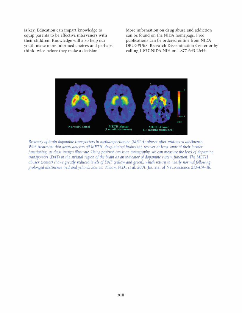
Recovery of brain dopamine transporters in methamphetamine (METH) abuser after protracted abstinence. With treatment that keeps abusers off METH, drug-altered brains can recover at least some of their former functioning, as these images illustrate. Using positron emission tomography, we can measure the level of dopamine transporters (DAT) in the striatal region of the brain as an indicator of dopamine system function. The METH abuser (center) shows greatly reduced levels of DAT (yellow and green), which return to nearly normal following prolonged abstinence (red and yellow). Source: Volkow, N.D., et al. 2001. Journal of Neuroscience 21:9414–18.
xiii

At a GlanceOverviewStudents examine images of human brains that illustrate that specific regions of the brain regulate specific functions. They extend that knowledge to learn that drugs of abuse activate a brain circuit known as the reward system. This same circuit is stimulated in response to basic survival needs, which produces feel ings of pleasure.
Major ConceptSpecific brain regions control specific brain functions.
ObjectivesBy the end of these activities, students will
understand that particular functions are localized to specific areas of the brain,appreciate that imaging techniques allow scientists to study activity in the brain, andrecognize that normal behaviors can activate the reward system in the brain and that drugs of abuse affect those same reward circuits.
Basic Science–Health ConnectionThe brain controls virtually everything humans experience, including move ment, sensing our environment, and regulating our involuntary body processes such as breathing, as well as controlling our emotions. Ongoing scientific research into the organization and function of the brain has led, and will con tinue to lead, to new treatments of diseases such as Parkinson’s disease, epilepsy, stroke, and mental illnesses (including depression and schizophrenia).
The brain is the organ of behavior. It is also the organ of our minds. Both overt behavior and consciousness are manifestations of the work of our brains. Other people can see an individual’s overt behaviors, whereas consciousness is appar ent only in our individual minds. The field of neuroscience studies how people control their behaviors, thoughts, and feelings, and how these actions some times get out of control.
The Brain: What’s Going On in There?
Source: NIDA. 1996. The Brain & the Actions of Cocaine, Opiates, and Marijuana. Slide Teaching Packet for Scientists.
L E S S O N 1Engage/Explore
19
The brain processes a huge amount of information in a remarkably efficient manner. Think about driving a car. It is something most of us do without much difficulty. But to do it properly, we must perform a remarkable number of tasks. First we have to make sure that our body is in working order: heart rate and breathing have to be properly regulated and body temperature held steady, and we certainly have to be sure we don’t fall asleep. Despite the complexity of these tasks, we carry them out with no conscious involvement on our part. Then, there are the things we are aware of. We have to see the road and hear the traf fic (or the radio), use information from our feet, legs, hands, and arms to know where the gas pedal and steering wheel are, and then generate the body move ments to control the direction and speed of the car. All of this often takes place while we are talking to someone else in the car, or even while talking on the phone (although this is not a good idea). The magnitude and speed of data processing needed to do this are stunning, but most of us consider driving to be an easy task.
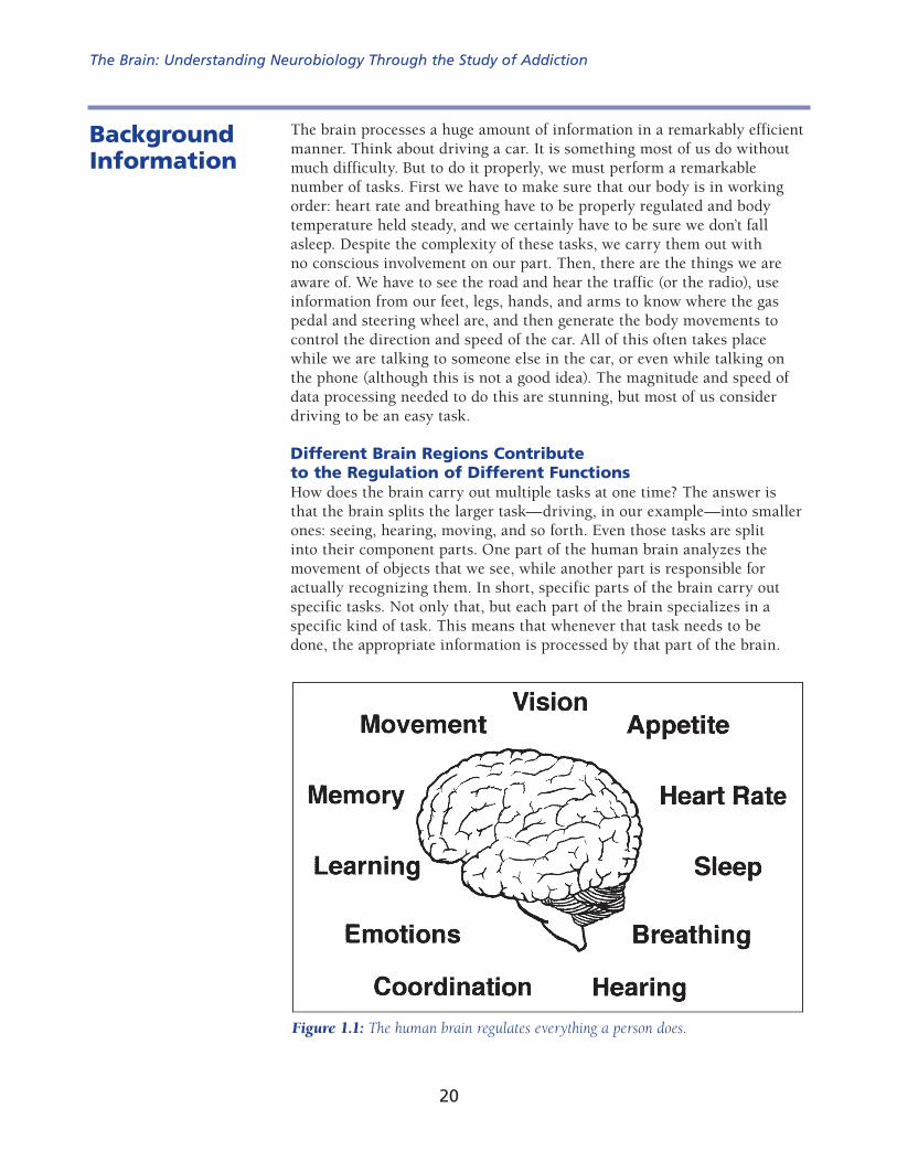
Different Brain Regions Contribute to the Regulation of Different FunctionsHow does the brain carry out multiple tasks at one time? The answer is that the brain splits the larger task—driving, in our example—into smaller ones: seeing, hearing, moving, and so forth. Even those tasks are split into their component parts. One part of the human brain analyzes the movement of objects that we see, while another part is responsible for actually recognizing them. In short, specific parts of the brain carry out specific tasks. Not only that, but each part of the brain specializes in a specific kind of task. This means that whenever that task needs to be done, the appropriate information is processed by that part of the brain.
Background Information
Figure 1.1: The human brain regulates everything a person does.
The Brain: Understanding Neurobiology Through the Study of Addiction
20

The flip side of this organizational scheme is that if a part of the brain is dam aged, then the job it used to undertake cannot be done. For example, damage to the occipital lobe at the back of the brain can cause blindness, but it has no effect on a person’s ability to hear or move. Because the job of seeing is highly compartmentalized, individuals who have lost one aspect of sight, such as the ability to see colors or to recognize faces, may still be able to do other visual tasks. Imagine being able to recognize someone by hearing his or her voice, but not being able to recognize his or her face when you see it.
The advantage of this localization of function is when larger jobs are parceled out throughout the brain, they all can be done at once. This “division of labor” adds great speed to our ability to perceive what is happening in the world around us, to analyze it, and then to generate appropriate responses. Dealing with information in this way is called parallel processing.1 (Superscript numbers refer to references listed by section on pages 153–156.) Computer scientists have used this concept in the development of computers.
The human brain consists of several large regions, each of which is responsible for some of the activities necessary for life. These include the brainstem, cere bellum, limbic system, diencephalon, and cerebral cortex.2,3
The brainstem is the part of the brain that connects the brain and spinal cord (Figure 1.2). This part of the brain is involved in coordinating many basic functions such as heart rate, breathing, eating, and sleeping.
Figure 1.2: This drawing of a brain cut in half illustrates some of the major regions of the brain. Source: National Institute on Drug Abuse (1997). Mind Over Matter: The Brain’s Response to Drugs, Teacher’s Guide.
Cerebral Cortex
Hypothalamus
Hippocampus
Spinal Cord
Midbrain
Cerebellum
Brainstem
Thalamus
Amygdala
Student Lesson 121

The cerebellum coordinates the brain’s instructions for skilled repetitive move ments and for maintaining balance and posture.
The limbic system, as discussed in the next section, is involved in regulating emotions, motivations, and movement. It includes the amygdala and hippocampus, which is important for memory formation.
The diencephalon contains the thalamus and hypothalamus. The thalamus is involved in sensory perception and regulating movement. The hypothal-amus is an important regulator of the pituitary gland, which directs the release of hormones throughout the body.
The cerebral cortex makes up the largest part of the brain mass and lies over and around most of the other brain structures. It is the part of the brain responsible for thinking, perceiving, and producing and understanding lan guage. The cortex can be divided into areas that are involved in vision, hearing, touch, movement, smell, and thinking and reasoning (Figure 1.3).
Drugs Act on the Reward System in the BrainJust as specific areas of the brain control seeing and hearing, specific brain areas also regulate emotions, motivations, and movement. These functions are carried out by a part of the brain called the limbic system. The limbic system influences how we respond to the world around us. Imagine a cool sunny day. You finish your work early and head to your favorite park for a leisurely walk with your dog. You are feeling so mellow that when the dog slobbers on your clean shirt, you merely scratch him behind the ears.
Figure 1.3: This drawing of a brain cut in half illustrates the lobes of the cerebral cortex and describes their main functions. Source: National Institute on Drug Abuse (1997). Mind Over Matter: The Brain’s Response to Drugs, Teacher’s Guide.
The Brain: Understanding Neurobiology Through the Study of Addiction
22

You might have a very different reaction on another day when you have to work late, traffic is backed up, and the dog runs away instead of coming to welcome you home. This time when the dog slobbers on you (after he finds his way home again), you shove him away and scold him.
The feelings you have in those two different situations are a result of your lim bic system at work. The limbic system uses memories, information about how your body is working, and current sensory input to generate your emotional responses to current situations.
The limbic system is involved in many of our emotions and motivations, partic ularly those related to survival, such as fear and anger. The system is also involved in pleasurable activities necessary for survival, such as eating and sex. If something is pleasurable, or rewarding, you want to do it again. Pleasurable activities engage the reward circuit (or system), so the brain notes that something important is happening that needs to be remembered and repeated.1,2 The reward system includes several interconnected structures—the ventral tegmental area (VTA), located at the top of the brain stem; the nucleus accumbens; and the prefrontal cortex (Figure 1.4). Neurons from the VTA relay messages to the nucleus accumbens and the prefrontal cortex. Information is also relayed back from the cortex to the nucleus accumbens and the VTA.
Most drugs of abuse activate these same VTA and nucleus accumbens neurons; that is why drugs produce pleasurable feelings to the drug user. And, because the feelings are pleasurable, the user wants to continue to experience the pleasure that he or she felt during previous drug use.
Figure 1.4: This drawing of a brain cut in half illustrates the brain areas and systems involved in the reward system, or pleasure circuit. Neurons in the ventral tegmental area (VTA) extend axons to the nucleus accumbens and part of the prefrontal cortex. Source: National Institute on Drug Abuse (1996). The Brain & the Actions of Cocaine, Opiates, and Marijuana. Slide Teaching Packet for Scientists.
Student Lesson 123

One of the reasons that drugs of abuse can exert such powerful control over our behavior is that they act directly on the more evolutionarily primitive brainstem and limbic structures, which can override the cortex in controlling our behavior.
Different drugs of abuse affect the neurons of the reward system in different ways. The activities in Lesson 3 in this module will elucidate the mechanisms by which drugs of abuse exert their effects.
Imaging the BrainScientists increasingly use newer technologies to learn more about how the brain works and how drugs of abuse change how the brain works. Historically, scientists could examine brains only after death, but new imaging procedures enable scientists to study the brain in living animals, including humans.
One of the most extensively used techniques to study brain activity and the effects of drugs on the brain is positron emission tomography (PET). PET mea sures the spatial distribution and movement of radioisotopes in tissues of living subjects. Because the patient is awake, the technique can be used to investigate the relationship between behavioral and physiological effects and changes in brain activity. PET scans can detect nanomolar concentrations of tracer mole cules and achieve spatial resolution of about 4 millimeters. In addition, comput ers can reconstruct images obtained from a PET scan in two or three dimensions.
PET requires the use of compounds labeled with positron-emitting iso topes.4,5 A cyclotron accelerates protons into the nucleus of nitrogen, carbon, oxygen, or fluorine to generate these isotopes. The additional proton
makes the isotope unstable. To become stable again, the proton must break down into a neutron and a positron. The unstable positron travels away from the site of generation and dissipates energy along the way. Eventually, the positron col lides with an electron, leading to the emission of two gamma rays at 180° from one another. The gamma rays reach a pair of detectors that record the event. Because the detectors respond only to simultaneous emis sions, scientists can precisely map the location where the gamma rays were generated. The labeled radioisotopes are very short-lived. The half-life (the time for half of the radioactive label to disintegrate) of the commonly used radioisotopes ranges from approximately two minutes to less than two hours, depending on the specific compound. Because a PET scan requires only small amounts (a few micrograms) of short-lived radioisotopes, pharmacological and radiological effects are negligible or even nonexistent.
Figure 1.5: When an unstable positron collides with an electron, the particles are annihilated and two gamma rays are emitted at 180° from each other. Detectors record gamma-ray emission to localize the site of positron annihilation.
The Brain: Understanding Neurobiology Through the Study of Addiction
24

PET scans can answer a variety of questions about brain function, including questions about the activity of neurons. Scientists use different radiolabeled compounds to investi gate different biological questions. For example, radiolabeled glucose can iden tify parts of the brain that become more active in response to a specific stimulus. Active neurons metabolize more glucose than inactive neurons. Active neurons will emit more positrons. This will show as red or yellow on PET scans compared with blue or purple in areas where the neurons are not highly active. PET also helps scientists investigate how drugs affect the brain by enabling them to
determine the distribution of a drug in the body,measure the local concentration of a drug at binding sites,estimate receptor occupancy or density,evaluate the effects of drugs on other neurotransmitter systems, andinvestigate the activity of enzymes that metabolize the drug.5
Although in the context of drug abuse, PET is currently used only as a research tool, it is a powerful diagnostic and monitoring tool for other diseases. For example, PET scans may be used to locate tumors in cancer patients, monitor the spread of cancer, and evaluate the effec tiveness of cancer treatment. PET scans are able to reveal the presence of tumors because of the rapid metabolism characteristic of cancerous cells. PET images reveal this increased glucose utilization by cells that have high meta bolic rates. PET is an accurate test for coronary heart disease because it can detect areas of diminished blood flow to the heart. Doctors also employ PET to reveal changes in the brain that occur with Alzheimer’s disease, Parkinson’s disease, or seizure disorders. PET is a valuable tool because it
is safe,replaces multiple testing procedures with a single exam,can detect diseases before they show up on other tests,can show the progress of disease, andreduces or eliminates the need for invasive procedures such as surgery.
Figure 1.6: Photograph of PET imaging equipment. Photo courtesy of UCLA School of Medicine.
Student Lesson 125

Different Neuroimaging Techniques Provide Different Information about the BrainPET scanning is a major neuroimaging technique used in drug abuse research. However, researchers also use other techniques when they are better for answering a specific question. Similar to PET, single photon emission computed tomography (SPECT), magnetic resonance imaging (MRI), and electroencephalography (EEG) are noninvasive procedures that can measure biological activity through the skull and reveal the living brain at work.4,6 Each technique has its own advantages, and each provides different information about brain structure and func tion. Scientists often use more than one technique when conducting their research studies.
Similar to PET, SPECT imaging uses radioactive tracers and a scanner to record data that a computer constructs into two- or three-dimensional images of active brain regions. Because the trac ers used in SPECT take longer to decay than those for PET, longer periods of time between tests are required for SPECT so a patient does not receive or accumulate too high a “load” of radioactivity. While PET is more versatile than SPECT and produces more detailed images with a higher degree of resolution, SPECT is much less expensive than PET and can address many of the same drug abuse research questions.
MRI uses magnetic fields and radio waves to produce high-quality two- or three-dimensional images of brain structures without inject ing radioactive tracers. In this procedure, a large cylindrical magnet creates a magnetic field around the research volunteer’s head, and radio waves are sent through the magnetic field. Sensors read the signals, and a computer uses the information to construct an image. Using MRI, scientists can image both surface
and deep brain struc tures with a high degree of anatomical detail, and they can detect minute changes in these structures over time. A modification of this technique, called functional MRI (fMRI), enables scientists to see images of blood flow in the brain as it occurs. fMRI provides supe rior image clarity along with the ability to assess blood flow and brain functions in just a few seconds. However, PET retains the advantage of being able to identify which brain receptors are being bound by neurotransmitters, abused drugs, and potential treatment compounds.
EEG uses electrodes placed on the scalp to detect and measure patterns of electrical activity in the brain. The greatest advantage of EEG is speed: it can record complex patterns of neural activity occurring within fractions of a second after a stimulus has been administered. The drawback to EEG is that it does not provide the spatial res olution of fMRI or PET. Researchers often combine EEG images of brain electrical activity with MRI scans to local ize brain activity more precisely.
Figure 1.7: MRI image of human brain. Photo courtesy of Penrad Imaging, Colorado Springs, CO.
The Brain: Understanding Neurobiology Through the Study of Addiction
26

L E S S O N 2Explore/Explain
OverviewStudents learn that the neuron is the functional unit of the brain. To learn how neurons convey information, students analyze a sequence of illustrations and watch an animation. They see that neurons communicate using electrical sig nals and chemical messengers called neurotransmitters that either stimulate or inhibit the activity of a responding neuron. Students then use the informa tion they have gained to deduce how one neuron influences the action of another.
Major ConceptNeurons convey information using electrical and chemical signals.
ObjectivesBy the end of these activities, the students will
understand the hierarchical organization of the brain, neuron, and synapse;understand the sequence of events involved in communication at the synapse; andunderstand that synaptic transmission involves neurotransmitters that may be either excitatory or inhibitory.
Basic Science–Health ConnectionCommunication between neurons is the foundation for brain function. Under standing how neurotransmission occurs is crucial to understanding how the brain processes and integrates information. Interruption of neural communi cation causes changes in cognitive processes and behavior.
Neurons, Brain Chemistry, and Neurotransmission
Source: NIDA. 1996. The Brain & the Actions of Cocaine, Opiates, and Marijuana. Slide Teaching Packet for Scientists.
At a Glance
41

The Brain Is Made Up of Nerve Cells and Glial CellsThe brain of an adult human weighs about 3 pounds and contains billions of cells. The two distinct classes of cells in the nervous system are neurons (nerve cells) and glia (glial cells).
The basic signaling unit of the nervous system is the neuron. The brain contains billions of neurons; the best estimates are that the adult human brain contains 1011 neurons. The interactions between neurons enable people to think, move, main tain homeostasis, and feel emotions. A neuron is a specialized cell that can pro duce different actions because of its precise connections with other neurons, sensory receptors, and muscle cells. A typical neuron has four morphologically defined regions: the cell body, dendrites, axons, and presynaptic, or axon, terminals.1,2,3
Background Information
Figure 2.1: The neuron, or nerve cell, is the functional unit of the nervous system. The neuron has processes called dendrites that receive signals and an axon that transmits signals to another neuron.
The cell body, also called the soma, is the metabolic center of the neuron. The nucleus is located in the cell body, and most of the cell’s protein synthesis occurs in the cell body.
A neuron usually has multiple processes, or fibers, called dendrites that extend from the cell body. These processes usually branch out somewhat like tree branches and serve as the main apparatus for receiving input into the neuron from other nerve cells.
The cell body also gives rise to the axon. Axons can be very long processes; in some cases, they may be up to 1 meter long. The axon is the part of the neuron that is specialized to carry messages away from the cell body and to relay messages to other cells. Some large axons are surrounded by a fatty insulating material called myelin, which enables the electrical signals to travel down the axon at higher speeds.
Near its end, the axon divides into many fine branches that have specialized swellings called axon, or presynaptic, terminals. These presynaptic terminals end in close proximity to the dendrites of another neuron. The dendrite of one neuron receives the message sent from the presynaptic terminal of another neuron.
The Brain: Understanding Neurobiology Through the Study of Addiction
42

The site where a presynaptic terminal ends in close proximity to a receiving dendrite is called the synapse. The cell that sends out information is called the presynaptic neuron, and the cell that receives the information is called the postsynaptic neuron. It is important to note that the synapse is not a physical con nection between the two neurons; there is no cytoplasmic continuity between the two neurons. The intercellular space between the presynaptic and postsy naptic neurons is called the synaptic space or synaptic cleft. An average neu ron forms approximately 1,000 synapses with other neurons. It has been estimated that there are more synapses in the human brain than there are stars in our galaxy. Furthermore, synaptic connections are not static. Neurons form new synapses or strengthen synaptic connections in response to life experi ences. This dynamic change in neuronal connections is the basis of learning.
Figure 2.2: Neurons transmit information to other neurons. Information passes from the axon of the presynaptic neuron to the dendrites of the postsynaptic neuron.
Figure 2.3: The synapse is the site where chemical signals pass between neurons. Neurotransmit ters are released from the presynaptic neuron terminals into the extracellular space, the synaptic cleft or synaptic space. The released neurotransmitter molecules can then bind to specific recep tors on the postsynaptic neuron to elicit a response. Excess neurotransmitter can then be reabsorbed into the presynaptic neuron through the action of specific reuptake molecules called transporters. This process ensures that the signal is terminated when appropriate.
Student Lesson 243

The brain contains another class of cells called glia. There are as many as 10 to 50 times more glial cells than neurons in the central nervous system. Glial cells are categorized as microglia or macroglia. Microglia are phago cytic cells that are mobilized after injury, infection, or disease. They are derived from macrophages and are unrelated to other cell types in the ner vous system. The three types of macroglia are oligodendrocytes, astrocytes, and Schwann cells. The oligodendrocytes and Schwann cells form the myelin sheaths that insulate axons and enhance conduction of electrical signals along the axons.
Scientists know less about the functions of glial cells than they do about the functions of neurons. Glial cells fulfill a variety of functions including as
support elements in the nervous system, providing structure and separating and insulating groups of neurons;oligodendrocytes in the central nervous system and Schwann cells in the peripheral nervous system, which form myelin, the sheath that wraps around cer tain axons;scavengers that remove debris after injury or neuronal death;helpers in regulating the potassium ion (K+) concentration in the extracel lular space and taking up and removing chemical neurotrans-mitters from the extracellular space after synaptic transmission;guides for the migration of neurons and for the outgrowth of axons during development; andinducers of the formation of impermeable tight junctions in endo- thelial cells that line the capillaries and venules of the brain to form the blood-brain barrier.3
The Blood-Brain BarrierThe blood-brain barrier protects the neurons and glial cells in the brain from substances that could harm them. Endothelial cells that form the capillaries and venules make this barrier, forming impermeable tight junc tions. Astrocytes surround the endothelial cells and induce them to form these junctions. Unlike blood vessels in other parts of the body that are relatively leaky to a variety of molecules, the blood-brain barrier keeps many substances, including toxins, away from the neurons and glia.
Most drugs do not get into the brain. Only drugs that are fat soluble can penetrate the blood-brain barrier. These include drugs of abuse as well as drugs that treat mental and neurological illness.
The blood-brain barrier is important for maintaining the environment of neurons in the brain, but it also presents challenges for scientists who are investigating new treatments for brain disorders. If a medication cannot get into the brain, it cannot be effective. Researchers attempt to circumvent the problems in different ways. Some techniques alter the structure of the drug to make it more lipid soluble. Other strategies attach potential therapeutic agents to molecules that pass through the blood-brain bar rier, while others attempt to open the blood-brain barrier.4
The Brain: Understanding Neurobiology Through the Study of Addiction
44

Neurons Use Electrical and Chemical Signals to Transmit Information*The billions of neurons that make up the brain coordinate thought, behavior, homeostasis, and more. How do all these neurons pass and receive information?
Neurons convey information by transmitting messages to other neurons or other types of cells, such as muscles. The following discussion focuses on how one neuron communicates with another neuron. Neurons employ electrical signals to relay information from one part of the neuron to another. The neu ron converts the electrical signal to a chemical signal in order to pass the information to another neuron. The target neuron then converts the message back to an electrical impulse to continue the process.
Within a single neuron, information is conducted via electrical signaling. When a neuron is stimulated, an electrical impulse, called an action potential, moves along the neuron axon.5 Action potentials enable signals to travel very rapidly along the neuron fiber. Action potentials last less than 2 milliseconds (1 millisecond = 0.001 second), and the fastest action potentials can travel the length of a football field in 1 second. Action potentials result from the flow of ions across the neuronal cell membrane. Neurons, like all cells, maintain a balance of ions inside the cell that differs from the balance outside the cell. This uneven distribution of ions creates an electrical poten tial across the cell membrane. This is called the resting membrane potential. In humans, the resting membrane potential ranges from –40 millivolts (mV) to –80 mV, with –65 mV as an average resting membrane potential. The resting membrane potential is, by convention, assigned a negative number because the inside of the neuron is more negatively charged than the outside of the neuron. This negative charge results from the unequal distribu tion of sodium ions (Na+), potassium ions (K+), chloride ions (Cl–), and other organic ions. The resting membrane potential is maintained by an energy-dependent Na+-K+ pump that keeps Na+ levels low inside the neuron and K+ levels high inside the neuron. In addition, the neuronal membrane is more permeable to K+ than it is to Na+, so K+ tends to leak out of the cell more readily than Na+ diffuses into the cell.
A stimulus occurring at the cell body starts an electrical change that travels like a wave over the length of the neuron. This electrical change, the action potential, results from a change in the permeability of the neuronal membrane. Sodium ions rush into the neuron, and the inside of the cell becomes more positive. The Na+-K+ pump then restores the balance of sodium and potassium to resting levels. However, the influx of Na+ ions in one area of the neuron fiber starts a similar change in the adjoining segment, and the impulse moves from the cell body toward the axon terminal. Action potentials are an all-or-none phenomenon. Regardless of the stimuli, the amplitude and duration of an action potential are the same. The action poten tial either occurs or it doesn’t. The response of the neuron to an action poten tial depends on how many action potentials it transmits and their frequency.
* “Electrical signals” are not actually electric because ions travel down the axon, not electrons. For the sake of simplicity, though, we use “electrical.”
Student Lesson 245

Electrical signals carry information within a single neuron. Communication between neurons (with a few exceptions in mammals) is a chemical process. When the neuron is stimulated, the electrical signal (action potential) travels down the axon to the axon terminals. When the electrical signal reaches the end of the axon, it triggers a series of chemical changes in the axon terminal. Cal cium ions (Ca++) flow into the axon terminal, which then initiates the release of neurotransmitters. A neurotransmitter is a molecule that is released from a neuron to relay information to another cell. Neurotransmitter molecules are stored in membranous sacs called vesicles in the axon terminal. Each vesicle contains thousands of molecules of a given neuro transmitter. For neurons to release their neurotransmitter, the vesicles fuse with the neuronal membrane and then release their contents, the neurotrans mitter, via exocytosis. The neurotransmitter molecules are released into the synaptic space and diffuse across the synaptic space to the postsynaptic neu ron. A neurotransmitter molecule can then bind to a special receptor on the membrane of the postsynaptic neuron. Receptors are membrane proteins that are able to bind a specific chemical substance,
Figure 2.4: (a) Recording of an action potential in an axon following stimulation due to changes in the permeability of the cell membrane to sodium and potassium ions. (b) The cell membrane of a resting neuron is more negative on the inside of the cell than on the outside. When the neuron is stimulated, the permeability of the membrane changes, allowing Na+ to rush into the cell. This causes the inside of the cell to become more positive. This local change starts a similar change in the adjoining segment of the neuron’s membrane. In this manner, the electrical impulse moves along the neuron. From: Molec ular Cell Biology, by Lodish et al. 1986, 1990 by Scientific American Books, Inc. Used with permission by W.H. Freeman and Company.
The Brain: Understanding Neurobiology Through the Study of Addiction
46

such as a neurotransmitter. For example, the dopamine receptor binds the neurotransmitter dopamine but does not bind other neurotransmitters such as serotonin. The interaction of a receptor and neurotransmitter can be thought of as a lock-and-key for regulat ing neuronal function. Just as a key fits only a specific lock, a neurotransmit ter only binds with high affinity to a specific receptor. The chemical binding of neurotransmitter and receptor initiates changes in the postsynaptic neuron that may facilitate or inhibit an action potential in the postsynaptic neuron. If it does trigger an action potential, the communication process continues.
Figure 2.5: Schematic diagram of a synapse. In response to an electrical impulse, neuro transmitter molecules released from the presynaptic axon terminal bind to the specific receptors for that neurotransmitter on the postsynaptic neuron. After binding to the recep tor, the neurotransmitter molecules either may be taken back up into the presynaptic neu ron through the transporter molecules for repackaging into vesicles or may be degraded by enzymes present in the synaptic space.
Figure 2.6: Like a lock that will open only if the right key is used, a receptor will bind only a molecule that has the right chemical shape. Molecules that do not have the right “fit” will not bind to the receptor and will not cause a response.
After a neurotransmitter molecule binds to its receptor on the postsynaptic neuron, it comes off (is released from) the receptor and diffuses back into the synaptic space. The released neurotransmitter, as well as any neurotransmitter that did not bind to a receptor, is either degraded by enzymes in the synaptic cleft or taken back up into the presynaptic axon terminal by active transport through a transporter or reuptake
Student Lesson 247

pump. Once the neurotransmitter is back inside the axon terminal, it is either destroyed or repackaged into new vesicles that may be released the next time an electrical impulse reaches the axon terminal. Different neurotransmit ters are inactivated in different ways.
Neurotransmitters Can Be Excitatory or InhibitoryDifferent neurotransmitters fulfill different functions in the brain. Some neu rotransmitters act to stimulate the firing of a postsynaptic neuron. Neuro transmitters that act this way are called excitatory neurotransmitters because they lead to changes that generate an action potential in the responding neu ron.1,6 Other neurotransmitters, called inhibitory neurotransmitters, tend to block the changes that cause an action potential to be generated in the responding cell. Table 2.1 lists some of the “classical neurotransmitters” used in the body and their major functions. In addition to the so-called classical neurotransmitters, there are many other peptide transmitters, sometimes called neuromodulators. They are similar to classical neurotransmitters in the way they are stored (in vesicles) and released, but they differ in how they are inactivated. Most neurons contain multiple transmitters, often a classical one (such as dopamine) and one or more peptides (such as neurotensin or endorphins).
The postsynaptic neuron often receives and integrates both excitatory and inhibitory mes sages. The response of the postsynaptic cell depends on which message is stronger. Keep in mind that a single neurotransmitter molecule cannot cause an action potential in the responding neuron. An action potential occurs when many neurotransmitter molecules bind to and activate their receptors. Each interaction contributes to the membrane permeability changes that generate the resultant action potential.
Table 2.1: Major Neurotransitters in the Body1,6,7
Neurotransmitter Role in the bodyAcetylcholine Used by spinal cord motor neurons to cause muscle contraction
and by many neurons in the brain to regulate memory. In most instances, acetylcholine is excitatory.
Dopamine Produces feelings of pleasure when released by the brain reward system. Dopamine has multiple functions depending on where in the brain it acts. It is usually inhibitory.
GABA (gamma-aminobutyric acid) The major inhibitory neurotransmitter in the brain. It is important in producing sleep, reducing anxiety, and forming memories.
Glutamate The most common excitatory neurotransmitter in the brain. It is important in learning and memory.
Glycine Used mainly by neurons in the spinal cord. It probably always acts as an inhibitory neurotransmitter.
Norepinephrine Acts as a neurotransmitter and a hormone. In the peripheral ner vous system, it is part of the fight-or-flight response. In the brain, it acts as a neurotransmitter regulating blood pressure and calmness. Norepinephrine is usually excitatory, but it is inhibitory in a few brain areas.
Serotonin Involved in many functions including mood, appetite, and sensory perception. In the spinal cord, serotonin is inhibitory in pain pathways.
The Brain: Understanding Neurobiology Through the Study of Addiction
48
OverviewStudents build upon their understanding of neurotransmission by learning how different drugs of abuse disrupt communication between neurons. Students then conduct an activity investigating the effect of caffeine on their heart rate. Finally, students analyze data on how the way a drug is taken into the body influences its effect.
Major ConceptDrugs affect the biology and chemistry of the brain.
ObjectivesBy the end of these activities, the students will
understand that certain drugs interfere selectively with neurotransmission andrealize that the effect of a drug is dependent upon dosage and route of administration.
Basic Science–Health ConnectionDrugs of abuse are valuable tools for investigations of brain function because they can mimic or block actions of neurotransmitters, and thus exert effects on homeostasis and behavior.
L E S S O N 3Explain/Elaborate
At a Glance
Drugs Change the Way Neurons Communicate
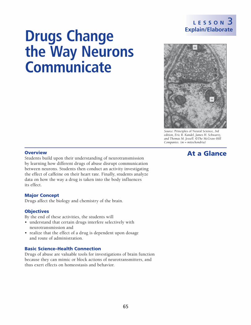
Source: Principles of Neural Science, 3rd edition, Eric R. Kandel, James H. Schwartz, and Thomas M. Jessell. ©The McGraw-Hill Companies. (m = mitochondria)
65
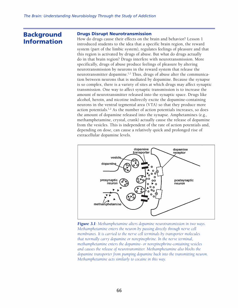
Drugs Disrupt NeurotransmissionHow do drugs cause their effects on the brain and behavior? Lesson 1 intro duced students to the idea that a specific brain region, the reward system (part of the limbic system), regulates feelings of pleasure and that this region is acti vated by drugs of abuse. But what do drugs actually do in that brain region? Drugs interfere with neurotransmission. More specifically, drugs of abuse pro duce feelings of pleasure by altering neurotransmission by neurons in the reward system that release the neurotransmitter dopamine.1,2 Thus, drugs of abuse alter the communica-tion between neurons that is mediated by dopamine. Because the synapse is so complex, there is a variety of sites at which drugs may affect synaptic transmission. One way to affect synaptic transmission is to increase the amount of neurotransmitter released into the synaptic space. Drugs like alcohol, heroin, and nicotine indirectly excite the dopamine-containing neu rons in the ventral tegmental area (VTA) so that they produce more action potentials.1,2 As the number of action potentials increases, so does the amount of dopamine released into the synapse. Amphetamines (e.g., methampheta mine, crystal, crank) actually cause the release of dopamine from the vesicles. This is independent of the rate of action potentials and, depending on dose, can cause a relatively quick and prolonged rise of extracellular dopamine levels.
Background Information
Figure 3.1: Methamphetamine alters dopamine neurotransmission in two ways. Methamphetamine enters the neuron by passing directly through nerve cell membranes. It is carried to the nerve cell terminals by transporter molecules that normally carry dopamine or norepinephrine. In the nerve terminal, methamphetamine enters the dopamine- or norepinephrine-containing vesicles and causes the release of neurotransmitter. Methamphetamine also blocks the dopamine transporter from pumping dopamine back into the transmitting neuron. Methamphetamine acts similarly to cocaine in this way.
The Brain: Understanding Neurobiology Through the Study of Addiction
66
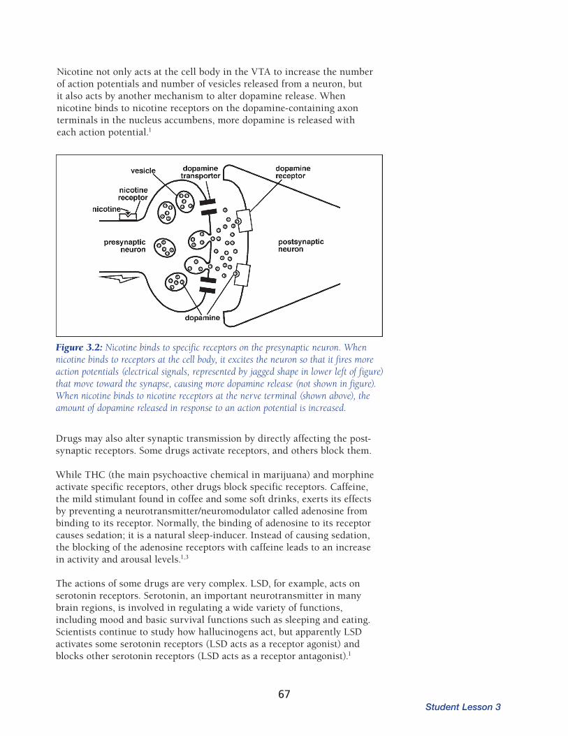
Nicotine not only acts at the cell body in the VTA to increase the number of action potentials and number of vesicles released from a neuron, but it also acts by another mechanism to alter dopamine release. When nicotine binds to nico tine receptors on the dopamine-containing axon terminals in the nucleus accumbens, more dopamine is released with each action potential.1
Figure 3.2: Nicotine binds to specific receptors on the presynaptic neuron. When nicotine binds to receptors at the cell body, it excites the neuron so that it fires more action potentials (electrical sig nals, represented by jagged shape in lower left of figure) that move toward the synapse, causing more dopamine release (not shown in figure). When nicotine binds to nicotine receptors at the nerve terminal (shown above), the amount of dopamine released in response to an action potential is increased.
Drugs may also alter synaptic transmission by directly affecting the post- synap tic receptors. Some drugs activate receptors, and others block them.
While THC (the main psychoactive chemical in marijuana) and morphine acti vate specific receptors, other drugs block specific receptors. Caffeine, the mild stimulant found in coffee and some soft drinks, exerts its effects by pre venting a neurotransmitter/neuromodulator called adenosine from binding to its receptor. Normally, the binding of adenosine to its receptor causes sedation; it is a natural sleep-inducer. Instead of causing sedation, the blocking of the adenosine receptors with caffeine leads to an increase in activity and arousal levels.1,3
The actions of some drugs are very complex. LSD, for example, acts on sero tonin receptors. Serotonin, an important neurotransmitter in many brain regions, is involved in regulating a wide variety of functions, including mood and basic survival functions such as sleeping and eating. Scientists continue to study how hallucinogens act, but apparently LSD activates some serotonin receptors (LSD acts as a receptor agonist) and blocks other serotonin receptors (LSD acts as a receptor antagonist).1
Student Lesson 367
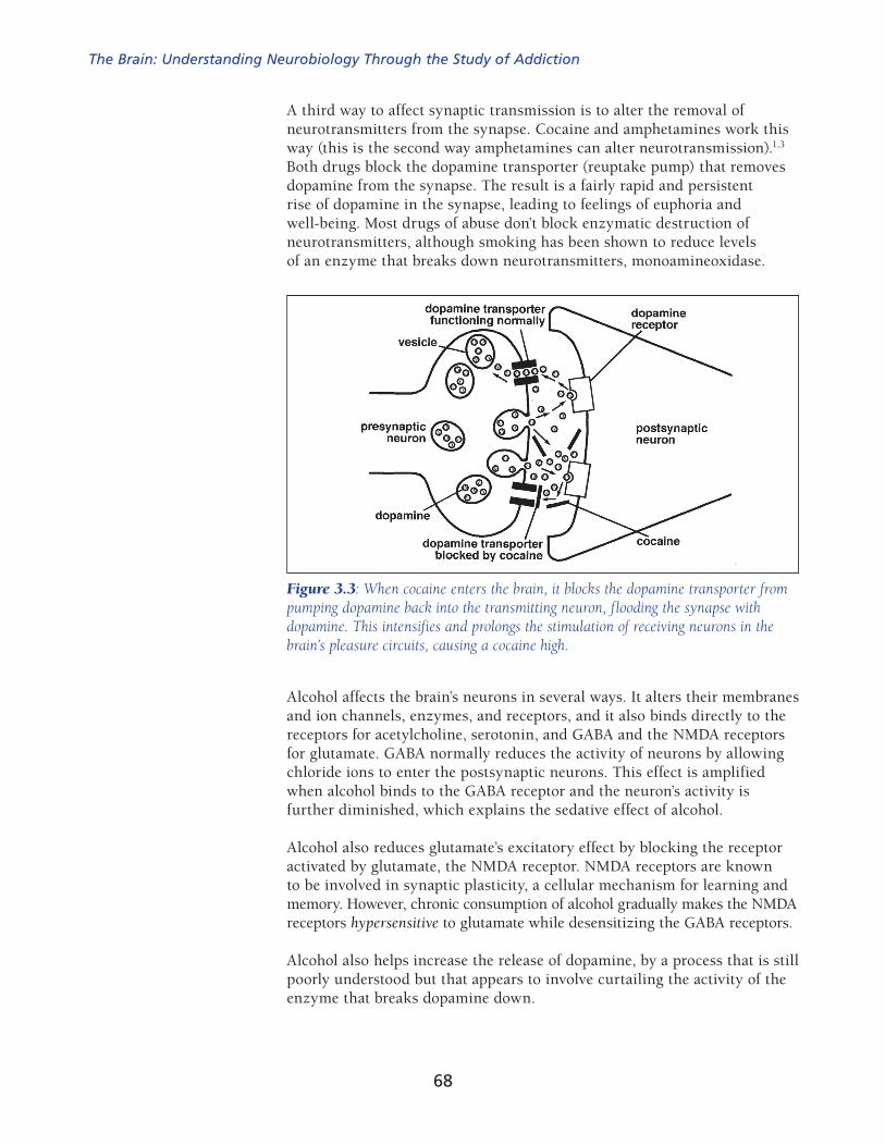
A third way to affect synaptic transmission is to alter the removal of neuro transmitters from the synapse. Cocaine and amphetamines work this way (this is the second way amphetamines can alter neurotransmission).1,3 Both drugs block the dopamine transporter (reuptake pump) that removes dopamine from the synapse. The result is a fairly rapid and persistent rise of dopamine in the synapse, leading to feelings of euphoria and well-being. Most drugs of abuse don’t block enzymatic destruction of neurotransmitters, although smoking has been shown to reduce levels of an enzyme that breaks down neurotransmitters, monoamineoxidase.
Figure 3.3: When cocaine enters the brain, it blocks the dopamine transporter from pumping dopamine back into the transmitting neuron, flooding the synapse with dopamine. This intensi fies and prolongs the stimulation of receiving neurons in the brain’s pleasure circuits, causing a cocaine high.
Alcohol affects the brain’s neurons in several ways. It alters their membranes and ion channels, enzymes, and receptors, and it also binds directly to the receptors for acetylcholine, serotonin, and GABA and the NMDA receptors for glutamate. GABA normally reduces the activity of neurons by allowing chloride ions to enter the postsynaptic neurons. This effect is amplified when alcohol binds to the GABA receptor and the neuron’s activity is further diminished, which explains the sedative effect of alcohol.
Alcohol also reduces glutamate’s excitatory effect by blocking the receptor activated by glutamate, the NMDA receptor. NMDA receptors are known to be involved in synaptic plasticity, a cellular mechanism for learning and memory. However, chronic consumption of alcohol gradually makes the NMDA receptors hypersensitive to glutamate while desensitizing the GABA receptors.
Alcohol also helps increase the release of dopamine, by a process that is still poorly understood but that appears to involve curtailing the activity of the enzyme that breaks dopamine down.
The Brain: Understanding Neurobiology Through the Study of Addiction
68

Drugs Mimic Natural Body ChemicalsThe ability of drugs to interrupt normal synaptic transmission may seem odd. After all, if receptors have such great specificity for a single type of binding partner, how can drugs disrupt the process? The answer lies in the similarity in conformation, or structure, of the drugs to natural body chemicals. For example, the receptors in the brain that bind mor phine and other opioids recognize natural opioid peptides called endorphins and enkephalins that are made by our brains and used as neurotransmitters.4 It is an evolutionary coincidence that these receptors recognize a plant-derived chemical (drug) as well. This coincidence is a double-edged sword. Opi oid compounds that come from plants are both the most potent analgesics (pain relievers) available and some of the most potent addictive drugs as well. Morphine continues to be one of the most effective drugs to relieve the pain associated with many chronic diseases. When abused, opioids are often taken at higher-than-prescribed doses or in ways other than as prescribed (for example, injected vs. orally), which, by stimulating the dopamine cells in the VTA, can cause profound feelings of pleasure (euphoria). Tetrahydocannabinol (THC), the active ingredient in marijuana, binds to specific receptors in the brain called cannabinoid receptors, which were discovered because scientists were trying to understand how marijuana works. Subsequently, natural (endogenous) transmitters that bind these receptors were identified—one of which is called anandamide. The cannabinoid system is distributed widely in the brain and the body and is thought to play a role in a wide variety of physiological activities, including memory, appetite, pain perception, and immune regulation. The discovery of this system may enable scientists to develop medications (without the abuse and other health liabilities of marijuana) for a variety of diseases, including obesity, schizophrenia, multiple sclerosis, and addiction.
Drugs of abuse share a common action: they act on the brain’s reward system. Within that system, they all (except perhaps for LSD) share the ability to increase the levels of dopamine in the nucleus accumbens. This almost certainly accounts for the rewarding (pleasurable) effects of abused drugs.
The effects of drugs are not limited to the reward pathway in the brain. Drugs can act in various regions of the brain to exert their effects, but their ability to alter dopamine neurotransmission in the ventral tegmental area (VTA) and the nucleus accumbens is the initial and one of the most important factors driving continued drug use.
Many factors determine how a drug affects an individual. Some of these are biological. For example, genetics can affect a person’s sensitivity to a drug or how quickly the drug is metabolized and cleared from the body. But environmental factors can also be important—stress or trauma can alter a person’s experience with drugs. Two factors that are especially important are the dose of the drug and the route of administration, which affects how fast it reaches the brain.
The Dose Changes the Drug’s EffectsFor a drug to work, it must be taken into the body, absorbed in the bloodstream, and delivered to the brain. Drugs can be taken in a range of doses—from low, having no detectable effect, to moderate, producing the drug’s desired effect, to large and unpleasant, or even toxic (Figure 3.4). Not everyone will respond the same way to a given drug dose—many factors can influence this, including those mentioned above, as well as age,
Student Lesson 369

gender, and the person’s history of using that drug or other related drugs. However, most drugs, when taken at high doses, produce effects that are both undesirable and potentially harmful to health (overdose).
Figure 3.4: Effects of a drug depend on the dose.
Figure 3.5: Drugs enter the brain by different routes.
Routes of Administration Ingestion Inhalation Injection Snorting/Snuffing Through the skin
Increasing dose of drug
Abovenormal
100%P
erce
nt o
f Bas
elin
e
Belownormal
Drugs Enter the Brain in Different WaysIn addition to dose, the manner in which a drug is taken can profoundly alter the response to the drug. A drug that is inhaled (smoked) reaches the brain very quickly. The inhaled drugs go directly from the lungs into the left side of the heart, where they enter the arte rial circulation that carries them to the brain. Marijuana and nicotine are examples of drugs that are commonly taken into the body by inhalation (smoking). The intensity of the effect of inhaled drugs may be slightly less than that for injected drugs because less of the drug gets into the brain; some of the drug will be exhaled with the rest of the components of the smoke. A drug that is injected intravenously also travels quickly to the brain, where it can exert its effects. The rapid passage of injected heroin, for exam ple, brings a high risk of overdose. In some cases, the heroin can reach lethal levels faster than medical help can be obtained to reverse the overdose. A third route of drug administration is by snorting or snuffing. A drug that is snorted or snuffed is taken in through the nose, where it is absorbed through the
The Brain: Understanding Neurobiology Through the Study of Addiction
70
mucous membranes lining the nasal passages. Television and movies often depict cocaine being snorted. The effects of drugs taken by this method will be less intense than by injection or inhalation because it takes longer for the drug to get into the brain.
Another route of administration is by oral ingestion. Most people are famil iar with taking a medicine, either as a solid or a liquid, by mouth. People can also take drugs of abuse this way. Drugs commonly taken orally include stim ulants and depressants. Drugs taken orally enter the bloodstream more slowly than by any of the other routes. The drugs that are swallowed reach the stomach and intestine, where they are absorbed into the bloodstream. Not only do they take longer to act, but the body begins to metabolize them before they can act on the brain. Enzymes in the stomach, intestines, and liver begin breaking down the drugs so they can be cleared from the body.
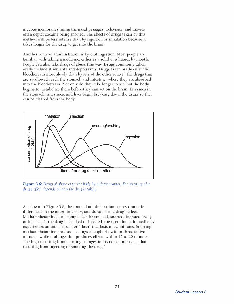
Figure 3.6: Drugs of abuse enter the body by different routes. The intensity of a drug’s effect depends on how the drug is taken.
As shown in Figure 3.6, the route of administration causes dramatic differences in the onset, intensity, and duration of a drug’s effect. Methamphetamine, for example, can be smoked, snorted, ingested orally, or injected. If the drug is smoked or injected, the user almost immediately experiences an intense rush or “flash” that lasts a few minutes. Snorting methamphetamine produces feelings of euphoria within three to five minutes, while oral ingestion produces effects within 15 to 20 minutes. The high resulting from snorting or ingestion is not as intense as that resulting from injecting or smoking the drug.5
Student Lesson 371

Web-Based ActivitiesActivity Web Component?
1 Yes
2 No
3 Yes
Photocopies
For the class For each student
1 transparency of Master 3.1, Cocaine Alters Neurotransmission
1 transparency of Master 3.2, Methamphetamine and Nicotine Disrupt Neurotransmission
1 transparency of Master 3.3, How Does Alcohol Affect Neurotransmission?
1 transparency of Master 3.7, What Should the Doctor Do?
1 copy of Master 3.4, Parent Letter
1 copy of Master 3.5, Caffeine: How Does Your Heart Respond?
1 copy of Master 3.6, How Do Drugs Get Into the Brain?
Materials Activity Materials
Activity 1 overhead projectorcomputers
Activity 2soft drinks, caffeinated and caffeine-free (see Preparation, below)1 watch or classroom clock with a second hand
Activity 3 computers
PreparationArrange for students to have access to the Internet for Activities 1 and 3, if possible.
At least one week before conducting Activity 2, send a copy of Master 3.4, Par ent Letter, home with each student to inform parents of the activity and get per mission for the students to consume a caffeinated or a caffeine-free soft drink during science class. You can also use the letter to ask each student to bring in his or her own can of the designated soft drink. Students who don’t drink soda can drink water as another control.
Decide on a brand of soft drink that is available with and without caffeine to use in the activity. Students should drink the same brand of soft drink because each brand contains a different amount of caffeine. If students drank different brands or flavors, the results would be difficult to interpret because each stu dent who drank a caffeinated soft drink would ingest a
In Advance
The Brain: Understanding Neurobiology Through the Study of Addiction
72

Content Standard A: Formulate and revise sci entific explanations and models using logic and evidence.Content Standard C: Cell functions are regulated.Content Standard C: Organisms have behav ioral responses to inter nal changes and to external stimuli.
ProcedureActivity 1: Drugs Alter Neurotransmission
1. Review neurotransmission with the students. It may be help ful to have the class watch the online animation of neuro transmission to refresh their memories. Have students refer to the summary of neurotransmission that they completed on Master 2.5.
After going to the supplement’s Web site, click on Lesson 2—Neurons, Brain Chem istry, and Neurotransmission.
2. Create a chart with the following headings on the board:
Change in neurotransmissionEffect on neurotransmitter
release or availability
3. Ask students if they think there are ways that neurotransmission could be altered. As students propose ideas, fill in the chart on the board. Probe for ideas by asking questions such as
What would happen if certain components in the process increased or decreased in amount?How would that change affect the response in the responding neuron?
Students may suggest a variety of ways in which neurotransmission can be altered. For example, maybe less neurotransmitter gets released, which would result in reduced (fewer) firings in the responding (postsynaptic) neuron. The postsynaptic neuron might have either more or fewer recep tors; changing the number of
different dose. You will need approximately half of the students to drink a caffeinated soft drink and half the students to drink a caffeine-free soft drink. Students who do not get parental permission can participate by drinking water, thereby providing a comparison to the control group. You may obtain the necessary soft drinks through one of the following ways:
purchase all the soft drinks yourself through your school budget,ask for parent or business donations to cover the cost, orrequest that each student bring in one can of soft drink, labeled with his or her name, for his or her consumption only. (If you use this approach, you will need to specify which drink each student brings to class.)
Before the day of Activity 2, have students practice taking a resting heart rate so they are used to finding their pulse, counting the beats for 15 seconds, and mul tiplying that number by four to get a resting heart rate for one minute (see Activity 2).
Student Lesson 373

receptors would cause an increased or decreased chance of postsynaptic neuron firing. The following chart out lines potential changes and their responses. Omit the third column on the chart at this time; you will complete that part in Step 4.
Change in neurotransmission Effect on neurotransmitter
release or availabilityDrug that acts this way
increase the number of impulses increased neurotransmitter release nicotine, alcohol,* opioids,* marijuana (THC)*
release neurotransmitter from vesicles with or without impulses
increased neurotransmitter release amphetaminesmethamphetamine
release more neurotransmitter in response to an impulse
increased neurotransmitter release nicotine
block reuptake more neurotransmitter present in synaptic cleft
cocaine, amphetamine
produce less neurotransmitter less neurotransmitter in synaptic cleft
no drug example
prevent vesicles from releasing neurotransmitter
less neurotransmitter released no drug example
block receptor with another molecule, or neurotransmitter cannot bind to its receptor on postsynaptic neuron
no change in amount of neurotransmitter released
LSD, caffeine
* These drugs cause an increase in dopamine release. However, both alcohol and opioids act indirectly. See Steps 10 and 11 on pages 76–77 for a more complete explanation of their actions.
4. When you have the first two columns completed on the chart, inform stu dents that certain drugs may cause the changes in the neurons that they have suggested. Write the name of the drug next to the change as indi cated in the third column on the chart.
Students will begin to see that drugs of abuse interfere with and disrupt the process of neurotransmission. When neurons do not communicate nor mally, the brain does not function normally, either.
5. Display a transparency of Master 3.1, Cocaine Alters Neurotransmission, showing cocaine’s effect on dopamine neurotransmission. Point out that cocaine blocks the dopamine transporters. Ask the following questions:
How does this blocking action of cocaine affect dopamine levels?What is the effect on the responding postsynaptic neuron?
Cocaine blocks the dopamine reuptake pumps (also called dopamine trans porters). Students should recall that transporters, or reuptake pumps, carry neurotransmitter, dopamine in this case, back into the presynaptic neuron, where it is repackaged into new vesicles. If the reuptake pumps cannot function, more dopamine will be present in
The Brain: Understanding Neurobiology Through the Study of Addiction
74
the synaptic space, where it can cause a greater stimulation of the postsynaptic neuron.
6. After the students understand how blocking the dopamine transporters alters neurotransmission, show the animation on the Web of cocaine’s effect on neurotransmission to the class, if possible.
To view the animation, go to the supplement’s Web site. Select Lesson 3—Drugs Change the Way Neurons Communicate.
7. Discuss the actions of another type of drug, methamphetamine, with the class. Display a transparency of Master 3.2, Methamphetamine and Nico tine Disrupt Neurotransmission (top half only). Explain that methamphet amine can act similarly to cocaine in blocking dopamine transporters (reuptake pumps). Methamphetamine also acts in another way to alter neurotransmission. Methamphetamine passes directly through the neu ron cell membrane and is carried to the axon terminals. In the terminals, methamphetamine enters the vesicles that contain dopamine. This then triggers the vesicles to be released, even without an electrical signal (action potential) to cause vesicle release. Ask students how this affects the postsynaptic neuron.
Methamphetamine acts in two ways to change dopamine neurotransmis sion. Both actions lead to an increase in the amount of dopamine in the synaptic cleft. When more dopamine is present in the synaptic cleft, it is more likely to bind to the dopamine receptors on the postsynaptic neuron.
8. Continue to assess the students’ understanding of how drugs can alter neurotransmission by asking them to consider how nicotine interferes with dopamine neurotransmission in the brain. Display a transparency of Master 3.2 (bottom half). Explain that nicotine binds to receptors on the transmitting (presynaptic) neuron and causes the neuron to release more neurotransmitter each time an electrical impulse (action potential) occurs. How does this affect the activity of the postsynaptic (receiving) neuron?
Nicotine binds to nicotine receptors on the presynaptic neuron. The binding of nicotine to its receptor stimulates the generation of action potentials in the neuron that cause dopamine to be released from the neuron. The released dopamine can then bind to its receptor on the postsynaptic neu ron. Nicotine also changes the amount of dopamine that is released. When the presynaptic neuron fires an action potential, more dopamine is released than normal. The increased amount of dopamine in the synaptic cleft will bind to dopamine receptors on the postsynaptic neuron.
Student Lesson 375
Now that students have expanded their understanding of neurotransmission to include how drugs of abuse can alter the process, they should be able to determine how another drug, alcohol, changes neurotransmission.
9. Display a transparency of Master 3.3, How Does Alcohol Affect Neuro-transmission? Inform the students that in the presence of alcohol, GABA activity is enhanced, resulting in greater Cl– influx into the postsynaptic neuron and, consequently, greater inhibition of the neuron. Ask students what other inhibitory signal they have learned.
This exercise is similar to Activity 4 in Lesson 2. Although the activity in Lesson 2 limited the signal molecules to being neurotransmitters, drugs can also be signal molecules that affect neuron activity.
Students may benefit from reviewing their work on Masters 2.7 and 2.8. Students have learned previously that GABA is an inhibitory neurotrans mitter.
10. Ask students to use what they have learned about neurotransmission to answer the following questions:
How does alcohol affect the activity of the neurons?
Alcohol affects the brain’s neurons in several ways, most of which are not fully understood. It alters their membranes as well as their ion channels, enzymes, and receptors.
GABA’s effect is to reduce neural activity by allowing Cl– ions to enter the postsynaptic neuron. These ions have a negative electrical charge, which helps make the neuron less excitable. This physiological effect is amplified when alcohol binds to the GABA receptor, probably because it enables the ion channel to stay open longer and thus let more Cl– ions into the cell. The neuron’s activity would be further diminished, thus explaining the sedative effect of alcohol. This effect is accentuated because alcohol also reduces glutamate’s excitatory effect on NMDA receptors.
In addition to these GABA-mediated effects, alcohol may bind to other receptors. It also helps increase the release of dopamine, by a process that is still poorly understood but that appears to involve curtailing the activity of the enzyme that breaks down dopamine.
If the presynaptic neuron releases GABA as its neurotransmitter, does the amount of GABA released increase or decrease when alcohol is present in the body?
If the activity of the presynaptic neuron is decreased, it releases less neuro transmitter.
How does this affect the release of dopamine from the postsynaptic neuron?
Because GABA is an inhibitory neurotransmitter, smaller quantities of it in the synaptic space create less inhibition of the postsynaptic neuron. There fore, the activity of the postsynaptic neuron increases and more dopamine is released when alcohol is present.
The Brain: Understanding Neurobiology Through the Study of Addiction
76

11. Now that students understand how alcohol affects neurotransmission in the brain, ask them to compare how alcohol and cocaine change neuro transmission. Use the following questions to guide the discussion.
How does the way alcohol alters dopamine neurotransmis sion differ from the way cocaine changes dopamine neuro transmission?
Unlike cocaine, alcohol does not act directly on the dopamine-producing neuron. Alcohol acts on another neuron that regulates the activity of a dopamine-producing neuron. In other words, alcohol acts indirectly on dopamine neurotransmission, whereas cocaine acts directly on the neuron that produces dopamine. (Opioids and tetrahydrocannabinol (THC), the active ingredient in marijuana, act by a mechanism similar to that of alcohol.)
Are there any similarities in how alcohol and cocaine change neuro transmission?
Both alcohol and cocaine change dopamine neurotransmission and increase the amount of dopamine present in the synaptic cleft. The increased amount of dopamine can inhibit or excite the activity of the postsynaptic neuron depending on the type of dopamine receptor present on the postsynaptic neuron.
Activity 2: How Does Caffeine Affect You?
In Activity 1, students learned that drugs change the communication between neurons. However, hands-on classroom investigations of drugs’ effects on the brain are impossible. The following activity is an exercise that students can do to learn more about how a drug, caffeine, affects their body.
Note: Before beginning this investigation, be sure to have permission forms signed by parents or guardians for the students to drink either a caffeinated or caffeine-free soft drink (use Master 3.4, Parent Letter). Those students who do not have permission can participate in the investigation by drinking water, thereby providing a comparison or second control for the activity.
Does thesignal moleculeexcite orinhibitNeuron #1?
Does theactivity ofNeuron #1increase ordecrease?
Does the amount of neuro- transmitter released fromNeuron #1 increase or decrease?
What isthe name ofthe neuro-transmitterreleased from Neuron #1?
Is the neuro-transmitterreleasedfrom Neuron#1 excitatoryor inhibitory?
Does theactivity ofNeuron #2increase ordecrease?
Does theamount ofdopaminereleased fromNeuron #2increase or decrease?
inhibit p p GABA inhibitory n n
Content Standard A: Design and conduct sci entific investigations. Content Standard A: Mathematics is essential in scientific inquiry. Content Standard C: Organisms have behav ioral responses to inter nal changes and to external stimuli.
If you complete a line for alcohol on the chart like the one on Master 2.8b, it would appear as follows:
Student Lesson 377

1. Several days prior to conducting Activity 2, decide which students will be in the group that drinks a caffeinated soft drink and which students will be in the group that drinks a caffeine-free soft drink. Tell students which group they will be a part of if you are asking them to bring a can of soft drink to class. Make sure students understand the need to bring only the specified type of drink.
Approximately half of the class should be assigned to each group. You should have permission letters specifying the type of drink for both of these groups. Any student who does not have parental permission can par ticipate in the activity by drinking water.
Tip from the field test: Knowing which beverage they are consuming may influence students’ results. To avoid this possibility, you can prepare cups of soda in advance. Cups labeled “A” could contain a noncaffeinated soft drink, and cups labeled “B” could contain a caffeinated soft drink. Reveal which cups contain each beverage type only after students have collected their data.
2. Because their heart rates might be elevated from their walk to class, spend several minutes allowing students to rest and talk quietly. Find out what students know about caffeine.
Caffeine is a mild stimulant contained in coffee and some soft drinks. People often report that mild doses of caffeine increase their alertness and their ability to concentrate. Higher doses can cause a person to feel jittery or nervous. High doses can cause sleeplessness.
Related chemicals theophylline (found in tea) and theobromine (found in cocoa and tea) are very mild stimulants also.
3. If you have not already done so, teach students how to find their pulse, count their heartbeats, and calculate their resting heart rate.
A student can find his or her pulse most easily by pressing two fingers against the artery in the neck or on the inside of the wrist. It is easiest to count for 15 seconds and then multiply that number by four to obtain the resting heart rate for one minute. Students should repeat the process sev eral times until they get a consistent resting heart rate.
4. Distribute one copy of Master 3.5, Caffeine: How Does Your Heart Respond?, to each student. On your signal, ask students to measure their heartbeats one more time for 15 seconds, stopping when you call time. Instruct students to calculate their resting heart rate for one minute by multiplying the number they counted by four. Direct them to record it on the data table on the master.
The Brain: Understanding Neurobiology Through the Study of Addiction
78

5. Ask students to work in pairs. Distribute cans of the appropriate soft drink, one to each student. Instruct students to follow the directions on the master, and remind them to continue to sit at rest. They can talk to their partner or work on Activity 3 in this lesson, but they should keep their bodies still so that they do not elevate their heart rate with activity.
6. When all the students have filled in their data tables and calculated the difference between their resting heart rate and their heart rate after drinking a soft drink, discuss their findings by asking:
Did your heart rate go up, down, or stay the same after you drank a caf feinated soft drink?If you drank a caffeine-free soft drink, how did your heart rate change?What happened if you drank water?
On average, most students should have seen their heart rate go up after drinking the caffeinated soft drink. Drinking a caffeinated soft drink increased the heart rate of students in a field-test class by an average of 15 beats per minute. Drinking either a caffeine-free soft drink or water should not change the heart rate significantly.
Scientists don’t know exactly how caffeine increases heart rate, but it is likely to work in two ways:
– It acts on parts of the brain that regulate the heart rate. – It acts directly on the heart.
Why was it important that some students drink the same amount of a caffeine-free soft drink? Why did some students drink water?
These questions address the need for controls in scientific investigations. Students will recognize that they are interested in determining the effect of caffeine on their heart rate. Because caffeine-free soft drinks generally con tain the same ingredients as caffeinated varieties except for the caffeine, the caffeine-free soft drink serves as a control to ensure that the response is due to the caffeine in the soft drink rather than some other ingredient. Water is a second control; it ensures that the effect on the heart rate of drinking a soft drink is not caused by an ingredient other than caffeine or by simply drinking something.
How long did the effect of caffeine last?
Most students will find that their heart rates are either back to the resting rate or very close to it after one hour.
Student Lesson 379

Did all the members of the class have exactly the same results when they drank a caffeinated soft drink?
While most members of the class will see their heart rate increase, the amount of increase will vary.
Why do people respond differently to caffeine?
Students differ from one another in gender, size, frequency of caffeine con sumption, metabolic rates, genetic makeup, and so on. This variability makes each student react differently to exposure to caffeine.
What could your results from the caffeine investigation tell you about how individuals respond to drugs of abuse?
Just as individuals vary in their response to caffeine, individuals will vary in their response to drugs of abuse. The same factors—gender, body size, fre quency of use (development of tolerance), genetics, and the individual’s metabolic rate—will influence a person’s response.
7. If you are conducting this activity in several classes, you may wish to pool the data from all classes to have a larger sample size.
8. Discuss the last item on the master that asks students to consider how different doses of caffeine might affect the response. Encourage students to design an experiment to investigate this.
Students likely will propose that drinking a small amount of soft drink will cause only a slight increase, if any, in a person’s heart rate, while drinking a large volume of soft drink will cause a larger increase in heart rate. This leads students to consider the concept of dose.
To investigate the effect of dose on the body’s response to caffeine, students may propose that different groups of students drink different amounts of caffeinated soft drink. For example, students could drink 1 ounce, 2 ounces, 4 ounces, 8 ounces, or 16 ounces of soft drink. The design should include appropriate controls. Caffeine-free soft drink again could serve as the con trol if it is consumed in equal amounts to the caffeinated variety.
The Brain: Understanding Neurobiology Through the Study of Addiction
80
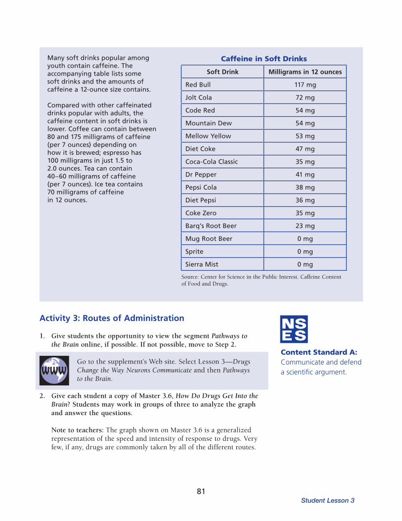
Content Standard A: Communicate and defend a scientific argument.
Activity 3: Routes of Administration
1. Give students the opportunity to view the segment Pathways to the Brain online, if possible. If not possible, move to Step 2.
Go to the supplement’s Web site. Select Lesson 3—Drugs Change the Way Neurons Communicate and then Pathways to the Brain.
2. Give each student a copy of Master 3.6, How Do Drugs Get Into the Brain? Students may work in groups of three to analyze the graph and answer the questions.
Note to teachers: The graph shown on Master 3.6 is a generalized repre sentation of the speed and intensity of response to drugs. Very few, if any, drugs are commonly taken by all of the different routes.
Many soft drinks popular among youth contain caffeine. The accompanying table lists some soft drinks and the amounts of caffeine a 12-ounce size contains.
Compared with other caffeinated drinks popu lar with adults, the caffeine content in soft drinks is lower. Coffee can contain between 80 and 175 milligrams of caffeine (per 7 ounces) depending on how it is brewed; espresso has 100 milligrams in just 1.5 to 2.0 ounces. Tea can contain 40–60 milligrams of caffeine (per 7 ounces). Ice tea contains 70 milligrams of caffeine in 12 ounces.
Caffeine in Soft Drinks
Soft Drink Milligrams in 12 ounces
Red Bull 117 mg
Jolt Cola 72 mg
Code Red 54 mg
Mountain Dew 54 mg
Mellow Yellow 53 mg
Diet Coke 47 mg
Coca-Cola Classic 35 mg
Dr Pepper 41 mg
Pepsi Cola 38 mg
Diet Pepsi 36 mg
Coke Zero 35 mg
Barq’s Root Beer 23 mg
Mug Root Beer 0 mg
Sprite 0 mg
Sierra Mist 0 mg
Source: Center for Science in the Public Interest. Caffeine Content of Food and Drugs.
Student Lesson 381

Sample Answers to Questions on Master 3.6Question 1. Four people who abuse drugs each take a drug. One person injects 100 mil ligrams of a drug into a vein, one person smokes 100 milligrams of the drug, one person snorts 100 milligrams of the drug, and one person swallows or ingests 100 milligrams of the drug. Who will experience the great est effect of the drug? The individual with the greatest concentration of drug in the brain will have the greatest effect.
The graph indicates that the individuals who inhale the drug or inject the drug into a vein will experience the greatest effect from the drug. These individuals will have a higher concentration of the drug in the brain than the people who snort (absorption through the mucous membranes) or ingest the drug. The concentration of drug in the brain will be slightly lower for inhalation than injection because some of the smoked drug is exhaled in the smoke.
Question 2. Who will experience the quickest effect from the drug?
The person who inhales the drug will experience the quickest effect from the drug (assuming the person inhales the whole 100 mg). The inhaled drug goes through the lungs and into the left side of the heart and then enters the arterial circulation to the brain, while injected drugs enter the venous circulation that returns the blood to the right side of the heart. The drug that enters the venous system takes longer to exert its effect because the blood must go to the lungs and then to the left side of the heart before it is pumped to the brain and the rest of the body.
Question 3. Who will experience the least behavioral effect from the drug?
The person who ingests, or swallows, the drug will experience the least effect.
Question 4. Who will experience the slowest effect from the drug?
The person who ingests, or swallows, the drug will also have the slowest effect.
Question 5. Tobacco smokers can use nicotine patches to help them quit smoking. The nicotine patches help the smoker slowly lower the amount of nicotine that enters the body. How does the nicotine in the patch enter the body?
Nicotine enters the body by absorption through the skin into capillaries.
The Brain: Understanding Neurobiology Through the Study of Addiction
82
Question 6. Explain why the different ways of taking drugs cause different behavioral responses.
Taking drugs by inhalation causes a very rapid increase in the level of drug in the brain. Inhaled drugs are absorbed into the arterial bloodstream in the lungs and then pumped to all parts of the body including the brain. Taking drugs by intravenous (IV) injection also causes a rapid increase in the drug level in the brain. It is slightly slower than inhalation because the drug goes first to the right side of the heart, is then pumped to the lungs where the blood is oxygenated, then goes back to the left side of the heart, and finally to the brain and body. Absorption through the skin or mucous membranes would be even slower because the drug has a longer path to travel before being circulated throughout the body. Drug response would be the slowest after ingestion because the drug goes into the digestive tract and then must pass through the walls of the stomach and intestine to enter the blood capillaries.
3. Display a transparency of Master 3.7, What Should the Doctor Do? Dis cuss the reasons why one action may be more appropriate than others.
On the basis of what you have learned about how drugs act in the body, how should morphine be given to the patient? Should the morphine be given as a pill, a shot, or an inhalant? Consider each alternative and explain why the different methods should or should not be chosen.
The question concerning how morphine should be administered to a patient to relieve pain is designed to assess whether students understand how different ways of getting drugs into the body changes their effects. The doctor’s goal is to relieve the patient’s pain quickly so that the fracture can be set.
On the basis of the graph that students analyzed on Master 3.6, the doctor should elect to give morphine as an inhalant or an injection. In each case, the drug reaches the brain quickly. Inhaled drugs can reach the brain even faster than injected drugs. Perhaps the main disadvantage of giving the morphine as an inhaled drug is the amount of drug that actually enters the blood stream is more variable. If the drug is injected, all of the drug is delivered into the bloodstream. The doctor knows how much morphine enters the bloodstream. Giving a pill to the patient would be less effective than the other means for pain relief because it would take longer for the drug to act and its concentration in the blood stream would be lower.
If students understand that taking drugs into the body by different routes causes different responses, they should be able to explain that the different ways of administering drugs can have advantages and dis-advantages. Use this sce nario to evaluate students’ understanding.
Student Lesson 383

L E S S O N 4Explain/Elaborate
OverviewStudents examine data from animal experiments, play a card game, and examine a case study. They learn that although the initial decision to take drugs of abuse is voluntary, continued use may lead to addiction, which is the continued compulsive abuse of drugs despite adverse consequences. Stu dents then watch a minidocumentary online to learn how drugs cause long-term changes in the brain.
Major ConceptAddiction is a brain disease.
ObjectivesBy the end of these activities, the students will
understand that drug abuse initially is a voluntary behavior,be able to define drug addiction as the continued compulsive drug abuse despite known adverse health or social consequences,understand that drug abuse and addiction are associated with long-term physical and functional changes in the brain, andrecognize that addiction is influenced by biological factors (for example, genetics and age) and by the social and behavioral context of drug use.
Basic Science–Health ConnectionDrug addiction is a complex brain disease. Preventing drug abuse and addic tion and treating the disease effectively require understanding the biological, genetic, social, psychological, and environmental factors that predispose indi viduals to drug addiction.
Drug Abuse and Addiction
At a Glance
Photo: Corel
91

Individuals make choices to begin using drugs. Some people begin using drugs to relieve a medical condition and then continue to use the drugs after the med ical need is over. Children or teens who are depressed or who have another psychiatric disorder sometimes begin using illicit drugs in an attempt to self-medicate. Other people begin taking drugs to feel pleasure, to escape the pressures of life, or to alter their view of reality. This voluntary initiation into the world of addictive drugs has strongly influenced society’s view of drug abuse and drug addiction and their treatment.
When does drug abuse become drug addiction? It rarely happens with the first use of a drug. Drug abuse and drug addiction can be thought of as points along a continuum. Any use of a mind-altering drug or the inappropri ate use of medication (either prescription or over-the-counter drugs) is drug abuse, but the point when drug abuse becomes drug addiction is less clear. Dif ferent people may reach the point of addiction at different stages. Scien tists continue to investigate the factors that contribute to the transition to drug addiction.
Background Information
Figure 4.1: The continuum of drug abuse and addiction.
no use drug abuse drug addiction
Drug addiction is defined as the continued compulsive use of drugs despite adverse health or social consequences.1 Drug-addicted people have lost con trol of their drug use. Individuals who are addicted to drugs often become iso lated from family or friends, have difficulty at work or school, may commit crimes, and become involved with the criminal justice system. For a person addicted to drugs, continuing to take them becomes the primary focus in life.
Certain drugs, including opioids and alcohol, cause strong physical reactions in the body when drug use stops. When a person addicted to heroin stops taking heroin, he or she can experience a variety of symptoms ranging from watery eyes and a runny nose to irritability and loss of appetite and then diarrhea, shivering, sweating, abdominal cramps, increased sensitivity to pain, and sleep problems.2 In general, withdrawal from heroin makes people feel miserable. Withdrawal from alcohol can cause serious effects such as seizures and even death. With drawal from other drugs, such as cocaine and amphetamines, does not lead to strong physical reactions, but it may make the person feel depressed or lethargic. For most drugs, physical withdrawal symptoms can usually be controlled effectively with medications. Even though withdrawal from some drugs does not cause the person abusing them to have physical reactions, stopping drug use is difficult because of the changes the drugs have caused in the brain. Once the drugs stop, the person will have cravings, or intense desire for the drugs.3 Craving arises from the brain’s need to maintain a state of homeostasis that now relies on the presence of the drug. A person may experience cravings at any stage of drug abuse or addiction, even early
The Brain: Understanding Neurobiology Through the Study of Addiction
92

in the experimentation phase of drug abuse. Cravings have a physical basis in the brain. Using PET imaging, scientists have shown that just seeing images of drug paraphernalia can stimulate the amygdala (part of the brain involved in emotional memory) in an addicted person.4
Drugs of addiction do not merely cause short-term changes in an individual’s cognitive skill and behavior. A drug “high” lasts a short time, ranging from less than an hour to 12 hours, depending on the drug, dose, and route of administration. The changes in the brain that result from continued drug use, however, can last a long time. Scientists believe that some of these changes disappear when drug use stops; some disappear within a short time after drug use stops, and other changes are potentially permanent. One of the first changes in the brain that may occur in response to repeated drug abuse is tolerance. Tolerance develops when a person needs increasing doses of a drug to achieve the same high or “rush” that previously resulted from a lower dose of the drug. Two primary mechanisms underlie the development of tolerance.3 First, the body may become more efficient at metabolizing the drug, thereby reducing the amount that enters the brain. Second, the cells of the body and brain may become more resistant to the effect of the drug. For example, after continued cocaine use, neurons decrease the number of dopamine receptors, which results in decreasing cocaine’s stimu latory effect. Opioids, on the other hand, do not cause a change in the number of receptors. Instead the opioid receptors become less efficient in activating associated cellular processes, thus reducing the effects of the opioids.
Drugs can cause other long-term changes in the anatomy and physiology of the brain’s neurons. Alcohol, methamphetamine, and MDMA (ecstasy) have been shown to be neurotoxic in animal studies.3 Unlike other types of cells in the body, neurons in many parts of the brain have little or no capacity to regenerate. (Recent studies have shown that the adult human brain can generate new neurons in the hippocampus, a part of the brain important for learning and memory.5 Other parts of the brain have not been shown to have this ability.) Alcohol kills neurons in a part of the brain that helps create new memories (the hippocampus and mammillary bodies). If those neurons die, the capacity for learning decreases. Methamphetamine is toxic to dopamine-containing neurons in animals and possibly in humans as well.6 MDMA has been shown in animal studies to damage the axon terminals of neurons that produce another neurotransmitter called serotonin.7 In addition to neurotoxic effects, drugs can significantly alter the activity of the brain. PET scans of people addicted to cocaine show that the metabolism of glucose, the primary fuel for cells, is drastically reduced in the brain; this decrease in metabolism can last for many months after drug abuse stops.8
In addition to the functional and anatomical changes in the brain, drug abuse puts people at higher risk for other health problems. For example, inhalant abuse can lead to disruption of heart rhythms, and snorting cocaine can lead to ulcerations in the mucous membranes of the nose. In addition, injection drug users (IDUs) are at higher risk of contracting HIV through the sharing of potentially contaminated needles. Similarly,
Student Lesson 493

Genetic, Behavioral, and Environmental Influences on Drug Addiction Drug addiction is not simply continuous drug abuse. Many more individuals will try an addictive drug than will become addicted. Most people know of sit uations in which two people use the same amount of alcohol or tobacco, but have very different responses to them. Environmental, social, behavioral, and genetic factors also contribute to the development of drug addiction.9 Stress can increase the susceptibility to addiction.
Figure 4.2: Photographs of serotonin axons in the cerebral cortex of nonhuman primates labeled with a fluorescent marker. The number of serotonin-labeled axons is dramatically reduced in the cerebral cortex at 2 weeks (B) and 18 months (C) after the last drug exposure. The brain of the control animal that did not receive MDMA (A) shows the dense network of labeled axons. Images E and F show changes caused by MDMA use on a different brain region, the hypothalamus. The control showing the hypo thalamus in the absence of MDMA is shown in D. Photographs courtesy of G.A. Ricaurte, with the permission of the Journal of Neuroscience.
Cerebral Cortex
Hypothalamus
Control MDMA 2 weeks MDMA 18 months
hepatitis B and hepatitis C are much more common among drug addicts than the general population. Tuberculosis is another concern. Drug abuse and addic tion also are contributing factors in motor vehicle accidents.
The Brain: Understanding Neurobiology Through the Study of Addiction
94
Scientists continue to investigate the factors that place one individual at greater risk of becoming addicted than another individual with a similar pattern of drug use. Individuals who have developed strong coping skills to deal with life’s pressures have less risk of becoming addicted to drugs. The younger a person is when he or she begins using drugs, the more likely he or she is to become addicted. This may be true because younger individuals have not developed the coping skills necessary to deal with life’s ups and downs. Additionally, the frontal cortex of the adolescent brain isn’t fully mature until age 24.10 This area of the brain is responsible for judgment and for inhibiting impulsivity and risk-taking behavior. In addition, genetic factors probably influence who engages in higher- risk behaviors.
The context in which a person uses an addictive drug greatly contributes to its behavioral effects and the risk of abuse and addiction. For example, some cancer patients take relatively large doses of morphine for extended periods to control pain without becoming addicted. It has been proposed that addiction is rare in these patients because, in contrast to addicted individuals, these patients are motivated not by a compulsive urge to seek a high but by a physiologic need to ease their pain and improve their quality of life.
Medical Uses of Addictive DrugsIt is well known that otherwise safe medications can turn harmful if abused or taken without prescription or supervision. The other side of this coin is that many drugs of abuse are themselves, or have been found to contain, active ingredients that can be therapeutic. A good example is morphine. During the Civil War, doctors gave morphine to wounded soldiers to relieve the pain of injuries. Doctors didn’t realize how addictive injected morphine was until many soldiers became addicted to the drug.2 Morphine addiction became known as “soldiers’ disease.” Today, morphine is a valuable medicine to relieve pain when administered with the appropriate medical supervision. Patients in hospitals receive morphine to ease their pain after surgery and during cancer and burn treatment. Very few of these patients become addicted to morphine even though they may take it for extended periods of time.
Another drug that has received considerable attention for its potential medical benefits is marijuana. Television and newspaper reports periodically present stories on the use of marijuana by terminal cancer or AIDS patients to ease their discomfort and pain. Following up on such anecdotal evidence, several scientific studies have been able to corroborate at least some of the claims about marijuana’s beneficial effects on appetite, nausea, and certain types of pain. However, marijuana’s addictive properties and its usual delivery by smoke inhalation—which exposes the lungs to many toxic chemicals—make it an unappealing candidate for medications development. Rather, it is likely that our understanding of the biology of marijuana’s active ingredients, such as tetrahydrocannabinol (THC), will lead to improved medications for a variety of conditions, ranging from obesity and addiction to neuropathic pain in multiple sclerosis (MS) patients, chronic pain in advanced cancer patients, nausea, and wasting syndrome.11
Student Lesson 495

The risk of becoming addicted to prescription pain medications is minimal in patients who are treated on a short-term basis; however, the risk for those with chronic pain is less well understood. Some studies have shown that those most vulnerable to becoming addicted to prescription pain medications have a history of psychological disorders, prior substance abuse problems, or a family history of these disorders. Pain management for patients who have substance abuse disorders is particularly challenging for the medical profession. However, these patients can still be successfully treated with opioid pain medications, although they may need to be admitted to a treatment or recovery program and monitored closely if controlled substances are prescribed for pain.
In the 1970s, news media reported the use of marijuana and heroin by soldiers who were serving in Vietnam. Combat stress, the easy availability of drugs, and the relaxation of taboos against drug use at the time all contributed to the prob lem. Although many soldiers did have drug problems while in Vietnam, 95 percent were not addicted to drugs after they returned to the United States.12 This illustrates the profound effect that environmental circumstances can have on drug taking and drug addiction.
In addition, scientists are working to identify genetic factors that contribute to drug abuse and addiction. Studies of identical twins indicate that as much as half of an individual’s risk of becoming addicted to nicotine, alcohol, or other drugs depends on his or her genes. Recent technical advances in DNA analysis have enabled researchers to untangle complex genetic interactions by examining a person’s entire genome at once. A series of studies has identified a certain variant in the gene for a nicotinic receptor subunit that more than doubles the risk for addiction among smokers, as well as increasing their vulnerability to lung cancer and peripheral arterial disease.
Animals as Research ModelsWhy do scientists study the brains of laboratory animals? Scientists use ani mals in research studies because the use of humans is either impossible or unethical. For example, when scientists investigate the effects of drugs of abuse on brain function, either the question they are asking cannot be answered in a living human or it would be inappropriate to give a person the drugs.
The use of animals as subjects in scientific research has contributed to many important advances in scientific and medical knowledge. Scientists must ana lyze the goals of their experiments in order to select an animal species that is appropriate. Scientists often use fruit flies (Drosophila melanogaster) when they want to learn more about genetics. However, fruit flies are not a very good model if a scientist is investigating muscle physiology or behavior; a mouse may be a better model for those experiments. Although scientists strive to develop nonanimal models for research, these models often do not duplicate the complex animal or human body. Continued progress toward a more complete under standing of human and animal health depends on the use of living animals.
The Brain: Understanding Neurobiology Through the Study of Addiction
96
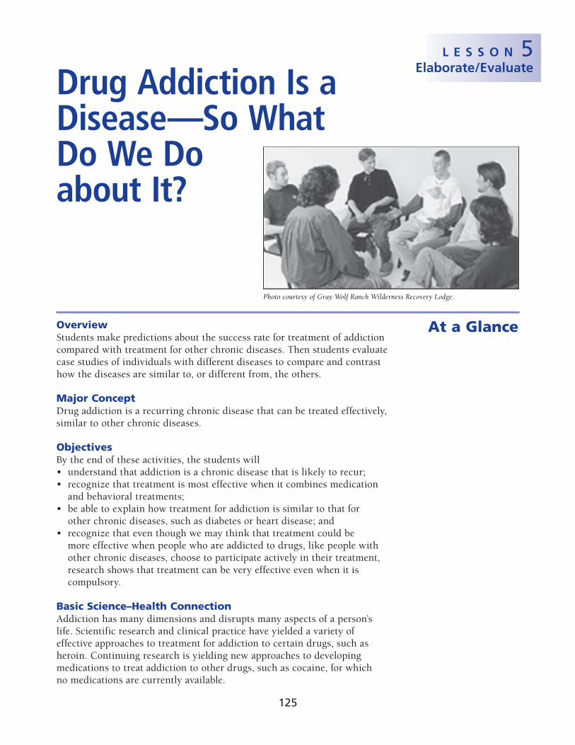
L E S S O N 5Elaborate/Evaluate
OverviewStudents make predictions about the success rate for treatment of addiction compared with treatment for other chronic diseases. Then students evaluate case studies of individuals with different diseases to compare and contrast how the diseases are similar to, or different from, the others.
Major ConceptDrug addiction is a recurring chronic disease that can be treated effectively, similar to other chronic diseases.
ObjectivesBy the end of these activities, the students will
understand that addiction is a chronic disease that is likely to recur;recognize that treatment is most effective when it combines medication and behavioral treatments;be able to explain how treatment for addiction is similar to that for other chronic diseases, such as diabetes or heart disease; andrecognize that even though we may think that treatment could be more effective when people who are addicted to drugs, like people with other chronic diseases, choose to participate actively in their treatment, research shows that treatment can be very effective even when it is compulsory.
Basic Science–Health ConnectionAddiction has many dimensions and disrupts many aspects of a person’s life. Sci entific research and clinical practice have yielded a variety of effective approaches to treatment for addiction to certain drugs, such as heroin. Continuing research is yielding new approaches to developing medications to treat addiction to other drugs, such as cocaine, for which no medications are currently available.
At a Glance
Photo courtesy of Gray Wolf Ranch Wilderness Recovery Lodge.
Drug Addiction Is a Disease—So What Do We Do about It?
125

A second medication prescribed for heroin addiction is naltrexone. Unlike methadone, naltrexone is an opioid-receptor antagonist. Instead of competing with or mimicking heroin for the opioid receptor, naltrexone prevents heroin from binding to the receptor, thereby preventing heroin
Drug abuse and addiction lead to long-term changes in the brain’s chemistry and physiology. The changes in the brain cause drug-addicted people not only to lose the ability to control their drug use, but their addiction also changes all aspects of their lives. People with drug addiction often become isolated from family and friends and have trouble in school or work. In addition, the compulsive need for drugs can lead to significant legal problems. While the biological foundation for drug addiction does not absolve an individual from the responsibility of his or her actions, the stigma of drug addiction needs to be lifted so individuals may receive proper medical treatment, similar to that for other chronic diseases.1
Addiction is a recurring chronic disease. No cure is available at this time, but addiction can often be treated effectively. Drug addiction is often viewed as a lapse in moral character. This value judgment influences how society deals with the disease, both socially and medically. Unfortunately, because people, including physicians, have often viewed addiction as a self-inflicted condition, drug-addicted people have not always received the medical treatment common for other chronic diseases. Treating addiction requires more than a “just say no” approach.2
Treatment for addiction can be very effective. Treatment is successful when the addicted person reduces or abstains from drug use, improves his or her personal health or social function, and becomes less of a threat to public health and safety.3 Certain addictions, such as heroin addiction, can be treated with medications.4,5 Methadone, the most common medication, prevents craving and withdrawal symptoms in heroin addiction. Methadone is an opioid-receptor agonist. That is, methadone binds to the opi oid receptor just as heroin does. Methadone, however, does not produce the euphoria or “high” that results from heroin use. When taken orally as indicated, it does not produce the rapid increase in opioid-receptor occupancy that comes from injecting or snorting heroin, but it does maintain sufficient opioid-receptor activity to prevent withdrawal and cravings for opioids.
Background Information
Figure 5.1: Methadone can be part of an effective treatment plan for addiction to opiates. Photo graph of pills by, and used with permission of, Roxane Laboratories, Inc. All Rights Reserved.
The Brain: Understanding Neurobiology Through the Study of Addiction
126

Figure 5.2: Agonists are chemicals that bind to a specific receptor to elicit a response, such as excitation or inhibition of action potentials. Methadone is an agonist that, like heroin, binds to opioid receptors. Unlike heroin, however, methadone does not produce the same level of eupho ria. Buprenorphine is a partial agonist that also binds to opioid receptors. Partial agonists are chemicals that are similar to full agonists, but at higher doses their effect is not as great as a full agonist’s. Buprenorphine does not produce the euphoria seen with heroin. Antagonists are chemicals that bind to a receptor and block it, producing no response and preventing other chemicals (drugs or receptor agonists) from binding or attaching to the receptor. Naltrexone is an antagonist that binds to the opioid receptor and blocks heroin from binding.
from eliciting the euphoric high (see Figure 5.2). Buprenorphine is also used to treat heroin addiction. It is a long-acting partial opioid-receptor agonist. It acts on the same receptors as heroin but does not produce the same intense “high” or dangerous side effects. Buprenorphine has some advantages over other medications for treating heroin addiction. Unlike methadone, buprenorphine can be prescribed in physicians’ offices. It is also less likely to be toxic or abused than methadone.
Table 5.1 outlines the different medications used to treat addic tion. The development of medications to treat drug addiction has been diffi cult because the brain, the main target of addictive drugs, is such a complex organ. Until scientists understand how drugs affect the chemistry of the brain, they cannot develop medicines that will alter their effects.
Table 5.1: Medications for Addiction4
Medication Treatment for addiction to Mechanism
Methadone Heroin Opioid-receptor agonist
Naltrexone Heroin Opioid-receptor antagonist
Naloxone Heroin, alcohol Opioid-receptor antagonist
Buprenorphine Heroin Mixed opioid-receptor agonist and antagonist
Nicotine gum, patches Nicotine Provide low doses of nicotine Medication, if available, is rarely sufficient for effective treat ment. Behavioral treatment in combination with medication is the most effective way to treat drug addiction.6,7 People recovering from drug addiction need to address the behavioral and social consequences of their drug use and learn to cope with the social and environmental factors that contribute to their illness.7 Behavioral treatments can be provided either individually or as a group.
Student Lesson 5127

Principles of Effective Drug Addiction Treatment1. Addiction is a complex but treatable disease that affects brain function and behavior. Drugs of
abuse alter the brain’s structure and function, resulting in changes that persist long after drug use has ceased. This may explain why drug abusers are at risk for relapse even after long periods of abstinence and despite the potentially devastating consequences.
2. No single treatment is appropriate for everyone. Matching treatment settings, interventions, and services to an individual’s particular problems and needs is critical to his or her ultimate success in returning to productive functioning in the family, workplace, and society.
3. Treatment needs to be readily available. Because drug-addicted individuals may be uncertain about entering treatment, taking advantage of available services the moment people are ready for treatment is critical. Potential patients can be lost if treatment is not immediately available or readily accessible. As with other chronic diseases, the earlier treatment is offered in the disease process, the greater the likelihood of positive outcomes.
4. Effective treatment attends to multiple needs of the individual, not just his or her drug abuse. To be effective, treatment must address the individual’s drug abuse and any associated medical, psychological, social, vocational, and legal problems. It is also important that treatment be appropriate to the individual’s age, gender, ethnicity, and culture.
5. Remaining in treatment for an adequate period of time is critical. The appropriate duration for an individual depends on the type and degree of his or her problems and needs. Research indicates that most addicted individuals need at least 3 months in treatment to significantly reduce or stop their drug use and that the best outcomes occur with longer durations of treatment. Recovery from drug addiction is a longterm process and frequently requires multiple episodes of treatment. As with other chronic illnesses, relapses to drug abuse can occur and should signal a need for treatment to be reinstated or adjusted. Because individuals often leave treatment prematurely, programs should include strategies to engage and keep patients in treatment.
6. Counseling—individual and/or group—and other behavioral therapies are the most commonly used forms of drug abuse treatment. Behavioral therapies vary in their focus and may involve addressing a patient’s motivation to change, providing incentives for abstinence, building skills to resist drug use, replacing drug-using activities with constructive and rewarding activities, improving problemsolving skills, and facilitating better interpersonal relationships. Also, participation in group therapy and other peer support programs during and following treatment can help maintain abstinence.
7. Medications are an important element of treatment for many patients, especially when combined with counseling and other behavioral therapies. For example, methadone and buprenorphine are effective in helping individuals addicted to heroin or other opioids stabilize their lives and reduce their illicit drug use. Naltrexone is also an effective medication for some opioid-addicted individuals and some patients with alcohol dependence. Other medications for alcohol dependence include acamprosate, disulfiram, and topiramate. For persons addicted to nicotine, a nicotine replacement product (such as patches, gum, or lozenges) or an oral medication (such as bupropion or varenicline) can be an effective component of treatment when part of a comprehensive behavioral treatment program.
8. An individual’s treatment and services plan must be assessed continually and modified as necessary to ensure that it meets his or her changing needs. A patient may require varying combinations of services and treatment components during the course of treatment and recovery. In addition to counseling or psychotherapy, a patient may require medication, medical services, family therapy, parenting instruction, vocational rehabilitation, and/or social and legal services. For many patients, a continuing care approach provides the best results, with the treatment intensity varying according to a person’s changing needs.
The Brain: Understanding Neurobiology Through the Study of Addiction
128

Relapse is a common event for people recovering form drug addiction. In many ways, relapse should be thought of as a normal part of the recovery process. A person in recovery is more likely to experience a relapse if he or she also has other psychiatric conditions, experiences stress, or lacks the support of family and friends.
Despite the preconceptions and value judgments many people place on addiction, it is, in many ways, similar to other chronic diseases such as dia betes and coronary artery disease. Genetic, environmental, and behavioral components contribute to each of these diseases. Some people may argue that drug addiction is different because it is “self-inflicted.” As presented in Les son 4, the initial choice to use drugs is voluntary, but, once addiction develops, drug use is compulsive—not voluntary. Moreover, voluntary choices do contribute to the onset or severity of other chronic diseases as well. For example, a person who chooses to eat an unhealthy diet and not exercise increases his or her risk for coronary heart disease.
9. Many drug-addicted individuals also have other mental disorders. Because drug abuse and addiction—both of which are mental disorders—often co-occur with other mental illnesses, patients presenting with one condition should be assessed for the other(s). And when these problems co-occur, treatment should address both (or all), including the use of medications as appropriate.
10. Medically assisted detoxification is only the first stage of addiction treatment and by itself does little to change long-term drug abuse. Although medically assisted detoxification can safely manage the acute physical symptoms of withdrawal and, for some, can pave the way for effective long-term addiction treatment, detoxification alone is rarely sufficient to help addicted individuals achieve long-term abstinence. Thus, patients should be encouraged to continue drug treatment following detoxification. Motivational enhancement and incentive strategies, begun at initial patient intake, can improve treatment engagement.
11. Treatment does not need to be voluntary to be effective. Sanctions or enticements from family, employment settings, and/or the criminal justice system can significantly increase treatment entry, retention rates, and the ultimate success of drug treatment interventions.
12. Drug use during treatment must be monitored continuously, as lapses during treatment do occur. Knowing their drug use is being monitored can be a powerful incentive for patients and can help them withstand urges to use drugs. Monitoring also provides an early indication of a return to drug use, signaling a possible need to adjust an individual’s treatment plan to better meet his or her needs.
13. Treatment programs should assess patients for the presence of HIV/ AIDS, hepatitis B and C, tuberculosis, and other infectious diseases as well as provide targeted risk-reduction counseling to help patients modify or change behaviors that place them at risk of contracting or spreading infectious diseases. Typically, drug abuse treatment addresses some of the drug-related behaviors that put people at risk of infectious diseases. Targeted counseling specifically focused on reducing infectious disease risk can help patients further reduce or avoid substance-related and other high-risk behaviors. Counseling can also help those who are already infected to manage their illness. Moreover, engaging in substance abuse treatment can facilitate adherence to other medical treatments. Patients may be reluctant to accept screening for HIV (and other infectious diseases); therefore, it is incumbent upon treatment providers to encourage and support HIV screening and inform patients that highly active antiretroviral therapy (HAART) has proven effective in combating HIV, including among drugabusing populations.
Source: NIDA. 2009. Principles of Drug Addiction Treatment: A Research-based Guide, 2nd edition. 2009. National Institute on Drug Abuse.
Student Lesson 5129

Successful treatment for any chronic disease necessitates patient compliance with the prescribed treatment regimen. Adhering to a treatment plan is diffi cult for those with any chronic disease. Less than 50 percent of people with diabetes fol low their routine medication plan, and only 30 percent follow their dietary guidelines.2 Problems adhering to a treatment plan lead to about 50 percent of diabetic people needing additional medical care within one year of diagnosis and initial treatment. Similar statistics hold true for other chronic diseases: approxi mately 40 percent of patients with hypertension need emergency room treat ment for episodes of extreme high blood pressure, and only about 30 percent of adult asthma sufferers take their medication as prescribed. People treated for drug addiction also commonly relapse during treat ment and recovery, resuming drug use. The difficulties in following a treatment plan and coping with the stresses of a chronic disease illustrate how difficult changing human behavior is. The challenge of adherence is particularly severe in the case of addiction because this disease implicates and coopts the very same brain substrates that underlie what we call free will.8 Activities 2 and 3 of this lesson provide more insight into this topic.
Scientific research is likely to change how drug addiction is treated. Research to understand how the brain works and how drugs cause changes in the chemistry and function of the brain may lead to new medications to treat dis ease. Scientists continue to work on developing medications that relieve the cravings experienced when drugs are withdrawn. Also, scientific advances may reveal ways to reverse the long-term functional changes to the brain that drugs inflict.
Web-Based ActivitiesActivity Web Component?
1 No
2 Yes
3 No
4 No
PhotocopiesFor each group of 3 students For each student
1 copy of Master 5.1, Ruth’s Story* 1 copy of Master 5.2, Mike’s Story* 1 copy of Master 5.3, Carol’s Story* 1 copy of Master 5.4, Disease
Reference Information*
1 copy of Master 5.5, Evaluating the Cases
* The Web version of Activity 2 is the preferred approach. Copies of Masters 5.1, 5.2, 5.3, and 5.4 are needed only if the Internet is unavailable for classroom use.
In Advance
The Brain: Understanding Neurobiology Through the Study of Addiction
130

This activity is intended to be a quick method to assess students’ prior conceptions about treat-ing drug addiction as a disease.
ProcedureActivity 1: Is Addiction Treatable?
1. Begin the activity by holding a classroom discussion about illness and disease. Ask, “What is a disease?” Ask students to name some diseases. Write responses on the board.
Students are likely to say a disease is some problem with the body that makes a person feel bad. They may also respond that a disease is something you see a doctor about or take medicine for. Students will list a variety of diseases and conditions. If they don’t include both short-term minor diseases (such as a cold or flu) and long-term complex diseases (such as diabetes or heart disease), prompt them with questions such as, Is a cold a disease? Is diabetes?
2. Introduce the terms chronic and acute and give examples of chronic and acute conditions. Categorize the diseases from Step 1 as either chronic or acute.
Chronic diseases are those that persist over a long period of time, whereas acute diseases last only a short time but may have a rapid onset and marked intensity. Diabetes, heart disease, asthma, and cancer are examples of chronic diseases. Colds, flu, and a broken bone are acute conditions.
3. Ask students to consider whether addiction is chronic or acute. Have them explain their answer based on what they have learned in the unit so far. After students recognize that addiction is a chronic disease, add it to the list of chronic diseases.
Students’ explanations should include something about the changes that occur in the brain as a result of drug use (Lessons 2, 3, and 4) and something about the compulsive, nonvoluntary nature of addiction.
Materials Activity Materials
Activity 1 overhead projector
Activity 2 computers (optional)
Activity 3 overhead projector
Activity 4 none
PreparationArrange for students to have access to computers for viewing the case studies in Activity 2.
Student Lesson 5131

4. Ask, “Do all diseases or illnesses affect people the same way?”
No. Some are longer lasting and require more intervention from healthcare providers than others. Some require medicines, others require psychological treatment, and some require both. Students may give a cold as an example of a short-term illness that doesn’t require a great deal of treatment and diabetes or heart disease as a longer-lasting illness that does require a lot of treatment. Students should realize that there are similarities as well as differences in disease treatment.
5. Hold a class discussion to find out what students know about treatments for addiction. Probe student understanding of what a person experiences in treatment, what types of treatments are available, how long treatment lasts, and whether it is successful. Have students justify their ideas. Accept all reasonable answers, and record ideas on the board or a blank transparency.
At this stage, students are likely to have many ideas about treatment for addiction. Some of their ideas will likely be drawn from stories they have seen on the Internet or from media coverage of celebrities. Their ideas may also reflect societal perceptions of addiction and may not include explanations based on the biology of addiction.
Note to Teachers: Save the list that students generate. They will revisit it in Activity 3.
6. Explain that in the next activity, students will learn about treatment for addiction and how it compares with treatment for other chronic diseases.
Activity 2: Evaluating the Case Studies
The following procedures describe how to conduct the Web version of this activity, which is the preferred method of instruc tion. Instructions for conducting the alternative print version follow (on page 136).
1. Divide the class into groups of three students. Give each student a copy of Master 5.5, Evaluating the Cases. Have the students complete the Web activity Dealing with a Chronic Disease. Each member of the group should answer questions 1–6 for a different case study. After they watch the three cases, the group should answer questions 7–11.
From the activities menu on the Web site, select Lesson 5—Drug Addiction Is a Disease, So What Do We Do about It? Then click to watch the video interviews.
2. As a class, discuss the case studies and the answers to Master 5.5.
Content Standard F: An individual’s mood and behavior may be modified by substances. Content Standard F: Personal choice concern ing fitness and health involves multiple factors. Content Standard F: Families serve basic health needs, especially for young children.
The Brain: Understanding Neurobiology Through the Study of Addiction
132

Sample Answers to Questions on Master 5.5
Case Study: Ruth
Question 1. What disease does the individual have? Is it chronic or acute?
Ruth is addicted to heroin. Addiction is a chronic disease.
Question 2. How did the disease change the individual’s life?
Ruth, like other drug-addicted people, was spending most of her energy focusing on how and where she was going to get her next drugs. She became isolated from her friends, lost her job, and got into trouble with the law.
Question 3. What is the recommended treatment?
The prescribed treatment for Ruth is a combination of medication (methadone or buprenorphine) and behavioral treatment.
Question 4. What did the individual do to improve his or her recovery?
Ruth followed her doctor’s advice and got medicine and psychological treatment to help her deal with the problems of addiction. She also worked to change her life by enrolling in college, making new friends, and getting involved in running. After a recurrence of her drug problem, she again started her medical and psychological treatment.
Question 5. What did the individual do that impaired his or her recovery?
Ruth thought she had conquered her disease and didn’t need to continue her treatment. Her life became very stressful, and she went back to the friends who started her on drugs in the first place.
Question 6. Are there other things the individual could do to help with the disease?
As long as Ruth continues her treatment plan, she should be able to man age her disease. If she ignores her treatment, her chance of having a recur rence increases.
Case Study: Mike
Question 1. What disease does the individual have? Is it chronic or acute?
Mike has diabetes, a chronic disease.
Question 2. How did the disease change the individual’s life?
After being diagnosed with diabetes, Mike had to check his blood glucose levels regularly, give himself insulin injections, and watch his diet.
Student Lesson 5133

Question 3. What is the recommended treatment?
Mike’s doctors placed him on insulin therapy. The doctors also prescribed behavioral treatments.
Question 4. What did the individual do to improve his or her recovery?
To help learn about diabetes, Mike attended a camp where he received information about coping with the disease. After some problems, Mike learned to control his blood sugar levels.
Question 5. What did the individual do that impaired his or her recovery?
Mike had trouble in social situations because he couldn’t do the same things his friends did. When he ignored his treatment, Mike had trouble in school and ended up in the hospital.
Question 6. Are there other things the individual could do to help with the disease?
Mike needs to continue to follow his treatment plan and monitor his blood glucose level.
Case Study: Carol
Question 1. What disease does the individual have? Is it chronic or acute?
Carol has hypertension. Hypertension is a chronic disease.
Question 2. How did the disease change the individual’s life?
Because of the disease, Carol had problems at work as well as with her family interactions. Her health problems became more severe, and she had a mild stroke.
Question 3. What is the recommended treatment?
Initially, the doctor prescribed medication as well as a change in Carol’s diet to reduce her salt intake. The doctor also told Carol that exercise would be beneficial.
After Carol had problems following the plan, the doctor recommended that Carol get additional help from other health professionals.
Question 4. What did the individual do to improve his or her recovery?
Carol followed the treatment plan for a while.
The Brain: Understanding Neurobiology Through the Study of Addiction
134

Question 5. What did the individual do that impaired his or her recovery?
Carol didn’t follow her doctor’s advice after the initial period and then ignored her doctor’s suggestion that she get additional help from other specialists.
Question 6. Are there other things the individual could do to help with the disease?
Carol needs to fit her treatment into her life.
Comparing the Cases
Question 7. Which individuals were successful in their treatment? Which individuals were not?
Ruth and Mike were both successful in their treatment. Although they had problems, both of them decided to again comply with their treat ment. Carol was not successful; she did not follow the recommended treatment.
Question 8. Who was cured of their disease? What is the difference between treatment and cure?
None of the individuals was cured of his or her disease. Treatment elimi nates or reduces the effects of the disease, but does not eliminate the dis ease. If a disease is cured, the problem is fixed and requires no additional treatment.
Question 9. How are the treatments for the different diseases similar?
In each case, the prescribed treatment included both medication and behavioral treatments. In each case, treatment is a long-term process.
Question 10. How are the treatments different?
Different medications are used to treat different diseases.
Question 11. Can you identify similarities and differences in the actions or strategies that individuals took to help them deal with their disease?
All three individuals initially complied with the prescribed treatment. All three individuals experienced a time when they ignored the treatment plan and had reoccurring problems with the disease. Ruth and Mike chose to get additional treatment and learned to cope with their disease. Carol, on the other hand, made the choice to continue to ignore the treat ment plan and her doctor’s advice.
Student Lesson 5135
The following procedure is for classes using the print version of this activity.
1. Break the class into groups of three students. Give one copy of each of the following masters to each group: Master 5.1, Ruth’s Story; Master 5.2, Mike’s Story; Master 5.3, Carol’s Story; and Master 5.4, Disease Ref erence Information. Each student in the group should read a different case. Give each student a copy of Master 5.5, Evaluating the Cases. Each student should answer questions 1–6 about the case study that he or she read. The students should answer questions 7–11 as a group. Give stu dents time to discuss and write answers to the questions. They may refer to the case studies for help.
2. After all the groups have finished the questions, discuss the cases with the class.
Sample answers for the questions on Master 5.5 are given in the procedures for the Web-based version of this activity (pages 133–135).
Activity 3: Is Treatment for Addiction Effective?
1. Display students’ ideas on addiction treatment from Activity 1, Step 5. Ask whether they now see these ideas as correct or incorrect based on what they learned from the case studies. Have students revise any incorrect statements and explain their changes.
Students should be able to use pieces of information to correct some common misconceptions that are probably on their list. For example, one misconception is that treatment for addiction doesn’t work and that once a person is addicted to drugs, there isn’t anything that can be done for them. From the case studies, students should recognize that treatment can be successful, and people can improve their lives if they follow the treatment plan, which could include behavioral therapies and medications. (Medications are available to treat addiction to some drugs (for example, opiates, nicotine, alcohol), but not others.)
If appropriate for the specific drug addiction, treatment that includes both behavioral therapy and medication is often more successful than treatment that uses only one approach. Students should recognize that the combination of behavioral therapy and medication helped the individual portrayed in the case study.
Treatment is most effective when adjusted for the individual’s needs and circumstances. The ultimate goal of drug addiction treatment is to enable an individual to achieve long-lasting abstinence, but the immediate goals are to reduce drug abuse, improve the patient’s ability to function, and minimize the medical and social complications of drug abuse and addiction.
Content Standard A: Formulate and revise scientific explanations and models using logic and evidence.
Now that students have evaluated the case stud ies, they should under stand that addiction is a disease that is treated as effectively as, or more effectively than, other chronic diseases.
The Brain: Understanding Neurobiology Through the Study of Addiction
136

Some initial ideas about drug addiction treatment may reflect the idea that simply stopping drug use means that treatment is effective. Students should realize after reading the case studies that drug addiction and other chronic diseases can have wide-ranging effects on a person, both physically and emotionally. Thus, addressing the person’s complex needs is imperative. For addiction, this may include helping with family problems, employment, legal concerns, and other co-occuring medical conditions. Reinforce to students that behavioral therapy, along with other services, can help individuals cope with the problems that can trigger a relapse. Just as treatment for diabetes or heart disease requires that people change their behaviors to adopt a healthier lifestyle, so does successful treatment for drug addiction.
2. Point out that the individual in the case study experienced relapse at one point. She started using drugs again after stopping for a while. Ask students if relapse means that treatment is not effective.
Relapse is common during recovery from drug addiction, as it is for other chronic diseases depicted in the case studies. If someone relapses, that does not mean that treatment failed. Rather, relapse signals that the person needs to go back to treatment or that the person’s treatment plan needs to be modified to better fit the individual’s needs.
Some students will suggest that relapse occurs because patients don’t always comply with their treatment. This is correct. Treatment is more effective when the patient participates actively in the process. It’s important for students to understand this. After all, therapies will not be effective if the patient chooses not to take the medicine or attend the counseling sessions.
3. Have students consider the problems of following a treatment plan. Ask them if they have ever made New Year’s resolutions. How long did they keep the resolution and why did they break it?
Each individual in the case studies experienced a relapse. The difficulties in making significant changes in lifestyle and behavior may be somewhat difficult for students to understand because they haven’t had to experience this personally during their young lives. One of the hardest things humans do is change their behaviors. This is as true for adhering to a treatment plan for a disease as it is for adhering to a plan for other types of behavior changes.
Student Lesson 5137
Activity 4: Addiction Is a Brain Disease
1. Read the following scenario to the class: Robert has been arrested several times for drug possession. After the first arrest, he was given probation. After the second and third arrests, he was sentenced to jail for one year each time. The police arrested him a fourth time, but instead of having Robert serve more time in jail, the judge ordered him to enter a drug treatment program.
2. Ask students to write a paper that provides scientific information that would support the judge’s decision to have Robert undergo drug treat ment instead of going to jail. Instruct the students to incor porate information they have learned from Lessons 1–5 to support their position.
Students may benefit from reviewing their work from all of the lessons. The crux of the paper should be that drug addiction is a brain disease and drugs cause long-term changes in the function of the brain.
This activity asks students to integrate the information they have learned in all of the lessons. Review their papers to evaluate their understanding.
The Brain: Understanding Neurobiology Through the Study of Addiction
138
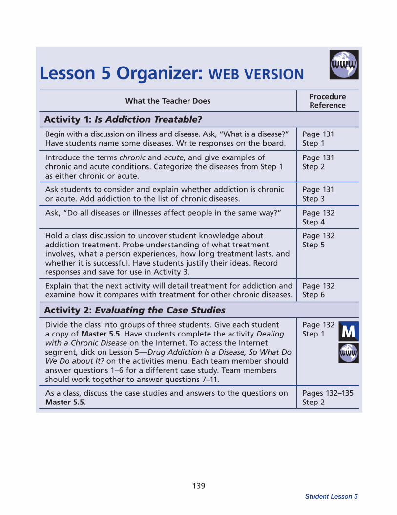
139
Lesson 5 Organizer: WEB VERSION
What the Teacher Does Procedure Reference
Activity 1: Is Addiction Treatable?Begin with a discussion on illness and disease. Ask, “What is a disease?” Have students name some diseases. Write responses on the board.
Page 131 Step 1
Introduce the terms chronic and acute, and give examples of chronic and acute conditions. Categorize the diseases from Step 1 as either chronic or acute.
Page 131Step 2
Ask students to consider and explain whether addiction is chronic or acute. Add addiction to the list of chronic diseases.
Page 131Step 3
Ask, “Do all diseases or illnesses affect people in the same way?” Page 132 Step 4
Hold a class discussion to uncover student knowledge about addiction treatment. Probe understanding of what treatment involves, what a person experiences, how long treatment lasts, and whether it is successful. Have students justify their ideas. Record responses and save for use in Activity 3.
Page 132 Step 5
Explain that the next activity will detail treatment for addiction and examine how it compares with treatment for other chronic diseases.
Page 132 Step 6
Activity 2: Evaluating the Case StudiesDivide the class into groups of three students. Give each student a copy of Master 5.5. Have students complete the activity Dealing with a Chronic Disease on the Internet. To access the Internet segment, click on Lesson 5—Drug Addiction Is a Disease, So What Do We Do about It? on the activities menu. Each team member should answer questions 1–6 for a different case study. Team members should work together to answer questions 7–11.
Page 132 Step 1
As a class, discuss the case studies and answers to the questions on Master 5.5.
Pages 132–135Step 2
Student Lesson 5

140
What the Teacher Does Procedure Reference
Activity 3: Is Treatment for Drug Addiction Effective?Display students’ ideas on addiction treatment from Activity 1, Step 5. Do they now see these ideas as correct or incorrect? Have students revise any incorrect statements and explain their changes.
Pages 136–137Step 1
The individual in the case study experienced relapse at one point. She started using drugs again after stopping for a while. Ask students if relapse means that treatment is not effective.
Page 137 Step 2
Have students consider the problems of following a treatment plan. Have they ever made New Year’s resolutions? How long did they keep the resolution and why did they break it?
Page 137 Step 3
Activity 4: Addiction Is a Brain DiseaseRead the following scenario to the class:
Robert has been arrested several times for drug possession. After the first arrest, he was given probation. After the second and third arrests, he was sentenced to jail for one year each time. The police arrested him a fourth time, but instead of having Robert serve more time in jail, the judge ordered him to enter a drug treatment program.
Page 138Step 1
Ask students to write a paper that provides scientific information that would support the judge’s decision to have Robert undergo drug treatment. Instruct students to incorporate information they have learned from Lessons 1–5 to support their position.
Page 138Step 2
= Involves using the Internet.
= Involves copying a master.
The Brain: Understanding Neurobiology Through the Study of Addiction
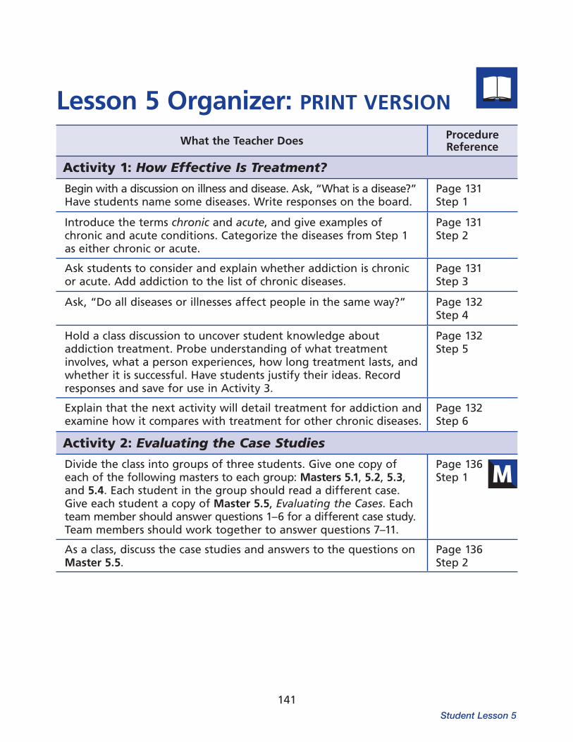
141
Lesson 5 Organizer: PRINT VERSION
What the Teacher Does Procedure Reference
Activity 1: How Effective Is Treatment?Begin with a discussion on illness and disease. Ask, “What is a disease?” Have students name some diseases. Write responses on the board.
Page 131 Step 1
Introduce the terms chronic and acute, and give examples of chronic and acute conditions. Categorize the diseases from Step 1 as either chronic or acute.
Page 131Step 2
Ask students to consider and explain whether addiction is chronic or acute. Add addiction to the list of chronic diseases.
Page 131Step 3
Ask, “Do all diseases or illnesses affect people in the same way?” Page 132 Step 4
Hold a class discussion to uncover student knowledge about addiction treatment. Probe understanding of what treatment involves, what a person experiences, how long treatment lasts, and whether it is successful. Have students justify their ideas. Record responses and save for use in Activity 3.
Page 132Step 5
Explain that the next activity will detail treatment for addiction and examine how it compares with treatment for other chronic diseases.
Page 132Step 6
Activity 2: Evaluating the Case StudiesDivide the class into groups of three students. Give one copy of each of the following masters to each group: Masters 5.1, 5.2, 5.3, and 5.4. Each student in the group should read a different case. Give each student a copy of Master 5.5, Evaluating the Cases. Each team member should answer questions 1–6 for a different case study. Team members should work together to answer questions 7–11.
Page 136 Step 1
As a class, discuss the case studies and answers to the questions on Master 5.5.
Page 136Step 2
Student Lesson 5

142
= Involves copying a master.
What the Teacher Does Procedure Reference
Activity 3: Is Treatment for Drug Addiction Effective?Display students’ ideas on addiction treatment from Activity 1, Step 5. Do students now see these ideas as correct or incorrect? Have students revise any incorrect statements and explain their changes.
Pages 136–137Step 1
The individual in the case study experienced relapse at one point. She started using drugs again after stopping for a while. Ask students if relapse means that treatment is not effective.
Page 137 Step 2
Have students consider the problems of following a treatment plan. Have they ever made New Year’s resolutions? How long did they keep the resolution and why did they break it?
Page 137 Step 3
Activity 4: Addiction Is a Brain DiseaseRead the following scenario to the class:
Robert has been arrested several times for drug possession. After the first arrest, he was given probation. After the second and third arrests, he was sentenced to jail for one year each time. The police arrested him a fourth time, but instead of having Robert serve more time in jail, the judge ordered him to enter a drug treatment program.
Page 138Step 1
Ask students to write a paper that provides scientific information that would support the judge’s decision to have Robert undergo drug treatment. Instruct students to incorporate information they have learned from Lessons 1–5 to support their position.
Page 138Step 2
The Brain: Understanding Neurobiology Through the Study of Addiction
143
Office of National Drug Control PolicyThe purpose of the Office of National Drug Control Policy (ONDCP) is to establish policies, priorities, and objectives for the nation’s drug control pro gram. The National Drug Control Policy is available on this Web site. This site also provides information about specific drugs (including statistics on their use), treatment, research, and enforcement.
Society for NeuroscienceThe Society for Neuroscience is the world’s largest organization of scientists and physicians dedicated to understanding the brain, spinal cord, and peripheral nervous system. This site provides a wide variety of information on topics related to the function of the brain and nervous system. The site also provides an opportunity to submit a spe cific question that may be answered online.
Partnership for a Drug-free AmericaInformation posted at this address includes infor mation about specific drugs and their effects.
The Dana FoundationThe Charles A. Dana Foundation is a private phil anthropic foundation with principal interests in health and education. Their Web site provides information for the general public on the latest research findings about the brain and brain disor ders. The Web site also provides access to their publications.
The following resources may provide additional background information for you or your students about neurobiology or drugs of abuse.
RESOURCES ON THE INTERNET
National Institute on Drug Abuse (NIDA)NIDA is the world’s leading supporter of research on the health aspects of drug abuse and addiction. This site provides current and authoritative infor mation about the latest research on drugs and addiction.
NIDA DRUGPUBS Research Dissemination CenterNIDA publications are available through NIDA DRUGPUBS. Included are the latest available student and teacher materials, prevention packets, booklets, posters, research reports, clinical reports, clinical reports, survey reports, and brochures. Most of these can be downloaded or ordered for free, by phone (1-877-NIDA-NIH, or 1-877-643-2644; TTY/TDD: 240-645-0228), fax (240-645-0227), or e-mail ([email protected]).
National Clearinghouse for Alcohol and Drug Information (NCADI)NCADI is part of the U.S. Department of Health and Human Services and functions as the informa tion service for the Center for Substance Abuse Prevention.
Additional Resources for Teachers

144
BOOKS AND VIDEOTAPE
Friedman, D.P., and Rusche, S. 1999. False Messen gers: How Addictive Drugs Change the Brain. Amster dam: Harwood Academic Publishers.
Kuhn, C., Swarzwelder, S., and Wilson, W. 1998. Buzzed: The Straight Facts about the Most Used and Abused Drugs from Alcohol to Ecstasy. New York: W.H. Norton & Company.
Gross de Núñez, G., and Schwartz-Bloom, R.D. 1998. Animated Neuroscience and the Action of Nico tine, Cocaine, and Marijuana in the Brain. Princeton, NJ: Films for the Humanities and Sciences.
The ReconstructorsThis Web game enables students to learn more about the history of opioids, club drugs, and inhalants. The activities incorporate aspects of chem istry, neuroscience, medicine, public policy, and history.
Office of Science EducationThis address takes you directly to the home page of the National Institutes of Health’s Office of Sci ence Education. This site provides access to a vari ety of resources for teachers and students, including NIH publications on drug abuse and brain function.
U.S. National Library of MedicineThe U.S. National Library of Medicine is the world’s largest medical library. This site provides extensive online information about health issues and includes access to Medline and MedlinePlus for searching for information about specific health topics.
The Brain: Understanding Neurobiology Through the Study of Addiction

145
alcohol: A psychoactively complex drug in bever ages such as beer, wine, and whiskey. Alcohol is a depressant drug with potential for abuse and addiction.
all-or-none phenomenon: Used to describe an action potential and the principle that a nerve fiber will respond maximally or not at all to a stimulus.
amphetamines: Stimulant drugs with effects very similar to cocaine’s.
amygdala: A component of the limbic system involved in the expression and perception of emotion.
anandamide: A neurotransmitter produced in the body that binds to the cannabinoid receptor.
antagonist: A chemical that, when it binds to a receptor, blocks the cell from responding. Antagonists prevent agonists from binding, or attaching, to the receptor. Antagonists include caffeine (for adenosine) and naloxone (for opioids).
astrocyte: A type of glial cell that provides nutri ents, support, and insulation for neurons of the central nervous system.
axon: The fiber-like extension of a neuron through which the cell carries information to target cells.
axon terminal: The structure at the end of an axon that produces and releases chemicals (neu rotransmitters) to transmit the neuron’s message across the synapse.
barbiturates: Depressant drugs that produce relaxation and sleep. Sleeping pills such as pento barbital and secobarbital are barbiturates.
Definitions for the following terms were adapted from a variety of sources. Specific sources are listed in the References section.
absorption: The process by which elements move from outside of the body into the blood and other tissues. Breakdown products of food are absorbed through the stomach and intestines. When tobacco is smoked, nicotine is absorbed through the lungs.
acetylcholine: A neurotransmitter that func tions in the brain to regulate memory and that controls the actions of skeletal and smooth mus cle in the peripheral nervous system.
action potential: The electrical part of a neuron’s two-part, electrical-chemical message. An action potential consists of a brief pulse of electrical cur rent that travels along the axon. When the action potential reaches the axon terminal, it triggers neurotransmitter release.
acute: Refers to an effect, disease, or condition that has a relatively rapid onset, marked intensity, and short duration.
addiction: A chronic, relapsing brain disease characterized by compulsive drug-taking despite adverse health, social, or legal conse quences.
adenosine: A neurotransmitter that binds to the adenosine receptor. Adenosine is a by-product of adenosine triphosphate (ATP) metabolism and is an important regulator of sleep. Caffeine is an adenosine antagonist.
agonist: A drug that binds to a receptor of a cell and triggers a response by the cell. An agonist often mimics the action of a naturally occurring substance. Opioids, THC, and nicotine are examples.
Glossary
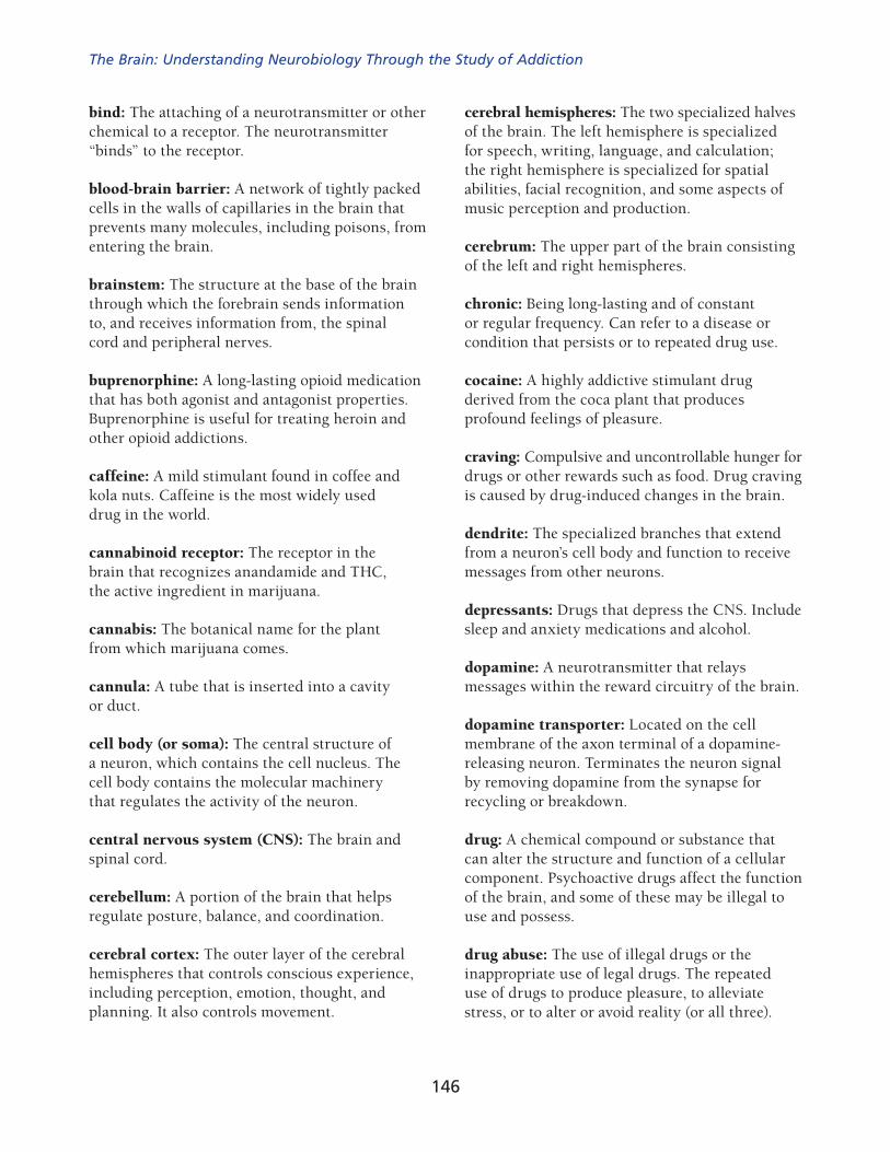
146
cerebral hemispheres: The two specialized halves of the brain. The left hemisphere is specialized for speech, writing, language, and calculation; the right hemisphere is specialized for spatial abili ties, facial recognition, and some aspects of music perception and production.
cerebrum: The upper part of the brain consisting of the left and right hemispheres.
chronic: Being long-lasting and of constant or regular frequency. Can refer to a disease or condition that per sists or to repeated drug use.
cocaine: A highly addictive stimulant drug derived from the coca plant that produces pro found feelings of pleasure.
craving: Compulsive and uncontrollable hunger for drugs or other rewards such as food. Drug craving is caused by drug-induced changes in the brain.
dendrite: The specialized branches that extend from a neuron’s cell body and function to receive messages from other neurons.
depressants: Drugs that depress the CNS. Include sleep and anxiety medications and alcohol.
dopamine: A neurotransmitter that relays messages within the reward circuitry of the brain.
dopamine transporter: Located on the cell membrane of the axon terminal of a dopamine-releasing neuron. Terminates the neuron signal by removing dopamine from the synapse for recycling or breakdown.
drug: A chemical compound or substance that can alter the structure and function of a cellular component. Psychoactive drugs affect the function of the brain, and some of these may be illegal to use and possess.
drug abuse: The use of illegal drugs or the inappropriate use of legal drugs. The repeated use of drugs to produce pleasure, to alleviate stress, or to alter or avoid reality (or all three).
bind: The attaching of a neurotransmitter or other chemical to a receptor. The neurotransmit ter “binds” to the receptor.
blood-brain barrier: A network of tightly packed cells in the walls of capillaries in the brain that prevents many molecules, including poisons, from entering the brain.
brainstem: The structure at the base of the brain through which the fore brain sends information to, and receives informa tion from, the spinal cord and peripheral nerves.
buprenorphine: A long-lasting opioid medication that has both agonist and antagonist prop erties. Buprenorphine is useful for treating heroin and other opioid addictions.
caffeine: A mild stimulant found in coffee and kola nuts. Caffeine is the most widely used drug in the world.
cannabinoid receptor: The receptor in the brain that recognizes anandamide and THC, the active ingredient in marijuana.
cannabis: The botanical name for the plant from which marijuana comes.
cannula: A tube that is inserted into a cavity or duct.
cell body (or soma): The central structure of a neuron, which contains the cell nucleus. The cell body contains the molecular machinery that reg ulates the activity of the neuron.
central nervous system (CNS): The brain and spinal cord.
cerebellum: A portion of the brain that helps reg ulate posture, balance, and coordination.
cerebral cortex: The outer layer of the cerebral hemispheres that controls conscious experience, including perception, emotion, thought, and planning. It also controls movement.
The Brain: Understanding Neurobiology Through the Study of Addiction
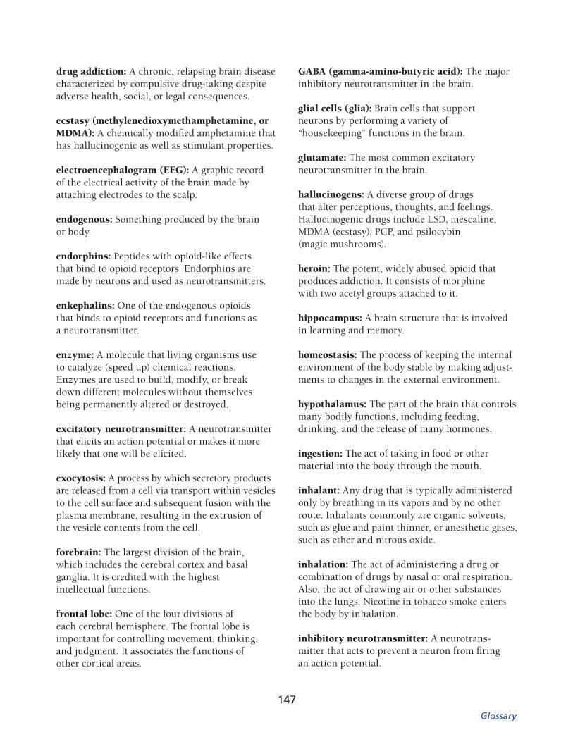
Glossary
147
GABA (gamma-amino-butyric acid): The major inhibitory neurotransmitter in the brain.
glial cells (glia): Brain cells that support neurons by performing a variety of “housekeeping” func tions in the brain.
glutamate: The most common excitatory neuro transmitter in the brain.
hallucinogens: A diverse group of drugs that alter perceptions, thoughts, and feelings. Hallu cinogenic drugs include LSD, mescaline, MDMA (ecstasy), PCP, and psilocybin (magic mush rooms).
heroin: The potent, widely abused opioid that produces addiction. It consists of morphine with two acetyl groups attached to it.
hippocampus: A brain structure that is involved in learning and memory.
homeostasis: The process of keeping the internal environment of the body stable by making adjust-ments to changes in the external environment.
hypothalamus: The part of the brain that controls many bodily functions, including feeding, drink ing, and the release of many hormones.
ingestion: The act of taking in food or other material into the body through the mouth.
inhalant: Any drug that is typically administered only by breathing in its vapors and by no other route. Inhalants commonly are organic sol vents, such as glue and paint thinner, or anes thetic gases, such as ether and nitrous oxide.
inhalation: The act of administering a drug or combination of drugs by nasal or oral respiration. Also, the act of drawing air or other substances into the lungs. Nicotine in tobacco smoke enters the body by inhalation.
inhibitory neurotransmitter: A neurotrans-mitter that acts to prevent a neuron from firing an action potential.
drug addiction: A chronic, relapsing brain disease characterized by compulsive drug-taking despite adverse health, social, or legal conse quences.
ecstasy (methylenedioxymethamphetamine, or MDMA): A chemically modified amphetamine that has hallucinogenic as well as stimulant properties.
electroencephalogram (EEG): A graphic record of the electrical activity of the brain made by attach ing electrodes to the scalp.
endogenous: Something produced by the brain or body.
endorphins: Peptides with opioid-like effects that bind to opioid receptors. Endorphins are made by neurons and used as neurotransmitters.
enkephalins: One of the endogenous opioids that binds to opioid receptors and functions as a neuro transmitter.
enzyme: A molecule that living organisms use to catalyze (speed up) chemical reactions. Enzymes are used to build, modify, or break down different molecules without themselves being permanently altered or destroyed.
excitatory neurotransmitter: A neurotransmitter that elicits an action potential or makes it more likely that one will be elicited.
exocytosis: A process by which secretory prod ucts are released from a cell via transport within vesicles to the cell surface and subsequent fusion with the plasma membrane, resulting in the extrusion of the vesicle contents from the cell.
forebrain: The largest division of the brain, which includes the cerebral cortex and basal ganglia. It is credited with the highest intellectual functions.
frontal lobe: One of the four divisions of each cerebral hemisphere. The frontal lobe is impor tant for controlling movement, thinking, and judgment. It associates the functions of other cortical areas.
148
naloxone: A short-acting opioid antagonist that binds to opioid receptors and blocks them, pre venting opioids from binding to these receptors.
naltrexone: Structurally similar to naloxone, an opioid antagonist used to treat heroin addiction and, more recently, alcohol addiction.
neuron (nerve cell): A unique type of cell found in the brain and body that is specialized to process and transmit information.
neurotransmission: The process that occurs when a neuron releases neurotransmitters to communi cate with another neuron across the synapse.
neurotransmitter: A chemical produced by neu rons to carry messages to other neurons.
nicotine: The addictive drug in tobacco. Nicotine activates a specific type of acetylcholine receptor.
NMDA (N-methyl-D-aspartate): A synthetic amino acid that is the defining agonist for the NMDA receptor, one of the glutamate receptors on neurons.
norepinephrine: A neurotransmitter and a hor mone. It is released by the sympathetic nervous system onto the heart, blood vessels, and other organs and by the adrenal gland into the blood stream as part of the fight-or-flight response. Norepinephrine in the brain is used as a neuro transmitter in normal brain processes.
nucleus: A cluster or group of nerve cells that is dedicated to performing its own special func tion(s). Nuclei are found in all parts of the brain but are called cortical fields in the cerebral cortex.
injection: A method of administering a substance such as a drug into the skin, subcutaneous tissue, muscle, blood vessels, or body cavities, usually by means of a needle.
limbic system: A set of brain structures that regulates our feelings, emotions, and motivations. It is also important in learning and memory.
LSD (lysergic acid diethylamide): A hallucino-genic drug that binds to and activates the serotonin receptor.
magnetic resonance imaging (MRI): An imaging technique that uses magnetic fields to generate images of the structure of the brain.
marijuana: A drug, usually smoked but it can be eaten, that is made from the leaves of the cannabis plant. The main psychoactive ingredient is THC.
medication: A drug that is used to treat an illness or disease according to established medical guidelines.
metabolism: The processes by which the body breaks things down or alters them so they can be eliminated.
methadone: A synthetic opioid used to treat pain and heroin addiction.
methamphetamine: A commonly abused, potent stimulant drug that is highly addictive and part of a larger family of amphetamines.
morphine: The most potent natural opiate com pound produced by the opium poppy. Morphine is a very effective medicine for treating pain.
myelin: Fatty material that surrounds and insu lates axons of some neurons.
The Brain: Understanding Neurobiology Through the Study of Addiction

Glossary
149
plasticity: The capacity of the brain to change its structure and function within certain limits. Plasticity underlies brain functions such as learning and allows the brain to generate nor mal, healthy responses to long-lasting environ mental changes.
positron: A positively charged particle having the same mass and spin as, but opposite charge of, an electron.
positron emission tomography (PET): An imaging technique for measuring brain function in living subjects by detecting the location and concentra tion of small amounts of radioactive chemicals.
postsynaptic neuron: The neuron that receives a given message from other neurons.
presynaptic neuron: The neuron that releases neu rotransmitters into the synaptic space to send messages to another neuron.
psychedelic drug: A drug that distorts perception, thought, and feeling. This term is typically used to refer to drugs with actions like those of LSD.
psychoactive drug: A drug that changes the way the brain works.
psychosocial therapy: Therapy that uses a combi nation of individual psychotherapy and group (social) therapy approaches to rehabilitate or pro vide the interpersonal and intrapersonal skills to help someone recover from drug addiction.
receptor: A protein that recognizes spe cific chemicals (normally neurotransmitters, hor mones, and similar endogenous substances) and transmits the message carried by the chemical into the cell on which the receptor resides.
nucleus accumbens: A part of the brain reward system that processes information related to motivation and reward. Virtually all drugs of abuse act on the nucleus accumbens to reinforce drug taking.
occipital lobe: The lobe of the cerebral cortex at the back of the head that includes the visual cortex.
opiate: Any of the psychoactive drugs that origi nate from the opium poppy or that have a chemi cal structure like the drugs derived from opium. Some opiates (such as opium, codeine, and mor phine) are derived from the plant, while others were first synthesized by chemists.
opioid: Any chemical that has opiate-like effects; commonly used to refer to endogenous neuro chemicals that activate opioid receptors but also includes natural, synthetic, and semisynthetic drugs.
opioid receptors: Receptors that recognize natural, synthetic, and endogenous opioids. When activated, they slow down or inhibit the activity of neurons on which they reside.
parallel processing: The division of an informa tion-processing job into smaller parts that are each handled simultaneously by various cortical fields and brain nuclei.
parietal lobe: One of the four subdivisions of the cerebral cortex; it is involved in sensory processes, attention, and language.
phencyclidine (PCP): Originally developed as an anesthetic, PCP may act as a hallucinogen, stim ulant, or sedative.
pituitary gland: An endocrine organ closely linked with the hypothalamus. The pituitary secretes a number of hormones that regulate the activity of other endocrine organs in the human body.

150
second messenger: A molecule produced inside neurons as a step in the process of communica tion between cells. The second messenger lets other parts of the cell know that a specific recep tor has been activated, thereby completing the message carried by the neurotransmitter that bound to the receptor. Some receptors (dopamine and opiate receptors, for example) use second messengers. Others (nicotine and GABA recep tors, for example) do not.
sensitization: An increased response to a drug caused by repeated administration. It is most commonly seen in some responses to stimulants.
serotonin: A neurotransmitter that regulates many functions, including mood, appetite, and sensory perception.
single photon emission computed tomography (SPECT): An imaging process that measures the emission of single photons of a given energy from radioactive tracers in the human body.
stimulants: A class of drugs that elevates mood, increases feelings of well-being, and increases energy and alertness. Stimu lants include nicotine, cocaine, methamphetamine, and methylphenidate (Ritalin).
synapse: The site where presynaptic and postsy naptic neurons communicate with each other.
synaptic space (or synaptic cleft): The intercellu lar space between the presynaptic and postsynap tic neurons.
temporal lobe: One of the four major subdivi-sions of each hemisphere of the cerebral cortex. It functions in auditory perception, speech, and visual perceptions.
relapse: In drug abuse, relapse is the resumption of drug use after stopping. Relapse is a common occurrence in many chronic disorders, including addiction.
resting membrane potential: The difference in electrical charge between the inside and the out side of a nerve cell when the cell is not firing. The inside of a resting neuron has a greater negative charge than the outside of the neuron.
reuptake: The process by which neurotransmit-ters are removed from the synapse by being “pumped” through transporters back into the axon terminals that first released them.
reuptake pump (transporter): The protein that actually transports neurotransmitter molecules back into the axon terminals that released them.
reward: The process that reinforces behavior, making it more likely to recur. It is mediated at least in part by the release of dopamine into the nucleus accumbens.
reward pathway (or brain reward system): A brain circuit that, when activated, reinforces behaviors. The circuit includes the dopamine-containing neurons of the ventral tegmental area, the nucleus accumbens, and part of the prefrontal cortex. The activation of this circuit causes feel ings of pleasure.
route of administration: The way a drug is introduced into the body. Drugs can enter the body by eat ing, drinking, inhaling, injecting, snorting, smok ing, or absorption through mucous membranes.
rush: Intense feelings of euphoria a drug pro duces when it is first injected or smoked.
The Brain: Understanding Neurobiology Through the Study of Addiction

Glossary
151
ventral tegmental area (VTA): The group of dopamine-containing cell bodies that make up a key part of the brain reward system. These neu rons extend axons to the nucleus accumbens and the prefrontal cortex.
vesicle: A membranous sac within an axon termi nal that stores and releases neurotransmitter.
withdrawal: Symptoms that occur when a person who is dependent on a drug abruptly stops using the drug.
tetrahydrocannabinol (THC): The active ingredi-ent in marijuana that is primarily responsible for producing the drug’s psychoactive effects.
thalamus: Located deep within the brain, the thalamus is the key relay station for sensory information flowing into the brain from the periphery. It also serves as a relay station for motor information leaving the brain to regulate function of the muscles.
tolerance: A physiological change resulting from repeated drug use that requires the user to take larger amounts of the drug to get the same effect initially felt from a smaller dose.
transporter: A large protein on the cell membrane of the axon terminals. It removes neurotransmit ter molecules from the synapse by carrying them back into the axon terminal that released them.


153
5. Gatley, S.J., and Volkow, N.D. 1998. Addiction and imaging of the living human brain. Drug and Alcohol Dependence, 51, 97-108.
6. National Institute on Drug Abuse. 1996. The basics of brain imaging. NIDA Notes.
7. Lincoln, A. The Gettysburg Address.
8. Damasio, H., Grabowski, T., Frank, R., Gal aburda, A.M., and Damasio, A.R. 1994. The return of Phineas Gage: Clues about the brain from the skull of a famous patient. Science, 264, 1102-1105.
9. Macmillan, M. The Phineas Gage information page.
Lesson 2—Neurons, Brain Chemistry, and Neurotransmission
1. Friedman, D.P., and Rusche, S. 1999. False mes sengers: How addictive drugs change the brain. Amsterdam: Harwood Academic Publishers.
2. National Institute on Drug Abuse. 1997. Mind Over Matter: The Brain’s Response to Drugs. NIH Publication No. 98-3592.
3. Kandel, E.R. 1991. Nerve cells and behavior. In E.R. Kandel, J.H. Schwartz, and T.M. Jessell (Eds.), Principles of Neural Science, 3rd edition (pp. 18-32). Norwalk, CT: Appleton & Lange.
Introduction to the Module
1. Loucks-Horsley, S., Love, N., Hewson, P.W., and Stiles, K.E. 1998. Designing Professional Development for Teachers of Science and Mathemat ics. Thousand Oaks, CA: Corwin Press.
Implementing the Module
1. National Research Council. 1996. National Sci ence Education Standards. Washington, DC: National Academy Press.
Lesson 1—The Brain: What’s Going On in There?
1. Friedman, D.P., and Rusche, S. 1999. False Mes sengers: How Addictive Drugs Change the Brain. Amsterdam: Harwood Academic Publishers.
2. National Institute on Drug Abuse. 1997. Mind Over Matter: The Brain’s Response to Drugs. NIH Publication No. 98-3592.
3. Kandel, E.R. 1991. Brain and behavior. In E.R. Kandel, J.H. Schwartz, and T.M. Jessell (Eds.), Principles of Neural Science, 3rd edition (pp. 5-17). Norwalk, CT: Appleton & Lange.
4. Martin, J.H., Brust, J.C.M., and Hilal, S. 1991. In E.R. Kandel, J.H. Schwartz, and T.M. Jes sell (Eds.), Principles of Neural Science, 3rd edi tion (pp. 309-324). Norwalk, CT: Appleton & Lange.
References
154
4. Gross de Núñez, G., and Schwartz-Bloom, R.D. 1998. Animated neuroscience and the action of nico tine, cocaine, and marijuana in the brain. Prince ton, NJ: Films for the Humanities and Sciences.
5. National Institute on Drug Abuse. 2006. Methamphetamine abuse and addiction. NIDA Research Report. NIH Publication No. 06- 4210.
Lesson 4—Drug Abuse and Addiction
1. Steindler, E.M. 1998. ASAM addiction termi-nology. In Principles of Addiction Medicine, 2nd edition (pp. 1301-1304). Chevy Chase, MD: American Society of Addiction Medicine.
2. Kuhn, C., Swarzwelder, S., and Wilson, W. 1998. Buzzed: The Straight Facts about the Most Used and Abused Drugs from Alcohol to Ecstasy. New York: W.H. Norton & Company.
3. Friedman, D.P., and Rusche, S. 1999. False Mes sengers: How Addictive Drugs Change the Brain. Amsterdam: Harwood Academic Publishers.
4. Childress, A.R., Mozley, P.D., Elgin, W., Fitzgerald, J., Reivich, M., and O’Brien, C.P. 1999. Limbic activation during cue-induced cocaine craving. American Journal of Psychiatry, 156, 11-18.
5. Eriksson, P.S., Perfilieva, E., Bjork-Eriksson, T., Alborn, A.M., Nordborg, C., Peterson, D.A., and Gage, F.H. 1998. Neurogenesis in the adult human hippocampus. Nature Medi cine, 4, 1313-1317.
4. Rowland, L.P., Fink, M.E. and Rubin, L. 1991. Cerebrospinal fluid: Blood-brain barrier, brain edema, and hydrocephalus. In E.R. Kandel, J.H. Schwartz, and T.M. Jessell (Eds.), Princi-ples of Neural Science, 3rd edition (pp. 1050-1060). Norwalk, CT: Appleton & Lange.
5. Society for Neuroscience. 1999. Blood-brain barrier.
6. Darnell, J., Lodish, H., and Baltimore, D. 1990. Nerve cells and the electric properties of cell membranes. In Molecular Cell Biology, 2nd edi tion (pp. 763-814). New York: Scientific Ameri can Books, W.H. Freeman and Company.
7. Guyton, A.C., and Hall, J.E. 1996. Organiza tion of the nervous system; basic functions of synapses and transmitter substances. In Text book of Medical Physiology, 9th edition (pp. 565-582). Philadelphia: W.B. Saunders Company.
8. Society for Neuroscience. 1999. The short answer: Definitions for common neuroscience terms.
Lesson 3—Drugs Change the Way Neurons Communicate
1. Friedman, D.P., and Rusche, S. 1999. False Mes sengers: How Addictive Drugs Change the Brain. Amsterdam: Harwood Academic Publishers.
2. National Institute on Drug Abuse. 2005. Mind Over Matter: The Brain’s Response to Drugs. NIH Publication No. 05-3592.
3. Kuhn, C., Swarzwelder, S., and Wilson, W. 1998. Buzzed: The Straight Facts about the Most Used and Abused Drugs from Alcohol to Ecstasy. New York: W.H. Norton & Company.
The Brain: Understanding Neurobiology Through the Study of Addiction

155
Lesson 5—Drug Addiction Is a Disease, So What Do We Do about It?
1. Leshner, A.I. 1997. Addiction is a brain dis ease, and it matters. Science, 278, 45-47.
2. Tobler, N.S., Roona, M.R., Ochshan, P., Marchall, D.G., Streke, A.V., Stackpole, K.M. 2000. School-based adolescent drug abuse prevention programs: 1998 Meta-analysis. Journal of Primary Prevention, 20, 275-335.
3. O’Brien, C.P., and McLellan, A.T. 1998. Myths about the treatment of addiction. In Principles of Addiction Medicine, 2nd edition (pp. 309-313). Chevy Chase, MD: American Society of Addiction Medicine.
4. National Institute on Drug Abuse. 2008. Treat ment medications. NIDA Infofacts.
5. Friedman, D.P., and Rusche, S. 1999. False Mes sengers: How Addictive Drugs Change the Brain. Amsterdam: Harwood Academic Publishers.
6. National Institute on Drug Abuse. 2009. Princi ples of Drug Addiction Treatment: A Research-Based Guide, 2nd edition. NIH Publication No. 09-4180.
7. National Institute on Drug Abuse. 2008. Treat ment approaches for drug addiction. NIDA Infofacts.
8. Baler, R.D., and Volkow, N.D. 2006. Drug addiction: the neurobiology of disrupted self-control. Trends in Molecular Medicine, 12, 559-66.
6. Villemagne, V., Yuan, J., Wong, D.F., Dannals, R.F., Hatzidimitriou, G., Matthews, W.B., Ravert, H.T., Musachio, J., McCann, U.D., and Ricaurte, G.A. 1998. Brain dopamine neuro toxicity in baboons treated with doses of methamphetamine comparable to those recre ationally abused by humans: Evidence from [11C]WIN-35,428 positron emission tomogra phy studies and direct in vitro determina tions. Journal of Neuroscience, 18, 418-427.
7. Fischer, C., Hatzidimitriou, G., Wlos, J., Katz, J., and Ricaurte, G. 1995. Reorganization of ascending 5-HT axon projections in animals previously exposed to the recreational drug (+)3,4-methylenedioxymethamphetamine (MDMA, “Ecstasy”). Journal of Neuroscience, 15, 5476-5485.
8. Volkow, N.D., Hitzemann, R., Wany, G.-J., Fowler, J.S., Wolf, A.P., and Dewey, S.L. 1992. Long-term frontal brain metabolic changes in cocaine abusers. Synapse, 11, 184-190.
9. National Institute on Drug Abuse. 1999. Drug abuse and addiction research: 25 years of discov ery to advance the health of the public.
10. Gogtay, N., Giedd, J.N., Lusk, L., et al. 2004. Dynamic mapping of human cortical development during childhood through early adulthood. Proceedings of the National Academy of Sciences USA, 101(21), 8174-9.
11. Joy, J.E., Watson, S.J., Jr., and Benson, J.A., Jr. (Eds.). 1999. Marijuana and medicine: Assessing the science base.
12. Stanton, M.D. 1976. Drugs, Vietnam, and the Vietnam veteran: An overview. American Jour nal of Drug and Alcohol Abuse, 3, 557-570.
13. National Institutes of Health. 1996. Public health service policy on humane care and use of laboratory animals.
References
156
Glossary
1. Friedman, D.P., and Rusche, S. 1999. False Messengers: How Addictive Drugs Change the Brain. Amsterdam: Harwood Academic Publishers.
2. Society for Neuroscience. 1999. The short answer: Definitions for common neuroscience terms.
3. Morris, C. (editor). 1999. Academic Press Dictio nary of Science and Technology. San Diego: Academic Press.
4. Brynie, F.H. 1998. 101 Questions Your Brain Has Asked about Itself but Couldn’t Answer Until Now. Brookfield, CT: Millbrook Press.
The Brain: Understanding Neurobiology Through the Study of Addiction

157
MastersRefer to the In Advance section in each lesson for more information about the number of copies required for each master. Masters marked with an asterisk (*) are not needed if you use the Web-based version of the activity.
Lesson 1—The Brain: What’s Going On in There?Master 1.1: Positron Emission Tomography (PET) Images*Master 1.2: Interpreting PET ImagesMaster 1.3: PET Image TasksMaster 1.4: Major Regions of the BrainMaster 1.5: Areas of the Cerebral Cortex and Their FunctionsMaster 1.6: What Happened to Phineas Gage?Master 1.7: The Reward System
Lesson 2—Neurons, Brain Chemistry, and NeurotransmissionMaster 1.7: The Reward System (from Lesson 1)Master 2.1: Anatomy of a NeuronMaster 2.2: Neurons Interact with Other Neurons through SynapsesMaster 2.3: How Do Neurons Communicate?Master 2.4: Neurons Communicate by Neurotransmission*Master 2.5: NeurotransmissionMaster 2.6: Recording the Activity of a NeuronMaster 2.7: Neurotransmitter ActionsMaster 2.8: Neurons in Series
Lesson 3—Drugs Change the Way Neurons CommunicateMaster 3.1: Cocaine Alters NeurotransmissionMaster 3.2: Methamphetamine and Nicotine Disrupt NeurotransmissionMaster 3.3: How Does Alcohol Affect Neurotransmission?Master 3.4: Parent LetterMaster 3.5: Caffeine: How Does Your Heart Respond?Master 3.6: How Do Drugs Get Into the Brain?Master 3.7: What Should the Doctor Do?
158
Lesson 4—Drug Abuse and AddictionMaster 4.1: Data for Rat Self-administration ExperimentMaster 4.2: Worksheet for Rat Experiment DataMaster 4.3: Evaluating the ExperimentMaster 4.4: Playing the GameMaster 4.5: Who Is Addicted?Master 4.6: Long-term Effects of Drugs on the Brain*
Lesson 5—Drug Addiction Is a Disease, So What Do We Do about It?Master 5.1: Ruth’s Story*Master 5.2, Mike’s Story*Master 5.3: Carol’s Story*Master 5.4: Disease Reference Information*Master 5.5: Evaluating the Cases
* Not needed if you use the Web-based version of the activity.
The Brain: Understanding Neurobiology Through the Study of Addiction

Set 1 Set 2 a b a b Set 4 Set 5
Positron Emission Tomography (PET) Images
Each set of PET images below contains four images of a human brain. The four images show cross-sections taken at different levels of the brain.
a b a b
c d
Set 6
a b
c d
Set 3 a b
c d
c d
highest activity
Cop
yrig
ht ©
200
0 by
BSC
S an
d V
ideo
disc
over
y, I
nc. P
erm
issi
on g
rant
ed fo
r cl
assr
oom
use
. Upd
ated
200
9.
lowest activity
Master 1.1a

Set 4 Set 5 a b a b
c d
Set 6 a b
c d
c d
PET images provided by:
Sanjiv S. Gambhir, M.D., Ph.D. UCLA School of Medicine
Crump Institute for Biological Imaging Copyright 1998
Regents of the University of California
Cop
yrig
ht ©
200
0 by
BSC
S an
d V
ideo
disc
over
y, I
nc. P
erm
issi
on g
rant
ed fo
r cl
assr
oom
use
. Upd
ated
200
9.
Master 1.1b

Interpreting PET Images C
opyr
ight
© 2
000
by B
SCS
and
Vid
eodi
scov
ery,
Inc
. Per
mis
sion
gra
nted
for
clas
sroo
m u
se. U
pdat
ed 2
009.
Name(s)______________________________________________________________ Date ______________
1. When you look at the images that make up Set #1 (Master 1.1), how do the four images differ from each other?
2. Why are four images shown in each set of PET images? Why would scientists need to examine more than one PET image taken of a subject’s brain?
3. When comparing the images in Set #1 with the images in Sets #2, 3, 4, 5, and 6, how is the activity of the brain in each of these sets different from Set #1’s?
Set Number
Identify the image that shows the greatest change
(a, b, c, or d) Describe the change in brain activity
2
3
4
5
6
Master 1.2a

4. The PET images shown in Set #1 show brain activity in a resting brain. The images in Sets #2 through 6 show activity in the brains of humans who are doing different tasks. When you look at the PET scans and the chart in question #3, what generalizations can you make about the activity of the brain when different tasks are performed?
5. Compare the tasks that the subject performed during each of the PET scans (as shown on the overhead transparency of Master 1.3) with the individual’s brain activity. Use the information from the over head and from the PET images to complete the following chart.
Cop
yrig
ht ©
200
0 by
BSC
S an
d V
ideo
disc
over
y, I
nc. P
erm
issi
on g
rant
ed fo
r cl
assr
oom
use
. Upd
ated
200
9.
Master 1.2b
Brain region that is Set more active in the This region is involved in
Number
2
3
4
5
6
PET image
auditory cortex
primary visual cortex
frontal cortex
hippocampus
motor cortex
processing information related to

PET Image Tasks
The tasks that the subject performed during each of the PET scans are as follows:
• Set #1 Subject is resting.
• Set #2 Subject is listening to music.
• Set #3 Subject is looking at a picture showing both pattern and color.
• Set #4 Subject is performing a thinking task.
• Set #5 Subject must remember an image for later recall.
• Set #6 Subject is hopping up and down on the right foot.
Cop
yrig
ht ©
200
0 by
BSC
S an
d V
ideo
disc
over
y, I
nc. P
erm
issi
on g
rant
ed fo
r cl
assr
oom
use
. Upd
ated
200
9.
Master 1.3

Major Regions of the Brain C
opyr
ight
© 2
000
by B
SCS
and
Vid
eodi
scov
ery,
Inc
. Per
mis
sion
gra
nted
for
clas
sroo
m u
se. U
pdat
ed 2
009.
Cerebral Cortex
Midbrain
Thalamus
Cerebellum
Hypothalamus
Amygdala Brainstem
Hippocampus
Spinal Cord
Drawing of a brain cut in half, showing the major regions.
Source: National Institute on Drug Abuse (1997). Mind Over Matter: The Brain’s Response to Drugs, Teacher’s Guide.
Master 1.4

Areas of the Cerebral Cortex and Their Functions
Cop
yrig
ht ©
200
0 by
BSC
S an
d V
ideo
disc
over
y, I
nc. P
erm
issi
on g
rant
ed fo
r cl
assr
oom
use
. Upd
ated
200
9.
Drawing of a brain cut in half, showing areas of the cerebral cortex and their functions.
Source: National Institute on Drug Abuse (1997). Mind Over Matter: The Brain’s Response to Drugs, Teacher’s Guide.
Master 1.5
What Happened to Phineas Gage? C
opyr
ight
© 2
000
by B
SCS
and
Vid
eodi
scov
ery,
Inc
. Per
mis
sion
gra
nted
for
clas
sroo
m u
se. U
pdat
ed 2
009.
Name(s)______________________________________________________________ Date ______________
Due to an accident while he was working, Phineas Gage made a contribution to the understanding of how the brain works. In 1848, 25-year-old Phineas Gage worked for the Rutland and Burlington Railroad Company laying railroad tracks across Vermont. Before railroad track could be laid, however, the uneven ground needed to be leveled. Gage and coworkers had to drill holes in the stone, put explosive in the holes, cover the explosive with sand, and then use a fuse and tamping iron to trigger an explosion. One day, an accident occurred that changed Gage’s life forever. The explosive went off early, sending the tamping iron, which was 1.25 inches in diameter and 43 inches long, shooting into Gage’s face, through his skull and brain, and out the top of his head. The tamping iron landed about 25 yards away. Gage regained consciousness within a few minutes. Amazingly, he not only survived the blast, but he was able to talk and to walk! His coworkers took him to the doctor, who cleaned and bandaged the wounds, the standard medical treatment at the time.
Although Gage survived the physical injuries from the blast, he was a changed man. He appeared to be just as intelligent as before the accident, and he did not have any impairment in movement, speech, or memory. But, something was different. Before the accident, he was a responsible, intelligent, and likeable person. After the accident, he was irresponsible, used profanity extensively, and demonstrated no respect for social customs. His friends commented that “Gage was no longer Gage.” He could not hold the responsible jobs that he had before the accident and apparently wandered for the next several years. Phineas Gage ended up in San Francisco in the custody of his family, where he died approximately 12 years after the accident.
Twenty years after the accident, the physician who treated Gage correlated the behavioral changes with damage to the frontal region of the brain. At the time, the brain was thought to control language and movement, but the suggestion that the brain functioned to process emotions and social behavior was new. In addition, scientists at the time believed the brain lacked localized functions. Unknowingly, Phineas Gage contributed to our understanding of how the brain processes information.
In the 1990s, scientists used their improved understanding of brain function, computer modeling techniques, and new data from Gage’s skull. On the basis of this information, they found that the acci-dent damaged both hemispheres of the frontal lobe, which is the part of the brain that influences social behavior. Today, physicians see patients with damage to the frontal lobe that has occurred through motor vehicle accidents, gun accidents, or major falls. These individuals, like Phineas Gage, often have dramatic changes in their emotional and decision-making abilities.
1. How did Phineas Gage change after the accident?
2. How did Phineas Gage’s accident change scientists’ understanding of the brain?
Master 1.6

The Reward System C
opyr
ight
© 2
000
by B
SCS
and
Vid
eodi
scov
ery,
Inc
. Per
mis
sion
gra
nted
for
clas
sroo
m u
se. U
pdat
ed 2
009.
Drawing of a brain cut in half, showing the reward system.
Source: National Institute on Drug Abuse (1997). Mind Over Matter: The Brain’s Response to Drugs, Teacher’s Guide.
Master 1.7

Anatomy of a Neuron C
opyr
ight
© 2
000
by B
SCS
and
Vid
eodi
scov
ery,
Inc
. Per
mis
sion
gra
nted
for
clas
sroo
m u
se. U
pdat
ed 2
009.
Master 2.1
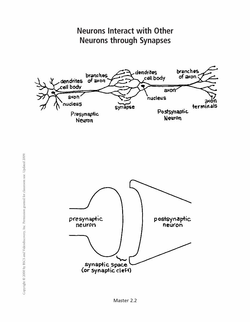
Neurons Interact with OtherNeurons through Synapses
Cop
yrig
ht ©
200
0 by
BSC
S an
d V
ideo
disc
over
y, I
nc. P
erm
issi
on g
rant
ed fo
r cl
assr
oom
use
. Upd
ated
200
9.
Master 2.2
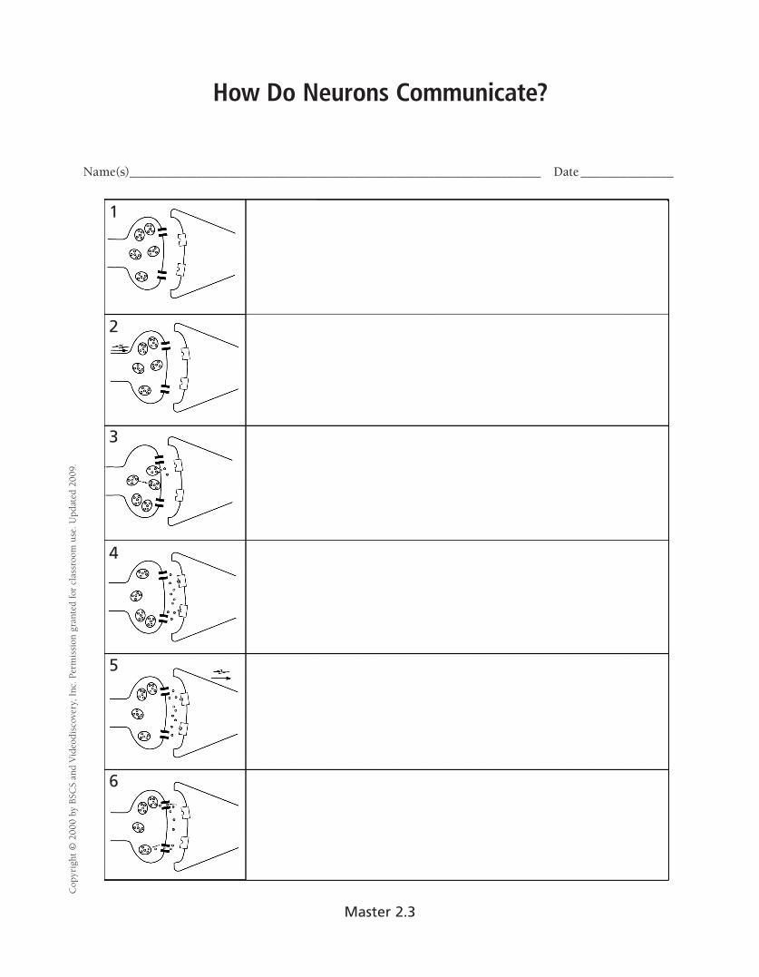
How Do Neurons Communicate?
Name(s)______________________________________________________________ Date ______________
Cop
yrig
ht ©
200
0 by
BSC
S an
d V
ideo
disc
over
y, I
nc. P
erm
issi
on g
rant
ed fo
r cl
assr
oom
use
. Upd
ated
200
9.
1
2
3
4
5
6
Master 2.3
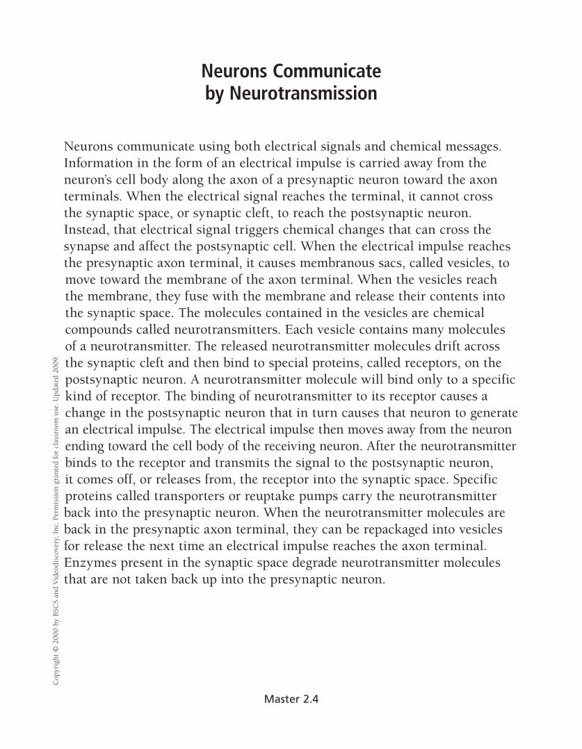
Cop
yrig
ht ©
200
0 by
BSC
S an
d V
ideo
disc
over
y, I
nc. P
erm
issi
on g
rant
ed fo
r cl
assr
oom
use
. Upd
ated
200
9.
Neurons Communicate by Neurotransmission
Neurons communicate using both electrical signals and chemical messages. Information in the form of an electrical impulse is carried away from the neuron’s cell body along the axon of a presynaptic neuron toward the axon terminals. When the electrical signal reaches the terminal, it cannot cross the synaptic space, or synaptic cleft, to reach the postsynaptic neuron. Instead, that electrical signal triggers chemical changes that can cross the synapse and affect the postsynaptic cell. When the electrical impulse reaches the presynaptic axon terminal, it causes membranous sacs, called vesicles, to move toward the membrane of the axon terminal. When the vesicles reach the membrane, they fuse with the membrane and release their contents into the synaptic space. The molecules contained in the vesicles are chemical compounds called neurotransmitters. Each vesicle contains many molecules of a neurotransmitter. The released neurotransmitter molecules drift across the synaptic cleft and then bind to special proteins, called receptors, on the postsynaptic neuron. A neurotransmitter molecule will bind only to a specific kind of receptor. The binding of neurotransmitter to its receptor causes a change in the postsynaptic neuron that in turn causes that neuron to generate an electrical impulse. The electrical impulse then moves away from the neuron ending toward the cell body of the receiving neuron. After the neurotransmitter binds to the receptor and transmits the signal to the postsynaptic neuron, it comes off, or releases from, the receptor into the synaptic space. Specific proteins called transporters or reuptake pumps carry the neurotransmitter back into the presynaptic neuron. When the neurotransmitter molecules are back in the presynaptic axon terminal, they can be repackaged into vesicles for release the next time an electrical impulse reaches the axon terminal. Enzymes present in the synaptic space degrade neurotransmitter molecules that are not taken back up into the presynaptic neuron.
Master 2.4

Neurotransmission
Name(s)______________________________________________________________ Date ______________
Cop
yrig
ht ©
200
0 by
BSC
S an
d V
ideo
disc
over
y, I
nc. P
erm
issi
on g
rant
ed fo
r cl
assr
oom
use
. Upd
ated
200
9.
1
2
3
4
5
6
Master 2.5

Recording the Activity of a Neuron C
opyr
ight
© 2
000
by B
SCS
and
Vid
eodi
scov
ery,
Inc
. Per
mis
sion
gra
nted
for
clas
sroo
m u
se. U
pdat
ed 2
009.
Master 2.6

Neurotransmitter Actions C
opyr
ight
© 2
000
by B
SCS
and
Vid
eodi
scov
ery,
Inc
. Per
mis
sion
gra
nted
for
clas
sroo
m u
se. U
pdat
ed 2
009.
Name(s)______________________________________________________________ Date ______________
The following diagrams represent recordings of the electrical activity of a neuron over a period of time. Each vertical line on the diagram represents an electrical impulse, or action potential, occurring in the neuron. The first diagram represents a neuron at rest. For the other recordings, a solution containing neurotransmitter was applied to the neuron.
1. Why is saline applied to the resting neuron?
2. When the neurotransmitter glutamate is applied to the neuron, how does its activity change?
3. How does the application of the two neurotransmitters, glutamate and GABA, change the activity of the neuron?
4. Predict how the activity of the neuron would change if only GABA was applied to the neuron.
5. Do all neurotransmitters affect a neuron in the same way?
6. How would the application of glutamate to a neuron change the amount of neurotransmitter released from that neuron? How would the application of GABA to a neuron change the amount of neurotransmitter released from that neuron?
Master 2.7

Neurons in Series C
opyr
ight
© 2
00V
ideo
disc
over
y, I
nc. P
erm
issi
on g
clas
sroo
m u
se. U
pdat
ed 2
009.
Name(s)______________________________________________________________ Date ______________
Using what you have learned about the effects of the neurotransmitters glutamate and GABA, determine how the different signals that affect Neuron #1 can change the release of the neurotransmitter dopamine from Neuron #2. Use the chart to help you work through the cases. You can use a down arrow to indicate a decrease or an up arrow to indicate an increase.
A. The signaling molecule is inhibitory. Neuron #1 releases glutamate as its neurotransmitter. Neuron #2 releases dopamine as its neurotransmitter.
r ofd e
B. The signaling molecule is excitatory. Neuron #1 releases glutamate as its neurotransmitter.
tna Neuron #2 releases dopamine as its neurotransmitter.
r
Master 2.8a
d naS
C. The signaling molecule is inhibitory. Neuron #1 releases GABA as its neurotransmitter.
CS Neuron #2 releases dopamine as its neurotransmitter.
By b
0
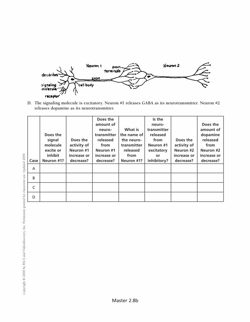
D. The signaling molecule is excitatory. Neuron #1 releases GABA as its neurotransmitter. Neuron #2 releases dopamine as its neurotransmitter.
Case
Does the signal
molecule excite or inhibit
Neuron #1?
Does the activity of Neuron #1 increase or decrease?
Does the amount of
neuro-transmitter
released from
Neuron #1 increase or decrease?
What is the name of the neuro- transmitter
released from
Neuron #1?
Is the neuro-
transmitter released
from Neuron #1 excitatory
or inhibitory?
Does the activity of Neuron #2 increase or decrease?
Does the amount of dopamine released
from Neuron #2 increase or decrease?
A
B
C
D
Cop
yrig
ht ©
200
0 by
BSC
S an
d V
ideo
disc
over
y, I
nc. P
erm
issi
on g
rant
ed fo
r cl
assr
oom
use
. Upd
ated
200
9.
Master 2.8b

Cocaine Alters Neurotransmission C
opyr
ight
© 2
000
by B
SCS
and
Vid
eodi
scov
ery,
Inc
. Per
mis
sion
gra
nted
for
clas
sroo
m u
se. U
pdat
ed 2
009.
Master 3.1
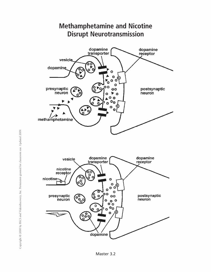
Methamphetamine and Nicotine Disrupt Neurotransmission
Cop
yrig
ht ©
200
0 by
BSC
S an
d V
ideo
disc
over
y, I
nc. P
erm
issi
on g
rant
ed fo
r cl
assr
oom
use
. Upd
ated
200
9.
Master 3.2

How Does Alcohol Affect Neurotransmission?
Cop
yrig
ht ©
200
0 by
BSC
S an
d V
ideo
disc
over
y, I
nc. P
erm
issi
on g
rant
ed fo
r cl
assr
oom
use
. Upd
ated
200
9.
Alcohol absent
GABA-releasing neuron
GABA receptorGABA
Cl–
Cl–
Cl–
Postsynaptic neuron
Alcohol present GABA receptor
Cl–
Cl–
Cl–
Cl–
Cl–
Cl–
Cl–
Cl–
Cl–
Cl–
GABA-releasing neuron
Master 3.3

Parent Letter C
opyr
ight
© 2
000
by B
SCS
and
Vid
eodi
scov
ery,
Inc
. Per
mis
sion
gra
nted
for
clas
sroo
m u
se. U
pdat
ed 2
009.
Dear Parents,
Next week in biology class, we will investigate the effect of caffeine on the body. Each student will need
to bring in a 12-ounce can of ______________________________. Please provide one can labeled with your
child’s name and class period.
During the activity, students will consume 12 ounces of the above-specified soft drink and measure what
effect it has, if any, on their heart rates.
Students are not to bring in any soft drink other than the one specified. Because the different brands and
flavors vary in their caffeine content, it is important that all students consume the same brand.
Students who choose not to bring in a soft drink, or those without signed permission forms, can
participate in the activity by drinking 12 ounces of water. They will be an important part of the activity
by serving as “controls.”
Thank you for your continued support.
Teacher’s Signature
My child, ______________________________________, has permission to participate in the caffeine activity
in class and will bring in a 12-ounce can of ______________________________ to consume as part of
the activity.
My child, ______________________________________, has permission to participate in the activity in class
and will bring in a 12-ounce can of caffeine-free ______________________________ to consume as part of
the activity.
My child, ______________________________________, will not drink a 12-ounce soft drink during the
activity, but will participate by drinking 12 ounces of water.
Parent’s or Guardian’s Signature: ______________________________________________
Date: _______________________
Master 3.4

Caffeine: How Does Your Heart Respond? C
opyr
ight
© 2
000
by B
SCS
and
Vid
eodi
scov
ery,
Inc
. Per
mis
sion
gra
nted
for
clas
sroo
m u
se. U
pdat
ed 2
009.
Name(s)______________________________________________________________ Date ______________
MATERIALS FOR EACH TEAM
2 cans of soft drink (caffeinated or caffeine-free) 1 watch or classroom clock with a second hand
PROCEDURE
Do Steps 1 to 3 with your teacher.
1. When your teacher directs you to do so, find your pulse. You can find it most easily by pressing two fingers against the artery in your neck or on the inside of your wrist. Practice counting the beats.
2. When your teacher directs you to start, count the number of beats you feel in 15 seconds. Your teacher will tell you when to stop. Record the number in the data table on the next page.
3. Multiply the number of beats you counted in 15 seconds by four to calculate your resting heart rate in beats per minute.
Complete the rest of the activity with your partner.
4. Predict what you think might happen to your heart rate after you drink a caffeinated soft drink. What might happen after drinking a caffeine-free soft drink? Write your predictions here:
5. At the same time as your partner, drink your can of soft drink. Write down the time when you started drinking it. For best results, try to drink it quickly, taking less than 10 minutes to finish the can. Write the type of soft drink at the top of the data table on the next page.
6. Watch the time. Sit quietly for 5 minutes. You can talk softly with your partner or read, but keep your body still so that you will not change your heart rate due to activity.
7. After 5 minutes, have one partner measure his or her pulse rate for 15 seconds. Record the number of beats in the data table. The other partner should be the timer, saying “Start” and then “Stop” when the 15-second period is over. Now the partners should switch roles.
8. Continue to take pulse rates every 2 minutes until you have measured your heart rate at least 10 times. Record each measurement in the data table.
Master 3.5a

9. Use the data that you collected to calculate your heart rate in beats per minute.
Cop
yrig
ht ©
200
0 by
BSC
S an
d V
ideo
disc
over
y, I
nc. P
erm
issi
on g
rant
ed fo
r cl
assr
oom
use
. Upd
ated
200
9.
Name of Drink: ___________________________________ Type (circle one): Caffeinated or Caffeine-Free Time ( inutes after drinking soft drink)
m Heartbeats counted in 15 seconds
Multiply by 4 Heart rate (beats per minute)
0 (resting heart rate) x 4
5 x 4
7 x 4
9 x 4
11 x 4
13 x 4
15 x 4
17 x 4
19 x 4
21 x 4
23 x 4
25 x 4
27 x 4
29 x 4
31 x 4
33 x 4
35 x 4
Difference between resting heart rate and the highest heart rate after drinking the soft drink: _________
Number of minutes after finishing the drink when the heart rate reached its peak: _________
Number of minutes after finishing the drink when the heart rate returned to resting rate: _________
Could you drink some amount of caffeinated soft drink without any effect on your heart rate? What would happen if you drank a large amount of caffeinated soft drink? Design an investigation to determine how the amount, or dose, of caffeine affects your heart rate.
Master 3.5b
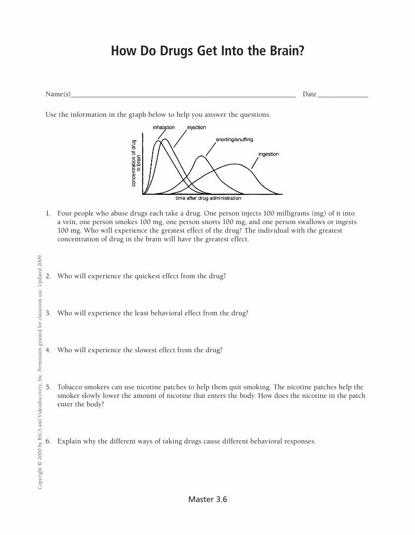
How Do Drugs Get Into the Brain? C
opyr
ight
© 2
000
by B
SCS
and
Vid
eodi
scov
ery,
Inc
. Per
mis
sion
gra
nted
for
clas
sroo
m u
se. U
pdat
ed 2
009.
Name(s)______________________________________________________________ Date ______________
Use the information in the graph below to help you answer the questions.
1. Four people who abuse drugs each take a drug. One person injects 100 milligrams (mg) of it into a vein, one person smokes 100 mg, one person snorts 100 mg, and one person swallows or ingests 100 mg. Who will experience the greatest effect of the drug? The individual with the greatest concentration of drug in the brain will have the greatest effect.
2. Who will experience the quickest effect from the drug?
3. Who will experience the least behavioral effect from the drug?
4. Who will experience the slowest effect from the drug?
5. Tobacco smokers can use nicotine patches to help them quit smoking. The nicotine patches help the smoker slowly lower the amount of nicotine that enters the body. How does the nicotine in the patch enter the body?
6. Explain why the different ways of taking drugs cause different behavioral responses.
Master 3.6
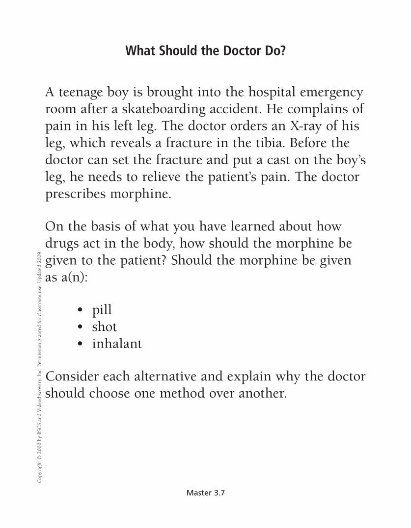
What Should the Doctor Do? C
opyr
ight
© 2
000
by B
SCS
and
Vid
eodi
scov
ery,
Inc
. Per
mis
sion
gra
nted
for
clas
sroo
m u
se. U
pdat
ed 2
009.
A teenage boy is brought into the hospital emergency room after a skateboarding accident. He complains of pain in his left leg. The doctor orders an X-ray of his leg, which reveals a fracture in the tibia. Before the doctor can set the fracture and put a cast on the boy’s leg, he needs to relieve the patient’s pain. The doctor prescribes morphine.
On the basis of what you have learned about how drugs act in the body, how should the morphine be given to the patient? Should the morphine be given as a(n):
• pill • shot • inhalant
Consider each alternative and explain why the doctor should choose one method over another.
Master 3.7
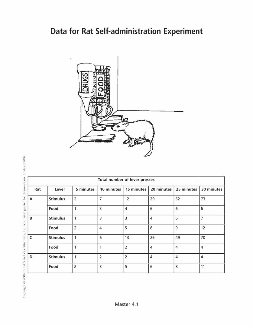
Data for Rat Self-administration Experiment C
opyr
ight
© 2
000
by B
SCS
and
Vid
eodi
scov
ery,
Inc
. Per
mis
sion
gra
nted
for
clas
sroo
m u
se. U
pdat
ed 2
009.
Total number of lever presses
Rat Lever 5 minutes 10 minutes 15 minutes 20 minutes 25 minutes 30 minutes
A Stimulus 2 7 12 29 52 73
Food 1 3 4 6 6 6
B Stimulus 1 3 3 4 6 7
Food 2 4 5 8 9 12
C Stimulus 1 6 13 26 49 70
Food 1 1 2 4 4 4
D Stimulus 1 2 2 4 4 4
Food 2 3 5 6 8 11
Master 4.1
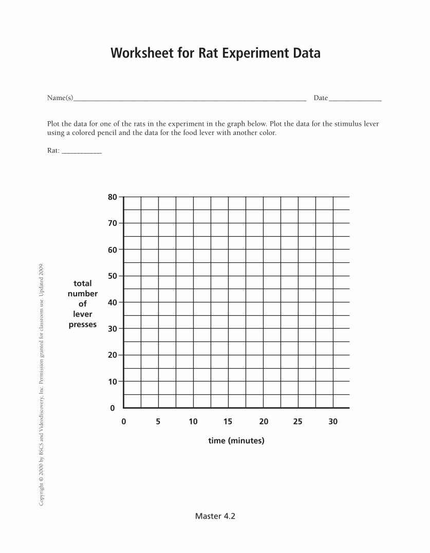
Worksheet for Rat Experiment Data
Name(s)______________________________________________________________ Date ______________
Plot the data for one of the rats in the experiment in the graph below. Plot the data for the stimulus lever using a colored pencil and the data for the food lever with another color.
Rat: ____________
Cop
yrig
ht ©
200
0 by
BSC
S an
d V
ideo
disc
over
y, I
nc. P
erm
issi
on g
rant
ed fo
r cl
assr
oom
use
. Upd
ated
200
9.
total number
of lever
presses
80
70
60
50
40
30
20
10
0 0
time (minutes)
5 10 15 20 25 30
Master 4.2
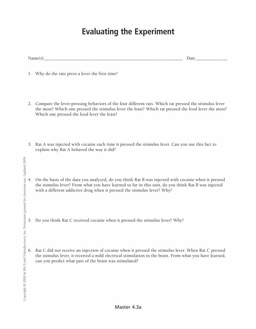
Evaluating the Experiment C
opyr
ight
© 2
000
by B
SCS
and
Vid
eodi
scov
ery,
Inc
. Per
mis
sion
gra
nted
for
clas
sroo
m u
se. U
pdat
ed 2
009.
Name(s)______________________________________________________________ Date ______________
1. Why do the rats press a lever the first time?
2. Compare the lever-pressing behaviors of the four different rats. Which rat pressed the stimulus lever the most? Which one pressed the stimulus lever the least? Which rat pressed the food lever the most? Which one pressed the food lever the least?
3. Rat A was injected with cocaine each time it pressed the stimulus lever. Can you use this fact to explain why Rat A behaved the way it did?
4. On the basis of the data you analyzed, do you think Rat B was injected with cocaine when it pressed the stimulus lever? From what you have learned so far in this unit, do you think Rat B was injected with a different addictive drug when it pressed the stimulus lever? Why?
5. Do you think Rat C received cocaine when it pressed the stimulus lever? Why?
6. Rat C did not receive an injection of cocaine when it pressed the stimulus lever. When Rat C pressed the stimulus lever, it received a mild electrical stimulation in the brain. From what you have learned, can you predict what part of the brain was stimulated?
Master 4.3a
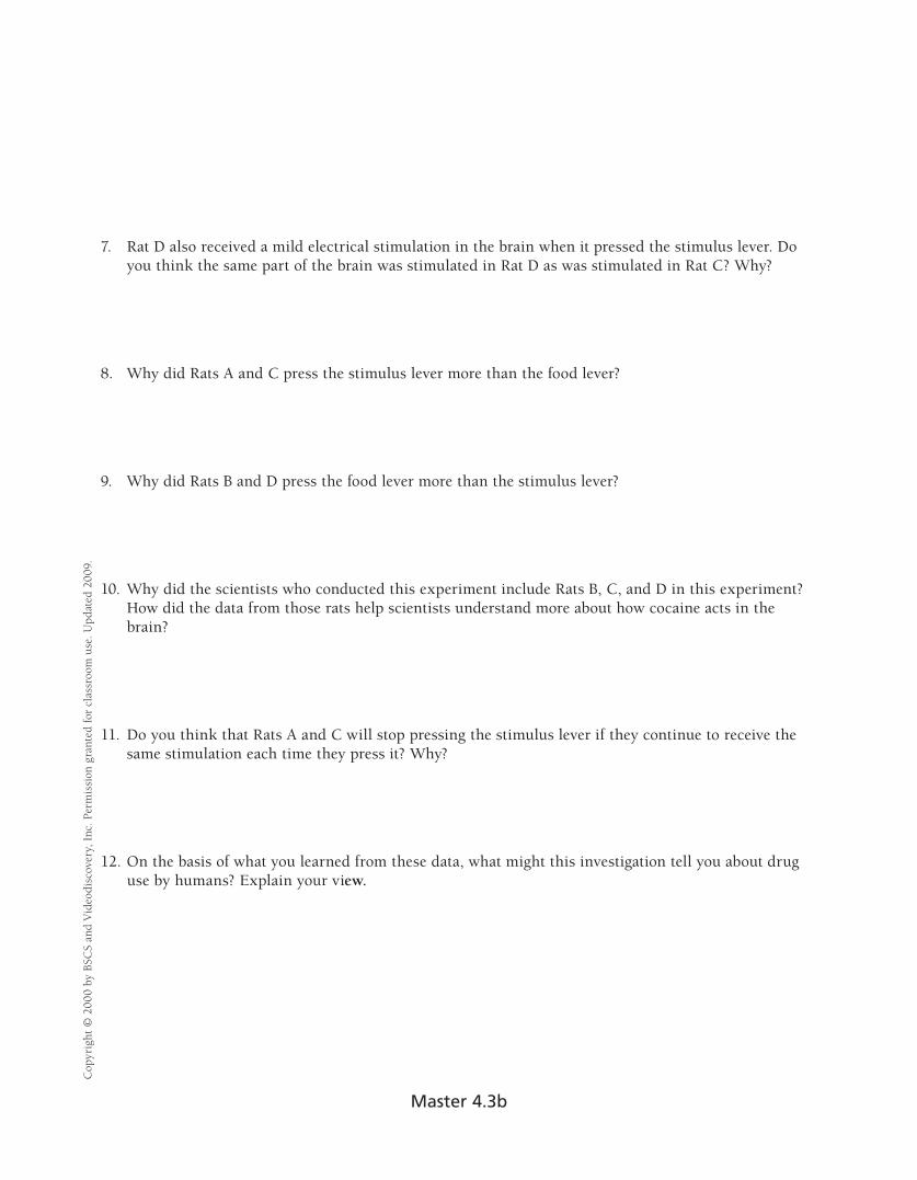
Cop
yrig
ht ©
200
0 by
BSC
S an
d V
ideo
disc
over
y, I
nc. P
erm
issi
on g
rant
ed fo
r cl
assr
oom
use
. Upd
ated
200
9.
7. Rat D also received a mild electrical stimulation in the brain when it pressed the stimulus lever. Do you think the same part of the brain was stimulated in Rat D as was stimulated in Rat C? Why?
8. Why did Rats A and C press the stimulus lever more than the food lever?
9. Why did Rats B and D press the food lever more than the stimulus lever?
10. Why did the scientists who conducted this experiment include Rats B, C, and D in this experiment? How did the data from those rats help scientists understand more about how cocaine acts in the brain?
11. Do you think that Rats A and C will stop pressing the stimulus lever if they continue to receive the same stimulation each time they press it? Why?
12. On the basis of what you learned from these data, what might this investigation tell you about drug use by humans? Explain your view.
Master 4.3b

Playing the Game C
opyr
ight
© 2
000
by B
SCS
and
Vid
eodi
scov
ery,
Inc
. Per
mis
sion
gra
nted
for
clas
sroo
m u
se. U
pdat
ed 2
009.
1. Each person draws one card from the small pile of cards. Place it face up in front of you. This is your switch card. Set the rest of the cards in the short deck aside. You won’t need them again.
• If you drew a jack, your switch value is 25. • If you drew a queen, your switch value is 35. • If you drew a king, your switch value is 45.
2. Draw a card face down from the larger pile that contains aces and the number cards. Don’t look at this card. Place it face down below your switch card. This is your risk card.
3. Draw cards from the large pile and place them face up next to the risk card. These are your choice cards. Draw as many choice cards as you wish, but keep in mind that you do not want the total of these cards plus the risk card to equal or go over your switch value.
• An ace = 1 point • Other cards = the number on the card
4. When you have finished drawing cards, turn over the risk card. Did you match or go over your switch value?
Master 4.4
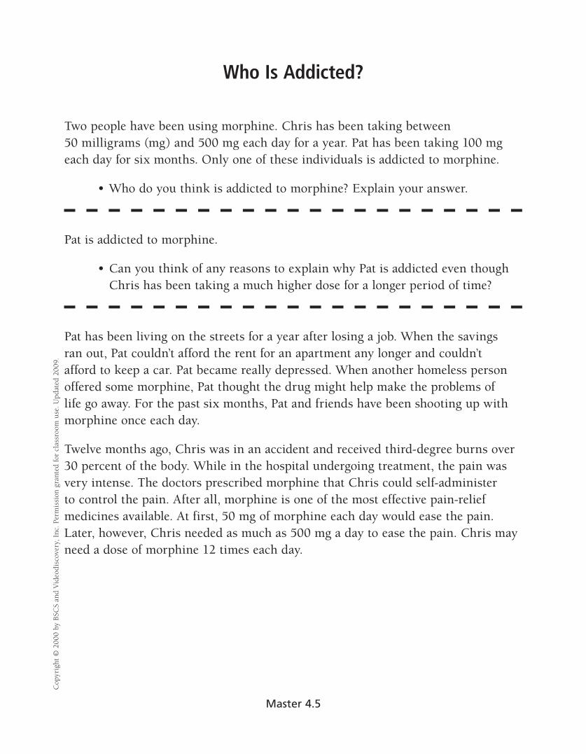
Cop
yrig
ht ©
200
0 by
BSC
S an
d V
ideo
disc
over
y, I
nc. P
erm
issi
on g
rant
ed fo
r cl
assr
oom
use
. Upd
ated
200
9.
Who Is Addicted?
Two people have been using morphine. Chris has been taking between 50 milligrams (mg) and 500 mg each day for a year. Pat has been taking 100 mg each day for six months. Only one of these individuals is addicted to morphine.
• Who do you think is addicted to morphine? Explain your answer.
Pat is addicted to morphine.
• Can you think of any reasons to explain why Pat is addicted even though Chris has been taking a much higher dose for a longer period of time?
Pat has been living on the streets for a year after losing a job. When the savings ran out, Pat couldn’t afford the rent for an apartment any longer and couldn’t afford to keep a car. Pat became really depressed. When another homeless person offered some morphine, Pat thought the drug might help make the problems of life go away. For the past six months, Pat and friends have been shooting up with morphine once each day.
Twelve months ago, Chris was in an accident and received third-degree burns over 30 percent of the body. While in the hospital undergoing treatment, the pain was very intense. The doctors prescribed morphine that Chris could self-administer to control the pain. After all, morphine is one of the most effective pain-relief medicines available. At first, 50 mg of morphine each day would ease the pain. Later, however, Chris needed as much as 500 mg a day to ease the pain. Chris may need a dose of morphine 12 times each day.
Master 4.5

Long-Term Effects of Drugs on the Brain C
opyr
ight
© 2
000
by B
SCS
and
Vid
eodi
scov
ery,
Inc
. Per
mis
sion
gra
nted
for
clas
sroo
m u
se. U
pdat
ed 2
009.
So, why are drugs so bad? After all, the high or rush only lasts a little while, right? What else could be happening in the brain of a person who abuses drugs? Consider that the brain is continuously changing. After all, learning occurs because neurons are forming new synapses. Scientists say that the brain is plastic and call this “neuroplasticity.” That doesn’t mean the brain is made of a chemical plastic like a credit card, but it refers to the brain’s ability to modify connections in response to experience. When a person learns something or has new experiences, some new synapses may form or existing synapses may get stronger. Other synapses may disappear.
When a person takes drugs repeatedly, the brain changes in response to this experience. If a person takes drugs and then stops, he or she will crave the drug. In other words, the individual will have a strong desire to take more of the drug. Scientists can actually see evidence of cravings in the brain. If someone addicted to cocaine sees pictures of drug paraphernalia, PET scans show that a part of the brain that is important for emotional memory (called the amygdala) is activated, and the person reports feelings of drug craving. If he or she sees a video with mountains, trees, and animals, the amygdala is not stimulated. Thus, just seeing pictures of drugs or things associated with drugs can trigger an uncontrollable urge for drugs.
After taking drugs for a period of time, a person may need to take a higher dose of the drug to have the same experience that he or she did when first taking the drug. This is called tolerance. The brain has adapted to having a certain amount of drug present and does not respond the same way it did initially. That is why people who abuse and who are addicted to drugs take increasingly higher amounts of an abused drug. Tolerance may develop because the body may become more efficient at eliminating the chemical from the body, or because the cells of the body and brain become less responsive to the effect of the drug.
Scientific studies have shown clearly that certain drugs can cause dramatic changes in the brain, but not all questions have been answered. Drugs can change the structure of the brain. Perhaps one of the most dramatic long-term effects of a drug is to kill neurons. Many people have heard that drinking alcohol will kill brain cells. It’s true. If alcohol is abused over a period of time, neurons in the brain can die. Some neurons in the brain are more sensitive to alcohol than others. Neurons that make up the mammillary bodies (small round structures on the brain’s undersurface) and hippocampus, areas in the brain that are important for memory, are more vulnerable to the effects of alcohol than are some other neurons in the brain. The neurons in the cerebral cortex, the part of the brain that controls most of our mental functions and endows us with consciousness, may also die if a person frequently abuses alcohol in high doses.
Another drug that can be toxic to neurons is an amphetamine derivative called MDMA, or ecstasy. In rats and nonhuman primates, MDMA damages the axon terminals of neurons that release serotonin, a neurotransmitter that is involved in regulating appetite, sleep, emotions, and so on. In some parts of the brain, the axons of some of these neurons may regenerate (or re-grow) after drug use is stopped, but the new growth of the neurons is not normal. Some areas are not reinnervated (nerve fibers do not grow back into the area), and some areas have abnormally high regrowth of the neurons. Either way, the neurons do not look normal. Studies have not yet been able to determine whether MDMA has this same effect on humans.
Master 4.6a

Cop
yrig
ht ©
200
0 by
BSC
S an
d V
ideo
disc
over
y, I
nc. P
erm
issi
on g
rant
ed fo
r cl
assr
oom
use
. Upd
ated
200
9.
Cocaine also changes the brain in ways that may last for a long time. PET scans of human brains have shown that glucose metabolism is reduced even three months after the last use of cocaine. Remember that glucose metabolism is an indicator of how active the brain cells are. If the neurons are using less glucose in certain areas, they are not as active. The changes that cocaine causes in the brain last much longer than the pleasurable feelings it produces. Other drugs cause similar decreases in brain activity. Even two years after the last use of amphetamines, PET images show that the brain of a person who has abused drugs is less active than the person’s who never used drugs.
Scientists, for many reasons, don’t know all of the effects that a drug has. First, the brain is such a complicated organ that, despite great scientific advances, understanding all that it does will take many more years. Second, individuals may respond differently to drugs due to genetic and other differences among people. Third, many people who abuse drugs abuse more than one drug. Many individuals who take cocaine, for example, also drink alcohol. The combination of the drugs makes it difficult to determine what the effect of one drug alone may be. Another complication is that people addicted to drugs may have other health problems in addition to their drug problem. People addicted to heroin, for example, spend most of their energy and activity trying to get their next “fix.” Consequently, they do not eat well and may have impaired immune systems. Also, drug-addicted people often suffer from mental illnesses, such as depression. The changes that occur in the brain because of mental illness make it difficult to determine what changes the drugs have caused.
The brain is an incredibly complex organ. This complexity will keep scientists working for many years to understand how the brain works. Someday, scientists will answer questions about what happens in the brain to cause addiction, which will then help scientists understand how to prevent addiction. On a separate sheet of paper, answer the following questions:
1. What are some of the ways that drugs cause long-term changes in the brain?
2. How does the brain adapt to the presence of drugs?
3. How may the abuse of drugs relate to the plasticity of the brain?
4. What are some problems that scientists have when they investigate the effects of drugs on the brain?
Master 4.6b

Ruth’s Story
Ruth is 24 years old and has a good job and a boyfriend. Everything seems to be going well in her life. But it hasn’t always been that way. When she was 14 years old, her friends began smoking cigarettes and drinking alcohol. Because she wanted to be part of the group, she also began smoking and drinking when she went to parties with her friends. One night when Ruth was 16, her friends had some marijuana and they all tried smoking it. After using marijuana for about a year, she began experimenting with other drugs and, by the time she was 18, Ruth was using heroin every day. Her drug habit was costing her $75 a day. After a while, her boyfriend left her, and the rest of her friends were tired of her asking for money to buy drugs. She was fired from her part-time job because she had missed work so many times. She was arrested several times for shoplifting items from local department and discount stores. She tried to quit using heroin several times, but she had strong cravings for the drug. Each time she began having symptoms of withdrawal, Ruth went back to abusing drugs.
When Ruth was 20, her brother convinced her to go to a drug rehabilitation center. The doctors at the center began treating her with methadone, and she participated in group behavioral treatments. She followed her treatment exactly as the doctors prescribed and, after six months, Ruth thought she had beaten her addiction. She enrolled in college and made new friends. Her friends got her involved in sports, and Ruth found that she enjoyed running. She even competed in a 10K run. She continued her methadone treatment and saw her therapist every two months.
When she was 22, Ruth was under a great deal of stress when she took on a new part-time job in addition to her school work. She ran into her old high school friends at a party and did some heroin with them. She thought she could handle it. Over the next couple of months, however, she quit her methadone treatment and began doing heroin more frequently, every couple of days. She was beginning to isolate herself from her friends and was having trouble at work. Ruth was scared. She called her doctors, and they started her treatments again. With her doctors’ help, Ruth realized that she needed to continue her medication and her counseling.
Cop
yrig
ht ©
200
0 by
BSC
S an
d V
ideo
disc
over
y, I
nc. P
erm
issi
on g
rant
ed fo
r cl
assr
oom
use
. Upd
ated
200
9.
Master 5.1

Mike’s Story
Mike grew up an active boy who loved participating in sports. When he was 14, he was diagnosed with Type I diabetes. Mike learned how to measure his blood glucose levels before meals and give himself insulin injections based on his blood glucose level. He also learned how he should change his diet. Mike learned what types and amounts of foods he could eat and how he should schedule the time interval between meals. But, actually making these changes was very difficult for him. After discussions with the family doctor, Mike and his family decided he would spend six weeks at a summer camp for teenagers who have diabetes. While at camp, Mike ate the correct diet and learned how other kids cope with their diabetes. He even made several friends there.
After he got home, Mike often e-mailed his friends from camp and they would talk about school, sports, and how diabetes changed their lives. Mike’s life was pretty normal for a teenager—school, sports, friends. He found that as long as he regulated his blood glucose levels, he could do most of what he wanted. When he was 16, he got his driver’s license. On weekends, he would sometimes forget his diet and eat hamburgers, french fries, and sodas with his friends. Because he only had a minor problem the first time he did this, he continued to ignore his diet when he was with his friends.
One Saturday night, Mike’s parents had to take him to the emergency room because his blood sugar level was over 600. Although this scared him, he recovered. After a few weeks, though, he went back to eating whatever he wanted instead of the proper diet, especially if he was with his friends. Mike only checked his blood glucose level if he thought he might have a problem. He ended up back in the hospital several more times that year. His grades fell from As to Cs because he could not keep up with the work. He had trouble concentrating and was tired a lot. He and his parents argued all the time about Mike’s failure to eat a healthy diet.
The last time Mike went into the hospital, the doctor warned him that he was at risk for permanent health problems if he didn’t control his blood glucose level: he could have kidney failure or could go blind. Mike’s doctor recommended a specialist who could help Mike learn to cope with diabetes and still maintain an active social life. Mike’s family also talked to the specialist to learn how they could help him. For the past four years, Mike has been able to control his blood sugar levels and has only had two minor episodes.
Cop
yrig
ht ©
200
0 by
BSC
S an
d V
ideo
disc
over
y, I
nc. P
erm
issi
on g
rant
ed fo
r cl
assr
oom
use
. Upd
ated
200
9.
Master 5.2
Carol’s Story
Carol is the mother of two high school students. Although she is only 42 years old, her doctor has told her that she has high blood pressure, or essential hypertension. On one visit to her doctor, her blood pressure was 160/105. When her doctor checked her blood pressure again on another day, her blood pressure was 150/95. Her doctor prescribed medicine to lower her blood pressure and told her to watch her diet and to begin exercising. The doctor also told Carol that she needed to be very careful in controlling the amount of salt that she ate in her diet.
Carol followed the doctor’s plan for about six months. Gradually she started skipping her exercise sessions and gave up making healthy eating choices. Carol had a difficult time skipping the potato chips and peanuts that she liked to eat for an afternoon snack. Often she forgot to take her medication. At her next appointment, Carol and her doctor discussed the problems she was having, and the doctor informed her that her blood pressure had actually gone up. The doctor talked to her about getting advice from a nutritionist, working with a personal trainer to help her establish an exercise plan, and seeing a psychologist who could help her make the needed changes. Carol decided that she didn’t need help from those people and tried again to diet and exercise on her own. But, with her long hours at work and her family to take care of, she found it difficult. Because she was missing work more often, Carol’s boss gave a promotion to someone else instead of her. Carol’s kids complained that she didn’t come to their football games and band concerts anymore.
One night, Carol complained that she was having another headache and her vision was blurry. Her kids commented that she was slurring her words when she spoke. Her husband immediately called an ambulance to take her to the emergency room. Carol received medical help in time, but the doctors told her that she had a mild stroke.
Cop
yrig
ht ©
200
0 by
BSC
S an
d V
ideo
disc
over
y, I
nc. P
erm
issi
on g
rant
ed fo
r cl
assr
oom
use
. Upd
ated
200
9.
Master 5.3
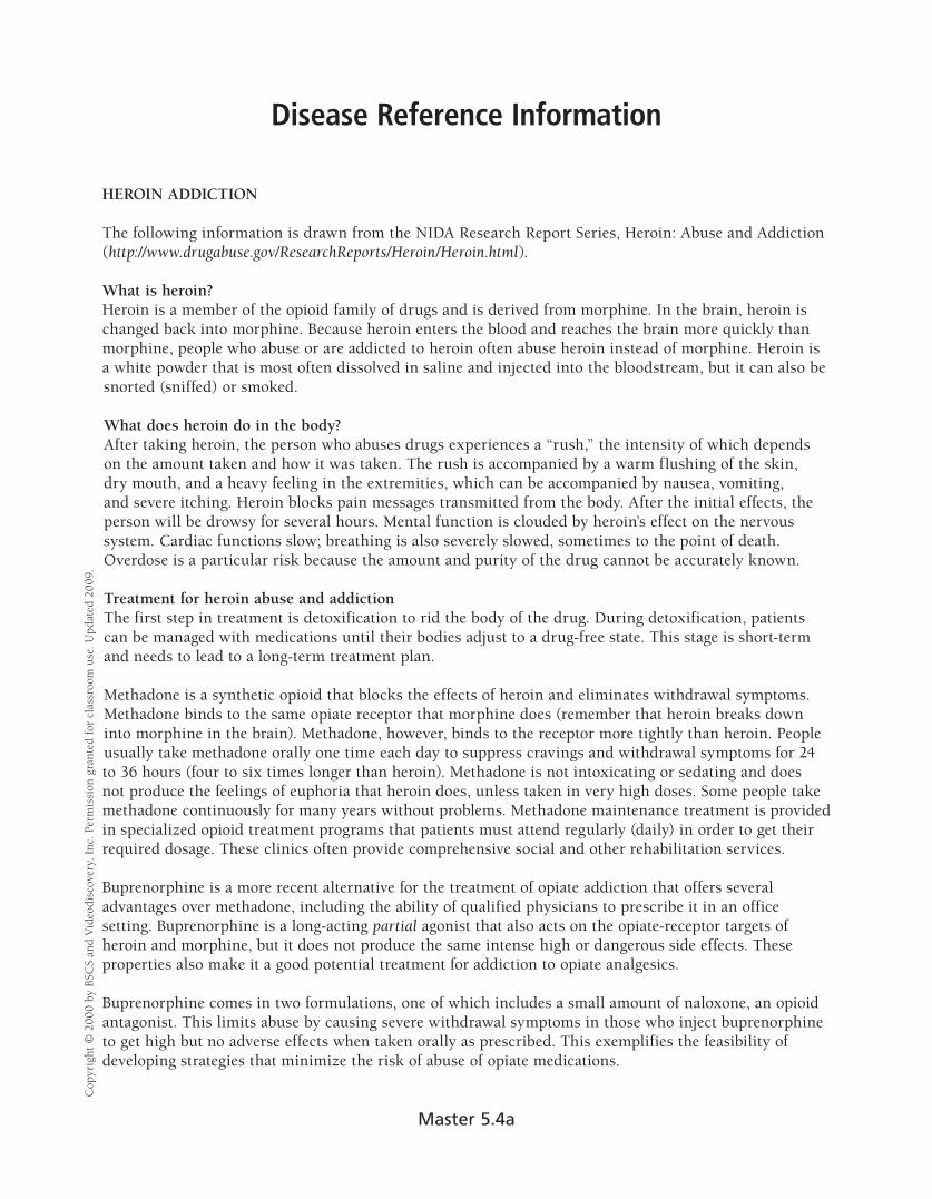
Disease Reference Information C
opyr
ight
© 2
000
by B
SCS
and
Vid
eodi
scov
ery,
Inc
. Per
mis
sion
gra
nted
for
clas
sroo
m u
se. U
pdat
ed 2
009.
HEROIN ADDICTION
The following information is drawn from the NIDA Research Report Series, Heroin: Abuse and Addiction (http://www.drugabuse.gov/ResearchReports/Heroin/Heroin.html).
What is heroin? Heroin is a member of the opioid family of drugs and is derived from morphine. In the brain, heroin is changed back into morphine. Because heroin enters the blood and reaches the brain more quickly than morphine, people who abuse or are addicted to heroin often abuse heroin instead of morphine. Heroin is a white powder that is most often dissolved in saline and injected into the bloodstream, but it can also be snorted (sniffed) or smoked.
What does heroin do in the body? After taking heroin, the person who abuses drugs experiences a “rush,” the intensity of which depends on the amount taken and how it was taken. The rush is accompanied by a warm flushing of the skin, dry mouth, and a heavy feeling in the extremities, which can be accompanied by nausea, vomiting, and severe itching. Heroin blocks pain messages transmitted from the body. After the initial effects, the person will be drowsy for several hours. Mental function is clouded by heroin’s effect on the nervous system. Cardiac functions slow; breathing is also severely slowed, sometimes to the point of death. Overdose is a particular risk because the amount and purity of the drug cannot be accurately known.
Treatment for heroin abuse and addiction The first step in treatment is detoxification to rid the body of the drug. During detoxification, patients can be managed with medications until their bodies adjust to a drug-free state. This stage is short-term and needs to lead to a long-term treatment plan.
Methadone is a synthetic opioid that blocks the effects of heroin and eliminates withdrawal symptoms. Methadone binds to the same opiate receptor that morphine does (remember that heroin breaks down into morphine in the brain). Methadone, however, binds to the receptor more tightly than heroin. People usually take methadone orally one time each day to suppress cravings and withdrawal symptoms for 24 to 36 hours (four to six times longer than heroin). Methadone is not intoxicating or sedating and does not produce the feelings of euphoria that heroin does, unless taken in very high doses. Some people take methadone continuously for many years without problems. Methadone maintenance treatment is provided in specialized opioid treatment programs that patients must attend regularly (daily) in order to get their required dosage. These clinics often provide comprehensive social and other rehabilitation services.
Buprenorphine is a more recent alternative for the treatment of opiate addiction that offers several advantages over methadone, including the ability of qualified physicians to prescribe it in an office setting. Buprenorphine is a long-acting partial agonist that also acts on the opiate-receptor targets of heroin and morphine, but it does not produce the same intense high or dangerous side effects. These properties also make it a good potential treatment for addiction to opiate analgesics.
Buprenorphine comes in two formulations, one of which includes a small amount of naloxone, an opioid antagonist. This limits abuse by causing severe withdrawal symptoms in those who inject buprenorphine to get high but no adverse effects when taken orally as prescribed. This exemplifies the feasibility of developing strategies that minimize the risk of abuse of opiate medications.
Master 5.4a
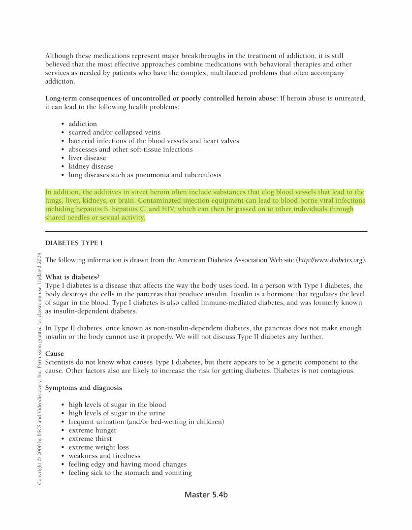
Cop
yrig
ht ©
200
0 by
BSC
S an
d V
ideo
disc
over
y, I
nc. P
erm
issi
on g
rant
ed fo
r cl
assr
oom
use
. Upd
ated
200
9.
Although these medications represent major breakthroughs in the treatment of addiction, it is still believed that the most effective approaches combine medications with behavioral therapies and other services as needed by patients who have the complex, multifaceted problems that often accompany addiction.
Long-term consequences of uncontrolled or poorly controlled heroin abuse: If heroin abuse is untreated, it can lead to the following health problems:
• addiction • scarred and/or collapsed veins • bacterial infections of the blood vessels and heart valves • abscesses and other soft-tissue infections • liver disease • kidney disease • lung diseases such as pneumonia and tuberculosis
In addition, the additives in street heroin often include substances that clog blood vessels that lead to the lungs, liver, kidneys, or brain. Contaminated injection equipment can lead to blood-borne viral infections including hepatitis B, hepatitis C, and HIV, which can then be passed on to other individuals through shared needles or sexual activity.
DIABETES TYPE I
The following information is drawn from the American Diabetes Association Web site (http://www.diabetes.org).
What is diabetes? Type I diabetes is a disease that affects the way the body uses food. In a person with Type I diabetes, the body destroys the cells in the pancreas that produce insulin. Insulin is a hormone that regulates the level of sugar in the blood. Type I diabetes is also called immune-mediated diabetes, and was formerly known as insulin-dependent diabetes.
In Type II diabetes, once known as non-insulin-dependent diabetes, the pancreas does not make enough insulin or the body cannot use it properly. We will not discuss Type II diabetes any further.
Cause Scientists do not know what causes Type I diabetes, but there appears to be a genetic component to the cause. Other factors also are likely to increase the risk for getting diabetes. Diabetes is not contagious.
Symptoms and diagnosis
• high levels of sugar in the blood • high levels of sugar in the urine • frequent urination (and/or bed-wetting in children) • extreme hunger • extreme thirst • extreme weight loss • weakness and tiredness • feeling edgy and having mood changes • feeling sick to the stomach and vomiting
Master 5.4b

Cop
yrig
ht ©
200
0 by
BSC
S an
d V
ideo
disc
over
y, I
nc. P
erm
issi
on g
rant
ed fo
r cl
assr
oom
use
. Upd
ated
200
9.
Treatment Treatment for Type I diabetes involves keeping the level of sugar in the blood as close to normal (80–120 mg/dL) as possible. Treatment usually includes
• Insulin injections to lower blood sugar. The number of injections required depends on the individual and the type of insulin treatment used.
• A meal plan to control changes in blood sugar levels. Food raises blood-sugar levels. A dietician can help develop a plan that lets the diabetic person eat the food he or she enjoys.
• Exercise to lower the blood sugar. • Blood and urine testing to determine if the blood-sugar level is low, normal, or high. The results
enable a diabetic person to modify his or her food intake, exercise, or insulin injections.
Long-term consequences of uncontrolled or poorly controlled diabetes
• blindness • kidney disease • nerve damage leading to abnormal sensations, including pain in the hands, feet, and legs • vascular (blood vessel) disease leading to heart disease and strokes
Long-term outlook for diabetes if treated and controlled People with Type I diabetes can live happy, healthy lives if they follow their treatment plans.
HYPERTENSION
The following is drawn from materials from the American Heart Association (http://www.americanheart.org) and the National Heart, Lung, and Blood Institute (http://www.nhlbi.nih.gov/health/public/heart/index.htm).
What is hypertension? Hypertension, or high blood pressure, is defined in an adult as a blood pressure greater than or equal to 140 mm Hg systolic pressure or greater than or equal to 90 mm Hg diastolic pressure. Hypertension does not refer to being tense, nervous, or hyperactive. Optimal blood pressure for an adult is 120 mm Hg systolic and 80 mm Hg diastolic. Blood pressures are normally written as systolic/ diastolic, such as 120/80.
Cause In most cases, the cause of high blood pressure is unknown. This type of high blood pressure is called essential hypertension.
In the remaining cases (5% to 10% of cases), high blood pressure, called secondary hypertension, is a result of another health problem such as a kidney abnormality, tumor of the adrenal gland, or congenital defect of the aorta. Blood pressure usually returns to normal when the underlying cause is corrected.
Master 5.4c

Symptoms and diagnosis: Diagnosis of high blood pressure is based on the average of two or more readings taken at each of two or more visits after an initial screening.
Hypertension usually has no symptoms. Many people have high blood pressure and don’t know it. If hypertension is severe, symptoms may include
• tiredness • confusion • headaches • anxiety • excessive perspiration • pale skin • muscle tremors • chest pain
Treatment The prescribed treatment depends on the severity of hypertension, but may involve the following components:
• taking medication • modifying diet to reduce sodium intake • increasing exercise • maintaining proper weight • limiting alcohol intake
Long-term consequences of uncontrolled hypertension High blood pressure directly increases the risk of coronary heart disease (which leads to heart attack) and stroke, especially along with other risk factors. Uncontrolled hypertension can also lead to renal failure.
Long-term outlook for hypertension if treated and controlled Hypertension is controllable with treatment, which may require periodic adjustment.
Cop
yrig
ht ©
200
0 by
BSC
S an
d V
ideo
disc
over
y, I
nc. P
erm
issi
on g
rant
ed fo
r cl
assr
oom
use
. Upd
ated
200
9.
Master 5.4d

Evaluating the Cases C
opyr
ight
© 2
000
by B
SCS
and
Vid
eodi
scov
ery,
Inc
. Per
mis
sion
gra
nted
for
clas
sroo
m u
se. U
pdat
ed 2
009.
Name(s)______________________________________________________________ Date ______________
As a team, decide which member of the group will watch or read each case study. When you finish with your case, answer questions 1 to 6. Then, discuss and answer questions 7 to 11 with your group members. If you wish, watch or read the case studies again to help with your answers.
Case Study: ________________________
1. What disease does the individual have? Is it chronic or acute?
2. How did the disease change the individual’s life?
3. What is the recommended treatment?
4. What did the individual do to improve his or her recovery?
5. What did the individual do that impaired his or her recovery?
6. Are there other things the individual could do to help with the disease?
Comparing the Cases
7. Which individuals were successful in their treatment? Which individuals were not?
8. Who was cured of their disease? What is the difference between treatment and cure?
9. How are the treatments for the different diseases similar?
10. How are the treatments different?
11. Can you identify similarities and differences in the actions or strategies that individuals took to help them deal with their disease?
Master 5.5