the bisecting glcnac on n-glycans inhibits growth factor signaling...
TRANSCRIPT

2010;70:3361-3371. Published online April 14, 2010.Cancer Res Yinghui Song, Jason A. Aglipay, Joshua D. Bernstein, et al. Signaling and Retards Mammary Tumor Progression
-Glycans Inhibits Growth FactorNThe Bisecting GlcNAc on
Updated Version 10.1158/0008-5472.CAN-09-2719doi:
Access the most recent version of this article at:
MaterialSupplementary
http://cancerres.aacrjournals.org/content/suppl/2010/04/13/70.8.3361.DC1.htmlAccess the most recent supplemental material at:
Cited Articles http://cancerres.aacrjournals.org/content/70/8/3361.full.html#ref-list-1
This article cites 49 articles, 28 of which you can access for free at:
Citing Articles http://cancerres.aacrjournals.org/content/70/8/3361.full.html#related-urls
This article has been cited by 2 HighWire-hosted articles. Access the articles at:
E-mail alerts related to this article or journal.Sign up to receive free email-alerts
SubscriptionsReprints and
[email protected] atTo order reprints of this article or to subscribe to the journal, contact the AACR Publications
To request permission to re-use all or part of this article, contact the AACR Publications
American Association for Cancer Research Copyright © 2010 on April 1, 2011cancerres.aacrjournals.orgDownloaded from
DOI:10.1158/0008-5472.CAN-09-2719
DOI:10.1158/0008-5472.CAN-09-2719
Tumor and Stem Cell Biology
CancerResearch
The Bisecting GlcNAc on N-Glycans Inhibits Growth FactorSignaling and Retards Mammary Tumor Progression
Yinghui Song1, Jason A. Aglipay1, Joshua D. Bernstein2, Sumanta Goswami2, and Pamela Stanley1
Abstract
Authors' ACollege of2Departme
Note: SupResearch O
CorresponAlbert EinsNY 10461.stanley@ein
doi: 10.115
©2010 Am
www.aacr
The branching of complex N-glycans attached to growth factor receptors promotes tumor progression byprolonging growth factor signaling. The addition of the bisecting GlcNAc to complex N-glycans by Mgat3 hasvarying effects on cell adhesion, cell migration, and hepatoma formation. Here, we show that Chinese hamsterovary cells expressing Mgat3 and the polyoma middle T (PyMT) antigen have reduced cell proliferation andgrowth factor signaling dependent on a galectin lattice. The Mgat3 gene is not expressed in virgin mammarygland but is upregulated during lactation and is expressed in mouse mammary tumor virus (MMTV)/PyMTtumors. Mice lacking Mgat3 that cannot transfer the bisecting GlcNAc to N-glycans acquire PyMT-inducedmammary tumors more rapidly and have an increased tumor burden, increased migration of tumor cells,and increased early metastasis to lung. Tumors and tumor-derived cells lacking Mgat3 exhibit enhanced sig-naling through the Ras pathway and reduced amounts of functionally glycosylated α-dystroglycan. Constitu-tive overexpression of an MMTV/Mgat3 transgene inhibits early mammary tumor development and tumor cellmigration. Thus, the addition of the bisecting GlcNAc to complex N-glycans of mammary tumor cell glyco-protein receptors is a cell autonomous mechanism serving to retard tumor progression by reducing growthfactor signaling. Cancer Res; 70(8); 3361–71. ©2010 AACR.
Introduction
N-glycans have a common core structure, and their branch-ing patterns are determined by differentN-acetylglucosaminyl-transferases (GlcNAcT; ref. 1). Loss of GlcNAcT-V (Mgat5),anN-acetylglucosaminyltransferase that initiates aβ1,6 branchof complex N-glycans, promotes tumorigenesis in the mam-mary glands ofmice carrying themousemammary tumor virus(MMTV) polyoma middle T (PyMT) oncogene (2). Mammarytumor cells expressing Mgat5 are more responsive to growthfactors due to enhanced interactions of their growth factorreceptors with galectins, leading to reduced endocytosis andprolonged signaling compared with cells lacking Mgat5 (3, 4).Human cancer cell lines with targeted silencing of the Mgat5gene also exhibit reduced epidermal growth factor (EGF)receptor (EGFR) signaling, although apparently by a galectin-independent mechanism (5).Mgat3 transfers a GlcNAc to generate the bisecting GlcNAc
in the core of complex and hybrid N-glycans (ref. 6; Fig. 1A).The presence of the bisecting GlcNAc alters glycan recogni-
ffiliations: 1Department of Cell Biology, Albert EinsteinMedicine of Yeshiva University, Bronx, New York and
nt of Biology, Yeshiva University, New York, New York
plementary data for this article are available at Cancernline (http://cancerres.aacrjournals.org/).
ding Author: Pamela Stanley, Department of Cell Biology,tein College of Medicine, 1300 Morris Park Avenue, Bronx,Phone: 718-430-3346; Fax: 718-430-8574; E-mail: pamela.stein.yu.edu.
8/0008-5472.CAN-09-2719
erican Association for Cancer Research.
journals.org
American Asso Copyright © 2010 cancerres.aacrjDownloaded from
tion reflected by changes in the binding of plant lectins andmammalian galectins. Thus, LEC10 Chinese hamster ovary(CHO) cells that express Mgat3 (7, 8) bind markedly less ricinand more erythrophytohemagglutinin (E-PHA) than wild-type CHO cells (Fig. 1A). LEC10 cells also bind less galectin-1and galectin-3 than parent CHO cells (9). These lectin-bindingproperties reflect changes in the number or accessibility ofGal residues on cell surface N-glycans with a bisecting GlcNAc.Glycomics profiling of LEC10 N-glycans by matrix-assistedlaser desorption/ionization–time-of-flight mass spectrometryshows that the bisecting GlcNAc is present on complex, multi-antennary N-glycans with many LacNAc units (10).Mgat3 has been overexpressed in a broad spectrum of cells
with consequences that may vary with cell type (11, 12).Thus, overexpression of Mgat3 in K562 cells causes anincrease in spleen colonization (13), whereas overexpressionin B16 melanoma cells causes a marked reduction in homingto the lung (14). In HeLa cells, overexpresion of Mgat3 causesincreased EGFR signaling and reduced cell adhesion, pro-moting metastasis (15). However in other experiments, HeLacells overexpressing Mgat3 had reduced cell migration onfibronectin, countering metastasis (16). When Mgat3 wasoverexpressed in MKN45 cells, E-cadherin was upregulated,cell adhesion was enhanced, and cell migration was inhi-bited (12, 17). The combined data indicate that Mgat3 maybehave as a promoter or suppressor of cell migration andcell adhesion. In liver tumors induced by a low dose of die-thylnitrosamine, ∼50% of males expressing Mgat3 under theserum amyloid protein promoter got fewer tumors (18). Bycontrast, Mgat3 expressed under the mouse urinary proteinpromoter was not inhibitory (19) when diethylnitrosamine
3361
ciation for Cancer Research on April 1, 2011ournals.org
Song et al.
3362
DOI:10.1158/0008-5472.CAN-09-2719
and phenobarbitol were used. In addition, males with inde-pendent, targeted mutations of the Mgat3 gene developedhepatomas more slowly than controls (19, 20), consistentwith the facilitation of hepatoma progression by Mgat3.We report here the effects of Mgat3 and the bisecting
GlcNAc on growth factor signaling in CHO cells expressing
Cancer Res; 70(8) April 15, 2010
American Asso Copyright © 2010 cancerres.aacrjDownloaded from
PyMT and in the mammary gland during tumor inductionby MMTV/PyMT (21). The MMTV/PyMT female developstumors at different rates in all mammary glands, dependingon genetic background (22). Progression to malignancy in thismodel appropriately reflects the stages of human breasttumorigenesis (23). The PyMT oncoprotein activates signaling
ciation for Cancer Researc on April 1, 201ournals.org
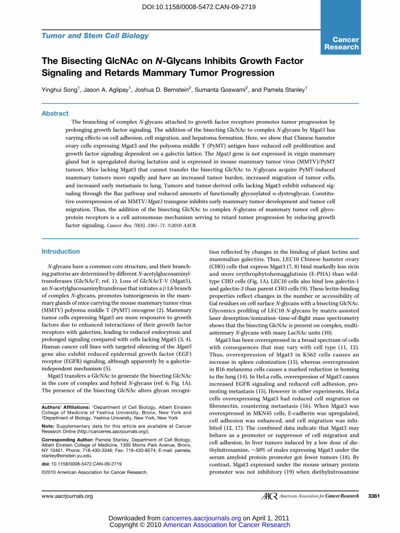
Figure 1. Mgat3 retards cellproliferation. A, complex N-glycansof CHO and LEC10 showing thereactions catalyzed by Mgat3and Mgat5 (top). LEC10 cellsare resistant to ricin andhypersensitive to E-PHA (bottom).B, glycoproteins expressingthe bisecting GlcNAc bind wellto biotinylated E-PHA (top) andbiotinylated L-PHA (bottom).C, proliferation of CHO, Lec4,Lec8, and LEC10 in medium with7.5% serum. D, proliferation ofCHO/PyMT, Lec4/PyMT, Lec8/PyMT, and LEC10B/PyMT inmedium with 7.5% serum. Bars,SD. ***, P < 0.0001; **, P < 0.01,two-tailed Student's t testcomparing CHO with LEC10B.
Cancer Research
h1
Mgat3 Suppresses Tumor Progression in Mammary Gland
DOI:10.1158/0008-5472.CAN-09-2719
pathways commonly amplified in human breast cancer, suchas phosphoinositide 3-kinase, leading to activation of Akt,Ras-Raf, and mitogen-activated protein kinases (MAPK;ref. 24). Here we show that Mgat3 inhibits growth factor sig-naling dependent on a cell surface galectin lattice in CHOcells and functions cell autonomously in the mammary glandto retard tumor progression, cell migration, and metastasis inMMTV/PyMT-induced tumors.
Materials and Methods
Cells and cell culture. Pro−5 CHO, Lec4 (Pro−Lec4.7B),Lec8 (Pro−Lec8.3D), and LEC10B (Pro−LEC10B.3) cells (25)validated by lectin resistance test and used within 6 mo ofcloning were transfected with pcDNA3.1-PyMT generatedfrom PJΩ-PyVMT (Elaine Lin; Albert Einstein College Medi-cine) and selected with 1 mg/mL G418 (Invitrogen). CHO andLEC10 cells were transfected with the Mgat3 coding exon orinactive Mgat3 (Mgat3T37; ref. 26) in pcDNA3.1. CHO cellswere cultured in α+-MEM (Invitrogen) containing 10% fetalbovine serum (FBS) and 2 mmol/L glutamine at 37°C in 5%CO2. Tumor epithelial cells (TEC) were derived from mincedtumors treated with 2 mg/mL collagenase (Sigma) and pas-saged ∼22 times to selectively remove fibroblasts. TECs werecultured in α+-MEM containing 10% heat-inactivated FBS,penicillin, and streptomycin.Lectin resistance test. Cells (2 × 10³) at 100 μL/well in a
96-well plate were incubated with 100 μL medium or medi-um with ricin (5 ng/mL; Vector Labs) or E-PHA (35 μg/mL;Vector Labs) for 4 d, stained with methylene blue in 50%methanol (2 g/L), and photographed.Western blot analysis and lectin blotting. Frozen tumor
(∼150 mg) homogenized in 1 mL 10 mmol/L Tris-HCl (pH7.4), 0.25 mol/L sucrose, and protease inhibitors (Complete;Roche) was centrifuged at 1,800 rpm for 10 min at 4°C.Tumor cells or washed cultured cells were solubilized in2% Triton-X-100, incubated on ice for 10 min, and centri-fuged at 3,000 rpm for 10 min at 4°C. Protein concentrationwas measured using the DC reagent (Bio-Rad). Lysates inloading buffer containing β-mercaptoethanol (5%) wereheated at 95°C for 5 min and separated by 12% SDS-PAGE.Proteins were transferred to a Polyscreen polyvinylidenedifluoride (PVDF) membrane (PerkinElmer) in Tris-glycinebuffer containing 5% methanol. For Western blot analysis,membranes were incubated in 5% nonfat milk and primaryantibody at room temperature for 1 h. Mouse anti-β-actinmonoclonal antibody (mAb; Abcam AC-15; 1:5,000), mouseanti-α-dystroglycan (α-DG) mAb IIH6C4 (Upstate Biotech-nology-Millipore; 1:1,000), mouse anti-β-DG mAb (43DAG1/8DG; Novocastra Laboratories; 1:300), horseradish peroxidase(HRP)–conjugated goat anti-mouse IgG (H+L; Thermo Scien-tific; 1:10,000), HRP goat anti-rabbit IgG-H+L (Zymed;1:10,000). After three washes with TBS-Tween [10 mmol/LTris-HCl (pH 7.4), 150 mmol/L NaCl, 0.05% Tween 20(Sigma)], secondary antibody HRP was incubated for 1 h.Bands were visualized using an enhanced chemilumines-cence (ECL) kit (Thermo Scientific) and quantitated byNIH Image/J. For lectin blotting, membranes were blocked
www.aacrjournals.org
American Asso Copyright © 2010 cancerres.aacrjDownloaded from
in 5% nonfat milk, incubated with biotinylated E-PHA or leu-kophytohemagglutinin (L-PHA; Vector Labs) at 5 μg/mL atroom temperature for 1 h, washed with TBS-Tween, incu-bated with streptavadin-HRP (1:5,000; Vector Labs) for 1 h,and visualized using an ECL kit.Signaling assays. Cells (85% to 90% confluent in 60-mm
dishes) were serum-starved for 24 h. After washing withα+-MEM, cells were stimulated with 10% FCS, 50 ng/mLhuman platelet-derived growth factor-AB (PDGF-AB; Invitro-gen), or 50 ng/mL EGF (R&D Systems) at 37°C. For sugartreatments after starvation, 1.5 mL α+-MEM or 0.5 mol/Llactose or 0.5 mol/L sucrose in α+-MEM was added for 1 hat 37°C, cells were washed twice with α+-MEM and treatedwith FCS, EGF, or PDGF-AB at 37°C. MAPK/extracellularsignal-regulated kinase kinase 1/2 (MEK1/2) inhibitor UO126(Cell Signaling) was dissolved in DMSO at 10 mmol/L, addedat ≤10 μmol/L for 2 h, and removed before adding growthfactor. Controls were treated with DMSO. After stimulation,cells were washed thrice with PBS (pH 7.4), lysed in EBC lysisbuffer [50 mmol/L Tris-HCl (pH 8.0), 120 mmol/L NaCl, 0.5%NP40, 100 mmol/L NaF, 200 μmol/L sodium orthovanadate]containing protease inhibitors (Complete, Roche), electro-phoresed and transferred to PVDF membrane. Membraneswere incubated with rabbit anti-phosphorylated p44/42MAPK antibody (Thr202/Tyr204; 1:1,000) and mouse anti-p44/42 MAPK mAb (L34F12; 1:2,000; Cell Signaling Technology) inOdyssey blocking buffer at 4°C overnight. Followingwashes withTBS-Tween, IRDye800-conjugated goat anti-rabbit IgG-H+L(MX10, Rockland Immunochemicals; 1:10,000), Alexa Fluor680
goat anti-mouse IgG-H+L (Invitrogen; 1:15,000) were addedfor 1 h at room temperature, membranes were washed,and bands were quantitated by ODYSSEY IR Imaging System(LI-COR BioSciences).Mice. Mgat3−/− mice (Mgat3tm1Jxm; ref. 27) backcrossed to
C57Bl/6 mice were mated with MMTV/PyMT transgenicmice (634 FVB; ref. 21; Jeffrey Pollard; Albert Einstein Collegeof Medicine). Mgat3+/− or Mgat3−/− females and Mgat3+/−
MMTV/PyMT males were mated to generateMgat3+/+/PyMT,Mgat3+/−/PyMT, and Mgat3−/−/PyMT littermates. The C57Bl/6/FVB background slowed the time of onset and progressionof mammary tumors (22, 28).The MMTV-SV40-BssK vector (Jeffrey Pollard) was used to
make the MMTV-Mgat3-CAGloxPCATloxP-EGFP transgene.The mouse Mgat3 coding region was inserted between theMMTV-LTR and the SV40-polyA addition site followedby the CAGloxPCATloxP-EGFP cassette (ref. 29; Jun-ichiMiyazaki; Osaka University Medical School). Plasmid linear-ized by Spe1 was microinjected into FVB fertilized eggs. Afounder with a single site of integration and several tandemcopies of the Mgat3 transgene was used to generate MMTV-Mgat3-PyMT mice. Mice were housed in a barrier facilitywith food and water ad libitum. Animal protocols wereapproved by the Animal Institute Committee of the AlbertEinstein College of Medicine.Tumor analysis. All 10 mammary glands of MMTV/PyMT
females were palpated (genotype-blinded) thrice a week,from 6 wk. The three largest mammary tumors were excised,weighed, and fixed in 10% formalin at room temperature for
Cancer Res; 70(8) April 15, 2010 3363
ciation for Cancer Research on April 1, 2011ournals.org

Song et al.
3364
DOI:10.1158/0008-5472.CAN-09-2719
24 h. Tumor tissue was also frozen in Trizol (Invitrogen) orstored at −80°C. Total RNA from tumors was analyzed byreverse transcription–PCR (RT-PCR) to determine expressionof PyMT, Mgat3, Mgat5, and β-actin (primers in Supplemen-tary Table S1).Lung metastasis. Formalin-fixed lungs were paraffin-
embedded and sectioned at 5 μm. Three sections per lungseparated by 50 μm were stained with H&E and examinedfor metastatic lesions. Total RNA extracted from lungs in Tri-zol (Invitrogen) was treated with amplification grade DNaseI(Invitrogen) and cDNA prepared using the SuperScript IIIfirst-strand synthesis system (Invitrogen). Real-time PCRwas performed with 5 ng cDNA and primers: PyMT, 5′-agc-cactcctatcccccaac-3′ (forward), 5′-ctcctcctcctcctcctcca-3′(reverse); β-actin, 5′-gtgggccgctctaggcacca-3′ (forward), 5′-tggccttagggttcaggggg-3′ (reverse). PCR products incorporatedSYBR Green dye (Qiagen) and were analyzed on a Prism 7700system (Applied Biosystems) as follows: 95°C 15min, then 94°C15 s, 59°C 30 s, 72°C 30 s for 40 cycles. PCR product formationwas measured continuously, and C(t) plots were generated.Plasmids TA-PyMT and TA-actin were used to determinethe absolute number of PyMT and mouse β-actin transcripts.In vivo invasion assay. Cell migration into microneedles
filled with 25 nmol/L EGF (Invitrogen) and Matrigel (BDBiosciences) and placed into tumors of live anesthetizedanimals was performed as described (30). Passive collectionof cells or tissue during insertion of needles was blocked.After 4 h, needles were removed and cell numbers weredetermined by 4′,6-diamidino-2-phenylindole staining. Cellmigration is required for cells to enter needles (31).Statistical analysis. Student's t test was from the Excel
Data Analysis Package. Tumor development was comparedby Mantel-Cox log-rank test. Univariate analysis was per-formed by the χ2 test.
Results
Mgat3 inhibits growth factor signaling in CHO/PyMTcells. To investigate effects of the bisecting GlcNAc andPyMT on growth factor signaling, we used PyMT-expressingCHO mutants whose glycosylation pathways are extremelywell characterized (10, 25). Wild-type CHO cells lack Mgat3but express Mgat5, LEC10B express Mgat3 and Mgat5,Lec4 lack both Mgat3 and Mgat5, and galectin binding isCHO>LEC10B∼Lec4 (9). Lec8 lacks Gal residues on all gly-cans and does not bind galectins (9). As expected, glycopro-teins with the bisecting GlcNAc from LEC10B/PyMT boundE-PHA and those without did not (Fig. 1B). However, glyco-proteins from LEC10 or CHO Mgat3 transfectants also boundL-PHA highly compared with cells expressing inactiveMgat3T37 (Fig. 1B). Therefore Mgat3 does not interfere withMgat5 in CHO cells.The effect of the bisecting GlcNAc on growth rate was
determined in medium with reduced FBS. All CHO cells ex-pressing PyMT grew at a faster rate (Fig. 1C and D). At 7.5%FBS LEC10B/PyMT with the bisecting GlcNAc on complexN-glycans proliferated more slowly than CHO/PyMT. Lec4
Cancer Res; 70(8) April 15, 2010
American Asso Copyright © 2010 cancerres.aacrjDownloaded from
with reduced N-glycan branching and Lec8 lacking Gal grewslower than CHO and LEC10B, whether they were expressingPyMT or not (Fig. 1C and D).Activation of the Ras pathway was also investigated. After
serum starvation for 24 h, cells were stimulated by 50 ng/mLPDGF-AB. All cells expressed similar cell surface levels of thePDGF receptor (PDGFR; Supplementary Fig. S1). The ratioof pErk-1/2/Erk-1/2 was greatest after 5 minutes in all cells(Fig. 2A). This ratio was reduced by ∼40% to 50% in LEC10B/PyMT and Lec4/PyMT and to an even greater extent in Lec8/PyMT cells that lack Gal on glycans (Fig. 2B). Similar resultswere obtained for 10% serum. Treatment with the MEKkinase inhibitor UO126 inhibited both Erk-1/2 activationand cell proliferation (Supplementary Fig. S2).The responses of PyMT transfectants to growth factors
correlated with their reduced ability to bind galectin-1 andgalectin-3 (CHO>LEC10B∼Lec4≫Lec8; ref. 9). Consistentwith a role for galectins, PDGF-induced Erk-1 activationwas strongly inhibited by treatment with lactose, whichremoves galectins from the CHO cell surface (9), whereassucrose had no effect (Fig. 2C and D). The same results wereobtained for Erk-2. Thus, galectins enhance signaling viaPDGFRs that carry wild-type complex N-glycans to a greaterextent than PDGFRs with bisected complex N-glycans(LEC10B) or complex N-glycans lacking a β1,6 branch(Lec4) or lacking Gal residues (Lec8).Mgat3 is expressed in lactating mammary glands and
PyMT tumors. RT-PCR on total RNA from the fourth mam-mary gland failed to detect Mgat3 expression in virgins butshowed robust expression during lactation (Fig. 3A). Reflect-ing active Mgat3, glycoproteins from lactating mammaryglands bound E-PHA much better than those from non-lactating mammary glands (Fig. 3B). In mammary tumors,the PyMT oncogene was expressed equivalently in control(Mgat3+/−/PyMT) and mutant (Mgat3−/−/PyMT) females(Fig. 3C). Mgat3 transcripts, although undetected in virginmammary glands, were present in mammary tumors ofMgat3+/−/PyMT virgins (Fig. 3C). Mgat5 transcripts werealso not detected in virgin mammary glands but were pres-ent in mammary tumors, irrespective of Mgat3 genotype(Fig. 3C). Glycoproteins from Mgat3+/−/PyMT tumors boundE-PHA better than those from Mgat3−/−/PyMT tumors orvirgin mammary glands (Fig. 3D). Mgat3 gene expressiondid not affect the expression of Mgat5 (Fig. 3C) nor L-PHAbinding to tumor glycoproteins.The absence of Mgat3 enhances tumor development.
Mammary tumor development in Mgat3+/+/PyMT (n = 4)and Mgat3+/−/PyMT (n = 23) females was shown to be equi-valent (days to first tumor, 74 ± 1.7 versus 75 ± 2.3; days tofirst five tumors, 90.5 ± 3.4 versus 91.6 ± 2.22; weight of largestthree tumors, 1.3 ± 0.2 g versus 1.2 ± 0.2 g, respectively, basedon mean ± SEM), allowing Mgat3+/−/PyMT females to serveas controls. Early tumor lesions were examined by whole-mount analysis of the fourth mammary gland. Expressionof Mgat3 correlated with a reduced primary tumor lesionin several 5-week littermate pairs (Supplementary Fig. S3).The average lesion area was 3.2 mm2 in 5-week Mgat3+/−/PyMT females (n = 8) compared with 4.5 mm2 in mutant
Cancer Research
ciation for Cancer Research on April 1, 2011ournals.org

Mgat3 Suppresses Tumor Progression in Mammary Gland
DOI:10.1158/0008-5472.CAN-09-2719
females (n = 9), but significance was P > 0.05. At 5 weeks allmammary tumors were adenomas.Tumor development was examined by palpation from
6 weeks. Mgat3−/−/PyMT mutants had a palpable tumor
www.aacrjournals.org
American Asso Copyright © 2010 cancerres.aacrjDownloaded from
∼7 days earlier than controls, and they were also ∼8 daysahead in having five palpable mammary tumors (Fig. 4A).Analysis of tumor development in all 10 mammary glandsshows that control females remained tumor-free for a
Figure 2. Galectin-regulated PDGF signaling is reduced by Mgat3. A, Western blot of pErk-1/2 and Erk-1/2 in PyMT CHO cells. The N-glycans are typicalof the cell line. Symbols in Fig. 1A. B, ratios of pErk-1/Erk-1 and pErk-2/Erk-2 after 50 ng/mL PDGF-AB (n = 5). Bars, SEM. **, P < 0.001, two-tailedStudent's t test; *, P < 0.05, one-tailed Student's t test comparing CHO to Lec4 or LEC10B. C, Western blot of pErk-1/2 and Erk-1/2 in CHO/PyMT cellsafter treatment with 0.5 mol/L lactose or sucrose. D, effects of lactose on PDGF signaling. Fold activation of pErk1/Erk1 at 5 and 10 min comparedwith 0 min after 50 ng/mL PDGF (n = 3). Bars, SEM.
Cancer Res; 70(8) April 15, 2010 3365
ciation for Cancer Research on April 1, 2011ournals.org

Song et al.
3366
DOI:10.1158/0008-5472.CAN-09-2719
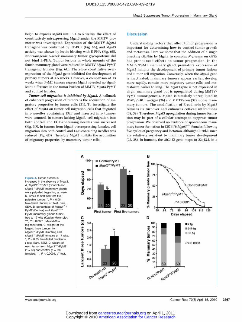
significantly longer time than mice lacking Mgat3 (Fig. 4B).At 17 weeks, 17 of 20 Mgat3−/−/PyMT females had tumors inall 10 mammary glands compared with only 9 of 23 Mgat3+/−/PyMT control mice.Tumor burden is increased in the absence of Mgat3.
The largest three tumors from 17-week mice were weighed.The absence of Mgat3 substantially affected tumor burden,increasing it by ∼1.7-fold (Fig. 4C). Among the 60 tumorsfrom mutant mice, ∼30% weighed >1 g, whereas from controlmice only ∼10% weighed >1 g (Fig. 4D). Body weight wassimilar for control and mutant females at 17 weeks.Erk-1/2 phosphorylation is increased in Mgat3−/−/PyMT
mammary tumors and TECs. Erk-1/2 activation was ana-lyzed in tumor tissue and compared by the ratios pErk-1/2/Erk-1/2. Tumors from 17-week Mgat3−/−/PyMT females ex-hibited greater levels of Erk-1/2 activation compared with con-trols (Fig. 5A). This was also found in tumors from 15-weekfemales. Similarly, TECs derived from Mgat3−/−/PyMT tumorsexhibited greater Erk-1/2 activation thanMgat3+/−/PyMTTECsfollowing EGF or PDGF-AB stimulation (Fig. 5B). IndependentTEC lines gave similar results with serum or EGF stimulation(Supplementary Fig. S4). Both signaling and proliferation ofTECs were inhibited by UO126 (Fig. 5B; SupplementaryFig. S5). Thus, the increased tumor progression of Mgat3−/−/PyMT tumors seems to be due in part to increased signalingvia the Ras pathway, consistent with results from LEC10B/PyMT cells (Fig. 2) showing that the bisecting GlcNAc onN-glycans of GFRs reduces growth factor signaling.
Cancer Res; 70(8) April 15, 2010
American Asso Copyright © 2010 cancerres.aacrjDownloaded from
Loss of Mgat3 causes increased pulmonary metastases.Western blot analyses showed that Mgat3−/−/PyMT tumorsfrom three 15-week females expressed low amounts of func-tionally glycosylated α-DG recognized by mAb IIH6 (Fig. 5C),indicating enhanced metastatic potential (32, 33). This loss ofIIH6 reactivity was confirmed in two Mgat3−/−/PyMT TEClines (Fig. 5C). Lung metastases in control and mutant fe-males were assayed by real-time PCR of PyMT transcriptsin lung (34, 35). Total RNA was isolated from whole lungsof 8-week mice when mammary tumors were at the adenomaor early carcinoma stage. The absolute copy number of PyMTand β-actin was determined and the PyMT/actin ratio calcu-lated. There was more PyMT expression in lungs of femaleslacking Mgat3 (Fig. 5D). This was also apparent in a plot ofPyMT/actin transcript ratio compared with tumor lesion ar-ea (Fig. 5D). In mammary glands with the least tumor size,Mgat3−/−/PyMT lungs generated more PyMT transcripts thancontrols, in which the number of PyMT transcripts was rel-atively constant in relation to tumor area. By contrast, lungPyMT transcripts generally increased with tumor area inMgat3−/−/PyMT mammary glands. Therefore, the absence ofMgat3 facilitates early lung metastasis from Mgat3−/−/PyMTtumors. By 17 weeks however, mutant and control lungs hadmany metastases in equivalent numbers based on histologiccomparisons of lung sections.Constitutive overexpression of Mgat3 retards early
tumor formation. Because virgin mammary glands do notexpress Mgat3 (Fig. 3) and Mgat3+/+/PyMT virgins do not
Figure 3. Mgat3 is expressed in lactating mammary gland and MMTV/PyMT tumors. A, RT-PCR of total RNA from the fourth mammary gland of4-month virgin or lactating females. B, glycoproteins (∼80 μg) from lactating mammary gland of the same females bound E-PHA. Lec4 glycoproteins lackand kidney glycoproteins (∼5 μg) carry the bisecting GlcNAc. C, representative RT-PCR of total RNA from tumors of Mgat3+/−/PyMT(n = 13) andMgat3−/−/PyMT(n = 7) females at 17 wks. D, glycoproteins (∼80 μg) with the bisecting GlcNAc bound E-PHA.
Cancer Research
ciation for Cancer Research on April 1, 2011ournals.org
Mgat3 Suppresses Tumor Progression in Mammary Gland
DOI:10.1158/0008-5472.CAN-09-2719
begin to express Mgat3 until ∼4 to 5 weeks, the effect ofconstitutively misexpressing Mgat3 under the MMTV pro-moter was investigated. Expression of the MMTV-Mgat3transgene was confirmed by RT-PCR (Fig. 6A), and Mgat3activity was shown by lectin blotting with E-PHA (Fig. 6B).Nontransgenic 5-week mammary tumor glycoproteins didnot bind E-PHA. Tumor lesions in whole mounts of thefourth mammary gland were reduced in MMTV-Mgat3-PyMTtransgenic females (Fig. 6C). Therefore constitutive over-expression of the Mgat3 gene inhibited the development ofprimary tumors at 4.5 weeks. However, a comparison at 13weeks when PyMT tumors express Mgat3 revealed no signif-icant difference in the tumor burden of MMTV-Mgat3-PyMTand control females.Tumor cell migration is inhibited by Mgat3. A hallmark
of enhanced progression of tumors is the acquisition of mi-gratory properties by tumor cells (31). To investigate theeffect of Mgat3 on tumor cell migration, cells that migratedinto needles containing EGF and inserted into tumorswere counted. In tumors lacking Mgat3, cell migration intoboth control and EGF-containing needles was increased(Fig. 6D). In tumors from Mgat3 overexpressing females, cellmigration into both control and EGF-containing needles wasreduced (Fig. 6D). Therefore Mgat3 inhibits the acquisitionof migratory properties by mammary tumor cells.
www.aacrjournals.org
American Asso Copyright © 2010 cancerres.aacrjDownloaded from
Discussion
Understanding factors that affect tumor progression isimportant for determining how to control tumor growthand metastasis. Here we show that the addition of a singlebisecting GlcNAc by Mgat3 to complex N-glycans on GFRshas pronounced effects on tumor progression. In theMMTV/PyMT mammary gland, premature expression ofMgat3 inhibits the development of primary tumor lesionsand tumor cell migration. Conversely, when the Mgat3 geneis inactivated, mammary tumors appear earlier, developmore rapidly, contain more migratory tumor cells, and me-tastasize earlier to lung. The Mgat3 gene is not expressed invirgin mammary gland but is upregulated during MMTV/PyMT tumorigenesis. Mgat3 is similarly upregulated inWAP/SV40 T antigen (36) and MMTV/neu (37) mouse mam-mary tumors. The modification of E-cadherin by Mgat3reduces its turnover and enhances cell-cell interactions(38, 39). Therefore, Mgat3 upregulation during tumor forma-tion may be part of a cellular attempt to suppress tumorprogression. We observed no evidence of spontaneous mam-mary tumor formation in C57Bl/6 Mgat3−/− females followingfive cycles of pregnancy and lactation, although C57Bl/6 miceare relatively resistant to mammary tumor development(22, 28). In humans, the MGAT3 gene maps to 22q13.1, in a
Figure 4. Tumor burden isincreased in the absence of Mgat3.A, Mgat3+/−/PyMT (Control) andMgat3−/−/PyMT mammary glandswere palpated beginning at week6. Times to first and first fivepalpable tumors. *, P < 0.05,two-tailed Student's t test. Bars,SEM. B, percentage of Mgat3+/−/PyMT (Control) and Mgat3−/−/PyMT mammary glands tumorfree to 17 wks (Kaplan-Meier plot;***, P < 0.0001, Mantel-Coxlog-rank test). C, weight of thelargest three tumors fromMgat3+/−/PyMT (Control) andMgat3−/−/PyMT females at 17 wks.*, P < 0.05, two-tailed Student'st test. Bars, SEM. D, weight ofeach tumor from Mgat3−/−/PyMT(n = 60) and control (n = 69)females. ***, P < 0.0001, χ2 test.
Cancer Res; 70(8) April 15, 2010 3367
ciation for Cancer Research on April 1, 2011ournals.org

Song et al.
3368
DOI:10.1158/0008-5472.CAN-09-2719
Cancer Res; 70(8) April 15, 2010
American Association for Cancer Researc Copyright © 2010 on April 1, 201cancerres.aacrjournals.orgDownloaded from
Figure 5. Increased expressionof pErk-1/2 and early pulmonarymetastases in the absence ofMgat3. A, Western blot of pErk-1/2and Erk-1/2 in tumors fromMgat3+/−/PyMT and Mgat3−/−/PyMT females. Ratios of pErk-1/Erk-1 and pErk-2/Erk-2 inhistograms. *, P < 0.05, two-tailedStudent's t test. Bars, SEM.B, EGF and PDGF-AB signaling inMgat3+/−/PyMT and Mgat3−/−/PyMT TECs in the presence andabsence of 10 μmol/L UO126.Bars, SEM. C, functionallyglycosylated α-DG (IIH6) in 15-wktumors and TEC lines. D, ratioabsolute number PyMT/actin(×103) transcripts. *, P < 0.05,two-tailed Student's t test. Bars,SEM (left). Ratio absolute numberPyMT/actin (×103) transcriptsversus tumor area from the fourthmammary gland of the samemice (right).
Cancer Research
h1

Mgat3 Suppresses Tumor Progression in Mammary Gland
DOI:10.1158/0008-5472.CAN-09-2719
region proposed to contain a tumor suppressor gene whoseloss of heterozygosity (LOH) correlates with human breastcancers (40, 41). Expression data from human breast cancershave not revealed changes in Mgat3 transcripts to date,perhaps because MGAT3 mutations do not alter the expres-sion of mutant alleles maintained by LOH. In human ovariancancer, however, upregulation of the MGAT3 gene was ob-served (42).To address how the loss of Mgat3 might promote tumor
progression, we examined growth factor signaling in CHO/PyMT cells, MMTV/PyMT tumors, and MMTV/PyMT TECcells. In LEC10B CHO cells with well-characterized bisectedN-glycans (10) that cause a reduction in cell surface galectinbinding (9), Mgat3 expression retards cell proliferation andinhibits galectin-promoted growth factor signaling. Impor-tantly, CHO/PyMT cell proliferation is driven in part byErk-1/2 activation as shown by the inhibition of cell growthby the MEK1/2 inhibitor UO126. Erk-1/2 activation is alsoregulated by Mgat3 in vivo, being greater in Mgat3−/−/PyMTmammary tumors. Tumor-derived TECs lacking Mgat3 alsoexhibited enhanced Erk-1/2 activation in response to serum,EGF, or PDGF. Therefore, whereas the MMTV/PyMT onco-gene was the driving force of mammary tumorigenesis,Mgat3 restrained growth factor signaling, and loss of Mgat3resulted in an increase in Erk-1/2 activation.PyMT is a scaffold protein that acts in the cytoplasm to
cause transformation (24). It cannot be directly affected byMgat3, which acts on N-glycans in the Golgi. This is the rea-son our investigations into how Mgat3 modulates mammary
www.aacrjournals.org
American Asso Copyright © 2010 cancerres.aacrjDownloaded from
tumor progression focused on its effects on growth factorsignaling via glycoprotein receptors such as PDGFR andEGFR known to have N-glycans modified by Mgat3 (15).Constitutive activation of EGF signaling due to activatingmutations in EGFRs is a well-characterized basis of poorprognosis in breast cancer (43). PDGF signaling has also beenimplicated in both autocrine and paracrine mechanisms ofpromoting breast cancer progression (44, 45). A new mecha-nism for modulating signaling through GFRs is through in-teractions of lactosamine units on their complex N-glycansvia a galectin lattice (46, 47). GFRs with more branchedN-glycans are retained longer at the cell surface in a galectinlattice, allowing them to signal longer before endocytosis anddownregulation. This is the mechanism that we propose isaffected by the addition of the bisecting GlcNAc. Thus, weshow that loss of Mgat3 reduces galectin-regulated growthfactor signaling and cell proliferation. Growth factor recep-tors with a bisecting GlcNAc are predicted to be less wellretained in a galectin lattice and to signal more weakly thantheir counterparts with N-glycans lacking the bisectingGlcNAc. We propose that reduced galectin lattice inter-actions caused by the bisecting GcNAc are due to reducedgalectin recognition of highly branched N-glycans carryinga bisecting GlcNAc. An alternative proposal that bisectedcomplex N-glycans are not substrates for Mgat5 and therebyhave reduced branching (13) seems unlikely because LEC10glycoproteins carrying the bisecting GlcNAc bind muchmore L-PHA (which recognizes the product of Mgat5) thanCHO glycoproteins and express N-glycans with many LacNAc
Figure 6. Constitutiveoverexpression of Mgat3inhibits early mammary tumordevelopment. A, RT-PCR of totalRNA from the fourth mammarygland of 5-wk virgin transgenic (Tg)or non-transgenic females;kidney cDNA, positive control.B, glycoproteins with bisectedN-glycans from the othermammary gland of the samefemales bound E-PHA. C, tumorlesion areas in control andMMTV-Mgat3-PyMT transgenicmice. *, P < 0.05, one-tailedStudent's t test. Bars, SEM. D,cells collected in control (n = 2) orEGF-containing needles (n = 4)inserted for 4 h into mammarytumors (n = 4–6) of ∼1.5 cm.
Cancer Res; 70(8) April 15, 2010 3369
ciation for Cancer Research on April 1, 2011ournals.org

Song et al.
3370
DOI:10.1158/0008-5472.CAN-09-2719
units (10), indicating that branched N-glyans are likely tohave been produced by Mgat5.Any growth factor or cytokine receptor or integrin with
complex N-glycans is a potential substrate for Mgat3 andmay have its signaling strength modulated by the additionof the bisecting GlcNAc. Thus, a broad spectrum of signalingpathways may be affected in MMTV/PyMT tumor cells. Inthis paper, we focus on Erk-1/2 activation and show a func-tional relationship to cell proliferation. It will be important inthe future to determine the hierarchy of growth-promotingversus growth-retarding pathways, as well as those involvedin epithelial-mesenchymal transition and metastasis that aremodulated by Mgat3 during MMTV/PyMT tumor progres-sion. For example, we observed that 15-week mammarytumors and TECs lacking Mgat3 express reduced levels offunctionally glycosylated α-DG, which results in reducedbinding to laminin and correlates with enhanced tumor pro-gression (32, 33). Loss of another GlcNAcT (β1,3GlcNAcT-1),which is essential to the generation of lactosamine units oncomplex N-glycans and also to the functional glycosylation ofα-DG, also leads to enhanced progression in a murine pros-tate cancer model (32). Mgat3 transfers the bisecting GlcNActo the same subset of complex N-glycans that are substratesfor β1,3GlcNAcT-1 and may act, in part, by inhibiting thefunctional glycosylation of N-glycans on α-DG, which areknown substrates of Large (48), a putative glycosyltransferasefor which β1,3GlcNAcT-1 is an essential partner (32).In investigations of mechanism, it will also be important to
identify which of the 10 mouse galectins promote the pro-gression of MMTV/PyMT mammary tumors through theirinteractions with complex N-glycans. Whereas galectin-3has been implicated in the regulation of growth factor signal-
Cancer Res; 70(8) April 15, 2010
American Asso Copyright © 2010 cancerres.aacrjDownloaded from
ing in MMTV/PyMT tumors (3), females lacking galectin-3generate equivalent numbers of MMTV/PyMT mammarytumors to controls (49). In addition, galectin-3 is downre-gulated and poorly expressed during lactation in the mouse(50). Therefore, one or more of the other mouse galectinsseem to be important for tumor progression in the murinemammary gland.In conclusion, it is apparent that addition of the bisecting
GlcNAc to complex N-glycans on mammary glycoproteinsserves to protect mammary epithelial cells from tumor pro-gression. Thus, loss of Mgat3 by LOH in human cancerswould be expected to promote tumor progression.
Disclosure of Potential Conflicts of Interest
No potential conflicts of interest were disclosed.
Acknowledgments
We thank Elaine Lin and Jeffrey Pollard for expert advice; RiddhiBattarycharrya, Peter Draber, David Gross, Yan Deng, and Wen Dong fortechnical assistance; Shira Landskorner-Eiger, Suzannah Williams, andParaic Kenny for helpful discussions; and all who kindly provided reagents.
Grant Support
National Cancer Institute grants RO130645 and RO136434 (P. Stanley) andP30CA013330 supporting the Albert Einstein Cancer Center and Young Inves-tigator Award Breast Cancer Alliance, Inc. (S. Goswami).
The costs of publication of this article were defrayed in part by the paymentof page charges. This article must therefore be hereby marked advertisement inaccordance with 18 U.S.C. Section 1734 solely to indicate this fact.
Received 07/22/2009; revised 01/05/2010; accepted 01/22/2010.
References
1. Stanley P, Schachter H, Taniguchi N. In: Varki A, Cummings RD,Esko JD, Freeze HH, Stanley P, Bertozzi CR, Hart GW, Etzler ME,editors. N-glycans. Essentials of gycobiology. 2nd ed Cold SpringHarbor: Cold Spring Harbor Laboratory Press; 2009, p. 101–14.
2. Granovsky M, Fata J, Pawling J, Muller WJ, Khokha R, Dennis JW.Suppression of tumor growth and metastasis in Mgat5-deficientmice. Nat Med 2000;6:306–12.
3. Partridge EA, Le Roy C, Di Guglielmo GM, et al. Regulation ofcytokine receptors by Golgi N-glycan processing and endocytosis.Science 2004;306:120–4.
4. Lau K, Partridge E, Grigorian A, et al. Complex N-glycan numberand degree of branching cooperate to regulate cell proliferationand differentiation. Cell 2007;129:123–34.
5. Guo HB, Johnson H, Randolph M, Lee I, Pierce M. Knockdown ofGnT-Va expression inhibits ligand-induced downregulation of theepidermal growth factor receptor and intracellular signaling by inhi-biting receptor endocytosis. Glycobiology 2009;19:547–59.
6. Narasimhan S. Control of glycoprotein synthesis. UDP-GlcNAc:glycopeptide β 4-N-acetylglucosaminyltransferase III, an enzymein hen oviduct which adds GlcNAc in β 1–4 linkage to the β-linkedmannose of the trimannosyl core of N-glycosyl oligosaccharides.J Biol Chem 1982;257:10235–42.
7. Campbell C, Stanley P. A dominant mutation to ricin resistancein Chinese hamster ovary cells induces UDP-GlcNAc:glycopeptideβ-4-N-acetylglucosaminyltransferase III activity. J Biol Chem 1984;259:13370–8.
8. Stanley P, Sundaram S, Tang J, Shi S. Molecular analysis of three
gain-of-function CHO mutants that add the bisecting GlcNAc toN-glycans. Glycobiology 2005;15:43–53.
9. Patnaik SK, Potvin B, Carlsson S, Sturm D, Leffler H, Stanley P.Complex N-glycans are the major ligands for galectin-1, -3, and-8 on Chinese hamster ovary cells. Glycobiology 2006;16:305–17.
10. North SJ, Huang H-H, Sundaram S, et al. Glycomics profiling ofChinese hamster ovary (CHO) cell glycosylation mutants revealsN-glycans of a novel size and complexity. J Biol Chem 2010;285:5759–75.
11. Zhao Y, Sato Y, Isaji T, et al. Branched N-glycans regulate the biologi-cal functions of integrins and cadherins. FEBS J 2008;275:1939–48.
12. Takahashi M, Kuroki Y, Ohtsubo K, Taniguchi N. Core fucose andbisecting GlcNAc, the direct modifiers of the N-glycan core: theirfunctions and target proteins. Carbohydr Res 2009;344:1387–90.
13. Yoshimura M, Ihara Y, Ohnishi A, et al. Bisecting N-acetylgluco-samine on K562 cells suppresses natural killer cytotoxicity and pro-motes spleen colonization. Cancer Res 1996;56:412–8.
14. Yoshimura M, Nishikawa A, Ihara Y, Taniguchi S, Taniguchi N.Suppression of lung metastasis of B16 mouse melanoma byN-acetylglucosaminyltransferase III gene transfection. Proc NatlAcad Sci U S A 1995;92:8754–8.
15. Sato Y, Takahashi M, Shibukawa Y, et al. Overexpression ofN-acetylglucosaminyltransferase III enhances the epidermal growthfactor-induced phosphorylation of ERK in HeLaS3 cells by up-regulation of the internalization rate of the receptors. J Biol Chem2001;276:11956–62.
16. Isaji T, Gu J, Nishiuchi R, et al. Introduction of bisecting GlcNAc into
Cancer Research
ciation for Cancer Research on April 1, 2011ournals.org

Mgat3 Suppresses Tumor Progression in Mammary Gland
DOI:10.1158/0008-5472.CAN-09-2719
integrin α5β1 reduces ligand binding and down-regulates cell adhe-sion and cell migration. J Biol Chem 2004;279:19747–54.
17. Akama R, Sato Y, Kariya Y, et al. N-acetylglucosaminyltransferaseIII expression is regulated by cell-cell adhesion via the E-cadherin-catenin-actin complex. Proteomics 2008;8:3221–8.
18. Ekuni A, Miyoshi E, Ko JH, et al. A glycomic approach to hepatictumors in N-acetylglucosaminyltransferase III (GnT-III) transgenicmice induced by diethylnitrosamine (DEN): identification of haptoglo-bin as a target molecule of GnT-III. Free Radic Res 2002;36:827–33.
19. Yang X, Bhaumik M, Bhattacharyya R, Gong S, Rogler CE, Stanley P.New evidence for an extra-hepatic role of N-acetylglucosaminyl-transferase III in the progression of diethylnitrosamine-induced livertumors in mice. Cancer Res 2000;60:3313–9.
20. Yang X, Tang J, Rogler CE, Stanley P. Reduced hepatocyte prolifer-ation is the basis of retarded liver tumor progression and liver regen-eration in mice lacking N-acetylglucosaminyltransferase III. CancerRes 2003;63:7753–9.
21. Guy CT, Cardiff RD, Muller WJ. Induction of mammary tumors byexpression of polyomavirus middle T oncogene: a transgenic mousemodel for metastatic disease. Mol Cell Biol 1992;12:954–61.
22. Qiu TH, Chandramouli GV, Hunter KW, Alkharouf NW, Green JE, LiuET. Global expression profiling identifies signatures of tumorvirulence in MMTV/PyMT-transgenic mice: correlation to human dis-ease. Cancer Res 2004;64:5973–81.
23. Lin EY, Jones JG, Li P, et al. Progression to malignancy in the poly-oma middle T oncoprotein mouse breast cancer model provides areliable model for human diseases. Am J Pathol 2003;163:2113–26.
24. Dilworth SM. Polyoma virus middle T antigen and its role in identify-ing cancer-related molecules. Nat Rev Cancer 2002;2:951–6.
25. Patnaik SK, Stanley P. Lectin-resistant CHO glycosylation mutants.Methods Enzymol 2006;416:159–82.
26. Bhattacharyya R, Bhaumik M, Raju TS, Stanley P. Truncated, inac-tive N-acetylglucosaminyltransferase III (GlcNAc-TIII) induces neuro-logical and other traits absent in mice that lack GlcNAc-TIII. J BiolChem 2002;277:26300–9.
27. Priatel JJ, Sarkar M, Schachter H, Marth JD. Isolation, characteri-zation and inactivation of the mouse Mgat3 gene: the bisectingN-acetylglucosamine in asparagine-linked oligosaccharides appearsdispensable for viability and reproduction. Glycobiology 1997;7:45–56.
28. Davie SA, Maglione JE, Manner CK, et al. Effects of FVB/NJ andC57Bl/6J strain backgrounds on mammary tumor phenotype in in-ducible nitric oxide synthase deficient mice. Transgenic Res 2007;16:193–201.
29. Kawamoto S, Niwa H, Tashiro F, et al. A novel reporter mousestrain that expresses enhanced green fluorescent protein uponCre-mediated recombination. FEBS Lett 2000;470:263–8.
30. Wyckoff JB, Segall JE, Condeelis JS. The collection of the motilepopulation of cells from a living tumor. Cancer Res 2000;60:5401–4.
31. Wyckoff J, Wang W, Lin EY, et al. A paracrine loop between tumorcells and macrophages is required for tumor cell migration in mam-mary tumors. Cancer Res 2004;64:7022–9.
32. Bao X, Kobayashi M, Hatakeyama S, et al. Tumor suppressorfunction of laminin-binding α-dystroglycan requires a distinct β3-N-acetylglucosaminyltransferase. Proc Natl Acad Sci U S A 2009;106:12109–14.
33. de Bernabé DB, Inamori K, Yoshida-Moriguchi T, et al. Loss of α-
www.aacrjournals.org
American Asso Copyright © 2010 cancerres.aacrjDownloaded from
dystroglycan laminin binding in epithelium-derived cancers is causedby silencing of LARGE. J Biol Chem 2009;284:11279–84.
34. Montagna C, Lyu MS, Hunter K, et al. The Septin 9 (MSF) gene isamplified and overexpressed in mouse mammary gland adenocarci-nomas and human breast cancer cell lines. Cancer Res 2003;63:2179–87.
35. Zheng H, Abdel Aziz HO, Nakanishi Y, et al. Oncogenic role of JCvirus in lung cancer. J Pathol 2007;212:306–15.
36. Klein A, Guhl E, Zollinger R, et al. Gene expression profiling: cellcycle deregulation and aneuploidy do not cause breast cancerformation in WAP-SVT/t transgenic animals. J Mol Med 2005;83:362–76.
37. Landis MD, Seachrist DD, Montanez-Wiscovich ME, Danielpour D,Keri RA. Gene expression profiling of cancer progression reveals in-trinsic regulation of transforming growth factor-β signaling in ErbB2/Neu-induced tumors from transgenic mice. Oncogene 2005;24:5173–90.
38. Iijima J, Zhao Y, Isaji T, et al. Cell-cell interaction-dependent reg-ulation of N-acetylglucosaminyltransferase III and the bisected N-glycans in GE11 epithelial cells. Involvement of E-cadherin-mediatedcell adhesion. J Biol Chem 2006;281:13038–46.
39. Yoshimura M, Ihara Y, Matsuzawa Y, Taniguchi N. Aberrant glycosyl-ation of E-cadherin enhances cell-cell binding to suppress metasta-sis. J Biol Chem 1996;271:13811–5.
40. Iida A, Kurose K, Isobe R, et al. Mapping of a new target region ofallelic loss to a 2-cM interval at 22q13.1 in primary breast cancer.Genes Chromosomes Cancer 1998;21:108–12.
41. Nagahata T, Hirano A, Utada Y, et al. Correlation of allelic losses andclinicopathological factors in 504 primary breast cancers. BreastCancer 2002;9:208–15.
42. Abbott KL, Nairn AV, Hall EM, et al. Focused glycomic analysis of theN-linked glycan biosynthetic pathway in ovarian cancer. Proteomics2008;8:3210–20.
43. Hynes NE, MacDonald G. ErbB receptors and signaling pathways incancer. Curr Opin Cell Biol 2009;21:177–84.
44. Jechlinger M, Sommer A, Moriggl R, et al. Autocrine PDGFR sig-naling promotes mammary cancer metastasis. J Clin Invest 2006;116:1561–70.
45. Roussidis AE, Theocharis AD, Tzanakakis GN, Karamanos NK. Theimportance of c-Kit and PDGF receptors as potential targets formolecular therapy in breast cancer. Curr Med Chem 2007;14:735–43.
46. Dennis JW, Lau KS, Demetriou M, Nabi IR. Adaptive regulation at thecell surface by N-glycosylation. Traffic 2009;10:1569–78.
47. Lajoie P, Goetz JG, Dennis JW, Nabi IR. Lattices, rafts, and scaffolds:domain regulation of receptor signaling at the plasma membrane.J Cell Biol 2009;185:381–5.
48. Patnaik SK, Stanley P. Mouse large can modify complex N- andmucin O-glycans on α-dystroglycan to induce laminin binding. J BiolChem 2005;280:20851–9.
49. Eude-Le Parco I, Gendronneau G, Dang T, et al. Genetic assessmentof the importance of galectin-3 in cancer initiation, progression, anddissemination in mice. Glycobiology 2009;19:68–75.
50. Ron M, Israeli G, Seroussi E, et al. Combining mouse mammarygland gene expression and comparative mapping for the identifica-tion of candidate genes for QTL of milk production traits in cattle.BMC Genomics 2007;8:183.
Cancer Res; 70(8) April 15, 2010 3371
ciation for Cancer Research on April 1, 2011ournals.org