the biology and pathology of lymphocyte fc receptors
TRANSCRIPT

American Journal ofPathology, Vol. 152, No. 3, March 1998Copyright American Societyfor Investigative Pathology
Rous*Whipple Award Lecture
The Biology and Pathology of Lymphocyte FcReceptors
Richard G. LynchFrom the Department ofPathology, University ofIowa College ofMedicine, Iowa City, Iowa
Molecular epitopes located in the constant region do-mains (Fc domains) of antibody molecules are recog-nized by cell surface receptors designated as Fc recep-tors (FcR). For each class of immunoglobulin heavy chainthere is a corresponding class of Fc receptors. Multiplephysiological functions are mediated by Fc receptors, areflection of their broad distribution on a diversity of celltypes. The focus of this article will be on the occurrenceand functions of Fc receptors on lymphocytes.1 ThoseFcR that specifically recognize the Fc region of IgG an-tibodies are termed Fc'yR. Likewise, IgA antibodies arerecognized by FcaR, IgM by Fc,R, IgE by FcER, and IgDby FcSR. Each class of FcR has one or more members,each encoded by distinct genes. Some FcR are mul-tichain molecules encoded by multiple genes, whereasothers are single chain polypeptides encoded by a singlegene. Structurally distinct FcR isoforms are generated atthe transcriptional level through differential exon usageand at the post-transcriptional level by alternative splic-ing of transcripts. The isoforms of each class of FcRperform distinct physiological functions. A large amountof structural information has been developed about FcyRand the genes that encode them in humans and mice.The molecular structures and the structure-functional re-lationships of FcyR have been reviewed.2-4Some Fc receptors have been appreciated for de-
cades because of their conspicuous involvement in hostdefense against infectious agents and in immunologicalmechanisms of disease (Table 1). An example of theformer are the IgG receptors on macrophages and neu-trophils that enhance the phagocytosis of IgG-coatedmicrobes.5 An example of the latter is the IgE receptorson mast cells, which when aggregated by antigen andIgE cause systemic anaphylaxis.6 During the past de-cade there has been an unprecedented growth in knowl-edge about these and other newly discovered FcR. Amajor area of interest has been the FcR that occur onlymphocytes. Research progress in this area has yieldeda new appreciation for the involvement of lymphocyte Fcreceptors in a broad array of normal physiological func-tions (Table 2) and has identified interesting alterations in
FcR expression on lymphocytes in an increasing numberof diseases.7-11
Investigations from multiple laboratories have estab-lished that in each hematopoietic lineage FcR expressionis regulated by developmental and environmental fac-tors. During ontogeny, different myeloid and lymphoidlineages show characteristic patterns of simultaneousand sequential display of combinations of the variousclasses of FcR. FcyR are the most broadly distributedFcR on hematopoietic cells, being found on cells of everyhematopoietic lineage. On T, B, and NK lymphocytes, thedifferent structural forms of FcyR have in common a lowaffinity binding for monomeric IgG but a high aviditybinding for IgG complexed with antigen. The literatureabout FcyR on mature myeloid and lymphoid cells isvoluminous because of the diversity of functions linked tothe receptors and the evident importance of those func-tions. These include the role of FcyR in phagocytosis bymacrophages and neutrophils,5 12,13 in antibody-depen-dent cellular cytotoxicity by certain T cells and NKcells,14 15 in the regulation of cellular activation in B lym-phocytes,16'17 in antibody-mediated feedback inhibi-tion,18619 in endocytosis and antigen presentation by sev-eral types of lymphoid cells,20 in the triggering of cytokineand superoxide production in mononuclear phagocytesand lymphocytes,20.21 and most recently in the inductionof apoptosis in eosinophils.21 In addition, FcyR are ex-pressed on basophils/mast cells, platelets, dendritic
Supported by research grants and training grants awarded by the Na-tional Institutes of Health. The author is supported by the Clement T. andSylvia H. Hanson Professorship in Immunology through the University ofIowa Foundation.
am deeply honored to receive this award named in memory of PeytonRous and George Whipple, Nobel laureates and master pathologists ofthe twentieth century. The Rous-Whipple Award is especially meaningfulbecause had the good fortune of being a student fellow in pathology atThe University of Rochester during 1963-64, a time when Dr. Whipple,although officially retired, was still very active and a regular attendee atdepartmental conferences and a wonderful role model for pathologists ofall ages.
Accepted for publication January 15, 1998.Address reprint requests to Dr. Richard Lynch, Department of Pathol-
ogy, University of Iowa College of Medicine, 144 Medical Labs, 200Hawkins Drive, Iowa City, IA 52242.
631
632 LynchAJP March 1998, Vol. 152, No. 3
Table 1. Classical Functions of Fc Receptors
* Phagocytosis by macrophages and neutrophils* Immunoglobulin transport by placenta* Antibody-dependent cell-mediated cytotoxicity (ADCC)* Release of inflammatory mediators by mast cells* Feedback inhibition of antibody production
cells, and Langerhans cells, a distribution that has at-tracted considerable research interest. Besides the largeamount of published information about the cellular andmolecular events that are linked to these normal functionsof FcyR, there is a substantial volume of literature thatdeals with the various pathological consequences thatresult from defects in FcyR structure and function.11The multiplicity of regulatory and effector functions
mediated by FcR on lymphocytes reflect the actions ofmultiple classes and isoforms of FcR that are displayedon the various subsets of lymphocytes. The material dis-cussed below will focus on Fc receptors that are presenton murine and human T and B lymphocytes and theircellular progenitors. Most of the information that will becovered was generated in experiments conducted bymany talented fellows, students, and staff in my labora-tory, which was initially in the Department of Pathology atWashington University in St. Louis and for the past 16years in the Department of Pathology at the University ofIowa.
Activation-Induced Expression of FcR onMature T CellsThe laboratory's interest in lymphocyte FcR began in the1970's during investigations of plasmacytoma cell differ-entiation22 and idiotype-specific plasmacytoma immunityinduced by immunization with the monoclonal immuno-globulin produced by the tumor.23 At the time of thosestudies the literature contained reports that lymphocytesin mice with plasmacytomas and in humans with multiplemyeloma expressed the idiotype of the monoclonal im-munoglobulin.24 27 It had been proposed by some inves-tigators that mRNA released from the tumor cells wasinternalized by host lymphocytes in which it was trans-lated and resulted in cell surface display of the variableregions of the monoclonal immunoglobulin.28 It was sug-gested by others that the circulating cells that expressedthe myeloma idiotype were lymphocytoid progenitors ofthe plasmacytic myeloma cells26 or were T-cells con-
Table 2. Functions Associated with Fc Receptors onLymphocytes
* Inhibition of antigen-induced activation of B cells* Enhancement of IgM production by mature B cells* Promotion of cellular homotypic aggregation* Facilitation of T-cell B-cell interaction* Processing and presentation of antigen by B cells* Production of soluble Fc receptors* Induction of B-cell tolerance* Regulation of immunoglobulin heavy chain isotype* Acceleration of differentiation of immature T cells and B
cells
MOI mm1Sb_r-4nds%A
I 12U-
PIC
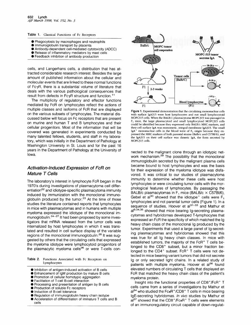
FlFigure 1. Experimental demonstration that the circulating mononuclear cellswith surface IgA315 were host lymphocytes and not small lymphocytoidMOPC315 cells. When the BALB/c plasmacytoma MOPC315 was passaged inF, mice, the large plasmacytoid and small lymphocytoid MOPC315 cellscould be identified because they expressed only BALB/c MHC markers, andtheir cell surface IgA was monomeric, integral membrane IgA315. The smallIgA+ mononuclear cells in the blood were of F1 origin because they ex-pressed the MHC markers of both parental strains (Balb/c and C57BU/6), andthe IgA315 on their cell surface was dimeric IgA, the form secreted byMOPC315 cells.
nected to the malignant clone through an idiotypic net-work mechanism.29 The possibility that the monoclonalimmunoglobulin secreted by the malignant plasma cellsbecame bound to host lymphocytes and was the basisfor their expression of the myeloma idiotype was disfa-vored. It was critical to our studies of plasmacytomaimmunity to determine whether these cells were hostlymphocytes or were circulating tumor cells with the mor-phological features of lymphocytes. By passaging theBALB/c plasmacytomas in F1 mice (BALB/c x C57BI/6),Gebel et a130 showed that the idiotype+ cells were F1lymphocytes and not parental tumor cells (Figure 1). In asequence of studies, Hoover et a131-33 and Mathur etal9,34,35 showed that mice bearing Ig-secreting plasma-cytomas and hybridomas developed T-lymphocytes thatexpressed an FcR the specificity of which matched the Igheavy chain class of the monoclonal Ig produced by thetumor. Experiments that used a large panel of Ig-secret-ing plasmacytomas and hybridomas showed that thiswas true for all Ig heavy chain classes. In mice withestablished tumors, the majority of the FcR+ T cells be-longed to the CD8+ subset, but a minor fraction be-longed to the CD4+ subset. FcR+ T cells were not de-tected in mice bearing variant tumors that did not secreteIg or only secreted light chains. In a related study ofpatients with multiple myeloma, Hoover et al36 foundelevated numbers of circulating T cells that displayed anFcR that matched the heavy chain class of the patient'smyeloma protein.
Insight into the functional properties of CD8+/FcR+ Tcells came from a series of investigations by Mathur eta134 who studied the FcER+/CD8+ T cells in mice bearingIgE-secreting hybridomas. In vivo studies by Mathur etal37 showed that the CD8+/FcER+ T cells were elementsof an immunoregulatory circuit capable of down-regulat-

Lymphocyte Fc Receptors 633AJP March 1998, Vol. 152, No. 3
ing E-heavy chain gene transcription in the hybridomacells. The molecular mechanism of transcriptional regu-lation involved a transacting factor that inhibited an in-tronic enhancer in the heavy chain gene but did not affectlight chain production.38 This mechanism of transcrip-tional regulation also operated spontaneously in vivo andwas responsible for the regular appearance of stableK-light chain (Bence-Jones) variants from the wild-typeIgEK parental hybridoma during sequential passage inmice.39 The finding that heavy chain gene transcriptionwas the site of control of immunoglobulin synthesis wasintriguing because earlier studies had shown that the siteof control of immunoglobulin synthesis in plasmacytomacells that was mediated by CD8+, idiotype-specific Tcells was at the level of the light chain gene.40 Thesestudies identified two pathways by which distinct popu-lations of CD8+ T-cells inhibited immunoglobulin synthe-sis in plasmacytoma and hybridoma cells. Both pathwaysinvolved transacting transcriptional inhibitors; in onepathway light chain gene transcription was selectivelyblocked, and in the other pathway heavy chain genetranscription was selectively blocked.
Definitive evidence for the role of the T-cell receptor(TCR) in the induction of FcR expression on T lympho-cytes came from a series of investigations by San-dor.41 42,43 Those studies showed that, with the exceptionof some murine intraepithelial eyI T cells in which cellsurface FcR expression is constitutive in the resting state,FcR are not displayed on resting murine T cells but areinduced on some subsets of T cells when activatedthrough their TCR.41'42 In a double-blind study, analysisof a large panel of CD4+ murine T-cell clones revealedthat activation through the TCR induced FcR preferen-tially on those clones that, by their pattern of lymphokineproduction, were members of the Th2 subset of CD4+cells.43 The biased induction of FcR on Th2 versus Thlcells was of particular interest because the Th2 subset ofCD4+ T cells contains the classic helper T cells for anti-body responses. All eight of the Th2 clones examinedexpressed high levels of the Fc,R when activatedthrough their TCR. The T-cell Fc,tR recognizes anepitope located in C,3, an IgM constant region domainthat is accessible on the surface of mature B cells.44 Thisinteresting set of circumstances raises the possibility thatin the collaboration between activated CD4+ T cells andB cells during the induction of antibody responses, inter-actions occur between the Fc,uR on the T cell and C,u3 onthe B cell. In addition to the induction of Fc,uR, Sandor eta143 also observed that multiple classes of FcR wereinitially induced on the same Th2 cell when it was acti-vated through its TCR. In related studies, Sandor et a145also found that the level of FcR displayed on the surfaceof T cells was enhanced and maintained in the presenceof the corresponding Ig ligand. Taken together, thesefindings identified a potential mechanism that could ac-count for the Ig heavy chain specificity of the FcR+ T cellsin mice with Ig-secreting plasmacytomas and for thefailure to detect FcR+ T cells in mice bearing variantplasmacytomas that did not produce an intact immuno-globulin.34 Conceivably, activation of CD8+ T cells inmice with plasmacytomas, possibly by major histocom-
patibility complex (MHC) class I-restricted TCR recogni-tion of idiotypic peptides, initially induces the expressionof multiple classes of FcR on the T cells. Persistent ex-pression of the class of FcR that corresponds to theheavy chain class of the myeloma protein might be ac-counted for by Ig ligand-induced up-regulation of thatFcR, whereas the other classes of FcR are only transientlydisplayed and disappear. The predominance of CD8+cells in the FcR+ T-cell response to plasmacytomasraises the possibility that Ig variable region peptides ofthe monoclonal immunoglobulin associate with MHCclass molecules in the plasmacytoma cells and thisresults in the tumor cells functioning as antigen-present-ing cells. The minor population of FcR+ T cells that areCD4+/CD8- in mice with plasmacytomas might reflectMHC class 11-restricted presentation of idiotypic peptidesby macrophages or other host cells that internalize andprocess the monoclonal Ig. Support for this possibilitycomes from the studies of Lauritzsen et al46 who havefound that MHC class Il-restricted, idiotype-specificCD4+ T cells are present in mice with plasmacytomas.
Several other observations established that the induc-tion of FcR on T lymphocytes in vivo is an event that is notconfined to mice with Ig-secreting tumors. In collabora-tion with Joel Weinstock's laboratory, studies were con-ducted in mice infected with Schistosoma mansoni inwhich a strong Th2 type immune response develops toantigens present in the eggs of the parasite. Adult wormsin the portal venous system produce eggs that lodge inthe liver where they elicit a granulomatous inflammatoryresponse. Sandor et aI41 found that the in vivo-activatedCD4+ T cells isolated from the egg-induced hepaticgranulomas expressed high levels of FcR on their cellsurface. In other experiments, CD4+ T cells from normalmice that had been injected with anti-TCR antibody wereshown to display cell surface FcR. Together, these ob-servations showed that activation of some populations ofT cells through their TCR induces the appearance of FcRon their cell surface. The finding of a linkage betweenTCR, T cell activation, and FcR is intriguing because, asdiscussed below, with B cells there is a linkage amongB-cell receptor (BCR), B-cell activation, and FcR.
Constitutive Expression of FcR on Mature BCellsIn contrast to resting T cells in which FcR are not present,resting mature B cells simultaneously express multipleclasses of FcR.1 There is a substantial literature on theFcyR and FcER on mature B cells.4753 Our laboratoryinvestigated the occurrence, regulation, and functions ofFcER,52 FcaR,54 Fc,55 and FcyR53 on B cells, andthese studies have previously been reviewed.1 Except fortheir occurrence, little information has been generatedabout the FcaR on murine B lymphocytes. Using a panelof IgM constant region domain deletional mutants,Mathur et al44'55 identified some of the structural require-ments in IgM for binding to occur to the Fc,uR on B cellsand T cells.

634 LynchAJP March 1998, Vol. 152, No. 3
The presence of FcERII(CD23) on B cells in mice andhumans has attracted considerable research attentionbecause of the multiple functions that have been as-cribed to CD23, including a proposed function in theregulation of IgE antibody responses.56 In contrast to thehigh affinity, multichain FcERI on mast cells and ba-sophils, FcERII(CD23), a low affinity binder of IgE, is a47-kd, single chain, type-Il membrane glycoprotein thatis a member of the C-type family of lectins.57 Our labo-ratory was interested in B-cell CD23 as an extension ofprevious studies of T-cell CD23 in mice with IgE-secret-ing hybridomas.34 Mathur et al58 compared the reactivityof T-cell and B-cell CD23 with a panel of anti-CD23antibodies and found evidence for both shared and un-shared epitopes. Waldschmidt et a159 and Conrad et a160showed that interleukin-4 regulated CD23 expression onB cells, and Noben et aI61 showed that B-cell CD23 wasmodulated by the protozoan parasite Leishmania dono-vani. Investigations by Nunez and Lynch62 establishedthat a and ,B isoforms of CD23 were produced in murinelymphocytes as had been previously shown in humanlymphocytes by Yokota et al.63 Nunez et a164 and Matsuiet aI65 showed that murine T and B cells also containedtruncated CD23 a and a transcripts lacking the mem-brane spanning segment of CD23. These were shown tobe functional transcripts that could be expressed whentransfected into CHO cells. The function(s) of CD23 on Tand B cells still remain an unsettled issue despite con-siderable research efforts by several groups and theavailability of CD23 transgenic and knockout mice.
Although multiple classes of FcR are constitutively ex-pressed on mature B cells, it is only in the case of theFcyR that there is a clearly established function and theavailability of detailed molecular structural information. Ithas been known for over 30 years that IgG antibody,when complexed with antigen, can mediate a feedbackinhibition effect by binding to the cells that produce theantibody.18 Paraskevas et a166 was the first to use theterm Fc receptor to designate the receptor on B cells thatwas involved in feedback inhibition. Over the years, mul-tiple investigators have progressively dissected themechanisms involved in antibody-mediated feedback in-hibition and the role that FcyR on the B cell plays in thisprocess. 16,17,19,20,47
In mice and humans three classes of FcyR have beenidentified on cells of hematopoietic origin. These havebeen designated as FcyRI (CD64), FcyRII (CD32), andFcyRIII (CD16). The structures of these receptors and oftheir encoding genes have been determined, and awealth of information has been published about theirfunctions and the factors that regulate their expres-sion.2'4'11'67'68-71 Each of the three classes of FcyR isencoded by distinct genes that belong to the Ig-superfamily of genes that encode proteins involved in cellularinteraction and immune recognition. Alternative splicingof gene transcripts generates functionally distinct iso-forms and soluble, truncated forms of these receptors.With regard to lymphocytes, the relevant FcyR areFcyRIII (CD16) and FcyRII (CD32). A brief summary oftheir salient features is presented below, and a simplifiedmolecular schematic is shown in Figure 2.
E 2FDCD10pi R aX
mM I
Y2
I-I
A ITAMFigure 2. Schematic models of murine FcyRII (CD32) and Fc-yRIII (CD16).CD32 is a single chain, transmembrane protein with a tyrosine-containing13-amino acid cytoplasmic sequence designated ITIM that negatively regu-lates cellular activation that is dependent on ITAM-containing immunorecep-tors, such as TCR, BCR, and FcR. The 131 form of CD32 contains a 47-aminoacid insertion that is absent from the ,32 form. The insertion mediatesaggregation-induced receptor capping and prevents receptor intemalization.When aggregated, the X32 form is intemalized. The ligand-binding extracel-lular domains of /31 and ,32 are identical and their amino acid sequence is95% identical to the extracellular domains of FcyRIII (CD16). In contrast toCD32, CD16 is a multichain FcyR. The CD16 a-chain associates in themembrane with a homodimer of the signal-transducing subunit FcR- y. The-y-chains contain ITAMs that are pairs of YxxL motifs separated by seven toeight variable amino acids, which are present on the transduction subunits ofimmunoreceptors. On aggregation, the y-chain ITAMs are rapidly phosphor-ylated and trigger a cascade of kinase-mediated biochemical reactions thatresult in the cellular activation events.
FcyRlll (CD16) consists of an a-chain transmembraneprotein (50 kd) containing two extracellular domains, atransmembrane segment and a short cytoplasmic do-main, and associates in the surface membrane with ahomodimer of a signaling protein that is designated theFcR-y chain.68 The latter is structurally homologous to the; and q chains of the TCR and is also present in othermultichain immune recognition receptors, including thehigh affinity IgE Fc receptor (FcERI), the IgA Fc receptor(FcaR), and the TCR complex of certain T cell sub-sets.70'72 The FcR-y chain associated with CD16 con-tains a cytoplasmic signal transduction motif termedITAM (immunoreceptor tyrosine-based activation mo-tif)73'74 that consists of two YxxL boxes separated byseven amino acids, which is the molecular structure thatlinks CD16 with intracellular activation cascades medi-ated by tyrosine kinases.68'75'76
FcyRII (CD32) is a transmembrane protein (40 kd) withan extracellular segment that has an amino acid se-quence that is 95% identical to the extracellular segmentof the CD16 a-chain.2'4 The Ig binding sites of CD16 andCD32 are located in their nearly identical extracellular
i__ ._

Lymphocyte Fc Receptors 635AJP March 1998, Vol. 152, No. 3
segments. CD32 does not associate with the FcR-ychain, and the sequences of the transmembrane andcytoplasmic segments of CD32 are totally different fromthose of CD16.77 The near identity of structure of theextracellular domains of CD16 and CD32 predicted quitesimilar ligand binding properties, and the structural dif-ferences between the transmembrane and cytoplasmicsegments of CD16 and CD32 predicted that the down-stream consequences of ligand binding would be differ-ent, predictions that have been confirmed. The cytoplas-mic domain of CD32 contains an inhibitory signaling motiftermed ITIM (immunoreceptor tyrosine-based inhibitorymotif) that is capable of blocking the initiation of signalactivation through ITAMs present on other signaling mol-ecules. Daeron et al78 have shown that CD32 can inhibitactivation signals generated through the TCR, the highaffinity IgE receptor on mast cells (FcERI), and throughthe BCR.
Structural considerations make it clear that the sameligand is capable of triggering quite different conse-quences depending on which isoform of FcyR it binds toand depending on what signaling is occurring in the cellthrough other immunoreceptors that contain ITAMs.Whereas numerous examples could be given, two will becited to demonstrate this principle: 1) the binding of IgGto CD16 on NK cells or skin y/6 T cells can initiate ITAM-dependent activation signals that result in the release ofthe cytotoxic granules that mediate antibody-dependentcellular cytotoxicity and 2) the binding of IgG to CD32 onB cells can result in ITIM-dependent inhibition of B-cellactivation signals that are mediated through the ITAMspresent on the Ig-a and Ig-f3 chains, signaling moleculesthat are associated with IgM in the cell surface mem-brane. In the first example aggregation of CD16 triggersthe ITAM-mediated effect, whereas in the second exam-ple there is a requirement for the ITIM-containing CD32 tobe co-crosslinked with the ITAM-containing BCR, a re-quirement that is met by IgG-antigen complexes. Thepolarity of the phenotypic effects, ie, activation versusinactivation, and the potential for linkage to multiple bio-chemical pathways are characteristics that complicatepredicting the functional significance of CD16 and CD32when they are simultaneously co-expressed on the samecell, a situation that occurs on developing T and B cells,as discussed below.
FcyR on Immature T-Lymphoid CellsThis laboratory's interest in FcyR on immature lymphoidcells was initially prompted by investigations of FcR ex-pression during T-cell activation.41 With the exceptionsmentioned above, resting adult T cells do not expressFcR, but on TCR-triggered activation FcR induction oc-curs, preferentially on the Th2 subset of CD4+ cells43 andon CD8+ T cells.58 The linkage of TCR to FcR up-regu-lation has functional implications that have been dis-cussed elsewhere.79When it was realized that FcR act as activation markers
on some mature T-cell populations, an obvious questionwas whether this also held for immature T cells, specifi-
cally for developing thymocytes during the activation thataccompanies positive and negative selection in the thy-mus gland. Sandor et al80 examined fetal thymocytesfrom days 13 through 20 of fetal life expecting that if FcRexpression was induced during selection, FcR would befound on thymocytes late in development after the onsetof a- and f3-TCR gene rearrangement, which begins onapproximately day 17. Interestingly, FcaR and Fc,uRwere present on day 17 and 18 on fetal thymocytes butnot at earlier times. Unexpectedly, we found that FcyRwere already present on day 13 on thymocytes, the ear-liest day that was feasible to harvest a fetal thymus. Atday 15 of fetal development, approximately 20% ofThyl + thymocytes were found to express FcyR as de-tected with 2.4G2, an mAb that recognizes an epitopepresent in the extracellular domain of FcyRII (CD32) andFc'yRIII (CD16). As 2.4G2 does not distinguish betweenFcyRII and FcyRIII, polymerase chain reaction analysiswas performed and revealed the presence of transcriptsfor both FcyRlIl(CD16) and FcyRll(CD32). Of particularinterest was the finding that FcyR were present on thy-mocytes early in development, but with the advent of TCRgene rearrangement FcyR were no longer detected. Thepresence of FcyR during this discrete developmentalwindow suggested that FcyR might function in thymocytematuration before the stage of TCR gene rearrangement.The finding of low affinity IgG Fc receptors on devel-
oping thymocytes at a time in fetal life when at most onlymicrogram levels of IgG are present raised the possibilityof an alternative, non-Ig ligand for the thymocyte Fc-yR.To investigate the possibility of a counterreceptor thatrecognized FcyR, the fetal thymus was searched for cellsthat could bind FcyR. Using a soluble recombinant formof the receptor (srFc-yRII) that was constructed by intro-ducing a stop codon near the junction of the extracellularand transmembrane segments of FcyRll(CD32), it wasfound that srFc-yRII bound to a small fraction of cells inthe fetal thymus. Interestingly, binding of srFcyRII wasnot detected after day 17 of fetal development.80 Thisfinding confirmed a total concordance of the times duringwhich FcyR+ thymocytes and srFcyR-binding cells werepresent in the fetal thymus. The cells that bound srFcyRIIdid not express Thy-1 or CD44, indicating that they werenot of hematopoietic origin and presumably were a formof thymic stromal cell. Considered together, these find-ings implied that FcyR might mediate a cellular interac-tion function early in T -cell development, a concept thathad not previously been proposed.
If FcyR functioned in early thymocyte development,such as by interacting with stromal cells that expressedan alternative, non-Ig ligand, it might be expected thatblocking the interaction would result in an abnormality ofthymocyte development. This hypothesis was tested inexperiments using thymic organ cultures from 13-day-oldfetuses. It was found that the addition of 2.4G2 orsrFcyRII to the cultures resulted in a dose-dependentincrease in the number of mature thymocytes (a/f3/TCR+,Thy-1bri9ht, single positive CD4+, or CD8+) after 12 daysof culture.80 It was not determined whether the increasein mature thymocytes resulted from increased prolifera-tion or decreased cell death. However, the effect ap-

636 LynchAJP March 1998, Vol. 152, No. 3
peared to be triggered at the stage of the early prothy-mocyte, and as 2.4G2 and srFcyRII each caused thesame effect, it was inferred that the effect was a result ofblocking an interaction between the FcyRIl' prothymo-cyte and the stromal cell that expressed the alternative,non-Ig ligand and not due simply to FcyR ligation by2.4G2.Two other reports are relevant to our findings of FcyR
on developing T cells. Rodewald et al81 found that FcyRwere present on a fraction of Thyl+ intrathymic pre-Tcells that contained precursors for both NK cells and a/,BT cells but which did not express CD2 or have rearrangedTCR-,B chain genes. In addition to a role for FcyR inthymocyte development, they may also play a role in thedevelopment of T cells that are generated outside of thethymus. In a recent publication, She et a182 showed thatFc,yR are expressed on the precursor cells of a subset(CD3+/CD8aa') of intraepithelial T cells of the intestinebut are not expressed on their mature T-cell progeny.Thus, our studies and the findings of Rodewald et aI81and She et a182 have shown that FcyR (CD16 and CD32)are constitutively expressed on developing T cells beforeTCR gene rearrangement, but with additional develop-ment these receptors disappear and are not expressedon mature, resting T cells. These independent findingssuggest a previously unsuspected role for Fc-yR in T-celldevelopment.
FcyR on Immature B-Lymphoid Cells
As the data from the thymic organ culture studies indi-cated that the FcyR on prothymocytes influenced thymo-cyte development, we were interested to determinewhether a comparable process occurred during B-celldevelopment.
Before 1992, there was a consensus in the field thatFc,yR were expressed in murine B-cell development onlyalong with or shortly after membrane-anchored surfaceIgM appeared and were fully displayed only on mature Bcells.83 However, using more sensitive methods, wefound that FcyR are expressed on all pre-B cells andimmature B cells in the neonatal murine spleen and adultbone marrow, on all mature B cells in peripheral lymphoidorgans, and on heavy chain class-switched B cells inPeyer's patches.53 These findings established that FcyRwere expressed on B-lineage cells at all stages of theirdevelopment, but they did not address which classesand isoforms of FcyR were present and whether thisvaried with the stage of development. At the time of ourpublication,53 there was also a consensus in the field thatthe FcyR expressed on murine B cells was FcyRII(CD32), a conclusion that was based on molecular anal-yses performed in several laboratories on mature B cells.Our findings with immature B cells were generated usinga sensitive flow cytometry method to detect FcyR. How-ever, the only available anti-FcyR monoclonal antibody(2.4G2) did not distinguish between FcyRII and FcyRIII,so it was not possible to conclude which FcyR isoform(s)was present on immature B cells. The issue of FcyRisoforms was an important consideration because, as
discussed above, there are fundamental differences inthe functions mediated by different isoforms of FcyR.Subsequent studies (M. Hagen, M. Sandor, S. Latour, M.Daeron, and R.G. Lynch, submitted for publication) haveestablished that both CD16 and CD32 are present ondeveloping B cells, a finding that parallels the earlierresults with developing thymocytes.80
Because Fc yR are present on B-cell precursors in thefetus before the rearrangement of Ig genes, we sus-pected that an alternative, non-Ig ligand might exist forthe FcyR on immature B cells. Investigations were con-ducted to test two hypotheses: 1) bone marrow stromalcells expressed an alternative, non-Ig ligand for the FcyRon immature B cells and 2) preventing interaction be-tween the putative alternative, non-Ig ligand and theFcyR would alter B-cell development. Experiments bySandor et al80 found evidence for an alternative, non-Igligand on normal bone marrow stromal cells84 and on abone marrow stromal cell line. Further studies (B deAndres, AL Mueller, S Verbeek, M Sandor, RG Lynch,submitted for publication) showed that B-cell develop-ment in bone marrow cultures was influenced by blockingthe interaction between the FcyR on the precursor B celland the alternative ligand present on bone marrow stro-mal cells. As was previously found by Sandor et a180 forprothymocytes, de Andres et al observed that experi-mentally blocking the putative interaction between theFc-yR on the pro/pre-B cells and the alternative ligand onthe stromal cells accelerated lymphocyte maturation.
As pre/pro-B cells co-express CD16(FcyRIII) andCD32(Fc-yRII) and the anti-FcyR monoclonal antibody(2.4G2) does not distinguish between CD16 and CD32,these experiments could not determine whether the ac-celerated development of B lymphocytes was dependenton CD16, CD32, or both. To address this issue, de An-dres et al conducted experiments in CD16-/- (geneknockout) mice to determine the relative contributions ofthe two forms of the FcyR. Those studies showed thatregulation of B-cell development through the FcyR isCD16 dependent. As experimental blockade of the inter-action between CD16 and the alternative ligand acceler-ates the maturation of developing T- and B-lymphoidcells, overexpression of CD16 would be predicted toretard thymocyte development. This prediction has beenconfirmed by Flamand et a185 in mice with a e-chaintransgene driven by a CD2 promoter.We were curious to know whether the findings in mice
might be relevant to human B-cell development. In col-laboration with Dr. Oskar Rokhlin, Department of Pathol-ogy, University of Iowa, cultures of bone marrow from firsttrimester-aborted human fetuses were examined. Ondays 2, 6, and 9 of culture, the human fetal bone marrowcells were analyzed by FACS for CD16 and CD32 ex-pression. Unlike the situation with murine FcyR, monoclo-nal antibodies specific for each human FcyR receptor areavailable. Rokhlin has observed (unpublished data) thatCD16 and CD32 are strongly expressed on early B-lineage cells (CD19+) and that the vast majority of theB-lineage cells that are CD16+ and CD32+ are immatureB cells that have not yet reached the developmentalstage in which surface Ig is expressed. These findings

Lymphocyte Fc Receptors 637AJP March 1998, Vol. 152, No. 3
Fc'yRll (CD32)
nmNa-chain Altemative,
S I i i I I I I IL w Wnon-Ig ligand
dimer j Fc*HI(CD 16)
Developing Lympho2giefi
Figure 3. Proposed model for regulatory influence of Fc-yR on the develop-ment of T- and B-lymphoid cells. FcyRIII(CD16) and FcyRII(CD32) ex-pressed on pro-B and pro-T cells interact with alternative, non-Ig ligands thatare expressed on stromal cells in the bone marrow and thymus, respectively.These interactions generate ITAM-dependent activation signals in the devel-oping lymphoid cell, which may be modulated by the level of ITIM activity.As the extracellular domains of CD32 and CD16 are not identical, there maydifferential sensitivity of CD16 and CD32 to the altemative ligand(s). Thebalance between activation and inhibition may be determined by the relativeamounts of CD16 and CD32 that are present on the developing lymphoidcell. Signals generated in the stromal cell by the interaction between theFc,yRs and the altemative ligand may influence the amount and type oflymphoid cell growth and differentiation factors that are produced andreleased by the stromal cell.
show that CD16 and CD32 are expressed on human fetalB-lineage cells before the stage of cell surface IgM ex-pression and suggest that the knowledge generated ininvestigations of FcyR on developing murine B cells islikely to be relevant to human B lymphopoiesis.
ProspectusThe recent studies just discussed identify two novel fea-tures in lymphoid cell development: 1) immature murine Tand B cells express CD16 and CD32 at stages of lym-phoid cell development before the onset of antigen re-ceptor gene rearrangements, and 2) thymic and bonemarrow stromal cells appear to express alternative,non-Ig ligands for FcyR. Figure 3 depicts a model for theproposed interactions between the developing lympho-cytes and the lymphopoietic stromal cells. A role for Fc-yRin the regulation of T- and B-cell development has impli-cations for lymphopoiesis and immune function in certaindisease states in which the FcyR on developing lymphoidcells might be inhibited from interacting with the alterna-tive, non-Ig ligand on the stromal cells. Blockade of thisinteraction could occur in diseases with circulating IgGimmune complexes or with extreme elevations of IgGsuch as in IgG myeloma; Blockade would also be pre-dicted in diseases accompanied by circulating solubleFcyR 86,87A role for FcyR in Iymphoid cell ontogeny is interesting
in the context of phylogenetic observations that havedetected Ig-binding moieties on cells of animal speciesthat appeared in evolution long before the existence ofimmunoglobulins.88 In the absence of information abouttheir molecular structures, one can only speculate about
the relationship, if any, between the Ig-binding moieties inprevertebrate animals and the FcR of vertebrates that areencoded by genes of the Ig super family. As ontogenyoften recapitulates phylogeny, it is tempting to considerthe possibility that the binding of IgG by certain membersof the Ig gene super family is a function that was adoptedlate in evolution and that the ancestral FcR were cellularinteraction molecules that bound to alternative, non-Igligands, a concept that has previously been proposed forother members of the Ig gene super family.89
AcknowledgmentsThe research findings from this laboratory that were dis-cussed above reflect the creative energies and dedica-tion of many fellows and students who have been mem-bers of the laboratory during the past 25 years. Theircontributions are identified in the text and cited in thebibliography. am indebted to Herman Eisen and the lateErnie Simms who introduced me to immunology researchand to murine plasmacytomas. Matyas Sandor has beena long-term colleague and a major contributor of dataand ideas to many of these studies. Richard Hoover,Ambika Mathur, and Brian Van Ness made major contri-butions to the work on T-cell FcR, and Tom Waldschmidtand Michael Hagen generated much of the data on thepresence of FcyR during B-cell ontogeny. Belen de An-dres conducted the studies that identified a regulatoryrole for FcyRIll in murine B-cell development, and OskarRokhlin conducted the studies of FcyR on human fetalB-cell precursors. Collaborations with Jeffrey Bluestone,Daniel Conrad, Mark Daeron, Frank Fitch, GeorgesKohler, Zoltan Ovary, Catherine Sautes, Sjef Verbeek,Joel Weinstock, and Junji Yodoi provided critical exper-imental and intellectual elements to various parts of thesestudies. Ruth Mordhorst, Allen Mueller, Marita Robinson,and Theresa Duehling provided excellent technical sup-port for the experiments. I want to thank Vicki Brown fortyping most of the manuscripts and grants connected tothis research. None of these investigations could havebeen carried out without the generosity of colleagueswho provided reagents, cell lines, mice, and valuablecriticism and advice. They are too numerous to list hereand are acknowledged in the original manuscripts. amindebted to Nancy Lynch for creating the figures and forher superb help with the bibliography.
References
1. Lynch RG, Sandor M: Fc receptors on T and B lymphocytes. FcReceptors and the Action of Antibodies. Edited by H Metzger. Wash-ington, DC, American Society for Microbiology, 1990, pp 305-334
2. Ravetch JV, Kinet JP: Fc receptors. Annu Rev Immunol 1991, 9:457-492
3. Unkeless JC, Seigliano E, Freedman VH: Structure and function ofhuman and murine receptors for IgG. Annu Rev Immunol 1988,6:251-281
4. Hulett MD, Hogarth PM: Molecular basis of Fc receptor function. AdvImmunol 1994, 57:1-127
5. Berken A, Benacerraf B: Properties of antibodies cytophilic for mac-rophages. J Exp Med 1966, 123:119-144

638 LynchAJP March 1998, Vol. 152, No. 3
6. Metzger H, Rivnay B, Henkart M, Kannev B, Kinet J-P, Perez-MonfortR: Analysis of the structure and function of the receptor for immuno-globulin E. Mol Immunol 1984, 21:1167-1173
7. Adachi M, Okumura K, Watanabe N, Noro N, Masuda T, Yodoi J:Altered expression of lymphocyte Fca receptor in selective IgA defi-ciency and IgA nephropathy. J Immunol 1983, 131:1246-1251
8. Lynch RG, Sandor M, Nunez R, Mathur A, Hagen M, Waldschmidt T,Van Ness B, Nelms K, Noben N, lbraghimov A, Mordue D, TeeraratkulP, Schaiff WT, lakoubov L: Lymphocyte Fc receptors: the immunobi-ology and pathology of CD23. Immunobiology 1992, 185:235-267
9. Mathur A, Van Ness B, Lynch RG: Fc-specific suppressor T cells. AdvExp Med Biol 1987, 216A:69-74
10. Sandor M, Lynch RG: The biology and pathology of Fc receptors.J Clin Immunol 1993, 13:237-246
11. van de Winkel JGJ, Hogarth PM (Eds): The immunoglobulin receptorsand their physiological and pathological roles in immunity. The Neth-erlands, Kluwer Academic Publications, 1998
12. Anderson CL, Shen L, Eicher DM, Wewers MD, Gill JK: Phagocytosismediated by three distinct Fcy receptor classes on human leuko-cytes. J Exp Med 1990, 171:1333-1345
13. Odin JA, Edberg JC, Painter CJ, Kimberly RP, Unkeless JC: Regula-tion of phagocytosis and [Ca2+]i flux by distinct regions of an Fcreceptor. Science 1991, 254:1785-1788
14. Fanger MW, Shen L, Graziano RF: Cytotoxicity mediated by humanFc receptors for IgG. Immunol Today 1989, 10:92-99
15. Kuziel WA, Lewis A, Nixon-Fulton J, Tigelaar RE, Tucker PW: Murineepidermal y/1 T cells express Fcy receptor 11 encoded by the FcYRagene. EurJ Immunol 1991, 21:1536-1566
16. Phillips NE, Parker DC: Cross-linking of B lymphocyte Fcy receptorsand membrane immunoglobulin inhibits anti-immunoglobulin-in-duced blastogenesis. J Immunol 1984, 132:627-632
17. Bijsterbosch MK, Klaus GB: Cross-linking of surface immunoglobulinand Fc receptors on B lymphocytes inhibits stimulation of inositolphospholipid breakdown via antigen receptors. J Exp Med 1985,162:1825-1 836
18. Uhr JW, Moller G: Regulatory effect of antibody on the immuneresponse. Adv Immunol 1968, 8:81-127
19. Sinclair NRSC: Regulation of the immune response: I. Reduction inability of specific antibody to inhibit long-lasting IgG immunologicalpriming after removal of the Fc fragment. J Exp Med 1969, 129:1183-1201
20. Amigorena S, Salamera J, Davoust J, Fridman WH, Bonnerot C:Tyrosine-containing motif that transduces cell activation signals alsodetermines internalization and antigen presentation via type IlIl recep-tors for IgG. Nature 1992, 358:337-341
21. de Andres B, Mueller AL, Blum A, Weinstock J, Verbeek S, Sandor M,Lynch RG. FcyRII (CD32) is linked to apoptotic pathways in murinegranulocyte precursors and mature eosinophils. Blood 1997, 90:1267-1 274
22. Rohrer JW, Vasa K, Lynch RG: Myeloma cell immunoglobulin expres-sion during in vivo growth in diffusion chambers: evidence for repet-itive cycles of differentiation. J Immunol 1977, 119:861-866
23. Lynch RG, Graff R, Sirisinha S, Simms ES, Eisen HN: Myeloma pro-teins as tumor-specific transplantation antigens. Proc Natl Acad SciUSA 1972, 69:1540-1544
24. Yakulis V, Bhoopalam N, Schade S, Heller P: Surface immunoglobu-lins of circulating lymphocytes in mouse plasmacytoma: I. Charac-terization of lymphocyte surface immunoglobulins. Blood 1972, 39:453-464
25. Holm G, Mellstedt H, Pettersson D, Biberfeld P: Idiotypic immuno-globulin structures on blood lymphocytes in human plasma cell my-eloma. Immunol Rev 1977, 34:139-164
26. Warner TFCS, Krueger RG: Circulating lymphocytes and the spreadof myeloma. Lancet 1978, 1:1174-1176
27. Lindstrom FD, Hardy WR, Eberle BJ, Williams RCJ: Multiple myelomaand benign monoclonal gammaopathy: differentiation by immunoflu-oresence of lymphocytes. Ann Intern Med 1973, 78:837-844
28. Heller P, Bhoopalam N, Chen Y, Yakulis V: The relationship of my-eloma RNA to the immune response. Immune RNA in Neoplasia.Edited by MS Fink. New York, Academic Press, 1976, pp 223-233
29. Preud'homme JL, Klein M, Labaume 5, Seligmann M: Idiotype-bear-ing and antigen-binding receptors produced by blood T lymphocytesin a case of human myeloma. Eur J Immunol 1977, 7:840-846
30. Gebel HM, Hoover RG, Lynch RG: Lymphocyte surface membrane
immunoglobulin in myeloma: I. M315-bearing T lymphocytes in micewith MOPC-315. J Immunol 1979, 123:1110-1116
31. Hoover RG, Lynch RG: Lymphocyte surface membrane immunoglob-ulin in myeloma: II. T cells with IgA-Fc receptors are markedly in-creased in mice with IgA plasmacytomas. J Immunol 1980, 125:1280-1 288
32. Hoover RG, Dieckgraefe BK, Lynch RG: T cells with Fc receptors forIgA: induction of T a cells in vivo and in vitro by purified IgA. J Im-munol 1981, 127:1560-1563
33. Hoover RG, Gebel HM, Dieckgraefe BK, Hickman S, Rebbe N,Hirayama N, Ovary Z, Lynch RG: Occurrence and potential signifi-cance of increased numbers of T cells with Fc receptors in myeloma.Immunol Rev 1981, 56:115-139
34. Mathur A, Maekawa S, Ovary Z, Lynch RG. Increased T epsilon cellsin BALB/c mice with an IgE-secreting hybridoma. Mol Immunol 1986,23:1193-1201
35. Mathur A, Lynch RG: Increased T , and T , cells in BALB/c mice withIgG, and IgM plasmacytomas, and hybridomas. J Immunol 1986,136:521-525
36. Hoover RG, Hickman S, Gebel HM, Rebbe N, Lynch RG: Expansionof Fc receptor-bearing T lymphocytes in patients with immunoglobu-lin G and immunoglobulin A myeloma. J Clin Invest 1981, 67:308-311
37. Mathur A, Kamat DM, Van Ness BG, Lynch RG: Thymus-dependent invivo suppression of IgE synthesis in a murine IgE-secreting hybrid-oma. J Immunol 1987, 139:2865-2872
38. Nelms K, Van Ness BG, Lynch RG, Mathur A: Enhancer mediatedsuppression of epsilon heavy-chain gene expression in a murineIgE-producing hybridoma. Mol Immunol 1991, 28:599-606
39. Mathur A, Van Ness BG, Lynch RG: In vivo and in vitro regulation ofIgE production in murine hybridomas. J Immunol 1990, 145:3610-3617
40. Parslow TG, Milburn GL, Lynch RG, Granner DK. Suppressor T cellaction inhibits the expression of an excluded immunoglobulin gene.Science 1983, 220:1389-1391
41. Sandor M, Cook GA, Sacco RE, Mathew R, lbraghimov A, WeinstockJ, Lynch RG: TCR induced expression of Fc receptors on murine Tcell subsets in vitro and in vivo. Immunobiology 1992, 185:268-280
42. Sandor M, Houlden B, Bluestone J, Hedrick SM, Weinstock J, LynchRG: In vitro and in vivo activation of murine y/delta T cells induces theexpression of IgA, IgM, and IgG Fc receptors. J Immunol 1992,148:2363-2369
43. Sandor M, Gajewski T, Thorson J, Kemp JD, Fitch FW, Lynch RG.CD4+ murine T cell clones that express high levels of immunoglob-ulin binding belong to the interleukin 4-producing T helper cell type 2subset. J Exp Med 1990, 171:2171-2176
44. Mathur A, Lynch RG, Kohler G: The contribution of constant regiondomains to the binding of murine IgM to Fc ,u receptors on T cells.J Immunol 1988, 140:143-147
45. Sandor M, Waldschmidt TJ, Williams KR, Lynch RG: IgA-inducedavidity maturation of IgA Fc receptors on murine T lymphocytes.J Immunol 1990, 144:4562-4570
46. Lauritzsen GF, Weiss S, Dembic Z, Bogen B: Native idiotype-specificCD4+ T cells and immunosurveillance of B-cell tumors. Proc NatlAcad Sci USA 1994, 91:5700-5704
47. Amigorena S, Bonnerot C, Choquet D, Fridman WH, Teilaud J-L.FcyRII expression in resting, and activated B lymphocytes. Eur J Im-munol 1989, 19:1379-1385
48. Anderson CL, Grey HM: Receptors for aggregated IgG on mouselymphocytes: their presence on thymocytes, thymus-derived andbone marrow-derived lymphocytes. J Exp Med 1974, 139:1175-1187
49. Gergely J: Multifunctional IgG and IgG-Binding Receptors. Budapest,Akademiai Kiado, 1988
50. Kikutani H, Suemura M, Owaki H, Nakamura H, Sato R, Yamasaki K,Barsumian EL, Hardy RR, Kishimoto T. FcE receptor, a specific dif-ferentiation marker transiently expressed on mature B cells beforeisotype switching. J Exp Med 1986, 164:1455-1469
51. Kehry MR, Yamashita LC: Low affinity IgE receptor (CD23) on mouseB cell: role in IgE dependent antigen focusing. Proc Natl Acad ScdUSA 1989, 86:7556-7560
52. Waldschmidt TJ, Conrad DH, Lynch RG: The expression of B cellsurface receptors: I. The ontogeny and distribution of the murine Bcell IgE Fc receptor. J Immunol 1988, 140:2148-2154
53. Foy TM, Lynch RG, Waldschmidt TJ: Ontogeny and distribution of themurine B cell Fc y Rll. J Immunol 1992, 149:1516-1523

Lymphocyte Fc Receptors 639AJP March 1998, Vol. 152, No. 3
54. Lynch RG, Sandor M, Waldschmidt TJ: Receptors for IgA and IgE onT and B lymphocytes: development, regulation and function. ResImmunol 1990, 141:241-248
55. Mathur A, Lynch RG, Kohler G: Expression, distribution and specific-ity of Fc receptors for IgM on murine B cells. J Immunol 1988,141:1855-1862
56. Gordon J, Flores-Romo L, Cairns JA, Millsum MJ, Lane PJ, JohnsonGD, MacLennan IC: CD23: a multi-functional receptor/lymphokine?Immunol Today 1989, 10:153-157
57. Conrad DH: Fc epsilon RII/CD23: the low affinity receptor for IgE.Annu Rev Immunol 1990, 8:623-645
58. Mathur A, Conrad DH, Lynch RG: Characterization of the murine Tcell receptor for IgE (Fc epsilon Rll): demonstration of shared andunshared epitopes with the B cell Fc epsilon RII. J Immunol 1988,141:2661-2667
59. Waldschmidt TJ, Conrad DH, Lynch RG: Expression of B cell surfacereceptors: II. IL-4 can accelerate the developmental expression of themurine B cell IgE Fc receptor. J Immunol 1989, 143:2820-2827
60. Conrad DH, Waldschmidt TJ, Lee WT, Rao M, Keegan AD, Noelle RJ,Lynch RG, Kehry MR: Effect of B cell stimulatory factor-1 (interleukin4) on Fc epsilon and Fc -y receptor expression on murine B lympho-cytes and B cell lines. J Immunol 1987, 139:2290-2296
61. Noben NN, Wilson ME, Lynch RG: Modulation of the low-affinity IgEFc receptor (Fc epsilon RII/CD23) by Leishmania chagasi. Int lmmu-nol 1994, 6:935-945
62. Nunez R, Lynch RG: CD23 isoforms in murine T, and B lymphocytes.Pathobiology 1993, 61:128-137
63. Yokota A, Kikutani H, Tanaka T, Sato R, Barsumian EL, Suemura M,Kishimoto T: Two species of human Fce receptor 11 (FceRII/CD23):tissue-specific and 11-4 specific regulation of gene expression. Cell1988, 55:611-618
64. Nunez R, Matsui M, Yodoi J, Lynch RG: Identification of novel CD23transcripts on human T and B lymphocytes and eosinophil cell line.Immunol Lett 1995, 44:169-174
65. Matsui M, Nunez R, Sachi Y, Lynch RG, Yodoi J: Alternative tran-scripts of the human CD23/Fc epsilon RII. A possible novel mecha-nism of generating a soluble isoform in the type-Il cell surface recep-tor. FEBS Lett 1993, 335:51-56
66. Paraskevas F, Lee ST, Orr KB, lsraels IG: A receptor for Fc in mouseB-lymphocytes. J Immunol 1972, 108:1319-1327
67. Unkeless JC, Scigliano E, Freedman V: Structure and function ofhuman and murine receptors for IgG. Annu Rev Immunol 1988,6:251-281
68. Wirthmueller U, Kursaki T, Murakami MS, Ravetch JV: Signal trans-duction by Fc-yRIII (CD16) is mediated through the -chain. J ExpMed 1992, 175:1381-1390
69. Letourner 0, Kennedy ICS, Brini AT, Ortaldo JR, O'Shea JJ, Kinet J-P:Characterization of the family of dimers associated with Fc receptors(FcERI and FclyRIII). J Immunol 1991, 147:2652-2656
70. Orloff DG, Ra C, Frank SJ, Klausner RD, Kinet J -P: Family of disulfide-linked dimers containing the C and i1 chains of the T-cell receptor andthe y chain of Fc receptors. Nature 1990, 347:189-191
71. Kurosaki T, Gander I, Ravetch JV: A subunit common to an IgG Fcreceptor and the T-cell receptor mediates assembly through differentinteractions. Proc Natl Acad Sci USA 1991, 88:3837-3841
72. Keegan AD, Paul WE: Multichain immune recognition receptors: sim-ilarities in structure and signaling pathways. Immunol Today 1992,13:63-68
73. Reth M: Antigen receptor tail clue. Nature 1989, 338:383-38474. Cambier JC, Daeron M, Fridman WH, Gergely J, Kinet J-P, Klausner
R, Lynch R, Malissen B, Pecht I, Reinherz E, Ravetch M, Samelson L,Sandor M, Schreiber A, Seed B, Terhorst C, Van de Winkel J, WeissA. A new nomenclature for the RETH motif (or ARH1/TAM/ARAM/YXXL). Immunol Today 1995, 16:1 10
75. Paolini R, Renard V, Vivier E, Ochiai K, Jouvin MH, Malissen B, KinetJ-P: Different roles for the FceRl -/-chain as a function of the receptorcontext. J Exp Med 1995, 118:247-255
76. Cambier JC: Antigen and Fc receptor signaling: the awesome powerof the immunoreceptor tyrosine-based acrtivation motif (ITAM). J Im-munol 1995, 155:3281-3285
77. Amigorena S, Bonnerot C, Drake JR, Choquet D, Hunziker W, GuilletJG, Webster P, Sautes C, Mellman I, Fridman WH: Cytoplasmicdomain heterogeneity and functions of IgG Fc receptors in B lympho-cytes. Science 1992, 256:1808-1812
78. Daeron M, Latour S, Malbec 0, Espinosa E, Pina P, Pasmans S,Fridman WH: The same tyrosine-based inhibition motif in the intracy-toplasmic domain of FcyRlIlb regulates negatively BCR-, TCR- andFcR-dependent activation. Immunity 1995, 3:635-646
79. Sandor M, Lynch RG: Lymphocyte Fc receptors: the special case ofT cells. Immunol Today 1993, 14:227-231
80. Sandor M, Galon J, Takacs L, Tatsumi Y, Mueller A, Sautes C, LynchRG: An alternative Fc y-receptor ligand: potential role in T-cell devel-opment. Proc NatI Acad Sci USA 1994, 91:12857-12861
81. Rodewald HR, Awad K, Moingeon P, D'Adamio L, Rabinowitz D,Shinkai Y, Alt FW, Reinherz EL. Fc y RII/I l, and CD2 expression markdistinct subpopulations of immature CD4-CD8- murine thymocytes: invivo developmental kinetics, and T cell receptor 8 chain rearrange-ment status. J Exp Med 1993, 177:1079-1092
82. She J, Simpson SJ, Gupta A, Hollander G, Levelt C, Liu CP, Allen D,van Houten N, Wang B, Terhorst C: CD16-expressing CD8aa+ Tlymphocytes in the intestinal epithelium: possible precursors of FcyR-CD8aa+ T cells. J Immunol 1997, 158:4678-4687
83. Vitetta ES, Brooks K, Isakson P, Layton J, Pure E, Yuan D: B lympho-cyte receptors. Fundamental Immunology. Edited by WE Paul. NewYork, Raven Press, 1984, pp 221-243
84. Sandor M, Hagen M, de Andres B, Lynch RG: Developmentallyregulated Fcy receptor expression in lymphopoiesis: FcyRIII (CD16)provides an ITAM motif for pro-T and pro-B cells. Immunol Lett 1996,54:123-127
85. Flamand V, Shores EW, Tran T, Huang K, Lee E, Grinberg A, KinetJ-P, Love PE: Delayed maturation of CD4(-)CD8(-)Fc- -RII/111+ Tand natural killer cell precursors in Fc-epsilon-RI--y transgenic mice. JExp Med 1996, 184:1725-1735
86. Khayat D, Soubrane C, Andrieu JM: Changes of soluble CD16 levelsin serum of HIV-infected patients: correlation with clinical and biolog-ical prognostic factors. J Infec Dis 1990, 161:430-435
87. Mathiot C, Teillaud J-L, Elmalek M: Correlation between soulble CD16(sCD16) levels and disease stage in patients with multiple myeloma.J Clin Immunol 1993, 13:41-48
88. Sandor M, Lynch RG: A possible role for FcyR in hematopoieticdevelopment. Human IgG Fc Receptors. Edited by J van de Winkel,P Capel. Austin, Texas, RG Landes Co, 1996
89. Edelman GM: CAMs, and Igs: cell adhesion and the evolutionaryorigins of immunity. Immunol Rev 1987, 100:11-45