the bdr gene families of the lyme disease and relapsing ... · the greatest threat to human health...
TRANSCRIPT
Synopses
Emerging Infectious Diseases Vol. 6, No. 2, March–April 2000110
Species of the genus Borrelia cause humanand animal infections (1). In North America,Lyme disease and endemic relapsing fever posethe greatest threat to human health and havereceived the most attention of the borrelialdiseases. Approximately 14,000 cases of Lymedisease are reported in the United States eachyear; however, the actual number of cases may be10-fold higher (2). Lyme disease was notrecognized as a distinct clinical entity in NorthAmerica until the 1970s (3). The causative agent,a previously uncharacterized spirochete trans-mitted through the bite of infected ticks of theIxodes ricinus complex (I. scapularis in theNortheast and Midwest and I. pacificus on theWest Coast) (4,5), was classified in the genusBorrelia and named B. burgdorferi. With theemergence of Lyme disease and the identificationof its etiologic agent, Borrelia research focused on
the development of reliable Lyme diseasediagnostic assays and vaccines, and the phenotypicand genotypic diversity of Borrelia was thor-oughly analyzed. Through modern moleculartaxonomic techniques, several newly describedspecies of Borrelia have emerged as possiblecausative agents of Lyme disease or at least asagents genetically related to B. burgdorferi (6-15).The B. burgdorferi sensu lato complex iscomposed of the following species: B. turdae, B.tanukii, B. bissettii, B. valaisiana, B. lusitaniae,B. bissettii, B. andersonii, B. japonica, B. garinii,and B. afzelii. Of these, B. burgdorferi, B. garinii,and B. afzelii are the dominant species associatedwith infection in humans.
Relapsing fever has been studied not only forits impact on human health but also as a modelsystem for antigenic variation. There are twogeneral forms of relapsing fever, epidemic (louseborne—Pediculus humanus) and endemic (tickborne—Ornithodoros spp.) (1). Epidemic relaps-ing fever tends to be associated with poor livingconditions and social disruption (famine and war)
The bdr Gene Families of the LymeDisease and Relapsing Fever
Spirochetes: Potential Influence onBiology, Pathogenesis, and Evolution
David M. Roberts,* Jason A Carlyon,† Michael Theisen,‡and Richard T. Marconi*
*Medical College of Virginia at Virginia Commonwealth University, Schoolof Medicine, Richmond, Virginia, USA; †Yale School of Medicine,
Yale University, New Haven, Connecticut, USA; and ‡Statens SerumInstitute, Copenhagen, Denmark
Address for correspondence: Richard T. Marconi, Departmentof Microbiology and Immunology, Medical College of Virginiaat VCU, Richmond, VA 23298-0678, USA; fax: 804-828-9946;e-mail: [email protected].
Species of the genus Borrelia cause human and animal infections, including Lymedisease, relapsing fever, and epizootic bovine abortion. The borrelial genome is uniqueamong bacterial genomes in that it is composed of a linear chromosome and a series oflinear and circular plasmids. The plasmids exhibit significant genetic redundancy andcarry 175 paralogous gene families, most of unknown function. Homologous alleles ondifferent plasmids could influence the organization and evolution of the Borrelia genomeby serving as foci for interplasmid homologous recombination. The plasmid-carriedBorrelia direct repeat (bdr) gene family encodes polymorphic, acidic proteins withputative phosphorylation sites and transmembrane domains. These proteins may playregulatory roles in Borrelia. We describe recent progress in the characterization of theBorrelia bdr genes and discuss the possible influence of this gene family on the biology,pathogenesis, and evolution of the Borrelia genome.

111
Synopses
Vol. 6, No. 2, March–April 2000 Emerging Infectious Diseases
and is rare in the United States. Endemicrelapsing fever is more prevalent, predominantlyin the western regions. Three closely relatedBorrelia species, B. hermsii, B. turicatae, andB. parkeri, are associated with this disease.Hallmark features of relapsing fever include cyclicfever and spirochetemia. The molecular basis forthese features can be attributed to the differentialproduction of dominant variable surface antigens ofthe Vmp protein families (16). The 40 or soplasmid-carried vmp related genes in theB. hermsii genome are expressed only one at atime. A single expression locus exists, and genesnot at this site lack a promoter element and aretherefore not transcribed (17). The expressedVmp becomes a primary target of a vigoroushumoral immune response that kills most of thespirochetal population. However, at a frequencyof approximately of 1 x 10⁻³ to 1 x 10⁻⁴ pergeneration, the identity of the expressed Vmpchanges (18) through gene conversion (19). Thenet effect of this nonreciprocal event is to replacethe gene located in the expression locus with onethat was previously silent. Production of a newantigenically distinct Vmp allows evasion of thehumoral immune response. This ongoing changein Vmp synthesis allows the relapsing feverspirochete population to reestablish itself in thehost, thus leading to spirochetemia and therelapse of fever. Antigenic variation systems havealso been identified in the Lyme diseasespirochetes; however, they appear to exert a moresubtle effect (20).
While clinical relapsing fever and Lymedisease differ from each other in many ways, theircausative agents share many similarities at boththe biologic and genetic levels. At the biologiclevel, they are host associated and undergosimilar environmental transitions in the course ofcycling between mammals and arthropods. Inview of the distinctly different characteristics ofthese environments, the spirochetes must be ableto adapt rapidly. Evidence suggests that therelapsing fever and Lyme disease spirochetes userelated proteins to adapt to or carry out similarfunctions in changing environments. For ex-ample, homologs of the plasmid-carried ospCgene of the Lyme disease spirochetes are carriedby several other Borrelia species, including therelapsing fever spirochetes (21). Both ospC andits relapsing fever spirochete homolog (vmp33)are selectively expressed during the early stagesof infection, which suggests that they play a
common functional role (22,23). The B. burgdorferiRep or Bdr protein family is also distributedgenuswide. Members of this polymorphic proteinfamily possess highly conserved putative func-tional motifs and structural properties, whichsuggests that they may also carry out animportant genuswide role (24,25).
The Borrelia GenomeAt the molecular level, a unique feature of
Borrelia is the unusual organization andstructure of their genome. Unlike most bacteria,which carry their genetic material in the form of asingle, circular DNA molecule, Borrelia have asegmented genome (26-28). Most genetic ele-ments carried by these bacteria are linear withcovalently closed termini or telomeres (27). Thetelomeres are characterized by short hairpinloops of DNA (29). If heat denatured, these linearmolecules relax to form a single-stranded circularmolecule. If reannealed, they base-pair uponthemselves to form a double-stranded linearmolecule that by physical necessity possesses ashort single-stranded hairpin loop at eachtelomere. Genetic elements of this structure arerare in bacteria and are reminiscent of certainviral genomes. In B. burgdorferi (isolate B31), thelargest of the linear genomic elements is the 911-kb chromosome (30). The chromosome carries 853putative ORFs, most of which are thought toencode housekeeping functions. The remaining12 linear and 8 circular genetic elements areplasmids. The plasmids might best be thought ofas mini-chromosomes, since as a group they areindispensable in situ and may carry genesencoding proteins involved in housekeepingfunctions (31). In addition, they may furtherdeviate from the true definition of a plasmid inthat their replication may not be independent andmay instead be tightly coordinated with thereplication of the chromosome (32,33).
Nearly 50% of the plasmid-carried ORFs lackhomology with known sequences, which suggeststhat their encoded proteins may define theunique biologic and pathogenetic aspects ofBorrelia (30). Several of the proteins derived fromthese plasmid-carried genes of unknown functionare antigenic or selectively expressed duringinfection, which indicates that they function inthe mammalian environment (20,34-37). Astriking feature of the plasmid-carried ORFs isthat they are organized into 175 paralogous genefamilies of two or more members (30). Hence, the
Synopses
Emerging Infectious Diseases Vol. 6, No. 2, March–April 2000112
Table 1. Borrelia species carrying bdr-related genes or expressing proteins immunoreactive with anti-Bdr antiseraSpecies Associated disease Arthropod vector Bdr-related informationB.burgdorferi Lyme disease, I. scapularis, All bdr-gene family members (18 total)
endemic worldwide I. ricinus, total) have been identified in isolate I. pacificus B31G (30), bdr-alleles that are
organized into 3 subfamilies (D,E,F) (25), the genes are carried on variably sized linear and circular plasmids (30)
B. afzelii Lyme disease, I. ricinus, Single bdr gene has been sequenced Eurasia I. persulcatus (43); several Bdr-related proteins have
been detected by immunoblot analysis (this report)
B. garinii Lyme disease, I. ricinus, Bdr proteins detected by immunoblot Eurasia I. persulcatus analyses only (this report)
B. tanukii Not associated I. tanuki Bdr proteins detected by with human disease immunoblot analyses only (this report)
B. turdae Not associated I. turdus Bdr proteins detected by immunoblot with human disease analyses only (this report)
B. bissettii Not associated I. pacificus, Bdr proteins detected by immunoblot with human disease I. scapularis, analyses only (this report)
I. spinipalpisB. andersonii Not associated I. dentatus bdr genes detected by hybridization
with human disease and Bdr-related proteins by immunoblot analyses (this report, data not shown)
B. valaisiana Not associated I. columnae, Bdr proteins detected by immunoblot with human disease I. ricinus analyses only (this report)
B. japonica Not associated I. ovatus Bdr proteins detected by immunoblot with human disease analyses only (this report)
B. hermsii Endemic relapsing Onithodoros hermsii Numerous bdr genes have been fever, United States described and are carried on both
linear and circular plasmids (25,46); several Bdr proteins have been detected by immunoblot analyses (this report) (44)
B. parkeri Endemic relapsing Onithodoros parkeri Two bdr-related genes have been fever, United States sequenced (25), and others have been
detected by hybridization with genes residing on both linear and circular plasmids (46); several Bdr proteins detected by immunoblot analyses (this report)
B. turicatae Endemic relapsing O. turicata At least nine bdr-related genes have fever, United States been described and are present on
linear plasmids ranging from 25 to 220 kb in size (24,46); several Bdr proteins have been detected by immunoblotting (this report) (24,44)
B. miyamotoi Relapsing fever? I. persulcatus Bdr proteins detected by immunoblotting only (this report)
B. coriaceae Epizootic bovine abor- O. coriaceus bdr-related genes and proteins tion, United States detected by hybridization (46) or
immunoblotting (this report)B. anserina Avian spirochetosis, Argas persicus bdr-related sequences have been
United States detected by hybridization (46); Bdr- related proteins were not detected in in vitro cultivated bacteria (this report)
DNA content of the plasmids is highly redundant.Since the maintenance of DNA is energeticallyexpensive, it is likely that this redundant DNA isof biologic importance to Borrelia. The paralogousgene families of Borrelia have been the focus ofintensive research as they are thought to playimportant roles in pathogenesis and to influencegenome organization and evolution (20,30,35,38-40).
Identification of Borrelia DirectRepeat (bdr) Related Genes
The bdr gene family is a large, polymorphic,plasmid-carried, paralogous gene family ofunknown function that was originally identifiedin B. burgdorferi (41,42). Members of this genefamily have been characterized in severalBorrelia species and isolates (Table 1) and have

113
Synopses
Vol. 6, No. 2, March–April 2000 Emerging Infectious Diseases
Table 2. Borrelia Bdr homology groups and gene nomenclatureSpecies/revised Accession
Bdr subfamily gene or TIGR Previousdesignation designation number gene names Ref.Subfamily A
B. turicatae OZ-1 bdrA1 AF062395 repA (46)B. turicatae OZ-1 bdrA2,A3,A4 AF128445-AF128447 none (25)B. hermsii YOR-1 bdrA1,A2,A3 AF143473-AF143475 none (25)B. hermsii HS1 bdrA1,A2 AF143457-AF143458 none (25)B. hermsii MAN bdrA1,A2 AF143465, AF143467 none (25)B. parkeri bdrA1 AF143455 none (25)
Subfamily BB. turicatae OZ-1 bdrB1,B2,B3,B4,B5 AF128448-AF128452 none (24)B. hermsii MAN bdrB1,B2,B3 AF143463, AF143464, none (25)
AF143466Subfamily C
B. parkeri bdrC1 AF143455 none (25)B. hermsii MAN bdrC1,C2,C3,C4,C5 AF143468-AF143472 none (25)B. hermsii HS1 bdrC1,C2,C3,C4 AF143459-AF143462 none (25)B. hermsii YOR-1 bdrC1 AF143476 none (25)B. parkeri bdrC2 AF143456 none (25)
Subfamily DB. burgdorferi B31G bdrD1,D2,D3, BBL35, BBM34, BBO34 bdrO, bdrK, bdrM (30)B. burgdorferi B31G bdrD4,D5,D6 BBP34, BBQ42, BBS37 bdrA, bdrV, bdrE (30)B. burgdorferi B31 bdrD7 X87201 ORF-E (lp50 allele) (41)B. burgdorferi B31 bdrD8 X87127 ORF-E (cp30.5 allele) (41)B. burgdorferi B31 bdrD9 U42599 ORF-E (cp18 allele) (41)B. burgdorferi B31 bdrD10 BBN34 bdrQ (30)B. burgdorferi B31 bdrD11 BBR35 bdrG (30)
Subfamily EB. burgdorferi B31G bdrE1,E2,E3 BBL27, BBN27, BBO27 bdrP, none, bdrN (30)B. burgdorferi B31G bdrE4,E5,E6 BBR27, BBS29, BBQ34 bdrH, bdrF, bdrW (30)B. burgdorferi 297 bdrE1,E2 U45421, U45422 rep+2.9-1, rep+2.9-2 (42)B. burgdorferi 297 bdrE3,E4 U45423, U45424 rep+2.9-3, rep+2.9-4 (42)B. burgdorferi 297 bdrE 5 U45425 rep+2.9-5 (42)B. burgdorferi 297 bdrE 6 AF046998 rep+2.9-8 (45)B. burgdorferi 297 bdrE 7 AF046999 rep+2.9-9 (45)
Subfamily FB. afzelii DK1 bdrF1 Y08143 p21 (43)B. burgdorferi B31G bdrF1,F2,F3 BBF03, BBG33, BBH13 bdrS, bdrT, bdrU (30)
been assigned various gene names (25,41-44)(Table 2). We have adopted the bdr designation inthe context of a nomenclature system (25),summarized below. Genes belonging to the bdrgene family were first identified through theanalysis of repeated DNA sequences in B.burgdorferi sensu lato complex isolates (41,42).Seven nonidentical but closely related copies of aplasmid-carried repeated element were identi-fied in B. burgdorferi 297 (42). Three additionalcopies of this repeated sequence were further
identified in B. burgdorferi 297 (45). These locicarry several ORFs that were designated as rep+,rep-, LPA, LPB (the LP genes have recently beenredesignated as mlp for multicopy lipoprotein[45]), rev, and the orfABCD operon (note: ORFs Aand B have been redesignated as blyA and blyB).Some of these genes, particularly rep and mlp,exhibit allelic variation and encode polymorphicproteins, the functions of which are underinvestigation. Focusing specifically on the rep orbdr genes, the rep designation was originally
Synopses
Emerging Infectious Diseases Vol. 6, No. 2, March–April 2000114
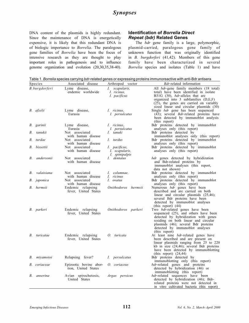
Figure 1. Restriction fragment length polymorphismpattern analysis of the rep or bdr genes of the Lymedisease spirochetes. Total DNA, isolated fromBorrelia cultures, was digested with Xba1, fraction-ated by electrophoresis, and transferred ontomembranes for hybridization. Hybridization wasperformed by the bdrAB-R1 oligonucleotide (46). Thespecies and isolates analyzed are indicated above eachlane. MW markers in kb are indicated.
chosen to reflect a central repeat motif carryingdomains in the deduced amino acid sequences.The + and - designations were assigned toindicate that the overlapping rep+ and rep- genesare located on opposing DNA strands. Plasmid-carried repeated DNA sequences were alsoidentified in B. burgdorferi B31 and found to carryeither all or a subset of seven ORFs, designated Athrough G (41). Of relevance to this discussion arethe ORF-E sequences that are rep or bdrhomologs. A bdr-related gene was also identifiedin B. afzelii DK1 and designated as p21 (43).B. afzelii causes Lyme disease in Europe andAsia. The rep+, ORF-E, and p21 designationshave recently been replaced with bdr genedesignations (24,25,44).
To assess and compare the composition andcomplexity of the bdr gene family among speciesand isolates of the B. burgdorferi sensu latocomplex, restriction fragment length polymor-phism (RFLP) patterns were determined (Appen-dix). Genomic DNA digested with Xba1 wasSouthern blotted and probed with an oligonucle-otide targeting the bdr genes (Figure 1). Avariable number of hybridizing bands of differentsize were detected. These analyses demonstratethat extensive bdr gene families are carried byB. burgdorferi sensu lato complex isolates andthat the RFLP patterns vary at the inter- andintraspecies level. Hybridization analyses ofother Borrelia species showed that they also carrybdr-related gene families (24,25,46). bdr-relatedgenes have been detected by hybridization inB. turicatae, B. hermsii, B. parkeri, B. coriaceae,and B. anserina (25,46). Isolates of these speciesalso exhibit substantial variation in their bdrRFLP patterns at the intraspecies level. Table 1lists the Borrelia species that carry bdr-relatedgenes and indicates the methods by which thesegenes or proteins were detected.
Sequences flanking some bdr alleles alsoappear to be distributed genus wide. Some bdralleles of B. turicatae, B. parkeri, and B. hermsiiare flanked by genes that are homologs of genescarried by the Lyme disease spirochetes (24,25).As a specific example, the B. turicatae bdrA1 geneis flanked by ORFs that are homologs of theBBG34 and BBG30 genes of B. burgdorferi(24,25). In the Lyme disease spirochetes, BBG34is part of a three-member paralogous gene family,while BBG30 is a single-copy gene (30). Locatedbetween BBG30 and BBG34 is BBG33, a memberof the bdr gene family (recently redesignated as
bdrF2) (25). Although these divergent Borreliaspecies carry related genes, their organizationdiffers (24), which indicates that rearrangementhas taken place in the ancestral plasmid thatcarried these homologs. Figure 2 compares the

115
Synopses
Vol. 6, No. 2, March–April 2000 Emerging Infectious Diseases
Figure 2. General organization of two bdr loci in Borrelia turicatae and B. burgdorferi. The gene arrangementdepicted for B. turicatae was determined through cloning and sequence analysis of a 2,217 base-pair XbaIrestriction fragment. The arrangement for the bdr-carrying locus of B. burgdorferi was previously determinedthrough the sequencing of the B. burgdorferi B31 genome (30). The arrows indicate the direction of transcription.Genes exhibiting homology are indicated by similar shading or hatch marks. Genes indicated by unfilled arrowsare not homologous. The numbering is indicated for scale and is not indicative of the positioning of these geneson the plasmids that carry them.
organization of two bdr loci from B. turicatae andB. burgdorferi.
Evolutionary Analyses of bdr-RelatedSequences: Revised Nomenclature forthe Bdr-Related Proteins
To simplify the complicated nomenclature ofbdr-related genes, a bdr nomenclature systemhas been developed that assigns gene names onthe basis of phylogenetic relationships inferredfrom comparative analysis of genetically stableregions of the bdr genes (25). This system, whichis applicable genuswide, allows for a readyassessment of relationships among bdr paralogsand orthologs. The rationale for this systemstemmed from the results of a comprehensiveevolutionary analysis of >50 bdr-related se-quences from five Borrelia species that demon-strated that bdr sequences are organized into sixdistinct subfamilies, designated A through F (25).Subfamilies are not necessarily species specific;some contain bdr alleles from different Borreliaspecies (25). Since members of a given subfamilyare closely related to one another with identityvalues for the N terminal domain being >95%,each member is assigned the same gene name
designation, and paralogs are distinguished by anumerical subscript. In B. turicatae OZ-1, two bdrsubfamilies, bdrA and bdrB, contain at least fourand five members, respectively (24). Members ofthe bdrA subfamily are designated bdrA1, bdrA2,bdrA3, and bdrA4, while members of the bdrBfamily are designated bdrB1 through bdrB5.This revised Bdr nomenclature scheme wasmodeled after that proposed for bacterialpolysaccharide synthesis genes (47) and is inaccordance with the nomenclature guidelinesestablished by Demerec (48).
The subfamily affiliation of bdr genes can bereadily determined through comparative se-quence analyses of the amino acid segmentpreceding the polymorphic repeat motif region ofthese proteins (described in detail below) (25).Relationship assessments based on the geneti-cally stable N terminal domain (vs. completesequences) are preferable because the calculatedevolutionary distances and clustering relation-ships are not artificially skewed by the variablenumber of repeat motifs present in the repeatmotif domain. Since the genetically unstablerepeat motif domain comprises as much as 50% ofthe total coding sequence in some alleles, it can
Synopses
Emerging Infectious Diseases Vol. 6, No. 2, March–April 2000116
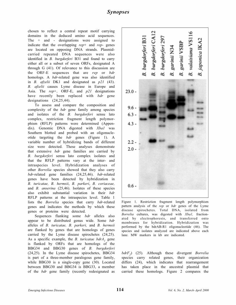
Figure 3. Key features and putative functional domains of the Bdr proteins. The schematic depicts a prototypeBdr protein with the characteristics of each domain indicated. The abbreviation, ID%, is for percentage aminoacid identity at either the inter- or intra-family level as indicated in the figure. Standard amino acidabbreviations are used in the figure to denote the conserved C-terminal lysine (K) or asparagine (N) residues,which are thought to be exposed in the periplasm and the cytoplasmically located core tripeptide of the repeatmotif (lysine-isoleucine-aspartic acid; KID).
have a substatial impact on inferred relation-ships. In addition, extensive sequence variationin the carboxyl termini of the Bdr proteins at theinter-species level makes it difficult to align thisdomain with confidence, which further influencesthe inferred relationships.
bdr evolutionary analyses show that Borreliaspecies carry members of at least two bdrsubfamilies (25,44). In fact, B. burgdorferi carriesthree distinct subfamilies. Multiple Bdr subfami-lies in diverse Borrelia species suggest that therehas been selective pressure to maintain multiplebdr alleles and bdr genetic diversity. This geneticdiversity may increase the functional diversity ofthe Bdr proteins.
Molecular Features and PhysicalProperties of the Bdr Proteins
While early analyses of Borrelia bdr genesdemonstrated their multicopy nature (41,42,46),the full extent of the complexity of the bdr genefamily in the Lyme disease spirochetes was notfully recognized until the B. burgdorferi genomesequence was determined (30). B. burgdorferiB31 was found to carry 17 distinct bdr-relatedgenes (and one truncated variant) distributedamong different linear and circular plasmids. B.turicatae, which carries at least nine different bdralleles, carries these genes exclusively on linearplasmids (24,25,46). Other relapsing feverspirochete species (B. parkeri and B. hermsii) aresimilar to the Lyme disease bacteria in that theycarry bdr genes on both linear and circular
plasmids (25). In the Lyme disease spirocheteseach of the 32-kb circular plasmids, with theexception of plasmids M and P, carry twodifferent bdr genes separated by seven or eightORFs. Each of these circular plasmids carries onebdrD subfamily member and one bdrE subfamilymember. The maintenance of genes belonging todifferent subfamilies on a single plasmid isconsistent with the possibility that each carriesout a different function. In contrast, in the Lymedisease spirochetes, the bdrF subfamily membersare localized to linear plasmids with only a singlebdr gene per plasmid. These observations suggestthat there has been selective pressure tomaintain the association of specific subfamilieswith specific types of plasmids. Less is known aboutthe bdr-carrying plasmids and the organization ofthe bdr genes and subfamilies in the relapsingfever borreliae. However, as in the Lyme diseasespirochetes, in B. turicatae most bdr-carryingplasmids carry two bdr genes, one from subfamilybdrA and one from subfamily bdrB (24).
The sequence of more than 50 bdr alleles fromfive different Borrelia species has been deter-mined (Table 2) (24,25,41-43,46). These exten-sive comparative sequence analyses led to theidentification of conserved features that provideinsight into the possible biologic roles of the Bdrproteins. For example, all bdr alleles carrycentrally located repeat motif domains (Figure 3).Although conserved in sequence, these domainsvary in length among alleles as a result of varyingnumbers of the repeat motif. The core tripeptide

117
Synopses
Vol. 6, No. 2, March–April 2000 Emerging Infectious Diseases
the repeat is the sequence KID. The repeat motifsencode consensus casein kinase 2 phosphoryla-tion (CK2P) motifs of the sequence T/SKID/E(43). While it may appear somewhat paradoxicalfor bacteria to carry casein kinases, casein kinaseis a descriptive term broadly applied to at leasttwo classes of ubiquitous protein kinases forwhich the substrates may include variousenzymes and noncatalytic proteins involved inimportant cellular regulatory functions (49).Most proteins phosphorylated by CK2-likekinases are highly acidic, as are the Borrelia Bdrproteins (isoelectric points between 5 and 6). Thephosphorylation site in CK2P motifs is either theSer or Thr residue of the motif. Althoughhistidine kinases have been known to exist insome bacteria, it has been widely held thatbacteria lack Ser - Thr kinases. However, Ser -Thr kinases have recently been identified inseveral bacterial species, including Myxococcus,Anabeana, Freymella, Yersinia, and Streptomy-ces (50). Most importantly, analysis of theB. burgdorferi genome sequence identified aputative Ser - Thr kinase designated BB0648(30,50). This ORF carries a domain that exhibitshomology with the active site of Ser - Thr kinases.B. burgdorferi also carries a homolog of the PPMfamily of eucaryotic protein Ser - Thr phos-phatases (30,50). The presence of these genes inB. burgdorferi suggests that the Borrelia possessthe machinery necessary for Ser - Thrphosphorylation and dephosphorylation.
Another important conserved feature identi-fied through sequence analyses is the hydropho-bic carboxyl terminal domain of approximately 20amino acids. Computer analyses conducted withthe TMpred program indicate that this domainhas a high propensity to form a transmembranehelix (24,25). The Tmpred values for the 20 aa C-terminal domains are 2,000 to 2,600. A value of500 or greater is considered significant (24,25).Comparison of the Bdr putative transmembranedomain sequences from the Lyme diseasespirochetes with those from the relapsing feverspirochetes indicates that, while there isconservation in physical properties, there isessentially no conservation of primary sequence.However, sequence conservation does exist at thesubfamily level (24,25). Since the Bdr proteinslack an obvious export signal, membraneassociation would most likely be with thespirochetal inner membrane, with the rest of theprotein, which is hydrophilic, extending into the
cytoplasm. The terminal residue of the protein isin almost all cases a positively charged aminoacid (lysine or asparagine). This residue couldextend into the periplasm and serve to anchor theBdr proteins to other cellular components, suchas the peptidoglycan.
Immunologic Analyses of the Bdr ProteinsThe presence of multiple bdr alleles and bdr
subfamilies within isogeneic populations hasprompted speculation that there may bedifferential expression at either the subfamily orindividual allele level, possibly in response toenvironmental stimuli (46). Limited studies ofbdr expression and production, based on eithermRNA detection or immunoblot analyses, havebeen performed. Porcella et al. (42) usedNorthern hybridization to determine if expres-sion of B. burgdorferi bdr-related genes occursduring cultivation in the laboratory understandard culture conditions (33°C in BSK media).Bdr transcripts were not detected by thisapproach. Similarly, in an earlier analysis, wealso conducted Northern hybridization experi-ments to assess bdr expression (46). We detectedexpression of B. turicatae OZ1 bdrA subfamilymembers in bacteria cultivated under standardlaboratory growth conditions (46). However,when reverse transcriptase (RT)-PCR methodswere applied, transcription of a single bdrA allelewas detected (46). B. turicatae OZ-1 was laterdemonstrated to carry at least nine bdr alleles,four of which belong to the bdrA subfamily.Analysis of the sequence of these alleles showedthat all four should have been readily amplifiedby the RT-PCR primer set because of theconservation of the primer binding sites (24). Thelack of detection of transcript derived from thesealleles suggested that only a subset of the bdr Asubfamily alleles is expressed. This raised thepossibility that other bdr alleles are eithernonfunctional genes or their expression requiresdifferent environmental stimuli. The transcrip-tional expression of the bdrB subfamily has notbeen specifically assessed. Thorough transcrip-tional analyses using allele-specific probes andprimers are an important step, since they allowspecific assessment of the expression ofindividual bdr alleles under differing environ-mental conditions. In addition, analyses of theupstream DNA sequences of individual bdr allelesand their genomic location may elucidate themolecular basis for bdr transcriptional regulation.

Synopses
Emerging Infectious Diseases Vol. 6, No. 2, March–April 2000118
Figure 4. Immunoblot analyses demonstrating the variation in Bdr protein expression in Borrelia species andisolates. Bacteria were cultivated and prepared for analysis as described in the methods. Proteins werefractionated by SDS-PAGE, immunoblotted and screened with anti-BdrF1-B.afzelii DK1 antisera. The species andisolates analyzed are indicated above each lane in panels A, B and C. The migration positions of the proteinstandards are indicated in each panel.
Immunologic analyses have provided a somewhatdifferent overall picture regarding Bdr produc-tion. Immunologic analyses described in thisreport and elsewhere (44) demonstrate thatseveral members of the bdr gene family areexpressed during in vitro cultivation. Weconducted a comprehensive analysis of theexpression of Bdr proteins among Borreliaspecies. When antisera raised against recombi-nant B. afzelii BdrF1 (24) were used inimmunoblot analyses, several immunoreactiveproteins were detected in cell lysates of allBorrelia species tested (Figure 4). The onlyexception was B. anserina, a causative agent ofavian spirochetosis. Although bdr-related se-quences have been detected in B. anserina byhybridization techniques (46), immunoreactiveproteins were not detected in immunoblotanalyses. Additional analyses are required todetermine if this indicates absence of transla-tional expression or the lack of epitopeconservation in this species. In any event, the factthat immunoreactive bands were not detected inthis species attests to the specificity of the anti-Bdr antisera. As a further demonstration of the
specificity of the antisera and to highlight the factthat the Bdr proteins are unique to Borrelia, a celllysate of Leptospira interrogans was included inthe immunoblot analyses. Immunoreactivitywith proteins in the Bdr size range was notobserved with the anti-Bdr antisera in thisspirochete species. Borrelia species that ex-pressed immunoreactive proteins includedB. garinii, B. burgdorferi, B. turdae, B. tanukii,B. japonica, B. valaisiana, B. afzelii, B. coriaceae,B. bissettii, B. miyamotoi, B. parkeri, B. hermsii,and B. turicatae (Table 1). Particularly strikingwas the extensive variation in the number andmolecular weight of the immunoreactive proteinsexpressed, with up to 12 distinct Bdr proteinsdetected. Variation in expression patterns wasobserved at both the inter- and intraspecies level.Analysis of three B. burgdorferi isolates (B31G,cN40, and CA12) demonstrated variability inboth the size and number of expressed Bdrproteins. Isolate B31G has been demonstrated bygenomic sequencing to carry 18 distinct bdr alleles.Immunoblot analyses show that not all alleles areexpressed during in vitro cultivation; therefore,some alleles may be differentially regulated.

119
Synopses
Vol. 6, No. 2, March–April 2000 Emerging Infectious Diseases
The broad immunoreactivity of the antiserawith diverse Borrelia species indicates that someepitopes are conserved genuswide. In view of thesequence divergence in the N and C terminaldomains of the Bdr proteins derived fromdifferent subfamilies, it is likely that the cross-reactive epitopes reside in the conserved repeatmotif region. Consistent with this, computeranalyses of the repeat domain of all determinedBdr protein sequences predict them to be alphahelical and to have a surface exposed on theprotein and a positive Jameson-Wolf antigenicindex (24,25,44). The conservation and synthesisof these polymorphic proteins in such a diversegroup of Borrelia species suggest that they playan important role in Borrelia biology genuswide.
The Bdr Proteins and BorreliaBiology: An Overview
Bdr genes and extensive bdr gene familieshave now been identified and characterized inseveral diverse Borrelia species (24,25,42-44,46).Comparative sequence analyses, which haveidentified conserved putative functional do-mains, have provided the basis for thedevelopment of hypotheses regarding Bdrfunction and cellular location. The Bdr proteins,which lack known consensus export signals, arelikely anchored to the cytoplasmic membranethrough their conserved, hydrophobic, putativetransmembrane spanning domain. The C-terminal positively charged amino acid may beexposed to the periplasm, where it may interactwith other cellular components that may includethe peptidoglycan. The repeat motif domain,which is predicted by computer analyses to behydrophilic and surface exposed on the protein,likely extends into the cytoplasm. The conservedrepeat motif domain that carries the putative Ser- Thr phosphorylation motifs may then beaccessible for phosphorylation or to interact withother cytoplasmic proteins or DNA to form amembrane anchored complex. As with numerousother proteins, phosphorylation and dephospho-rylation could play a regulatory role, perhaps insignaling or sensing.
Multiple polymorphic bdr alleles mayincrease the functional range and diversity of theBdr proteins. Functional partitioning among Bdrproteins could offer a possible explanation of whyBorrelia expend such biologic energy to maintainthese genes in large gene families and expressvariants of these proteins. The homology among
bdr alleles may also allow or lead to the continualmodification of these genes through homologousrecombination. In fact, the variable nature of therepeat motif region, which is clearly notevolutionarily stable, has likely arisen fromslipped-strand mispairing, recombination, orrearrangement. In view of the extensive geneticredundancy of the plasmid component of theBorrelia genome, recombination in and amongrelated sequences on different plasmids couldaffect the organization and evolution of thegenome and ultimately host-pathogen interac-tion. Inter- or intra-plasmid exchange of DNAsequences could provide a mechanistic basis forthe extensive genetic variability that has beenwidely described for Borrelia plasmids (28,29,51-59). In spite of the apparent necessity for at leastmost of the plasmids for survival, as inferred fromtheir ubiquitous distribution among Borreliaisolates, these bacteria are able to tolerateremarkable genomic variability. Diversity in theplasmids and the genes they carry may actuallybe exploited as a tool for phenotypic diversity andrapid environmental adaptation.
AcknowledgmentsWe thank Michael Norgard, Steve Porcella, Justin
Radolf, and the Molecular Pathogenesis research group atVirginia Commonwealth University for helpful discussions.
This work was supported in part by a grant from theJeffress Memorial Trust.
Mr. Roberts is a 3rd-year Ph.D. student, Departmentof Microbiology and Immunology, Medical College of Vir-ginia at Virginia Commonwealth University. His scien-tific interests are centered on the study of the molecularmechanisms of pathogenesis and adaptive responses ofarthropod-carried spirochetes, specifically on the role ofplasmid-carried gene families in Borrelia biology andpathogenesis.
References 1. Barbour AG, Hayes SF. Biology of Borrelia species.
Microbiological Reviews 1986;50:381-400. 2. Young JD. Underreporting of Lyme disease. N Engl J
Med 1998;338:1629. 3. Steere AC, Malawista SE, Snydman DR, Shope RE,
Andiman WA, Ross MR, et al. Lyme arthritis: anepidemic of oligoarticular arthritis in children andadults in three Connecticut communities. ArthritisRheum 1977;20:7-17.
4. Burgdorfer W, Barbour AG, Hayes SF, Benach JL,Grunwaldt E, Davis JP. Lyme disease—a tick-bornespirochetosis? Science 1982;216:1317-9.

Synopses
Emerging Infectious Diseases Vol. 6, No. 2, March–April 2000120
5. Benach JL, Bosler EM, Hanrahan JP, Coleman JL,Bast TF, Habicht GS, et al. Spirochetes isolated fromthe blood of two patients with Lyme disease. N Engl JMed 1983;308:740-2.
6. Baranton G, Postic D, Saint Girons I, Boerlin P,Piffaretti J-C, Assous M, et al. Delineation of Borreliaburgdorferi sensu stricto, Borrelia garinii sp. nov., andgroup VS461 associated with Lyme borreliosis. Int JSyst Bacteriol 1992;42:378-83.
7. Marconi RT, Garon CF. Identification of a thirdgenomic group of Borrelia burgdorferi throughsignature nucleotide analysis and 16S rRNA sequencedetermination. Journal of General Microbiology1992;138:533-6.
8. Marconi RT, Garon CF. Phylogenetic analysis of thegenus Borrelia: a comparison of North American andEuropean isolates of B. burgdorferi. J Bacteriol1992;174:241-4.
9. Marconi RT, Liveris D, Schwartz I. Identification ofnovel insertion elements, restriction fragment lengthpolymorphism patterns, and discontinuous 23S rRNAin Lyme Disease spirochetes: phylogenetic analyses ofrRNA genes and their intergenic spacers in Borreliajaponica sp. nov. and genomic group 21038 (Borreliaandersonii sp. nov.) isolates. J Clin Microbiol1995;33:2427-34.
10. Fukunaga M, Hamase A, Okada K, Inoue H, TsurutaY, Miyamoto K, et al. Characterization of spirochetesisolated from ticks (Ixodes tanukin, I. turdus andIxodes columnae) and comparision of the sequenceswith those of Borrelia burgdorferi sensu lato strains.Appl Environ Microbiol 1996;62:2338-44.
11. Fukunaga M, Hamase A, Okada K, Nakao M. Borreliatanuki sp. nov. and Borrelia turdae sp. nov. found fromIxodid ticks in Japan: rapid species identification by16S rRNA gene—targeted PCR analysis. MicrobiolImmunol 1996;40:877-81.
12. Fukunaga M, Takahashi Y, Tsuruta Y, Matsushita O,Ralph D, McClelland M, et al. Genetic and phenotypicanalysis of Borrelia miyamotoi sp. nov., isolates fromthe Ixodid tick Ixodes persulcatus, the vector for Lymedisease in Japan. Int J Syst Bacteriol 1995;45:804-10.
13. Wang G, van Dam AP, Le Flecha A, Postic D, Peter O,Baranton G, et al. Genetic and phenotypic analysis ofBorrelia valaisiana sp. nov. (Borrelia genomic groupsVS116 and M19). Int J Syst Bacteriol 1997;47:926-32.
14. Postic D, Belfazia J, Isogai E, Saint Girons I, GrimontPAD, Baranton G. A new genomic species in Borreliaburgdorferi sensu lato isolated from Japanese ticks.Res Microbiol 1993;144:467-73.
15. Postic D, Edlinger C, Richaud C, Grimont F, DufresneY, Perolat P, et al. Two genomic species in Borreliaburgdorferi. Res Microbiol 1990;141:465-75.
16. Barbour AG. Antigenic variation of a relapsing feverBorrelia species. Annu Rev Microbiol 1990;44:155-71.
17. Barbour AG, Byrman N, Carter CJ, Kitten T,Berstrom S. Variable antigen genes of the replasingfever agent Borrelia hermsii are activated by promoteraddition. Mol Microbiol 1991;5:489-93.
18. Stoenner HG, Dodd T, Larsen C. Antigenic variation ofB. hermsii. J Exp Med 1982;156:1297-311.
19. Plasterk RHA, Simon MI, Barbour AG. Transpositionof structural genes to an expression sequence on a
linear plasmid causes antigenic variation in thebacterium B. hermsii. Nature 1985;318:257-63.
20. Zhang J-R, Hardham JM, Barbour AG, Norris AG.Antigenic variation in Lyme disease Borreliae bypromiscuous recombination of vmp like sequencecassettes. Cell 1997;89:275-85.
21. Marconi RT, Samuels DS, Schwan TG, Garon CF.Identification of a protein in several Borrelia specieswhich is related to OspC of the Lyme diseasespirochetes. J Clin Microbiol 1993;31:2577-83.
22. Schwan TG, Piesman J, Golde WT, Dolan MC, RosaPA. Induction of an outer surface protein on Borreliaburgdorferi during tick feeding. Proc Natl Acad Sci U SA 1995;92:2909-13.
23. Schwan TG, Hinnebusch BJ. Bloodstream- versustick-associated variants of a relapsing fever bacterium.Science 1998;280:1938-40.
24. Carlyon JA, Roberts DM, Theisen M, Marconi RT.Molecular analyses of the B. turicatae bdr genes: apolymorphic, linear plasmid carried, paralogous genefamily. In press 1999.
25. Carlyon JA, Roberts DM, Marconi RT. Evolutionaryand molecular analyses of the Borrelia rep super genefamily: delineation of six distinct sub-families anddemonstration of the genus wide conservation ofputative functional domains, structural properties andrepeat motifs. Microb Pathog. In press 2000.
26. Ferdows MS, Barbour AG. Megabase-sized linear DNA inthe bacterium Borrelia burgdorferi, the Lyme diseaseagent. Proc Natl Acad Sci U S A 1989;86:5969-73.
27. Barbour AG, Garon CF. Linear plasmids of thebacterium Borrelia burgdorferi have covalently closedends. Science 1987;237:409-11.
28. Barbour AG. Plasmid analysis of Borrelia burgdorferi, theLyme disease agent. J Clin Microbiol 1988;26:475-8.
29. Hinnebusch J, Barbour AG. Linear plasmids ofBorrelia burgdorferi have a telomeric structure andsequence similar to those of a eukaryotic virus. JBacteriol 1991;173:7233-9.
30. Fraser C, Casjens S, Huang WM, Sutton GG, ClaytonR, Lathigra R, et al. Genomic sequence of a Lymedisease spirochaete, Borrelia burgdorferi. Nature1997;390:580-6.
31. Barbour AG. Linear DNA of Borrelia species andantigenic variation. Trends Microbiol 1993;1.
32. Hinnebusch J, Tilly K. Linear plasmids and chromosomesin bacteria. Mol Microbiol 1993;10:917-22.
33. Hinnebusch J, Barbour AG. Linear- and circular-plasmid copy numbers in Borrelia burgdorferi. JBacteriol 1992;174:5251-7.
34. Champion CI, Blanco DR, Skare JT, Haake DA, GiladiM, Foley D, et al. A 9.0 kilobase-pair circular plasmid ofBorrelia burgdorferi encodes an exported protein:evidence for expression only during infection. InfectImmun 1994;62:2653-61.
35. Akins D, Porcella SF, Popova TG, Shevchenko D, BakerSI, Li M, et al. Evidence for in vivo but not in vitroexpression of a Borrelia burgdorferi outer surface proteinF (OspF) homologue. Mol Microbiol 1995;18:507-20.
36. Suk K, Das S, Sun W, Jwang B, Barthold SW, FlavellRA, et al. Borrelia burgdorferi genes selectivelyexpressed in the infected host. Proc Natl Acad Sci U SA 1995;92:4269-73.
121
Synopses
Vol. 6, No. 2, March–April 2000 Emerging Infectious Diseases
37. Skare JT, Foley DM, Hernandez SR, Moore DC, BlancoDR, Miller JN, et al. Cloning and molecularcharacterization of plasmid-encoded antigens ofBorrelia burgdorferi. Infect Immun 1999;67:4407-17.
38. Marconi RT, Sung SY, Hughes CN, Carlyon JA.Molecular and evolutionary analyses of a variableseries of genes in Borrelia burgdorferi that are relatedto ospE and ospF, constitute a gene family, and share acommon upstream homology box. J Bacteriol1996;178:5615-26.
39. Sung S-Y, LaVoie C, Carlyon JA, Marconi RT.Evolutionary instability of ospE related members ofthe UHB gene family in Borrelia burgdorferi sensulato complex isolates. Infect Immun 1998;66:4656-68.
40. Zhang JR, Norris SJ. Genetic variation of the Borreliaburgdorferi gene vlsE involves cassette-specific,segmental gene conversion. Infect Immun1998;66:3698-704.
41. Zuckert WR, Meyer J. Circular and linear plasmids ofLyme Disease spirochetes have extensive homology:characterization of a repeated DNA element. JBacteriol 1996;178:2287-98.
42. Porcella SF, Popova TG, Akins DR, Li M, Radolf JR,Norgard MV. Borrelia burgdorferi supercoiled plasmidsencode multicopy open reading frames and a lipoproteingene family. J Bacteriol 1996;178:3293-307.
43. Theisen M. Molecular cloning and characterization ofnlpH, encoding a novel surface exposed, polymorphic,plasmid-encoded 33kD lipoprotein of Borrelia afzelii. JBacteriol 1996;178:6435-42.
44. Zuckert WR, Meyer J, Barbour AG. Comparativeanalysis and immunological characterization of theBorrelia Bdr protein family. Infect Immun1999;67:3257-66.
45. Yang X, Popova TG, Hagman KE, Wikel SK, SchoelerGB, Caimano MJ, et al. Identification, characterization,and expression of three new members of the Borreliaburgdorferi Mlp (2.9) lipoprotein gene family. InfectImmun 1999;67:6008-18.
46. Carlyon JA, Marconi RT. Cloning and molecularcharacterization of a multi-copy, linear plasmid-carried, repeat motif-containing gene from Borreliaturicatae, a causative agent of relapsing fever. JBacteriol 1998;180:4974-81.
47. Reeves PR, Hobbs M, Valvano MA, Kido N, Klena J,Maskell D, et al. Bacterial polysaccharide synthesis andgene nomenclature. Trends Microbiol 1996;4:498-503.
48. Demerec M. A proposal for a uniform nomenclature inbacterial genetics. Genetics 1966;54:61-76.
49. Pinna LA. Casein kinase 2: an “eminence grise” incellular regulation. Biochim Biophys Acta1990;1054:267-84.
50. Shi L, Potts M, Kennelly PJ. The serine, threonine and/or tyrosine protein kinases and protein phosphatasesof procaryotic organisms: a family portrait. FEMSMicrobiol Rev 1998;22:229-53.
51. Xu Y, Kodner C, Coleman L, Johnson RC. Correlationof plasmids with infectivity of Borrelia burgdorferisensu stricto type strain B31. Infect Immun1996;64:3870-6.
52. Xu Y, Johnson RC. Analysis and comparision ofplasmid profiles of Borrelia burgdorferi sensu latostrains. J Clin Microbiol 1995;33:2679-85.
53. Stålhammar-Carlemalm M, Jenny E, Gern L,Aeschlimann A, Meyer J. Plasmid analysis andrestriction fragment length polymorphisms ofchromosomal DNA allow a distinction betweenBorrelia burgdorferi strains. Zentralbl Bakteriol1990;274:28-39.
54. Simpson WJ, Garon CF, Schwan TG. Analysis ofsupercoiled circular plasmids in infectious and non-infectious Borrelia burgdorferi. Microb Pathog1990;8:109-18.
55. Schwan TG, Schrumpf ME, Karstens RH, Clover JR,Wong J, Daugherty M, et al. Distribution andmolecular analysis of Lyme disease spirochetes,Borrelia burgdorferi, isolated from ticks throughoutCalifornia. J Clin Microbiol 1993;31:3096-108.
56. Marconi RT, Casjens S, Munderloh UG, Samuels DS.Analysis of linear plasmid dimers in Borreliaburgdorferi sensu lato isolates: implications concerningthe potential mechanism of linear plasmid replication.J Bacteriol 1996;178:3357-61.
57. Marconi RT, Samuels DS, Landry RK, Garon CF.Analysis of the distribution and molecular heterogeneityof the ospD gene among the Lyme disease spirochetes:evidence for lateral gene exchange. J Bacteriol1994;176:4572-82.
58. Ferdows MS, Serwer P, Griess GA, Norris SJ, BarbourAG. Conversion of a linear to a circular plasmid in therelapsing fever agent Borrelia hermsii. J Bacteriol1996;178:793-800.
59. Carlyon JA, LaVoie C, Sung SY, Marconi RT. Analysisof the organization of multi-copy linear and circularplasmid carried open reading frames in Borreliaburgdorferi sensu lato isolates. Infect Immun1998;66:1149-58.
Appendix
Bacterial Cultivation, DNA Isolation, and SouthernHybridization Analyses
Isolates belonging to the Borrelia burgdorferi sensu latocomplex were cultivated in complete BSK-H media (Sigma)at 33°C. To cultivate the relapsing fever borreliae and otherBorrelia species, the complete BSK-H media weresupplemented with additional rabbit sera (Sigma) to a finalconcentration of 12% (vol/vol). Bacteria were harvested bycentrifugation and washed with phosphate buffered saline(pH 7.0), and DNA was extracted (25). For Southernhybridization analyses, 5 �g of DNA from each isolate wasdigested under standard conditions with Xba1 andfractionated by electrophoresis in 0.8% GTG agarose gels.(The DNA was transferred onto membranes for hybridiza-tion by vacuum blotting using the VacuGene system asdescribed by the manufacturer (Pharmacia). All otherSouthern hybridization methods were as previouslydescribed (39).

Synopses
Emerging Infectious Diseases Vol. 6, No. 2, March–April 2000122
Immunoblot AnalysesBacterial cultures were grown and harvested as
described above. One OD600 equivalent of cells was pelletedand resuspended in 100 �l of standard SDS-sample bufferwith reducing agents. The cell lysates (7 �l) were fractionatedby electrophoresis in 15% SDS-PAGE gels and electroblottedonto Immobilon P membranes (38). The immunoblots wereblocked overnight in blocking buffer (1X PBS, 0.2% Tween,0.002% NaCl, and 5% nonfat dry milk) and then incubatedwith a 1:1,000 antisera dilutions. ImmunoPure Goat anti-mouse IgG (H+L) peroxidase conjugate served as thesecondary antibody. The secondary antibody was incubatedwith the blots for 1 hour at room temperature at a 1:40,000-
fold dilution and then the blots were washed three times withwash buffer. For chemiluminescent detection, the SupersignalWest Pico Stable Peroxide solution and the Supersignal WestPico Luminol/Enhancer solution were used. Both reagentswere from Pierce Chemical Company, Rockford, IL and wereused as described by the manufacturer. The immunoblotswere exposed to film for time frames of 5 to 30 seconds.