the axial skelton activity 1. human anatomy and physiology, 7e by elaine marieb & katja hoehn...
TRANSCRIPT

The Axial Skelton
• Activity 1

Human Anatomy and Physiology, 7eby Elaine Marieb & Katja Hoehn
Copyright © 2007 Pearson Education, Inc.,publishing as Benjamin Cummings.
Bones of the axial skeleton
Skull
Ribs
Lumbar vertebrae
Sacrum
Coccyx
Sternum
Costalcartilages
Vertebrae
Thoracic
Lumbar
Anterior view Posterior view
Cervical

Human Anatomy and Physiology, 7eby Elaine Marieb & Katja Hoehn
Copyright © 2007 Pearson Education, Inc.,publishing as Benjamin Cummings.
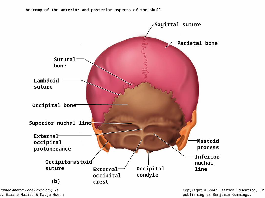
Anatomy of the anterior and posterior aspects of the skull
(a)
Parietal bone
Frontal squamaof frontal bone
Nasal bone
Sphenoid bone(greater wing)Temporal boneEthmoid bone
Lacrimal bone
Zygomatic bone
Maxilla
Mandible
Infraorbital foramen
Mentalforamen
Mandibular symphysis
Frontal bone
Glabella
Frontonasal suture
Supraorbital foramen(notch)
Supraorbital marginSuperior orbitalfissure
Inferior orbitalfissure
Middle nasal conchaPerpendicular plate
Inferior nasal concha
Vomer bone
Optic canal
Ethmoidbone
Human Anatomy and Physiology, 7eby Elaine Marieb & Katja Hoehn
Copyright © 2007 Pearson Education, Inc.,publishing as Benjamin Cummings.
Anatomy of the anterior and posterior aspects of the skull
(b)
Lambdoidsuture
Occipital bone
Occipitomastoidsuture
Superior nuchal line
Externaloccipitalprotuberance
InferiornuchallineOccipital
condyle
Mastoidprocess
Parietal bone
Sagittal suture
Externaloccipitalcrest
Suturalbone

Human Anatomy and Physiology, 7eby Elaine Marieb & Katja Hoehn
Copyright © 2007 Pearson Education, Inc.,publishing as Benjamin Cummings.
Anatomy of the lateral aspects of the skull
(a)
Coronal suture Frontal bone
Sphenoid bone(greater wing)
Ethmoid bone
Lacrimal bone
Lacrimal fossa
Nasal bone
Zygomatic bone
Maxilla
Alveolar margins
MandibleMental foramen
Parietal bone
Lambdoidsuture
Squamous suture
Occipital bone
Occipitomastoid suture
External acoustic meatus
Mastoid process
Styloid process
Mandibular condyle
Mandibular notch
Mandibular ramus
Mandibular angle Coronoid process
Zygomatic process
Temporal bone

Human Anatomy and Physiology, 7eby Elaine Marieb & Katja Hoehn
Copyright © 2007 Pearson Education, Inc.,publishing as Benjamin Cummings.
Anatomy of the lateral aspects of the skull
(b)
Parietal boneCoronal suture
Frontal bone
Frontal sinus
Sphenoid bone(greater wing)
Crista galli
Nasal bone
Sphenoid sinusEthmoid bone(perpendicular plate)Vomer bone
Maxilla
Mandible
Alveolar margins
Incisive fossa
Lambdoid suture
Occipitalbone
OccipitomastoidsutureExternal occipitalprotuberance
Internal acousticmeatus
Sella turcicaof sphenoidbone
Pterygoidprocess of sphenoid bone
Mandibularforamen
Palatinebone
Squamoussuture
Temporalbone
Palatineprocess ofmaxilla

Human Anatomy and Physiology, 7eby Elaine Marieb & Katja Hoehn
Copyright © 2007 Pearson Education, Inc.,publishing as Benjamin Cummings.
Anatomy of the inferior portion of the skull
(a)
Maxilla(palatine process)
Hardpalate
Zygomatic bone
Incisive fossaMedial palatine sutureInfraorbital foramen
Maxilla
Sphenoid bone(greater wing)
Foramen ovale
ForamenlacerumCarotid canal
External acoustic meatus
Stylomastoidforamen
Jugular foramen
Foramen magnum
Occipital condyle
Inferior nuchal line
Superior nuchal line
Temporal bone(zygomatic process)
Mandibularfossa
Vomer
Styloid process
External occipital crest
External occipitalprotuberance
Mastoid process
Temporal bone(petrous part)
Pharyngealtubercle ofbasioccipital
Parietal bone
Palatine bone(horizontal plate)

Human Anatomy and Physiology, 7eby Elaine Marieb & Katja Hoehn
Copyright © 2007 Pearson Education, Inc.,publishing as Benjamin Cummings.
Figure 7.4b-c: Anatomy of the inferior portion of the skull, p. 208.
(b)
(c)
Frontal boneOlfactory foramina
Lesser wingSphenoid
Anterior cranial fossa
Hypophyseal fossa
Middle cranialfossaTemporal bone(petrous part)
Internalacoustic meatus
Posteriorcranial fossa
Parietal bone
Occipital bone
Foramen magnum
Greater wing
Cribriform plateEthmoidboneCrista galli
Optic canalAnterior clinoid processForamen rotundum
Foramen ovale
Foramen spinosum
Jugular foramen
Hypoglossal canal
Anteriorcranialfossa
Middlecranialfossa
Posteriorcranialfossa
Foramen lacerum
Tuberculum sellae
Dorsum sellaeSellaturcica Posterior clinoid process

Human Anatomy and Physiology, 7eby Elaine Marieb & Katja Hoehn
Copyright © 2007 Pearson Education, Inc.,publishing as Benjamin Cummings.
The temporal bone
Mastoidregion
External acousticmeatus
Mastoid process
Styloid processTympanicregion
Mandibularfossa
Zygomaticprocess
Squamousregion

Human Anatomy and Physiology, 7eby Elaine Marieb & Katja Hoehn
Copyright © 2007 Pearson Education, Inc.,publishing as Benjamin Cummings.
The sphenoid bone
(b)
(a)
(c)
Opticcanal
Greaterwing
Body of sphenoid
Greaterwing
Greaterwing
Hypophysealfossa ofsellaturcica
Lesser wing Superiororbitalfissure
Foramenrotundum
Posterior view
Superior view
Foramen rotundumForamen ovaleForamen spinosum
Body of sphenoidDorsum sellae
Anterior clinoidprocess
Posterior clinoid process
Posterior clinoidprocess
Chiasmatic groove
Lesser wing
HypophysealfossaGreater wing
Foramen ovale
Sphenoid sinuses
Pterygoidprocesses
Lesserwing

Human Anatomy and Physiology, 7eby Elaine Marieb & Katja Hoehn
Copyright © 2007 Pearson Education, Inc.,publishing as Benjamin Cummings.
The sphenoid bone
(a)
Opticcanal
Greaterwing
Greaterwing
Hypophysealfossa ofsella turcica
Lesser wing
Superior view
Foramen rotundum
Foramen ovaleForamen spinosum
Body of sphenoidDorsum sellae
Anterior clinoidprocess
Posterior clinoidprocess
Chiasmatic groove

Human Anatomy and Physiology, 7eby Elaine Marieb & Katja Hoehn
Copyright © 2007 Pearson Education, Inc.,publishing as Benjamin Cummings.
The sphenoid bone
(b)
Body of sphenoid
Greaterwing
Superiororbitalfissure
Foramenrotundum
Posterior view
Posterior clinoid process
Lesser wing
Pterygoidprocesses

Human Anatomy and Physiology, 7eby Elaine Marieb & Katja Hoehn
Copyright © 2007 Pearson Education, Inc.,publishing as Benjamin Cummings.
(c)
Lesser wing
HypophysealfossaGreater wing
Foramen ovale
Sphenoid sinuses
The sphenoid bone
Human Anatomy and Physiology, 7eby Elaine Marieb & Katja Hoehn
Copyright © 2007 Pearson Education, Inc.,publishing as Benjamin Cummings.
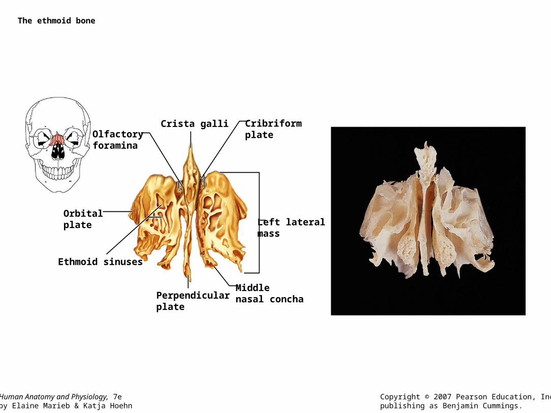
The ethmoid bone
Orbitalplate
Ethmoid sinuses
Perpendicularplate
Middlenasal concha
Cribriformplate Olfactory
foramina
Crista galli
Left lateralmass

Human Anatomy and Physiology, 7eby Elaine Marieb & Katja Hoehn
Copyright © 2007 Pearson Education, Inc.,publishing as Benjamin Cummings.
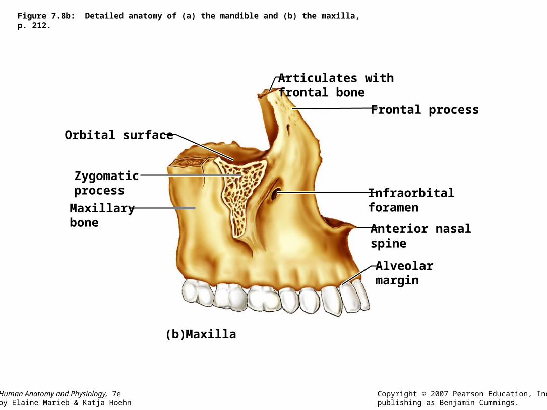
Detailed anatomy of (a) the mandible and (b) the maxilla
(b)
(a)
Frontal process
Articulates with frontal bone
Anterior nasalspine
Coronoidprocess
Mandibular foramen
Mentalforamen
Mandibularangle
Ramusofmandible
Mandibularcondyle
Mandibular notch
Mandibular fossaof temporal bone
Body of mandible
Alveolarmargin
Infraorbitalforamen
Alveolarmargin
Maxilla
Mandible
Orbital surface
ZygomaticprocessMaxillarybone
Temporomandibularjoint

Human Anatomy and Physiology, 7eby Elaine Marieb & Katja Hoehn
Copyright © 2007 Pearson Education, Inc.,publishing as Benjamin Cummings.
Detailed anatomy of (a) the mandible and (b) the maxilla
(a) Mandible
Coronoidprocess
Mandibular foramen
Mentalforamen
Mandibularangle
Ramusofmandible
Mandibularcondyle
Mandibular notch
Mandibular fossaof temporal bone
Body of mandible
Alveolarmargin
Temporomandibularjoint
Human Anatomy and Physiology, 7eby Elaine Marieb & Katja Hoehn
Copyright © 2007 Pearson Education, Inc.,publishing as Benjamin Cummings.
Figure 7.8b: Detailed anatomy of (a) the mandible and (b) the maxilla, p. 212.
(b)
Frontal process
Articulates with frontal bone
Anterior nasalspine
Infraorbitalforamen
Alveolarmargin
Maxilla
Orbital surface
Zygomaticprocess
Maxillarybone

Human Anatomy and Physiology, 7eby Elaine Marieb & Katja Hoehn
Copyright © 2007 Pearson Education, Inc.,publishing as Benjamin Cummings.
(b)
Roof of orbit
Lesser wing ofsphenoid bone
Medial wall
Orbital plateof ethmoid bone
Sphenoid body
Supraorbital foramen Optic canal
Floor of orbit
Orbital process ofpalatine bone
Orbital surface ofmaxillary bone
Lacrimal bone
Nasal bone
Frontal processof maxilla
Orbital plate offrontal bone
Lateral wall of orbit
Zygomatic processof frontal bone
Greater wing ofsphenoid bone
Orbital surface ofzygomatic bone
Zygomatic bone
Zygomatic bone
Inferior orbital fissure
Infraorbital groove
Infraorbitalforamen
Superiororbital fissure
Special anatomical characteristics of the orbits

Human Anatomy and Physiology, 7eby Elaine Marieb & Katja Hoehn
Copyright © 2007 Pearson Education, Inc.,publishing as Benjamin Cummings.
Special anatomical characteristics of the nasal cavity
(a)
Frontal sinus
Superiornasal concha
Middlenasal concha
Ethmoid bone
Inferior nasal concha
Nasal bone
Maxillary bone(palatine process)
Palatine bone(perpendicularplate)
Palatine bone (horizontal plate)
Pterygoid process
Sphenoid sinusSphenoid bone
Superior, middle, andinferior meatus
Anterior nasal spine

Human Anatomy and Physiology, 7eby Elaine Marieb & Katja Hoehn
Copyright © 2007 Pearson Education, Inc.,publishing as Benjamin Cummings.
Special anatomical characteristics of the nasal cavity
(b)
Vomer
Crista galliCribriformplate
Ethmoidbone
Frontal sinus
Nasal bone
Septal cartilage
Alveolar marginof maxilla
Perpendicular plateof ethmoid bone
Sella turcica
Sphenoid sinus
Palatine bone
Palatine processof maxilla

Human Anatomy and Physiology, 7eby Elaine Marieb & Katja Hoehn
Copyright © 2007 Pearson Education, Inc.,publishing as Benjamin Cummings.
Paranasal sinuses
(a) (b)
Frontal sinusEthmoid sinus
Maxillary sinus
Sphenoid sinus
Frontal sinus
Ethmoid sinus
Maxillary sinus
Sphenoid sinus

Hyoid bone

Human Anatomy and Physiology, 7eby Elaine Marieb & Katja Hoehn
Copyright © 2007 Pearson Education, Inc.,publishing as Benjamin Cummings.
The vertebral column
Cervical curvature (concave)7 vertebrae, C1– C7
Thoracic curvature(convex)12 vertebrae,T1– T12
Lumbar curvature(concave)5 vertebrae, L1– L5
Sacrum (convex)5 fused vertebrae
Coccyx4 fused vertebrae
Anterior view Right lateral view
C1
T1
2345
678
9
10
11
12L1
2
3
4
5
234567
Spinousprocess
Transverseprocess
Intervertebraldiscs
Intervertebralforamen

Human Anatomy and Physiology, 7eby Elaine Marieb & Katja Hoehn
Copyright © 2007 Pearson Education, Inc.,publishing as Benjamin Cummings.
Ligaments and fibrocartilage discs uniting the vertebrae
(a)
(b)
Supraspinous ligamentIntervertebraldiscAnteriorlongitudinalligament
Intervertebral foramen
Posterior longitudinalligamentAnulus fibrosus
Nucleus pulposus
Sectioned bodyof vertebra
Transverse process
Sectionedspinous process
Ligamentum flavum
Interspinousligament
Inferior articular process
Vertebral spinousprocess (posterioraspect of vertebra)
Spinal nerveroot
Anulusfibrosusof disc
Herniatedportion ofdisc
Nucleuspulposusof disc
Spinal cord
Human Anatomy and Physiology, 7eby Elaine Marieb & Katja Hoehn
Copyright © 2007 Pearson Education, Inc.,publishing as Benjamin Cummings.
Structure of a typical vertebra
Posterior
Anterior
Lamina
Superior articularprocess
Transverseprocess
Pedicle
Spinousprocess Vertebral
arch
Vertebralforamen
Body(centrum)

Human Anatomy and Physiology, 7eby Elaine Marieb & Katja Hoehn
Copyright © 2007 Pearson Education, Inc.,publishing as Benjamin Cummings.
The first and second cervical vertebrae
(a)
(b) (c)
Anterior arch
C1C2 Superior articular
facet
Transverse foramen
Posterior arch
Posterior tubercle
Anterior tubercleAnterior
Lateralmasses
Facet for dens
Transverseprocess
Lateralmasses
Transverse foramen
Posterior arch Posterior tubercle
AnteriorAnterior tubercle
Anterior arch
Inferiorarticularfacet
AnteriorBody
Superior view of atlas (C1)
Inferior view of atlas (C1) Superior view of axis (C2)
Inferiorarticularprocess
Dens
Superiorarticularfacet
Transverseforamen
Transverseprocess
Pedicle
Lamina
Spinous process


Human Anatomy and Physiology, 7eby Elaine Marieb & Katja Hoehn
Copyright © 2007 Pearson Education, Inc.,publishing as Benjamin Cummings.
Posterolateral views of articulated vertebrae
(a)
Dens of axis
Transverse ligament of atlas
C1 (atlas)
C2 (axis)
Inferior articularprocess
Bifid spinousprocess
Transverse processes
C7 (vertebra prominens)
Cervical vertebrae
C3

Human Anatomy and Physiology, 7eby Elaine Marieb & Katja Hoehn
Copyright © 2007 Pearson Education, Inc.,publishing as Benjamin Cummings.
Posterolateral views of articulated vertebrae
(b)
Transverse process
Lamina
Spinousprocess
Superiorarticularprocess
Transversecostal facet(for tubercleof rib)
Body
Intervertebraldisc
Pedicle
Inferior costalfacet (for headof rib)
Inferiorarticularprocess
Thoracic vertebrae
Human Anatomy and Physiology, 7eby Elaine Marieb & Katja Hoehn
Copyright © 2007 Pearson Education, Inc.,publishing as Benjamin Cummings.
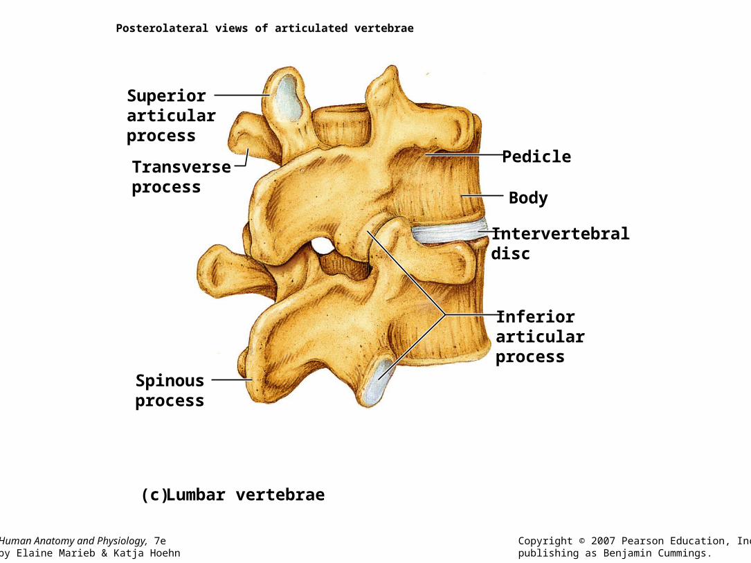
Posterolateral views of articulated vertebrae
(c)
Superiorarticularprocess
Transverseprocess
Spinousprocess
Intervertebraldisc
Body
Pedicle
Inferiorarticularprocess
Lumbar vertebrae

Human Anatomy and Physiology, 7eby Elaine Marieb & Katja Hoehn
Copyright © 2007 Pearson Education, Inc.,publishing as Benjamin Cummings.
The sacrum and coccyx
(a) (b)
Body offirstsacralvertebra
Transverse ridges(site of vertebralfusion)
Coccyx Coccyx
Anteriorsacralforamina
ApexPosteriorsacralforamina
Mediansacralcrest
Sacralpromontory
Sacralcanal
Sacralhiatus
Body
Facet ofsuperiorarticularprocess
Lateralsacralcrest
Auricularsurface
Ala
Anterior view Posterior view

Human Anatomy and Physiology, 7eby Elaine Marieb & Katja Hoehn
Copyright © 2007 Pearson Education, Inc.,publishing as Benjamin Cummings.
The thoracic cage
(a)
Intercostalspaces
True ribs(1–7)
Falseribs(8–12)
Jugular notchClavicular notch
Manubrium
Body
XiphisternaljointXiphoidprocess
L1
Vertebra
Floatingribs(11, 12)
Sternum
Costal cartilageCostal margin
Sternal angle

Human Anatomy and Physiology, 7eby Elaine Marieb & Katja Hoehn
Copyright © 2007 Pearson Education, Inc.,publishing as Benjamin Cummings.
The thoracic cage
(b)
Aorta
Diaphragm
Xiphisternaljoint
Heart
Sternal angle
Left brachiocephalicvein
Jugular notch
Left common carotid artery
T1
T4
T9
T12
Human Anatomy and Physiology, 7eby Elaine Marieb & Katja Hoehn
Copyright © 2007 Pearson Education, Inc.,publishing as Benjamin Cummings.
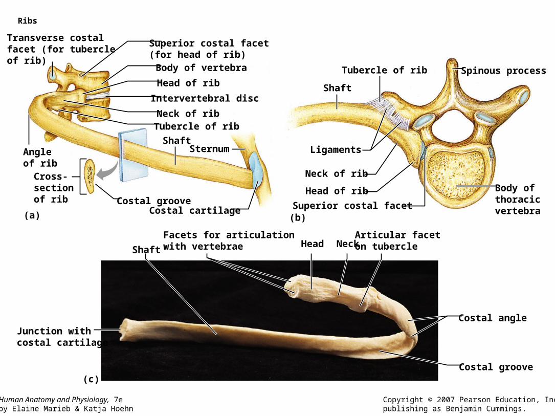
Ribs
(a) (b)
(c)
Transverse costalfacet (for tubercleof rib)
Superior costal facet (for head of rib)Body of vertebra
Head of rib
Intervertebral disc
Tubercle of ribNeck of rib
ShaftSternumAngle
of ribCross-sectionof rib Costal groove
Costal cartilage
Spinous processTubercle of rib
Shaft
Ligaments
Neck of rib
Head of rib Body ofthoracicvertebraSuperior costal facet
Junction withcostal cartilage
ShaftHead Neck
Articular faceton tubercle
Costal angle
Costal groove
Facets for articulationwith vertebrae

The Appendicular Skeleton
• Activity 1

Human Anatomy and Physiology, 7eby Elaine Marieb & Katja Hoehn
Copyright © 2007 Pearson Education, Inc.,publishing as Benjamin Cummings.
The appendicular skeleton
Humerus
Radius
Ulna
Carpals
Phalanges
Metacarpals
MetatarsalsPhalanges
Tarsals
Femur
Patella
Tibia
Fibula
Clavicle
Scapula

Human Anatomy and Physiology, 7eby Elaine Marieb & Katja Hoehn
Copyright © 2007 Pearson Education, Inc.,publishing as Benjamin Cummings.
Bones of the pectoral girdle
(a)
(b)
(c)
Clavicle
Scapula
Articulated pectoral girdle
Acromio-clavicularjoint
Acromial (lateral)end
Right clavicle, superior view
Posterior Sternal (medial)end
Anterior
Acromial end
Conoid tubercle
Anterior
Posterior
Right clavicle, inferior view
Sternal end

Human Anatomy and Physiology, 7eby Elaine Marieb & Katja Hoehn
Copyright © 2007 Pearson Education, Inc.,publishing as Benjamin Cummings.
(d)(e)
Acromion
Coracoidprocess
Suprascapular notchSuperior border
Superiorangle
Subscapularfossa
Medial border
Inferior angle
Glenoidcavity
Lateralborder
Right scapula, anterior aspect
Coracoid process
Suprascapular notch
Acromion
Lateralangle
Lateral border
Medial border
Infraspinousfossa
Spine
Superior angle
Right scapula,posterior aspect
Supraspinousfossa
Bones of the pectoral girdle

Human Anatomy and Physiology, 7eby Elaine Marieb & Katja Hoehn
Copyright © 2007 Pearson Education, Inc.,publishing as Benjamin Cummings.
Bones of the pectoral girdle
(f)
Supraspinousfossa
Infraspinousfossa
Subscapularfossa
Posterior Anterior
Coracoidprocess
Glenoidcavity
Acromion
Infraspinousfossa
Right scapula, lateral aspect
Infraglenoidtubercle
Subscapularfossa
Inferior angle

Human Anatomy and Physiology, 7eby Elaine Marieb & Katja Hoehn
Copyright © 2007 Pearson Education, Inc.,publishing as Benjamin Cummings.
Photographs of selected bones of the pectoral girdle and right upper limb
Acromion AcromionCoracoidprocess
Glenoidcavity
Subscapularfossa
Lateralborder
Medialborder
Inferiorangle
Supraspinousfossa
Body
Anterior view Posterior view
Scapula
Superiorborder
Superiorangle
Coracoidprocess
Superiorborder
Infraspinousfossa
Lateralborder
Spine ofscapula

Human Anatomy and Physiology, 7eby Elaine Marieb & Katja Hoehn
Copyright © 2007 Pearson Education, Inc.,publishing as Benjamin Cummings.
The humerus of the right arm
(a) (b)
GreatertubercleLessertubercle
Inter-tubercularsulacus
Lateralsupracondylarridge
Radialfossa
Capitulum
Head ofhumerus
Anatomicalneck
Radial groove
Deltoid tuberosity
Coronoidfossa
Olecranonfossa
MedialepicondyleTrochlea
Surgicalneck
Deltoidtuberosity
Lateralepicondyle
Medialsupracondylarridge
Anterior view Posterior view

Human Anatomy and Physiology, 7eby Elaine Marieb & Katja Hoehn
Copyright © 2007 Pearson Education, Inc.,publishing as Benjamin Cummings.
Radius and ulna of the right forearm
(a) Anterior view (b) Posterior view
(c)
Radialnotch
OlecranonprocessTrochlearnotchCoronoid process
Proximalradioulnarjoint
Distalradioulnar joint
Styloid processof radius
Radius
Neck ofradius
Head ofradius
Ulnar notch
Head of ulna
Styloid processof ulna
Interosseousmembrane
Ulna
HeadNeck
Radialtuberosity
Radius
Styloid processof radius
Colle’sfracture

Human Anatomy and Physiology, 7eby Elaine Marieb & Katja Hoehn
Copyright © 2007 Pearson Education, Inc.,publishing as Benjamin Cummings.
Photographs of selected bones of the pectoral girdle and right upper limb (continued)
Posterior
Anterior
Right clavicle, inferior view
Acromial end Sternal end
Impression forcostoclavicularligament
Conoidtubercle
Posterior
Anterior
Acromial (lateral) end
Sternal (medial) end
Right clavicle, superior view
Intertubercularsulcus
Lessertubercle
Greatertubercle
Head
Anatomicalneck
Surgicalneck
Deltoidtuberosity
Coronoidfossa
Medialepicondyle
Shaft
Lateralepicondyle
TrochleaCapitulum
Humerus, anterior view Radius (left) and ulna (right), anterior view
Ulnarhead
Distalradioulnarjoint
Ulna
Radialnotch
Trochlearnotch
Coronoidprocess
Head of radiusNeck of radius
Radialtuberosity
Radius
Location of interosseousmembrane
Styloidprocessof radius
Olecranonprocess

Human Anatomy and Physiology, 7eby Elaine Marieb & Katja Hoehn
Copyright © 2007 Pearson Education, Inc.,publishing as Benjamin Cummings.
Bones of the hand
(a) (b)
Trapezoid
Trapezium
Scaphoid
Capitate
Radius
Distal
Middle
Proximal
Hamate
Triquetral
Lunate
Ulna
Pisiform
5
43 2
1
Phalanges(fingers)
Metacarpals(palm)
Carpals(wrist)

Human Anatomy and Physiology, 7eby Elaine Marieb & Katja Hoehn
Copyright © 2007 Pearson Education, Inc.,publishing as Benjamin Cummings.
Bones of the bony pelvis
(a)
Coxal bone(os coxae
or hip bone)
llium
Sacroiliacjoint
Iliac fossa
Pubic bone
Ischium
Sacrum
Base of sacrum
Sacralpromontory
Pelvic brim
Ischial spine
Acetabulum
Pubic crest
Pubic symphysis
Iliac crest
Coccyx
Pubic arch

Human Anatomy and Physiology, 7eby Elaine Marieb & Katja Hoehn
Copyright © 2007 Pearson Education, Inc.,publishing as Benjamin Cummings.
Bones of the bony pelvis
(b)
Ilium
Ala
Tubercle ofthe iliaccrest
Anterior glutealline
Posterior glutealline
PosteriorsuperioriIiacspine
Greater sciaticnotch
Posterior inferioriliac spine
Ischial body
Ischial spine
Lesser sciatic notch
Ischialtuberosity
Ischium
Ischial ramus
Inferiorgluteal line
Acetabulum
Pubic body
Iliac crest
Anterior superioriliac spine
Anterior inferioriliac spine
Pubis
Inferior ramusof pubis

Human Anatomy and Physiology, 7eby Elaine Marieb & Katja Hoehn
Copyright © 2007 Pearson Education, Inc.,publishing as Benjamin Cummings.
Bones of the bony pelvis
(c)
Iliac fossaIlium
Iliac crest
Anterior superioriliac spine
Anterior inferioriliac spine
Arcuateline
PubictuberclePubiccrest
Superior ramusof pubis
Inferior ramusof pubis
Posteriorsuperioriliacspine
Obturatorforamen
Body
Ischium
Ischial ramus
Auricularsurface
Ischial spine
Posteriorinferioriliac spine
Articular surface ofpubis (at pubic symphysis)
Human Anatomy and Physiology, 7eby Elaine Marieb & Katja Hoehn
Copyright © 2007 Pearson Education, Inc.,publishing as Benjamin Cummings.
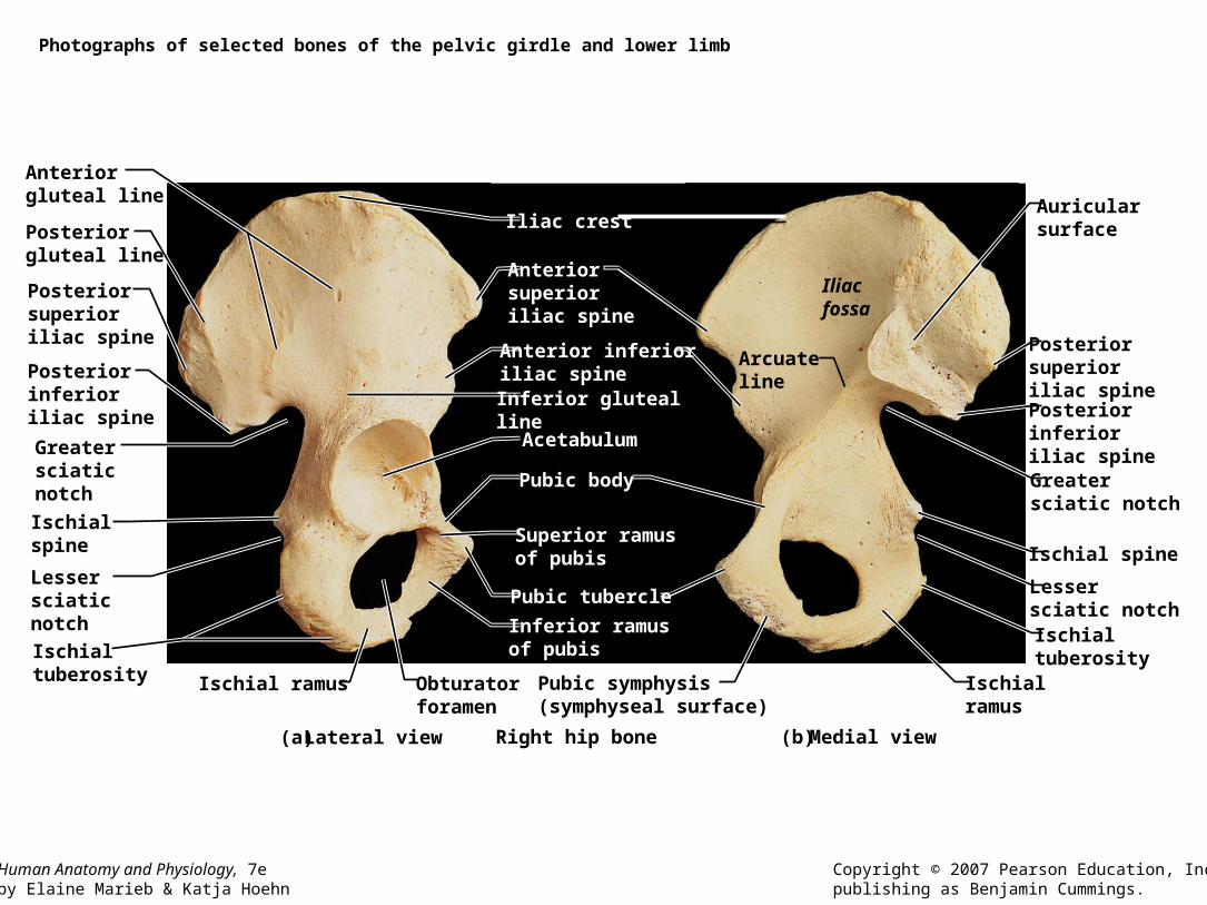
Photographs of selected bones of the pelvic girdle and lower limb
(a) (b)
Anteriorgluteal line Auricular
surface
Posterior superioriliac spinePosterior inferioriliac spineGreatersciatic notch
Ischial spine
Lessersciatic notchIschial tuberosity
Iliac crest
Anteriorsuperioriliac spine
Anterior inferioriliac spineInferior gluteal lineAcetabulum
Pubic body
Superior ramusof pubis
Pubic tubercle
Inferior ramusof pubis
Pubic symphysis(symphyseal surface)
Ischialramus
Iliacfossa
Arcuateline
Posteriorgluteal line
Posteriorsuperioriliac spine
Posteriorinferioriliac spine
Greater sciaticnotch
Ischial spine
Lessersciatic notch
Ischialtuberosity Ischial ramus
Right hip boneLateral view Medial view
Obturatorforamen

Human Anatomy and Physiology, 7eby Elaine Marieb & Katja Hoehn
Copyright © 2007 Pearson Education, Inc.,publishing as Benjamin Cummings.
Bones of the right thigh and knee
(a) Patella
(b) Femur
Apex
Anterior
Posterior
Facet formedialcondyleof femur
Facet forlateral condyleof femur
Surface forpatellarligament
Neck Foveacapitis
Greatertrochanter
Intertrochantericcrest
Lateralcondyle
Lateralepicondyle
Head
Intertrochantericline
Lesser trochanter
Gluteal tuberosity
Linea aspera
Intercondylar fossa
Medial andlateral supra-condylar lines
Medial condyle
Medialepicondyle
Adductor tubercle
Anterior view Posterior view
Lateralepicondyle
Patellarsurface

Human Anatomy and Physiology, 7eby Elaine Marieb & Katja Hoehn
Copyright © 2007 Pearson Education, Inc.,publishing as Benjamin Cummings.
The tibia and fibula of the right leg
Parts offracturedfibula
Intercondylareminence
Distaltibiofibularjoint
Lateralmalleolus
Articular surface
Anterior view Posterior view
Medialmalleolus
Tibia
Fibula
Lateralmalleolus
Anteriorborder
Tibialtuberosity
Medialcondyle
Articular surfaceof medial condyle
Articularsurface oflateral condyle
Head offibula
Interosseous membrane
Proximaltibiofibularjoint
LateralcondyleHead
Fibula

Human Anatomy and Physiology, 7eby Elaine Marieb & Katja Hoehn
Copyright © 2007 Pearson Education, Inc.,publishing as Benjamin Cummings.
Photographs of selected bones of the pelvic girdle and lower limb (continued)
LateralcondyleFibulaarticulateshere
Right femur, anterior surface
Neck
Greatertrochanter
Patellarsurface
Lateral epicondyleLateral condyle
Medialepicondyle Medialcondyle
Adductortubercle
Lessertrochanter
Intertrochantericline
Head
Right tibia and fibula, anterior view
Lateralmalleolusof fibula
Distaltibiofibularjoint
Fibula
Tibia
Anteriorborder
Head offibula
Lateralcondyleof tibia
Medial condyleof tibia
Intercondylareminence
Tibialtuberosity
Medialmalleolus
Inferiorarticular surface
Proximal right tibia,posterior view

Human Anatomy and Physiology, 7eby Elaine Marieb & Katja Hoehn
Copyright © 2007 Pearson Education, Inc.,publishing as Benjamin Cummings.
Bones of the right foot
(a)
Medialcuneiform
Phalanges
Metatarsals
Tarsals
Navicular
Intermediatecuneiform
Talus
Calcaneus
Cuboid
Lateralcuneiform
Proximal
MiddleDistal
Trochlea ofthe talus
Superior view

Human Anatomy and Physiology, 7eby Elaine Marieb & Katja Hoehn
Copyright © 2007 Pearson Education, Inc.,publishing as Benjamin Cummings.
Bones of the right foot
(b)
Facet formedialmalleolus
Calcanealtuberosity
Intermediatecuneiform
Susten-taculumtali
Talus
Navicular
First metatarsal
Medialcuneiform
Calcaneus
Medial view

Human Anatomy and Physiology, 7eby Elaine Marieb & Katja Hoehn
Copyright © 2007 Pearson Education, Inc.,publishing as Benjamin Cummings.
Bones of the right foot
(c) Lateral view
Intermediate cuneiform
Lateral cuneiform
Fifth metatarsal
Facet forlateral malleolus
Talus
Navicular
CuboidCalcaneus

Arches in the foot

Fetal skeleton
• Characteristic– Fontanels
• Anterior fontanel
• Sphenoidal fontanel
• Mastoid Fontanel
• Posterior fontanel

Human Anatomy and Physiology, 7eby Elaine Marieb & Katja Hoehn
Copyright © 2007 Pearson Education, Inc.,publishing as Benjamin Cummings.
The fetal skull
(a) (b)
Frontal bone
Ossificationcenter
Occipitalbone
Superior view
Posterior fontanel
Parietal bone
Anteriorfontanel
Frontal suture
Lateral view
Posteriorfontanel
Mastoidfontanel
Parietal bone
Ossificationcenter
Occipital bone
Temporal bone (squamous portion)
Frontal bone
Sphenoidalfontanel