the autonomic nervous system - carlo capelli · the autonomic nervous system was described at the...
TRANSCRIPT
The Autonomic Nervous System 1. Anatomy and general organisation
• Carlo Capelli, MD • Department of Neurological, Neuropsychological, Morphological and
Movement Sciences, University of Verona, Italy
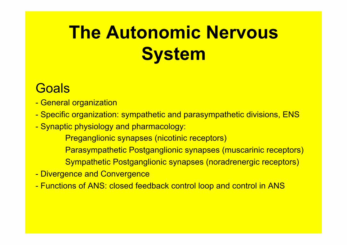
The Autonomic Nervous System
Goals - General organization - Specific organization: sympathetic and parasympathetic divisions, ENS - Synaptic physiology and pharmacology:
Preganglionic synapses (nicotinic receptors) Parasympathetic Postganglionic synapses (muscarinic receptors) Sympathetic Postganglionic synapses (noradrenergic receptors)
- Divergence and Convergence - Functions of ANS: closed feedback control loop and control in ANS
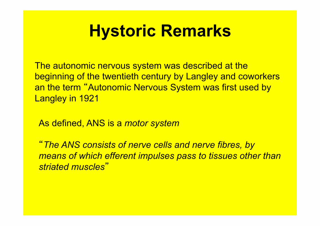
Hystoric Remarks
The autonomic nervous system was described at the beginning of the twentieth century by Langley and coworkers an the term “Autonomic Nervous System was first used by Langley in 1921
As defined, ANS is a motor system “The ANS consists of nerve cells and nerve fibres, by means of which efferent impulses pass to tissues other than striated muscles”
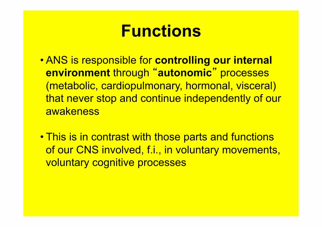
Functions • ANS is responsible for controlling our internal environment through “autonomic” processes (metabolic, cardiopulmonary, hormonal, visceral) that never stop and continue independently of our awakeness
• This is in contrast with those parts and functions of our CNS involved, f.i., in voluntary movements, voluntary cognitive processes
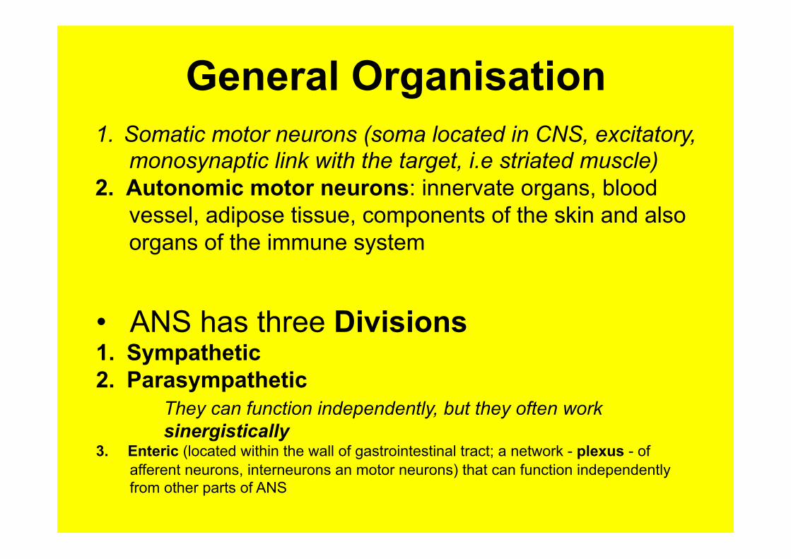
General Organisation 1. Somatic motor neurons (soma located in CNS, excitatory,
monosynaptic link with the target, i.e striated muscle) 2. Autonomic motor neurons: innervate organs, blood
vessel, adipose tissue, components of the skin and also organs of the immune system
• ANS has three Divisions 1. Sympathetic 2. Parasympathetic
They can function independently, but they often work sinergistically
3. Enteric (located within the wall of gastrointestinal tract; a network - plexus - of afferent neurons, interneurons an motor neurons) that can function independently from other parts of ANS
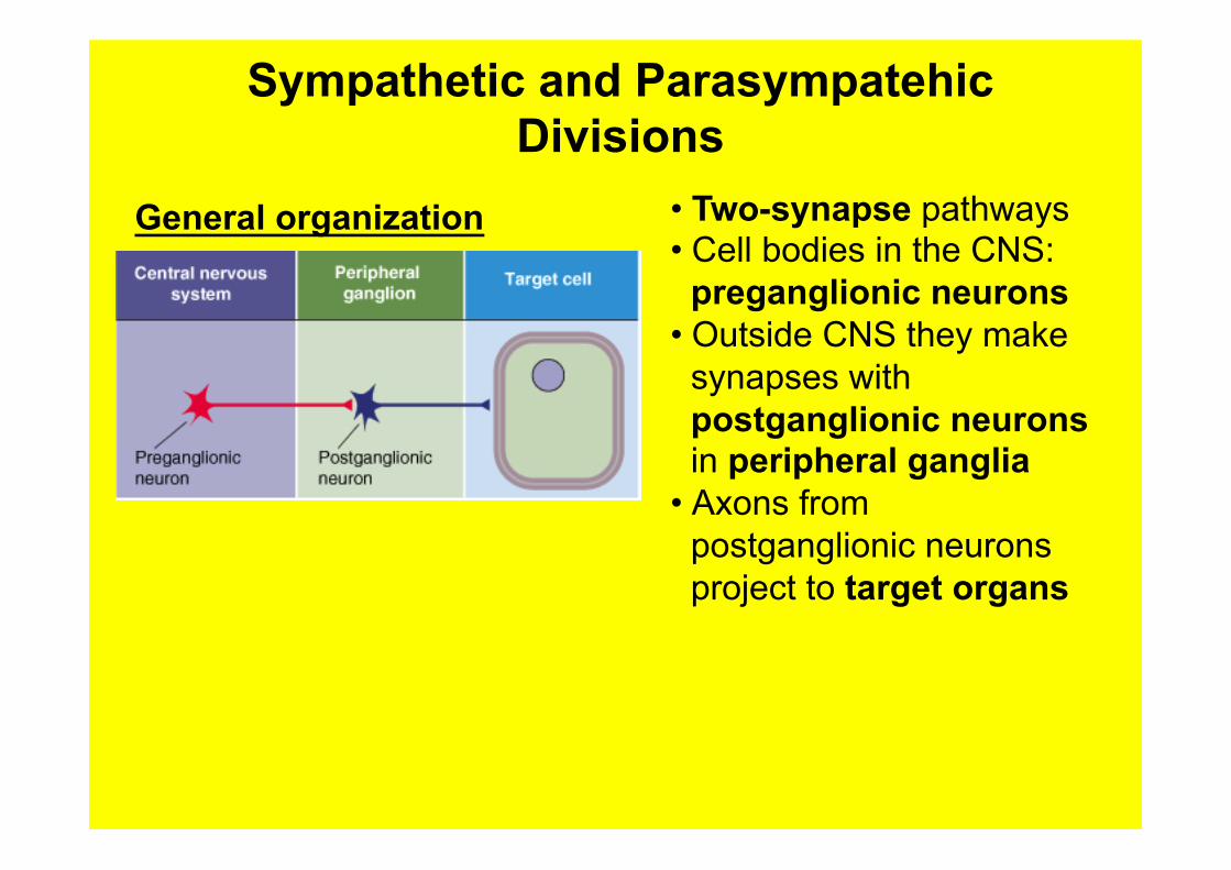
Sympathetic and Parasympatehic Divisions
• Two-synapse pathways • Cell bodies in the CNS:
preganglionic neurons • Outside CNS they make
synapses with postganglionic neurons in peripheral ganglia
• Axons from postganglionic neurons project to target organs
General organization
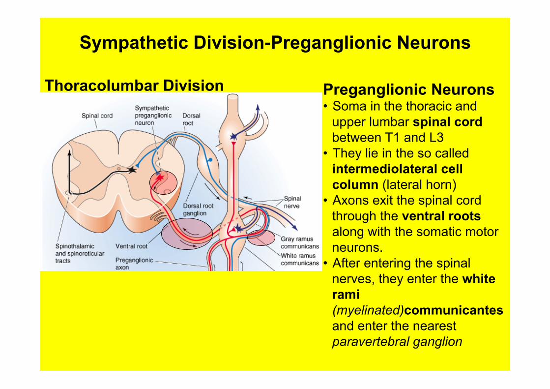
Sympathetic Division-Preganglionic Neurons
Preganglionic Neurons • Soma in the thoracic and
upper lumbar spinal cord between T1 and L3
• They lie in the so called intermediolateral cell column (lateral horn)
• Axons exit the spinal cord through the ventral roots along with the somatic motor neurons.
• After entering the spinal nerves, they enter the white rami (myelinated)communicantes and enter the nearest paravertebral ganglion
Thoracolumbar Division
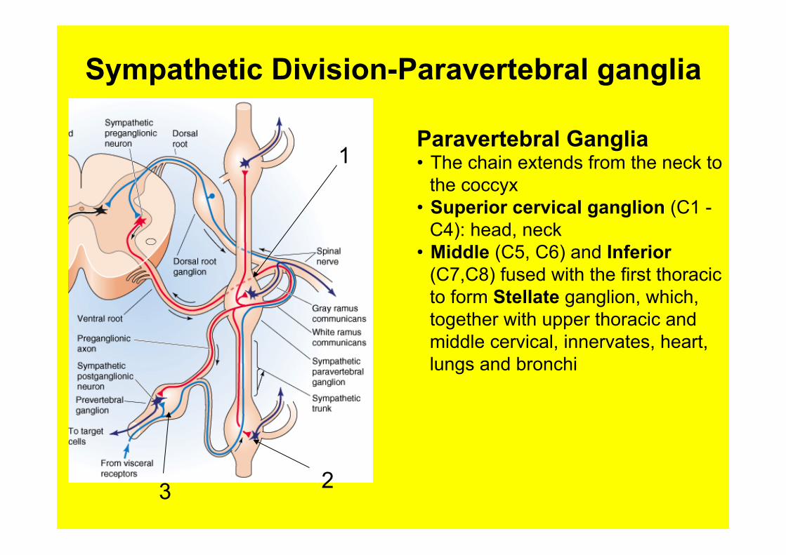
Sympathetic Division-Paravertebral ganglia
Paravertebral Ganglia • The chain extends from the neck to
the coccyx • Superior cervical ganglion (C1 -
C4): head, neck • Middle (C5, C6) and Inferior
(C7,C8) fused with the first thoracic to form Stellate ganglion, which, together with upper thoracic and middle cervical, innervates, heart, lungs and bronchi
1
2 3
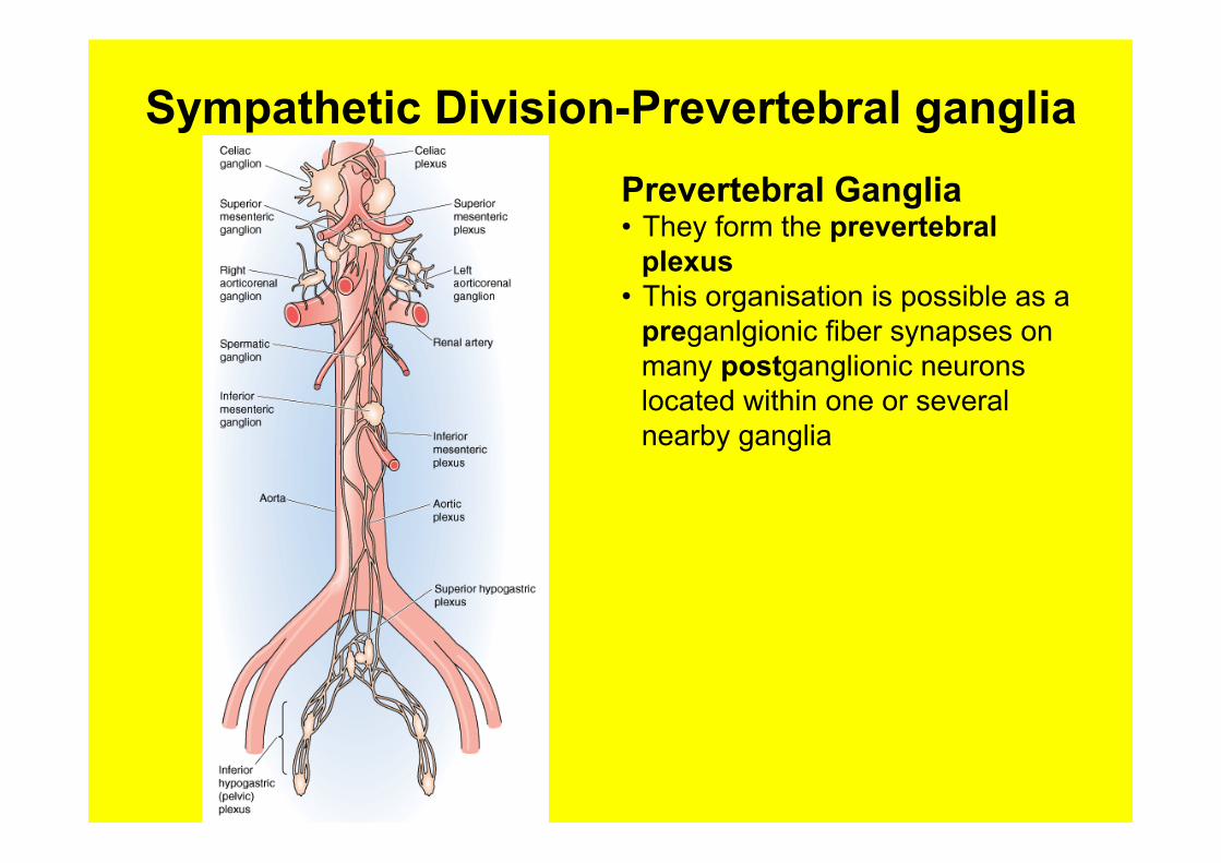
Sympathetic Division-Prevertebral ganglia Prevertebral Ganglia • They form the prevertebral
plexus • This organisation is possible as a
preganlgionic fiber synapses on many postganglionic neurons located within one or several nearby ganglia
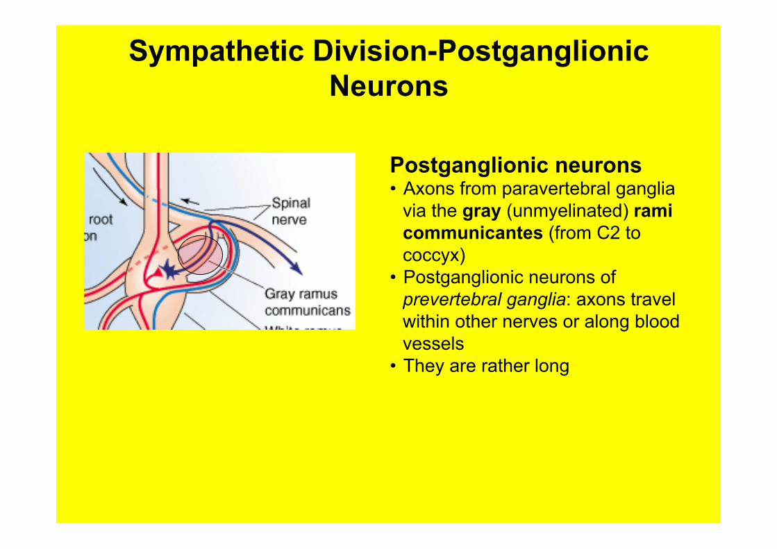
Sympathetic Division-Postganglionic Neurons
Postganglionic neurons • Axons from paravertebral ganglia
via the gray (unmyelinated) rami communicantes (from C2 to coccyx)
• Postganglionic neurons of prevertebral ganglia: axons travel within other nerves or along blood vessels
• They are rather long
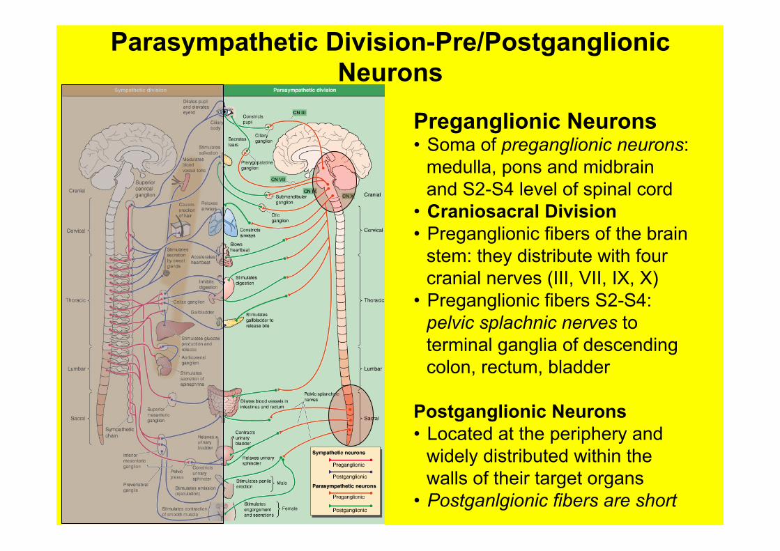
Parasympathetic Division-Pre/Postganglionic Neurons
Preganglionic Neurons • Soma of preganglionic neurons:
medulla, pons and midbrain and S2-S4 level of spinal cord
• Craniosacral Division • Preganglionic fibers of the brain
stem: they distribute with four cranial nerves (III, VII, IX, X)
• Preganglionic fibers S2-S4: pelvic splachnic nerves to terminal ganglia of descending colon, rectum, bladder
Postganglionic Neurons • Located at the periphery and
widely distributed within the walls of their target organs
• Postganlgionic fibers are short
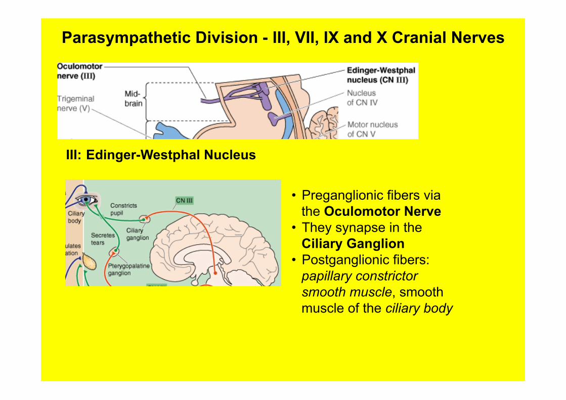
Parasympathetic Division - III, VII, IX and X Cranial Nerves
III: Edinger-Westphal Nucleus
• Preganglionic fibers via the Oculomotor Nerve
• They synapse in the Ciliary Ganglion
• Postganglionic fibers: papillary constrictor smooth muscle, smooth muscle of the ciliary body
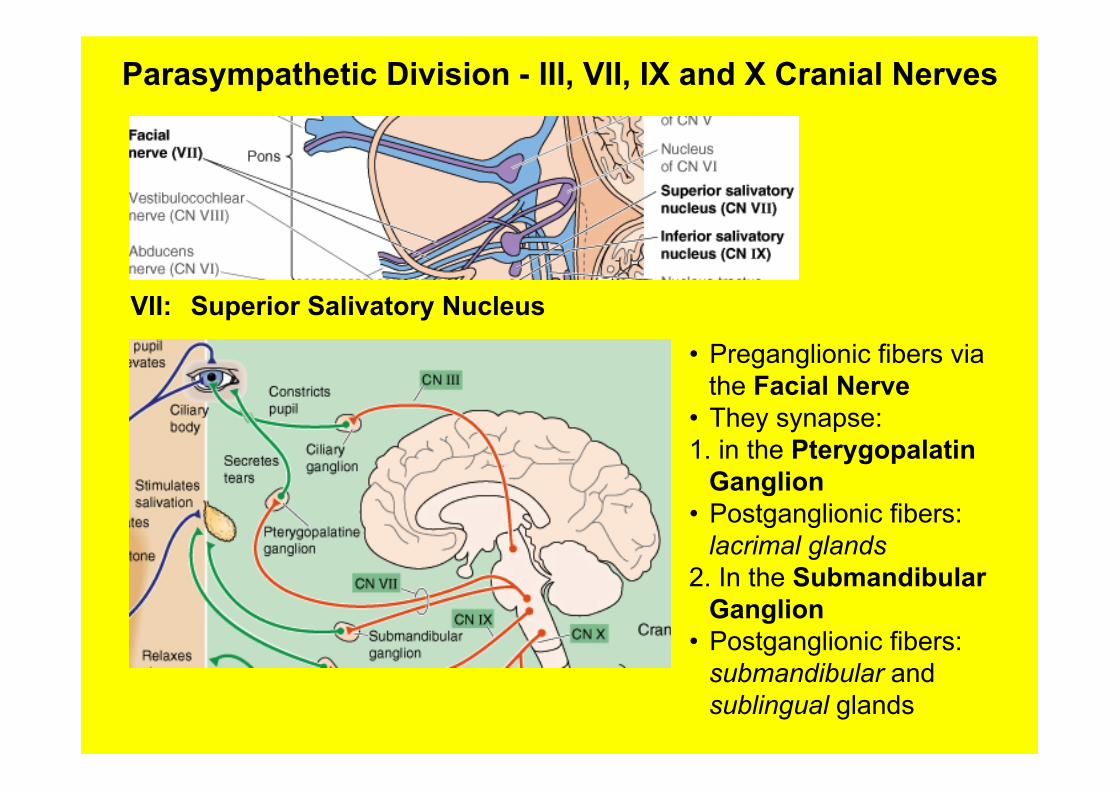
Parasympathetic Division - III, VII, IX and X Cranial Nerves
VII: Superior Salivatory Nucleus
• Preganglionic fibers via the Facial Nerve
• They synapse: 1. in the Pterygopalatin
Ganglion • Postganglionic fibers:
lacrimal glands 2. In the Submandibular
Ganglion • Postganglionic fibers:
submandibular and sublingual glands
Parasympathetic Division - III, VII, IX and X Cranial Nerves
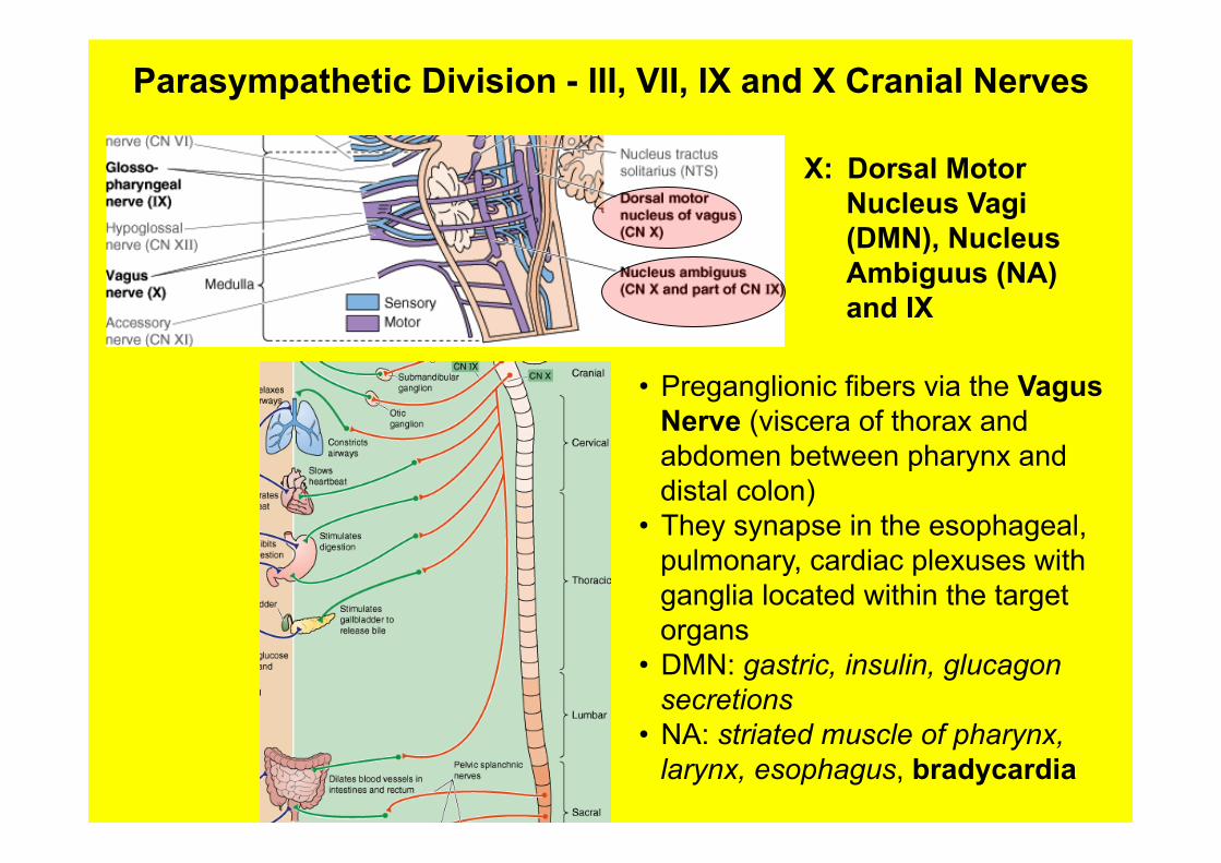
X: Dorsal Motor Nucleus Vagi (DMN), Nucleus Ambiguus (NA) and IX
• Preganglionic fibers via the Vagus Nerve (viscera of thorax and abdomen between pharynx and distal colon)
• They synapse in the esophageal, pulmonary, cardiac plexuses with ganglia located within the target organs
• DMN: gastric, insulin, glucagon secretions
• NA: striated muscle of pharynx, larynx, esophagus, bradycardia
Convergence and Divergence
• Convergence: Many preganglionic axons may synapse on a single postganglionic neurons (4-15 pre to one post)
• A single synaptic event is not sufficient to initiate an action potential in the postganglionic neurons, but the summation of multiple events is required to initiate it
• Divergence: relatively few preganglionic neurons synapse with many postganglionic neurons located within one or several nearby ganglia (1:10; 1:100)
• It allows for massive activation by few spinal centers of multiple sympathetic targets under extreme conditions (flight or fight)
• However, any impulse crosses a single synapse between pre and postganglianic neurons
Synaptic physiology of ANS
General concepts • Many visceral targets receive both inhibitory and
excitatory synapses • These antagonistic synapses arise form the two divisions of
ANS 1. organs activated during exercise: a. sympathetic: excitatory b. parasympathetic: inhibitory 2. organs whose activity increases at rest a. parasympathetic: excitatory b. sympathetic: inhibitory
• Exception: sweat glands, piloeroector muscles and most peripheral blood vessels receive only sympathetic inputs
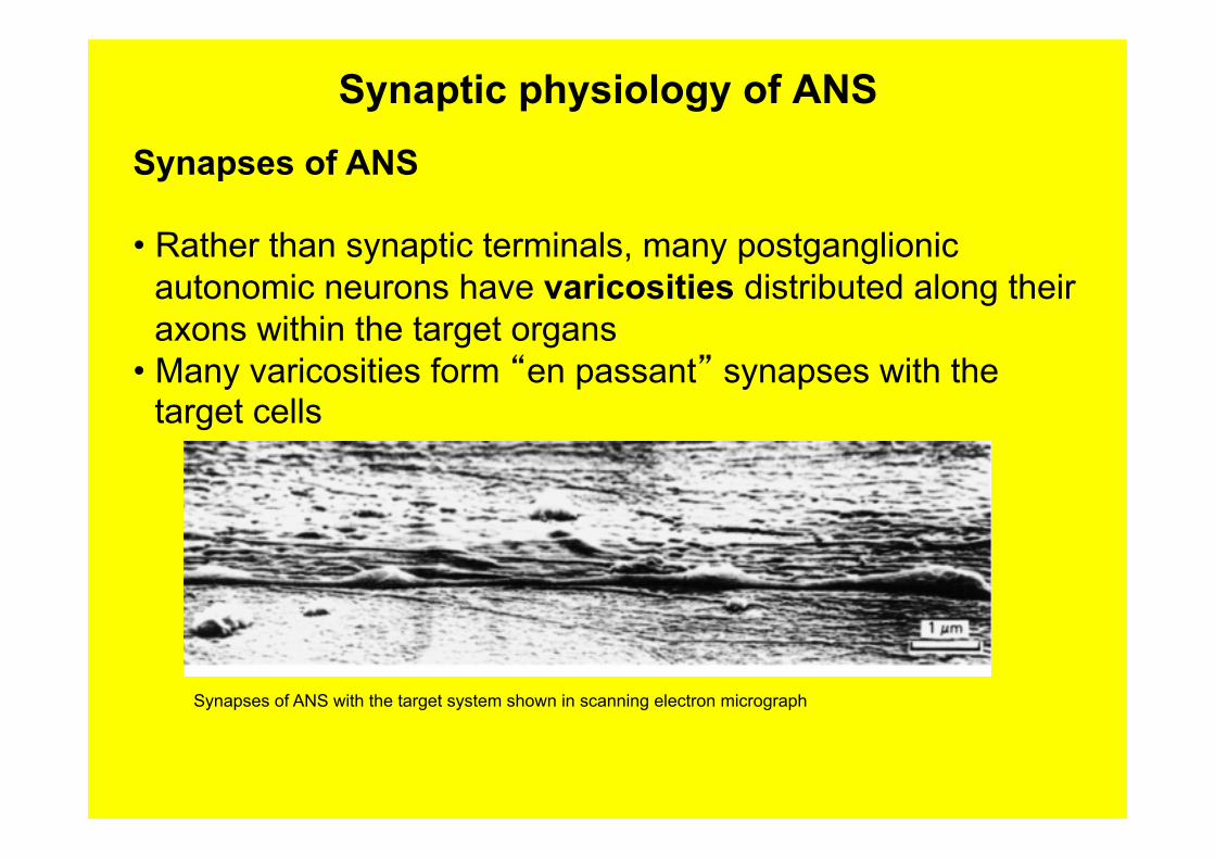
Synaptic physiology of ANS
Synapses of ANS • Rather than synaptic terminals, many postganglionic
autonomic neurons have varicosities distributed along their axons within the target organs
• Many varicosities form “en passant” synapses with the target cells
Synapses of ANS with the target system shown in scanning electron micrograph
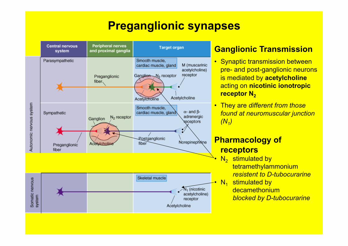
Preganglionic synapses
Ganglionic Transmission • Synaptic transmission between
pre- and post-ganglionic neurons is mediated by acetylcholine acting on nicotinic ionotropic receptor N2
• They are different from those found at neuromuscular junction (N1)
Pharmacology of receptors
• N2 stimulated by tetramethylammonium
resistent to D-tubocurarine • N1 stimulated by
decamethonium blocked by D-tubocurarine
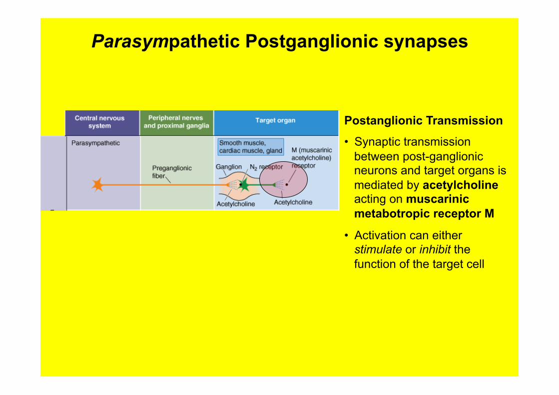
Parasympathetic Postganglionic synapses
Postanglionic Transmission • Synaptic transmission
between post-ganglionic neurons and target organs is mediated by acetylcholine acting on muscarinic metabotropic receptor M
• Activation can either stimulate or inhibit the function of the target cell
Muscarinic receptors
Physiology • They are metabotropic receptors that interact with heterotrimetric G proteins
• Their actions are mediated by second messengers and are slow and prolonged
• The interactions occur by 1. Stimulating the hydrolysis of phosphoinositide (PIP2) and increase [CA++] and activate protein kinase
C
2. Inhibiting adenylate cyclase and decreasing the levels of cAMP
3. Directly modulating K+ channels via the G-protein βγ complex
Pharmacology • Five different subtypes (M1 to M5) coded by five different genes
• They are stimulated by Ach and blocked by atropine
• M1, M3, M5: via the hydrolisis of PIP2
• M2, M4: inhibition of adenylate cyclase and decrease the levels of cAMP
• The five subtypes are heterogeneously distributed among tissues, they are found both pre and postsynaptically, many smooth muscles coexpress multiple muscarinic receptors
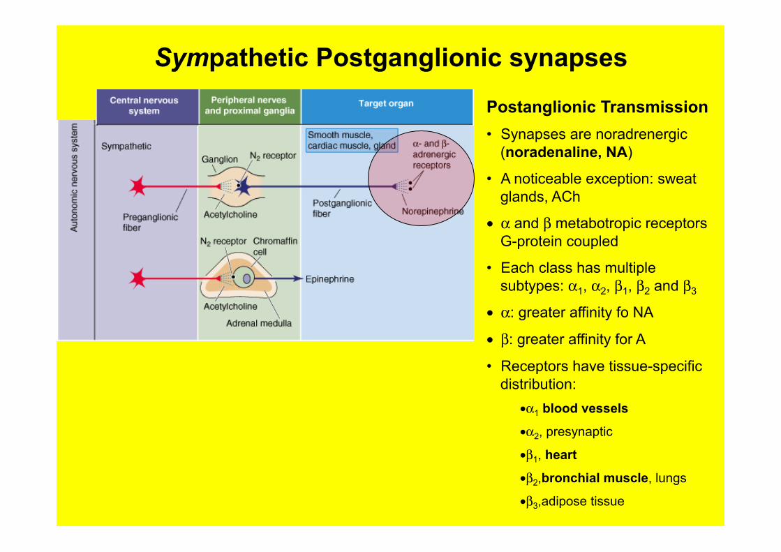
Sympathetic Postganglionic synapses
Postanglionic Transmission • Synapses are noradrenergic
(noradenaline, NA)
• A noticeable exception: sweat glands, ACh
• α and β metabotropic receptors G-protein coupled
• Each class has multiple subtypes: α1, α2, β1, β2 and β3
• α: greater affinity fo NA
• β: greater affinity for A
• Receptors have tissue-specific distribution:
• α1 blood vessels
• α2, presynaptic
• β1, heart
• β2,bronchial muscle, lungs
• β3,adipose tissue
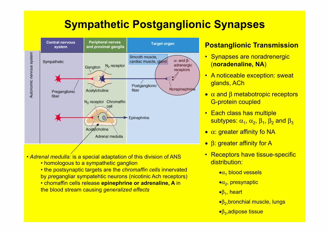
Sympathetic Postganglionic Synapses
Postanglionic Transmission • Synapses are noradrenergic
(noradenaline, NA)
• A noticeable exception: sweat glands, ACh
• α and β metabotropic receptors G-protein coupled
• Each class has multiple subtypes: α1, α2, β1, β2 and β3
• α: greater affinity fo NA
• β: greater affinity for A
• Receptors have tissue-specific distribution:
• α1 blood vessels
• α2, presynaptic
• β1, heart
• β2,bronchial muscle, lungs
• β3,adipose tissue
• Adrenal medulla: is a special adaptation of this division of ANS • homologous to a sympathetic ganglion • the postsynaptic targets are the chromaffin cells innervated by pregangliar sympatehtic neurons (nicotinic Ach receptors) • chomaffin cells release epinephrine or adrenaline, A in the blood stream causing generalized effects
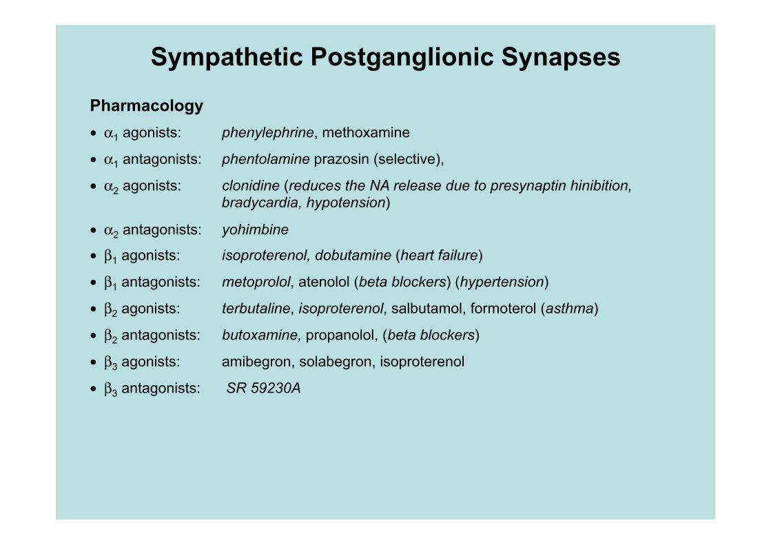
Sympathetic Postganglionic Synapses
Pharmacology • α1 agonists: phenylephrine, methoxamine
• α1 antagonists: phentolamine prazosin (selective),
• α2 agonists: clonidine (reduces the NA release due to presynaptin hinibition, bradycardia, hypotension)
• α2 antagonists: yohimbine
• β1 agonists: isoproterenol, dobutamine (heart failure)
• β1 antagonists: metoprolol, atenolol (beta blockers) (hypertension)
• β2 agonists: terbutaline, isoproterenol, salbutamol, formoterol (asthma)
• β2 antagonists: butoxamine, propanolol, (beta blockers)
• β3 agonists: amibegron, solabegron, isoproterenol
• β3 antagonists: SR 59230A
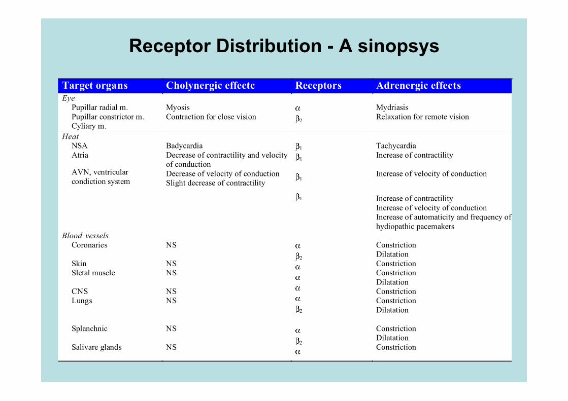
Receptor Distribution - A sinopsys
Target organs Cholynergic effectc Receptors Adrenergic effects Eye
Pupillar radial m. Pupillar constrictor m. Cyliary m.
Myosis Contraction for close vision
2
Mydriasis Relaxation for remote vision
Heat NSA Atria
AVN, ventricular condiction system
Badycardia Decrease of contractility and velocity of conduction Decrease of velocity of conduction Slight decrease of contractility
1 1
1
1
Tachycardia Increase of contractility Increase of velocity of conduction Increase of contractility Increase of velocity of conduction Increase of automaticity and frequency of hydiopathic pacemakers
Blood vessels Coronaries
Skin Sletal muscle CNS Lungs Splanchnic Salivare glands
NS NS NS NS NS NS NS
2 2
2
Constriction Dilatation Constriction Constriction Dilatation Constriction Constriction Dilatation Constriction Dilatation Constriction
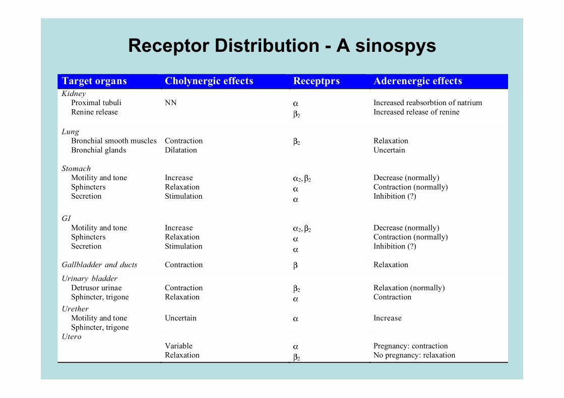
Receptor Distribution - A sinospys Target organs Cholynergic effects Receptprs Aderenergic effects Kidney
Proximal tubuli Renine release
NN
2
Increased reabsorbtion of natrium Increased release of renine
Lung Bronchial smooth muscles Bronchial glands
Contraction Dilatation
2
Relaxation Uncertain
Stomach Motility and tone Sphincters Secretion
Increase Relaxation Stimulation
2, 2
Decrease (normally) Contraction (normally) Inhibition (?)
GI Motility and tone Sphincters Secretion
Increase Relaxation Stimulation
2, 2
Decrease (normally) Contraction (normally) Inhibition (?)
Gallbladder and ducts Contraction Relaxation
Urinary bladder Detrusor urinae Sphincter, trigone
Contraction Relaxation
2
Relaxation (normally) Contraction
Urether Motility and tone Sphincter, trigone
Uncertain
Increase
Utero
Variable Relaxation
2
Pregnancy: contraction No pregnancy: relaxation
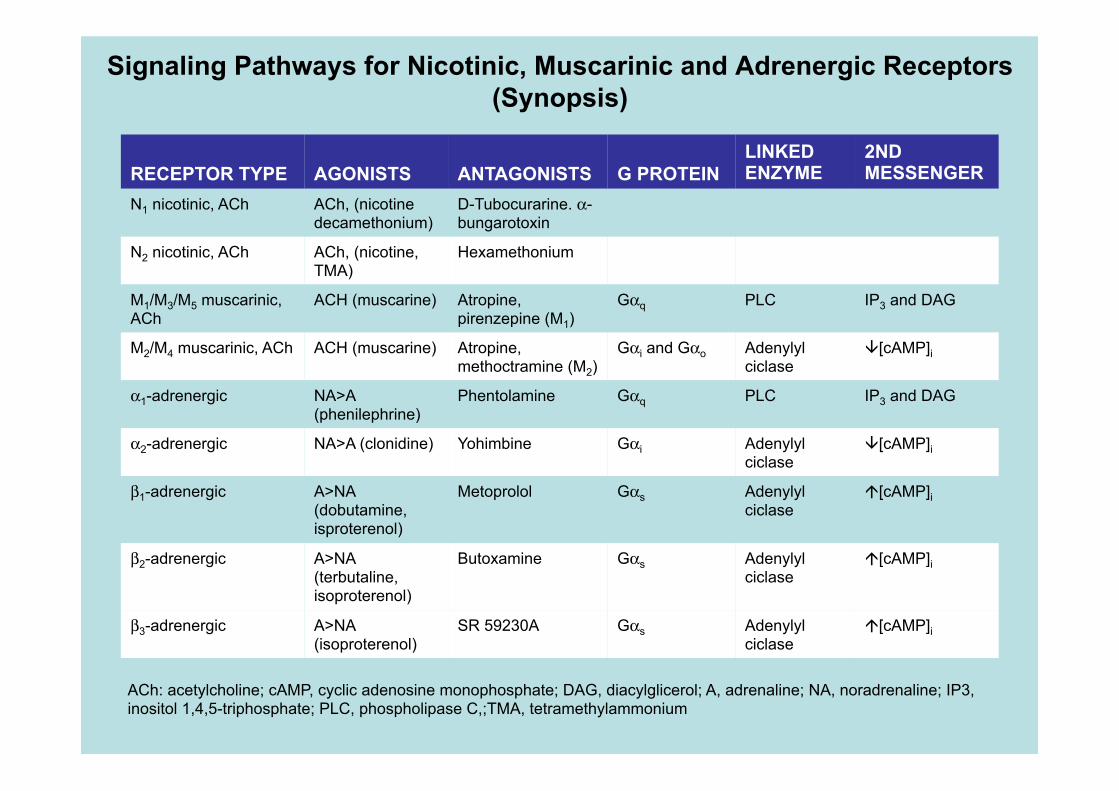
Signaling Pathways for Nicotinic, Muscarinic and Adrenergic Receptors (Synopsis)
RECEPTOR TYPE AGONISTS ANTAGONISTS G PROTEIN LINKED ENZYME
2ND MESSENGER
N1 nicotinic, ACh ACh, (nicotine decamethonium)
D-Tubocurarine. α-bungarotoxin
N2 nicotinic, ACh ACh, (nicotine, TMA)
Hexamethonium
M1/M3/M5 muscarinic, ACh
ACH (muscarine) Atropine, pirenzepine (M1)
Gαq PLC IP3 and DAG
M2/M4 muscarinic, ACh ACH (muscarine) Atropine, methoctramine (M2)
Gαi and Gαo Adenylyl ciclase
[cAMP]i
α1-adrenergic NA>A (phenilephrine)
Phentolamine Gαq PLC IP3 and DAG
α2-adrenergic NA>A (clonidine) Yohimbine Gαi Adenylyl ciclase
[cAMP]i
β1-adrenergic A>NA (dobutamine, isproterenol)
Metoprolol Gαs Adenylyl ciclase
[cAMP]i
β2-adrenergic A>NA (terbutaline, isoproterenol)
Butoxamine Gαs Adenylyl ciclase
[cAMP]i
β3-adrenergic A>NA (isoproterenol)
SR 59230A Gαs Adenylyl ciclase
[cAMP]i
ACh: acetylcholine; cAMP, cyclic adenosine monophosphate; DAG, diacylglicerol; A, adrenaline; NA, noradrenaline; IP3, inositol 1,4,5-triphosphate; PLC, phospholipase C,;TMA, tetramethylammonium

Bibliography
• Boron WF, Boulpaep EL, Medical Physiology, Saunders
• Fisiologia dell’Uomo, autori vari, Edi.Ermes, Milano – Capitolo 4: Il Sistema nervoso vegetativo