the aspen retinal detachment society - medconfs.com · the aspen retinal detachment society ......
TRANSCRIPT
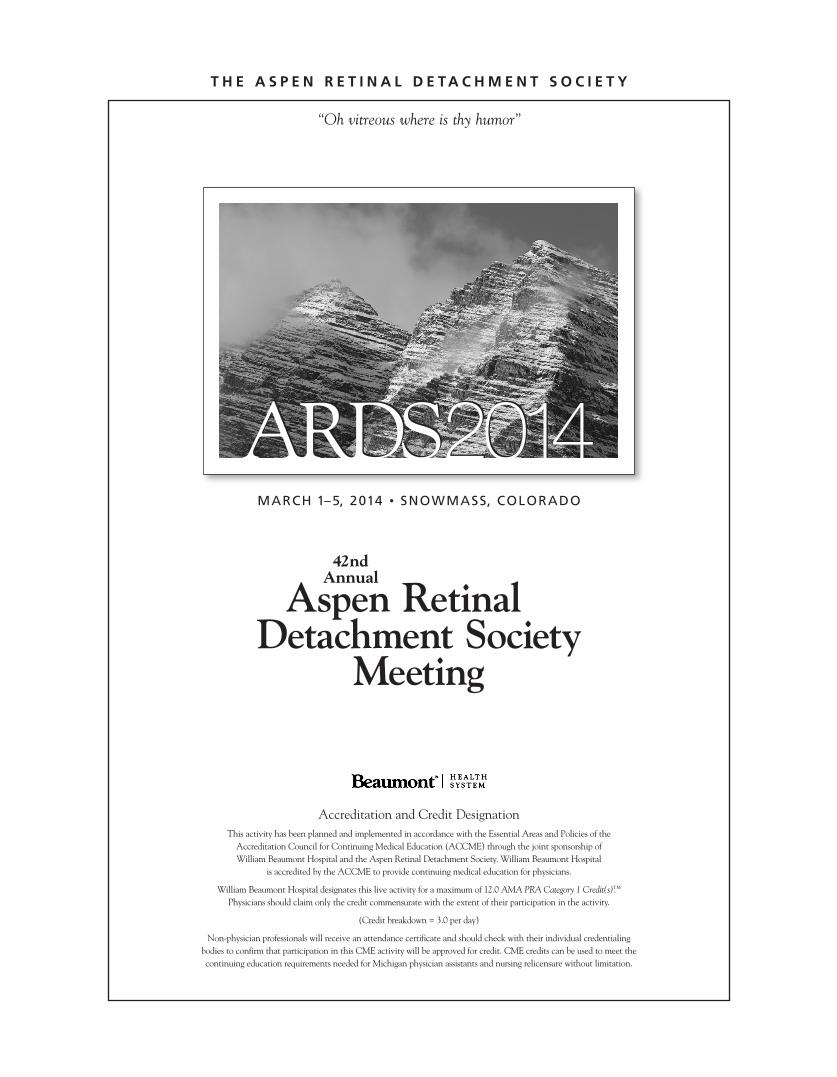
T H E A S P E N R E T I N A L D E TA C HM E N T S O C I E T Y
“Oh vitreous where is thy humor”
42ndAnnual
Aspen RetinalDetachment Society
Meeting
Accreditation and Credit Designation This activity has been planned and implemented in accordance with the Essential Areas and Policies of the Accreditation Council for Continuing Medical Education (ACCME) through the joint sponsorship of William Beaumont Hospital and the Aspen Retinal Detachment Society. William Beaumont Hospital
is accredited by the ACCME to provide continuing medical education for physicians.
William Beaumont Hospital designates this live activity for a maximum of 12.0 AMA PRA Category 1 Credit(s).™
Physicians should claim only the credit commensurate with the extent of their participation in the activity.
(Credit breakdown = 3.0 per day)
Non-physician professionals will receive an attendance certificate and should check with their individual credentialingbodies to confirm that participation in this CME activity will be approved for credit. CME credits can be used to meet thecontinuing education requirements needed for Michigan physician assistants and nursing relicensure without limitation.
ARDS2014ARDS2014MARCH 1–5, 2014 • SNOWMASS, COLORADO

MARCH 1–5, 2014 • SNOWMASS, COLORADO 3
. . . . . . . . . . . . . . . . . . . . . . . . . . . . . . . . . . . . . . . . . . . . . . . . . . . . . . . . . . . . . . . . . . . . . . . . . . . . . . . . . . . . . . . . . . . . .. . . . . . . . . . . . . . . . . . . . . . .
. . . . . . . . . . . . . . . . . . . . . . . . . . . . . . . . . . . . . . . . . . . . . . . . . . . . . . . . . . . . . . . . . . . . . . . . . . . . . . . . . . . . . . . . . . . . .. . . . . . . . . . . . . . . . . . . . . . .
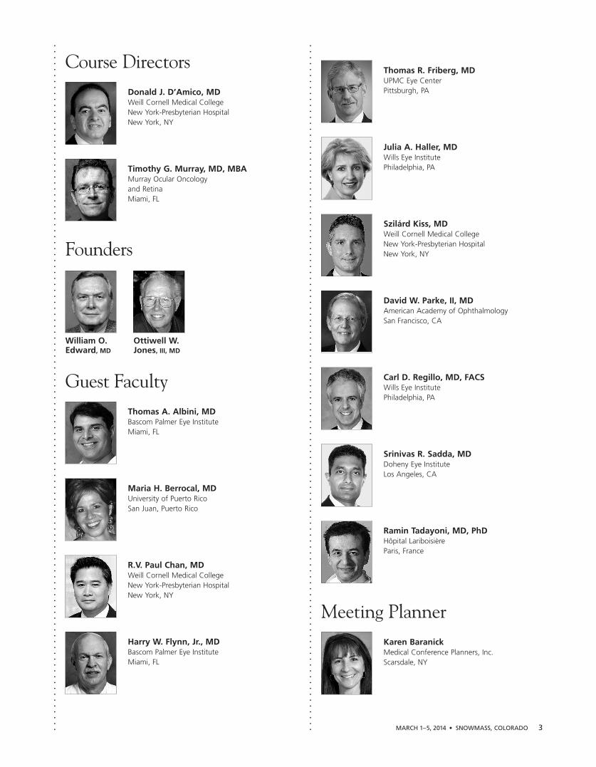
Course DirectorsDonald J. D’Amico, MDWeill Cornell Medical CollegeNew York-Presbyterian HospitalNew York, NY
Timothy G. Murray, MD, MBAMurray Ocular Oncology and RetinaMiami, FL
Founders
William O. Ottiwell W. Edward, MD Jones, III, MD
Guest FacultyThomas A. Albini, MD Bascom Palmer Eye InstituteMiami, FL
Maria H. Berrocal, MDUniversity of Puerto RicoSan Juan, Puerto Rico
R.V. Paul Chan, MD Weill Cornell Medical CollegeNew York-Presbyterian HospitalNew York, NY
Harry W. Flynn, Jr., MDBascom Palmer Eye InstituteMiami, FL
Thomas R. Friberg, MD UPMC Eye CenterPittsburgh, PA
Julia A. Haller, MD Wills Eye InstitutePhiladelphia, PA
Szilárd Kiss, MD Weill Cornell Medical CollegeNew York-Presbyterian HospitalNew York, NY
David W. Parke, II, MD American Academy of OphthalmologySan Francisco, CA
Carl D. Regillo, MD, FACSWills Eye InstitutePhiladelphia, PA
Srinivas R. Sadda, MD Doheny Eye InstituteLos Angeles, CA
Ramin Tadayoni, MD, PhD Hôpital Lariboisière Paris, France
Meeting PlannerKaren BaranickMedical Conference Planners, Inc.Scarsdale, NY

Dr. Regillo was a Phi Kappa Phi graduate of Northeastern University, College ofPharmacy, where he was the class valedic -torian. He received his medical degree fromHarvard Medical School on a full militaryscholarship. He performed his internship in internal medicine at Harvard’s Brighamand Women’s Hospital in Boston, and both hisophthalmology residency and vitreoretinalfellowship at Wills Eye Hospital in Philadelphia.He was appointed the Co-Chief Resident andawarded a Heed Ophthalmic Fellowship during hispostgraduate training.
After training, he was commissioned a Major in the United States Air Force and stationed fouryears as a vitreoretinal surgeon at the San DiegoNaval Medical Center. There he was Co-Directorof the Ophthalmology Department’s Retina Service, Assistant Professor of Ophthalmology atthe University of California, San Diego, andrecipient of a Naval Commendation medal and the Ophthal mology Department’s annual teachingaward. In 1998, he returned to Philadelphia to joinMid Atlantic Retina and the Retina Service ofWills Eye Hospital.
Dr. Regillo is currently the Director of the Retina Service of Wills Eye Hospital, Professor ofOphthalmology at Thomas Jefferson UniversitySchool of Medicine, and Principle Investigator ofseveral major international clinical trials investigatingnew forms of treatment for macular degeneration,diabetic retinopathy, and a variety of other retinalconditions. He is the founder and former Directorof the Wills Eye Clinical Retina Research Unit,
prior Chairman of the Wills EyeInstitutional Review Board and priorChairman of the American Academy’sBasic and Clinical Science Course(BCSC) Retina Section committee. He is the former Wills Retina Fellowship Program Director.
Dr. Regillo has authored over 100 scientific papers,lectured nationally and abroad, and has publishedeight major textbooks in the field. He has served on the scientific editorial board for the AmericanJournal of Ophthalmology, Review of Ophthalmology,Current Opinion in Ophthalmology, Yearbook ofOphthalmology, Retinal Physician, and Retina Today.He is a fellow of the American College of Surgeonsand an active member of the American Academy of Ophthalmology, The Retina Society, The Macula Society, and the American Society ofRetina Specialists.
He is a former examiner for the American Board of Ophthalmology and a recipient of the AmericanAcademy of Ophthalmology Achieve ment, SeniorAchievement, and Secretariat Awards along withthe America Society of Retina Specialists Honorand Senior Honor Awards. He was named one of the “150 Top Innovators in Retina” by OcularSurgery News. He also has been listed multipletimes through the years as a retina “Top Doctor” in the Philadelphia Magazine, Philadelphia Life,Mainline Today and South Jersey Magazine.
Founders Honorees
2012 Steve Charles, MD
2013 Joan W. Miller, MD
4 ASPEN RETINAL DETACHMENT SOCIETY MEETING
. . . . . . . . . . . . . . . . . . . . . . . . . . . . . . . . . . . . . . . . . . . . . . . . . . . . . . . . . . . . . . . . . . . . . . . . . . . . . . . . . . . . . . . . . . . . .. . . . . . . . . . . . . . . . . . . . . . .
FOUNDERS LECTURE
Monday, March 3, 2014 • 4:05 pm
Anti-VEGF Maintenance Therapy for Neovascular AMD
Carl D. Regillo, MD, FACS
. . . . . . . . . . . . . . . . . . . . . . . . . . . . . . . . . . . . . . . . . . . . . . . . . . . . . . . . . . . . . . . . . . . . . . . . . . .

MARCH 1–5, 2014 • SNOWMASS, COLORADO 5
. . . . . . . . . . . . . . . . . . . . . . . . . . . . . . . . . . . . . . . . . . . . . . . . . . . . . . . . . . . . . . . . . . . . . . . . . . . . . . . . . . . . . . . . . . . . .. . . . . . . . . . . . . . . . . . . . . . .
Dr. Haller is Ophthalmologist-in-Chief ofthe Wills Eye Institute, where she holds theWilliam Tasman, MD Endowed Chair. Sheserves as Professor and Chair of the Dept. of Ophth. at Jefferson Medical College. Shewas educated at the Bryn Mawr School inBaltimore, Princeton University, andHarvard Medical School. After a surgical internshipat Hopkins and a fellowship in ocular pathologywith Frederick A. Jakobiec, MD at Manhattan Eye& Ear, she entered the residency program at WilmerEye Institute at Johns Hopkins Hospital.
Following her retina fellowship at Hopkins, directedby Ronald G. Michels, MD, she became Wilmer’sfirst female Chief Resident in 1986. She then joined the faculty, became the inaugural KatharineGraham Professor of Ophthalmology in 2002, andwas installed as the inaugural Robert Bond Welch,MD Professor of Ophthalmology in 2006. AtWilmer, she directed the Retina Fellowship TrainingProgram. She assumed leadership of the Wills EyeInstitute in 2007.
Dr. Haller is the recipient of the 2013 Heed Award.Additional honors include the Bryn Mawr Schoolscholarship award for valedictorian, National MeritScholarship, her A.B. in philosophy magna cumlaude, Alpha Omega Alpha, Heed FoundationFellowship award, AAO Honor Award and Senior
Achievement Awards, ASRS SeniorHonor Award and Crystal Apple Awardfor teaching and mentorship, EURETINAKreissig Award, Women in Ophthal mologyPresident’s Award, AAO SecretariatAward, Retina Research Foundation andASRS Pyron Award, AAO Lifetime
Achievement Award, and the Louis Braille Awardfrom Associated Services for the Blind.
Dr. Haller has published over 300 scientific articlesand book chapters and has research interest in retinal pharmacology, macular surgery, retinal venousocclusive disease, diabetic retinopathy, AMD,complicated retinal detachment repair, and healthcare disparities. She currently serves as principalinvestigator on $9m in grant-funded projects.
Dr. Haller is president of the Retina Society, pastpresident of the ASRS, member of the Board ofTrustees of AUPO, an editorial board member ofRETINA, Retinal Physician, Retina Times, OcularSurgery News, Retina Today, Ophthalmology Times,EyeWorld, and Evidence-Based Eye Care. She is alsoa member of numerous scientific advisory boardsand data and safety monitoring committees. Shecurrently sits on the Board of the ARVO Foundationfor Eye Research, Women in Retina, the HarvardMedical School Alumni Council, and the JohnsHopkins Medical and Surgical Association.
TAYLOR SMITH LECTURE
Tuesday, March 4, 2014 • 6:55 pm
Nothing inVein: The Evolution of Management of Retinal Vein Occlusions
Julia A. Haller, MD
Taylor Smith Honorees1983 Thomas M. Aaberg, Sr., MD
1984 Robert E. Morris, MD
1985 Michael Shea, MD
1986 Alexander Ray Irvine, Jr., MD
1987 William H. Spencer, MD
1988 Victor T. Curtin, MD
1989 Alan Bird, MD
1990 J. Donald M. Gass, MD
1991 Robert J. Brockhurst, MD
1992 Stephen J. Ryan, MD
1993 Wayne E. Fung, MD
1994 Charles P. Wilkinson, MD
1995 George W. Blankenship, MD
1996 Mary Lou Lewis, MD
1997 Donald J. D’Amico, MD
1998 Stanley Chang, MD
1999 Harry W. Flynn, Jr., MD
2000 Ian J. Constable, MD
2001 Thomas R. Friberg, MD
2002 William S. Tasman, MD
2003 Evangelos S. Gragoudas, MD
2004 Steve Charles, MD
2005 Thaddeus P. Dryja, MD
2006 Jerry A. Shields, MD
2007 Mark S. Blumenkranz, MD
2008 Allan E. Kreiger, MD
2009 Alexander R. Gaudio, MD
2010 Carmen A. Puliafito, MD, MBA
2011 David W. Parke, II, MD
2012 J. Brooks Crawford, MD
2013 Michael T. Trese, MD
. . . . . . . . . . . . . . . . . . . . . . . . . . . . . . . . . . . . . . . . . . . . . . . . . . . . . .

PROGRAMA T A G L A N C E
SaturdayM A R C H 1
4:00–9:00 PM
RegistrationViceroy Snowmass(Spa Level – Salon 2)
6:00–9:00 PM
Welcome Dinner Viceroy Snowmass(Spa Level – Salon 2)
SundayM A R C H 2
3:30–4:00 PM
Après Ski Refreshments
3:30–7:30 PM
Exhibits
4:00–4:20 PM
Future Optical CoherenceTomography (OCT) Imaging Srinivas R. Sadda, MD
4:20–4:35 PM
Discussion
4:35–4:55 PM
The Peripheral Retina and its Implications for Age-RelatedMacular Degeneration Thomas R. Friberg, MD
4:55–5:10 PM
Discussion
5:10–5:30 PM
Is Fluorescein Angiography Still the Sole Gold Standard for the Diagnosis of Neovascular AMD? Ramin Tadayoni, MD, PhD
5:30–5:45 PM
Discussion
5:45–6:15 PM
Break
6:15–6:35 PM
Latest Observations on Wide-FieldImaging of Retinal Diseases Szilárd Kiss, MD
6:35–6:50 PM
Discussion
6:50–7:30 PM
PANEL 1: Advances in Retinal Imaging and Ultrasound Moderator: Thomas R. Friberg, MDPanelists: Szilárd Kiss, MD, Timothy G. Murray, MD, MBA, Srinivas R. Sadda, MD, Ramin Tadayoni, MD, PhD
MondayM A R C H 3
3:30–4:00 PM
Après Ski Refreshments
3:30–7:30 PM
Exhibits
4:00–4:05 PM
Introduction of Carl D. Regillo, MD
4:05–4:25 PM
FOUNDERS LECTUREAnti-VEGF Maintenance Therapy for Neovascular AMD Carl D. Regillo, MD
4:25–4:40 PM
Discussion
4:40–5:00 PM
Compounding Pharmacies and Endophthalmitis Harry W. Flynn, Jr., MD
5:00–5:15 PM
Discussion
5:15–5:35 PM
Local Risk Factors for Developmentof Geographic Atrophy: NewInsights into AMD Pathogenesis Srinivas R. Sadda, MD
5:35–5:50 PM
Discussion
5:50–6:20 PM
Break
6:20–6:40 PM
The Law of UnintendedConsequences in Health Care Reform David W. Parke, II, MD
6:40–6:55 PM
Discussion
6:55–7:30 PM
PANEL 2: Controversial Aspects of AMD Management Moderator: Szilárd Kiss, MDPanelists: Harry W. Flynn, Jr., MD,Thomas R. Friberg, MD, Carl D. Regillo, MD, Srinivas R. Sadda, MD
8:00–10:00 PM
Faculty Dinner
6 ASPEN RETINAL DETACHMENT SOCIETY MEETING
. . . . . . . . . . . . . . . . . . . . . . . . . . . . . . . . . . . . . . . . . . . . . . . . . . . . . . . . . . . . . . . . . . . . . . . . . . . . . . . . . . . . . . . . . . . . .. . . . . . . . . . . . . . . . . . . . . . .
. . . . . . . . . . . . . . . . . . . . . . . . . . . . . . . . . . . . . . . . . . . . . . . . . . . . . . . . . . . . . . . . . . . . . . . . . . . . . . . . . . . . . . . . . . . . .. . . . . . . . . . . . . . . . . . . . . . .
. . . . . . . . . . . . . . . . . . . . . . . . . . . . . . . . . . . . . . . . . . . . . . . . . . . . . . . . . . . . . . . . . . . . . . . . . . . . . . . . . . . . . . . . . . . . .. . . . . . . . . . . . . . . . . . . . . . .
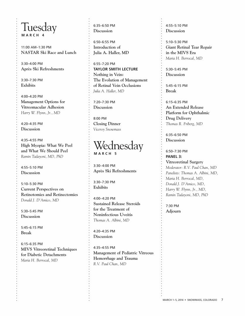
TuesdayM A R C H 4
11:00 AM–1:30 PM
NASTAR Ski Race and Lunch
3:30–4:00 PM
Après Ski Refreshments
3:30–7:30 PM
Exhibits
4:00–4:20 PM
Management Options forVitreomacular Adhesion Harry W. Flynn, Jr., MD
4:20–4:35 PM
Discussion
4:35–4:55 PM
High Myopia: What We Peel and What We Should Peel Ramin Tadayoni, MD, PhD
4:55–5:10 PM
Discussion
5:10–5:30 PM
Current Perspectives onRetinotomies and Retinectomies Donald J. D’Amico, MD
5:30–5:45 PM
Discussion
5:45–6:15 PM
Break
6:15–6:35 PM
MIVS Vitreoretinal Techniques for Diabetic Detachments Maria H. Berrocal, MD
6:35–6:50 PM
Discussion
6:50–6:55 PM
Introduction of Julia A. Haller, MD
6:55–7:20 PM
TAYLOR SMITH LECTURENothing in Vein: The Evolution of Management of Retinal Vein OcclusionsJulia A. Haller, MD
7:20–7:30 PM
Discussion
8:00 PM
Closing Dinner Viceroy Snowmass
WednesdayM A R C H 5
3:30–4:00 PM
Après Ski Refreshments
3:30–7:30 PM
Exhibits
4:00–4:20 PM
Sustained Release Steroids for the Treatment of Noninfectious Uveitis Thomas A. Albini, MD
4:20–4:35 PM
Discussion
4:35–4:55 PM
Management of Pediatric VitreousHemorrhage and Trauma R.V. Paul Chan, MD
4:55–5:10 PM
Discussion
5:10–5:30 PM
Giant Retinal Tear Repair in the MIVS Era Maria H. Berrocal, MD
5:30–5:45 PM
Discussion
5:45–6:15 PM
Break
6:15–6:35 PM
An Extended Release Platform for Ophthalmic Drug Delivery Thomas R. Friberg, MD
6:35–6:50 PM
Discussion
6:50–7:30 PM
PANEL 3: Vitreoretinal Surgery Moderator: R.V. Paul Chan, MDPanelists: Thomas A. Albini, MD, Maria H. Berrocal, MD, Donald J. D’Amico, MD, Harry W. Flynn, Jr., MD, Ramin Tadayoni, MD, PhD
7:30 PM
Adjourn
. . . . . . . . . . . . . . . . . . . . . . . . . . . . . . . . . . . . . . . . . . . . . . . . . . . . . . . . . . . . . . . . . . . . . . . . . . . . . . . . . . . . . . . . . . . . .. . . . . . . . . . . . . . . . . . . . . . .
. . . . . . . . . . . . . . . . . . . . . . . . . . . . . . . . . . . . . . . . . . . . . . . . . . . . . . . . . . . . . . . . . . . . . . . . . . . . . . . . . . . . . . . . . . . . .. . . . . . . . . . . . . . . . . . . . . . .
. . . . . . . . . . . . . . . . . . . . . . . . . . . . . . . . . . . . . . . . . . . . . . . . . . . . . . . . . . . . . . . . . . . . . . . . . . . . . . . . . . . . . . . . . . . . .. . . . . . . . . . . . . . . . . . . . .
MARCH 1–5, 2014 • SNOWMASS, COLORADO 7

PROGRAM SUMMARIES
SundayM A R C H 2
4:00–4:35 PM
Future Optical Coherence Tomography (OCT) Imaging Srinivas R. Sadda, MD
It is clear that we are currently in a golden age of retinalimaging, particularly in optical coherence tomography(OCT). The rapid advances in this technology over thepast decade have dramatically transformed the practiceof ophthalmology and retina. We can expect theseadvances to accelerate over the next decade. Thispresentation focuses on those advances that we canexpect over the next several years and how they willimpact our care of patients.
The morphologic biopsy of the retina provided by OCTwill likely be enhanced by the incorporation of robustlow-cost adaptive optics solutions, which should makecommercial clinical applications feasible. The improve-ments in resolution will enable better visualization andunderstanding of the various retinal bands. As a result,one can expect continued refinement of the classifi-cation of these structures over time. In addition toachieving isotropic image resolution, one can expectadvances in the speed of OCT instruments. Commercialswept source devices are already available and featureimproved sensitivity in addition to a dramatic increase in speed.
These speed advantages allow very dense sampling of themacula yielding megapixel OCT fundus images whichrival traditional fundus photographs, but offer theadditional advantage of being depth resolved. As a result,the quality of C-scan en face imaging through the retinais dramatically improved. En face OCT-based applica-tions can be expected to continue to rise as a result ofthis technology. The new swept source OCT devices alsofeature light sources with longer imaging wavelengths.As a result, deeper penetration into ocular structures ispossible, including the choroid, optic nerve head laminacribosa, and the angle recess.
Functional imaging with OCT is also an eagerly antici-pated future technology. Coupling precise motiontracking with high-speed, high resolution imaging at anon-visible wavelength, measurements of reflectivitychanges in retinal layers in response to stimulation
should be possible. This technique, known as opto -physiology, may provide new avenues for early diagnosisand high-precision monitoring of the progression ofretinal disease.
Vascular imaging with OCT will be another area of rapid future imaging development. In addition toDoppler OCT, a variety of OCT angiography techniquesare becoming available to selectively isolate the retinaland choroidal microvasculature from the surroundingnon-vascular tissue. Both phase-contrast and split-spectrum amplitude de-correlation techniques haveshown promise.
A final expected advance in imaging is improvements inautomation. Automated segmentation and classificationof features on imaging data will increase with a morecomplete delineation and quantification of all diseasefindings. Automated diagnosis and monitoring of diseasemay become possible. In addition to automated analysis,techniques for automated acquisition of images, particu-larly OCT images, will be advanced. These approachesmay offer opportunities to streamline ophthalmicpractices and also have applications as part of diseasescreening programs.
In summary, we are in the midst of a golden era of OCTimaging, and can expect advances in OCT to proceed at a dramatic pace.
4:35–5:10 PM
The Peripheral Retina and its Implications for Age-Related Macular Degeneration Thomas R. Friberg, MD
Age-related macular degeneration (AMD) is charac-terized by the presence of yellow deposits at the level ofthe retinal pigment epithelium called drusen, which arethe hallmarks of AMD and may represent byproducts ofphotoreceptor outer segment recycling, and proteinsassociated with inflammation and immune-mediatedprocesses. In addition, pigmentary changes in themacular retinal pigment epithelial layer are common.These changes constitute dry AMD, and are seenincreasingly as a patient ages. Neovascular AMD isassociated with severe loss of visual acuity leading inmany instances to blindness within the year if leftuntreated. While new strategies of therapy include theuse of anti-vascular endothelial growth factor (VEGF)drugs injected directly into the eye to prevent growth ofnew vessels, early detection of patients with neovascularAMD is paramount to optimize the visual prognosis.
8 ASPEN RETINAL DETACHMENT SOCIETY MEETING
. . . . . . . . . . . . . . . . . . . . . . . . . . . . . . . . . . . . . . . . . . . . . . . . . . . . . . . . . . . . . . . . . . . . . . . . . . . . . . . . . . . . . . . . . . . . .. . . . . . . . . . . . . . . . . . . . . . .
. . . . . . . . . . . . . . . . . . . . . . . . . . . . . . . . . . . . . . . . . . . . . . . . . . . . . . . . . . . . . . . . . . . . . . . . . . . . . . . . . . . . . . . . . . . . .. . . . . . . . . . . . . . . . . . . . . . .

The other form of advanced AMD is geographic atrophy, usually involving the center of the macula. This is usually a slower deterioration with the loss of thephotoreceptors, the retinal pigment epithelium, andeventually the choriocapillaris and other layers of theretina. There are no known treatments to prevent theprogression to central geographic atrophy. In approxi-mately 30% of cases with geography, neovascular formmay also occur. Therefore, assessment of the risk that a particular patient will convert to neovascular AMD or central geographic atrophy over a certain time periodbecomes very relevant.
5:10–5:45 PM
Is Fluorescein Angiography Still the Sole Gold Standard for the Diagnosis of Neovascular AMD? Ramin Tadayoni, MD, PhD
The increasing clinical use of optical coherence tomog-raphy (OCT) has progressively changed the diagnosisapproach of exudative age-related macular degeneration(AMD). Although fluorescein angiography (FA) is stillconsidered by many as a gold standard, in current clinicalpractice, the diagnosis of choroidal neovascularization(CNV) is often anticipated before obtaining the resultsof FA, which is then used as a complementary test. Weaimed to compare the relative performances of SD-OCTand FA for establishing the diagnosis of CNV in patientsreferred for suspicious exudative AMD. To determine thesensitivity and specificity of different retinal imagingcombinations a prospective, observational, multicenterstudy was conducted.
148 patients over 50 years referred for suspicious recent-onset CNV related to AMD were included and 110 hadCNV (74.3%). For the diagnosis of CNV, the sensitivityof SD-OCT+ color fundus photographs (CFP), FA+CFP,and their combination (OCT+FA+CFP) were respectively90.9%, 90.9% and 97.3% and their specificity respec-tively 88.2%, 87.7%, and 93.4%. Classic CNV werediagnosed in 98-100% of cases with SD-OCT+CFP orFA+CFP. SD-OCT+CFP showed a slightly better repro-ducibility: inter-rater kappa = 0.82 vs. 0.74 for FA+CFP,and intra-rater kappa = 0.85 vs. 0.63, respectively.
CONCLUSIONS: When used as first diagnostic test, SD-OCT and FA combined with CFP have similar sensitivity and specificity for the diagnosis of CNV inAMD and allow diagnosing of almost all classic CNV.
6:15–6:50 PM
Latest Observations on Wide-FieldImaging of Retinal Diseases Szilárd Kiss, MD
The retinal periphery is the site of pathology innumerous eye diseases. Imaging of the peripheral retinaoffers a way to diagnose, monitor, and evaluate responsesto the treatment of these conditions. Traditional funduscameras have offered a 30- to 50-degree field of view.Recent technology has advanced to provide up to a 200-degree field of view in a single image. The utility of thistechnology in clinical practice continues to be investi-gated; wide-field color photography, autofluorescenceimaging, fluorescein and indocyanine green angiographyhave been used for imaging a wide variety of peripheralretinal diseases.
Examples of specific retinal abnormalities and the utilityof ultra widefield imaging include: diabetic retinopathy –screening, classification, and follow-up of patients; age-related macular degeneration – classification; uveitis –monitoring of vasculitis, retinitis and choroiditis; retinaldetachment – determining the extent and prognosis forvisual recovery; central serous chorioretinopathy – evaluating the extent of disease and possible underlyingpathophysiology. Due to the continued limitations of thisimaging technology and the lack of ‘normative’ data, theclinical role of wide-field imaging continues to evolvebut is undoubtedly becoming the standard-of-care inmany retinal disorders.
6:50–7:30 PM
PANEL 1: Advances in Retinal Imaging and Ultrasound Moderator: Thomas R. Friberg, MDPanelists: Szilárd Kiss, MD, Timothy G. Murray,MD, MBA, Srinivas R. Sadda, MD, Ramin Tadayoni, MD, PhD
The panel will give their individual views on whichimaging techniques are most appropriate for thediagnosis and management of a variety of retinal diseasecases. Each panelist has both broad and specificexpertise in OCT and retinal imaging.
MARCH 1–5, 2014 • SNOWMASS, COLORADO 9
. . . . . . . . . . . . . . . . . . . . . . . . . . . . . . . . . . . . . . . . . . . . . . . . . . . . . . . . . . . . . . . . . . . . . . . . . . . . . . . . . . . . . . . . . . . . .. . . . . . . . . . . . . . . . . . . . . . .
. . . . . . . . . . . . . . . . . . . . . . . . . . . . . . . . . . . . . . . . . . . . . . . . . . . . . . . . . . . . . . . . . . . . . . . . . . . . . . . . . . . . . . . . . . . . .. . . . . . . . . . . . . . . . . . . . . . .

MondayM A R C H 3
4:05–4:40 PM
FOUNDERS LECTUREAnti-VEGF Maintenance Therapy for Neovascular AMD Carl D. Regillo, MD
The functional and anatomic outcomes achieved in thepivotal ranibizumab phase III trials with monthly injec-tions set the standard for comparison for the treatment ofneovascular age-related macular degeneration (nAMD)with intravitreal, pan-VEGF inhibitors. There is nowlevel I evidence from the CATT and IVAN studies thatdemonstrated non-inferiority of bevacizumab comparedto ranibizumab in this setting. Aflibercept has demon-strated equivalency to ranibizumab in its phase III VIEWstudies when dosed monthly and bimonthly in themaintenance phase of therapy. Although all three agentshave been shown to perform well to treat nAMD whendosed in a frequent and fixed fashion, the drugs aretypically administered in a more individualized mannerin clinical practice worldwide.
Individualizing anti-VEGF therapy for nAMD includesthe as needed (PRN) and the treat-and-extend (TAE)approaches. The former has been tested in several largescale, comparative trials and the results over two years of using pan-VEGF blockers PRN have not shown to benon-inferior to continuous, frequent dosing. The TAEstyle of therapy has evolved into the most frequentlyused treatment approach in the US and is gaining inpopularity elsewhere. Although there are no studiesavailable that directly compare TAE to either PRN orfixed regimens, there is mounting evidence from retro-spective and prospective studies to support its use.Evidence to date using the various treatment strategies in nAMD will be reviewed.
4:40–5:15 PM
Compounding Pharmacies andEndophthalmitis Harry W. Flynn, Jr., MD
Compounding is defined as “the combining, mixing, oraltering of ingredients to create a customized medicationfor an individual patient” (FDA definition). This definitiondoes not include mixing, reconstituting, or similar actsthat are performed in accordance with the directionscontained in an approved labeling. (USP <797> doesinclude those acts even if in the package insert).
Retinal specialists rely on compounded medications. In the case of “bevacizumab”, the individual patientreceives a small dose of the drug which is prepared byvarious compounding pharmacies.
Various outbreaks of endophthalmitis associated with useof compounded bevacizumab have occurred. In SouthFlorida, 12 patients developed severe endophthalmitisfrom compounded medication prepared by a subsidiary ofWalgreens. Many lessons were learned from this outbreakwhich can be incorporated into physician practices:
Does the pharmacy have proper licensed pharmacists, upto date inspection forms and certifications, and testingresults. Current good practice requires that compoundingpharmacies are compliant with USP <797> guidelinesand that the facility is accredited by the PharmacyCompounding Accreditation Board (PCAB). In generala “beyond use” date is 3 months for bevacizumabassuming it has been sterility tested. The medication iskept in a light resistant storage bag and stored in a refrigerator (2-8 � C or 36-46 � F) but not in a freezer.
Due to recent congressional action, legislation in manystates now requires an individual patient prescription foreach syringe prepared by compounding pharmacy. Thisadministrative burden often makes it difficult for retinalspecialists to acquire the medication in a timely manner.As a result, many retinal specialists have turned tocommercially available anti-VEGF alternatives eventhough the price is substantially higher. Increasingoversight of compounding pharmacies and morestandardized routines of preparation have reduced ratesof endophthalmitis after intravitreal bevacizumabinjection.
5:15–5:50 PM
Local Risk Factors for Development of Geographic Atrophy: New Insightsinto AMD Pathogenesis Srinivas R. Sadda, MD
PURPOSE: Systematic review of cases of incidentgeographic atrophy (GA) from large clinical studies (e.g.Age-related Eye Diseases Study) has demonstrated thatlarge drusenoid pigment often precede the developmentof atrophy. To better understand features of drusenoidlesions which specifically conferred an increased risk forthe development of GA, we used longitudinal spectraldomain optical coherence tomography (SDOCT) toinvestigate risk factors predictive for development ofatrophy from drusenoid lesions (drusen and drusenoidpigment epithelium detachment) in eyes with non-neovascular age-related macular degeneration(NNVAMD).
10 ASPEN RETINAL DETACHMENT SOCIETY MEETING
. . . . . . . . . . . . . . . . . . . . . . . . . . . . . . . . . . . . . . . . . . . . . . . . . . . . . . . . . . . . . . . . . . . . . . . . . . . . . . . . . . . . . . . . . . . . .. . . . . . . . . . . . . . . . . . . . . . .
. . . . . . . . . . . . . . . . . . . . . . . . . . . . . . . . . . . . . . . . . . . . . . . . . . . . . . . . . . . . . . . . . . . . . . . . . . . . . . . . . . . . . . . . . . . . .. . . . . . . . . . . . . . . . . . . . . . .

METHODS: Forty-one eyes from 29 patients with NNVAMDwho underwent registered SDOCT imaging over aminimum period of six months, were reviewed. Drusenoidlesions that were accompanied by new atrophy onset at 6 months or last follow up were further analyzed. Featuresof individual drusenoid lesions were consecutively eval -uated, including: maximum lesion height, lesion diameter,lesion internal reflectivity, presence and extent ofoverlying intraretinal hyperreflective features (HRF).Choroidal thickness was also measured below each lesion,and subfoveally. Changes in the lesion characteristicsthough the course of the study were also noted. Oddsratios (OR) and risk for new local atrophy onset werecalculated.
RESULTS: 543 individual drusenoid lesions were identifiedat baseline, while 28 lesions developed during follow-up.The mean follow-up time was 21.3 ± 8.6 (range,6-44)months. 3.2% (18/571) of drusenoid lesions progressed toatrophy within 18.3±9.5 (range: 5-28) months of theinitial visit. Drusenoid lesions with heterogenous internalreflectivity were significantly associated with new atrophyonset at 6 month (OR=6.549, 95% confidence interval(CI) =1.406-30.500 and new atrophy onset at last followup (OR=6.869, CI=2.22-21.236). Lesions with presenceof HRF were also significant predictors for new atrophyonset at 6 month (OR=9.354, CI=1.933-45.273) and atlast follow up (OR=11.211, CI=2.513-50.019). Lesionswith a baseline maximum height over 80 microns orchoroidal thickness less than 135 microns showedpositive association with the new atrophy onset at lastfollow up (OR=7.886, CI=2.105-29.538 and OR=3.796,CI=1.154-12.481, respectively).
CONCLUSIONS: In this study, the presence of HRF over -lying drusenoid lesions, and a heterogeneous internalreflectivity of these lesions, was found to be predictive of local atrophy onset in the ensuing months. Thesefindings provide further insight into the natural historyof anatomical changes occurring in patients with AMD.
6:20–6:55 PM
The Law of Unintended Consequencesin Health Care Reform David W. Parke, II, MD
Charles Darwin famously said, “It is not the strongest or the most intelligent who will survive, but those whocan best manage change.” Physicians (including ophthal -mologists) are in a period of seemingly unendinghealthcare change.
With change come unintended consequences. We havealready seen unintended consequences for the adminis-tration (job loss), for the American people (“If you likeyour current health insurance, you can keep it”), for the
payers (pricing set with enrollment expectations that areunlikely to be met), for the drug and device industry(‘Gee, I thought they’d leave me alone in exchange formy support’), and for physicians (narrow networks—among others). We are beginning a process that will becharacterized by failed experiments, perverse incentives,and business disruption.
The main driver of change is cost. The first, and biggest,cost-related trend facing physicians is both vertical andhorizontal healthcare integration. In many markets bothare happening simultaneously. Hundreds of billions ofdollars have been invested in integration. Having spentthose dollars, integrated systems are not going to ignorethose investments. You might say, “Well ACO’s are justHMO’s by another name, and they aren’t going to survive.”Large, integrated systems with alternative physicianpayment systems will survive for the fore seeable futurejust because too much money has already been invested.Flexibility to manage the unforeseen consequences willhelp determine our collective and individual future.
6:55–7:30 PM
PANEL 2:Controversial Aspects of AMDManagement Moderator: Szilárd Kiss, MDPanelists: Harry W. Flynn, Jr., MD, Thomas R. Friberg, MD, Carl D. Regillo, MD,Srinivas R. Sadda, MD
The treatment of neovascular AMD has been revolu-tionized by the development of anti-VEGF agentsincluding the three FDA approved medications – pegap-tanib (Macugen) ranibizumab (Lucentis) and aflibercept(Eylea). A fourth agent, bevacizumab (Avastin), is notspecifically approved for intraocular use but is frequentlyused off-label for this indication. Large prospectiveclinical trials (including ANCHOR, MARINA, PEIR,VIEW 1, VEIW 2, CATT, IVAN, HARBOR, amongouthers) suggest that monthly monitoring with frequentanti-VEGF injections maximizes the likelihood for visual improvement. Nonetheless, according to the mostrecent ASRS PAT survey, a majority of practicing retinalspecialists follow a ‘treat-and-extend’ injection andpatient follow-up protocol (this being really the onlytreatment paradigm not to have undergone rigorousprospective evaluation.) The panel discussion willexamine the diagnosis of wet AMD (use of dilated exam,OCT, FA, ICGA), the treatment paradigm (includingchoice of specific medication, re-injection criteria, etc.)as well as patient follow-up patterns (e.g., fixed monthlyfollow-up vs. treat-and-extend vs PRN).
MARCH 1–5, 2014 • SNOWMASS, COLORADO 11
. . . . . . . . . . . . . . . . . . . . . . . . . . . . . . . . . . . . . . . . . . . . . . . . . . . . . . . . . . . . . . . . . . . . . . . . . . . . . . . . . . . . . . . . . . . . .. . . . . . . . . . . . . . . . . . . . . . .
. . . . . . . . . . . . . . . . . . . . . . . . . . . . . . . . . . . . . . . . . . . . . . . . . . . . . . . . . . . . . . . . . . . . . . . . . . . . . . . . . . . . . . . . . . . . .. . . . . . . . . . . . . . . . . . . . . . .

TuesdayM A R C H 4
4:00–4:35 PM
Management Options for Vitreomacular Adhesion Harry W. Flynn, Jr., MD
The clinical course of patients with vitreomacularadhesion (VMA) is not well established, especially whenthe degree of visual disturbance is mild. Managementoptions include observation, pneumatic vitreolysis withintravitreal injection of a gas bubble, enzymatic vitre-olysis with intravitreal ocriplasmin, and surgical releaseof the attachment with pars plana vitrectomy.
A study by John et al (Retina 2013) showed a spon -taneous release rate of 32% in patients (n=106) withsymptomatic VMA managed by observation initially.Visual acuity was stable between initial and follow upexaminations, and the rate of progression requiringvitrectomy was 4%. The follow up study to this obser -vational series shows a similar spontaneous release rate(35.6%), a median time to release of 9.7 months, andstable visual acuity through time. Recently, Rodrigues et al (AJO 2013) reported a 40% release rate of sympto-matic VMA one month after intravitreal injection ofexpansile perfluoropropane (C3F8), with no associatedadverse events.
In 2012, the Food and Drug Administration approvedocriplasmin for the treatment of symptomatic VMA. The MIVI-TRUST study (Stalmans 2012) showed aVMA release rate of 26.5% in patients injected withocriplasmin compared to a rate of 10% in the intravitrealsaline group. Post marketing surveillance on the safety of this new therapy continues, and the types of adverseevents encountered are slowly coming to light withincreased use and exposure. Surgical treatment with parsplana vitrectomy has been the contemporary solution forvitreomacular adhesion. Davis et al (OSLI 2010)reported in a series of 36 patients with an anatomicsuccess rate of 86% and improvement in visual acuitythat correlated with a shorter time of symptoms beforepars plana vitrectomy.
4:35–5:10 PM
High Myopia: What We Peel and What We Should Peel Ramin Tadayoni, MD, PhD
Advances in vitrectomy techniques, use of dyes andvisualization agents such as triamcinolone in myopic eyeshave shown that many patients, who were believed tohave a complete posterior vitreous detachment (PVD),still had plaques of cortical vitreous adherent to theretina. Proper dissection of this vitreous cortex seems tohave improved surgical results, as has done appropriatedissection of internal limiting membrane (ILM).However, any dissection in these eyes may also have side effects.
We reviewed the PVD status in 96 consecutive highlymyopic eyes (mean -13,9D) requiring vitreoretinalsurgery, using intraoperative observation. The rate ofcomplete PVD assessed by this intraoperative evaluationwas 52.1%. This rate was significantly different amongdiseases (p < 0.001). It was higher in reghmatogenousretinal detachments (85%) and epiretinal membranes(74.2%), and lowest in foveoschisis (14.3%) and macularholes (10%). It was intermediate in retinal detachmentsdue to a macular hole (42.9%). Pre-operative exami-nation (slit lamp helped by SD-OCT or ultrasound) indetection of PVD showed a 75% positive predictivevalue and 70% negative predictive value.
CONCLUSION: This first study reporting the PVD rate inhighly myopic eyes using intraoperative examinationshowed a complete PVD rate lower than the completePVD prevalence previously estimated by biomicroscopyand SD-OCT examination. Preoperative examinationmay falsely suggest a PVD and an intraoperative control,in particular for myopic foveoschisis and macular holes,with visualization agents seems advisable.
12 ASPEN RETINAL DETACHMENT SOCIETY MEETING
. . . . . . . . . . . . . . . . . . . . . . . . . . . . . . . . . . . . . . . . . . . . . . . . . . . . . . . . . . . . . . . . . . . . . . . . . . . . . . . . . . . . . . . . . . . . .. . . . . . . . . . . . . . . . . . . . . . .
. . . . . . . . . . . . . . . . . . . . . . . . . . . . . . . . . . . . . . . . . . . . . . . . . . . . . . . . . . . . . . . . . . . . . . . . . . . . . . . . . . . . . . . . . . . . .. . . . . . . . . . . . . . . . . . . . . . .
5:10–5:45 PM
Current Perspectives on Retinotomies and Retinectomies Donald J. D’Amico, MD
Retinotomy and retinectomy were introduced almostthree decades ago, and quickly became a criticalmaneuver in vitreoretinal surgery. The evolution ofrelaxing retinotomy for proliferative vitreoretinopathyhas passed through several phases, beginning with theearliest (“classical”) approach in which relaxingretinotomy was performed as a last resort and only ifextensive buckling and membrane peeling were insuffi-cient to permit retinal reattachment; inevitably, it wascombined with silicone oil tamponade in this period.
However, the subsequent use and refinement ofretinotomy was strongly influenced by several factors: 1) the increased availability of endolaser, 2) sophisticationin the use of varied intraocular tamponades, 3) the intro-duction of perfluorochemicals for the manipulation ofgiant tears and large retinotomies, 4) advances in wide-angle intraoperative visualization of the retina, and 5) experience gained by the procedure of macular translo-cation in which 360 degree retinotomies were routinelyperformed in eyes with a normal retinal periphery.
These influences had the effect of broadening the use of relaxing retinotomy in PVR to include retinotomy inthe absence of a scleral buckle for some authors, or theabsence of silicone oil tamponade for others, or therelatively less aggressive membrane peeling prior toconsidering relaxing retinotomy; this may be termed the“modern” period. Finally, many surgeons have entered an“avant garde” period, in which relaxing retinotomy isfavored for very early or even primary use as the maintechnique for PVR repair.
6:15–6:50 PM
MIVS Vitreoretinal Techniques for Diabetic Detachments Maria H. Berrocal, MD
A video presentation of novel techniques utilizing 25g and27g vitrectomy cutters for the removal of fibrovasculartissue in traction and traction and rhegmatogenousdiabetic detachments. The utilization of the vitreouscutter to lift, peel, dissect and shave will be shown, aswell as a description of the advantages of the smallergauges in these challenging cases, in particularly theability of these smaller instruments of getting betweentissue planes and shave the retinal surface.
6:55–7:30 PM
TAYLOR SMITH LECTURENothing in Vein: The Evolution ofManagement of Retinal Vein OcclusionsJulia A. Haller, MD
Retinal venous occlusions (RVOs) produce a chronicretinal vasculopathy, with ischemia and macular edema.Retinal vein occlusion is the most common retinalvascular disease after diabetic retinopathy. Its prevalenceapproaches that of neovascular age-related maculardegeneration.
The treatment of retinal vein occlusion has evolveddramatically in the last five years and with it our abilityto evaluate the associated ocular pathologies and todesign and conduct clinical trials targeting therapies.
Prior to about five years ago, our treatment of RVO wasbased on studies performed by the Branch Retina VeinOcclusion Study Group in the early 1980s and theCentral Retinal Vein Occlusion Study Group in the midto late 1990s.
The next major advances in the treatment of vein occlu-sions had to wait until about five years ago. From 2009 totoday, very significant well-designed clinical trials havetotally altered the landscape for the treatment of RVOs.
These include the SCORE study of intravitreal triamci-nolone, the GENEVA study of the dexamethasonebioerodable intravitreal implant, the BRAVO/CRUISEstudies of ranibizumab, and the COPERNICUS,GALILEO, and VIBRANT studies of aflibercept.
These advances hinge on several developments thatenabled their success, including the development of newdrugs and drug delivery systems, the acceptance ofintravitreal drug delivery, and the development of opticalcoherence tomography.
We have also developed a more sophisticated awarenessof the differences between pharmacological agents, evenones within the same class.
For example steroids may differ in chemical structure,effect on anti-inflammatory potency, effect on activationof genes, and may have different cytotoxicity. Anti-VEGF agents, similarly, are not all created identical intheir mode of activity, duration, or side effects.
Once therapies became available, our approach to RVOevolved. As a result of the SCORE study, the standard ofcare for the treatment of CRVO became intravitrealtriamcinolone, with the 1 mg dose having a lower
MARCH 1–5, 2014 • SNOWMASS, COLORADO 13
. . . . . . . . . . . . . . . . . . . . . . . . . . . . . . . . . . . . . . . . . . . . . . . . . . . . . . . . . . . . . . . . . . . . . . . . . . . . . . . . . . . . . . . . . . . . .. . . . . . . . . . . . . . . . . . . . . . .
. . . . . . . . . . . . . . . . . . . . . . . . . . . . . . . . . . . . . . . . . . . . . . . . . . . . . . . . . . . . . . . . . . . . . . . . . . . . . . . . . . . . . . . . . . . . .. . . . . . . . . . . . . . . . . . . . . . .
complication rate in terms of elevated intraocularpressure, glaucoma, and cataract development. BRVO bycontrast seemed to do better with grid laser than steroid.
The dexamethasone intravitreal implant studies showedthat eyes treated with the 700ug dose gained three linesof vision faster than those treated with sham. Theimprovement of visual acuity correlated with OCT–documented drying of macular edema as well as withreduced fluorescein angiographic leakage. Side effects ofIOP elevation and cataract formation were more minimalwith this steroid formulation. Additional analysis of datafrom these trials showed that duration of macular edemahad an effect on clinical outcome. Fewer eyes gained ≥15 letters of visual acuity when treatment was delayedfor six months and more patients had a ≥3 line loss ofvision with the delay.
Next on the scene were the BRAVO and CRUISEstudies documenting marked visual gain and retinalthinning with intravitreal ranibizumab given monthly,and confirming the benefit of earlier treatment. Morerecently a different class of antiVEGF agent, aflibercept,has been shown in both CRVO and BRVO eyes toimprove vision and dry up edema, with the added benefitof improvement in perfusion, and the possibility that lessthan monthly treatment may be effective.
There is still much to understand and learn. Challengesinclude customization of treatment, combining therapiesin eyes that become less responsive or have residualedema, and reducing the treatment burden, as well asoptimizing visual acuity, reducing ischemia, andpreventing occlusion primarily.
Wednesday M A R C H 5
4:00–4:35 PM
Sustained Release Steroids for theTreatment of Noninfectious Uveitis Thomas A. Albini, MD
Sustained release steroids have greatly improved the careof uveitis patients in the last decade. While they havenot replaced traditional systemic steroid-sparing agents,these devices offer alternatives for patients that cannottolerate complex systemic immunosuppression. Localcomplications, such as ocular hypertension, glaucomaand cataract formation are common. The three implantsto be discussed include the anchored fluocinoloneimplant (Retisert, Bausch and Lomb), the injectabledexamethasone implant (Ozurdex, Allergan) and theinjectable fluocinolone implant (Iluvien, Alimera).
The Multicenter Uveitis Steroid Treatment (MUST)Trial demonstrated not only noniferiority of visual acuityoutcomes following anchored fluocinolone implant ascompared to standard systemic uveitis therapy, but alsodemonstrated better inflammatory control with theimplant. Subgroup analysis of the original pivotal trialdata for the anchored fluocinolone implant shows thatthe visual acuity improvement seen with the implant isstrongly associated with resolution of vascular leakage,suggesting that eyes with vascular leakage stronglybenefit from this local treatment. Patient selection isextremely important in the use of these implants. Best-case and worst-case patients for the implants will bediscussed.
4:35–5:10
Management of Pediatric VitreousHemorrhage and Trauma R.V. Paul Chan, MD
The management of pediatric vitreous hemorrhage andtrauma has improved significantly with advances inimaging and surgical technology. However, even thoughthe etiology of pediatric vitreous hemorrhage and itsadverse effects including tractional retinal detachment,epiretinal membrane, myopia, and amblyopia, have all
14 ASPEN RETINAL DETACHMENT SOCIETY MEETING
. . . . . . . . . . . . . . . . . . . . . . . . . . . . . . . . . . . . . . . . . . . . . . . . . . . . . . . . . . . . . . . . . . . . . . . . . . . . . . . . . . . . . . . . . . . . .. . . . . . . . . . . . . . . . . . . . . . .
. . . . . . . . . . . . . . . . . . . . . . . . . . . . . . . . . . . . . . . . . . . . . . . . . . . . . . . . . . . . . . . . . . . . . . . . . . . . . . . . . . . . . . . . . . . . .. . . . . . . . . . . . . . . . . . . . . . .

been previously reported, there is currently no consensuson optimal management of vitreous hemorrhage inchildren. With the advent of ultra-widefield retinalimaging (e.g. OPTOS, Marlborough, MA) and RetCam(Clarity Medical Systems, Pleasanton, CA), decisions formedical and surgical intervention may now be moreeasily defined. The OPTOS and RetCam have made itpossible to better evaluate peripheral retinal pathology inthe pediatric population both in the outpatient andhopital setting.
Timing of surgery for vitreous hemorrhage and trauma in the pediatric population has unique considerationsand dense, non-clearing vitreous hemorrhage may lead toirreversible vision loss from amblyopia due to mediaopacification and sensory deprivation. Successful reversalof amblyopia has been demonstrated with long-termpatching once the media has been cleared. Therefore,earlier intervention with improved techniques andmicroincision vitrectomy surgery (MIVS) may beindicated.
The current advances in surgical and medical technologymake it necessary for us to reevaluate how we havehistorically managed pediatric retinal disease. Thechallenges and changing trends we now face formanaging pediatric retina patients, specifically thosewith vitreous hemorrhage and trauma, will be discussed.In addition, the role of ultra-widefield imaging,fluorescein angiography, optical coherence tomography,endoscopic vitrectomy, pharmacotherapy, and MIVS will be highlighted through case presentations.
5:10–5:45 PM
Giant Retinal Tear Repair in the MIVS Era Maria H. Berrocal, MD
The challenges and advantages of MIVS in themanagement of giant retinal tear detachments will be presented. Video will present the advantages ofchandelier utilization, perfluotocarbon liquid injectionthrough 25g cannulas, and aspiration of fluid from theedge of the detachment. Important steps include theremoval of the anterior retina, adequate photocoagulationof the edges of the tear and meticulous aspiration of fluidanterior to the edge of the retina. Different tamponadingtechniques, i.e., gas and silicone oil, will be explained.
6:15–6:50 PM
An Extended Release Platform forOphthalmic Drug Delivery Thomas R. Friberg, MD
A number of polymeric, intravitreal delivery systemshave been considered by researchers, including micropar-ticles, which are well-tolerated in the eye and arecapable of delivering drugs over a longer period of time.For example, microparticle-encapsulated or PEGylatedbevacizumab are more effective than bevacizumab alonein treating CNV in rats. However, direct intravitrealinjection of such microparticle formulations may resultin rapid particle dispersion and cause turbidity of thevitreous humor, affecting vision. In situ-forming,injectable hydrogels are thus an attractive alternative forintravitreal delivery as they can be formulated to encap-sulate drugs and sustain release locally.
Studies with non-degradable, thermally-responsivehydrogels have demonstrated excellent protein encapsu-lation, minimal toxicity, and easy injectability with nolong-term effects on retinal function in vivo. Thesestudies established that thermoresponsive hydrogels areexcellent candidates for intraocular drug delivery. Wehave used such a gel to deliver bevacizumab at thera-peutic levels for more than 60 days in animals after asingle 0.05 cc injection through a 31 gauge needle. Thebiocompatibility appears excellent.
6:50–7:30 PM
PANEL 3: Vitreoretinal Surgery Moderator: R.V. Paul Chan, MDPanelists: Thomas A. Albini, MD, Maria H. Berrocal, MD, Donald J. D’Amico, MD, Harry W.Flynn, Jr., MD, Ramin Tadayoni, MD, PhD
We will discuss the panelists’ approach to the evaluationand surgical management of various vitreoretinal condi-tions including complex diabetic retinopathy, macularpathology, proliferative vitreoretinopathy, uveitis, andpediatric vitreoretinal disease. The discussion will befocused on current surgical technologies and the use ofpharmacologic adjuvants in vitreoretinal surgery.
MARCH 1–5, 2014 • SNOWMASS, COLORADO 15
. . . . . . . . . . . . . . . . . . . . . . . . . . . . . . . . . . . . . . . . . . . . . . . . . . . . . . . . . . . . . . . . . . . . . . . . . . . . . . . . . . . . . . . . . . . . .. . . . . . . . . . . . . . . . . . . . . . .
. . . . . . . . . . . . . . . . . . . . . . . . . . . . . . . . . . . . . . . . . . . . . . . . . . . . . . . . . . . . . . . . . . . . . . . . . . . . . . . . . . . . . . . . . . . . .. . . . . . . . . . . . . . . . . . . . . . .

EXHIBITORSThe Aspen Retinal Detachment Society
gratefully acknowledges generous contributions
from the following companies:
BLACK DIAMOND
Genentech, Inc.
DIAMOND
Insight Instruments, Inc.
Regeneron Pharmaceuticals, Inc.
PLATINUM
Bausch + Lomb
GOLD
Alcon Laboratories, Inc.
Allergan, Inc.
Dutch Ophthalmic, USA
MedOne Surgical, Inc.
Notal Vision
OCULUS Surgical, Inc.
Synergetics, Inc.
ThromboGenics, Inc.
VisionCare Ophthalmic Technologies
SILVER
Blue Star Radiology