the art & science of clinical...
TRANSCRIPT

The Art & Science of Clinical Problem-Solving
Sanjay Saint, MD, MPH Professor of Internal Medicine and Hospitalist
Ann Arbor VAMC University of Michigan Medical School
Your 72-year-old mother calls complaining of sudden-onset difficulty breathing and chest pain for the last several hours. She was feeling fine during her recent vacation in Australia (she returned to Florida a few days ago) but noticed left thigh pain yesterday.
Patient Presentation
• She denies fever, chills, or abdominal pain.
• Her chest only hurt when she took a breath
• She did not have enough frequent flier miles to upgrade to Business Class…
Patient Presentation, continued

Question: What is the most likely diagnosis?
a) Pneumococcal pneumonia
b) Acute myocardial infarction
c) Panic attack
d) Pulmonary embolism
e) Aortic dissection
• Pattern recognition: The example of PE
• Diagnostic error: Cognitive biases and heuristics
• How can we become better clinical problem-solvers?
• Case presentation
Clinical Problem-Solving: Outline
• Pattern recognition: The example of PE
• Diagnostic error: Cognitive biases and heuristics
• How can we become better clinical problem-solvers?
• Case presentation
Clinical Problem-Solving: Outline

Diagnostic Error
No Fault Cognitive Errors
Systems Errors
(Graber, M. L. et al. Arch Intern Med 2005;165:1493-1499)
Diagnostic Error
No Fault Cognitive Errors
Systems Errors
1. Poor knowledge
– Unaware of heparin-induced thrombocytopenia
– Unaware of Fournier’s gangrene
Diagnostic Error
No Fault Cognitive Errors
Systems Errors
2. Incomplete data gathering – Didn’t consider endocarditis due to a brief cardiac exam – Didn’t consider the correct diagnosis because did not
obtain a complete history

Question for the Audience
• When comparing the relative value of the history, exam, and labs in making medical diagnoses, studies reveal that the correct diagnosis is determined after only history in what percent of patients?
A) 20% or less of the time
B) Between 20% and 70% of the time
C) Above 70% of the time
D) Not sure: I no longer waste time obtaining a history
History is The Key • 6 decades ago: the correct diagnosis can be made after
history-taking alone in 74% of patients (Platt R. Manchester University Medical School Gazette 1947; 27:139-145)
• Comparing the relative value of history, exam, and labs in making medical diagnoses: correct diagnosis determined after only history in 82% of patients
(Hampton JR, Br Med J 1975;2:486-489)
• In 1992, Petersen reproduced the above study: found that the history led to the correct diagnosis 76% of the time
(Peterson MC, West J Med 1992;156:163-165)

Diagnostic Error
No Fault Cognitive Errors
Systems Errors
3. Succumbed to a cognitive bias – Availability bias – Anchoring bias – Premature closure – Framing
(Kassirer and Kopelman. Learning Clinical Reasoning. 1991) (Gurpreet Dhaliwal in Clinical Problem-Solving. 2006)
“Space Shuttle”
Availability Bias
Availability Bias
• Judge probability by ease of recall
• Humans remember the remarkable or unusual
• If sued re: missing dissection, every pt with chest pain has dissection until proven otherwise
• Specialists also susceptible (Gulati et al. “Impatient Inpatient Care” N Engl J Med 2000)

Anchoring Bias
• Fall in love with the first diagnosis
Anchoring Bias: Classic Example
Randomized trial:
a) What does 8 x 7 x 6 x 5 x 4 x 3 x 2 x 1 = ?
b) What does 1 x 2 x 3 x 4 x 5 x 6 x 7 x 8 = ?
Answers:
a) 2250
b) 512
Anchoring Bias
• Fall in love with the first diagnosis
• Medical example: admitting a patient already worked up in the Emergency Room

Premature Closure • Tendency to stop considering other possibilities once you
have reached a diagnosis
• We see what we are looking for (confirmation bias)
Focus on the supporting data
Dismiss the conflicting data
• Label persistent GI symptoms as refractory IBD and proceed with colectomy without ruling out ……………..
(C Goulet et al. “The Unturned Stone” N Engl J Med 2005)
Framing • Framing effect: a different conclusion drawn from the
same set of facts, depending on how the facts are presented
• Some believe that humans tend to be “cognitive misers” preferring to do as little thinking as possible
• “Frames” provide people a quick way to process information
Framing • 82-year-old man with mild dementia and aortic stenosis
brought to the ED by his daughter with several weeks of anorexia, weight loss, subjective fever, and malaise.
• The ED doc calls up the admission:
“82 yo with failure-to-thrive, likely needs nursing home placement…”
VS
“82 yo with unexplained fevers at home and a heart murmur…”

Cognitive Errors
Availability
Anchoring
Premature Closure
Framing Effects

Pattern recognition: The example of PE
Diagnostic error: Cognitive biases and heuristics
• How can we become better clinical problem-solvers?
• Case presentation
Clinical Problem-Solving: Outline
Reducing Cognitive Errors
1. Acknowledge
2. Metacognition
Acknowledge
• Overall, physicians do an excellent job
• Errors are common… so is overconfidence
“The fool doth think he is wise, but the wise man knows himself to be a fool.” -- Shakespeare (As You
Like, Act 5, Scene 1)
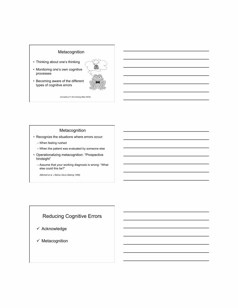
Metacognition
• Thinking about one’s thinking
• Monitoring one’s own cognitive processes
• Becoming aware of the different types of cognitive errors
(Croskerry P. Ann Emerg Med 2003)
Metacognition • Recognize the situations where errors occur:
– When feeling rushed
– When the patient was evaluated by someone else
• Operationalizing metacognition: “Prospective hindsight”
– Assume that your working diagnosis is wrong: “What else could this be?”
(Mitchell et al. J Behav Decis Making 1989)
Reducing Cognitive Errors
Acknowledge
Metacognition

Conclusions
Clinical problem-solving is an essential function of the physician
Pattern recognition works well for some diagnoses
Cognitive errors are common
We can (hopefully) become better problem-solvers!
Pattern recognition: The example of PE
Diagnostic error: Cognitive biases and heuristics
How can we become better clinical problem-solvers?
• Case presentation
Clinical Problem-Solving: Outline
Clinical Problem-Solving Exercise

Presentation Chief Complaint: Cough and Shortness of Breath History of Present Illness: • A 47-year-old woman was in her usual state of excellent
health until 2 weeks prior to presentation at an outside hospital when she developed headache, clear rhinorrhea, myalgias and arthralgias.
• 1 week prior to presentation she developed subjective fever and chills, dyspnea, and a nonproductive cough.
• She visited an urgent care clinic and was prescribed azithromycin which did not improve her symptoms.
Presentation PMH: Mild Iron Deficiency Anemia PSH: Lasik, Tonsillectomy, Right Knee Arthroscopy Meds: Multi-Vitamin Allergies: Penicillin and Sulfa: hives FH: Father with laryngeal cancer SH: Married with two children. Works in finance in
southeastern Michigan. No history of tobacco use, alcohol or illicit drug use.
Presentation: Physical Exam Temp 97.6°F, BP 77/45, HR 86, RR 16, 96% RA Gen: alert and cooperative, coughing. HEENT: PERRLA. Anicteric sclerae. EOMI. Dry mucosa. Neck: Supple. No JVD. No bruit. Lungs: Scattered rhonchi. Rales at the right base. Heart: Regular rate without murmur. Breast: No masses. Abdomen: Soft. Nontender. No organomegaly. Extremity: No pedal edema. No calf tenderness. No
cyanosis. Neuro: CN II-XII grossly intact. Deep tendon reflexes 2+
and equal bilaterally. No motor or sensory deficits.

Presentation Admission Data:
11.3
31.3 420
137 104 5
3.8 24 0.8 10.4
Seg 72% Mono 13% Lymph 9% Eo 5.4%
135
MCV 90.6 RDW 11.8
Chest x-ray: right basilar airspace c/w pneumonia.
Question
What are your initial diagnostic considerations in this 47-year-old previously
healthy woman who now has a 2-week illness of non-productive cough, subjective fever,
dyspnea, headache, and myalgias, along with a right lower lobe density?

Hospital Course
• While in the ED, the patient’s temperature rose to 101.1°F.
• She was admitted with a diagnosis of community- acquired pneumonia and started on levofloxacin and nebulized breathing treatments.
• Despite therapy, she continued to have fever 2 to 3 times per day, to a maximum of 103.2 °F.
Hospital Day #4 • The patient develops diarrhea; started on
metronidazole. Fecal leukocytes and C.difficile toxin negative.
• ID is consulted who elicits more history: – No birds or pets at home – No known exposure to TB – She traveled to California several weeks prior to
presentation • ID recommended urine legionella antigen testing.
Hospital Day #7 • Her symptoms have not improved and she continues to
be febrile. Vancomycin is added.
• While her white count remains ~ 9 K/µL, her peripheral eosinophilia reaches 12.1%.
• ESR is 97. Numerous serologies subsequently sent.
• Neurology recommends MRI given the continuing headache, result is a normal scan.
• A CT of the chest, abdomen and pelvis is ordered due to the continued symptoms.

CT Results
• Scattered air space opacities in the left lower, right upper and right middle lobes with more focal consolidation in the right lower lobe.
• Right paratracheal, subcarinal and right hilar lymphadenopathy.
• The abdomen and pelvis are unremarkable.
Hospital Day #8 • Bronchoscopy is performed
– BAL fluid with numerous eosinophils
– Gram stain and fungal smears are negative
– Biopsy without evidence of malignancy
• ANA, ANCA, C3, C4 are all normal.
• Cold agglutinins, monospot, urine legionella antigen and lyme titres are negative.
• Numerous blood cultures are negative. BAL bacterial culture is negative.
Question: What is the most likely diagnosis?
a) Pneumococcal pneumonia
b) Legionella pneumonia
c) Acute Eosinophilic pneumonia
d) Tuberculosis
e) Fungal pneumonia

Hospital Day #9
• The patient is diagnosed with Acute Eosinophilic Pneumonia.
• All antibiotics are discontinued.
• Solumedrol 40 mg IV twice a day given.
• Patient is kept hospitalized and observed.
Question: Do you agree with this management plan?
a) Yes
b) No
c) Not sure – prefer to punt the decision to the incoming hospitalist
“Error is inevitable in a field premised on the art of balancing probabilities.”
Sir William Osler

Hospital Days 10-13
• The patient’s pulmonary symptoms improve dramatically.
• Infiltrates on the chest radiograph also improve.
• Peripheral eosinophilia resolves, 0.5% at discharge.
• Headache controlled by over-the-counter meds.
• She is discharged to home on prednisone 20 mg daily for the next 4 weeks.
Question: How do you think the patient will do?
a) Well
b) Poorly
From Complications by Atul Gawande (2002):
“The core predicament of medicine – the thing that makes being a patient so
wrenching, being a doctor so difficult, and being a part of a society that pays the bills
they run up so vexing – is uncertainty.”

Second Presentation Chief Complaint: Fever, Headache, Blurry Vision
History of Present Illness:
• Five weeks after discharge, ~ 1 week after steroid course completed, she comes to UM with continued headache.
• She developed regular daily fever to 101°F; also reports episodes of nausea and vomiting.
• She has had persistent blurry vision for one week which improves by covering one eye.
• Her original cough and dyspnea have resolved.
Exposure History Obtained at University of Michigan Medical Center
• The patient grew up and always resided in Michigan
• No pets or exposure to birds, rodents or mold
• Travel history
– Tijuana, Mexico: 10 years ago
– Scottsdale, Arizona: 2 years ago
– New Orleans: 18 months ago, just before Katrina
– California: ~2 weeks prior to original symptom onset
Exposure History: California Dreamin’
• Flew with her husband to LA, spent several days on the beach
• Drove to SF along the coast
• While in SF’s Chinatown, reports being coughed on heavily by a stranger
• On return from San Francisco to LA, they drove down the I-5
• They made two stops: one for gas and another at Taco Bell; windows were up, air conditioning was on

Second Presentation
Physical Exam: Temp 98°F, BP 110/80, HR 70, RR 20, 98% RA General: Oriented and conversant. No acute distress. HEENT: Pupils equal and reactive. R CN6 palsy. Neck: Supple without lymphadenopathy. Lungs: Clear to auscultation. Heart: Regular rate, without murmur, rub or gallop. Abdomen: Soft, nontender and nondistended. Extremity: Without clubbing, cyanosis or edema.
Second Presentation
Admission Laboratory Data:
5.7 11.3
33.3 347
135 102 11
3.5 26 0.8 102
Eo 6.1%
CSF Tube #1 Clear RBC: 4 cm3
WBC: 383 cm3
CSF Tube #4 Clear RBC: 3 cm3 WBC: 287 cm3
Seg 18% Lymph 42% Histio 39% Eo 1%
CSF Protein 195 mg/dL CSF Glucose 46 mg/dL
Question: What is the most likely diagnosis?
a) Pneumococcal disease
b) Acute Eosinophilic pneumonia
c) Tuberculosis
d) Fungal infection
e) None of the above

The Answer: “Valley Fever”
• Coccidioidomycosis immunodiffusion and complement fixation on both serum and CSF return positive.
• She was started on fluconazole 800 mg daily.
• As of five months after her presentation to UM, she is improving: no fevers, no headache, mild esotropia and increased energy.
• Her course of therapy may run months to years and includes follow-up lumbar punctures and MRI.
What is Coccidioidimycosis? • Coccidioidomycosis is a fungal disease caused by
Coccidioides species.
• These organisms live in the soil of semiarid areas.
• People at risk live in or visit places where the fungus is in the soil and engage in activities that expose them to dust (e.g., construction, agricultural work, archeology ).
• Endemic in areas are the southwestern United States, parts of Mexico and South America.

Coccidioidimycosis • People get infected with Coccidioides by inhaling airborne
spores often after disturbance of contaminated soil by humans or disasters (e.g., dust storms, earthquakes).
• Symptoms, if present, usually occur 1 to 3 weeks after exposure.
• About 60% of infections do not cause any symptoms!
Clinical Manifestations of Coccidioidimycosis
• Some may experience a flu-like illness, with fever, cough, headache, rash and muscle aches.
• Most people recovery fully, within weeks to months of symptom onset.
• A small number of people may develop chronic pulmonary infection or widespread disseminated infection (~ 1% have disseminated disease).

Dissemination of Coccidioidimycosis • When the infection spreads outside of the lungs, it most
commonly results in skin lesions, CNS infection (e.g., meningitis), and bone and joint infection.
• Some are at increased risk for developing disseminated infection: – African-Americans – Asians – Filipinos – Pregnant women – Immunocompromised persons
Diagnosis • Can be challenging…
• Often misdiagnosed as community-acquired pneumonia
• Serologies, biopsy, culture
• But first must consider the diagnosis!
Teaching Points • An exposure history is important but was obtained
and then largely dismissed.
• Types of Cognitive Errors:
– Incomplete data gathering
– Incomplete knowledge
– Premature closure
• Prescribing steroids without adequately excluding infection is done at one’s own peril.

Thank you!