the architecture of black and white facial skin
TRANSCRIPT
Clinical and laboratorv s t u d i e s I I l
The architecture of black and white facial skin William Montagna, PhD, and Kay Carlisle, MS Beaverton, Oregon
This study yielded the following findings on the morphologic facial skin differences between black and white women: The epidermis of black skin has more and larger singly distributed melanosomes in the keratinocytes and corneoeytes than that of white skin. The stratum lu- eidum in black skin is not altered by sunlight exposure. The epidernfis of black skin rarely shows atrophied areas. The elaunin and oxytalan fibers in black skin are not disposed in can- delabra-like formations. Black skin has minimal elastosis and elastic fibers stain pink or red with the hernatoxylin and Lee procedure; none stain lilac or blue. The dermis of black skin contains many more fiber fragments composed of collagen fibrils and glycoproteins. Fibro- blasts are more numerous, larger, have more biosynthetic organelles than white skin, and are often binucleated and multinucleated. The dermis of black skin has many binucleated and multinucleated macrophages and multinucleated giant cells. Black skin has many more mixed apocrine-eccrine sweat glands than does white skin and more blood and Lymphatic vessels. (J AM ACAD DERMATOL 1991;24:929-37.)
This study compares the morphology of the facial skin of black and white women 22 to 50 years of age, all of whom had lived in Tucson, Arizona, for 2 or more years. Other authors have concluded that, other than the distribution and size of melanosomes, there are no structural differences between white and black skin. 1 Andersen and Maibach, 2 who arrived at the same conclusion but found some pharmacologic differences, stated that: "Although color is the most striking racial skin difference, it is but one piece of a biologic mosaic." In our studies, we found that the epidermis of black skin is largely spared the gross photodamage we have reported in white skin. 3 The dermis of black skin shows the greatest amount of difference from that of white skirl.
We believe, as others do, 4, 5 that the larger quan- tity of melanin and its distribution in black skin does protect it from sunlight, but only to a degree. Black skin may be endowed with properties in addition to pigment that protect it from sunlight and other en- vironmental hazards.
MATERIAL AND METHODS
Punch biopsy specimens (4 mm) were obtained from the malar eminences of the faces of 19 black and 19 white
From the Oregon Regional Primate Research Center. Supported by grants from the Proctor & Gamble Company. Accepted for publication Nov. 12, 1990.
Reprint requests: William Montagna, PhD, 505 NW 185th Ave., Bea- verton, OR 97006.
16/1/26808
women, all of whom had lived for some time in Tucson, Arizona. All subjects provided informed consent. The specimens were grouped according to age of the subject: (1) 22 to 29 years, (2) 30 to 39 years, and (3) 40 to 50 years. The specimens were obtained in November 1988 from clinically normal skin of the malar eminence with the use of lidocaine anesthesia and were fixed immedi- ately in 10% buffered cold formalin (pH 7,0), for light microscopy and embedded in glycol methacrylate, All specimens were treated alike. Sections 2 ~m in thickness were stained wkh the hematoxylin and Lee procedure, Giemsa, Fontana-Masson ammoniacal silver nitrate and picro-sirius red; sections 4 ,m thick were stained with pe- riodic acid-Schiff, the original Weigert technique for elastic fibers, the Prussian blue technique for iron, the colloidal iron method for mucopolysaccharides, and for reticulin. 3 For transmission electron microscopy small pieces of skin were fixed in 0.75% glutaraldehyde, 3.0% formaldehyde, 0.001 mol/L collidine, 0.05 mol/L HEPES buffer, sodium hydroxide and 0.005 mol/L hy- drogen chloride, postfixed in osmium tetroxide, 6 and em- bedded in Epon, Ultrathin sections, cut from the Epon- embedded blocks, were stained with uranyl acetate and lead citrate, and examined with a transmission electron microscope (model 200, Philips Electronics, Eindhoven, The Netherlands).
FINDINGS
Epidermis
Regardless of the person's age, the epidermis of black or white skin can be thick or thin, and the b as al cells may or may not show conspicuous basal projections that extend into the papillary dermis. The basal cytoplasmic extensions are present in all epidermis but they may or may not be evident in tis-
929

930 Montagna and Carlisle
Journal of the American Academy of
Dermatology
Fig. 1. h and B. Facial skin of a 40-year-old, fair-skinned white woman. A, Note pink-stained elastotic fibers, and the clear grenz zone. (Hematoxylin-Lee; • B, Argen- tophilic melanosomes have sparse and patchy distribution throughout all epidermal layers. (Fontana-Masson stain; X630.) C, Facial skin from a 20-year-old white woman stained with the Weigert technique for elastic fibers. Observe candelabra formations of the elaunin and oxytalan fibers in white skin. D and E, Facial skin of a 41-year-old black woman. D, Dermis shows light-stained pink, delicate elastic fibers; there is no elastosis. Compare with A. (He- matoxylin-Lee stain; • E, Many melanosomes extend throughout epidermal layers. (Fontana-Masson stain; • F, Facial skin of a 22-year-old black woman stained with the Weigert technique. Oxytalan and elaunin fibers are not arranged in distinct candelabra for- mations. Conpare with C. (X 1260.)
sue sections. The horny layer may be compact, loosely woven, or have a basket-weave pattern (Fig. 1, A and D). Under the light microscope, the stratum lucidum in undamaged skin, white or black, consists
of one or two thin layers, and the stratum granulo- sum rarely exceeds three layers. We have reported earlier that in white skin, the epidermal stratum lu- cidum is usually distorted on exposure to sun. 3 In
Volume 24 Number 6, Part 1 June 1991
Black and white skin 931
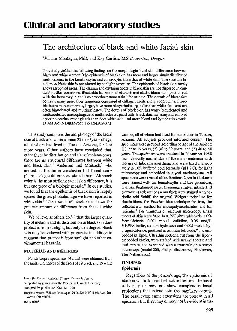
Fig. 2. Mixed apocrine-eccrine glands in 4 #m thick sections of facial skin from a black woman. PAS technique counterstained with iron hematoxylin. Tubule at upper right is lined with apocrine cells and contains particulate material mildly reactive to PAS. The dilated tu- bule at lower left is mostly lined with clear, reticulated cells and contains densely PAS-pos- itive homogeneous material surrounded by granular, mildly PAS-reactive material. (X 1200.)
contrast, the stratum lucidum in black skin rarely shows any signs of alteration.
In Fontana-Masson preparations, the entire black skin epidermis, including the stratum granulosum, the stratum lucidum, and the stratum corneum, contains melanosomes. In very light-colored white skin only a few melanosomes can be seen in the basal keratinocytes and in the malpighian layer. In dark- colored white skin the distribution of melanosomes resembles that of black skin. The distribution of malanosomes in black skin was similar, regardless of age. Melanosomes are larger and more numerous in black than in white skin. In Fig. 1, B and E, we compare the melanosome density and distribution in black and in white skin.
Regardless of age, in some specimens of black skin the epidermis is free of histologic flaws, but in oth- ers vacuoles and dyskeratosis are present in the ke- ratinocytes of the malpighian layer. These alter- ations are similar to those that we reported in white skin3; however, two observations can be made. White skin shows frequent focal areas of atrophy and/or necrosis but only one of the t9 black women in our study had minor atrophic spots in the epider- mis. Second, the stratum lucidum, which in white skin is often swollen and distinctly cellular on expo- sure to sun, remains compact and unaltered in black skin, regardless of age.
Our electron microscopic observations confirm those of others] Regardless of age, each kerati- nocyte in the epidermis of black skin is replete with
mostly large, single, and membrane-bound mature melanosomes; melanosomes may occasionally form small aggregates. In white skin we find membrane- bound aggregates of small melanosomes.
Epidermal melanocytes in both white and black skin are mostly sandwiched between the basal kera- tinocytes and are often found below the basal layer as pendulous cells. The melanocytes contain some argentophilic melanosomes scattered in their perikaryons and are often riddled with vacuoles as they are in white skin.
Cutaneous appendages
In histologic sections, the appendages in black women are essentially similar to those in white women, but they have more pigment. Melanosomes descend to a deeper level along the side of hair fol- licles. The bulbs of vellus hair follicles, which do not contain melanin in white skin are often pigmented in black skin, and the outer root sheath cells always contain some melanosomes. 8 Hair follicles seem to be anchored by fewer elastic fibers than are those in white skin.
We encountered what at first we thought to be apocrine glands more frequently in black than in white facial skin, as did Schiefferdecker, 9 who first distinguished and named sweat glands. The glands are found in all levels of the dermis. These "apocrine" glands, however, possess the characteristics of both apocrine and eccrine glands (Fig. 2). Some of the secretory cells resemble the clear cells of eccrine

932 Montagna and Carlisle
Journal of the American Academy of
Dermatology
Fig. 3. A, TEM of blood vessel lined with endothelial cells (E) and veil cell from dermis of a 26-year-old black woman. There are many fiber fragments around this vessel. A binucleate fibroblast (F) is at bottom. C, Collagen bundle (X3900.) B, Fiber fragments consisting of col- lagen fibrils intermixed with darker staining glycoprotein particles in dermis of black skin. Arrowheads indicate structures in the ground substance that resemble glycoprotein mole- cules. El, Elastic fiber. (•
sweat glands whereas others are cuboidal or colum- nar as in apocrine glands. These are mixed glands. Their secretory cells often have clusters of argento- philic pigment granules that, like melanosomes, contain no iron. This pigment is a characteristic fea- ture of the secretory cells of eccrine sweat glands, and we have never found it in the secretory cells of apocrine glands. The large lumen of these glands is usually full of a granular, or flocculent, and often particulate material, all of which are PAS positive. Minamitani, t~ I to] t and other Japanese scholars of sweat glands have called these mixed sweat glands but Sato et al. 12 have chosen the name apoeccrine.
Concerning the structure and staining properties of eccrine gland cells, we note that in some subjects, black or white, what appear to be clear secretory cells are sometimes predominant, with few or no visible dark cells. Holyoke and Lobitz 13 reported similar findings in white skin and described the glands as having a "reticulated" cytoplasm. When stained with colloidal iron technique, the dark cells
of common, normal eccrine sweat glands in black or white skin do not stain and we propose that the practice of calling these cells mucoid be abandoned. All mucoid materials stain so readily with the colloidal iron technique that we use sections of colon as controls to check the validity of our positive results.
Dermis
Black skin has a thick and compact dermis in which the distinction between papillary and reticu- lar layers is even less clear than it is in white skin. We saw only islands of an intermediate layer, a layer which is thick and distinct in white facial skin. 3 A characteristic feature of the dermis in black skin is the close stacking of the collagenous fiber bundles, which, although not precisely layered orthogonally, run mostly parallel to the epidermis. This arrange- ment of the collagen fiber bundles can be seen par- ticularly well in preparations that show the elastic fibers between them.

Volume 24 Number 6, Part 1 June 1991 Black and white skin 933
Collagenous fiber bundles in the dermis appear to be smaller than those found in white skin, although no morphometric studies were made. The ultra- structure of these fiber bundles shows densely stained material that we believe to be proteoglycans, ad- mixed with collagen fibrils (Fig. 3). The ground substance surrounding the collagen bundles contains structures that resemble proteoglycan molecules. A peculiarity of black skin is the presence of many fiber fragments in the dermal interstices; similar frag- ments are also found throughout the dermis but can be seen best in perivascular areas. Fiber fragments are present in all of the black skin specimens we studied but are sparse in the dermis of white skin.
Superficial, subepidermal blood vessels appear to be more numerous in black than in white skin, in agreement with the observations of Basset et al.~4 Regardless of age, black skin also contains many di- lated empty lymph channels. These vessels often show valves composed primarily of endothelial cells. Lymph channels are usually, but not always, sur- rounded by masses of elastic fibers. The panniculus adiposus makes frequent incursions into the reticu- lar dermis and single islands or streamers of fat cells may rise up to the mid reticular layer.
There is a surprisingly large number of myeli- hated nerves and nerve endings in the dermis of black skin; under the transmission electron micro- scope we rarely encountered a field in the papillary dermis that did not contain at least one section of a nerve.
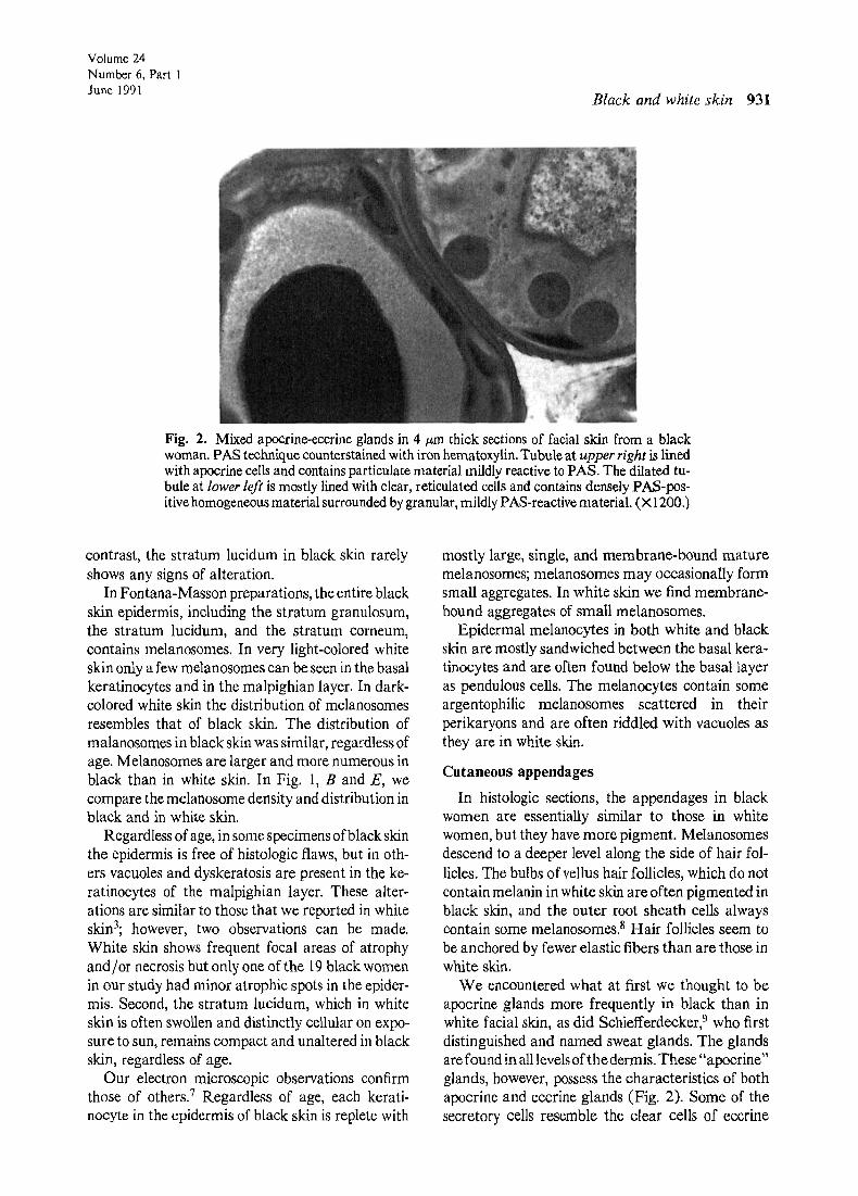
The papillary dermis of black skin has more crisply outlined larger fibroblasts and macrophages, many of which have two or more nuclei (Fig. 4) than there are in white skin. In addition, multinucleated giant cells are common in black skin. Our ultra- structural examinations confirm and expand the histologic studies. The majority of the fibroblasts are hypertrophied and contain extensive rough endo- plasmic reticulum, Golgi bodies, and various vesi- cles. We have often seen fibroblasts closely associ- ated with basal keratinocytes.
The melanosomes spilled into the dermis by the epidermal melanocytes are phagocytized by mac- rophages in both white and black skin. The melano- phages in black skin always contain membrane- bound complexes of melanosomes, as they often do in white skin. Melanophages are usually more numerous and larger in black than in white skin. Whereas the papillary dermis of black and white skin contains both small and large melanophages,
the large ones are found almost exclusively in the upper part of the papillary dermis close to the epi- dermis. Melanophages become progressively smaller in the deeper dermis and some small melanosome- containing macrophages can be seen almost any- where in the dermis of black skin. Multinucleated giant cells may contain some melanosomes but these cells do not appear to be principally involved in the phagocytosis of melanosomes.
Perivascular and periappendageal infiltration is encountered as often in black as it is in white skin. 15 There also appears to be an equal number of mast cells in black and white skin, and no difference is ev- ident in the staining properties of mast cell granules. The mast cell microvilli lie close to fibroblasts 16 and melanophages. The mast cells in black skin have dense and relatively homogeneous granules, which in our preparations do not have swirling or "fingerprint" patterns. However, many of the gran- ules have a dense core.
The colloidal iron technique shows larger ex- panses of extracellular blue-staining amorphous material in both papiUary and reticular layers of the dermis in black than in white skin.
White skin always has more elastic fibers in the dermis than does black skin, even in persons in their 20s. Furthermore, the elaunin fibers in black skin do not form the characteristic candelabra-like struc- tures found in faces of young white persons' skin (Fig. 1, C and F), but they are disposed parallel, or at an angle, to the epidermis. Long and short, branching, fine oxytalan fibers rise at intervals from the apparently haphazardly distributed elaunin fi- bers. We have also found oxytalan fibers in the skin of 50-year-old black subjects, in contrast with the skin of white persons of this age in which such fibers are usually lacking. In the facial skin of white sub- jects oxytalan fibers abound only in their 20s and early 30s but become rarified and disappear in sub- jects 40 years or older.
We saw no bona fide elastosis in any of our spec- imens of black skin. In the older subjects (50 years), there appeared to be an increase in the number and thickness of elastic fibers separating the collagenous fiber layers in the reticular dermis. Elastic fibers, which are single-stranded in younger subjects, look like braids in the 50-year-old ones. The amount and distribution of elastic fibers in the skin of a 45-year- old light-skinned black woman resemble those in white skin. Even so, we found in this specimen only small foci of incipient elastic fibrolysis in the elastic
934 Montagna and Carlisle
Journal of the American Academy of
Dermatology
Fig. 4. A and B. Dermis of skin of a 32-year-old black woman. A, There are many cells and a lymph vessel (Lie) at left. Note multinucleated cells. (Hematoxylin-Lee stain; X500.) B, Binucleated and multinucleated cells in dermis. (Hematoxylin-Lee stain; •
fibers that anchor the hair follicles in that specimen. The elastic fibers in the facial skin of the black
women in our study stain differently with the hema- toxylin and Lee stain than those in white skin. In photodamaged white skin only the elastic fibers in the papillary dermis (including elaunin and oxytalan fibers and the elastotic material), and those in the reticular dermis stain pink; the wide, ribbonlike fibers in the recesses of the intermediate dermis stain lilac or deep blue. 3 In black skin all dermal elastic fibers stain pink as they do in the sun-protected skin of white persons. The elastic fibers in the skin of a light-colored 45-year-old black woman stain similar to those in white skin.
DISCUSSION
All specimens of facial skin used in this study were obtained during November from black and white women in Tucson, Arizona. This study was con- ducted specifically to verify whether or not there are substantive morphologic differences between black and white skin in the age group of 20 to 50 years and whether there are alterations in black skin that could be attributed to sunlight exposure. We uncovered many interesting details in the morphology of black skin that we report herein (Table I).
Whereas most of the older white women in our study (45 to 50 years old) had wrinkles beside the lateral canthi of the eyes (crow's feet) and on the
Volume 24 Number 6, Part 1 June 1991 Black and white skin 935
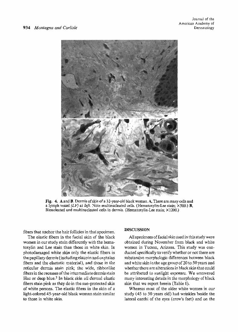
Table I. Structural differences between black and white skin with transmission electron microscopy and light microscopy
Epidermal melanosomes
Hair follicle melanosomes
Elaunin and oxytalan fibers
Elastic fibers
Fiber fragments
Fibroblasts
Macrophages (melanophages)
Multinucleated giant cells
Blood vessels (superficial)
Lymphatic vessels Glycoprotein molecules in
matrix Mixed eccrine, apocrine
glands
Large, membrane-bound, and distributed primarily singly inside keratinocytes; numerous even in stratum eorneum
In outer root sheath and in bulb of vellus hairs
No subepidermal candelabra formations
Stain pink (Hematoxylin-Lee) throughout dermis; no overt evidence of elastosis
Many, easily seen in perivascular areas; composed of collagen fibrils and glycoprotein
Numerous and large; many binucleated and multinucleated ones
Numerous; large in papillary dermis, many binucleated and multinucleated; smaller ones elsewhere; melanosomes singly and in complexes.
Numerous
Numerous, mostly dilated
Numerous, dilated Numerous throughout
dermis Many
Small and distributed primarily in aggregates; fewer than in black skin; in dark subjects similar in distribution to black skin; in lighter subjects absent from upper layers of epidermis
Neither in outer root sheath nor in bulb of vellus hair follicles
Candelabra formations
Variable amounts of moderate to extensive elastosis; fibers in mid dermis always stain lilac to dark blue (Hematoxylin-Lee) after sun exposure
Occasional
Variable numbers; some binucleated and multinucleated ones
Many; large in papillary dermis, small elsewhere; some binucleated and multinucleated; melanosomes in complexes
Rare
Sparse to moderate numbers
Moderate numbers, dilated Variable
Variable
corners of the mouth, none of the 19 black women had obvious wrinkles.
We were unable to confirm the observation of Weigand et a1.17 that the stratum corneum of black epidermis is more compact than that of white facial epidermis and that it has more cell layers. In another large sample of white skin, the layers of the stratum corneum show many modes of stacking. Our obser- vations agree more with those of Thomson, t8 who found no differences in the thickness of the stratum corneum of "Europeans" and "Africans."
Most authors who studied the histologic and ultrastructural features of black skin agree that there are no structural differences other than the "packaging" and the number of the melanosomes. 19
Even in the pigmentation, however, things are not as exact as was originally believed; for example, Toda et al. 2~ found that whereas the darker skinned blacks possess "non-aggregated" large melanosomes, the lighter colored ones have both nonaggregated large melanosomes and aggregated smaller ones. The size of the melanosomes is a determinant factor for the way in which they are distributed. We have con- firmed these observations in the present study. In sections stained with the Masson-Fontana tech- nique, the melanosomes in both black and white skin are dispersed throughout the epidermis, even inside the corneocytes.
After long-term exposure to sunlight the epider- mis of black skin shows only minor changes corn-
936 Montagna and Carlisle
Journal of the American Academy of
Dermatology
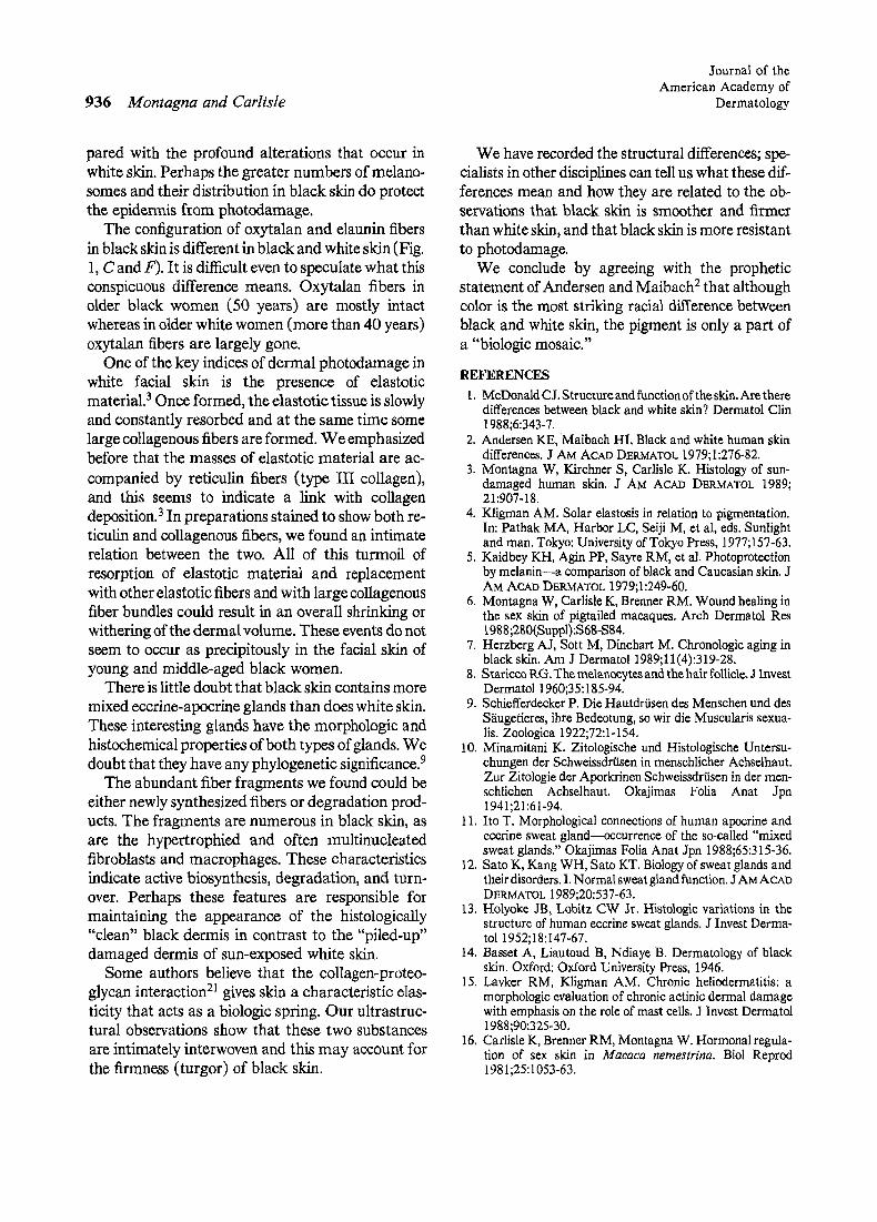
pared with the profound alterations that occur in white skin. Perhaps the greater numbers of melano- somes and their distribution in black skin do protect the epidermis from photodamage.
The configuration of oxytalan and elaunin fibers in black skin is different in black and white skin (Fig. 1, C and F). It is difficult even to speculate what this conspicuous difference means. Oxytalan fibers in older black women (50 years) are mostly intact whereas in older white women (more than 40 years) oxytalan fibers are largely gone.
One of the key indices of dermal photedamage in white facial skin is the presence of elastotic material. 3 Once formed, the elastotic tissue is slowly and constantly resorbed and at the same time some large collagenous fibers are formed. W e emphasized before that the masses of elastotic material are ac- companied by reticulin fibers ( type III collagen), and this seems to indicate a link with collagen deposition.3 In preparations stained to show both re- ticulin and collagenous fibers, we found an intimate relation between the two. All of this turmoil of resorption of elastotic material and replacement with other elastotic fibers and with large collagenous fiber bundles could result in an overall shrinking or withering of the dermal volume. These events do not seem to occur as precipitously in the facial skin of young and middle-aged black women.
There is little doubt that black skin contains more mixed eccrine-apocrine glands than does white skin. These interesting glands have the morphologic and histochemical properties of both types of glands. We doubt that they have any phylogenetic significance. 9
The abundant fiber fragments we found could be either newly synthesized fibers or degradation prod- ucts. The fragments are numerous in black skin, as are the hypertrophied and often multinucleated fibroblasts and macrophages. These characteristics indicate active biosynthesis, degradation, and turn- over. Perhaps these features are responsible for maintaining the appearance of the histologically "clean" black dermis in contrast to the "piled-up" damaged dermis of sun-exposed white skin.
Some authors believe that the collagen-proteo- glycan interaction 21 gives skin a characteristic elas- ticity that acts as a biologic spring. Our ultrastruc- rural observations show that these two substances are intimately interwoven and this may account for the firmness (turgor) of black skin.
We have recorded the structural differences; spe- cialists in other disciplines can tell us what these dif- ferences mean and how they are related to the ob- servations that black skin is smoother and firmer than white skin, and that black skin is more resistant to photodamage.
We conclude by agreeing with the prophetic statement of Andersen and Maibach 2 that although color is the most striking racial difference between black and white skin, the pigment is only a part of a "biologic mosaic."
REFERENCES 1. McDonald CJ. Structure and function of the skin. Are there
differences between black and white skin? Dermatol Clin 1988;6:343-7.
2. Andersen KE, Maibach HI. Black and white human skin differences. J AM ACAD DERMATOL 1979;1:276-82.
3. Montagna W, Kirchner S, Carlisle K. Histology of sun- damaged human skin. J AM ACAD DERMATOL 1989; 21:907-18.
4. Kligman AM. Solar elastosis in relation to pigmentation. In: Pathak MA, Harbor LC, Seiji M, et al, eds. Sunlight and man. Tokyo: University of Tokyo Press, 1977;157-63.
5. Kaidbey KH, Agin PP, Sayre RM, et al. Photoproteetion by melanin--a comparison of black and Caucasian skin. J AM ACAD DI~RMATOL 1979;1:249-60.
6. Montagna W, Car/isle K, Brenner RM. Wound healing in the sex skin of pigtailed macaques. Arch Dermatol Res 1988;280(Suppl):S68-S84.
7. Herzberg A J, Sott M, Dinehart M. Chronologic aging in black skin. Am J Dermatol 1989;11(4):319-28.
8. Starlcco RG. The melanoeytes and the hair follicle. J Invest Dermatol 1960;35:185-94.
9. Schiefferdecker P. Die Hautdrfisen des Menschen und des S~iugetieres, ihre Bedeotung, so wir die Muscularis sexua- lis. Zoologica 1922;72:1-154.
10. Minamitani K. Zitologische und Histologische Untersu- chungen der Schweissdriisen in menschlicher Achselhaut. Zur Zitologie der Aporkrinen Schweissdrtisen in der men- schlichen Achselhaut. Okajimas Folia Anat Jpn 1941;21:61-94.
11. Ito T. Morphological connections of human apocrine and ecerine sweat gland----occurrence of the so-called "mixed sweat glands." Okajimas Folia Anat Jpn 1988;65:315-36.
12. Sato K, Kang WH, Sato KT. Biology of sweat glands and their disorders. I. Normal sweat gland function. J AM ACAD DERMATOL 1989;20:537-63.
13. Holyoke JB, Lobitz CW Jr. Histologic variations in the structure of human eccrine sweat glands. J Invest Derma- tol 1952;18:147-67.
14. Basset A, Liautoud B, Ndiaye B. Dermatology of black skin. Oxford: Oxford University Press, 1946.
15. Lavker RM, Kligman AM. Chronic heliodermatitis: a morphologic evaluation of chronic actinic dermal damage with emphasis on the role of mast ceils. J Invest Dermatol 1988;90:325-30.
16. Carlisle K, Brenner RM, Montagna W. Hormonal regula- tion of sex skin in Macaca nemestrina. BiN Reprod 1981;25:1053-63.

Volume 24 Number 6, Part 1 June 1991 Black and white skin
17. Weigand DA, Haygood C, Gaylor J. Cell layers and den- sity of Negro and Caucasian stratum corneum. J Invest Dermatol 1974;62:563-8.
18. Thomson DA. Relative efficiency of pigment and horny layer-thickness in protecting the skin of Europeans and Africans against solar ultraviolet radiation. J Physiol (Lon- don) 1955;127:236-46.
19. Szabb G, Gerald AB, Pathak MA, et al. Racial differences in the fate of melanosomes in human epidermis. Nature (London) 1969;222:181-2.
20. Toda K, Pathak MA, Parrish JA, et al. Alteration of racial differences in melanosome distribution in human epidermis after exposure to ultraviolet light. Nature (London) 1972;236:143-5.
21. Montes GS, Bezerra MSF, Junqueira LCU. Collagen dis- tribution in tissues. In: Ruggeri A, Motta PM, eds. Ultra- structure of the connective tissue matrix. Boston: Martinus Nijhoff Publishers, 1984:79-81.
I II ! III I I
Classic Kaposi's sarcoma: T-lymphocyte subsets, T4/T8 ratio, and NK cell activity Rachel Friedman-Birnbaum, MD, a Sara Weltfriend, MD, a and Shimon Pollack, M D b Haifa, Israel
Lymphocyte function as expressed by T-lymphocyte subsets and natural killer cell activity was evaluated in a group of Israeli patients with classic Kaposi's sarcoma. T-cell subsets were examined in 28 patients, 14 with lesions limited to the lower extremities and 14 with diffuse cutaneous or systemic involvement. CD4 and CD8 lymphocytes and CD4/CD8 ratio were in the normal range in all patients, and mean values of the entire group were similar to a con- trol group. However, CD8 was in the upper limits of the normal range in some patients with diffuse cutaneous or systemic involvement. This factor led to a significantly decreased CD4/ CD8 ratio in that group as compared with the group of patients with the disease limited to the lower extremities. Mean values of natural killer cell activity in three effector/target cell ratios were significantly decreased in 13 of the patients with Kaposi's sarcoma with lesions limited to the lower extremities and in all of the patients with normal T-cell subsets and CD4/CD8 ratios. (J AM ACAD DERMATOL 1991;24:937-40.)
From its first description in 1872,1 Kaposi's sar- coma (KS) in its classic form has been considered a rare disease with an increased incidence in Jews of Ashkenazi and Mediterranean ancestry and in per- sons of Italian origin. 2 The classic form of KS is usually chronic) Since the 1960s an increasing fre- quency of KS has been recognized in patients who are receiving immunosuppressive treatment and since 1979 the endemic form has emerged in patients with the acquired immunodeficiency syndrome
From the Departments of Dermatology a and Clinical Immunology, b Rambam Medical Center, Technion-Israel Institute of Technology.
Accepted for publication Dec. 27, 1990. Reprint requests: R. Friedman-Birnbaum, MD, Dept. of Dermatology,
Rarnbam Medical Center, Haifa 35254, Israel. 16/1/27608
(AIDS). 4 In immunodeficiency conditions, the dis- ease usually runs a much more aggressive course compared with that seen in the classic form.
Little is known about the factors involved in the development, the course, and the prognosis of KS. Its more aggressive course in immunosuppressed trans- plant recipients, the regression of the process with discontinuation ofimmunosuppressive therapy, 5 and the aggressive course in AIDS-related KS suggest a role for host defense mechanisms in the development and progression of this process. In patients with AIDS-related KS, various abnormalities of the im- mune functions are constant findings. Among them are abnorrhal CD4/CD8 ratios 6, 7 and a decreased NK cell function. 8 The course of the disease in AIDS-related KS is mainly influenced by the rate at which the patient's immune system deteriorates, 9 In
937