the arabidopsis floral homeotic proteins apetala3 … · the arabidopsis floral homeotic proteins...
TRANSCRIPT

The Arabidopsis Floral Homeotic Proteins APETALA3 andPISTILLATA Negatively Regulate the BANQUO GenesImplicated in Light Signaling W
Chloe D. Mara,1 Tengbo Huang, and Vivian F. Irish2
Department of Molecular, Cellular, and Developmental Biology, Yale University, New Haven, Connecticut 06520-8104
The Arabidopsis thaliana MADS box transcription factors APETALA3 (AP3) and PISTILLATA (PI) heterodimerize and are
required to specify petal identity, yet many details of how this regulatory process is effected are unclear. We have identified
three related genes, BHLH136/BANQUO1 (BNQ1), BHLH134/BANQUO2 (BNQ2), and BHLH161/BANQUO3 (BNQ3), as being
directly and negatively regulated by AP3 and PI in petals. BNQ1, BNQ2, and BNQ3 encode products belonging to a family of
atypical non-DNA binding basic helix-loop-helix (bHLH) proteins that heterodimerize with and negatively regulate bHLH
transcription factors. We show that bnq3 mutants have pale-green sepals and carpels and decreased chlorophyll levels,
suggesting that BNQ3 has a role in regulating light responses. The ap3 bnq3 double mutant displays pale second-whorl
organs, supporting the hypothesis that BNQ3 is downstream of AP3. Consistent with a role in light response, we show that
the BNQ gene products regulate the function of HFR1 (for LONG HYPOCOTYL IN FAR-RED1), which encodes a bHLH protein
that regulates photomorphogenesis through modulating phytochrome and cryptochrome signaling. The BNQ genes also
are required for appropriate regulation of flowering time. Our results suggest that petal identity is specified in part through
downregulation of BNQ-dependent photomorphogenic and developmental signaling pathways.
INTRODUCTION
Arabidopsis thaliana petals are simple laminar floral organs; the
white petal blades lack chlorophyll and, at maturity, possess
characteristic conical epidermal cells on their adaxial surfaces
(Irish, 2008). The appropriate specification of petal identity de-
pends on the activities of two MADS box–containing transcrip-
tion factors, APETALA3 (AP3) and PISTILLATA (PI) (Bowman
et al., 1989; Jack et al., 1992; Goto andMeyerowitz, 1994; Krizek
and Meyerowitz, 1996). The expression patterns of AP3 and PI
depend upon the activity of the meristem identity genes LEAFY
and AP1, which encode transcription factors, in conjunction with
the activity ofUNUSUALFLORALORGANS, encoding an F-box–
containing protein (Ng and Yanofsky, 2001; Lamb et al., 2002;
Chae et al., 2008). In turn, AP3 and PI form an obligate hetero-
dimer necessary for DNA binding, nuclear localization, and
consequent transcriptional regulation of suites of downstream
target genes (McGonigle et al., 1996; Riechmann et al., 1996b;
Yang et al., 2003b). The AP3/PI heterodimer appears to act
together with other MADS box proteins, presumably as compo-
nents of higher-order protein complexes, to regulate organ-
specific differentiation processes (Pelaz et al., 2000; Honma and
Goto, 2001). In petals, these processes appear to depend on the
combined activities of AP3 and PI in conjunction with the AP1
and SEPALLATA (SEP) MADS box proteins (Pelaz et al., 2000,
2001; Honma and Goto, 2001).
AP3 andPI are expressed throughout the petal until late stages
of petal differentiation, and continued and ubiquitous expression
of these organ identity genes appears to be required throughout
the petal for normal development to ensue (Bowman et al., 1989;
Goto and Meyerowitz, 1994; Jack et al., 1994; Jenik and Irish,
2001). These observations imply that AP3 and PI act to regulate
spatially and temporally distinct subsets of target genes during
petal development and differentiation. Although many putative
AP3 and PI targets have been identified through microarray and
other analyses (Sablowski and Meyerowitz, 1998; Zik and Irish,
2003;Wellmer et al., 2004; Sundstrom et al., 2006; Alves-Ferreira
et al., 2007; Peiffer et al., 2008), only a few such target genes
have been experimentally verified. These include AP3 and PI
themselves, which are autoregulated in a positive feedback loop
(Goto and Meyerowitz, 1994; Jack et al., 1994). Regulation of
AP3 is direct, since the AP3/PI heterodimer can bind toCArGbox
consensus sequences in the AP3 promoter and AP3 can be
activated by AP3 and PI without de novo protein synthesis (Jack
et al., 1992; Goto and Meyerowitz, 1994; Hill et al., 1998; Tilly
et al., 1998; Sundstrom et al., 2006). PI regulation, however, is
likely to be indirect since de novo protein synthesis is required for
AP3/PI-dependent regulation of PI (Honma and Goto, 2000).
NAP (for NAC-LIKE, ACTIVATED BY AP3/PI), a gene that is
involved in the transition between the cell division and cell
expansion phases during the growth of petals and stamens
and in promoting senescence, has also been shown to be
positively regulated by AP3 and PI (Sablowski and Meyerowitz,
1 Current address: Department of Cell Biology and Molecular Genetics,University of Maryland, College Park, MD 204742.2 Address correspondence to [email protected] author responsible for distribution of materials integral to thefindings presented in this article in accordance with the policy describedin the Instructions for Authors (www.plantcell.org) is: Vivian F. Irish([email protected]).WOnline version contains Web-only data.www.plantcell.org/cgi/doi/10.1105/tpc.109.065946
The Plant Cell, Vol. 22: 690–702, March 2010, www.plantcell.org ã 2010 American Society of Plant Biologists

1998; Guo and Gan, 2006). In addition to positive regulation, AP3
and PI have also been shown to act as negative regulators of
AP1, suggesting that complex feedback regulatory mechanisms
are important for appropriate specification of organ identity
(Sundstrom et al., 2006). AP3 and PI also negatively regulate
the expression of two GATA-type zinc finger genes, GNC
(for GATA, NITRATE-INDUCIBLE, CARBON-METABOLISM-
INVOLVED) and GNC-LIKE (GNL), which in turn regulate a
suite of sugar response and nitrate metabolism genes, pro-
viding a link between organ development and nutrient sensing
(Mara and Irish, 2008).
In this study, we have identified three closely related genes,
BANQUO1 (BNQ1), BNQ2, and BNQ3, that are negatively reg-
ulated by AP3 and PI.BNQ1,BNQ2, andBNQ3 encode products
that are members of the basic helix-loop-helix (bHLH) family of
transcriptional regulators. The Arabidopsis genome encodes
>160 bHLH proteins that have been variously grouped into 15
to 25 subfamilies (Heim et al., 2003; Toledo-Ortiz et al., 2003; Li
et al., 2006; Pires and Dolan, 2010). These proteins are charac-
terized by a basic domain of;15 to 17 amino acids responsible
for DNA binding and an HLH region required for dimerization and
consisting of two amphipathic a-helices joined by a loop of
variable length (Ellenberger et al., 1994; Jones, 2004). However,
the BNQ1, BNQ2, and BNQ3 gene products have fewer basic
amino acids in their basic domains and lack the amino acids (Glu-
13/Arg-17) that are critical for DNA binding of canonical bHLH
proteins (Toledo-Ortiz et al., 2003). This class of non-basic bHLH
proteins, as exemplified by the human Id-1 (Inhibitor of DNA
binding-1) protein, is thought to act as dominant-negative reg-
ulators of DNA binding bHLH transcription factors (Massari and
Murre, 2000; Norton, 2000).
Here, we show, using loss-of-function and gain-of-function
approaches, thatBNQ1,BNQ2, andBNQ3 have a variety of roles
in regulating light responses as well as developmental transi-
tions. These roles include the ability to heterodimerize with, and
regulate the activity of, the bHLH protein HFR1 (for LONG
HYPOCOTYL IN FAR-RED LIGHT1) that is a critical regulator of
light signaling and shade avoidance (Fairchild et al., 2000; Soh
et al., 2000; Duek and Fankhauser, 2003; Sessa et al., 2005;
Zhang et al., 2008; Hornitschek et al., 2009). Together, our data
support a model whereby AP3 and PI influence petal morpho-
genesis in part through the negative regulation of a family of
atypical bHLH proteins that in turn modulate the activity of a
number of other signaling pathways, providing amechanistic link
between developmental and physiological responses.
RESULTS
The BNQ Genes Are Targets of AP3 and PI
To identify genes directly regulated by AP3/PI, we previously
conducted a genome-wide screen using the Affymetrix ATH1
GeneChip array to identify genes whose expression was altered
in response to steroid-inducible activation of AP3. We used 35S:
AP3-GR 35S:PI ap3-3 transgenic plants that constitutively ex-
pressPI aswell as constitutively express a steroid-inducible form
of AP3 in an ap3-3mutant background. Prior to dexamethasone
(dex) induction, these transgenic plants show an ap3-3 pheno-
type. After induction, these plants display a rescue of the ap3-3
mutant phenotype, as well as partial homeotic conversions of
sepals to petals and carpels to stamens, reflecting the combined
ectopic expression of AP3 and PI (Sablowski and Meyerowitz,
1998). Application of dex to 35S:AP3-GR 35S:PI ap3-3 plants
results in transcriptional upregulation of direct targets of AP3/PI
within 4 to 6 h of treatment (Sundstrom et al., 2006; Mara and
Irish, 2008).
Previously, we used this transgenic line to conduct microarray
experiments (Mara and Irish, 2008). One hundred putative AP3/
PI targets, genes whose expression profiles changed in a sta-
tistically significant manner after 4 h of dex treatment, were
identified (Mara and Irish, 2008) and included BNQ1. Previously
known as BHLH136, BNQ1 encodes one of;33 predicted non-
DNA binding bHLH proteins in the Arabidopsis genome; these
proteins are thought to inhibit the function of DNA binding bHLH
transcription factors through heterodimerization (Fairman et al.,
1993; Bailey et al., 2003; Heim et al., 2003; Toledo-Ortiz et al.,
2003; Li et al., 2006). The microarray data indicated that BNQ1 is
downregulated 2.1-fold after induction of AP3 activity, suggest-
ing that BNQ1 is negatively regulated by AP3 and PI. RT-PCR
data corroborate the microarray data, indicating that BNQ1
expression decreases significantly 4 h after dex treatment of
35S:AP3-GR, 35S:PI, ap3-3 transgenic plants and increases in
ap3-3 and pi-1 mutant flowers compared with the wild type
(Figure 1).
BNQ1 encodes a member of a small subfamily of six atypical
bHLH proteins (Figures 2A and 2B) that together form a strongly
supported subclade within the larger bHLH family (see Supple-
mental Figure 1 online). Included in this subclade are BNQ2,
BNQ3 (previously called BHLH134 and BHLH161, respectively),
At3g28857, KIDARI, and BHLH135. This subfamily of bHLH
proteins shows considerable conservation of the HLH protein
interaction domain but do not possess the stereotypical basic
amino acids of DNA binding bHLH proteins (Figure 2C).
We tested if these other members of this bHLH subfamily were
also targets of AP3 and PI. We found that BNQ2 and BNQ3
expression levels decreased rapidly, within 1 h, after dex treat-
ment of 35S:AP3-GR, 35S:PI, ap3-3 transgenic plants (Figures
1B and 1C). Consistent with this, BNQ2 and BNQ3 expression
increased in ap3-3 and pi-1 mutant plants compared with the
wild type (Figures 1A and 1D). BNQ2 and BNQ3 were not
recovered in our microarray screen due to fact that BNQ3 was
not represented on the array, and BNQ2 was listed as below the
threshold of detection. Thus, we focused our subsequent anal-
yses onBNQ1,BNQ2, andBNQ3 that are all negatively regulated
by AP3 and PI. Furthermore, the downregulation of the tran-
scription of all three genes occurs rapidly in response to induc-
tion of AP3 activity (Figure 1B), suggesting that the AP3/PI
heterodimer may be binding directly to the promoters of each of
these bHLH genes.
BNQGenesAreNegativelyRegulatedbyAP3andPI inPetals
Digital gene expression analyses using the Arabidopsis eFP
browser, a tool for visualizing publicly available microarray data
sets (Winter et al., 2007), indicated that BNQ1 and BNQ2 are
BANQUO genes are targets of AP3/PI 691

expressed at low but detectable levels in most plant tissues and
have substantially overlapping expression patterns based on
analyses of ATH1 microarray data sets (Schmid et al., 2005) (see
Supplemental Figures 2A and 2B online). Similar digital profiling
of BNQ3 expression has been performed using whole-genome
tiling arrays (Laubinger et al., 2008) and indicates that BNQ3 is
also expressed in most plant tissues at low but detectable levels
(see Supplemental Figure 2C online).
To examine further the mechanisms by which AP3 and PI
regulate BNQ gene expression, we used in situ hybridizations to
characterize the patterns ofBNQ1,BNQ2, andBNQ3 expression
in floral tissues. BNQ1 expression is detectable in the sepals of
wild-type flowers at stage 5 (Figure 3B). Prior to stage 5, BNQ1
transcripts cannot be detected in the flowers, although expres-
sion is strong in cauline leaves (Figures 3A and 3D). Sepal
expression continues throughout floral development until stage
12 (Figures 3B to 3F). Weak expression is also detectable in the
anthers at later stages (Figure 3F). BNQ3 expression overlaps
considerably with that of BNQ1, although BNQ3 is expressed
more broadly in flowers. BNQ3 is expressed ubiquitously
throughout stage 4 floral organ primordia (Figure 3M). From
stage 5 onward, BNQ3 is expressed most strongly in the sepals
with some expression detectable in the inner whorls (Figures 3N
and 3O). In late stages, BNQ3 is also strongly expressed in
anthers and carpels (Figures 3P to 3R). By contrast, BNQ2
is expressed weakly throughout the inner whorls of stage 4
wild-type flowers (see Supplemental Figure 3 online). Weak
Figure 1. BNQ1, BNQ2, and BNQ3 Are Targets of AP3/PI.
(A) Relative expression levels of BNQ1, BNQ2, and BNQ3 and other
atypical bHLH family members assayed by RT-PCR in 0 and 4 h dex- and
mock-treated 35S:AP3-GR 35S:PI ap3-3 flowers and in wild-type
(Landsberg erecta [Ler]), ap3-3, and pi-1 mutant flowers. ACTIN expres-
sion was used as a control.
(B) Time course of relative expression of atypical bHLH genes by RT-
PCR in dex-treated 35S:AP3-GR 35S:PI ap3-3 flowers. Dex was applied
at time 0 and tissues collected for analysis at times indicated.
(C) Relative expression levels of BNQ1, BNQ2, and BNQ3 and family
members by RT-PCR in 0 and 4 h dex- and mock-treated flowers.
Average expression levels from three biological replicates were normal-
ized to ACTIN with 0 h scaled to 1. Standard deviations are shown.
(D) Relative expression levels of BNQ1, BNQ2, and BNQ3 and family
members by RT-PCR in the linear range in wild-type (Ler), ap3-3, and
pi-1 mutant flowers. Average expression levels from three biological
replicates were normalized to ACTIN with the wild type scaled to 1.
Standard deviations are shown.
Figure 2. Gene Structure and Amino Acid Sequences of BNQ Family
Members.
(A) Neighbor-joining analysis of the BNQ subclade (see Supplemental
Figure 1 online for complete analysis). Bootstrap values of 1000 repli-
cates are shown.
(B) Gene structure of BNQ1. Black boxes represent the two exons.
(C) Alignment of amino acid sequences of BNQ related family members.
The HLH domain is indicated by black and gray boxes.
692 The Plant Cell

expression of BNQ2 persists in the stamen and carpel primordia
until stage 8, but by stage 9, expression is no longer detectable
(see Supplemental Figure 3 online).
To test whether AP3 and PI restrict the spatial domains of
BNQ1 and BNQ3 expression, we examined their expression
patterns in ap3-3 and pi-1 mutant flowers. In stages 6 to 8 of
ap3-3 and pi-1 flowers, BNQ1 expression is observed in the first
whorl of sepals (Figures 3G and 3I). In stage 12 ap3-3 and pi-1
flowers, BNQ1 expression is also found in the second-whorl
organs (Figures 3H and 3J). To determine if BNQ1 expression is
position dependent or tissue specific, we monitored its expres-
sion in ag-1 mutant flowers in which stamens are transformed
into petals and the fourth whorl differentiates into a new flower
consisting only of sepals and petals (Bowman et al., 1989). We
found thatBNQ1 is expressed in each whorl of sepals regardless
of position (Figures 3Kand3L). Similarly, in ap3-3 andpi-1mutant
flowers,BNQ3 is expressed in the first whorl, and by stage 12, its
expression domain expands into the second whorl of sepals,
indicating that AP3 and PI repress BNQ3 in the second whorl
(Figures 3S to 3V). This repression is tissue specific and not whorl
specific, since in ag-1mutant flowers,BNQ3 is expressed both in
first-whorl sepals and in ectopic fourth-whorl sepals (Figures 3W
and 3X). Thus, these data indicate that AP3 and PI repress the
expression of both BNQ1 and BNQ3 in developing petals.
To determine if the AP3/PI heterodimer binds to the promoters
of theBNQ genes,weperformed chromatin immunoprecipitation
(ChIP) assays. The AP3/PI heterodimer has been shown to bind
to a 10-bp conserved DNA region called the CArG box [CC(A/
T)6GG] (Schwarz-Sommer et al., 1992; Riechmann et al., 1996a;
Hill et al., 1998; Tilly et al., 1998). Allowing for a 1-bp mismatch,
we identified a number of CArG-like boxes present in the pro-
moter regions of BNQ1, BNQ2, and BNQ3 and tested if PI can
bind to these sequences (Figure 4). We extracted nuclei from
wild-type and 35S:PI-HA epitope-tagged transgenic plants and
immunoprecipitated with either a-HA antibody or normal mouse
serum. Immunoprecipitated DNA from three independent bio-
logical replicates was used in ChIP-PCR reactions with primers
designed around each CArG-like box to monitor enrichment
(Figure 4A). As a positive control, we confirmed binding of PI to
CArG3, an autoregulatory region in the AP3 promoter (Hill et al.,
1998). No enrichment was detected in the negative controls, PI
(an indirect target of AP3/PI; Honma and Goto, 2000) or AST101
(a root-specific gene; Takahashi et al., 2000) (Figures 4B and 4C).
We could detect an enrichment of a 250-bp fragment in the
Figure 3. In Situ Expression Analyses of BNQ Family Members.
(A) to (F) Expression (indicated by purple color) of BNQ1 in wild-type (Ler) flowers at stage 4 (A), stage 5 (B), stage 6 (C), stage 7 (D), stage 8 (E), and
late-stage (F) flowers.
(G) to (L) Expression of BNQ1 in various mutant backgrounds: in approximately stage 8 (G) and stage 12 (H) ap3-3 mutant flowers, in approximately
stage 6 (I) and stage 8 (J) pi-1 mutant flowers, and in approximately stage 8 (K) and stage 12 (L) ag-1 mutant flowers.
(M) to (R) Expression of BNQ3 in wild-type flowers at stage 4 (M), stage 5 (N), stage 6 (O), stage 7 (P), stage 8 (Q), and late-stage (R) flowers.
(S) to (X) Expression of BNQ3 in various mutant backgrounds: in approximately stage 6 (S) and stage 12 (T) ap3-3 mutant flowers, in approximately
stage 6 (U) and stage 12 (V) pi-1 mutant flowers, and in approximately stage 8 (W) and stage 12 (X) ag-1 mutant flowers.
BANQUO genes are targets of AP3/PI 693

BNQ1promoter region that contains theCArG-like box in 35S:PI-
HA extracts precipitated with a-HA antibodies compared with
controls (Figure 4C).We also detected an enrichment of a 169-bp
region spanning CArG box 2 present in theBNQ2 promoter and a
slight enrichment of a 216-bp region containing CArG box 2
present in the BNQ3 promoter (Figure 4C). However, we could
not detect any enrichment of any of the other CArG-like boxes
present in the promoters of BNQ2 and BNQ3 (Figure 4C). Thus,
BNQ1, BNQ2, and BNQ3 appear to be direct targets of PI,
presumably through binding of the AP3/PI heterodimer to aCArG
box sequence in the promoters of each of these genes.
Roles of BNQ Genes in Chlorophyll Accumulation and
Floral Induction
A T-DNA insertional mutation in the second predicted helix of the
BNQ3 coding region was obtained from the SALK collection
(Alonso et al., 2003) and backcrossed four times to remove
exogenous lesions. Homozygous bnq3 (SALK 098881) mutants
have undetectable levels of transcripts, suggesting that the
mutation is a complete loss-of-function allele (Figure 7A).
The sepals and carpels of homozygous bnq3mutant plants are
pale yelloworwhite,while the inflorescence stemsandsiliquesare
purple (Figure 5). The floral organs appear to be morphologically
normal but are somewhat smaller than wild-type organs (Figures
5C to 5F). To test the genetic relationship of AP3 and BNQ3, we
generated ap3-3 bnq3 double mutant plants. While ap3-3 single
mutant plants display green sepaloid organs, the ap3-3 bnq3
double mutant flowers have pale yellowish second-whorl organs,
consistent with negative regulation of BNQ3 by AP3 (Figure 6).
Consistent with the pale phenotype of bnq3mutants, we found
that flowers from such plants had decreased levels of chlorophyll
compared with wild-type flowers (Figure 7B). We also detected
lower amounts of chlorophyll in the cauline leaves, stems, and
siliques of the bnq3mutant plants (Figure 7B). Complementation
tests in which homozygous bnq3 plants were transformed with a
35S:BNQ3 construct produced normal flowers with green sepals
(see Supplemental Figure 4 online), demonstrating that the
T-DNA insertion in BNQ3 is responsible for the pale phenotype.
We used RNA interference (RNAi) to knock down BNQ1 and
BNQ2 expression.We generated 23BNQ1RNAi lines to see if we
could detect a loss-of-function phenotype; there were no obvi-
ous phenotypes in any of these lines (Figure 7A; data not shown).
Twenty-one BNQ2 RNAi transgenic lines were generated, and
none of the BNQ2 RNAi lines showed an obvious mutant phe-
notype (Figure 7A; data not shown). We assayed the relative
expression of BNQ1 (or 2) in the corresponding RNAi lines
Figure 4. AP3/PI Proteins Are Associated with Promoter Elements of the
BNQ1, BNQ2, and BNQ3 Genes.
(A) CArG boxes in the promoters of BNQ1, BNQ2, and BNQ3. Black
triangles indicate the position of the CArG box, and the black boxes
indicate the regions amplified by the primers used for ChIP-PCR.
Sequences shown below each schematic indicate the sequence present
in the specific CArG box indicated.
(B) ChIP-PCR for each of the regions indicated in (A), as well as for CArG
2 in the AP3 promoter and control promoter regions of the PI and AST101
genes. Nuclear extracts from 35S:PI-HA and wild-type (Ler) plants were
immunoprecipitated with anti-HA antibody (HA) or normal serum (N).
(C) Relative enrichment levels in the BNQ1, BNQ2, BNQ3, AP3, PI, and
AST101 promoters. Black bars indicate the enrichment fold change
based on three replicates that were normalized to wild-type samples
immunoprecipitated with normal serum scaled to 1. Standard deviations
are shown.
694 The Plant Cell

compared with the expression in the wild type normalized to
actin and used lines with a significant reduction in BNQ gene
expression for further experiments (22% of wild-type levels for
BNQ1i and 19% of wild-type levels for BNQ2i). To determine if
the BNQ genes act redundantly, we generated triple BNQ1i
BNQ2i bnq3 knockdown combinations. However, the triple
mutants showed no enhancement of the bnq3 chlorophyll-
deficient mutant phenotype or any other obvious morphological
phenotypes (Figure 7B). These results suggest that BNQ3 has a
nonredundant role in chlorophyll deposition and that functions of
the other BNQ family members may be obscured by redundancy
with other, as yet uncharacterized, atypical bHLH proteins.
We also observed that bnq3mutant plants flower later than the
wild type (Figure 7C). On the other hand, transgenic plants
containing the 35S:BNQ3 construct resulted in bolting and
flowering considerably earlier than the wild type (Figures 8B
and 8D). Similarly, constitutive expression of either BNQ1 or
BNQ2 under control of the 35S promoter also resulted in early
flowering (Figures 8B and 8D). Despite early flowering, these
BNQ1, BNQ2, and BNQ3 overexpression lines showed no ob-
vious defects in floral morphology. Thus, in addition to BNQ3,
BNQ1, and BNQ2 may also normally function to promote the
transition to flowering.
BNQ Genes Act as Regulators of Light Signaling
The 35S:BNQ1, 35S:BNQ2, and 35S:BNQ3 lines also displayed
phenotypes that suggested that these bHLH genes have a role in
light signal transduction. Transgenic seedlings constitutively
expressing each of the BNQ genes had elongated hypocotyls
compared with the wild type when grown under white light
(Figures 8A to 8C). These phenotypes were predominantly red
light dependent (Figure 8C), indicating that this response is likely
to be mediated to a large extent by phytochromes A and B
(Smith, 2000; Tepperman et al., 2004).
In Arabidopsis seedlings, both the cryptochromes, which
absorb blue/UV-A light, and the phytochromes, which sense
red/far-red light, are necessary for normal hypocotyl develop-
ment (Briggs and Olney, 2001; Wang and Deng, 2004). HFR1
encodes a bHLH protein that is required for both phytochrome-
and cryptochrome-dependent light signal transduction in
seedlings and during shade avoidance responses (Fairchild
et al., 2000; Duek and Fankhauser, 2003; Sessa et al., 2005;
Hornitschek et al., 2009). To investigate the potential relationship
between theBNQ andHFR1 gene products, we performed yeast
two-hybrid analyses. We found that BNQ1, BNQ2, and BNQ3
each were capable of physically interacting with HFR1 (Table 1).
In addition to HFR1, several other bHLH proteins have been
implicated in light signal transduction in Arabidopsis, including
PHYTOCHROME INTERACTING FACTOR 3-LIKE1 (PIL1), PIL5,
PHYTOCHROME INTERACTING FACTOR3 (PIF3), and PIF4
(Huq and Quail, 2002; Kim et al., 2003; Toledo-Ortiz et al.,
2003; Huq et al., 2004; Oh et al., 2004; Castillon et al., 2007). We
also examined whether BNQ1, BNQ2, and BNQ3 could interact
in yeast two-hybrid assays with PIL1, PIL5, PIF3, or PIF4 (Table
1). No such interactions could be detected, suggesting that
BNQ1, BNQ2, and BNQ3 specifically interact with the bHLH
protein HFR1 to modulate light signaling.
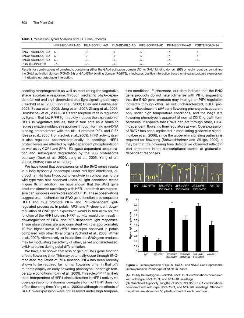
To test further this possibility, we examined whether over-
expression of the BNQ proteins could repress HFR1 function in
vivo. We constructed 35S:BNQ; 35S:HFR1 doubly transgenic
plants (i.e., 35S:BNQ1 35S:HFR1, 35S: BNQ2 35S:HFR1, and
35S:BNQ3 35S:HFR1) and measured the resulting hypocotyl
lengths. The 35S:HFR1 plants have slightly shorter hypocotyls
compared with the wild type, while 35S:BNQ1, 35S:BNQ2, or
35S:BNQ3 plants have elongated hypocotyls (Figures 9A and
9B). We found that the doubly transgenic plants had hypocotyls
that were similar in length to 35S:BNQ1, 35S:BNQ2, or 35S:
BNQ3 plants and longer than 35S:HFR1 or wild-type plants
(Figures 9A and B). This suppression of the 35S:HFR1 hypocotyl
phenotype indicates that the BNQ proteins can interact with
HFR1 in vivo, presumably by heterodimerizing with, and repres-
sing, HFR1 activity. Furthermore, these data suggest that the
BNQ gene products interact with other, presumably as yet
uncharacterized, bHLH proteins to regulate hypocotyl growth
since the 35S:BNQ35S:HFR seedlings display longer hypocotyls
than do hfr1 mutants.
DISCUSSION
AP3andPINegativelyRegulate theExpressionof aFamilyof
bHLHGenes
In this study, we identified BNQ1, BNQ2, and BNQ3 as genes
that are negatively regulated by AP3 and PI. We demonstrated
that, in the absence of AP3 or PI activity, BNQ1 and BNQ3
become ectopically expressed in the second whorl. These
Figure 5. Mutational Analysis of BNQ3.
(A) Wild-type (Columbia [Col]) flower buds.
(B) bnq3 mutant flower buds.
(C) Sepals from wild-type (left) and bnq3 mutant (right) flowers.
(D) Petals from wild-type (left) and bnq3 mutant (right) flowers.
(E) Stamens from wild-type (left) and bnq3 mutant (right) flowers.
(F) Carpels from wild-type (left) and bnq3 mutant (right) flowers.
BANQUO genes are targets of AP3/PI 695

observations suggest that the AP3/PI heterodimeric transcrip-
tion factor may have important roles in repressing a family of
atypical bHLH proteins to ensure the proper development of
petals in the second whorl. Furthermore, this repression appears
to be direct, based on both the rapid repression of expression of
all three BNQ genes upon the activation of AP3 function, as well
as through ChIP assays that demonstrate that PI can directly
associate with CArG boxes present in the BNQ1, BNQ2, and
BNQ3 promoters.
Although AP3 and PI act to specify both petal and stamen
identity, the AP3- and PI-dependent negative regulation of the
BNQ genes appears to be petal specific, in thatBNQ1 andBNQ3
expression can be observed in late stage stamens. Presumably,
this is due to the fact that the AP3/PI heterodimer can form
higher-order transcriptional complexes with the AP1 and SEP
MADS box proteins to direct petal development specifically
(Pelaz et al., 2000, 2001; Honma and Goto, 2001). Furthermore,
negative regulation by the AP3/PI heterodimer may be achieved
via formation of transcription complexes containing corepres-
sors or through affecting histone modifications of target gene
promoter regions. Although potential AP3/PI corepressors have
not yet been identified, SEUSS and LEUNIG encode compo-
nents of a corepressor complex that acts in conjunction with
other floral organ identity MADS box gene products to regulate
petal development (Franks et al., 2006; Sridhar et al., 2006).
Furthermore, the recruitment of a histone deacetylase complex
is necessary for the MADS domain protein, AGL15, to act as a
transcriptional repressor in vivo (Hill et al., 2008). Since relatively
few petal-specific genes have been identified despite multiple
microarray analyses (Zik and Irish, 2003; Wellmer et al., 2004),
repression as opposed to activation of specific genes by the
AP3/PI heterodimer may be a predominant means of petal
specification.
Our data suggest that AP3 and PI spatially repress BNQ1,
BNQ2, and BNQ3 expression in whorl 2 to promote the correct
specification of petals. Furthermore, bnq3 mutants have de-
creased chlorophyll levels associated with a pale-white pheno-
type, indicating a requirement for BNQ3 to promote chlorophyll
accumulation. Thus, it appears that AP3 and PI may function in
part to abrogate chlorophyll accumulation in the petals to ensure
the proper differentiation of these organs. Nonetheless, it is clear
that expression of BNQ3 is not sufficient for chlorophyll accu-
mulation since ectopic expression of BNQ3 does not result in
greening of the petals. Furthermore, ectopic expression of all
three BNQ genes does not result in petal greening. These results
imply that in ap3 or pi mutants, other factors in addition to the
BNQ gene products are necessary to promote chlorophyll ac-
cumulation in the second whorl. Such factors could potentially
correspond to the products of other genes that have been shown
to be negatively regulated by AP3 and PI, such as GNC or GNL,
which have also been shown to be required for chlorophyll
biosynthesis (Bi et al., 2005; Mara and Irish, 2008).
Based on the loss of chlorophyll autofluorescence, rediffer-
entiation of green chloroplasts to colorless leucoplasts in
developing petals occurs around stage 12 ofArabidopsis flower
development (Pyke and Page, 1998). Our observations that
BNQ gene expression expands into the second whorl only at
later stages of flower development in ap3-3 and pi-1 flowers is
consistent with this observation and suggest that AP3 and PI
have specific regulatory roles at later stages of petal organo-
genesis. The NAC family transcription factor NAP, a previously
identified direct target of AP3 and PI, has also been proposed to
act at later stages of petal differentiation (Sablowski and
Meyerowitz, 1998). Similarly, the floral organ identity gene
AGAMOUS has been shown to regulate directly SPOROCYTE-
LESS, encoding a putative transcription factor required for
Figure 6. Floral Phenotypes of ap3-3 bnq3 Double Mutant.
(A) to (E) Individual flowers of Ler (A) and Col (B) with green sepals; by contrast, bnq3 (C) displays pale-yellow sepals. The ap3-3 mutant (D) has green
first and second whorl organs compared with the ap3-3 bnq3 double mutant (E), which is pale.
(F) to (J) The second-whorl organs of Ler (F) and Col (G) have morphologically normal petals. The bnq3 (H) petals also appear normally shaped. The
second-whorl organs of ap3-3 (I) are sepaloid and green, while the ap3-3 bnq3 (J) second-whorl organ is pale.
696 The Plant Cell

microsporogenesis during late stages of flower differentiation
(Ito et al., 2004). Together, these observations underscore the
idea that the floral organ identity genes regulate different
aspects of organogenesis throughout development by regulat-
ing the expression of subsidiary transcription factors required
for specific differentiation processes.
The BNQ Genes Regulate a Variety of Physiological and
Developmental Responses
The BNQ genes that we have identified belong to an atypical
class of bHLH proteins that lack the basic DNA binding domain
and the critical amino acids for DNA binding (Toledo-Ortiz et al.,
2003). As a consequence, such proteins can form inactive
heterodimers with other bHLH proteins, thus modulating activity
of their binding partners (Norton, 2000).
At least one BNQ interacting partner appears to be the
product ofHFR1, which itself encodes an atypical bHLH protein
(Fairchild et al., 2000). HFR1 has been shown to be required for
Figure 7. Analysis of BNQ Loss-of-Function Phenotypes.
(A) Relative expression of BNQ1, BNQ2, and BNQ3 in wild-type (Col)
flowers and from corresponding RNAi or mutant flowers. ACTIN levels
are shown in comparison.
(B) Chlorophyll levels in different tissues from wild-type (Col), bnq3, and
bnq triple mutant (bnq3 BNQ1-RNAi BNQ2-RNAi) plants. Standard
deviations using three replicates are shown.
(C) Days to bolting and to appearance of first flower are shown for wild-
type (Col), bnq3, BNQ1-RNAi, BNQ2-RNAi, and triple mutant plants.
Standard deviations from 25 plants scored for each genotype are shown.
Figure 8. Overexpression Analyses of BNQ Genes.
(A) Representative 35S:BNQ1, 35S:BNQ2, and 35S:BNQ3 seedlings
have elongated hypocotyls compared with the wild type (Col) (two plants
each).
(B) BNQ1, BNQ2, and BNQ3 expression is increased in the correspond-
ing overexpression lines; ACTIN amplification is shown as a control.
(C) Quantified hypocotyl lengths of 35S:BNQ1, 35S:BNQ2, and 35S:
BNQ3 seedlings under white, blue, red, and far-red light conditions.
Standard deviations are shown for 50 plants scored of each genotype.
(D) 35S:BNQ1, 35S:BNQ2, and 35S:BNQ3 plants flower earlier than the
wild type; days to forming a 1-cm bolt and to first flower opening are
shown. Standard deviations are shown for 50 plants scored of each
genotype.
BANQUO genes are targets of AP3/PI 697
seedling morphogenesis as well as modulating the vegetative
shade avoidance response, through mediating phyA-depen-
dent far-red and cry1-dependent blue light signaling pathways
(Fairchild et al., 2000; Soh et al., 2000; Duek and Fankhauser,
2003; Sessa et al., 2005; Jang et al., 2007; Zhang et al., 2008;
Hornitschek et al., 2009). HFR1 transcription itself is regulated
by light, in that low R/FR light rapidly induces the expression of
HFR1 in vegetative tissues, that in turn acts as a brake to
repress shade avoidance responses through forming non-DNA
binding heterodimers with the bHLH proteins PIF4 and PIF5
(Sessa et al., 2005; Hornitschek et al., 2009). HFR1 activity itself
is also regulated posttranscriptionally. In seedlings, HFR1
protein levels are affected by light-dependent phosphorylation
as well as by COP1 and SPA1 E3 ligase-dependent ubiquitina-
tion and subsequent degradation by the 26S proteasome
pathway (Duek et al., 2004; Jang et al., 2005; Yang et al.,
2005a, 2005b; Park et al., 2008).
We have found that overexpression of the BNQ genes results
in a long hypocotyl phenotype under red light conditions, al-
though a mild long hypocotyl phenotype in comparison to the
wild type was also observed under all light conditions tested
(Figure 8). In addition, we have shown that the BNQ gene
products dimerize specifically with HFR1, and their overexpres-
sion can suppress overexpression of HFR1. These observations
suggest one mechanism for BNQ gene function is to sequester
HFR1 and thus promote PIF4- and PIF5-dependent light-
regulated processes. In petals, AP3- and PI-dependent down-
regulation of BNQ gene expression would in turn allow for the
function of the HFR1 protein; HFR1 activity would then result in
downregulation of PIF4- and PIF5-dependent light responses.
These observations are also consistent with the approximately
10-fold higher levels of HFR1 transcripts observed in petals
compared with other floral organs (Schmid et al., 2005; Winter
et al., 2007). Alternatively, or in addition, the BNQ gene products
may be modulating the activity of other, as yet uncharacterized,
bHLH proteins during petal differentiation.
We have also shown that loss or gain of BNQ gene function
affects flowering time. Thismay potentially occur throughBNQ-
mediated regulation of PIF4 function. PIF4 has been recently
shown to be required for normal flowering time, in that pif4
mutants display an early flowering phenotype under high tem-
perature conditions (Koini et al., 2009). This role of PIF4 is likely
to be independent of HFR1 since alteration in HFR1 activity via
overexpression of a dominant-negative form of HFR1 does not
affect flowering time (Yang et al., 2003a), although the effects of
HFR1 overexpression were not assayed under high tempera-
ture conditions. Furthermore, our data indicate that the BNQ
gene products do not heterodimerize with PIF4, suggesting
that the BNQ gene products may impinge on PIF4 regulation
indirectly through other, as yet uncharacterized, bHLH pro-
teins. Also, since the pif4 early flowering phenotype is apparent
only under high temperature conditions, and the bnq1 late
flowering phenotype is apparent at normal (228C) growth tem-
peratures, it appears that BNQ1 can act through other, PIF4-
independent, flowering time regulators as well. Overexpression
of BNQ1 has been implicated in modulating gibberellin signal-
ing (Lee et al., 2006); since the gibberellin signaling pathway is
required for flowering (Schwechheimer and Willige, 2009), it
may be that the flowering time defects we observed reflect in
part alterations in the transcriptional control of gibberellin-
dependent responses.
Table 1. Yeast Two-Hybrid Analyses of bHLH Gene Products
HFR1-BD/HFR1-AD PIL1-BD/PIL1-AD PIL5-BD/PIL5-AD PIF3-BD/PIF3-AD PIF4-BD/PIF4-AD PGBT9/PGAD424
BNQ1-AD/BNQ1-BD +/+ �/� �/� +/� +/� �/�BNQ2-AD/BNQ2-BD +/� �/� �/� +/� +/� �/�BNQ3-AD/BNQ3-BD +/� �/� �/� +/� +/� �/�PGAD424/PGBT9 �/� �/� �/� +/� +/�Results for combinations of constructs containing either the GAL4 activation domain (AD) or GAL4 binding domain (BD) or vector controls containing
the GAL4 activation domain (PGAD424) or GAL4DNA binding domain (PGBT9). + Indicates positive interaction based on b-galactosidase expression;
� indicates no detectable interaction.
Figure 9. Overexpression of BNQ1, BNQ2, and BNQ3 Can Repress the
Overexpression Phenotype of HFR1 in Planta.
(A) Doubly heterozygous 35S:BNQ 35S:HFR1 combinations compared
with wild-type, 35S:HFR1, and hfr1-201 seedlings.
(B) Quantified hypocotyl lengths of 35S:BNQ 35S:HFR1 combinations
compared with wild-type, 35S:HFR1, and hfr1-201 seedlings. Standard
deviations are shown for 50 plants scored of each genotype.
698 The Plant Cell

Overexpression of the BNQ genes also has been shown
recently to abrogate the vegetative phenotype of the brassino-
steroid receptor mutant bri1-301, suggesting that the BNQ gene
products also participate in regulating brassinosteroid signaling
through modulating the activity of regulatory bHLH proteins
(Wang et al., 2009).
Together, these results imply that the BNQ genes are likely
acting to modulate multiple exogenous and endogenous cues
that are necessary for different aspects of plant development.
Given that only a handful of the >160 bHLH genes in Arabidopsis
have been functionally characterized, our analyses of the BNQ
genes have uncovered a new role for this subfamily of atypical
bHLH proteins. The BNQ genes have both overlapping and
independent functions in different parts of the plant. These
functions include regulation of light signal transduction, chloro-
phyll accumulation, and the regulation of the floral transition. We
suggest that the BNQ gene products can regulate these pro-
cesses in part through modulating the activity of the HFR1
atypical bHLH protein. This regulatory mechanism of modulating
the activity of non-DNA binding HLH protein activity through
sequestering such proteins through heterodimerization with
other non-DNA binding HLH proteins may represent a general
strategy to titrate the regulatory roles of such proteins. The
downregulation of BNQ gene expression specifically in petals
through the action of AP3 and PI alters this homeostasis,
promoting petal differentiation. Thus, a cascade of transcrip-
tional and postranscriptional negative regulation appears to be
one mechanism by which petal morphogenesis is achieved.
METHODS
Plant Material and Growth Conditions
Arabidopsis thaliana plants were grown on 12:3:1 mix of vermiculite:soil:
sand at 228C under 16-h-light/8-h-dark conditions. The mutant lines
(ap3-3, pi-1, and ag-1) and transgenic lines (ap3-3 35S:PI 35S:AP3-GR,
35S:AP3 and 35S:PI-HA) are in the Ler background. Mutant lines were
obtained from the ABRC (Ohio State University). The ap3-3 35S:PI 35S:
AP3-GR line was a gift from Robert W. M. Sablowski (John Innes Centre,
Norwich, UK) (Sablowski and Meyerowitz, 1998). The 35S: PI-HA line
was a gift from Naomi Nakayama (Yale University, New Haven, CT)
(Sundstrom et al., 2006).
For dex induction, plants were treated with dex (0.015% silwet, 0.1%
ethanol, and 5 mM dex) or mock (0.015% silwet and 0.1% ethanol),
collected at various timepoints, and snap frozen in liquid nitrogen. Total
RNA was extracted using Trizol (GibcoBRL) according to the manufac-
turer’s instructions, purified using the Qiagen Rneasy kit, and used in
subsequent analyses.
The BNQ3 insertional mutation was identified as a SALK T-DNA
insertion line (SALK 098881) and is in the Col background; the mutant
was backcrossed four times to remove exogenous mutations. To gener-
ate the ap3-3 bnq3 double mutant, ap3-3 was crossed with bnq3
homozygous plants. Following self-pollination of the F1 plants, pheno-
typic characterization of the double mutants was performed in three F2
families, with 10 ap3-3 bnq3 double mutants identified out of 144 plants.
Representative flowers were dissected and photographed.
For light treatments, seedlings were placed in a standard continuous-
white-light growth chamber at 228C. After 12 h of incubation, seedlings
were transferred to various light conditions in growth chambers (Percival
Scientific) with fluence rates of 111.0 mmol m–2 s–1 for far-red light, 150.6
mmol m–2 s–1 for white light, 172.6 mmol m–2 s–1 for red light, and 8.1 mmol
m–2 s–1 for blue light.
ChIP and Expression Analyses
For ChIP assays, nuclear extracts were prepared using MC, M1, M2, and
M3 buffers as described by Ito et al. (1997) and immunoprecipitations
performed as byMara and Irish (2008). Fractions corresponding to bound
and unbound DNA samples were used as templates for ChIP-PCR using
primers flanking the CArG-like boxes identified in the promoter regions of
each gene using the RSA tools software (rsat.ulb.ac.be/rsat/) (Thomas-
Chollier et al., 2008). For ChIP and expression analyses, DNA band
intensities from ethidium bromide–stained gels were measured with
Molecular Imaging Software 4.0 (Eastman KODAK Company). This soft-
ware used a Gaussian Curve method with background subtraction to
normalize the DNA band intensity, which significantly increases the
accuracy of measuring extremely strong or weak DNA bands. This
software also directly converts band intensity into DNA content (mg) in a
specific DNA band by comparing it to a standard (DNA size marker),
which also ensures that bands were measured in the linear range for DNA
quantification in a gel image. For RT-PCR, cycle numbers were varied
between 20 and 35, and the linear phase of amplification was determined
empirically for each reaction by assessing band intensities for different
cycle numbers. Gene-specific primers used to analyze expression are
listed in Supplemental Table 1 online. The primers used for ChIP-PCR are
listed in Supplemental Table 2 online.
In Situ Hybridization
In situ probes were generated by PCR amplification of cDNA using gene-
specific primers containing T7 RNA polymerase binding sites. Procedures
for probe preparation, sectioning, in situ prehybridization, hybridization,
and detection were performed as described previously (Zondlo and Irish,
1999; Mara and Irish, 2008). The primers used for in situ probes are listed in
Supplemental Table 3 online.
Transgenic and SALK Line Analyses
All RNAi lines were generated in the Col background using the Gateway
system (Invitrogen) vectors pK7GWIWG2 (II) and pH7GWIWG2 (II) and
protocol. Gene-specific primers used to amplify;300-bp coding regions
of BNQ1 and BNQ2 to insert into the vectors are listed in Supplemental
Table 4 online. Reduction in expression of RNAi transgenic lines was
assessed by comparing the relative expression of BNQ1 (or 2) in the
corresponding RNAi line to the expression in the wild type (scaled to 1)
and normalized to actin, using three replicates. To construct the plasmids
used in genetic complementation and overexpression analysis, the full-
length cDNA of each BNQ gene was amplified with the corresponding
primers listed in Supplemental Table 5 online. The PCR fragment was
digested with BamHI and XbaI and inserted into the binary vector p235
(Jenik and Irish, 2001), a derivative of pPZP221 (Hajdukiewicz et al., 1994)
containing the 35S promoter from the cauliflower mosaic virus. These
constructs were introduced into Agrobacterium tumefaciens and trans-
formed into Arabidopsis plants by floral dip. For genetic complementa-
tion, both p235 and 35S:BNQ3 were transformed into bnq3 mutants,
whereas Ler plants were used for the overexpression analysis of BNQ
genes. Transgenic plants were selected on half-strength Murashige and
Skoog plates containing gentamicin.
Homozygous SALK lines were identified by PCR genotyping for the
presence of the T-DNA insertion. RNA was extracted from homozygous
plants using Trizol (GibcoBRL) according to the manufacturer’s instruc-
tions. RT-PCR analysis, as described above, was used to check for
abolishment of the transcript. The primers used to verify the BNQ3
insertion (SALK 098881) are listed in Supplemental Table 4 online.
BANQUO genes are targets of AP3/PI 699

Chlorophyll Extraction and Measurement
Tissue was snap frozen in liquid nitrogen and then chlorophyll was
extracted using 80% acetone as previously described (Lichtenthaler,
1987). Absorbance was measured at 645 and 657 nm, and chlorophyll
content was calculated using (20.23A645 + 8.023A657)/g freshweight.
Yeast Two-Hybrid Assay
The Matchmaker GAL4 two-hybrid system (Clontech) was used for the
yeast two-hybrid assay. The pGBT9 and pGAD424 vectors were used for
making DNA binding domain and activation domain fusion constructs.
The open reading frames of each gene were amplified from cDNA using
gene-specific primers and inserted into pGBT9 and pGAD424 vectors
using EcoRI and/or BamHI restriction sites. Gene-specific primers are
listed in Supplemental Table 6 online. b-Galactosidase liquid assays, for
five colonies per construct, were performed using the protocol available
at http://www.fhcrc.org/science/labs/gottschling/yeast/Bgal.html where
U ¼ 10003 ½ðOD420Þ2 ð1:753OD550Þ�=½ðtimeÞ3 ðvolÞ3OD600�:
Phylogenetic Analyses
A total of 154 Arabidopsis bHLH sequences were identified based on
BLAST searches and previously published data (Toledo-Ortiz et al.,
2003). Full-length amino acid sequences were aligned using ClustalW
(Thompson et al., 1994) with default values and refined by hand using
MacClade 4.03 (Maddison and Maddison, 2000). The alignment is
presented as Supplemental Data Set 1 online. Unrooted trees were gen-
erated using the neighbor-joining algorithm as implemented in PAUP 4.0
(Swofford, 2000) with default values. Bootstrap values for resolved nodes
were derived from 1000 replicates using the neighbor-joining algorithm.
Accession Numbers
Sequence data from this article can be found in the Arabidopsis
Genome Initiative or GenBank/EMBL databases under the following ac-
cession numbers: AP3, At3g54340; PI, At5g20240; AST101, At4g08620;
BNQ1 (BHLH136), At5g39860; BNQ2 (BHLH134), At5g15160; BNQ3
(BHLH161), At3g47710; KIDARI, At1g26945; and BHLH135, At1g74500.
Accession numbers for all other sequences used are shown in Sup-
plemental Figure 1 online.
Supplemental Data
The following materials are available in the online version of this article.
Supplemental Figure 1. Neighbor-Joining Analysis of 154 Arabidop-
sis bHLH Proteins.
Supplemental Figure 2. Digital Expression Profiling of BNQ1, BNQ2,
and BNQ3.
Supplemental Figure 3. In Situ Expression Analyses of BNQ2.
Supplemental Figure 4. Complementation Analysis of bnq3.
Supplemental Table 1. RT-PCR Primer Sequences.
Supplemental Table 2. ChIP-PCR Primer Sequences.
Supplemental Table 3. In Situ Probe Primer Sequences.
Supplemental Table 4. SALK Line and RNAi Line Primer Sequences.
Supplemental Table 5. Overexpression Line Primer Sequences.
Supplemental Table 6. Yeast Two-Hybrid Primer Sequences.
Supplemental Data Set 1. Text File of the Alignment Used for the
Phylogenetic Analysis Shown in Supplemental Figure 1.
ACKNOWLEDGMENTS
We thank Koen Geuten (Yale University) for help with phylogenetic
analyses and Xing Wang Deng (Yale University) for comments on the
manuscript. This work was supported by a grant from the National
Science Foundation (IOS-0817744) to V.F.I.
Received January 25, 2009; revised February 17, 2010; accepted
March 7, 2010; published March 19, 2010.
REFERENCES
Alonso, J.M., et al. (2003). Genome-wide insertional mutagenesis of
Arabidopsis thaliana. Science 301: 653–657.
Alves-Ferreira, M., Wellmer, F., Banhara, A., Kumar, V., Riechmann,
J.L., and Meyerowitz, E.M. (2007). Global expression profiling ap-
plied to the analysis of Arabidopsis stamen development. Plant
Physiol. 145: 747–762.
Bailey, P.C., Martin, C., Toledo-Ortiz, G., Quail, P.H., Huq, E., Heim,
M.A., Jakoby, M., Werber, M., and Weisshaar, B. (2003). Update on
the basic helix-loop-helix transcription factor gene family in Arabi-
dopsis thaliana. Plant Cell 15: 2497–2502.
Bi, Y.M., Zhang, Y., Signorelli, T., Zhao, R., Zhu, T., and Rothstein, S.
(2005). Genetic analysis of Arabidopsis GATA transcription factor
gene family reveals a nitrate-inducible member important for chloro-
phyll synthesis and glucose sensitivity. Plant J. 44: 680–692.
Bowman, J.L., Smyth, D.R., and Meyerowitz, E.M. (1989). Genes
directing flower development in Arabidopsis. Plant Cell 1: 37–52.
Briggs, W.R., and Olney, M.A. (2001). Photoreceptors in plant photo-
morphogenesis to date. Five phytochromes, two cryptochromes, one
phototropin, and one superchrome. Plant Physiol. 125: 85–88.
Castillon, A., Shen, H., and Huq, E. (2007). Phytochrome interacting
factors: Central players in phytochrome-mediated light signaling
networks. Trends Plant Sci. 12: 514–521.
Chae, E., Tan, Q.K., Hill, T.A., and Irish, V.F. (2008). An Arabidopsis
F-box protein acts as a transcriptional co-factor to regulate floral
development. Development 135: 1235–1245.
Duek, P.D., Elmer, M.V., van Oosten, V.R., and Fankhauser, C.
(2004). The degradation of HFR1, a putative bHLH class transcription
factor involved in light signaling, is regulated by phosphorylation and
requires COP1. Curr. Biol. 14: 2296–2301.
Duek, P.D., and Fankhauser, C. (2003). HFR1, a putative bHLH
transcription factor, mediates both phytochrome A and cryptochrome
signalling. Plant J. 34: 827–836.
Ellenberger, T., Fass, D., Arnaud, M., and Harrison, S.C. (1994).
Crystal structure of transcription factor E47: E-box recognition by a
basic region helix-loop-helix dimer. Genes Dev. 8: 970–980.
Fairchild, C.D., Schumaker, M.A., and Quail, P.H. (2000). HFR1
encodes an atypical bHLH protein that acts in phytochrome A signal
transduction. Genes Dev. 14: 2377–2391.
Fairman, R., Beran-Steed, R.K., Anthony-Cahill, S.J., Lear, J.D.,
Stafford III, W.F., DeGrado, W.F., Benfield, P.A., and Brenner, S.L.
(1993). Multiple oligomeric states regulate the DNA binding of helix-
loop-helix peptides. Proc. Natl. Acad. Sci. USA 90: 10429–10433.
Franks, R.G., Liu, Z., and Fischer, R.L. (2006). SEUSS and LEUNIG
regulate cell proliferation, vascular development and organ polarity in
Arabidopsis petals. Planta 224: 801–811.
Goto, K., and Meyerowitz, E.M. (1994). Function and regulation of the
Arabidopsis floral homeotic gene PISTILLATA. Genes Dev. 8: 1548–
1560.
Guo, Y., and Gan, S. (2006). AtNAP, a NAC family transcription factor,
has an important role in leaf senescence. Plant J. 46: 601–612.
700 The Plant Cell

Hajdukiewicz, P., Svab, Z., and Maliga, P. (1994). The small, versatile
pPZP family of Agrobacterium binary vectors for plant transformation.
Plant Mol. Biol. 25: 989–994.
Heim, M.A., Jakoby, M., Werber, M., Martin, C., Weisshaar, B., and
Bailey, P.C. (2003). The basic helix-loop-helix transcription factor
family in plants: A genome-wide study of protein structure and
functional diversity. Mol. Biol. Evol. 20: 735–747.
Hill, K., Wang, H., and Perry, S.E. (2008). A transcriptional repression
motif in the MADS factor AGL15 is involved in recruitment of histone
deacetylase complex components. Plant J. 53: 172–185.
Hill, T.A., Day, C.D., Zondlo, S.C., Thackeray, A.G., and Irish, V.F.
(1998). Discrete spatial and temporal cis-acting elements regulate
transcription of the Arabidopsis floral homeotic gene APETALA3.
Development 125: 1711–1721.
Honma, T., and Goto, K. (2000). The Arabidopsis floral homeotic gene
PISTILLATA is regulated by discrete cis-elements responsive to
induction and maintenance signals. Development 127: 2021–2030.
Honma, T., and Goto, K. (2001). Complexes of MADS-box proteins are
sufficient to convert leaves into floral organs. Nature 409: 469–471.
Hornitschek, P., Lorrain, S., Zoete, V., Michielin, O., and Fankhauser,
C. (2009). Inhibition of the shade avoidance response by formation of
non-DNA binding bHLH heterodimers. EMBO J. 28: 3893–3902.
Huq, E., Al-Sady, B., Hudson, M., Kim, C., Apel, K., and Quail, P.H.
(2004). Phytochrome-interacting factor 1 is a critical bHLH regulator of
chlorophyll biosynthesis. Science 305: 1937–1941.
Huq, E., and Quail, P.H. (2002). PIF4, a phytochrome-interacting bHLH
factor, functions as a negative regulator of phytochrome B signaling in
Arabidopsis. EMBO J. 21: 2441–2450.
Irish, V.F. (2008). The Arabidopsis petal: A model for plant organogen-
esis. Trends Plant Sci. 13: 430–436.
Ito, T., Takahashi, N., Shimura, Y., and Okada, K. (1997). A serine/
threonine protein kinase gene isolated by an in vivo binding procedure
using the Arabidopsis floral homeotic gene product, AGAMOUS. Plant
Cell Physiol. 38: 248–258.
Ito, T., Wellmer, F., Yu, H., Das, P., Ito, N., Alves-Ferreira, M.,
Riechmann, J.L., and Meyerowitz, E.M. (2004). The homeotic
protein AGAMOUS controls microsporogenesis by regulation of
SPOROCYTELESS. Nature 430: 356–360.
Jack, T., Brockman, L.L., and Meyerowitz, E.M. (1992). The homeotic
gene APETALA3 of Arabidopsis thaliana encodes a MADS box and is
expressed in petals and stamens. Cell 68: 683–697.
Jack, T., Fox, G.L., and Meyerowitz, E.M. (1994). Arabidopsis homeotic
gene APETALA3 ectopic expression: Transcriptional and posttrans-
criptional regulation determine floral organ identity. Cell 76: 703–716.
Jang, I.C., Yang, J.Y., Seo, H.S., and Chua, N.H. (2005). HFR1 is
targeted by COP1 E3 ligase for post-translational proteolysis during
phytochrome A signaling. Genes Dev. 19: 593–602.
Jang, I.C., Yang, S.W., Yang, J.Y., and Chua, N.H. (2007). Indepen-
dent and interdependent functions of LAF1 and HFR1 in phytochrome
A signaling. Genes Dev. 21: 2100–2111.
Jenik, P.D., and Irish, V.F. (2001). The Arabidopsis floral homeotic gene
APETALA3 differentially regulates intercellular signaling required for
petal and stamen development. Development 128: 13–23.
Jones, S. (2004). An overview of the basic helix-loop-helix proteins.
Genome Biol. 5: 226.
Kim, J., Yi, H., Choi, G., Shin, B., Song, P.S., and Choi, G. (2003).
Functional characterization of phytochrome interacting factor 3 in
phytochrome-mediated light signal transduction. Plant Cell 15: 2399–
2407.
Koini, M.A., Alvey, L., Allen, T., Tilley, C.A., Harberd, N.P., Whitelam,
G.C., and Franklin, K.A. (2009). High temperature-mediated adapta-
tions in plant architecture require the bHLH transcription factor PIF4.
Curr. Biol. 19: 408–413.
Krizek, B.A., and Meyerowitz, E.M. (1996). The Arabidopsis homeotic
genes APETALA3 and PISTILLATA are sufficient to provide the B
class organ identity function. Development 122: 11–22.
Lamb, R.S., Hill, T.A., Tan, Q.K., and Irish, V.F. (2002). Regulation of
APETALA3 floral homeotic gene expression by meristem identity
genes. Development 129: 2079–2086.
Laubinger, S., Zeller, G., Henz, S.R., Sachsenberg, T., Widmer, C.K.,
Naouar, N., Vuylsteke, M., Scholkopf, B., Ratsch, G., and Weigel,
D. (2008). At-TAX: A whole genome tiling array resource for develop-
mental expression analysis and transcript identification in Arabidopsis
thaliana. Genome Biol. 9: R112.
Lee, S., Lee, S., Yang, K.Y., Kim, Y.M., Park, S.Y., Kim, S.Y., and Soh,
M.S. (2006). Overexpression of PRE1 and its homologous genes
activates gibberellin-dependent responses in Arabidopsis thaliana.
Plant Cell Physiol. 47: 591–600.
Li, X., et al. (2006). Genome-wide analysis of basic/helix-loop-helix
transcription factor family in rice and Arabidopsis. Plant Physiol. 141:
1167–1184.
Lichtenthaler, H. (1987). Chlorophyll and carotenoids: pigments of the
photosynthetic membranes. Methods Enzymol. 148: 350–382.
Maddison, W.P., and Maddison, D.R. (2000). MacClade, Analysis of
Phylogeny and Character Evolution. (Sunderland, MA: Sinauer Asso-
ciates).
Mara, C.D., and Irish, V.F. (2008). Two GATA transcription factors are
downstream effectors of floral homeotic gene action in Arabidopsis.
Plant Physiol. 147: 707–718.
Massari, M.E., and Murre, C. (2000). Helix-loop-helix proteins: regu-
lators of transcription in eucaryotic organisms. Mol. Cell. Biol. 20:
429–440.
McGonigle, B., Bouhidel, K., and Irish, V.F. (1996). Nuclear localization
of the Arabidopsis APETALA3 and PISTILLATA homeotic gene
products depends on their simultaneous expression. Genes Dev.
10: 1812–1821.
Ng, M., and Yanofsky, M.F. (2001). Activation of the Arabidopsis B
class homeotic genes by APETALA1. Plant Cell 13: 739–753.
Norton, J.D. (2000). ID helix-loop-helix proteins in cell growth, differ-
entiation and tumorigenesis. J. Cell Sci. 113: 3897–3905.
Oh, E., Kim, J., Park, E., Kim, J.I., Kang, C., and Choi, G. (2004). PIL5,
a phytochrome-interacting basic helix-loop-helix protein, is a key
negative regulator of seed germination in Arabidopsis thaliana. Plant
Cell 16: 3045–3058.
Park, H.J., Ding, L., Dai, M., Lin, R., and Wang, H. (2008). Multisite
phosphorylation of Arabidopsis HFR1 by casein kinase II and a
plausible role in regulating its degradation rate. J. Biol. Chem. 283:
23264–23273.
Peiffer, J.A., Kaushik, S., Sakai, H., Arteaga-Vazquez, M., Sanchez-Leon,
N., Ghazal, H., Vielle-Calzada, J.P., and Meyers, B.C. (2008). A
spatial dissection of the Arabidopsis floral transcriptome by MPSS.
BMC Plant Biol. 8: 43.
Pelaz, S., Ditta, G.S., Baumann, E., Wisman, E., and Yanofsky, M.F.
(2000). B and C floral organ identity functions require SEPALLATA
MADS-box genes. Nature 405: 200–203.
Pelaz, S., Tapia-Lopez, R., Alvarez-Buylla, E.R., and Yanofsky, M.F.
(2001). Conversion of leaves into petals in Arabidopsis. Curr. Biol. 11:
182–184.
Pires, N., and Dolan, L. (2010). Origin and diversification of basic-helix-
loop-helix proteins in pants. Mol. Biol. Evol. 27: 862–874.
Pyke, K.A., and Page, A.M. (1998). Plastid ontogeny during petal
development in Arabidopsis. Plant Physiol. 116: 797–803.
Riechmann, J.L., Krizek, B.A., and Meyerowitz, E.M. (1996b). Dimer-
ization specificity of Arabidopsis MADS domain homeotic proteins
APETALA1, APETALA3, PISTILLATA, and AGAMOUS. Proc. Natl.
Acad. Sci. USA 93: 4793–4798.
BANQUO genes are targets of AP3/PI 701

Riechmann, J.L., Wang, M., and Meyerowitz, E.M. (1996a). DNA-
binding properties of Arabidopsis MADS domain homeotic proteins
APETALA1, APETALA3, PISTILLATA and AGAMOUS. Nucleic Acids
Res. 24: 3134–3141.
Sablowski, R.W.M., and Meyerowitz, E.M. (1998). A homolog of NO
APICAL MERISTEM is an immediate target of the floral homeotic
genes APETALA3/PISTILLATA. Cell 92: 93–103.
Schmid, M., Davison, T.S., Henz, S.R., Pape, U.J., Demar, M.,
Vingron, M., Scholkopf, B., Weigel, D., and Lohmann, J.U.
(2005). A gene expression map of Arabidopsis thaliana development.
Nat. Genet. 37: 501–506.
Schwarz-Sommer, Z., Hue, I., Huijser, P., Flor, P.J., Hansen, R.,
Tetens, F., Lonnig, W.-E., Saedler, H., and Sommer, H. (1992).
Characterization of the Antirrhinum floral homeotic MADS-box gene
deficiens: Evidence for DNA binding and autoregulation of its persistent
expression throughout flower development. EMBO J. 11: 251–263.
Schwechheimer, C., and Willige, B.C. (2009). Shedding light on
gibberellic acid signalling. Curr. Opin. Plant Biol. 12: 57–62.
Sessa, G., Carabelli, M., Sassi, M., Ciolfi, A., Possenti, M.,
Mittempergher, F., Becker, J., Morelli, G., and Ruberti, I. (2005). A
dynamic balance between gene activation and repression regulates
the shade avoidance response in Arabidopsis. Genes Dev. 19:
2811–2815.
Smith, H. (2000). Phytochromes and light signal perception by plants–
An emerging synthesis. Nature 407: 585–591.
Soh, M.S., Kim, Y.M., Han, S.J., and Song, P.S. (2000). REP1, a basic
helix-loop-helix protein, is required for a branch pathway of phyto-
chrome A signaling in Arabidopsis. Plant Cell 12: 2061–2074.
Sridhar, V.V., Surendrarao, A., and Liu, Z. (2006). APETALA1 and
SEPALLATA3 interact with SEUSS to mediate transcription repression
during flower development. Development 133: 3159–3166.
Sundstrom, J.F., Nakayama, N., Glimelius, K., and Irish, V.F. (2006).
Direct regulation of the floral homeotic APETALA1 gene by APETALA3
and PISTILLATA in Arabidopsis. Plant J. 46: 593–600.
Swofford, D.L. (2000). PAUP*: Phylogenetic Analysis Using Parsimony
(and Other Methods). (Sunderland, MA: Sinauer Associates).
Takahashi, H., Watanabe-Takahashi, A., Smith, F.W., Blake-Kalff,
M., Hawkesford, M.J., and Saito, K. (2000). The roles of three
functional sulphate transporters involved in uptake and translocation
of sulphate in Arabidopsis thaliana. Plant J. 23: 171–182.
Tepperman, J.M., Hudson, M.E., Khanna, R., Zhu, T., Chang, S.H.,
Wang, X., and Quail, P.H. (2004). Expression profiling of phyB mutant
demonstrates substantial contribution of other phytochromes to red-
light-regulated gene expression during seedling de-etiolation. Plant J.
38: 725–739.
Thomas-Chollier, M., Sand, O., Turatsinze, J.V., Janky, R., Defrance,
M., Vervisch, E., Brohee, S., and van Helden, J. (2008). RSAT:
Regulatory sequence analysis tools. Nucleic Acids Res. 36:
W119–127.
Thompson, J.D., Higgins, D.G., and Gibson, T.J. (1994). CLUSTAL W:
Improving the sensitivity of progressive multiple sequence alignment
through sequence weighting, position-specific gap penalties and
weight matrix choice. Nucleic Acids Res. 22: 4673–4680.
Tilly, J., Allen, D.W., and Jack, T. (1998). The CArG boxes in the
promoter of the Arabidopsis floral organ identity gene APETALA3
mediate diverse regulatory effects. Development 125: 1647–1657.
Toledo-Ortiz, G., Huq, E., and Quail, P.H. (2003). The Arabidopsis
basic/helix-loop-helix transcription factor family. Plant Cell 15:
1749–1770.
Wang, H., and Deng, X.-W. (2004). Phytochrome signaling mech-
anisms. In The Arabidopsis Book, C.R. Somerville and E.M.
Meyerowitz, eds (Rockville, MD: American Society of Plant Biologists),
doi/10.1199/tab.0074.1, http://www.aspb.org/publications/arabidopsis/.
Wang, H., Zhu, Y., Fujioka, S., Asami, T., and Li, J. (2009). Regulation
of Arabidopsis brassinosteroid signaling by atypical basic helix-loop-
helix proteins. Plant Cell 21: 3781–3791.
Wellmer, F., Riechmann, J.L., Alves-Ferreira, M., and Meyerowitz,
E.M. (2004). Genome-wide analysis of spatial gene expression in
Arabidopsis flowers. Plant Cell 16: 1314–1326.
Winter, D., Vinegar, B., Nahal, H., Ammar, R., Wilson, G.V., and
Provart, N.J. (2007). An “electronic fluorescent pictograph” browser
for exploring and analyzing large-scale biological data sets. PLoS One
2: e718.
Yang, J., Lin, R., Hoecker, U., Liu, B., Xu, L., and Wang, H. (2005a).
Repression of light signaling by Arabidopsis SPA1 involves post-
translational regulation of HFR1 protein accumulation. Plant J. 43:
131–141.
Yang, J., Lin, R., Sullivan, J., Hoecker, U., Liu, B., Xu, L., Deng, X.W.,
and Wang, H. (2005b). Light regulates COP1-mediated degradation
of HFR1, a transcription factor essential for light signaling in Arabi-
dopsis. Plant Cell 17: 804–821.
Yang, K.Y., Kim, Y.M., Lee, S., Song, P.S., and Soh, M.S. (2003a).
Overexpression of a mutant basic helix-loop-helix protein HFR1,
HFR1-deltaN105, activates a branch pathway of light signaling in
Arabidopsis. Plant Physiol. 133: 1630–1642.
Yang, Y., Fanning, L., and Jack, T. (2003b). The K domain mediates
heterodimerization of the Arabidopsis floral organ identity proteins,
APETALA3 and PISTILLATA. Plant J. 33: 47–59.
Zhang, X.N., Wu, Y., Tobias, J.W., Brunk, B.P., Deitzer, G.F., and Liu,
D. (2008). HFR1 is crucial for transcriptome regulation in the crypto-
chrome 1-mediated early response to blue light in Arabidopsis
thaliana. PLoS One 3: e3563.
Zik, M., and Irish, V.F. (2003). Global identification of target genes
regulated by APETALA3 and PISTILLATA floral homeotic gene action.
Plant Cell 15: 207–222.
Zondlo, S.C., and Irish, V.F. (1999). CYP78A5 encodes a cytochrome
P450 that marks the shoot apical meristem boundary in Arabidopsis.
Plant J. 19: 259–268.
702 The Plant Cell

DOI 10.1105/tpc.109.065946; originally published online March 19, 2010; 2010;22;690-702Plant Cell
Chloe D. Mara, Tengbo Huang and Vivian F. Irish Genes Implicated in Light SignalingBANQUO
Floral Homeotic Proteins APETALA3 and PISTILLATA Negatively Regulate the ArabidopsisThe
This information is current as of May 31, 2018
Supplemental Data /content/suppl/2010/03/15/tpc.109.065946.DC1.html
References /content/22/3/690.full.html#ref-list-1
This article cites 81 articles, 40 of which can be accessed free at:
Permissions https://www.copyright.com/ccc/openurl.do?sid=pd_hw1532298X&issn=1532298X&WT.mc_id=pd_hw1532298X
eTOCs http://www.plantcell.org/cgi/alerts/ctmain
Sign up for eTOCs at:
CiteTrack Alerts http://www.plantcell.org/cgi/alerts/ctmain
Sign up for CiteTrack Alerts at:
Subscription Information http://www.aspb.org/publications/subscriptions.cfm
is available at:Plant Physiology and The Plant CellSubscription Information for
ADVANCING THE SCIENCE OF PLANT BIOLOGY © American Society of Plant Biologists