the ant protein handbook
TRANSCRIPT

8/2/2019 The ant Protein Handbook
http://slidepdf.com/reader/full/the-ant-protein-handbook 1/112
18-1142-75
Edition AB
Protein Amplificationand Simple Purification
The Recombinant
Protein Handbook

8/2/2019 The ant Protein Handbook
http://slidepdf.com/reader/full/the-ant-protein-handbook 2/112
Antibody PurificationHandbook18-1037-46
The Recombinant Protein HandbookProtein Amplification and Simple Purification18-1142-75
Protein PurificationHandbook18-1132-29
Ion Exchange ChromatographyPrinciples and Methods18-1114-21
Affinity ChromatographyPrinciples and Methods18-1022-29
Hydrophobic Interaction ChromatographyPrinciples and Methods18-1020-90
Gel FiltrationPrinciples and Methods18-1022-18
Handbooksfrom Amersham Biosciences
Reversed Phase ChromatographyPrinciples and Methods18-1134-16
Expanded Bed AdsorptionPrinciples and Methods18-1124-26
Chromatofocusingwith Polybuffer and PBE50-01-022PB
Microcarrier cell culturePrinciples and Methods18-1140-62

8/2/2019 The ant Protein Handbook
http://slidepdf.com/reader/full/the-ant-protein-handbook 3/112
1
The Recombinant Protein Handbook
Protein Amplification
and Simple Purification

8/2/2019 The ant Protein Handbook
http://slidepdf.com/reader/full/the-ant-protein-handbook 4/112
2
Contents
Introduction ............................................................................................................. 5Symbols and abbreviations .............................................................................................................. 5
CHAPTER 1 .......... ............... ............... ................ ............... ............... ............... ......... 6Choice of host for protein amplification ......................................................................... 6
Choice of vectors ......................................................................................................... 7Vectors for non-fusion proteins ......................................................................................................... 7
Vectors for fusion proteins ............................................................................................................... 8
Choice of fusion tag ........................................................................................................................ 8
CHAPTER 2 .......... ............... ............... ................ ............... ............... ............... ......... 9Protein amplification ................................................................................................... 9Sample extraction .......................................................................................................................... 9
Troubleshooting protein amplification ............................................................................................... 9
CHAPTER 3 ............................................................................................................ 13GST fusion proteins ................................................................................................... 13Amplification ............................................................................................................................... 13
Purification .................................................................................................................................. 14
Detection of GST fusion proteins .................................................................................................... 21
Purification and detection troubleshooting ...................................................................................... 28
Tag removal by enzymatic cleavage ................................................................................................. 30
PreScission Protease cleavage and purification ................................................................................ 31
Thrombin cleavage and purification ................................................................................................ 35
Factor Xa cleavage and purification ................................................................................................ 37
Removal of thrombin, Factor Xa or other serine proteases ................................................................. 39
CHAPTER 4 ............................................................................................................ 41(His)
6fusion proteins ................................................................................................. 41
Amplification ............................................................................................................................... 41
Purification .................................................................................................................................. 41
Detection of (His)6
fusion proteins .................................................................................................. 53
Purification and detection troubleshooting ...................................................................................... 56
Tag removal by enzymatic cleavage ................................................................................................. 58
CHAPTER 5 ............................................................................................................ 59Handling inclusion bodies .......................................................................................... 59Solubilization of inclusion bodies ................................................................................................... 59
Refolding of solubilized recombinant proteins ................................................................................. 60
CHAPTER 6 ............................................................................................................ 63Harvesting and extraction of recombinant proteins ........................................................ 63
CHAPTER 7 ............................................................................................................ 67Buffer exchange and desalting of recombinant proteins ................................................. 67
CHAPTER 8 ............................................................................................................ 71Simple purification of other recombinant proteins ......................................................... 71Ready to use affinity purification columns ....................................................................................... 71
Making a specific purification column ............................................................................................ 73
Purification .................................................................................................................................. 75

8/2/2019 The ant Protein Handbook
http://slidepdf.com/reader/full/the-ant-protein-handbook 5/112
3
CHAPTER 9 ............................................................................................................ 77Multi-step purification of recombinant proteins (fusion and non-fusion) .......................... 77
Selection and combination of purification techniques....................................................................... 78
Appendix 1 ............................................................................................................ 86Map of the GST fusion vectors showing reading frames and main features ....................... 86
Glutathione S-transferase (GST) .................................................................................. 87
Appendix 2 ............................................................................................................ 88Amino acids table ...................................................................................................... 88
Appendix 3 ............................................................................................................ 90Protein conversion data .............................................................................................. 90
Appendix 4. ........................................................................................................... 90Centrifuges, rotors and carriers for use with MicroPlex 24 .............................................. 90
Appendix 5 ............................................................................................................ 91Characteristics, cleaning and storage of Glutathione Sepharose ...................................... 91
Characteristics, cleaning and storage of Chelating Sepharose ......................................... 92
Appendix 6 ............................................................................................................ 93Column packing and preparation ................................................................................. 93
Appendix 7 ............................................................................................................ 95Converting from linear flow (cm/hour) to volumetric flow rates (ml/min) and vice versa ...... 95
Appendix 8 ............................................................................................................ 96Selection of purification equipment ............................................................................. 96
Appendix 9 ............................................................................................................ 97Principles and standard conditions for purification techniques ....................................... 97Affinity Chromatography (AC) ......................................................................................................... 97
Ion Exchange (IEX) ....................................................................................................................... 97
Hydrophobic Interaction Chromatography (HIC) ............................................................................... 99
Gel Filtration (GF) Chromatography .............................................................................................. 100
Reversed Phase Chromatography (RPC) ........................................................................................ 101
Additional reading and reference material .................................................................. 104
Ordering information ................................................................................................ 105

8/2/2019 The ant Protein Handbook
http://slidepdf.com/reader/full/the-ant-protein-handbook 6/112
4
Native
conditions
Binding buffer
Binding buffer
Denaturing
conditions
Cell lysis
Binding buffer including
8 M Urea or
6 M Gua-HCl
Binding to
affinity media
Wash
Elute
Pure denatured
fusion proteinPure fusion
protein
Refolding
Binding buffer(as above) added
10-50 mM imidazole
for (His) fusion
protein6
Elution buffer: Binding buffer (as above)
with increased amount of imidazole((His) fusion proteins)6
Elution buffer: Binding buffer
with increased amount ofimidazole ((His) fusion proteins) or
glutathione (GST fusion proteins)6
General Purification of fusion proteins
Fusion protein
Cell protein

8/2/2019 The ant Protein Handbook
http://slidepdf.com/reader/full/the-ant-protein-handbook 7/112
5
IntroductionThis handbook is intended for the general reader interested in the amplification andpurification of recombinant pr oteins and for everyday use at t he laborato ry bench.
The use of recombinant proteins has increased greatly in recent years, as has the wealth of techniques and p roducts used for their amplification and purification. The advantages of using a fusion protein to facilitate purification and detection o f the recombinant p roteinsare now widely recognised. This handbook introduces the reader to the initial considerationsto be made when deciding upon host, vector and use of a fusion or non-fusion p rotein andcovers general guidelines for successful protein amplification. General advice is also givenon harvesting and extraction, hand ling of inclusion bod ies, tag removal and removal of unwanted salts and small molecules.
The more tha t is known about the characteristics of a pro tein, the more easily it can beisolated and purified. Consequently, fusion proteins are simple and convenient to workwith and, for many applications, a single purification step, using a commercially availableaffinity chro matography column, is sufficient. Th is is clearly demonstrated in the specificchapters on the amp lification, purification and detection of the two most common fusionproteins (GST and (His)6 tagged proteins) which include simple practical protocols for usein the laboratory. The handbook also gives suggestions for the successful purification of other fusion proteins by a single affinity chromatography step.
In situations where no fusion system is available, or when a higher degree of purity isrequired, a multi-step purification will be necessary. This can also become a straightforwardtask b y following a Three Phase Purification Strat egy reviewed in the final chapter.
Symbols and abbreviationsthis symbol gives general advice that can improve procedures and providesrecommendations for action under specific situations.
this symbol denotes advice that should be regarded as mandatory and gives a war ningwhen special care should be taken in a procedure.
this symbol gives troub leshoo ting advice to help analyse and resolve any d ifficulties whichmay occur.
reagents and equipment required.
experimental p rotocol.
PBS phospha te buffered sa line.

8/2/2019 The ant Protein Handbook
http://slidepdf.com/reader/full/the-ant-protein-handbook 8/112
6
CHAPTER 1
Choice of host for protein amplificationSeveral host systems are available including bacteria, yeast, plants, filamentous fungi, insect
or mammalian cells grown in culture and transgenic animals. The final choice of host willdepend upon the specific requirements and applications for the recombinant p rotein. Table 1reviews commonly used host systems with their advantages and disadvantages.
The choice of host affects not only the amplification of the protein, but also the way inwhich the product can be subsequently purified. In order to decide which host is mostsuitable the amount and the degree of purity of the product as well as its biological integrityand potential toxicity should be considered. For example, bacterial expression systems arenot suitable if post-translational modification is required to produce a fully functionalrecombinant product.
The location of product within the host will affect the choice of methods for isolation andpurification of the product. For example, a bacterial host may secrete the protein into thegrowth media, transport it to the periplasmic space or store it as insoluble inclusion bodieswithin the cytoplasm.
Host Advantages DisadvantagesBacteria Many references and much No post-translational modificatione.g.Escherichia coli experience available
Wide choice of cloning vectors
Gene expression easily controlled Biological activity and immunogenicitymay differ from natural protein
Easy to grow with high yields (product can High endotoxin content in gram negative
form up to 50% of total cell protein) bacteriaProduct can be designed forsecretion into the growth media
Bacteria Secretes fusion proteins into the Does not express such high levels as E. coli e.g. Staphylococcus growth mediaaureus
Pathogenic
Mammalian cells Same biological activity as native proteins Cells can be difficult and expensive to grow
Mammalian expression vectors available Cells grow slowly
Can be grown in large scale cultures Manipulated cells can be genetically unstable
Low productivity as compared to micro-organisms
Yeasts Lacks detectable endotoxins Gene expression less easily controlled
Generally Regarded As Safe (GRAS) Glycosylation not identical to mammalian systems
Fermentation relatively inexpensiveFacilitates glycosylation and formationof disulphide bonds
Only 0.5% native proteins are secreted soisolation of secreted product is simplified
Well established large scale productionand downstream processing
Cultured insect cells Faci li tates glycosylation and Lack of information on glycosylation mechanismsBaculovirus vector formation of disulphide bonds
Safe, since few arthropods are adequate Product not always fully functionalhosts for baculovirus
Baculovirus vector received FDA approval Few differences in functional and antigenicfor a clinical trial properties between product and native protein
Virus stops host protein amplification.High level expression of product
Table 1 (continued).

8/2/2019 The ant Protein Handbook
http://slidepdf.com/reader/full/the-ant-protein-handbook 9/112
7
Advantages DisadvantagesFusion proteins
Targetting information can be incorporated into a tag Tag may interfere with protein structure andaffect folding and biological activity
Provide a marker for expression Cleavage site is not always 100% specific if tagneeds to be removed
Simple purification using affinity chromatographyunder denaturing or non-denaturing conditions
Easy detection
Refolding achievable on a chromatography column
Ideal for secreted proteins as the product is easilyisolated from the growth media
Non- fusion proteins
No cleavage steps necessary Purification and detection not as simple
Problems with solubility may be difficult to overcome,reducing potential yield
Choice of vectorsIn order to clone the gene of interest all engineered vectors have a selection of unique restrictionsites downstream of a transcription promotor sequence. The choice of vector family isgoverned by the host. Once the host has been selected, many different vectors are availablefor consideration, from simple expression vectors to those that secrete fusion proteins.
However, as for t he selection of a suitable host system, the final choice of vector should
take into consideration the specific requirements of the application and will, of course, beinfluenced by the behaviour of the target protein. O ne key factor that has led to the increaseduse of fusion protein vectors is that amplification of a fusion protein containing a tag of known size and biological function can greatly simplify subsequent isolation, purificationand detection. In some cases the pro tein yield can a lso be increased. Table 2 reviews someof the features of fusion protein amplification that may influence the final choice of vector.
Maintenance and cloning protocols are highly specific for each vector and the instructionsprovided by the supplier should be followed carefully.
Host Advantages DisadvantagesFungi Well established systems for fermentation High level of expression not yet achievede.g.Aspergillus sp. of filamentous fungi
Growth inexpensive Genetics not well characterized
A.niger is GRAS No cloning vectors available
Can secrete large quantities of product into growth
media, source of many industrial enzymesPlants Low transformation efficiencyLong generation time
Vectors for non-fusion proteinsTable 3 shows examples of non-fusion vectors.
Vector family CommentspTrc 99 A E. coli vector for expression of proteins encoded by inserts lacking a start codon, inducible by IPTG
pKK223-3 For over-expression of proteins under the control of the strong tac promotor in E. coli
pSVK 3 For in vivo expression in mammalian cell lines
PSVL SV40 For high level transient expression in mammalian cells
pMSG For inducible expression in mammalian cells
Table 1.
Table 2.
Table 3.

8/2/2019 The ant Protein Handbook
http://slidepdf.com/reader/full/the-ant-protein-handbook 10/112
8
Choice of fusion tagThe two most commonly used tags are glutathione S-transferase (GST tag) and 6 x histidineresidues (His)6 tag. As for the selection of host and vectors, the decision to use either a GSTor a (His)6 tag must be made according to the needs of the specific application. Table 5highlights some key features of these tags that should be considered.
GST tag (His)6 tagCan be used in any expression system Can be used in any expression system
Purification procedure gives high yields of pure product Purification procedure gives high yields of pure product
Selection of purification products available for any scale Selection of purification products available for any scale
pGEX6P PreScission™ protease vectors enable cleavage and Small tag may not need to be removed e.g. tag is poorlypurification in a single step immunogenic so fusion partner can be used directly as
an antigen in antibody production
Site-specific proteases enable cleavage of tag if required Site-specific proteases enable cleavage of tag if required.N.B. Enterokinase sites that enable tag cleavagewithout leaving behind extra amino acids are preferable
GST tag easily detected using an enzyme assay or (His)6 tag easily detected using an immunoassayan immunoassay
Simple purification. Very mild elution conditions Simple purification, but elution conditions are not asminimize r isk of damage to functional ity and mild as for GST fusion proteins. Puri fication can beantigenicity of target protein performed under denaturing conditions if required.
N.B. Neutral pH but imidazole may cause precipitationDesalting to remove imidazole may be necessary
GST tag can help stabilize folding of recombinant proteins (His)6 - dihydrofolate reductase tag stabilizes smallpeptides during expression
Fusion proteins form dimers Small tag is less likely to interfere with structure andfunction of fusion partner
Mass determination by mass spectrometry not alwaysaccurate for some (His)6 fusion proteins*
Vectors for fusion proteinsTable 4 shows examples of vectors for fusion proteins together with the requiredpurification product.
Vector family Tag Purification ProductspGEX Glutathione S-transferase GST MicroSpin™ Purification Module
GSTrap™ FFGlutathione Sepharose™ Fast Flow
PQE 6 x Histidine His MicroSpin Purification ModuleHisTrap™ KitHiTrap™ Chelating HPChelating Sepharose Fast Flow
pET 6 x Histidine His MicroSpin Purification ModuleHisTrap KitHiTrap Chelating HPChelating Sepharose Fast Flow
pEZZ 18 (non-inducible expression) IgG binding domain of protein A IgG Sepharose 6 Fast Flow
pRIT2T(expression inducible IgG binding domain of protein A IgG Sepharose 6 Fast Flowby temperature change)
Table 4.Please refer to Chapter 3 and 4 for further details of purification products for GST and (His)
6fusion proteins.
Table 5.
*Geoghegan, K.F., et al., Anal Biochem 267 (1), 169–184 (1999).
Polyhistidine tags such a s (His)4 or (H is)10 are also used. They may provide useful
alternatives to (His)6 if there are specific requirements for pur ification, as discussed onpage 42.
8/2/2019 The ant Protein Handbook
http://slidepdf.com/reader/full/the-ant-protein-handbook 11/112
9
CHAPTER 2
Protein amplification
Cell culture conditions are dependent upon the host system. Follow the instructions of the supplier. Before performing a large scale pur ification, check prot ein amplification inthe culture or do a small pilot experiment to establish optimum conditions for expression.
Monitor expression during growth and induction by one or more of the detection methodsreferred to in this handbook .
Retain small samples at key steps in all procedures for analysis of the purification method.
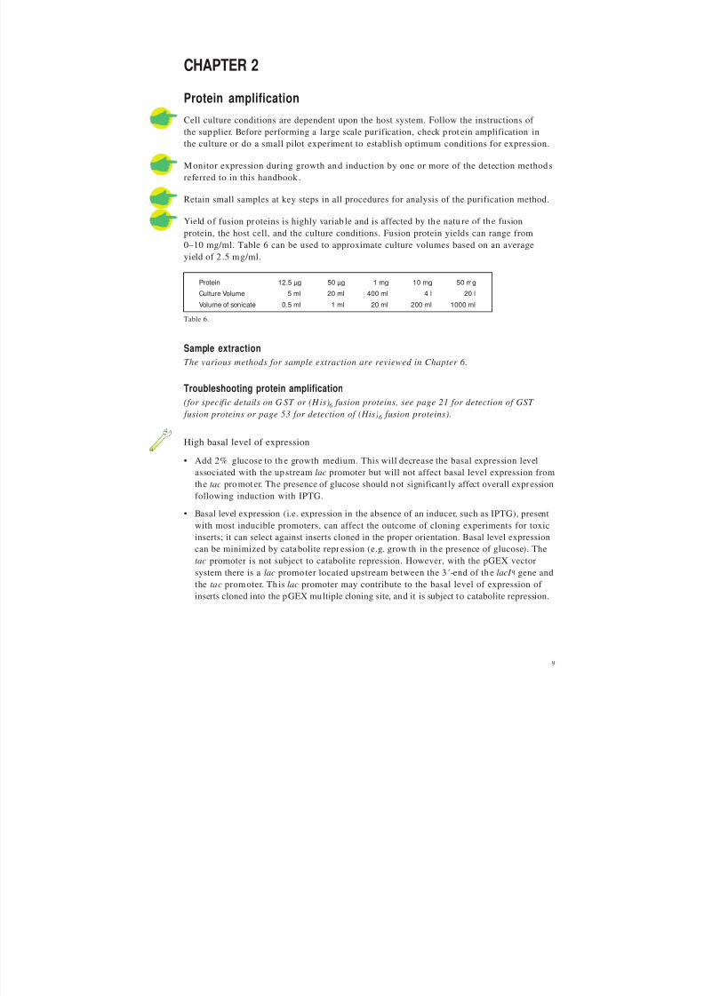
Yield of fusion proteins is highly variab le and is affected by the natu re of the fusionprotein, the host cell, and the culture conditions. Fusion protein yields can range from
0–10 mg/ml. Table 6 can be used to approximate culture volumes based on an averageyield of 2 .5 mg/ml.
Protein 12.5 µg 50 µg 1 mg 10 mg 50 mg
Culture Volume 5 ml 20 ml 400 ml 4 l 20 l
Volume of sonicate 0.5 ml 1 ml 20 ml 200 ml 1000 ml
Sample extractionThe various methods for sample extraction are reviewed in Chapter 6.
Troubleshooting protein amplification(for specific details on G ST or (His)6 fusion proteins, see page 21 for detection of GST
fusion proteins or page 53 for detection of (His)6 fusion proteins).
High basal level of expression
• Add 2% glucose to th e growth medium. This will decrease the basal expression levelassociated with the upstream lac promoter but will not affect basal level expression fromthe tac promoter. The presence of glucose should not significant ly affect overall expression
following induction with IPTG.
• Basal level expression (i.e. expression in the absence of an inducer, such as IPTG), presentwith most inducible promoters, can affect the outcome of cloning experiments for toxicinserts; it can select against inserts cloned in the proper orientation. Basal level expressioncan be minimized by catabolite repression (e.g. growth in the presence of glucose). Thetac promoter is not subject to catabolite repression. However, with the pGEX vectorsystem there is a lac promo ter located upstream between the 3´-end of th e lacI q gene andthe tac promoter. Th is lac promoter may contribute to the basal level of expression of inserts cloned into the pGEX multiple cloning site, and it is subject to catabolite repression.
Table 6.

8/2/2019 The ant Protein Handbook
http://slidepdf.com/reader/full/the-ant-protein-handbook 12/112
10
No protein is detected in bacterial sonicate
• Check DNA sequences. It is essential that pro tein-coding DNA sequences are cloned inthe proper tr anslation frame in the vectors. Cloning junctions should be sequenced toverify tha t inserts are in-frame.
• Optimize culture conditions to improve yield. Investigate the effect of cell strain, mediumcomposition, incubation temperature and induction conditions. Exact conditions willvary for each fusion protein expressed.
• Analyse a small aliquot of an overnight culture by SDS-PAGE. Generally, a highlyexpressed protein will be visible by Coomassie™ blue staining when 5–10 µl of aninduced culture whose A600 is ~1.0 is loaded on the gel. N on-transformed host E. coli
cells and cells transformed with the parental vector should be run in parallel as negativeand positive cont rols, respectively. The presence of the fusion p rot ein in this to tal cellpreparation and its absence from a clarified sonicate may indicate the presence of inclusion bod ies.
• Check for expression by immunoblotting. Some fusion proteins may be masked on anSDS-polyacrylamide gel by a bacterial protein of approximately the same molecularweight. Immunoblotting can be used to identify fusion proteins in these cases. Run anSDS-polyacrylamide gel of induced cells and transfer the proteins to a n itrocellulose orPVDF membrane (such as H ybond™-C or Hybond-P). Detect fusion p rotein usinganti-GST or anti-His antibody.
Most of fusion protein is in the post-sonicate pellet
• Check cell disrupt ion procedure. Cell disruption is seen by partial clearing of thesuspension or by microscopic examination. Addition of lysozyme (0.1 volume of a10 mg/ml lysozyme solution in 2 5 mM Tris-HCl, pH 8.0) prior to sonication mayimprove results. Avoid frothing as this may denature the fusion protein.
• Reduce sonication since over-sonication can lead to co-purification of host proteins withthe fusion protein.
• Fusion protein may be produced as insoluble inclusion bodies. Try altering the growthconditions to slow the rate of translation, as suggested below. It may be necessary tocombine these approaches. Exact conditions must be determined empirically for eachfusion p rotein.
- Lower the growth temperature (within the range of +20 to +30 °C) toimprove solubility.
- Decrease IPTG concentration to < 0.1 mM to a lter induction level.
- Alter time of induction.
- Induce for a shorter period of time.
- Induce at a higher cell density for a short period of time.
- Increase aeration. High oxygen tran sport can help prevent the formation of inclusion bod ies.

8/2/2019 The ant Protein Handbook
http://slidepdf.com/reader/full/the-ant-protein-handbook 13/112
11
It may be necessary to combine the above approaches. Exact conditions must be determinedempirically for each fusion protein.
• Alter extraction conditions to improve solubilization of inclusion bodies (see Chap ter 5).
Quantification of fusion proteins
Fusion proteins must be pur ified to homogeneity and q uantified using a stand ardpro tein a ssay.
• The relative yield of fusion protein can often be determined by measuring the absorbanceat 280 nm (suitable for bo th GST and (His)6 fusion proteins).
• The yield of protein may also be determined by standard chromogenic methods(e.g. Lowry, BCA, Bradford, etc.).
• Immunoassays can be used for quan tification if a suitable standard curve can be
produced. In this case, the fusion prot ein does not have to be pu rified for quantificationas long as a purified standard is available. The immunoassay technique is also particularlysuitable for screening large numbers of samp les when a simple yes/no answer is required,as, for example, when testing fractions from a purification.

8/2/2019 The ant Protein Handbook
http://slidepdf.com/reader/full/the-ant-protein-handbook 14/112
12

8/2/2019 The ant Protein Handbook
http://slidepdf.com/reader/full/the-ant-protein-handbook 15/112
13
CHAPTER 3
GST fusion proteins
AmplificationGlutathione S-transferase (GST) Gene Fusion System is an integrated range of products forthe amplification, purification and detection of GST fusion proteins in E. coli. The charac-teristics of GST are shown in Table 7 and Figure 1 shows the structure of GlutathioneSepharose used in the purification steps.
Glutathione S-transferase Naturally occurring Mr 26 000 proteinCan be expressed in E. coli with full enzymatic activity
Properties as determined in pGEX-1N
Dimer Molecular Weight Mr 58 500
Km (glutathione) 0.43 ± 0.07 mM
Km
(CDNB) 2.68 ± 0.77 mM
pI (chromatofocusing) 5.0
GST class hybrid of Alpha and Mu characteristics
Table 7.
Genotype F-, omp T, hsd S (rB-, mB
-), gal (52, 53)
Growth conditions Resuspend lyophilized cultures in 1 ml of L-broth. Grow overnight before plating onto L-brothmedia plates
Long term storage Mix equal volumes of stationary phase culture (grown in L-broth) and glycerol. Store at -70 °C.Revive frozen glycerol stocks by streaking onto L-broth media plates
Use an alternative strain for cloning and maintenance of the vector (e.g. JM105) as BL21does not transform well.
Using E. coli strains that are not protease-deficient may result in proteolysis of the fusionpro tein, seen as multiple ban ds on SDS-PAGE or Western blots.
CH2
C
CH2
H OH
S
C O
NH
C
OO
N H
C O
C
O
NH 3+
O
O
Fig. 1. Glutathione is attached to Sepharose by coupling tothe oxirane group using epoxy-activation. The structure of
glutathione is complementary to the binding site of theglutathion e S-tran sferase binding site.
General considerations for the amplification of fusion proteins are discussed in Chapter 2.
In the GST gene fusion system expression is under control of the tac promoter, which isinduced using the lactose analogue isopropyl b-D-thiogalactoside (IPTG). Induced culturesshould be left to express GST fusion proteins for several hours before the cells are harvested.
The host
E. coli BL21 is a protease-deficient strain specifically selected to give a high level of expression of GST fusion proteins.
Table 8.

8/2/2019 The ant Protein Handbook
http://slidepdf.com/reader/full/the-ant-protein-handbook 16/112
14
Table 9.
Column (prepacked) Amount of GST Comment
or M edia* * fusion protein fora single purification
GST MicroSpin Purification Module Up to 400 µg Ready to use, prepacked columns, buffers and chemicalsHigh throughput when used with MicroPlex™ 24 Vacuum(up to 48 samples simultaneously)
GSTrap FF 1 ml 10–12 mg Prepacked column, ready to use
GSTrap FF 5 ml 50–60 mg Prepacked column, ready to use
Glutathione Sepharose 4B 8 mg per ml For packing small columns and other formats
Glutathione Sepharose 4 Fast Flow 10–12 mg per ml For packing high performance columns for use withpurification systems and scaling up
Re-use of purification columns depends upon the nature of the sample and should only beperformed with identical samples to prevent cross contamination.
Batch preparation p rocedures are frequently mentioned in the literatur e. How ever theavailability of prepacked columns and easily packed high flow rate Glutathione Sepharoseprovide faster, mor e convenient alterna tives. Batch p reparation s are occasionally used if itappears that the tag is not fully accessible or when the protein in the lysate is at very lowconcentrations (both could appear to give a low yield from the first purification step).A more convenient alternative to improve yield is to decrease the flow rate or pass thesample through the column several times.
pGEX-6P-1, pGEX-6P-2, pGEX-6P-3 PreScission Protease
pGEX-4T-1, pGEX-4T-2, pGEX-4T-3 Thrombin
pGEX-5X-1, pGEX-5X-2, pGEX-5X-3 Factor Xa
PGEX-2TK Thrombin, c-AMP dependent protein kinaseAllows detection of expressed proteinsby direct labelling in vitro
pGEX6P PreScission Prot ease vector s offer t he most efficient method for cleavage and
pur ification of GST fusion prot eins. Site specific cleavage is performed with simultaneousimmobilization of th e protease on th e column. The pro tease has a high activity at a lowtemperature so that all steps can be performed in the cold room to prot ect p rotein integrity.Cleavage enzyme and GST tag are removed in a single step.
PurificationFor simple, one step purification of GST fusion proteins, several products have beendesigned to meet specific purification needs, as shown in Table 10.
Table 10. Summary of purification options for GST fusion proteins.
**Characteristics of GSTrap FF and Glutathione Sepharose are given in Appendix 5.
The vectors
pGEX vectors (pGEX-T, pGEX-P, pGEX-X, pGEX-2TK) are available in all three readingframes with a range of cleavage recognition sites as shown in Table 9. The same multiplecloning sites in each vector ensure easy transfer of inserts. The vector s carry the lacI q gene,so there are no specific host requirements for expression of fusion proteins. Vector control
regions and the reading frame of the multiple cloning site for each pGEX vector are shownin Appendix 1.

8/2/2019 The ant Protein Handbook
http://slidepdf.com/reader/full/the-ant-protein-handbook 17/112
15
Monitor purification steps by using one or more of the detection methods referred to in t hishandbook. The choice of purification equipment should also be made according to the needsof the purification. Appendix 8 provides a guide to aid in the selection of the correctpurification solution and key points to consider are highlighted here.
• For a single purification of a small quant ity of product or for high throu ghput screeningMicroSpin columns using centrifugation or MicroPlex 24 Vacuum are convenient andsimple to use.
• For purification of larger quantities of fusion pro teins GSTrap FF columns provide theideal solution and can be used with a syringe, a peristaltic pump or a chromatograph ysystem.
• To increase capacity use several GSTrap FF columns (1 ml or 5 ml) in series or, for evenlarger capacity requirements, pack Glutathione Sepharose 4 Fast Flow into a suitablecolumn (details of column packing procedures are outlined in Appendix 6).
• For simple and reproducible purification a chromatography system such as ÄKTA™ primeis a significant advantage, recording the purification process and eliminating manualerrors.
• For laboratory environments in which all experimental data must be recorded andtraceable, where method development, optimization and scale up are needed, a computercontrolled ÄKTAdesign chromatography system is recommended.
• Experiments such as protein refolding or method opt imization require linear gradientelution steps that can only be performed by a chromatography system.
GST MicroSpin Purification Module
The GST MicroSpin Purification Module is useful forscreening small or large numbers of lysates and forchecking samples during the optimization of amplification or purification conditions.Each module conta ins reagents sufficientfor 50 purifications.
• 10X PBS: 1.4 M NaCl, 27 mM KCl, 101 mM Na2HPO4, 18 mM KH2PO4, pH 7.3
• Reduced glutathione: 0.154 g• Dilution buffer: 50 mM Tris-HCl, pH 8.0
• IPTG: 500 mg
• MicroSpin columns: 50 units
Reagents are prepared as follows:
1X PBS: Dilute 10X PBS with sterile water. Store at +4 °C.
Glutathione elution buffer: Pour the entire contents of dilution buffer into the bottle containing thereduced glutathione.Shake until completely dissolved.Store as 1–20 ml aliquots at -20 °C.
IPTG 100 mM: Dissolve contents of the IPTG vial in 20 ml sterile water.Store as 1 ml aliquots at -20 °C.

8/2/2019 The ant Protein Handbook
http://slidepdf.com/reader/full/the-ant-protein-handbook 18/112
16
Alternative 1. H igh th roughput purification using MicroPlex Vacuum
Do not apply more than 600 µl of sample at atime to a MicroSpin column. This procedure willaccommoda te lysates from 2 to 12 ml of culture.
Also required:• Vacuum source capable of providing 220 mm Hg (e.g. a water vacuum).
• Side arm flask, 500 ml or 1 litre.
• Single or double hole rubber stop.
• Vacuum tubing.
• MicroPlex 24 Vacuum apparatus (one or two).
1. Assemble the MicroPlex 24 Vacuum following the instructions supplied.
2. Resuspend the Glutathione Sepharose in each MicroSpin column by vortexing gently.
3. Remove the caps and snap off the bottom closures from the MicroSpin columns. Place the columns in themanifold, filling any unused holes with the plugs provided with MicroPlex 24 Vacuum.
4. Ensure the stopcock is in the closed position (i.e. perpendicular to the vacuum tubing) and that themanifold is placed squarely on the gasket.
5. Turn on vacuum supply at source. Open the stopcock (i.e. parallel to the vacuum tubing). After the columnstorage buffer has been drawn through all the columns into the collection tray, close the stopcock.
6. Allow 10–15 seconds for the vacuum pressure to dissipate. Remove the manifold and place it on apaper towel.
7. Apply up to 600 µl of lysate to the column and incubate at room temperature for 5–10 minutes.
8. Open the stopcock. After the lysates have been drawn through all the columns into the collection tray,close the stopcock.
9. Add 600 µl of 1X PBS wash buffer to each column. Open the stopcock. After buffer has been drawn throughall the columns into the collection tray, close the stopcock.
10. Allow 10–15 seconds for the vacuum pressure to dissipate. Remove the manifold and reassemble theapparatus with a clean collection tray. Additional 600 µl washes may be performed if desired.
11. Add 200 µl of Glutathione elution buffer to each column. Incubate at room temperature for 5–10 minutes.
12. Open the stopcock. After elution buffer has been drawn through all the columns into the collection tray,close the stopcock.
13. Allow 10–15 seconds for the vacuum pressure to dissipate. Remove the manifold. Cover eluates with sealingtape until required for analysis.
Note: Yields of fusion protein may be increased by repeating the elution step two or three times and pooling
the eluates.
Troubleshooting
See Purification and D etection Troubleshoot ing page 28.
Alternative 2. Purification of multiple samples using a microcentrifuge
Do no t apply more than 600 µl of sample at a time to a M icroSpin column. This procedurewill accommodate lysates from 2 to 12 ml of culture.

8/2/2019 The ant Protein Handbook
http://slidepdf.com/reader/full/the-ant-protein-handbook 19/112
17
1. Resuspend the Glutathione Sepharose in each column by vortexing gently.
2. Loosen the column caps one-fourth turn. Remove (and save) bottom closures.
3. Place each column into a clean 1.5 or 2 ml microcentrifuge tube. Spin for 1 minute at 735 g.
4. Discard the buffer from each centrifuge tube and replace the bottom closures.
5. Apply up to 600 µl of lysate to the column.
6. Recap each column securely and mix by gentle, repeated inversion. Incubate at room temperature for5–10 minutes.
7. Remove (and save) the top caps and bottom closures. Place each column into a clean, pre-labelled 1.5 or2 ml microcentrifuge tube.
8. Spin for 1 minute at 735 g to collect flow through.
9. Place each column into a clean, pre-labelled 1.5 or 2 ml microcentrifuge tube.
10. Apply 600 µl of 1X PBS wash buffer to each column and repeat spin procedure. Additional 600 µl washeswith 1X PBS may be performed if desired.
11. Add 100–200 µl of Glutathione elution buffer to each column. Replace top caps and bottom closures.Incubate at room temperature for 5–10 minutes.
12. Remove and discard top caps and bottom closures and place the column into a clean 1.5 or 2 ml
microcentrifuge tube.13. Spin all columns again to collect eluate. Save for analysis.
Note: Yields of fusion protein may be increased by repeating the elution step two or three times and pooling the
eluates.
Troubleshooting
See Purification and D etection T roubleshooting page 28.
Alternative 3. Purification using MicroPlex Centrifugation
Do no t apply more than 600 µl of sample at a time to a GST MicroSpin column. Thisprocedure will accommodate lysates from 2 to 12 ml of culture.
See Appendix 4 for recommended centrifugation systems.
1. Assemble the MicroPlex 24 unit following the instructions supplied. Two units can be processedsimultaneously to handle 48 samples.
2. Resuspend the Glutathione Sepharose in each column by vortexing gently.
3. Remove the caps from the MicroSpin columns and snap off the bottom closures. Place the columns in themanifold.
4. Centrifuge the unit for 2 minutes following the instructions supplied.
5. Add up to 600 µl of lysate to each column. Incubate at room temperature for 5–10 minutes.6. Centrifuge the unit for 2 minutes following the instructions supplied. If desired remove the manifold from
each collection try and place on a clean paper towel. Reassemble each unit with a fresh collection tray.
7. Apply 600 µl of 1X PBS wash buffer to each column and repeat spin procedure. Additional 600 µl washes with1X PBS may be performed if desired. Remove the manifold from each collection tray and place it on clean paper.
8. Add 100–200 µl of glutathione elution buffer to each column. Incubate at room temperature for 5–10 minutes.
9. Centrifuge the unit for 2 minutes following the instructions supplied. Cover the eluted samples with sealingtape until required for analysis.
Note: Yields of fusion protein may be increased by repeating the elution step two or three times and pooling the
eluates.
TroubleshootingSee Purification and D etection T roubleshooting page 28.

8/2/2019 The ant Protein Handbook
http://slidepdf.com/reader/full/the-ant-protein-handbook 20/112
18
Equilibrate column
with
binding buffer
Apply sample
wash with
binding buffer
Waste Collect
Elute
with
elution buffer
Collect fractions
3 min 5-15 min 2 min
Fig. 2. Simple pur ification of GST fusion prot eins using GSTrap FF.
Re-use of any purification column depends on t he natu re of the sample and should only beperformed with identical fusion proteins to prevent cross-contamination.
GSTrap FF columns (1 ml or 5 ml) can be connected in series to increase binding capacityand hence scale of purification. Larger columns can be packed with Glutathione Sepharose 4Fast Flow (see Appendix 6 for column packing).
Sample and buffer preparation
Use high quality water and chemicals. Filtration through 0.45 µm filters is recommended.
Samples should be centrifuged immediately before use and/or filtered through a 0.45 µmfilter. If the sample is too viscous, dilute with binding buffer.
Sample binding properties can be improved by adjusting the sample to the composition of the binding buffer: dilute in binding buffer or perform a buffer exchange using a desaltingcolumn (see Chapter 7).
Purification using GSTrap FF 1 ml or 5 ml columns
GSTrap FF columns can be operated with a syringe, a peristaltic pump or a liquidchromatography system such as ÄKTA prime. Figure 2 shows a schematic of the simplesteps needed for successful purification using a 1 ml GSTrap FF column.

8/2/2019 The ant Protein Handbook
http://slidepdf.com/reader/full/the-ant-protein-handbook 21/112
19
Alternative 1. Manual purification with a syringe
Fig. 3. Using GSTrap FF with a syringe. A Prepare buffers and sample. Remove the column’s top cap andtwist off the end. B Load the sample and begin collecting fractions. C Wash and elute and continuecollecting fractions.
A B C
1. Fill the syringe with binding buffer.
2. Connect the column to the syringe using the adapter supplied ("drop to drop" to avoid introducing air intothe column).
3. Remove the twist-off end.
4. Equilibrate the column with 5 column volumes of binding buffer.
5. Apply the sample using the syringe. For best results, maintain a flow rate of 0.2–1 ml/min (1 ml column)and 1–5 ml/min (5 ml column) as the sample is applied.*
6. Wash with 5–10 column volumes of binding buffer. Maintain flow rates of 1–2 ml/min (1 ml column)and 5–10 ml/min (5 ml column) during the wash.*
7. Elute with 5–10 column volumes of elution buffer. Maintain flow rates of 1–2 ml/min (1 ml column) and
5–10 ml/min (5 ml column) during elution.** One ml/min corresponds to approximately 30 drops/min when using a syringe with a HiTrap 1 ml column and
five ml/min corresponds to approximately 120 drops/min when using a HiTrap 5 ml column.
For large sample volumes a simple peristaltic pump can be used to apply sample and buffers.
Alternative 2. Simple purification with ÄKTA prime
ÄKTA prime contains a pr e-programmed template for pur ification of GST fusion pro teinsusing a single GSTrap FF column, as shown below. This provides a standard purificationprotocol which can be followed exactly or optimized as required.
Binding buffer: 1X PBS, pH 7.3 (140 mM NaCl, 2.7 mM KCl, 10 mM Na2HPO4, 1.8 mM KH2PO4, pH 7.3).
Elution buffer: 50 mM Tris-HCl, 10 mM reduced glutathione, pH 8.0.
ReequilibrationWash
Elution
System preparation &column equilibration
100
50
11 10 11 6 Min
Sample
Total separation time = 37 min + sample application time
% Elution buffer
8/2/2019 The ant Protein Handbook
http://slidepdf.com/reader/full/the-ant-protein-handbook 22/112
20
Connecting the column. Preparing the fraction collector.
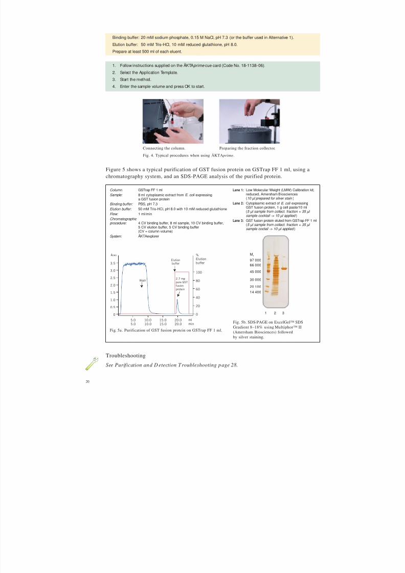
Fig. 5a . Purification of GST fusion protein on GSTrap FF 1 ml.
Fig. 5b. SDS-PAGE on ExcelGel™ SDSGradient 8–18% using Multiphor™ II(Amersham Biosciences) followedby silver staining.
Binding buffer: 20 mM sodium phosphate, 0.15 M NaCl, pH 7.3 (or the buffer used in Alternative 1).
Elution buffer: 50 mM Tris-HCl, 10 mM reduced glutathione, pH 8.0.
Prepare at least 500 ml of each eluent.
1. Follow instructions supplied on the ÄKTAprime cue card (Code No. 18-1138-06).
2. Select the Application Template.
3. Start the method.
4. Enter the sample volume and press OK to start.
Column: GSTrap FF 1 mlSample: 8 ml cytoplasmic extract from E. coli expressing
a GST fusion proteinBinding buffer: PBS, pH 7.3Elution buffer: 50 mM Tris-HCl, pH 8.0 with 10 mM reduced glutathione
Flow: 1 ml/minChromatographic procedure: 4 CV binding buffer, 8 ml sample, 10 CV binding buffer,
5 CV elution buffer, 5 CV binding buffer(CV = column volume)
System: ÄKTAexplorer
Lane 1: Low Molecular Weight (LMW) Calibration kit,reduced, Amersham Biosciences(10 µl prepared for silver stain )
Lane 2: Cytoplasmic extract of E. coli expressingGST fusion protein, 1 g cell paste/10 ml
(5 µl sample from collect. fraction + 35 µl sample cocktail -> 10 µl applied )Lane 3: GST fusion protein eluted from GSTrap FF 1 ml
(5 µl sample from collect. fraction + 35 µl sample coctail -> 10 µl applied )
Fig. 4. Typical procedures when using ÄKTA prime.
Figure 5 shows a typical purification of GST fusion protein on GSTrap FF 1 ml, using achromatography system, and an SDS-PAGE analysis of the purified protein.
0
20
40
60
80
100
%Elutionbuffer
0
0.5
1.0
1.5
2.0
2.5
3.0
3.5
5.0 10.0 15.0 20.0
ml
min
Wash
Elution
buffer
2.7 mg
pure GST
fusion
protein
5.0 10.0 15.0 20.0
A280
1 2 3
Mr
97 00066 000
20 100
30 000
45 000
14 400
TroubleshootingSee Purification and D etection Troubleshooting page 28.

8/2/2019 The ant Protein Handbook
http://slidepdf.com/reader/full/the-ant-protein-handbook 23/112
21
Optimization of GST fusion protein purification
Following the instructions supplied for each prepacked GSTrap FF column will generallyprovide very good results.
Dimer formation is inevitable with GST fusion pro teins since GST itself is a homodimer
when folded. Use gel filtration to remove the dimers. A column prepacked with Superdex™will give the highest possible resolution between two mo lecules of similar molecular weight.
One of the most important parameters affecting the binding of GST fusion pro teins toGlutathione Sepharose is the flow rate. Since the binding kinetics between glutathione andGST are relatively slow, it is important to keep the flow rate low during sample applicationto achieve maximum binding capacity.
Volumes and times used for elution may vary among fusion proteins. Further elution withhigher concentrations o f glutathione (20–50 mM) may improve yield. At concentrationsabove 15 mM glutathione the buffer concentration should also be increased to ma intain the
pH within the range 8–9.
Detection of GST fusion proteinsTable 11 reviews the methods available for detection of GST fusion proteins. These methodscan be selected accord ing to the experimental situa tion, for example, SDS-PAGE analysis,performed frequently during amplification and purification to monitor results, may not b ethe method of choice for rou tine monitoring of samples from high throughput screening.Functional assays based on the properties of the protein of interest (and not the GST tag)are useful, but need to be developed for each specific protein.
Detection method CommentsGST 96 Well Detection module for ELISA assay Ideal for screening expression systems and chromatographicfractions.Useful when amount of expressed protein is unknown or whenincreased sensitivity is required.
GST Detection Module for enzymatic assay Rapid assay, ideal for screening.
Western blot analysis using anti-GST antibody Highly specific, detects only GST fusion protein.and ECL™ detection systems Little or no background detectable when used with optimized
concentrations of secondary HRP conjugated antibody.ECL detection systems enhance detection in Western blot.ECL provides adequate sensitivity for most recombinantexpression applications.For higher sensitivity use ECL Plus™.
SDS-PAGE with Coomassie or silver staining Provides information on size and % purity.Detects fusion protein and contaminants.
Functional assays Useful to assess activity of the purified GST fusion protein, but mayrequire development and optimization.
Table 11. Detection methods for GST fusion proteins.

8/2/2019 The ant Protein Handbook
http://slidepdf.com/reader/full/the-ant-protein-handbook 24/112
22
Alternative 1. SDS-PAGE Analysis
For information and advice on electrophoresis techniques, please refer to the sectionAdditional reading and reference material.
Troubleshooting
• If the fusion protein is absent, it may be insoluble or expressed at very low levels: referto Troubleshooting protein amplification (page 9).
• If no fusion protein is detected by Coo massie Blue, try silver staining or Western blotting
to enhance sensitivity.
Transformants expressing the fusion protein will be identified by the absence from totalcellular proteins of the parental M r 29 000 GST and by the presence of a novel, largerfusion protein. Parental pGEX vectors pr oduce a 29 kDa GST fusion pro tein containingamino acids that are coded for by the pGEX multiple cloning site. In some cases both theM r 29 000 GST and fusion p rotein may be present. Th is can be caused by translationalpausing at the junction between GST and the fusion partner or a mixed culture betweencells with parental plasmid and cells with fusion plasmid.
Interpretation is sometimes complicated when fusion proteins break down and release theGST moiety M r 26 000. Such cases are usually recognized by the appearance of theM r 26 000 species, and a series of larger, partial proteolytic fragments above it.
% Acrylamide in resolving gel Separation size range (M r x 1 0 -3 )
Single percentage:5% 36–200
7.5% 24–200
10% 14–200
12.5% 14–100
15% 14–601
Gradient:
5–15% 14–200
5–20% 10–200
10–20% 10–1501The larger proteins fail to move significantly into the gel.
6X SDS loading buffer: 0.35 M Tris-HCl, 10.28% (w/v) SDS, 36% (v/v) glycerol, 0.6 M dithiothreitol(or 5% 2-mercaptoethanol), 0.012% (w/v) bromophenol blue, pH 6.8.Store in 0.5 ml aliquots at -80 °C.
1. Add 2 µl of 6X SDS loading buffer to 5–10 µl of supernatant from crude extracts, cell lysates or purifiedfractions as appropriate.
2. Vortex briefly and heat for 5 minutes at +90 to +100 °C.
3. Load the samples onto an SDS-polyacrylamide gel.
4. Run the gel for the appropriate length of time and stain with Coomassie Blue (Coomassie Blue R Tablets) orsilver (PlusOne Silver Staining Kit, Protein).
The percentage of acrylamide in the SDS-gel shou ld be selected according to t he expectedmolecular weight of the pr otein of interest (see Table 12).
Table 12.

8/2/2019 The ant Protein Handbook
http://slidepdf.com/reader/full/the-ant-protein-handbook 25/112
23
• 10X Reaction buffer: 1 M potassium phosphate buffer, pH 6.5.
• CDNB: 100 mM 1-chloro-2,4-dinitrobenzene (CDNB) in ethanol.
• Reduced glutathione powder for glutathione solution. Dissolve 100 mM reduced glutathione in steriledistilled water. Aliquot into microcentrifuge tubes.Store at -20 °C. Avoid more than five freeze/thaw cycles.
• Goat/anti-GST antiserum is also supplied for use in Western blots.
Measurement of GST activity by CDNB assay
CDNB is toxic. Avoid contact with eyes, skin and clothing. In case of accidental contact,flush affected area with water. In case of ingestion, seek immediate medical att ention.
pGEX-bearing cells must be lysed b efore performing a CDN B assay.
1. In a microcentrifuge tube, combine the following:- Distilled water 880 µl- 10X Reaction buffer 100 µl- CDNB 10 µl
- Glutathione solution 10 µl- Total Volume 1000 µl
2. Cap and mix by inverting the tube several times.
CDNB may cause the solution to become slightly cloudy. The solution should clearupon mixing.
Alternative 2. GST Detection Module
The GST Detection Module is designed for the rapid enzymatic detection of GST fusionproteins produced using the pGEX vectors using the GST substrate 1-chloro-2,4 dinitrobenzene(CDNB). The GST-mediated reaction of CDNB with glutathione produces a conjugate thatis measured by absorbance at 340 nm using a UV/vis spectrophotometer, such as an
Ultrospec™ 1000, or a plate reader. The CDNB assay is performed in less than 10 minuteson crude bacterial sonicates, column eluates, or purified GST fusion protein. Figure 6 showstypical results from a CDNB assay. Each detection module contains reagents sufficient for50 detections.
Fig. 6. Typical results of a CDN B assay for GSTfusion proteins. 53 µg of total protein from an E. coli TG1/pGEX-4T-Luc sonicate and 0.8 µg of total protein eluted from Glutathione Sepharosewere assayed according to the instructions for theGST Detection Module.
1 2 3 4Time (minutes)
0.2
0.4
0.6
Sonicate (53 µg)
Eluate (0.8 µg)
A340

8/2/2019 The ant Protein Handbook
http://slidepdf.com/reader/full/the-ant-protein-handbook 26/112
24
Alternative 3: GST 96 Well Detection Module
The GST 96 Well Detection Module providesa highly sensitive ELISA assay for testingclarified lysates and intermediate pur ificationfractions. Each detection module containsreagents sufficient for 96 detections:
3. Transfer 500 µl volumes of the above solution into two UV-transparent cuvettes. Add sample (5–50 µl) tothe "sample cuvette". To the "blank cuvette", add a volume of 1X reaction buffer equal to the samplevolume in the sample cuvette.
4. Cover each cuvette with wax film and invert to mix.
5. Place the blank cuvette in the spectrophotometer and blank at 340 nm. Measure the absorbance of thesample cuvette at 340 nm and simultaneously start a stopwatch or other timer.
6. Record absorbance readings at 340 nm at one-minute intervals for 5 minutes by first blanking thespectrophotometer with the blank cuvette and then measuring the absorbance of the sample cuvette.
7. Calculate the A340 /min/ml sample
Calculations
DA340 /min/ml =
Where: A340 (t2) = absorbance at 340 nm at time t2 in minutes
A340 (t1) = absorbance at 340 nm at time t1 in minutes
DA340 /min/ml values can be used as a relative comparison of GST fusion protein content between samples of agiven fusion protein.
Adapt the assay to give absolute fusion protein concentrat ions by constructing a standardcurve of DA340 /min versus fusion protein amount.
The activity of the GST moiety can be affected by the folding of the fusion partner.Absorbance readings obtained for a given fusion protein may not reflect the actual amountof fusion protein present.
Troubleshooting
• The reaction rate is linear provided that an A340 of approximately 0.8 is not exceeded
during the five-minute time course. Plot initial results to verify that the reaction rate islinear over the time course. Adjust the amount of sample containing the GST fusionprotein to maintain a linear reaction rat e.
• If a low absorbance is obtained using the CDN B assay, a Western blot using the Anti-GSTAntibod y may reveal high levels of pro tein expression.
• Under standard assay conditions at +22 °C and in the absence of GST, glutathione andCDNB react spontaneously to form a chemical moiety that produces a baseline drift atDA340 /min of approximately 0.003 (or 0 .015 in 5 minutes). Correct for baseline drift byblanking the spectrophotometer with the blank cuvette before each reading of the
sample cuvette.
A340 (t2) - A340 (t1)
(t2 - t1)(ml sample added)

8/2/2019 The ant Protein Handbook
http://slidepdf.com/reader/full/the-ant-protein-handbook 27/112
25
• GST 96 Well detection plates in which each well is coated with anti-GST antibody, blocked and dried.
• Horse-radish peroxidase conjugated to goat polyclonal anti-GST antibody.
• Purified recombinant glutathione S-transferase test protein.
Additional reagents to be prepared:
PBS: 140 mM NaCl, 2.7 mM KCl, 10 mM Na2HPO4, 1.8 mM KH2PO4, pH 7.4.
Wash buffer: 0.05% Tween™ 20 in PBS (500 ml/96 well plate).Store at room temperature until needed.
Blocking buffer: 1 x conc. 3% non-fat dry milk in PBS with 0.05% Tween 20 (10 ml/96 well plate).2 x conc. 6% non-fat dry milk in PBS with 0.1% Tween 20 (5 ml/96 well plate).
Prepare fresh buffers daily.
As each fusion protein is captured uniquely, prepare standards of rGST protein and thetarget fusion p rotein using a dilution series from 100 ng/100 µ l to 10 pg/µl in 1X b lockingbuffer if quantification is required. Run recombinant GST (rGST) protein as a standardcontrol in every assay.
Screening of GST expression clones or chromatograph ic fractions
1. Bring each test sample to a final volume of 50 µl with 1X PBS.
2. Mix with 50 µl of 2X blocking buffer.
3. For screening: dilute rGST protein standard to 1 ng/100 µl in 1X blocking buffer.
4. For quantification: use dilution series from 100 ng/100 µl to 10 pg/µl in 1X blocking buffer for rGST proteinand for the target fusion protein.
5. Remove one 96-well plate from the foil pouch. If using less than 96 wells, carefully remove the well strips
from the holder by pushing up on the wells from below. Store unused swell strips in the pouch with thedessicant provided.
6. Pipette 100 µl of sample into each well.
7. Incubate for 1 hour at room temperature in a humidified container or incubator.
8. Empty contents of the well by flicking the inverted plate.(Biohazardous material should be pipetted or aspirated into a suitable container.)
9. Blot the inverted well or strips on to a paper towel to remove excess liquid.
10. Wash each well 5 times with wash buffer (inverting and flicking out the contents each time).
11. Blot the inverted well or strips on to a paper towel to remove excess wash buffer.
12. Dilute HRP/anti-GST conjugate 1:10 000 (1 µl:10 ml) in 1X blocking buffer. One 96 well plate will require10 ml of the diluted solution.
13. Add 100 µl of diluted HRP/anti-GST conjugate to each well and incubate for 1 hour at room temperature ina humidified container or incubator.
14. Empty well contents and wash twice with wash buffer as previously described.
15. Add soluble horseradish peroxidase substrate* to each well and incubate according to supplier's instructions.
*3,3' ,5,5' -tetramethyl benzidine (A450) or 2',2'-azino-bis (3-ethylbenzthiazoline-6-sulphonicacid) diammonium salt (ABTS™) (A410) have been used successfully.
16. Read plate absorbance in a microplate reader or spectrophotometer.
Troubleshooting
See also Purification and D etection Troubleshooting page 28.

8/2/2019 The ant Protein Handbook
http://slidepdf.com/reader/full/the-ant-protein-handbook 28/112
26
Anti-GST Antibody
Blocking/Incubationbuffer: 5% (w/v) non-fat dry milk and 0.1% (v/v) Tween 20 in PBS
(140 mM NaCl, 2.7 mM KCl, 10 mM Na2HPO4, 1.8 mM KH2PO4, pH 7.3)
Wash buffer: 0.1% v/v Tween 20 in PBS (as above)
Secondary Antibody to detect the anti-GST antibody (such as anti-goat IgG HRP conjugate).
1. Separate the protein samples by SDS-PAGE.
Although anti-GST antibody from Amersham Biosciences has been cross-adsorbedwith E. coli proteins, low levels of cross-reacting antibodies may remain. It is recommendedalways to run samples of E. coli sonicates that do not contain a recombinant pGEX plasmidand samples that conta in the parental pGEX plasmid as contro ls.
2. Transfer the separated proteins from the electrophoresis gel to an appropriate membrane, such asHybond ECL (for subsequent ECL detection) or Hybond P (for subsequent ECL or ECL Plus detection).
Electrophoresis and protein transfer may be accomplished using a variety of equipment andreagents. For further details, refer to the Protein Electrophoresis Technical Manual and Hybond ECL Instruction Manual from Amersham Biosciences.
Blocking of membrane
1. Transfer the membrane onto which the proteins have been blotted to a container such as a Petri dish.
2. Add 50–200 ml of blocking/incubation buffer.
3. Incubate for 1–16 hours at ambient temperature with gentle shaking.
Longer incubation times with blocking buffer may reduce background signal.
4. Decant and discard the buffer.
Low absorbance detected in samples
• Check that samples were sufficiently induced and lysed (see Troubleshooting proteinamplification page 9).
• If clarified lysate is being tested, mix initial GST sample with 2X blocking buffer to give
a final concentration of 1X blocking buffer.
Poor day to day reproducibility between identical samples
• Ensure that all incubation times are consistent. Reduction in GST capture incubation
time can be reduced to > 30 minutes with slightly reduced signal, bu t H RP/anti-GST
conjugate incubat ion t ime can significant ly reduce signal with every 15 minute decrease.
Alternat ive 4. Western blot analysis
Amplification and pur ification can also be mon itored by Western blot analysis, using ECL
or ECL Plus detection systems to enhance sensitivity.

8/2/2019 The ant Protein Handbook
http://slidepdf.com/reader/full/the-ant-protein-handbook 29/112
27
Anti-GST antibody
1. Prepare an appropriate dilution of anti-GST antibody with blocking/incubation buffer, e.g. 5–10 µl of antibodyto 50 ml of buffer. Refer to Amersham Biosciences Application Note 18-1139-13 for furtherinformation on optimization.
2. Pour the antibody-buffer mixture into the container with the membrane.
3. Incubate for 1 hour at ambient temperature with gentle shaking.4. Decant and discard the antibody-buffer.
5. Rinse twice with 20–30 ml of blocking or wash buffer to remove most of the unbound antibody.
6. Decant and discard the rinses.
7. Wash the membrane with 20–30 ml of blocking or wash buffer for 10–60 minutes at ambient temperaturewith gentle shaking.
8. Discard the wash and repeat.
Secondary antibody
1. Dilute an appropriate anti-goat secondary antibody with blocking/incubation buffer according to the
manufacturer's recommendation. Refer to Amersham Biosciences Application Note 18-1139-13 forfurther information on optimization.
2. Pour the antibody-buffer mixture into the container with the membrane.
3. Incubate for 1 hour at ambient temperature with gentle shaking.
4. Decant and discard the antibody-buffer.
5. Rinse twice with 20–30 ml of blocking or wash buffer to remove most of the unbound antibody.
6. Decant and discard the rinses.
7. Wash the membrane with 20–30 ml of blocking or wash buffer for 10–60 minutes at ambient temperaturewith gentle shaking.
8. Discard the wash and repeat.
9. Develop the blot with the appropriate substrate for the conjugated secondary antibody.
ECL and ECL Plus detection systems require very little antibod y to achieve a sufficientsensitivity so the amount of antibody (primary and secondary) used in the protocols can beminimized. Smaller quantities of antibody-buffer mixtures can be used by scaling down theprotocol and performing the incubations in sealable plastic bags.
Troubleshooting
See also Purification and D etection Troubleshooting page 28.
Multiple bands seen on Western b lot analysis
• Anti-GST antibody from Amersham Biosciences has been cross-absorbed against
E. coli proteins and tested for its lack of non-specific background binding in a Western
blot. Some sources of the anti-GST antibody may contain antibodies that react with
various E. coli proteins present in the fusion protein sample. Cross-adsorb the antibody
with an E. coli sonicate to remove anti- E. coli antibodies. This E. coli must not contain
the pGEX plasmid.

8/2/2019 The ant Protein Handbook
http://slidepdf.com/reader/full/the-ant-protein-handbook 30/112
28
Purification and detection troubleshooting
Column has clogged
• Cell debris in the sample may clog the column. Clean the column according to Appendix 5and ensure that samples have been filtered or centrifuged.
Fusion protein does not bind to purification column
• Over-sonication may have denatured the fusion protein. Check by using a microscope tomonitor cell breakage. Use mild sonication conditions during cell lysis.
• Sonication may be insufficient: Check using a microscope or monitor by measuring therelease of nucleic acids at A260 . Addition of lysozyme (0.1 volume of a 10 mg/ml lysozymesolution in 25 mM Tris-HC l, pH 8.0) prior to sonication may improve results.
• Add 5 mM DTT prior to cell lysis. This can significantly increase binding of some GST
fusion p roteins to Glutathione Sepharose.• Check that the column has been equilibrated with a buffer 6.5 < pH < 8.0 (e.g. PBS) before
application of the fusion protein. The correct pH range is critical for efficient binding.
• Decrease the flow rate to improve binding.
• If re-using a column, check that the column has been regenerated correctly (seeAppendix 5). Replace with fresh Glutathione Sepharose or a new column if bindingcapacity does not r eturn after regeneration.
• Check the binding of a cell sonicate prepared from the parental pGEX plasmid. If GSTproduced from the par ental plasmid b inds with high affinity, then the fusion partn er mayhave altered the conformation of GST, thereby reducing its affinity. Try reducing thebinding temperature to +4 °C and limit the number of washes.
• Column capacity may have been exceeded. If using GSTrap FF columns (1 ml or 5 ml)link 2 or 3 columns in series to increase capacity or pack a larger column.
• Fusion protein may be in the inclusion bodies, although using a GST-tag reduces thechance of this problem occurring.
Fusion protein is poorly eluted
• Increase concentra tion of glutathione in the elution buffer. Above 15 mM glutathione thebuffer concentration should b e increased to maintain pH .
• Increase pH o f the elution buffer. Values up to pH 9 may improve elution withoutrequiring an increase in the concentration of glutathione.
• Increase ionic strength of the elution buffer by addition of 0.1–0.2 M N aCl. Note thatvery hydrophobic proteins may precipitate under high salt conditions. If this is the case,addition of a non-ionic detergent may improve results (see below).
• Decrease the flow rate to improve elution.

8/2/2019 The ant Protein Handbook
http://slidepdf.com/reader/full/the-ant-protein-handbook 31/112
29
• Add a non-ionic detergent (0.1% Triton™ X-100 or 2% N-octyl glucoside) to the elutionbuffer to reduce non-specific hydrophobic interactions that may prevent solubilizationand elution o f fusion pro teins
• Try over-night elution at room temperature or +4 °C.
Multiple bands seen on SDS-PAGE or Western blot analysis
Multiple bands result from partial degradation of fusion proteins by proteases, or denaturationand co-purification of host proteins with the GST fusion protein due to over-sonication.
• Check that a protease-deficient host such as E. coli B21 has been used.
• Add protease inhibitors such as 1 mM PMSF to the lysis solution. A non-tox ic, watersoluble alternat ive to PMSF is 4-(2-amino-ethyl)- benzenesulfonyl fluoride hydr ochloride(AEBSF), commercially available as Pefabloc™ SC from Boehringer Mannheim.
• Use prepacked GSTrap FF columns or Glutathione Sepharose 4 Fast Flow. These can beused at higher flow rates to process samples more quickly and so avoid degradation.
• Decrease sonication. Addition of lysozyme (0.1 volume of a 10 mg/ml lysozyme solutionin 25 mM Tris-HCl, pH 8.0) prior to sonication may improve results. Avoid frothing asthis may denature the fusion p rotein.
• Include an additional purification step (see Chapter 9). A variety of proteins known a schaperonins that are involved in the correct folding of nascent proteins in E. coli mayco-purify with GST fusion proteins, including a M r 70 000 protein (see below).
Serine protease inhibitors must be removed prior to cleavage by thrombin or Factor Xa.
Use HiTrap Benzamidine FF (high sub) (see page 39).
M r 70 000 protein co-purifies with the GST-fusion pro tein
Pre-incubate the protein solution with 2 mM ATP, 10 mM MgSO 4, 50 mM Tris-HCl(pH 7.4) for 10 minutes at +37 °C prior to p urification in order to d issociate the complex.This M r 70 000 protein is probably a protein product of the E. coli gene dn aK and involvedin the degradation of "abnormal" proteins in E. coli. Report s suggest that this protein canbe removed by ion exchange chromatography (Analects and Separations p24 , 199 6,Amersham Biosciences and (http://bionet.hgmp.mrc.ac.uk/hypermail/methods/
methods.199406/0813.html)) or by passage of the sample over ATP agarose(Myers, M., BIOSCI posting, 7 July 1993).Thain, A., et al. Trends Genet . 12 , 209–210 (1996) and Sherman, M . and Goldberg, A.L., J. Biol. Chem , 269, 31479–31483, (1994) suggest washing the column with ATP or Gr oESrather than using a subsequent IEX step.

8/2/2019 The ant Protein Handbook
http://slidepdf.com/reader/full/the-ant-protein-handbook 32/112
30
PreScission Protease Mr 46 000Bovine thrombin Mr 37 000
Bovine Factor Xa Mr 48 000
Tag removal by enzymatic cleavageIn most cases, functional tests can be performed using the intact fusion with GST. If removalof the GST tag is necessary, it is highly recommended to produce the fusion proteins with aPreScission Protease cleavage site. The GST tag then can be removed and the protein purifiedin a single step on the column (see Figure 7). This protease also has the useful property of
being maximally active at +4 °C thus allowing cleavage to be performed at low temperaturesand so improving the stability of the target prot ein.
Thrombin or Factor Xa recognition sites may be cleaved either while bound on the columnor in solution after elution from the column (see Figure 8). The protease used for cleavagecan be removed using Benzamidine Sepharose, a purification medium with a high specificityfor serine proteases (see page 39 ).
On-column cleavage is generally recommended a s the method of choice since manypotential contaminants can be washed through the column and the target protein elutedwith a higher level of purity. For the removal of thrombin and Factor Xa, a GSTrap FF and
a H iTrap Benzamidine FF (high sub) column can be conn ected in series so that cleavedproduct passes directly from the GSTrap FF into the HiTrap Benzamidine FF (high sub).Samples are cleaved and pro teases removed in a single step (see page 39).
The amount of enzyme, temperature and length of incubation required for completedigestion varies according to the specific GST fusion protein produced. Determine optimalconditions in pilot experiments.
Remove samples at var ious t ime point s and ana lyse by SDS-PAGE to estimate the yield,pur ity and extent o f digestion. Approx imate molecular w eights for SDS-PAGE analysis:
If protease inhibitors have been used in the lysis solution, they must be removed prior tocleavage by PreScission Protease, thrombin or Factor Xa (the inhibitors will usually beeluted in the flow-through when sample is loaded onto a GSTrap FF column).
Enzyme Inhibitor
PreScission protease 100 mM ZnCl2 (> 50% inhibition)100 µM chymostatin
4 mM PefablocFactor Xa and thrombin AEBSF, APMSF, antithrombin III, Antipain,a1-antitrypsin, aprotinin, chymostatin,hirudin, leupeptin, PMSF
Factor Xa only Pefabloc FXa
Thrombin only Pefabloc THBenzamidine
Cleavage of fusion proteins is most commonly performed on milligram quantities of fusionprotein suitable for purification on GSTrap FF. The following protocols describe a manualcleavage and purification using a syringe and a 1 ml GSTrap FF column. The protocols canbe adapted for use with GST MicroSpin columns to work at smaller scales or scaled up
onto larger columns to run on ÄKTAdesign systems.

8/2/2019 The ant Protein Handbook
http://slidepdf.com/reader/full/the-ant-protein-handbook 33/112
31
PreScission Protease cleavage and purificationPreScission Protease is a fusion protein of GST and human rhinovirus 3C protease. Theprotease specifically recognizes the amino acid sequence Leu-Glu-Val-Leu-Phe-Gln¡Gly-Procleaving between the Gln and Gly residues. Since the protease is fused to GST, it is easilyremoved from cleavage reactions using GSTrap FF or Glutathione Sepharose. This protease
also has the useful property of being maximally active at +4 °C thus allowing cleavage tobe performed at low temperatures and so improving the stability of the target protein.
Enzymatic cleavage
PreScission cleavage buffer: 50 mM Tris-HCl, 150 mM NaCl, 1 mM EDTA, 1 mM dithiothreitol, pH 7.0.
PreScission Protease.
Cleavage should be complete following a 4 hour treatment at +5 ºC with at least 10 units/mgof fusion protein.
Incubation times may be reduced by adding a greater amount of PreScission Protease.(continued on page 34)

8/2/2019 The ant Protein Handbook
http://slidepdf.com/reader/full/the-ant-protein-handbook 34/112
32
Cleavage of GST tag using PreScission Protease
Add cell lysate to prepacked
Glutathione Sepharose
(GST MicroSpin or
GSTrap FF columns)
Wash
Cleave fusion
protein withPreScission
Protease
1
2
Elute with
reduced
Glutathione
Cleave eluted fusion
protein with
PreScission
Protease
3 4
3
Off column cleavage
On column cleavage
Fig. 7. Flow chart of the affinity purification procedur e and PreScission Protease cleavage of glutathione S-transferase fusion proteins.
Cleavage of GST tag using Thrombin or Factor Xa
Add cell lysateto prepackedGST MicroSpin orGSTrap FF columns
Wash
Cleave fusionprotein with
site-specificprotease(Thrombinor Factor Xa)
Cleave eluted fusionprotein with site-specificprotease (Thrombin
or Factor Xa)
If using GSTrap FF, connect the columndirectly to a HiTrap Benzamidine FF
(high sub) before elution. Cleavedproduct passes directly from the GSTrap FFinto the HiTrap Benzamidine FF (high sub).Samples are cleaved and the proteaseremoved in a single step (see page 39).
1
2
Elute withreducedGlutathione
3 4
3
Off column cleavage
On column cleavage
GSTrap FF
HiTrap Benzamidine FF(high sub)
Fig. 8. Flow chart of the affinity purification procedure and Thrombin orFactor Xa cleavage of glutathione S-transferase fusion proteins.
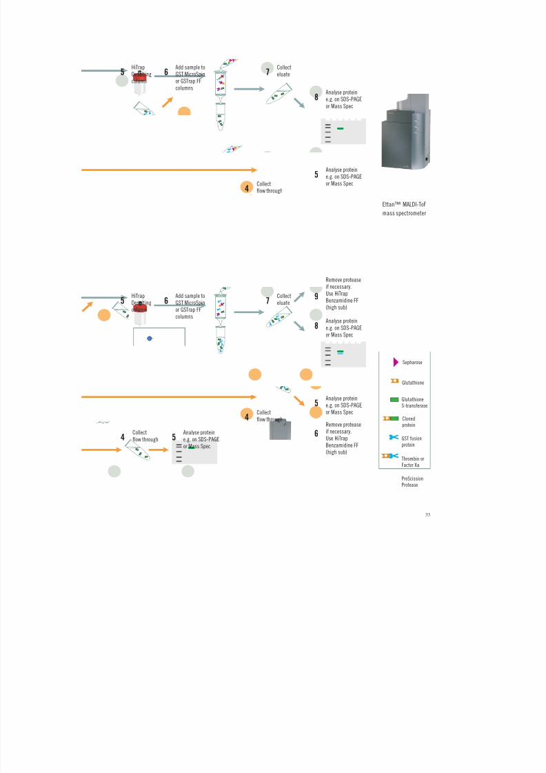
8/2/2019 The ant Protein Handbook
http://slidepdf.com/reader/full/the-ant-protein-handbook 35/112
33
Glutathione
S-transferase
Cloned
protein
GST fusion
protein
Thrombin or
Factor Xa
PreScissionProtease
Glutathione
Sepharose
Ettan™ MALDI-ToF
mass spectrometer
Collect
flow through
Collect
eluate
Analyse protein
e.g. on SDS-PAGE
or Mass Spec
Analyse protein
e.g. on SDS-PAGE
or Mass Spec
Add sample to
GST MicroSpin
or GSTrap FF
columns
HiTrap
Desalting
column
5 6 7
8
4
5
Analyse proteine.g. on SDS-PAGEor Mass Spec
Collect
flow through4 5
Collectflow through
Collecteluate
Analyse protein
e.g. on SDS-PAGEor Mass Spec
Remove protease
if necessary.Use HiTrapBenzamidine FF(high sub)
Remove proteaseif necessary.Use HiTrapBenzamidine FF(high sub)
Analyse proteine.g. on SDS-PAGEor Mass Spec
Add sample toGST MicroSpinor GSTrap FFcolumns
HiTrapDesaltingcolumn
5 6 7
8
9
6
4
5

8/2/2019 The ant Protein Handbook
http://slidepdf.com/reader/full/the-ant-protein-handbook 36/112
34
Alternative 1. On-column cleavage and purification
Assume: 8 mg GST fusion protein bound/ml gel, using GSTrap FF 1 ml column (adjust
volum es for larger columns)
1. Fill the syringe with binding buffer.
2. Connect the column to the syringe using the adapter supplied ("drop to drop" to avoid introducing air intothe column).
3. Remove the twist-off end.
4. Equilibrate the column with 5 column volumes of binding buffer.
5. Apply the sample using the syringe. For best results, maintain a flow rate of 0.2–1 ml/min as the sampleis applied.
6. Wash with 5–10 column volumes of binding buffer. Maintain flow rates of 1–2 ml/min during the wash.
7. Wash the column with 10 column volumes of PreScission cleavage buffer.
8. Prepare the PreScission Protease mix: mix 80 µl (160 units) of PreScission Protease with 920 µl ofPreScission cleavage buffer at +5 °C.
9. Load the PreScission Protease mix onto the column using a syringe and the adapter supplied.Seal the column with the top cap and the domed nut supplied.
10. Incubate the column at +5 °C for 4 hours.
11. Fill a syringe with 3 ml of PreScission cleavage buffer. Remove the top cap and domed nut.Avoid introducing air into the column. Begin elution and collect the eluate (0.5 ml–1 ml/tube).
N.B. The eluate will contain the protein of interest, while the GST moiety of the fusion protein and the
PreScission Protease will remain bound to GSTrap FF.
Alternative 2. O ff-column cleavage and purification
Assume: 8 mg GST fusion protein bound/ml gel, using GSTrap FF 1 ml column (adjust
volum es for larger columns)
1. Follow steps 1–6 above.
2. Elute with 5–10 column volumes of elution buffer. Maintain flow rates of 1–2 ml/min during elution.
3. Remove the reduced glutathione from the eluate using a quick buffer exchange on HiTrap Desalting orHiPrep™ 26/10 Desalting depending on the sample volume.
4. Add 1 µl (2 units) of PreScission Protease for each 100 µg of fusion protein in the eluate. If the amount offusion protein in the eluate has not been determined, add 80 µl (160 units) of PreScission Protease.
5. Incubate at +5 °C for 4 hours.
6. Once digestion is complete, apply the sample to an equilibrated GSTrap FF column to remove the GSTmoiety of the fusion protein and the PreScission Protease.
NB. The protein of interest will be found in the flow-through, while the GST moiety of the fusion protein and the
PreScission Protease will remain bound to the column.
Troubleshooting
Incomplete PreScission Pro tease cleavage
• Check that the PreScission Protease to fusion protein ratio is correct (although saturationof the purification column is rarely a problem).
• Increase the incubation time to 20 hours or longer at +5 °C and increase the amount of PreScission Protease used in the reaction.
• Verify presence of the PreScission Protease cleavage site. Compare the DN A sequence of
the constr uct with the kno wn PreScission Protease cleavage sequence. Verify tha t theoptimal PreScission Protease recognition site, Leu-Glu-Val-Leu-Phe-Gln¡Gly-Pro, hasnot been altered.

8/2/2019 The ant Protein Handbook
http://slidepdf.com/reader/full/the-ant-protein-handbook 37/112
35
• Remove possible PreScission Protease inhibitors by extensive washing of the purificationcolumn before cleaving with PreScission Prot ease. The presence of Z n2+ as well asPefabloc SC or chymostat in may interfere with PreScission Protease activity.
Multiple bands seen on SDS gel after cleavage
• Determine when the bands appear. Additional bands seen prior to PreScission Proteasecleavage may be the result of proteolysis in the host bacteria. E. coli BL21 is a recommended protease-deficient strain.
• Check the sequence of the fusion partner for the presence of additional PreScissionProtease recognition sites. PreScission Pro tease opt imally recognizes the sequenceLeu-Glu-Val-Leu-Phe-Gln¡Gly-Pro and cleaves between the Gln and Gly residues butsimilar secondary sites may exhibit some propensity for cleavage. Adjusting reactionconditions (e.g. time, temperature, salt concentration) may result in selective cleavage atthe desired site. If adjustment of the conditions does not correct the problem, reclone the
insert into a pGEX T (thrombin) or pGEX X (Factor Xa) expression vector.
Fusion par tner is contaminated with PreScission Protease after purification
• Pass the sample over fresh Glutathione Sepharose to remove residual PreScission Protease(the Glutathione Sepharose may have been saturated with GST fusion protein in the firstpurification). Alternatively, a conventional ion exchange chromatography separation canbe developed to remove the PreScission Protease and other minor contaminants(see Appendix 9).
Thrombin cleavage and purificationEnzymatic cleavage
PBS: 140 mM NaCl, 2.7 mM KCl, 10 mM Na2HPO4, 1.8 mM KH2PO4, pH 7.3.
Thrombin Solution: Dissolve 500 units in 0.5 ml of PBS pre-chilled to +4 °C. Swirl gently.Store solution in small aliquots at -80 °C to preserve activity.
With a specific activity > 7 500 units/mg protein one unit of thrombin will digest > 90% of 100 µg of a test fusion protein in 16 hours at +22 °C in elution buffer. A unit is approximatelyequal to 0.2 N IH units.
Cleavage should be complete following overnight treatment with < 10 units/mg of fusion p rotein.
Thrombin can be removed using Benzamidine Sepharose, a purification medium with ahigh specificity for serine proteases (see page 39). A GSTrap FF and a H iTrap Benzamidine FF(high sub) column can be connected in series so that cleaved product passes directly fromthe GSTrap FF into the H iTrap Benzamidine FF (high sub). Samples are cleaved and thethrombin removed in a single step (see page 39).

8/2/2019 The ant Protein Handbook
http://slidepdf.com/reader/full/the-ant-protein-handbook 38/112
36
Alternative 1. On-column cleavage and purification
Assume: 8 mg GST fusion protein bound/ml gel, using GSTrap FF 1 ml column (adjust
volum es for larger columns)
1. Follow steps 1–6 under on-column Prescission Protease cleavage.
2. Prepare the thrombin mix: mix 80 µl thrombin solution (1 unit/µl ) with 920 µl PBS.
3. Load the thrombin mix onto the column using a syringe and the adapter supplied. Seal the column withthe top cap and the domed nut supplied.
4. Incubate the column at room temperature (+22 to +25 °C) for 2–16 hours.
5. Fill a syringe with 3 ml PBS. Remove the top cap and domed nut. Avoid introducing air into the column.Begin elution and collect the eluate (0.5 ml–1 ml/tube).
N.B. The eluate will contain the protein of interest and thrombin, while the GST moiety of the fusion protein will
remain bound to GSTrap FF.
Alternative 2. O ff-column cleavage and purification
Assume: 8 mg GST fusion protein bound/ml gel, using GSTrap FF 1 ml column (adjust
volum es for larger columns)
1. Follow steps 1–6 under on-column Prescission Protease cleavage.
2. Elute with 5–10 column volumes of elution buffer. Maintain flow rates of 1–2 ml/min during elution.
3. Add 10 µl (10 units) of thrombin solution for each mg of fusion protein in the eluate. If the amount of fusionprotein in the eluate has not been determined, add 80 µl (80 units) thrombin solution.
4. Incubate at room temperature (+22 to +25 °C) for 2–16 hours.
5. Once digestion is complete, remove the reduced glutathione using a quick buffer exchange on HiTrapDesalting or HiPrep 26/10 Desalting depending on the sample volume.
6. Apply the sample to an equil ibrated GSTrap FF column to remove the GST moiety of the fusion protein.
N.B.The purified protein of interest and thrombin will be found in the flow-through.
Troubleshooting
Incomplete thrombin cleavage
• Check that the thrombin to fusion protein ratio is correct.
• Increase the reaction time to 20 hou rs at +22 to +25 °C and increase the amount of thrombin used in the reaction.
• Check the DN A sequence of the construct to verify the presence of the thrombin site.Verify that the thrombin site has not been altered.
• Check that protease inhibitors have been removed.
Multiple bands seen on SDS-PAGE analysis after cleavage
• Determine when the bands appear. Additional bands seen prior to cleavage may be theresult of proteolysis in the host bacteria. E. coli BL21 is a protease-deficient strain that isrecommended.

8/2/2019 The ant Protein Handbook
http://slidepdf.com/reader/full/the-ant-protein-handbook 39/112
37
• Check the sequence of the fusion partner for the presence of additional thrombinrecognition sites. Optimum cleavage sites for thrombin are given in 1) and 2) below. Ref: Chang, J-Y. , Eur. J. Biochem. 151, 217 (1985).
1) P4-P3-Pro-Arg/Lys¡P1´-P2´ where P3 and P4 are hydrophobic amino acids and P1´and P2´ are non-acidic amino acids. The Arg/Lys¡P1´ bond is cleaved.
Examples:
P2 R/K¡P1 ´
A) Ala Arg¡Gly
B) Gly Lys¡Ala
P4 P3 Pro R/K¡P1 ´ P2 ´
A) Met Tyr Pro Arg¡Gly Asn
B) Ile Arg Pro Lys¡Leu Lys
C) Leu Val Pro Arg¡Gly Ser
In A, the Arg¡Gly bond is cleaved very quickly by thrombin. In B, the Lys¡Leu bond iscleaved. C is the recognition sequence found on the thrombin series of pGEX plasmids, theArg¡Gly bond is cleaved.
2) P2-Arg/Lys¡P1´, where either P2 or P1' is Gly. The Arg/Lys¡P1´ bond is cleaved.
Examples:
In A, the Arg¡Gly bond is cleaved efficiently. In B, the Lys¡Ala bond is cleaved. Adjustingtime and temperature of digestion can result in selective cleavage at the desired thrombinsite. If adjustment of conditions does not correct the problem, reclone the insert into a
pGEX-6P (PreScission) or pGEX (Factor Xa) expression vector.
Factor Xa cleavage and purification
Enzymatic cleavage
Factor Xa cleavage buffer: 50 mM Tris-HCl, 150 mM NaCl, 1 mM CaCl2, pH 7.5.
Factor Xa solution: Dissolve 400 units of Factor Xa in +4 °C water to give a final solution of 1 unit/µl.Swirl gently.
Store solution in small aliquots at -80 °C to preserve activity.
With a specific activity of > 800 units/mg protein, one unit of Factor Xa will digest > 90% of 100 µg of a test fusion protein in 16 hours at +22 °C in Factor Xa cleavage buffer.
Cleavage should be complete following overnight treatment at +22 °C with a Factor Xa tosubstrate ratio of at least 1% (w/w).
Factor Xa can be removed using Benzamidine Sepharose, a purification medium with ahigh specificity for serine proteases (see page 39). A GSTrap FF and a H iTrap Benzamidine FF(high sub) column can be connected in series so that cleaved product passes directly fromthe GSTrap FF into the H iTrap Benzamidine FF (high sub). Samples are cleaved and theFactor Xa removed in a single step (see page 39). Heparin Sepharose has been used for this
application but, since benzamidine has a higher specificity for Factor Xa, the protease willbe removed more effectively.

8/2/2019 The ant Protein Handbook
http://slidepdf.com/reader/full/the-ant-protein-handbook 40/112
38
Factor Xa consists of two subunits linked by disulphide bridges. Since glutathione candisrupt disulphide bridges, it should be removed from the sample prior to the cleavagereaction. Glutathione can be easily and rapidly removed from the fusion protein using adesalting column (Chapter 7) with Factor Xa cleavage buffer as eluent.
Alternative 1. On-column cleavage and purification
Assume: 8 mg GST fusion protein bound/ml gel, using GSTrap FF 1 ml column (adjust
volum es for larger columns)
1. Follow steps 1–6 under on-column Prescission Protease cleavage.
2. Wash GSTrap FF with 10 column volumes of Factor Xa cleavage buffer.
3. Prepare the Factor Xa mix: Mix 80 µl Factor Xa solution with 920 µl Factor Xa cleavage buffer.
4. Load the Factor Xa mix onto the column using a syringe and the adapter supplied. Seal the column with thetop cap and the domed nut.
5. Incubate the column at room temperature (+22 to +25 °C) for 2–16 hours.
6. Fill a syringe with 3 column volmes of Factor Xa cleavage buffer. Remove the top cap and domed nut. Avoidintroducing air into the column. Begin elution and collect the eluate (0.5 ml–1 ml/tube).
N.B. The eluate will contain the protein of interest and Factor Xa, while the GST moiety of the fusion protein will
remain bound to GSTrap FF.
Alternative 2. O ff-column cleavage and purification
Assume: 8 mg GST fusion protein bound/ml gel, using GSTrap FF 1 ml column (adjust
volum es for larger columns)
1. Follow steps 1–6 under on-column Prescission Protease cleavage.
2. Elute with 5–10 column volumes of elution buffer. Maintain flow rates of 1–2 ml/min during elution.
3. Remove the reduced glutathione from the eluate using a quick buffer exchange on HiTrap Desalting orHiPrep 26/10 Desalting depending on sample volume.
4. Add 10 units of Factor Xa solution for each mg fusion protein in the eluate. If the amount of fusion protein inthe eluate has not been determined, add 80 µl (80 units) of Factor Xa solution.
5. Incubate the column at room temperature (+22 to +25 °C) for 2–16 hours.
6. Once digestion is complete, apply the sample to an equilibrated GSTrap FF column to remove the GSTmoiety of the fusion.
N.B. The protein of interest will be found in the flow-through together with Factor Xa.
Troubleshooting
Incomplete Factor Xa cleavage
• Factor Xa requires activation of Factor X with Russell's viper venom. Factor Xa fromAmersham Biosciences has been preactivated, but other sources may not beactivated. Activation conditions: Incubate 1% (w/w) of Russell's viper venom to Factor Xin 8 mM Tris-HCl (pH 8.0), 70 mM NaCl, 8 mM CaCl2 at +37 °C for 5 minutes.
• Check the DNA sequence of the construct to verify the presence of the Factor Xa site.Verify that the Factor Xa site has not been altered. The recognition sequence forFactor Xa is Ile-Glu-Gly-Arg¡X, where X can be any amino acid except Arg or Pro.
• Check that the Factor Xa to fusion protein ratio is correct.
• Check that glutathione has been removed as recommended.

8/2/2019 The ant Protein Handbook
http://slidepdf.com/reader/full/the-ant-protein-handbook 41/112
39
• In some cases increasing substrate concentration up to 1 mg/ml may improve results.
• Add < 0.5% w/v SDS to th e reaction buffer. This can significantly improve Factor Xacleavage with some fusion pro teins. Variou s concentrat ions of SDS should be tested tofind th e optimum concentrat ion.
• Increase incubation time to 20 hours or longer at +22 °C and increase the amount of Factor Xa (for some fusion pro teins, Factor Xa can be increased up to 5 % ).
• Check that protease inhibitors have been removed.
Multiple bands on SDS-PAGE gel after cleavage
• Determine when the bands appear. Additional bands seen prior to cleavage may be theresult of proteolysis in the host bacteria. E. coli BL21 is a protease-deficient strain that isrecommended.
• Check the sequence of the fusion partner for the presence of Factor Xa recognition sites.
Factor Xa is highly specific for the recognition sequence Ile-Glu-Gly-Arg¡. The bondfollowing the Arg residue is cleaved. Adjusting t ime and tempera ture of digestion canresult in selective cleavage at the desired Factor Xa site. If adjustment of conditions doesnot correct the problem, reclone the insert into a pGEX-6P (PreScission) or pGEX T(thrombin) expression vector.
Removal of thrombin, Factor Xa or other serine proteases
Before enzymatic cleavage it may be necessary to remove pro teases from the samp le andafter enzymatic cleavage it may be necessary to remove thrombin or Factor Xa.
Benzamidine 4 Fast Flow (high sub) provides a convenient an d highly specific medium forthe removal of serine proteases not only from enzymatic digests, but also from cell culturesupernatant, bacterial lysate or serum.
Purification Options
Binding buffer: 0.05 M Tris-HCl, 0.5 M NaCl, pH 7.4
Elution buffer alternatives:
- 0.05 M glycine-HCl, pH 3.0
- 10 mM HCl, 0.5 M NaCl, pH 2.0
- 20 mM para-aminobenzamidine in binding buffer (competitive elution)
- 8 M guanidine hydrochloride or 6 M urea (denaturing solutions)
Column (prepacked) or M edium Binding capacity Commentsfor serine proteases
HiTrap Benzamidine FF (high sub), 1 ml > 35 mg trypsin Prepacked 1 ml column
HiTrap Benzamidine FF (high sub), 5 ml > 175 mg trypsin Prepacked 5 ml column
Benzamidine Sepharose 4 Fast Flow (high sub) > 35 mg trypsin/ml medium For column packingand scale-up

8/2/2019 The ant Protein Handbook
http://slidepdf.com/reader/full/the-ant-protein-handbook 42/112
40
1. Equilibrate the column with 5 column volumes of binding buffer.
2. Apply the sample.
3. Wash with 5–10 column volumes of binding buffer or until no material appears in the effluent (monitored byUV absorption at A280 nm).
4. Elute with 5–10 column volumes of elution buffer. Collect fractions in neutralization buffer*. The purifiedfractions can be buffer exchanged using desalting columns (see Chapter 7).
*Since elution conditions are quite harsh, collect fractions into neutralization buffer (60 µl–200 µl 1 M Tris-HCl,
pH 9.0 per ml fraction), so that the final pH of the fractions will be approximately neutral.
Figure 9 shows the isolation of a GST fusion protein on a GSTrap FF column and on-column cleavage with thrombin (as described previously). After the thrombin incubation aHiTrap Benzamidine FF (high sub) column is placed in series below the GSTrap FF column .As binding buffers are passed through the two columns the cleaved fusion protein andthrombin are washed from the GSTrap FF column onto the HiTrap Benzamidine FF (high sub)column. This column binds the th rombin enabling the collection of pur e thrombin-freeprotein in the eluent.
Sample: 2 ml clarified E.coli homogenate expressing a Mr 37 000 SH2-GST fusion proteinwith a thrombin protease site
Columns: GSTrap FF, 1 ml and HiTrap Benzamidine FF (high sub), 1 mlBinding buffer: 20 mM sodium phosphate, 0.15 M NaCl, pH 7.5High salt wash buffer: 20 mM sodium phosphate, 1.0 M NaCl, pH 7.5Benzamidine elution buffer: 20 mM p-aminobenzamidine in binding bufferGST elution buffer: 20 mM reduced glutathione, 50 mM Tris, pH 8.0Flow: 0.5 ml/minSystem: ÄKTAprime
Protease treatment: 20 units/ml thrombin protease (Amersham Biosciences) for 2 hours at room temperatureThrombin activity: S-2238 (Chromogenix, Haemochrom Diagnostica AB) was used as a substrate and its absorbance at
405 nm was measured
Gel: ExcelGel SDS Gradient 8-18%,Coomassie staining
Lane 1. Low Molecular WeightCalibration Kit (LMW)
Lane 2. Clarified E.coli homogenate expressingSH2-GST fusion protein
Lane 3. Flow-through from GSTrap FF (Fraction 2)Lane 4. SH2 GST-tag cleaved, washed out with
binding buffer through both columns(Fraction 6)
Lane 5. as above (Fraction 7)Lane 6. as above (Fraction 8)Lane 7. Elution of thrombin from
HiTrap Benzamidine FF (high sub)Lane 8. Elution of GST-tag and some non-cleaved
SH2-GST from GSTrap FF (Fraction 21)Lane 9. as above (Fraction 22)
1 2 3 4 5 6 7 8 9
Mr
97 00066 000
20 100
30 000
45 000
14 400
Fig. 9. On-column cleavage of a GST fusion protein and removal of thrombin after on-column cleavage, usingGSTrap FF and H iTrap Benzamidine FF (high sub).
0 0
0.100.20
0.20
0.30
0.40
0.80
0.60
0
f r .
6
Cleaved SH2protein
Thrombin
A)
B)
GSTrap FF, 1 ml
HiTrap Benzamidine FF (high sub), 1 ml
Thrombin
High saltbuffer wash
Elution of HiTrap
Benzamidine FF(high sub)
Elution ofGSTrap FF
GST-tag
f r .
7
f r .
8
f r .
1 4
f r .
2 1
f r .
2 2
f r .
2
10 20 50 ml15 25
A280 nm A405 nm
A) B) A)
B)
B) A)

8/2/2019 The ant Protein Handbook
http://slidepdf.com/reader/full/the-ant-protein-handbook 43/112
41
CHAPTER 4
(His)6
fusion proteins
AmplificationGeneral considerations for the amplification of fusion proteins are discussed in Chapter 2.
The vectors and hosts
There is a wide variety of hosts and vectors for the amplification of (His) 6 fusion proteins.The factors that should be considered when selecting the host and vector are discussed inChapter 1.
Purification
Figure 10 gives an overview of a typical purification flow scheme for (His)6 fusion proteins,including purification under denatu ring conditions. On -column purification and refoldingof (His)6 fusion proteins is described in Chapter 5.
Native
conditions
Binding buffer
Binding buffer
Denaturing
conditions
Cell lysis
Binding buffer including
8 M Urea or6 M Gua-HCl
Binding to
affinity media
Wash
Elute
Pure denaturedfusion protein
Pure fusion
protein
Refolding
Binding buffer
(as above) including10-50 mM imidazole
Elution buffer: Binding buffer (as above)
with increased amount of imidazole
Elution buffer: Binding buffer with
increased amount of imidazole
Fusion protein
Cell protein
Fig. 10.

8/2/2019 The ant Protein Handbook
http://slidepdf.com/reader/full/the-ant-protein-handbook 44/112
42
For simple, one step purification of (His)6 fusion proteins, there is a range of productsdesigned to meet specific pur ification needs, as shown in Table 13. These products can alsobe used for the purification of fusion proteins containing shorter or longer polyhistidinetags, such as (His)4 or (H is)10 . Under the standard binding and elution conditions describedin this handbook , the shorter (His)4 will bind more weakly and the longer (His)10 will bind
more strongly as compared to (His)6. This difference in binding strength can be used toadvantage during purification. For example, since (His)10 binds more strongly, a higherconcentration of imidazole can be used during the washing step before elution. This canfacilitate the removal of contaminants which may otherwise be co-purified with a (His)6
fusion p rotein.
Chelating Sepharose, when charged with Ni2+ ions, selectively retains proteins if complex-forming amino acid residues, in particular histidine, are exposed on the protein surface.(His)6 fusion proteins can be easily desorbed with buffers containing imidazole.
If the (His)6 fusion proteins are expressed as inclusion bodies, see Chapter 5 forsolubilization, refolding and purification information.
Column (prepacked) Amount of protein for Commentor Media* * a single purification
His MicroSpin Purification Module Up to 100 µg Ready to use, prepacked columns, buffers andchemicals
High throughput when used with MicroPlex 24
Vacuum (up to 48 samples simultaneously)
HiTrap Chelating HP 1 ml Up to 12 mg* Prepacked column, ready to use
HisTrap Kit Up to 12 mg/column* As above, but includes buffers for up to 12purifications using a syringe
HiTrap Chelating HP 5 ml Up to 60 mg* Prepacked column, ready to use
Chelating Sepharose Fast Flow 12 mg (His)6 fusion protein* For packing columns and scale upper ml
*estimate for a protein ofMr 27 600, binding capacityvaries according to specificprotein
Re-use of purification columns depends upon the nature of the sample and should only beperformed with identical samples to prevent cross contamination.
Batch preparation p rocedures are frequently mentioned in the literatur e. How ever theavailability of prepacked columns and easily packed high flow rate Chelating Sepharoseprovide faster, mor e convenient alterna tives. Batch p reparation s are occasionally used if itappears that the tag is not fully accessible or when the protein in the lysate is at very lowconcentrations (both could appear to give a low yield from the first purification step).A more convenient alternative to improve yield is to decrease the flow rate or pass thesample through the column several times.
Table 13. Summary of purification options for (His)6
fusion proteins.
**Characteristics of HiTrap Chelating HP and Chelating Sepharose Fast Flow are given in Appendix 5.

8/2/2019 The ant Protein Handbook
http://slidepdf.com/reader/full/the-ant-protein-handbook 45/112
43
Monitor purification steps by one or more of the detection methods referred to in th is book.The choice of purification equipment should also be made according to the needs of thepurification. Appendix 8 provides a guide to aid in the selection of the correct purificationsolution and key points to consider are highlighted below.
• For a single purification of a small quan tity of product or for high throu ghput screening,MicroSpin columns, using centrifugation or MicroPlex 24 Vacuum respectively, areconvenient and simple to use.
• For purification of larger quantities of fusion proteins HisTrap Kit or HiTrap Chelating HPcolumns are ready to use with a syringe, a peristaltic pump or a chromatography system.
• To increase capacity use several HiTrap Chelating HP columns (1 ml or 5 ml) in series or,for even larger capacity requirements, pack Chelating Sepharose Fast Flow into a suitablecolumn (details of column packing procedures are outlined in Appendix 6).
• For simple and reproducible purification a chromatography system such as ÄKTA prime
is a significant advantage, recording the purification process and eliminating manualerrors.
• For laboratory environments in which all experimental data must be recorded and traceableor where method d evelopment, optimization and scale up are r equired, a computercontrolled ÄKTAdesign chromatography system is recommended.
• Experiments such as protein refolding or method opt imization require linear gradientelution steps that can only be performed by a chromatography system.
His MicroSpin Purification ModuleThe His MicroSpin Purification Module is usefulfor screening small or large numbers of lysates andfor checking samples during the optimization of amplification or purification conditions.Each module conta ins reagents sufficientfor 50 purifications.
• 10X PBS: 1.4 M NaCl, 27 mM KCl, 101 mM Na2HPO4, 18 mM KH2PO4, pH 7.3
• 8X Phosphate/NaCl buffer: 160 mM phosphate, 4 M NaCl, pH 7.4
• 4 M Imidazole elution buffer (avoid contact with skin or eyes)
• IPTG: 500 mg
• His MicroSpin columns: 50 units
Reagents are prepared as follows:
1X PBS: Dilute 10X PBS with sterile water. Store at +4 °C.
PNI20 wash buffer: Mix 6.25 ml of 8X phosphate/NaCl buffer with 0.25 ml imidazole elution buffer.Add distilled water to a final volume of 50 ml.
PNI400 elution buffer: Mix 6.25 ml of 8X phosphate/NaCl buffer with 5 ml imidazole elution buffer.Add distilled water to a final volume of 50 ml.
IPTG 100 mM: Dissolve contents of the IPTG vial in 20 ml sterile water.Store as 1 ml aliquots at -20 °C.

8/2/2019 The ant Protein Handbook
http://slidepdf.com/reader/full/the-ant-protein-handbook 46/112
44
Alternative 1. H igh th roughput purification using MicroPlex Vacuum
Do not apply more than 400 µl of sample at a timeto a H is MicroSpin column. Th is procedure willaccommodate lysates from 2 to 8 ml of culture.
Also required:
• Vacuum source capable of providing 220 mm Hg (e.g. a water vacuum).
• Side arm flask, 500 ml or 1 litre.
• Single or double hole rubber stop.
• Vacuum tubing.
• MicroPlex 24 Vacuum apparatus (one or two).
1. Assemble the MicroPlex 24 Vacuum following the instructions supplied.
2. Remove the caps from the MicroSpin columns. Place the columns in the manifold, filling any unused holeswith the plugs provided with MicroPlex 24 Vacuum.
3. Apply 400 µl of lysate to the column.
If using < 400 µl adjust volume to 400 µl with 1X PBS beforehand.
4. Recap each column securely and mix by gentle, repeated inversion. Incubate at room temperaturefor 5–10 minutes.
5. Remove columns from the manifold and remove top caps and bottom closures. Return columns tothe manifold.
6. Ensure the stopcock is in the closed position (i.e. perpendicular to the vacuum tubing) and that themanifold is placed squarely on the gasket.
7. Turn on vacuum supply at source. Open the stopcock (i.e. parallel to the vacuum tubing). After lysates havebeen drawn through all the columns into the collection tray, close the stopcock.
8. Allow 10–15 seconds for the vacuum pressure to dissipate. Remove the manifold and place it on a paper towel.
9. Discard eluate and re-use collection plate or save eluate in the collection plate for later analysis and use anew collection plate for the next step.
10. Add 600 µl of PNI20 wash buffer to each column. Open the stopcock. After buffer has been drawn throughall the columns into the collection tray, close the stopcock.
11. Allow 10–15 seconds for the vacuum pressure to dissipate. Remove the manifold and reassemble theapparatus with a clean collection tray. Additonal 600 µl washes with PI 20 may be performed if desired.Washes with higher concentrations of imidazole may remove impurities, but decrease yield of thefusion protein.
12. Add 200 µl of PNI400 elution buffer to each column. Incubate at room temperature for 5–10 minutes.
13. Open the stopcock. After elution buffer has been drawn through all the columns into the collection tray,close the stopcock.
14. Allow 10–15 seconds for the vacuum pressure to dissipate. Remove the manifold. Cover eluates with sealingtape until required for analysis.
Note: Yields of fusion protein may be increased by repeating the elution step two or three times and pooling
the eluates.
Troubleshooting
See Purification and D etection Troubleshoot ing page 56.

8/2/2019 The ant Protein Handbook
http://slidepdf.com/reader/full/the-ant-protein-handbook 47/112
45
Alternative 2. Purification of multiple samples using a microcentrifuge
Do no t app ly more than 400 µl of sample at a t ime to a H is MicroSpin column. Thisprocedure will accommodate lysates from 2 to 8 ml of culture.
1. Remove (and save) each column cap.
2. Apply 400 µl of lysate to the column.
If using < 400 µl adjust volume to 400 µl with 1X PBS beforehand.
3. Recap each column securely and mix by gentle, repeated inversion. Incubate at room temperaturefor 5–10 minutes.
4. Remove (and save) the top cap and bottom closure from each column. Place column into a clean1.5 or 2 ml microcentrifuge tube. Spin for 1 minute at 735 g.
5. Remove the MicroSpin columns and place into a clean 1.5 or 2 ml microcentrifuge tube.
6. Apply 600 µl of PNI20 wash buffer to each column. Repeat spin procedure. Additonal 600 µl washes withPI20 may be performed if desired. Washes with higher concentrations of imidazole may remove impurities,but decrease yield of the fusion protein.
7. Replace the bottom closure on each column. Add 100–200 µl of PNI400 elution buffer to each column.Replace top cap and incubate at room temperature for 5–10 minutes.
8. Remove and discard top cap and bottom closure from each column and place the column into a clean1.5 or 2 ml microcentrifuge tube.
9. Spin all columns again to collect eluate. Save for analysis.
Note: Yields of fusion protein may be increased by repeating the elution step two or three times and pooling the
eluates.
Troubleshooting
See Purification and D etection T roubleshooting page 56.

8/2/2019 The ant Protein Handbook
http://slidepdf.com/reader/full/the-ant-protein-handbook 48/112
46
Alternative 3. Purification using MicroPlex Centrifugation
Do no t app ly more than 400 µl at a t ime to a H is MicroSpin column. This procedure willaccommodate lysates from 2 to 8 ml of culture.
See Appendix 4 for recommended centrifugation systems.
1. Assemble the MicroPlex 24 unit following the instructions supplied. Two units can be processedsimultaneously to handle 48 samples.
2. Remove the caps from the MicroSpin columns and place the columns in the manifold.
3. Apply 400 µl of lysate to each column.
If using < 400 µl adjust volume to 400 µl with 1X PBS beforehand.
4. Recap each column securely and mix by gentle, repeated inversion. Incubate at room temperaturefor 5–10 minutes.
5. Remove columns from the manifold and remove top caps and bottom closures. Return columns tothe manifold.
6. Centrifuge the unit for 2 minutes following the instructions supplied.
7. Add 600 µl of PNI20 wash buffer to each column.
8. Centrifuge the unit for 2 minutes following the instructions supplied. Remove the manifold from eachcollection tray and place on a clean paper towel. Reassemble each unit with a fresh collection tray.Additonal 600 µl washes with PI20 may be performed if desired. Washes with higher concentrations ofimidazole may remove impurities, but decrease yield of the fusion protein.
9. Add 100–200 µl of PNI400 elution buffer to each column. Incubate at room temperature for 5–10 minutes.
10. Centrifuge the unit for 2 minutes following the instructions supplied. Cover the eluted samples with sealingtape until required for analysis.
Note: Yields of fusion protein may be increased by repeating the elution step two or three times and pooling the
eluates.
Troubleshooting
See Purification and D etection Troubleshoot ing page 56.

8/2/2019 The ant Protein Handbook
http://slidepdf.com/reader/full/the-ant-protein-handbook 49/112
47
Re-use of any purification column depends on the natu re of the sample and should only beperformed with identical fusion proteins to prevent cross-contamination.
HiTrap Chelating HP columns (1 ml and 5 ml) can be connected in series to increase thebinding capacity. At larger scale, columns can be packed with Chelating Sepharose FastFlow (for column packing see Appendix 6).
Sample and buffer preparation
Use high quality water and chemicals. Filtration through 0.45 µm filters is recommended.
Samples should be centrifuged immediately before use and/or filtered through a 0.45 µmfilter. If the sample is too viscous, dilute with binding buffer.
Sample binding properties can be improved by adjusting the sample to the composition of the binding buffer: dilute in binding buffer or perform a buffer exchange using a desaltingcolumn (see Chapter 7).
If the fusion protein is expressed as an inclusion body, add 6 M guanidine hydrochloride or8 M urea to a ll buffers.
During purification there is a balance between the amount of imidazole needed to preventnon-specific binding of contaminants and the amount of imidazole needed to elute the(His)6 fusion protein, conditions may need to be optimized.
Fig. 11 . HiTrap Chelating HP (1 ml) and a schematic overview of (His)6
fusion protein purification.
Equilibrate columnwith
binding buffer
Waste
3 min
Waste
3 min
Apply samplewash with
binding buffer
Collect
5-15 min
Elutewith
elution buffer
Collect fractions
2 min
Prepare columnwash with H Oload with NiSO
wash with H O
2
2
4
Purification using HiTrap Chelating HP 1 ml or 5 ml columns
HiTrap Chelating HP columns are prepacked and ready for use.They can be operated with a syringe, a peristalticpump or a liquid chromatography system such
as ÄKTA prime. Figure 11 illustrates thesimple steps required for purificationof a (His)6 fusion protein.

8/2/2019 The ant Protein Handbook
http://slidepdf.com/reader/full/the-ant-protein-handbook 50/112
48
Alternative 1. Purification using HisTrap Kit
HisTrap Kit includes everything needed for 12 preparationsusing a syringe. Three ready-to-use HiTrap Chelating HP1 ml columns and ready-made buffer concentratesare supplied with easy-to-follow instructions.
• HiTrap Chelating HP columns 3 x 1 ml• Phosphate buffer, 8X stock solution, pH 7.4 2 x 50 ml
• 2 M imidazole, pH 7.4 50 ml
• 0.1 M NiSO4 10 ml
• Syringe 5 ml
• All necessary connectors
Purification pro tocols
Column preparation: loading with nickel ions
1. Fill syringe with distilled water. Remove stopper and connect the column to the syringe with the adapterprovided ("drop to drop" to avoid introducing air into the column).
2. Remove the twist-off end.
3. Wash the column with 5 ml distilled water, using the syringe.
Use water, not buffer, to wash away the storage solution (precipitation of nickel salts mayoccur if the nickel solution comes into contact with the 20% ethanol in the storage solution).If air is trapped in the column, wash the column with distilled water until the air d isappears.
4. Disconnect syringe from the column, fill syringe with 0.5 ml of the 0.1 M nickel solution supplied and loadonto the column.
5. Wash column with 5 ml distilled water, using the syringe.
The column is now ready for use. When high yield is more important than highest purity, follow Protocol A onpage 49. To achieve the highest purity, follow Protocol B on page 50.

8/2/2019 The ant Protein Handbook
http://slidepdf.com/reader/full/the-ant-protein-handbook 51/112
49
Samples: 2.5 ml GST-(His)6 cell extract
Binding buffer: 1X binding buffer, 20 mM imidazole, pH 7.4Elution buffer: 1X binding buffer, 500 mM imidazole, pH 7.4Flow: 2 ml/min, 312 cm/hNote: No Ni
2+ re-loading of the column between the runs
Result:Run No. Eluted GST-(His)6, total mg
1 2.762 2.823 2.834 2.725 2.716 2.657 2.648 2.639 2.54
10 2.59
Fig. 12 . 10 repetitive purifications of GST-(His)6
without reloading the HiTrap Chelating HP 1 m l column with N i2+
between the runs.
Troubleshooting
See Purification and D etection T roubleshooting page 56.
0
1 2 43 5 6Run No. 7 8 9 10
0.5
1.0
1.5
2.5
3.0
2.0
6
mgelutedGST-(His)
A. Basic protocol for high yield
In all steps use the syringe supplied. Note that 1 ml/min corresponds to approximately 30 drops/min whenusing a syringe with a HiTrap 1 ml column and 5 ml/min corresponds to approximately 120 drops/min whenusing a HiTrap 5 ml column.
1. Prepare 24 ml binding buffer. Mix 3 ml phosphate buffer 8X stock solution with 0.12 ml 2 M imidazole and
add water up to 24 ml. Check pH and adjust to pH 7.4–7.6 if necessary. This buffer contains20 mM phosphate, 0.5 M NaCl and 10 mM imidazole.
2. Prepare 8 ml elution buffer. Mix 1 ml phosphate buffer 8X stock solution with 2 ml 2 M imidazole and adddistilled water up to 8 ml. Check pH and adjust to pH 7.4–7.6 if necessary. This buffer contains20 mM phosphate, 0.5 M NaCl and 500 mM imidazole.
3. Equilibrate the column with 10 ml binding buffer. Ensure that the column has been loaded with nickel ionsbefore this step.
4. Apply sample at a flow rate 1–3 ml/min. Collect the flow-through fraction. A pump is more suitable forapplication of sample volumes greater than 15 ml.
5. Wash with 10 ml binding buffer. Collect wash fraction.
6. Elute with 5 ml elution buffer. Avoid dilution of the purified product by collecting the eluate in 1 ml fractions.
7. Check the purification by analysing aliquots of starting material, flow through and eluent, for example, bySDS-PAGE. The purified protein is most likely to be found in the 2nd + 3rd ml of the elution step.
Imidazole absorbs at 280 nm. Use elution buffer as the blank when monitoring absorban ce.If imidazole needs to be removed use a desalting column (see Chapt er 7).
8. After the protein has been eluted, regenerate the column by washing with 10 ml of binding buffer.The column is now ready for a new purification if the same (His)6 protein is to be purified and there is rarelya need to reload with metal (see Figure 12).

8/2/2019 The ant Protein Handbook
http://slidepdf.com/reader/full/the-ant-protein-handbook 52/112
50
2. Load the column with nickel ions according to "Column preparation" (page 48).
3. Equilibrate the column with 10 ml binding buffer (1X phosphate buffer, 10 mM imidazole, pH 7.4).
4. Apply the sample. Collect the flow-through fraction.
5. Wash with 10 ml binding buffer. Collect the wash fraction.
6. Begin elution with 5 ml 1X phosphate buffer containing 20 mM imidazole. Avoid dilution by collecting theeluate in 1 ml fractions.
7. Proceed with the next imidazole concentration, i.e. elute with 5 ml 1X phosphate buffer containing
40 mM imidazole. Collect the eluate in 1 ml fractions as above.8. Proceed with the buffers of increasing imidazole concentration, as described in steps 6 and 7.
The purified protein is most likely to be found in the 2nd + 3rd ml of one of the elution steps.
9. Check the collected fractions, for example, by SDS-PAGE.
10. Regenerate column by washing with 10 ml binding buffer.
From the results select the optimal binding and elution buffers. The optimum elution bufferis the one which eluted the (His)6 fusion protein. The optimum binding buffer is the elutionbuffer used in the step before, with a lower concentration of imidazole. Using the highestpossible concentration of imidazole in the binding buffer will give the highest purity. Thesebuffers can be used for any purification of an identical protein.
Imidazole absorbs at 280 nm. Use elution bu ffer as the blank when mon itoring absorbance.If imidazole needs to be removed use a desalting column (see Chap ter 7).
Perform a blank run to elute non-specifically bound metal ions. Add 5 column volumesbinding buffer followed by 5 column volumes elution buffer. Re-equilibrate the columnwith 5–10 column volumes of binding buffer before sample application.
For large sample volumes a simple peristaltic pump or an ÄKTAdesign chromatographysystem can be used to apply sample and buffers.
Troubleshooting
See Purification and Detection Troubleshoot ing page 56.
Imidazole concentration Phosphate buffer 2 M Imidazole Final volumein buffer (mM) 8X stock solution pH 7 .4 (ml) pH 7 .4 (ml) (ml)*
10 3.0 0.12 24
20 1.0 0.08 8
40 1.0 0.16 8
60 1.0 0.24 8
100 1.0 0.40 8
300 1.0 1.20 8
500 1.0 2.00 8
*Use distilled water to adjust to final volume
B. Optimization pro tocol for high purity
In all steps use the syringe supplied. Note that 1 ml/min corresponds to approximately 30 drops/min whenusing a syringe with a HiTrap 1 ml column and 5 ml/min corresponds to approximately120 drops/min whenusing a HiTrap 5 ml column.
1. Prepare buffers according to Table 14. Use 1X phosphate buffer including 10 mM imidazole as binding
buffer and 6 steps ranging up to 500 mM imidazole as elution buffers. Check pH after mixing and adjust topH 7.4–7.6 if necessary.
Table 14. General mixing table using HisTrap Kit buffers for one purification.

8/2/2019 The ant Protein Handbook
http://slidepdf.com/reader/full/the-ant-protein-handbook 53/112
51
Alternative 2. Simple purification with ÄKTA prime
ÄKTA prime contains a pre-programmed template for automat ic column preparation andpurification of (His)6 fusion proteins on a single HiTrap Chelating HP column, as shownbelow. This provides a standard purification protocol which can be followed exactly oroptimized as required.
Binding buffer: 20 mM sodium phosphate, 0.5 M NaCl, pH 7.4
Elution buffer: 20 mM sodium phosphate, 0.5 M NaCl, 0.5 M imidazole, pH 7.4
Eluent: Distilled water
Metal loading solution: 0.1 M NiSO4
Prepare at least 500 ml of each buffer.
1. Follow the instructions supplied on ÄKTAprime cue card Code No: 18-1138-02.
2. Select the Application Template.
3. Start the method.
4. Enter the sample volume and press OK.
Connecting the column. Preparing the fraction collector.
Fig. 13. Typical procedures when using ÄKTA prime.
625 11 11 20 17 5 Min
100
50
Sample
System preparation
& water wash
Wash
Elution
Total separation time = 77 min + sample application time
Water wash
Buffer wash
Reequilibration
Ni-loading
Equilibration
% Elution buffer

8/2/2019 The ant Protein Handbook
http://slidepdf.com/reader/full/the-ant-protein-handbook 54/112
52
Figure 14 shows an example of the purification of a (His)6 fusion protein using aHiTrap C helating HP 1 ml column.
Troubleshooting
See Purification and D etection Troubleshooting page 56.
An example of the purification and refolding of an insoluble (His)6 fusion protein is shownon page 61 and clearly demonstrates the advantage of using a chromatography system forthis type of work.
Fig. 14 a. Purification of histidine-tagged GlutathioneS-tran sferase from a cytoplasmic extract.
Sample: 5 ml cytoplasmic extract containing (His)-taggedGlutathione S-transferase, GST-(His)6The clone was a kind gift from Dr. J. Lidholm,Amersham Biosciences, Sweden
Column: HiTrap Chelating HP 1 ml, Ni2+-loaded according tothe instructions
Binding buffer: 1X phosphate buffer, 20 mM imidazole, pH 7.4
Elution buffer: 1X phosphate buffer, 500 mM imidazole, pH 7.4Flow: 2 ml/min, 312 cm/hResult: Eluted GST-(His)6, 4 ml, A280: 1.65
Total amount: 4.46 mg
Flow through
Sampleapplication
Bindingbuffer
Elutionbuffer
Bindingbuffer
A
A
280 nm
280
A
A
405 nm
405
1.0
0.5
0.0
0.70
0.60
0.50
0.40
0.30
0.20
0.10
0.0 5.0 10.0 15.0 20.0 Volume (ml)
Elutedpool
Wash
Fig. 14b . SDS electropho resis on PhastSystem™using PhastGel™ 10–15, silver staining.
Lane 1: Low Molecular Weight Calibration Kit (LMW)Lane 2: Starting material, cytoplasmic extract, dil. 1:20Lane 3: Flow-through, dil. 1:10Lane 4: WashLane 5: Eluted GST-(His)6, dil 1:20Lane 6: Eluted GST-(His)6, dil 1:10
Lane 7: GST standard, 0.5 mg/mlLane 8: LMW
1 2 3 4 5 6 7 8
Fig. 14 .
Mr
97 000
30 000
45 000
14 400
66 000
20 100

8/2/2019 The ant Protein Handbook
http://slidepdf.com/reader/full/the-ant-protein-handbook 55/112
53
Alternative 1: SDS-PAGE analysis
6X SDS loading buffer: 0.35 M Tris-HCl, 10.28% (w/v) SDS, 36% (v/v) glycerol, 0.6 M dithiothreitol(or 5% 2-mercaptoethanol), 0.012% (w/v) bromophenol blue, pH 6.8.Store in 0.5 ml aliquots at -80 °C.
1. Add 2 µl of 6X SDS loading buffer to 5–10 µl of supernatant from crude extracts, cell lysates or purified
fractions as appropriate.2. Vortex briefly and heat for 5 minutes at +90 to +100 °C.
3. Load the samples onto an SDS-polyacrylamide gel.
4. Run the gel for the appropriate length of time and stain with Coomassie Blue (Coomassie Blue R Tablets) orsilver (PlusOne Silver Staining Kit, Protein).
The percentage of acrylamide in the SDS-gel should be selected accord ing to the expectedmolecular weight o f the protein of interest (see Table 16).
Detection method Comments
ELISA assay using anti-His antibody Highly specific, detects only (His)6 fusion protein.
Western blot analysis using Highly specif ic, detects only (His)6 fusion protein.anti-His antibody and ECL Little or no background when used at optimized concentrations withdetection systems secondary HRP conjugated antibody.
ECL detection systems enhance detection in Western blot.ECL provides adequate sensitivity for most recombinant expression applications.For higher sensitivity use ECL Plus.
SDS-PAGE with Coomassie Provides information on size and % purity.or silver staining Detects fusion protein and contaminants.
Functional assays Useful to assess if the purified (His)6 fusion protein is active.
Not always available.May require development and optimization.
Detection of (His)6
fusion proteins
Table 15 reviews the methods available for detection of (His)6 fusion proteins. These methodscan be selected accord ing to the experimental situa tion, for example, SDS-PAGE analysis,performed frequently during amplification and purification to monitor results, may not b ethe method of choice for rou tine monitoring of samples from high throughput screening.
Functional assays specific for the protein of interest are useful but not often available.
Table 16.
% Ac ryl ami de in resol vi ng gel Se pa rati on si ze range (M r x 1 0-3
)
Single percentage:
5% 36–2007.5% 24–200
10% 14–200
12.5% 14–100
15% 14–601
Gradient:
5–15% 14–200
5–20% 10–200
10–20% 10–1501The larger proteins fail to move significantly into the gel.
Table 15. Detection methods for (His)6
fusion proteins.

8/2/2019 The ant Protein Handbook
http://slidepdf.com/reader/full/the-ant-protein-handbook 56/112
54
If there appears to be aggregation of protein in the gel, use at least 7.5 mM 2-mercaptoethanolin th e SDS-PAGE sample buffer. Th is may be caused by aggregation between N termina lhistidine helices.
For information and advice on electrophoresis techniques, please refer to the section
Additional reading and reference material.
Troubleshooting
• If the fusion protein is absent, it may be insoluble or expressed at very low levels; refer to
protein amplification troubleshooting (page 9).
• If no fusion protein is detected by Coomassie Blue, try silver staining or Western blotting
to enhance sensitivity.
Alternat ive 2. Western blot analysisAmplification and pur ification can be mon itored by Western b lot analysis using ECL or
ECL Plus detection systems to enhance sensitivity, if required.
Anti-His Antibody
Blocking/Incubation buffer: 5% (w/v) non-fat dry milk and 0.1% (v/v) Tween 20 in PBS (140 mM NaCl,
2.7 mM KCl, 10 mM Na2HPO4, 1.8 mM KH2PO4, pH 7.3)
Wash buffer: 0.1% v/v Tween 20 in PBS (as above)
Secondary Antibody to detect the anti-His antibody (such as antibody to mouse Ig, HRP-linked Whole Ab, NA931).
1. Separate the protein samples by SDS-PAGE.
Anti-His antibody from Amersham Biosciences is a monoclonal preparation avoiding
the presence of low levels of cross-reacting ant ibodies. However, it is recommended to
always run a sample of an E. coli sonicate that does not conta in a recombinant (His)6
plasmid as a control.
2. Transfer the separated proteins from the electrophoresis gel to an appropriate membrane, such as
Hybond ECL (for subsequent ECL detection) or Hybond P (for subsequent ECL or ECL Plus detection).
Electrophoresis and protein transfer may be accomplished using a variety of equipment and
reagents. For further details, refer to the Protein Electrophoresis Technical M anual and the
Hybond ECL instruction m anual from Amersham Biosciences.
Blocking of membrane
1. Transfer the membrane onto which the proteins have been blotted to a container such as a Petri dish.
2. Add 50–200 ml of blocking/incubation buffer.
3. Incubate for 1–16 hours at ambient temperature with gentle shaking.
Longer incubation times (up to 16 hours) with blocking buffer may reduce background signal.
4. Decant and discard the buffer.

8/2/2019 The ant Protein Handbook
http://slidepdf.com/reader/full/the-ant-protein-handbook 57/112
55
Anti-His antibody
1. Prepare an appropriate dilution of anti-His antibody with blocking/incubation buffer e.g. 5–10 µl of antibodyto 50 ml of buffer. Refer to Amersham Biosciences Application Note 18-1139-13 for furtherinformation on optimization.
2. Pour the antibody-buffer mixture into the container with the membrane.
3. Incubate for 1 hour at ambient temperature with gentle shaking.4. Decant and discard the antibody-buffer.
5. Rinse twice with 20–30 ml of blocking or wash buffer to remove most of the unbound antibody.
6. Decant and discard the rinses.
7. Wash the membrane with 20–30 ml of blocking or wash buffer for 10–60 minutes at ambient temperaturewith gentle shaking.
8. Discard the wash and repeat.
Secondary antibody
1. Dilute an appropriate anti-mouse secondary antibody with blocking/incubation buffer according to themanufacturer's recommendation. Refer to Amersham Biosciences Application Note 18-1139-13 forfurther information on optimization.
2. Pour the antibody-buffer mixture into the container with the membrane.
3. Incubate for 1 hour at ambient temperature with gentle shaking.
4. Decant and discard the antibody-buffer.
5. Rinse twice with 20–30 ml of blocking or wash buffer to remove most of the unbound antibody.
6. Decant and discard the rinses.
7. Wash the membrane with 20–30 ml of blocking or wash buffer for 10–60 minutes at ambient temperaturewith gentle shaking.
8. Discard the wash and repeat.
9. Develop the blot with the appropriate substrate for the conjugated secondary antibody.
ECL and ECL Plus detection systems require very little ant ibody t o achieve a sufficientsensitivity so the amount of antibody (primary and secondary) used in the protocols can beminimized. Smaller quantities of antibody-buffer mixtures can be used by scaling down theprotocol and performing the incubations in sealable plastic bags.
Troubleshooting
See also Purification and D etection Troubleshooting page 56.
Multiple bands seen on Western b lot analysis
Anti-His antibody from Amersham Biosciences is a monoclonal preparation and hasbeen tested for its lack of non-specific background binding in a Western blot. Some sourcesof the anti-His antibody may contain ant ibodies that react with various E. coli proteinspresent in the fusion p rotein sample. Cross-adsorb the ant ibody with an E. coli sonicate toremove anti- E. coli antibodies. This E. coli should not contain a H is-tag encoding plasmid.

8/2/2019 The ant Protein Handbook
http://slidepdf.com/reader/full/the-ant-protein-handbook 58/112
56
Purification and detection troubleshooting
Column has clogged
• Cell debris in the sample may clog the column. Clean the column according to Appendix 5and ensure that samples have been filtered or centrifuged.
Protein is precipitating in solution or on the column
• Solubilizing agents include: 2 M NaCl, 50 mM CHAPS, 50% glycerol, 8 M urea,6 M guanidine hydrochloride, 0.1–2% Triton X-100, 0.1–2% Tween 20 . Mix gently for30 minutes. Reducing agents such as 2-mercaptoethanol may also help the solubilizationprocess.
Triton X-100 has a high absorban ce at A280 and cannot be removed by buffer exchangeprocedures.
Fusion protein does not bind to the column
• Over-sonication may have denatured the fusion protein. Check by using a microscope tomonitor cell breakage. Use mild sonication conditions during cell lysis.
• Sonication may be insufficient: Check using a microscope or monitor by measuring therelease of nucleic acids at A260 . Addition of lysozyme (0.1 volume of a 10 mg/ml lysozymesolution in 25 mM Tris-HC l, pH 8.0) prior to sonication may improve results.
• Check that the correct buffers and pH have been used. Ensure that no chelating orreducing agents are present in the sample.
• If re-using a column check tha t it has been regenerated correctly (see Appendix 5).Replace with fresh Chelating Sepharose or a new column if the binding capacity doesnot r eturn after regeneration.
• Decrease the concentration of imidazole in the binding buffer (MicroSpin columns areprepacked with 20 mM imidazole, wash the column once with phosphat e/NaCl bufferand re-equilibrate with buffer containing the desired concentration of imidazole).
• Tag may be inaccessible. Purify under denatu ring conditions or move tag to opp ositeend of the protein. For denatu ring conditions use 20 mM sodium phosphate, 8 M ureaor 6 M guanidine hydrochloride and imidazole concentrations in the range 10–500 mM ,
pH 7.4.
• Tag may be degraded. Check that tag is not associated with any part of the proteinof interest.
• Column capacity is exceeded. Join 2 or 3 H iTrap Chelating HP 1 ml or 5 ml columns inseries or use an even larger column.
• Fusion protein may be in inclusion bod ies. Add solubilization agents.
• Column may not have been correctly charged with nickel ions. Repeat columnpreparation steps.

8/2/2019 The ant Protein Handbook
http://slidepdf.com/reader/full/the-ant-protein-handbook 59/112
57
Fusion protein is poorly eluted
• The elution conditions are too mild: increase concentration of imidazole in the elutionbuffer. Concentrations > 400 mM may be more effective. Elute with an increasingimidazole gradient or decreasing pH to determine the optimal elution conditions.
• Fusion protein may be precipitating (see Protein is precipitating).
• Try over-night elution at room temperature or +4 °C.
Do not go below p H 3.5, as metal ions will be stripped off the column.
Purification needs to be optimized
• Following the instructions supplied with H iTrap Chelating HP columns will generallygive good results. Further optimization for a specific fusion protein may be possible by
adjusting the imidazole concentrat ion in the start and elution buffers.• Optimize the imidazole concentration in the binding and elution buffers: Follow instructions
on page 50 (HisTrap Kit protocols). Higher concentrations of imidazole in the bindingbuffer will remove more impurities from the column resulting in a higher purity of fusion protein in the elution buffer.
• Yields of (His)6 fusion proteins may sometimes be increased by repeating the elution steptwo or t hree times and pooling the eluents or by reducing the flow rate of elution.
Increasing the purity by increasing the imidazole concentration may decrease the yield of protein as some of the (His)6 fusion proteins may be washed away. Always determine the
optimum combination of purity and yield for the specific application.
Multiple bands seen on SDS-PAGE or Western blot analysis
• Multiple bands result from partial degradation of fusion pro teins by proteases, ordenaturat ion and co-purification o f host pro teins with the GST fusion protein due toover-sonication.
• Over-sonication can lead to co-purification of host proteins with the fusion protein.Check conditions as described earlier. Avoid frothing. Decrease sonication times.Add lysozyme.
• Follow optimization instructions on page 50 (HisTrap Kit protocols).
• Increase detergent levels (e.g. up to 2% Triton X-100 o r 2% Tween 20) or add glycerol(up to 50% ) to the binding buffer to disrupt non-specific interactions.
• Add 2-mercaptoethanol (up to 20 mM ) to disrupt disulphide bonds. N.B. this can affectbinding properties.
• If contaminants are truncated forms of fusion pro tein, check for premature terminationsites (N-terminal tag) or internal translation starts (C-terminal tag). Work at +4 °C oradd protease inhibitors to prevent prot eolysis.

8/2/2019 The ant Protein Handbook
http://slidepdf.com/reader/full/the-ant-protein-handbook 60/112
58
• If contaminants have a high affinity for nickel ions add imidazole to the sample at thesame concentration as in the binding buffer.
• Use further chromatographic purification steps (see Chapter 9).
• Use a protease deficient host or add protease inhibitors.
Tag removal by enzymatic cleavage
In most cases, functional tests can be performed using the intact (His)6 fusion protein. If removal of the tag is necessary, then procedures similar to GST tag removal can be followedi.e. specific recognition sites are incorpora ted to allow subsequent enzymatic cleavage. Theprecise protocols required for cleavage and purification will depend upon the originalvectors and the properties of the specific enzymes used for cleavage.
There is no PreScission Prot ease recognition site available for use with (His)6 fusionproteins.
rTEV protease (Life Technologies Cat. No. 10127-017) has a (His)6 tag and recognizes theamino acid sequence Glu-Asn-Leu-Tyr-Phe-Gln¡Gly. Glu, Tyr, Gln and Gly are needed forcleavage between the Gln and Gly residues(¡). N-terminal (His)6 tags can be removed. Theadvantage of this enzymatic cleavage is that the protein of interest can be repurified usingthe same Chelating Sepharose. The (His)6 tag and the (His)6 tag rTEV protease will bothbind to the column and the pro tein of interest can be collected in th e flow through.
The amount of enzyme, temperature and length of incubation required for completedigestion varies according to the specific fusion protein produced. Determine optimalconditions in preliminary experiments.
Remove samples at var ious t ime point s and ana lyse by SDS-PAGE to estimate the yield,pur ity and extent o f digestion. Approx imate molecular w eights for SDS-PAGE analysis:
* for the removal of C-terminal (His)6
tags.
rTEV protease Mr 29 000
Carboxypeptidase A* Mr 94 000
Some cleavage procedures will require a second purification step to be performed to removethe prot ease or other contaminants. Conventional chromatographic separat ion techniques
such as ion exchange or hydrophobic interaction chromatography will need to be developed(see Appendix 9).

8/2/2019 The ant Protein Handbook
http://slidepdf.com/reader/full/the-ant-protein-handbook 61/112
59
CHAPTER 5
Handling inclusion bodiesAmplification can often be controlled so t hat recombinant protein accumulates in the
intracellular space or is secreted into the periplasmic space or out into the culture medium.While secretion is advantageous in terms of protein folding, solubility and disulphidebonding, the yield is generally much higher when using intracellular expression.
However, recombinant protein accumulated intracellularly is frequently laid down in theform o f inclusion bodies, insoluble aggregates of mis-folded protein lacking biologicalactivity. So, whilst the presence of inclusion bodies can make preliminary isolation stepsvery simple, the isolation of proteins from inclusion bod ies often leads to difficulties withre-folding and full recovery of b iological activity. Table 17 summarizes the advantages anddisadvantages of working with recombinant products expressed as inclusion bodies.
Solubilization of inclusion bodies
The solubility of a r ecombinant protein can be made mor e favourab le by modification of culture conditions (see Chapter 1 and 2).
If culture modifications do not significantly improve the yield of soluble fusion proteins,then common denaturant s such as 4–6 M guanidine hydrochloride, 4–8 M urea, detergents,alkaline pH (> 9), organic solvents or N-lauroyl-sarcosine can be used to solubilizeinclusion bod ies.
For each denaturant the success of solubilization will be affected by time, temperature,
ionic strength, th e ratio o f denaturan t t o p rotein and the presence of reducing reagents.
Solubilized proteins can often be purified at this stage by using a suitable purificationtechnique tha t will also remove the denatu rant and allow refolding of the pr otein(see below).
Success of affinity purification in the presence of denaturing agents will depend on thenature of the fusion protein. For example: GST fusion proteins from inclusion bodies havebeen solubilized and purified in the presence of 2–3 M guanidine hydrochloride or urea.Other denaturants (up to 2% Tween 20 ; 1% CTAB; or 0.03% SDS) have also been used.
Advantages Disadvantages
High expression levels can reduce fermentation costs Re-folding shifts difficulties and costs downstream
Easily monitored by SDS-PAGE or immunoblotting Amplification cannot be monitored directlyby functional assays
Cytoplasmic proteins are easily washed away Minor contaminants are often hydrophobic, poorlysoluble membrane proteins and cell wall fragments
Major contaminants are oligomers and misfolded orproteolyzed forms of the protein which can be difficultto separate
If the protein does not fold well, another expressionsystem will be needed
Table 17.

8/2/2019 The ant Protein Handbook
http://slidepdf.com/reader/full/the-ant-protein-handbook 62/112
60
On-column refolding
Using a (His)6 fusion protein enables the use of a simple, but efficient, purification and on-column refolding procedure that produces soluble protein exhibiting the desired biologicalactivity. The pro tocol shown in Figure 15 has been used successfully for several d ifferent(His)6 fusion proteins.
Fig. 15. General scheme for the extraction,solubilization a nd refolding of (His)
6fusion
proteins produced as inclusion bodies in E. coli cells.
Refolding techniques Advantages/Disadvantages
Step dialysis Takes several daysUses large volumes of buffer
Dilution into near neutral pH Dilutes the protein of interest
Gel filtration SlowRequires a second column to be runOnly small volumes can be processed per column
On-column refolding Fast and simpleNo sample volume limitations
Refolding of solubilized recombinant proteins
Following solubilization, proteins must be properly refolded to regain function. Denaturingagents must a lways be removed to allow refolding of the pro tein and formation of the correctintramolecular associations. Critical parameters during refolding include pH, presence of reducing reagents, the speed of denaturant removal, and the relative concentrations of host
proteins and recombinant protein. Table 17 compares conventional methods for refoldingof insoluble recombinant proteins with on-column affinity purification and refolding.
Table 17. Comparison of methods for protein refolding.
High concentrations of chaotropic agents (such as urea or guanidine hydrochloride) enhancethe binding of the histidine tract to immobilized divalent metal ions. Consequently, (His)6
fusion proteins can be solubilized by chaotropic extraction and bound to Chelating Sepharose.Removal of contaminating proteins and refolding by exchange to n on-denaturing bufferconditions can be performed before elution of the protein from the column (Colangeli et al.,
J. Chrom. B, 714, 223–235 (1998)).
E. coli culture
Cell paste
Cell disruption
Centrifugation (5 –10 000 g)
Pellet
Wash & Centrifugation
Isolated Inclusion Bodies
Solubilization
Purification & Refolding on
Ni -loaded HiTrap Chelating HP2+

8/2/2019 The ant Protein Handbook
http://slidepdf.com/reader/full/the-ant-protein-handbook 63/112
61
Once refolded, the protein may be purified further by other techniques (see Chapter 9) if ahigher degree of purity is required.
Purification and on-column refolding of an insoluble (His)6 fusion protein froma 100 ml E. coli culture
Resuspension buffer: 20 mM Tris-HCl, pH 8.0
Isolation buffer: 2 M urea, 20 mM Tris-HCl, 0.5 M NaCl, 2% Triton X-100, pH 8.0
Binding buffer: 6 M guanidine hydrochloride, 20 mM Tris-HCl, 0.5 M NaCl, 5 mM imidazole,1 mM 2-mercaptoethanol, pH 8.0
Wash buffer: 6 M urea, 20 mM Tris-HCl, 0.5 M NaCl, 20 mM imidazole, 1 mM 2-mercaptoethanol,pH 8.0
Refolding buffer: 20 mM Tris-HCl, 0.5 M NaCl, 20 mM imidazole, 1 mM 2-mercaptoethanol, pH 8.0
Elution buffer: 20 mM Tris-HCl, 0.5 M NaCl, 0.5 M imidazole, 1 mM 2-mercaptoethanol, pH 8.0
For column preparation: Distilled water100 mM NiSO4
Phosphate buffers can also be used instead of Tris-HCl (see earlier purification protocolsfor (His)6 fusion proteins).
Use high purity water and chemicals. Filter buffers through a 0.45 µm filter before use.Prepare at least 500 ml of each buffer when using ÄKTA prime.
After preliminary investigation s it may be possible to increase the concentra tion o f imidazoleand narrow the elution gradient. The optimum concentrat ion and gradient elution will bedependent o n the fusion pro tein. See the optimization protocol for high purity on page 50.
Disruption, wash and isolation of inclusion bodies
1. Resuspend the cell paste from 100 ml culture in 4 ml resuspension buffer.
2. Disrupt cells with sonication on ice (e.g. 4 × 10 sec.).
3. Centrifuge at high speed for 10 min. at +4 °C.
4. Resuspend pellet in 3 ml cold isolation buffer and sonicate as above.
5. Centrifuge at high speed for 10 min. at +4 °C.
6. Repeat steps 4 and 5.
At this stage the pellet material can be washed once in buffer lacking urea and stored
frozen for later processing.
Solubilization and sample preparation
1. Resuspend pellet in 5 ml binding buffer.
2. Stir for 30–60 min. at room temperature.
3. Centrifuge for 15 min. at high speed, +4 °C.
4. Remove remaining particles by passing sample through a 0.45 µm filter.
Optimal concentrat ion of r educing 2-mercaptoethanol (0–5 mM ) must be determined
experimentally for each individual protein.

8/2/2019 The ant Protein Handbook
http://slidepdf.com/reader/full/the-ant-protein-handbook 64/112
62
Imidazole is easily removed using a desalting column (see Chap ter 7 ).
Refolding and pur ification
The requirement for linear gradient formation for refolding and elution makes the use of achromatography system essential.
When using ÄKTA prime, select the Application Template for automatic refolding and
purification of His-tagged proteins and follow the instructions on the cue card(code no: 18-1139-43). The following steps will be performed automatically.
Preparation of the column
1. Wash HiTrap Chelating HP 1 ml column with 5 ml distilled water.
2. Load 0.5 ml 0.1 M NiSO4 solution and continue to wash with 5 ml distilled water.
3. Equilibrate column with 5–10 ml binding buffer.
Loading and washing
1. Load sample and wash column with 10 ml binding buffer.
2. Wash with 10 ml wash buffer.
Refolding
1. Refolding of the bound protein is performed by the use of a linear 6–0 M urea gradient, starting with thewash buffer above and finishing with the refolding buffer. A gradient volume of 30 ml or higher and a flowrate of 0.1–1 ml/min can be used. The optimal refolding rate should be determined experimentally foreach protein.
2. Continue to wash with 5 ml of refolding buffer after the gradient has come to its endpoint.
Elution and Purification
1. Elute refolded recombinant protein using a 10–20 ml linear gradient starting with refolding buffer andending with the elution buffer.
2. Check collected fractions with, for example, SDS-PAGE and pool as suitable. Figure 16 illustrates anexample of this on-column refolding and purification procedure.
Column: Ni2+-loaded HiTrap Chelating HP 1 mlSample: N-terminal (His)6 recombinant protein
produced in E. coli
Flow: 0.1–1 ml/min, sample loading and refolding1 ml/min, wash and elution
Binding buffer: 20 mM Tris-HCl, 0.5 M NaCl, 5 mM imidazole,6 M guanidine hydrochloride,1 mM 2-mercaptoethanol, pH 8.0
Washing buffer: 20 mM Tris-HCl, 0.5 M NaCl, 20 mM imidazole,6 M urea, 1 mM 2-mercaptoethanol, pH 8.0
Refolding buffer: 20 mM Tris-HCl, 0.5 M NaCl, 20 mM imidazole,1 mM 2-mercaptoethanol, pH 8.0
Refolding gradient: 30 mlElution buffer: 20 mM Tris-HCl, 0.5 M NaCl, 500 mM imidazole,
1 mM 2-mercaptoethanol, pH 8.0Elution gradient: 10 mlFraction volumes: 3 ml sample loading, wash and refolding
1 ml elution
Fig. 16 .
1.0
0.75
0.5
0.25
0
10 20 30 40 50 60 65 ml
Manually usinga syringe: Sample loading• Gua-HCl wash• Urea wash
Startelution
fr.38
fr.42
fr.46
fr.49
fr.40
Startrefold-ing
A280

8/2/2019 The ant Protein Handbook
http://slidepdf.com/reader/full/the-ant-protein-handbook 65/112
63
CHAPTER 6
Harvesting and extraction of recombinant proteinsThis section reviews the most common harvesting and extraction procedures for
recombinant proteins. Samples should be clear and free from particles before beginningpurification. Extraction procedures should be selected according to the source of theprotein, such as bacterial, plant or mammalian, intracellular or extracellular. Selection of an extraction technique is dependent as much upon the equipment available and scale of operation as on the type of sample. Examples of common extraction processes are shownin Table 18.
Use procedur es which are as gentle as possible since disrup tion of cells or t issues leads tothe release of pro teolytic enzymes and general acidification.
Extraction should be performed quickly, at sub-ambient temperatures, in the presence of a
suitable buffer to maintain pH and ionic strength and stabilize the sample.
If lysates are too viscous to handle (caused by the presence of a high concentration of hostnucleic acid) continue to sonicate for a longer period or follow one of the followingprocedures:
• Add DNase I to a final concentration of 10 µg/ml and incubate for 10–15 min.
• Add RNase A to a final concentration of 10 µg/ml and DNase I to a final concentration of 5 µg/ml, andincubate on ice for 10–15 min.
• To avoid the use of enzymes, draw the lysate through a syringe needle several times.
To achieve optimal conditions for cell growth, induction of your r ecombinant fusion proteinand cell lysis, refer to recommended protocols. The following is a general protocol forsample preparat ion.
Extraction process Typical conditions Protein source Comment
Gentle Cell lysis (osmotic shock) 2 volumes water to 1 volume E. coli periplasm: lower product yield but
packed pre-washed cells intracellular proteins reduced protease release
Enzymatic digestion lysozyme 0.2 mg/ml, bacteria: intracellular lab scale only, often+37 °C, 15 mins. proteins and periplasm combined with mechanical
disruption
Moderate follow equipment instructions bacteria, cells/tissuesGrinding withabrasive e.g. sand
Vigorous “ cell suspensions: small scale, release ofUltrasonication or intracellular proteins nucleic acids may causebead milling in cytoplasm, periplasm, viscosity problems, inclusion
inclusion bodies bodies must be resolubilized
Manton-Gaulin homogeniser “ cell suspensions large scale only
French press “ bacteria, cells/tissues
Fractional precipi tation see section on fractional extracel lular: secreted precipitates must beprecipitation recombinant proteins, resolubilized
cell lysates
Table 18. Common sample extraction processes.

8/2/2019 The ant Protein Handbook
http://slidepdf.com/reader/full/the-ant-protein-handbook 66/112
64
Extraction buffer: PBS, pH 7.4 or other recommended buffer.
Additives such as 8 M urea or 6 M guanidine hydrochloride can be included if solubilization of the protein isneeded (e.g. if the protein is expressed as an inclusion body).
To prevent non-specific binding of host cell proteins, 5–50 mM imidazole can be included
in the extraction buffer when working with (His)6 fusion proteins.1. Harvest cells by centrifugation (e.g. at 7 000–8 000 g for 10 minutes or 1 000–1 500 g for 30 minutes
at +4 °C).
2. Discard supernatant. Place bacterial pellet on ice.
3. Using a pipette, resuspend the cell pellet by adding 50 µl of ice-cold extraction buffer pH 7–8.5 per ml ofcell culture.
4. Disrupt suspended cells e.g. sonicate on ice in short 10 seconds bursts. Save an aliquot of the sonicate foranalysis by SDS-PAGE.
Sonicate for the minimum time necessary to disrupt the cells. Prolonged sonication maydestroy protein functionality. Avoid frothing as this may denature the fusion protein and
can lead to co-purification of host p roteins with the fusion protein.
5. Sediment cell debris by centrifugation (e.g. at 12 000 g for 10 minutes at +4 °C).
6. Carefully transfer the supernatant, without disturbing the pellet, to a fresh container. Save aliquots of thesupernatant and the cell debris pellet for analysis by SDS-PAGE.
Samples containing 8 M urea can be analysed directly, but, if 6 M guanidine hydrochlorideis present, this must be exchanged for 8 M urea by using a prepacked desalting column (seeChapter 7) before loading onto an electrophoresis gel.
7. The sample should be fully dissolved prior to loading the column and adjusted to pH 7–8 by dilution orbuffer exchange on a desalting column.
8. Store sample at -20 °C when not in use.
Fractional precipitation
Fractional precipitation is frequently used at laboratory scale to remove gross impuritiesfrom small sample volumes, and occasionally used in small-scale commercial production.
When using a H iTrap affinity purification column (such as GSTrap FF or H iTrap Chelating HP)at laboratory scale, it is unlikely that fractional precipitation will be required.
Precipitat ion t echniqu es separat e fractions by the p rinciple of differential solub ility. Because
pro tein species differ in their degree of hydroph obicity, increased salt concentrat ions canenhance hydrophobic interactions between the proteins and cause precipitation. Fractionalprecipitation can be applied to remove gross impurities in three different ways, as shownin Figure 17.
Precipitation techniques are affected by temperature, pH and sample concentration. Theseparameters must be controlled to ensure reproducible results.
Not all proteins are easy to redissolve, yield may be reduced.
Most precipitation techniques are not suitable for large-scale preparation.

8/2/2019 The ant Protein Handbook
http://slidepdf.com/reader/full/the-ant-protein-handbook 67/112
65
Fig. 17 . Three ways to use precipitation.
Precipitation agents are reviewed in Table 19 and the most common method is described inmore detail.
Precipitation agent Typical conditions for use Sample type Comment
Ammonium sulphate as described on page 66 > 1 mg/ml proteins stabilizes proteins, noespecially immuno- denaturation, supernatantglobulins can go directly to HIC
Dextran sulphate Add 0.04 ml 10% dextran samples with high levels precipitates lipoproteinsulphate and 1 ml 1 M CaCl of lipoprotein e.g ascites
per ml sample, mix 15 min.,centrifuge 10 000 g, discardpellet
Polyvinylpyrrolidine Add 3% (w/vol), stir 4 hours, as above alternative to dextrancentrifuge, discard pellet sulphate
Polyethylene glycol up to 20% w/vol plasma proteins no denaturation, supernatant(PEG, Mr > 4 000) goes direct to IEX or AC,
complete removal may bedifficult
Acetone (cold) up to 80% vol/vol at +0 °C useful for peptide may denature proteincollect pellet after precipitation or concentration irreversiblycentrifugation at full speed of sample for electrophoresisin an Eppendorf centrifuge
Polyethyleneimine 0.1% w/vol precipitates aggregated
nucleoproteinsProtamine sulphate 1% w/vol as above
Streptomycin sulphate 1% w/vol precipitation of nucleic acids
Table 19. Examples of precipitation techniques.
Details taken from:Protein Purification, Principles and Practice. Scopes, R.K., Springer (1994).Protein Purification, Principles, High Resolution Methods and Applications. Janson, J.C. and Rydén, L., 2nd ed. W iley
Inc. (1998).Other personal communications.
For general advice on bu ffer selection and the use of detergents please refer to the SamplePreparat ion chapter in the Affinity Chromatography, Principles and Methods Handbook
from Amersham Biosciences.
Clarification
Bulk proteins and
particulate matter
precipitated
Extraction Clarification
Concentration
Target protein precipitated
with proteins of similar
solubility
Extraction Clarification
Bulk proteins and
particulate matter
precipitated
Supernatant
Redissolve
pellet*
Redissolve
pellet*
Chromatography
Concentration
Target protein
precipitated
with proteins
of similar
solubility
Remember: if precipitating agent is incompatible with next purification step,use Sephadex ™ G-25 for desalting and buffer exchange e.g. HiTrap Desalting or PD-10 columns
*Remember: not all proteins are easy to redissolve, yield may be reduced

8/2/2019 The ant Protein Handbook
http://slidepdf.com/reader/full/the-ant-protein-handbook 68/112
66
Ammonium sulphate precipitation
Solutions required
Saturated ammonium sulphate: add 100 g ammonium sulphate to 100 ml distilled water, stir to dissolve.
1 M Tris-HCl, pH 8.0.
Buffer for first purification step.
1. Filter (0.45 µm) or centrifuge (10 000 g, refrigerated at +4 °C) sample.
2. Add 1 part 1 M Tris-HCl, pH 8.0 to 10 parts sample volume to maintain pH.
3. Stir gently. Add saturated ammonium sulphate solution, drop by drop (solution becomes milky at about20% saturation). Add up to 50% saturation*. Stir for 1 hour.
4. Centrifuge 20 minutes at 10 000 g.
5. Discard supernatant. Wash pellet twice by resuspension in an equal volume of ammonium sulphate solutionof the same concentration (i.e. a solution that will not redissolve the precipitated protein or cause furtherprecipitation). Centrifuge again, as in step 4.
6. Dissolve pellet in a small volume of the purification buffer.
7. Ammonium sulphate is removed during clarification/buffer exchange steps with Sephadex G-25 (see Chapter 7)or during hydrophobic interaction separations.
*The % saturation can be adjusted either to precipitate a target molecule or to precipitate contaminants.
The quan tity of ammonium sulphate required to reach a given degree of saturation variesaccording to temperature. Table 20 shows the quan tities required at +20 °C.
Final percent saturation to be obtained
2 0 25 3 0 3 5 4 0 4 5 50 5 5 6 0 6 5 7 0 75 8 0 8 5 90 9 5 1 0 0
Starting percent Amount of ammonium sulphate to add (grams) per l itre of solution a t +20 °Csaturation
0 113 144 176 208 242 277 314 351 390 430 472 516 561 608 657 708 7615 85 115 146 179 212 246 282 319 358 397 439 481 526 572 621 671 723
10 57 86 117 149 182 216 251 287 325 364 405 447 491 537 584 634 685
15 28 58 88 119 151 185 219 255 293 331 371 413 456 501 548 596 647
20 0 29 59 89 121 154 188 223 260 298 337 378 421 465 511 559 609
25 0 29 60 91 123 157 191 228 265 304 344 386 429 475 522 571
30 0 30 61 92 125 160 195 232 270 309 351 393 438 485 533
35 0 30 62 94 128 163 199 236 275 316 358 402 447 495
40 0 31 63 96 130 166 202 241 281 322 365 410 457
45 0 31 64 98 132 169 206 245 286 329 373 419
50 0 32 65 99 135 172 210 250 292 335 381
55 0 33 66 101 138 175 215 256 298 34360 0 33 67 103 140 179 219 261 305
65 0 34 69 105 143 183 224 267
70 0 34 70 107 146 186 228
75 0 35 72 110 149 190
80 0 36 73 112 152
85 0 37 75 114
90 0 37 76
95 0 38
Table 20. Quantities of ammonium sulphate required to reach given degrees of saturation at +20 °C.

8/2/2019 The ant Protein Handbook
http://slidepdf.com/reader/full/the-ant-protein-handbook 69/112
67
CHAPTER 7
Buffer exchange and desalting of recombinant proteinsDialysis is frequently mentioned in the literature as a technique used to remove salt or
other small molecules and exchange the buffer composition of a sample. However dialysisis generally a slow technique, requiring large volumes of buffer and with a risk of losingmaterial during handling or because of proteolytic breakdown or non-specific binding of samples to the dialysis membranes.
A simpler and much faster technique is to use desalting columns that perform a groupseparation between high and low molecular weight substances. The columns are packedwith Sephadex G-25, a gel filtration product that separates molecules on the basis of size.
In a single step, the sample is desalted, transferred into a new buffer, and low molecularweight materials are removed.
Desalting columns are used not only for the removal of low molecular weight contaminantssuch as salt, but also to transfer the sample into a different buffer before or after pur ification.They are also used for the rapid removal of reagents to terminate a reaction.
Sample volumes up to 30% of the total volume of the desalting column can be processed.The high speed and high capacity of the separation allows even large sample volumes to beprocessed rapidly and efficiently. Sample concentration does not influence the separation aslong as the concentra tion of proteins does not exceed 70 mg/ml when using normal aqueousbuffers. Figure 18 shows a typical desalting and buffer exchange separation, monitored byfollowing changes in UV absorption and conductivity.
Table 21 shows a selection guide for prepacked, ready to use desalting and bufferexchange columns.
Table 21. Selection guide for desalting columns.
0.0 1.0 2.00.00
0.05
0.10
0.15
0.20
0.25
A280 nm
5.0
10.0
(mS/cm)
protein salt
(min)Time
Column Column volumn Sample volume Volume after elution
MicroSpin G-25 0.5 ml 0.1–0.15 ml 0.1–0.15 ml
HiTrap Desalting 5 ml 0.25–1.5 ml 1.0–2.0 ml
HiPrep 26/10 Desalting 53 ml 2.5–15 ml 7.5–20 ml
Fig. 18. Desalting and buffer exchange of mouse plasma (10 ml),using a HiPrep 26/10 Desalting column.

8/2/2019 The ant Protein Handbook
http://slidepdf.com/reader/full/the-ant-protein-handbook 70/112
68
To desalt larger sample volumes
- connect up to 5 H iTrap Desalting columns in series to increase the sample volumecapacity e.g. 2 columns: sample volume 3 ml, 5 columns: sample volume 7.5 ml.
- connect up t o 4 HiPrep 26/10 Desalting columns in series to increase the sample volumecapacity, e.g. 2 columns: sample volume 30 ml, 4 columns: sample volume 60 ml. Evenwith 4 columns in series the sample can be processed in 20 to 30 minutes.
Instructions are supplied with each column. Desalting and buffer exchange can take lessthan 5 minutes per sample with greater than 95% recovery for most pro teins.
To prevent possible ionic interactions the presence of a low salt concentration (25 mM NaCl)is recommended during desalting and in the final sample buffer.
The sample should be fully dissolved. Centrifuge or filter (0.45 µm filter) to removepar ticulate material if necessary.
The protocols below describe desalting and buffer exchange using HiTrap Desalting.These procedures can be adapted to process smaller sample volumes (on MicroSpin G-25columns) or larger sample volumes (on HiPrep 26/10 Desalting columns using achromatography system).
Alternative 1. Desalting with H iTrap Desalting using a syringe or pipette
1. Fill the syringe with buffer. Remove the stop-plug. To avoid introducing air into the column, connect thecolumn "drop to drop" to the syringe (via the adapter provided).
2. Remove the twist-off end.
3. Wash the column with 25 ml buffer at 5 ml/min to completely remove the storage buffer which contains20% ethanol. If air is trapped in the column, wash with degassed buffer until the air disappears. Airintroduced onto the column by accident during sample application does not influence the separation.
Note: 5 ml/min corresponds to approximately 120 drops/min when using a HiTrap 5 ml column.
To deliver more precise volumes, a multi-dispensing pipette (Eppendor f model 4780Multipipette™) can also be used for sample application and elution. Use the M6 threadedstopper from the HiTrap column as an adapter by piercing a hole throu gh the bottom-endof the stopper. Connect the modified " stopper" to the top o f the column and, by usinggentle force, drive the pipette tip (Combitip with a pipette tip mounted) into the stopper.
When dispensing liquid with the Mu ltipipette, do not exceed the maximum flow rate for
the column. Take care that all liquid is dispensed for each stroke before a new stroke isdelivered.

8/2/2019 The ant Protein Handbook
http://slidepdf.com/reader/full/the-ant-protein-handbook 71/112
69
A simple peristaltic pump can also be used to apply sample and buffers.
Alternative 2. Simple desalting with ÄKTA prime
ÄKTA prime contains pre-programmed templates for individual HiTrap Desalting andHiPrep 26/10 Desalting columns.
Prepare at least 500 ml of the required buffer.
1. Follow the instructions supplied on ÄKTAprime cue cards, Desalting on HiTrap Desalting (code no: 18-1138-03)or Desalting on HiPrep 26/10 Desalting (code no: 18-1138-04).
2. Select the Application Template.
3. Start the method.4. Enter the sample volume (up to 1.5 ml) and press OK.
Figure 19 shows a typical desalting procedure using ÄKTA prime. The UV and conductivitytraces enable the appropriate desalted fractions to be pooled.
Fig. 19 . Desalting of a (His)6
fusion protein on ÄKTA prime.
Sample load Add buffer Elute and collect Yield % Remaining Dilution factorml ml ml salt %
0.25 1.25 1.0 >95 0.0 4.0
0.50 1.0 1.5 >95 <0.1 3.0
1.00 0.5 2.0 >95 <0.2 2.0
1.50 0.0 2.0 >95 <0.2 1.3
Sample: His tagged protein eluted with sodium phosphate 20 mM,sodium chloride 0.5 M, imidazole 0.5 M, pH 7.4
Column: HiTrap Desalting 5 mlBuffer: Sodium phosphate 20 mM, sodium chloride 0.15 M, pH 7.0
Table 22. Recommended sample and elution volumes using a syringe or Multipipette.
4. Apply the sample using a 2–5 ml syringe or a Multipipette at a flow rate between 1–10 ml/min. Discard theliquid eluted from the column. If the sample volume is less than 1.5 ml, change to buffer and proceed withthe injection until a total of 1.5 ml has been eluted. Discard the eluted liquid.
5. Elute the protein with the appropriate volume selected from Table 22.Collect the desalted protein in the volume indicated.
The maximum recommended sample volume is 1.5 ml. See Table 22 for the effect of reducing the sample volume applied to the column.
0
0.05
0.10
0.15
AU280 nm –– UV 280 nm–– Conductivity
Inject
min10

8/2/2019 The ant Protein Handbook
http://slidepdf.com/reader/full/the-ant-protein-handbook 72/112
70

8/2/2019 The ant Protein Handbook
http://slidepdf.com/reader/full/the-ant-protein-handbook 73/112
71
CHAPTER 8
Simple purification of other recombinant proteinsGST MicroSpin and His MicroSpin Purification Modules, GSTrap FF, HiTrap Chelating HP
and HisTrap Kit are products tha t use affinity chroma tography to isolate and purify aspecific fusion protein. However, many other fusion and non-fusion proteins can also beisolated to a satisfactory degree of purity by a single step purification using affinitychromatography.
Affinity chromatography isolates a specific protein or a group of proteins with similarcharacteristics. The technique separates proteins on the basis of a reversible interactionbetween t he protein(s) and a specific ligand attached to a chroma tographic matr ix. When-
ever a suitable ligand is available for the protein(s) of interest, a single affinity purificationstep offers high selectivity, hence high resolution, and usually high capacity for th e targetprotein(s). The basic principles of affinity chromatography are outlined in Appendix 9.
Ready to use affinity purification columns
Table 23 shows the applications for which affinity purification with HiTrap columns arealready available. All columns are supplied with a detailed protocol that outlines the buffersand steps required for op timum results. For larger scale work HiTrap columns can o ften belinked together in series to increase the capacity of a single purification step. Media are alsoavailable for packing larger columns.
Single step p urification u sing specific affinity chroma tograph y.
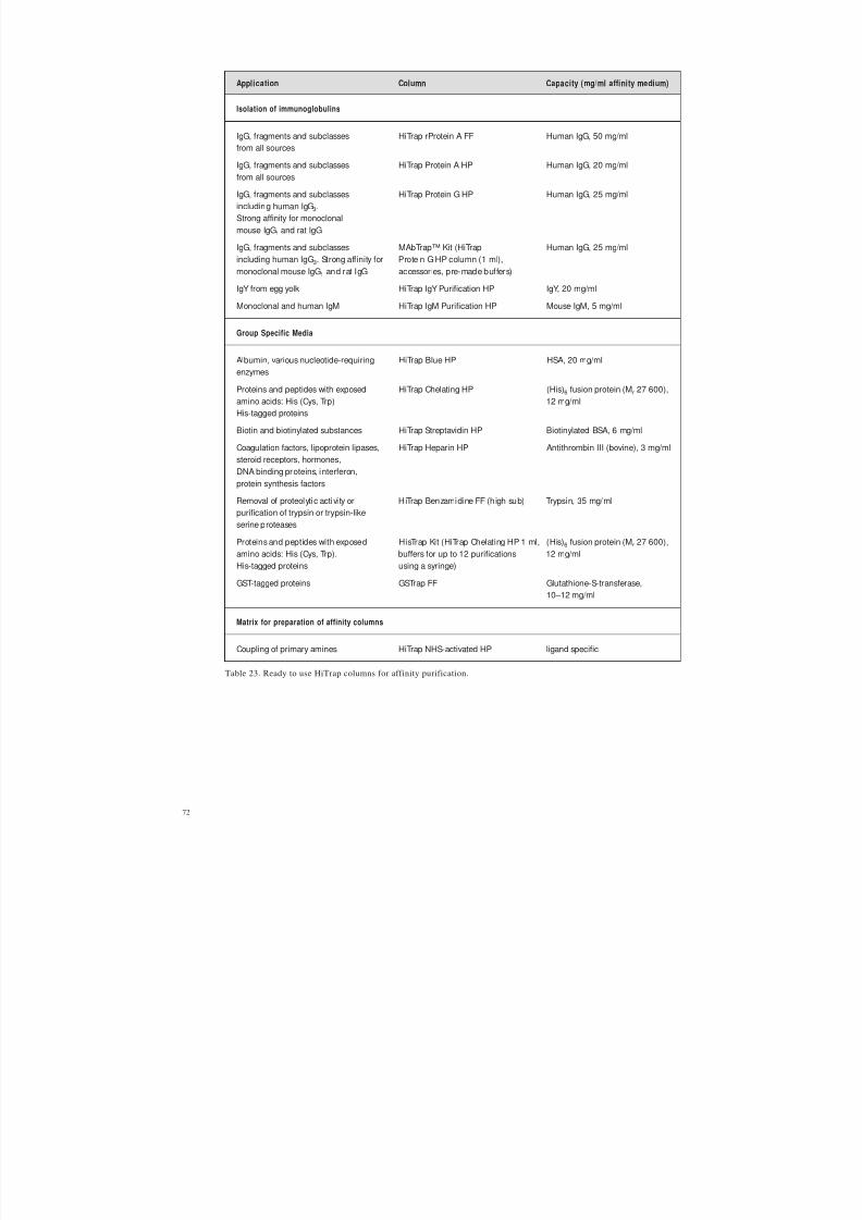
8/2/2019 The ant Protein Handbook
http://slidepdf.com/reader/full/the-ant-protein-handbook 74/112
72
Table 23. Ready to use HiTrap columns for affinity purification.
Application Column Capacity (mg/ml affinity medium)
Isolation of immunoglobulins
IgG, fragments and subclasses HiTrap rProtein A FF Human IgG, 50 mg/ml
from all sourcesIgG, fragments and subclasses HiTrap Protein A HP Human IgG, 20 mg/ml
from all sources
IgG, fragments and subclasses HiTrap Protein G HP Human IgG, 25 mg/mlincluding human IgG3.Strong affinity for monoclonal
mouse IgG1 and rat IgG
IgG, fragments and subclasses MAbTrap™ Kit (HiTrap Human IgG, 25 mg/mlincluding human IgG3. Strong affinity for Protein G HP column (1 ml),
monoclonal mouse IgG1 and rat IgG accessories, pre-made buffers)
IgY from egg yolk HiTrap IgY Purification HP IgY, 20 mg/ml
Monoclonal and human IgM HiTrap IgM Purification HP Mouse IgM, 5 mg/ml
Group Specific Media
Albumin, various nucleotide-requiring HiTrap Blue HP HSA, 20 mg/mlenzymes
Proteins and peptides with exposed HiTrap Chelating HP (His)6 fusion protein (Mr 27 600),amino acids: His (Cys, Trp) 12 mg/mlHis-tagged proteins
Biotin and biotinylated substances HiTrap Streptavidin HP Biotinylated BSA, 6 mg/ml
Coagulation factors, lipoprotein lipases, HiTrap Heparin HP Antithrombin III (bovine), 3 mg/mlsteroid receptors, hormones,DNA binding proteins, interferon,
protein synthesis factors
Removal of proteolytic activi ty or HiTrap Benzamidine FF (high sub) Trypsin, 35 mg/mlpurification of trypsin or trypsin-likeserine proteases
Proteins and peptides with exposed HisTrap Kit (HiTrap Chelating HP 1 ml, (His)6 fusion protein (Mr 27 600),amino acids: His (Cys, Trp). buffers for up to 12 purifications 12 mg/mlHis-tagged proteins using a syringe)
GST-tagged proteins GSTrap FF Glutathione-S-transferase,10–12 mg/ml
Matrix for preparation of affinity columns
Coupling of primary amines HiTrap NHS-activated HP ligand specific

8/2/2019 The ant Protein Handbook
http://slidepdf.com/reader/full/the-ant-protein-handbook 75/112
73
Making a specific purification column
In cases when a ready made affinity medium is unavailable, it may be considered worth-while to develop a “home-made” affinity purification column, for example, when a specificrecombinant protein needs to be prepared efficiently on a regular basis.
The ligand must be prepared (following procedures that are specific for the type of ligande.g. by raising antibodies), tested for affinity to the target protein and purified before linkingto a chromatograph ic matrix. For further details on general purification strategies forproteins see Chapter 9 and the Protein Purification Handbook from Amersham Biosciences.A detailed account of the principles of affinity chromatography can be found inthe Affinity Chromatography, Principles and Methods Handbook also available fromAmersham Biosciences.
Preparation of HiTrap NHS-activated HP to create a simple affinity purification column
NHS-activated Sepharose High Performance is a chromatographic matrix specificallydesigned for the covalent coupling of ligands containing primary amino groups (the mostcommon form of attachment). The matrix is based on highly cross-linked agarose beadswith 10-atom spacer arms att ached to the matrix by epichlorohydrine and activated byN-hydroxysuccinimide. The substitution level is ~10 µmol NHS-groups/ml gel. Non-specificadsorption of proteins (which can reduce binding capacity of the target protein) is negligibledue to the excellent hydrophilic properties of the base matrix.
The protocol below describes the preparation of a pr epacked H iTrap N HS-activated HPcolumn and is generally applicable to all NHS-activated Sepharose products.
Optimum binding and elution conditions for pur ification of the target protein must bedetermined separately for each ligand.
This procedure can be performed using a HiTrap column with a syringe, a peristaltic pumpor a liquid chromatography system such as ÄKTA prime.
The activated mat rix is supplied in 100% isopropanol to preserve stability prior tocoupling. Do not replace the isopropanol until it is time to couple the ligand.
Buffer preparation
Acidification solution: 1 mM HCl (ice-cold)
Coupling buffer: 0.2 M NaHCO3
, 0.5 M NaCl, pH 8.3
Use high quality water and chemicals. Filtration through 0.45 µm filters isrecommended.
Coupling within pH range 6.5–9, maximum yield is achieved at pH ~8.

8/2/2019 The ant Protein Handbook
http://slidepdf.com/reader/full/the-ant-protein-handbook 76/112
74
Ligand and column preparation
1. Dissolve desired ligand in the coupling buffer to a concentration of 0.5–10 mg/ml (for protein ligands) orperform a buffer exchange using a desalting column (see Chapter 7). The optimum concentration dependson the ligand. Optimum sample volume is equivalent to one column volume.
2. Remove top-cap and apply a drop of ice-cold 1 mM HCl to the top of the column to avoid air bubbles.
3. Connect the top of the column to the syringe or system.4. Remove the twist-off end.
Ligand coupling
1. Wash out the isopropanol with 3 x 2 column volumes of ice-cold 1 mM HCl.
Do not use excessive flow rates (maximum recommended flow rates are 1 ml/min (equivalentto approximately 30 drops/min when using a syringe) with H iTrap 1 ml and 5 ml/min(equivalent to appro ximately 120 dro ps/min when using a syringe) with HiTrap 5 ml). Thecolumn con tents can be irreversibly compressed.
2. Immediately inject one column volume of ligand solution onto the column.
3. Seal the column. Leave to stand for 15–30 minutes at +25 °C (or 4 hours at +4 °C).
If larger volumes of ligand solution are used, re-circulate the solution. For example, whenusing a syringe connect a second syringe to the outlet of the column and gently pump thesolution back and forth for 15–30 minutes or, if using a peristaltic pump, simply re-circulatethe sample through the column.
If required, the coupling efficiency can be measured at this stage. These procedures aresupplied with each HiTrap NHS-activated H P column.
Washing and deactivation
This procedure deactivates any excess active groups that have not coupled to the ligand andwashes out non-specifically bound ligands.
Buffer A: 0.5 M ethanolamine, 0.5 M NaCl, pH 8.3
Buffer B: 0.1 M acetate, 0.5 M NaCl, pH 4
1. Inject 3 x 2 column volumes of buffer A.
2. Inject 3 x 2 column volumes of buffer B.
3. Inject 3 x 2 column volumes of buffer A.4. Let the column stand for 15–30 min.
5. Inject 3 x 2 column volumes of buffer B.
6. Inject 3 x 2 column volumes of buffer A.
7. Inject 3 x 2 column volumes of buffer B.
8. Inject 2–5 column volumes of a buffer with neutral pH.
The column is now ready for use.
Store the column in storage solution, e.g. 0.05 M Na2HPO 4, 0.1% NaN 3, pH 7.
Sodium azide can interfere with many coupling methods and some biological assays. It canbe removed by using a desalting column (see Chapter 7).

8/2/2019 The ant Protein Handbook
http://slidepdf.com/reader/full/the-ant-protein-handbook 77/112
75
Purification
Optimum binding and elution conditions for pur ification of the target protein must bedetermined separately for each ligand. Literature references and textbooks may give goodguidelines. Below is a general protocol that can be used initially.
Use high quality water and chemicals. Filtration through 0.45 µm filters is recommended.
Perform a blank run t o ensure that loosely bound ligand is removed.
Samples should be centrifuged immediately before use and/or filtered through a 0.45 µmfilter. If the sample is too viscous, dilute with binding buffer. Sample binding p rop erties canbe improved by adjusting the sample to the composition of the binding buffer. Dilute inbinding buffer or perform a buffer exchange using a desalting column (see Chapter 7).
Prepare the column (blank run)
1. Wash with 3 column volumes of binding buffer.
2. Wash with 3 column volumes of elution buffer.
3. Equilibrate with 10 column volumes of binding buffer.
Purification
1. Apply sample. Optimal flow rate is dependent on the binding constant of the ligand, but a recommendedflow rate range is, for example, 0.2–1 ml/min on a HiTrap 1 ml column.
2. Wash with 5–10 column volumes of binding buffer, or until no material appears in the eluent.
Avoid excessive washing if the interaction between the protein of interest and the ligand isweak, since this may decrease the yield.
3. Elute with 1–3 column volumes of elution buffer (larger volumes may be necessary).
4. If required purified fractions can be desalted and exchanged into the buffer of choice using prepackeddesalting columns (see Chapter 7).
Re-equilibrate the column
Re-equilibrate the column by washing with 5–10 column volumes of binding buffer.

8/2/2019 The ant Protein Handbook
http://slidepdf.com/reader/full/the-ant-protein-handbook 78/112
76

8/2/2019 The ant Protein Handbook
http://slidepdf.com/reader/full/the-ant-protein-handbook 79/112
77
Step
Purity
Capture
Intermediate purification
Polishing
Preparation,
extraction,
clarification
Achieve final high level purity
Remove bulk impurities
Isolate, concentrate and stabilize
CHAPTER 9
Multi-step purification of recombinant proteins (fusion and non-fusion)Fusion systems are simple and convenient and, for many applications, a single purification
step using affinity chromatography is sufficient to achieve the desired level of pur ity.However, if there is no suitable fusion system so that affinity chromatography cannot beused, or if a higher degree of purity is required, a multi-step pur ification will be necessary.
A significant advantage when working with recombinant products is that th ere is oftenconsiderable information available about the product and contaminants. With thisinformation, detection assays and sample preparation and extraction procedures in place,a purification strategy of Capture, Intermediate Purification and Polishing (C IPP) can beapplied (Figure 20). This strategy is used in both the pharmaceutical industry and in theresearch laboratory to ensure faster method development, a shorter time to p ure prod uctand good economy.
This section gives a brief overview of the approach recommended for any multi-step proteinpurification. The Protein Purification Handbook (from Amersham Biosciences) ishighly recommended as a guide to planning efficient and effective protein purificationstrategies.
Fig. 20. Preparation and CIPP.
CIPP is applied as follows:
Imagine the purification has three phases Capture, Intermediate Purification and Polishing.
Assign a specific objective to each step with in th e pur ification process.
The pur ification p roblem associated with a particular step will depend greatly upon theproperties of the starting material. Thus, the objective of a purification step will varyaccording to its position in the process i.e. at the beginning for isolation of product fromcrude sample, in the middle for further p urification of part ially purified sample, or at theend for final clean up of an almost pure prod uct.
In the capture phase the objectives are to isolate, concentrate and stabilize the target product.
The product should be concentrat ed and t ransferred to an environment that will conservepotency/activity.
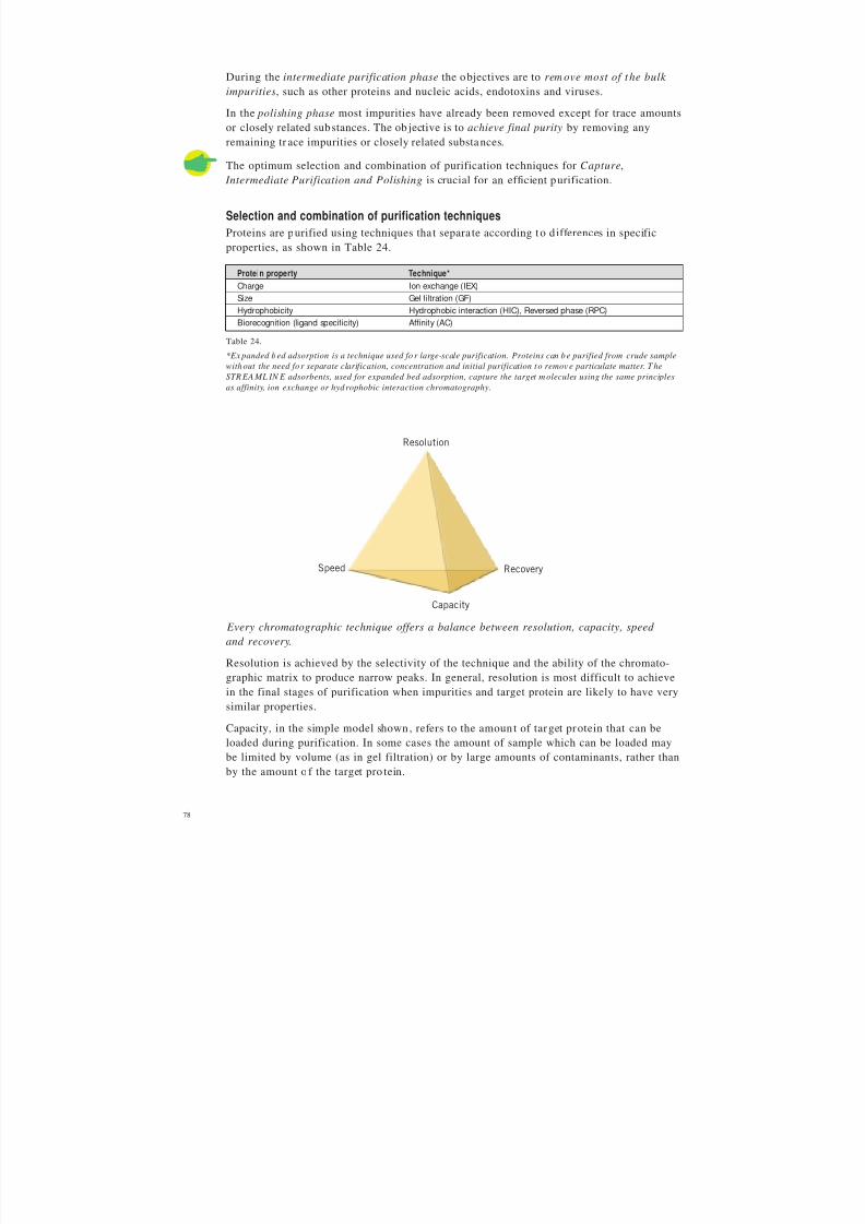
8/2/2019 The ant Protein Handbook
http://slidepdf.com/reader/full/the-ant-protein-handbook 80/112
78
During the intermediate purification phase the objectives are to remove most of t he bulk
impurities, such as other proteins and nucleic acids, endotoxins and viruses.
In the polishing phase most impurities have already been removed except for trace amountsor closely related substances. The ob jective is to achieve final purity by removing anyremaining tr ace impurities or closely related substances.
The optimum selection and combination of purification techniques for Capture, Intermediate Purification and Polishing is crucial for an efficient purification.
Selection and combination of purification techniques
Proteins are purified using techniques tha t separa te according to differences in specificproperties, as shown in Table 24.
Every chromatographic technique offers a balance between resolution, capacity, speed
and recovery.
Resolution is achieved by the selectivity of the technique and the ability of the chromato-graphic matrix to produce narrow peaks. In general, resolution is most difficult to achievein the final stages of purification when impurities and target protein are likely to have verysimilar properties.
Capacity, in the simple model shown, refers to the amount of target protein that can beloaded during purification. In some cases the amount of sample which can be loaded may
be limited by volume (as in gel filtration) or by large amounts of contaminants, rather thanby the amount o f the target pro tein.
Protein property Technique*
Charge Ion exchange (IEX)
Size Gel filtration (GF)
Hydrophobicity Hydrophobic interaction (HIC), Reversed phase (RPC)
Biorecognition (ligand specificity) Affinity (AC)
Table 24.
*Ex panded bed adsorption is a technique used fo r large-scale purification. Proteins can be purified from crude sample
without the need fo r separate clarification, concentration and initial purification to remov e particulate matter. T he
STR EA ML IN E adsorbents, used for expanded bed adsorption, capture the target m olecules using the same principles
as affinity, ion exchange or hydrophobic interaction chromatography.
Resolution
Capacity
RecoverySpeed

8/2/2019 The ant Protein Handbook
http://slidepdf.com/reader/full/the-ant-protein-handbook 81/112
79
Speed is of the highest importance at the beginning of purification where contaminantssuch as pro teases must be removed as qu ickly as possible.
Recovery becomes increasingly important as the purification proceeds because of theincreased value of the purified prod uct. Recovery is reduced by destructive processes in the
sample and unfavourable conditions on the column.Select a chromatographic technique to meet the objectives for the purification step.
Choose logical combinations of purification techniques based on the main benefits of thetechnique and the condition of the sample at the beginning or end of each step.
A guide to the suitab ility of each pur ification t echniqu e for the stages in C IPP is shownin Table 25.
Table 25. Suitability of purification techniques for the CIPP.
Minimize sample handling between purification steps by combining techniques to avoid theneed for sample conditioning. The pro duct should be eluted from the first column in abuffer suitable for the start conditions required for the next technique (see Table 25).
Ammonium sulphate p recipitation is a common sample clarification and concentrat ion stepand so HIC (which requires high salt to enhance binding to the media) is ideal as the capturestep. The salt concentration and the total sample volume will be significantly reduced afterelution from the H IC column. Dilution of the fractionated sample or rapid b uffer exchangeusing a desalting column will prepare it for the next IEX or AC step.
GF is well suited for use after any of the concentrating techniques (IEX, HIC, AC) since thetarget protein will be eluted in a r educed volume and the components from the elution bufferwill not affect the gel filtration separation. Gel filtration is a non-binding technique withlimited volume capacity and is unaffected by buffer conditions.
Selection of the final stra tegy will always depend upon specific sample properties and therequired level of purification. Logical combinations of techniques are shown in Figure 21.
Technique Main features Capture Intermediate Pol ishing Sample start Sample end condition condition
IEX high resolution low ionic strength high ionic high capacity sample volume strength or high speed not limiting pH change
concentrated sample
HIC good resolution high ionic strength low ionic good capacity sample volume strength high speed not limiting concentrated
sample
AC high resolution specific binding specific high capacity conditions elution high speed sample volume conditions
not limiting concentrated sample
GF high resolution limited sample buffer using Superdex volume (<5% total exchanged
column volume) (if r equired) and flow rate diluted
range sample
RPC high resolution sample volume usually not limiting additives may be required
in organic solvent, risk loss of biological activity

8/2/2019 The ant Protein Handbook
http://slidepdf.com/reader/full/the-ant-protein-handbook 82/112
80
For any capture step, select the technique showing the strongest binding to the target
protein while binding as few of the contaminants as possible, i.e. the technique with thehighest selectivity and/or capacity for the protein of interest.
A sample is pur ified using a combination of techniques and alterna tive selectivities. Forexample, in an IEX-HIC-GF strat egy the capture step selects according to d ifferences incharge (IEX), the intermediate purification step according to differences in hydrophobicity(HIC) and the final polishing step according to d ifferences in size (GF).
If nothing is known about the target p rotein use IEX-HIC-GF. This combination o f techniques can be regarded as a standard protocol.
Consider the use of both anion and cation exchange chromatography to give differentselectivities within the same purification strategy.
Proteins with low solubility
SDS
extraction
SDS
extraction
Solubilizing agents
(urea, ethylene glycol non-ionic detergents)
GF
(in non-ionic detergent)
HIC HIC
GF GF
Clear or very dilute samples
AC IEX IEX Precipitation
(e.g. in high ionic strength)
HIC Resolubilize
GF Treat as for
sample in high salt
concentration
Capture
Intermediate
Purification
Polishing GF
or
RPC
GF
or
RPC
Crude sample or sample in high salt concentration
GF
desalt mode
GF
desalt mode
GF
desalt mode
AC IEX HIC
dilution may be needed
IEX
IEX HIC
GF
or
RPC
GF
or
RPC
GF GF
Sample
clarification
Capture
Intermediate
Purification
Polishing
Fig. 21 . Logical combinations of chromatographic steps.

8/2/2019 The ant Protein Handbook
http://slidepdf.com/reader/full/the-ant-protein-handbook 83/112
81
IEX is a technique which offers different selectivities using either an ion o r cation exchangers.The pH can be modified to alter the charge characteristics of the sample components. It istherefore possible to use IEX more than once in a purification strategy, for capture, inter-mediate purification or polishing. IEX can be used effectively both for rapid separation inlow resolution mode dur ing captur e, and in high resolution mode dur ing polishing in the
same pur ification scheme.
Consider RPC for a polishing step pro vided that the target protein can withstand therun conditions.
Reversed phase chromatography (RPC) separates proteins and peptides on the basis of hydropho bicity. RPC is a h igh selectivity (high resolution) technique, requiring the use of organ ic solvents. The technique is widely used for pu rity check analyses when r ecovery of activity and tertiary structure are not essential. Since many proteins are denatured byorganic solvents, the technique is not generally recommended for protein purificationwhere recovery of activity and return to a native tertiary structure may be compromised.
However, in the polishing phase, when the majority of protein impurities have beenremoved, RPC can be excellent, particularly for small target proteins that are not oftendenatured by organic solvents.
CIPP does not mean that all strategies must have three purification steps. For example, captureand intermediate purification may be achievable in a single step, as may intermediatepurification and polishing. Similarly, purity demands may be so low that a rapid capturestep is sufficient to achieve the desired result. For purification of therapeutic proteins a fourthor fifth purification step may be required to fulfil the highest purity and safety demands.The number of steps used will always depend up on the pur ity requirements and intendeduse for t he protein.
The following example demonstrates the successful application of C IPP in the pu rification of a recombinant protein.
Three step purification o f a recombinan t enzyme using ÄKTAFPLC
This example demonstrates one of the most common purification strategies used when highpurity levels are required: IEX for capture, HIC for intermediate purification and GF forthe polishing step.
The objective was to obtain highly purified deacetoxycephalosporin C synthase (DAOCS),an oxygen-sensitive enzyme that had been produced by over-expression in soluble form in
the cytoplasm of E. coli bacteria.
A more detailed description of this work can be found in Application N ote 18-1128 -91.
Sample extraction and clarification
Cells were suspended in Tris-based lysis buffer, pH 7.5 and lysed using ultrasonication .Streptomycin sulphate and polyethyleneimine were added to precipitate DNA. The extractwas clarified by centrifugation. EDTA, DTT, benzamidine-HCl and PMSF were used in thelysis buffer to inh ibit proteases and minimize damage to the oxygen sensitive-enzyme.Keeping the sample on ice also reduced p rot ease activity.

8/2/2019 The ant Protein Handbook
http://slidepdf.com/reader/full/the-ant-protein-handbook 84/112
82
Capture
The capture step focused on the rapid removal of the most harmful contaminant s from therelatively unstable target protein. This, together with the calculated isoelectric point of DAOCS (pI = 4.8), led to the selection of an anion exchange purification. A selection of anion exchange columns, including those from the H iTrap IEX Selection Kit, was screened
to find the optimum medium (results not shown). O ptimization o f the capture step (inFigure 22) allowed the use of a step elution at high flow rate to speed up the purification.
System: ÄKTAFPLC
Column: HiPrep 16/10 Q XLSample: Clarified E. coli extractSample volume: 40 mlBinding buffer: 50 mM Tris-HCl, 1 mM EDTA, 2 mM DTT,
0.2 M benzamidine-HCl, 0.2 mM PMSF, pH 7.5Elution buffer: Binding buffer + 1.0 M NaCl
Flow: 10 ml/min (300 cm/h)
System: ÄKTAFPLC
Column: SOURCE™ 15ISO, packed in HR 16/10 columnSample: DAOCS pool from HiPrep 16/10 Q XLSample volume: 40 mlBinding buffer: 1.6 M ammonium sulphate, 10% glycerol,
50 mM Tris-HCl, 1 mM EDTA, 2 mM DTT,0.2 mM benzamidine-HCl, 0.2 mM PMSF, pH 7.5
Elution buffer (B): 50 mM Tris-HCl, 10% glycerol, 1 mM EDTA,2 mM DTT, 0.2 mM benzamidine-HCl,0.2 mM PMSF, pH 7.5
Gradient: 0–16%B in 4 CV, 16-24%B in 8 CV,24–35%B in 4 CV, 100%B in 4 CV
Flow: 5 ml/min (150 cm/h)CV = column volume
0 100 200
1000
2000
3000
0 0
20
40
60
80
ml
mS/cmA280 nm
0 100 200
100
200
300
400
0ml
A280 nm
Fig. 22 . Capture using IEX. The elution position of DAOCS is shaded.
Intermediate Purification
Hydrophob ic interaction chromatography (H IC) was selected b ecause the separat ionprinciple is complementary to ion exchange and because a minimum amount of sample
conditioning was required. H ydrophobic properties are difficult t o p redict and it is alwaysrecommended to screen different media. After screening, RESOURCE™ ISO was selectedon the basis of the resolution achieved. In this intermediate step, shown in Figure 23, themaximum possible speed for separation was sacrificed in order to achieve higher resolutionand allow significant reduction of impurities.
Fig. 23 . Intermediate purification using HIC. The elution position of DAOCS is shaded.

8/2/2019 The ant Protein Handbook
http://slidepdf.com/reader/full/the-ant-protein-handbook 85/112
83
Fig. 24 . Polishing step using gel filtration. The elution p osition of DAO CS is shaded.
System: ÄKTAFPLC
Column: HiLoad™ 16/60 Superdex 75 prep gradeSample: Concentrated DAOCS pool from SOURCE 15ISOSample volume: 3 mlBuffer: 100 mM Tris-HCl, 1 mM EDTA, 2 mM DTT,
0.2 mM benzamidine-HCl, 0.2 mM PMSF, pH 7.5Flow: 1 ml/min (30 cm/h)
0 80604020 100
200
400
600
800
1000
0ml
A280 nm
Polishing
The main goal of the polishing step, shown in Figure 24, was to remove aggregates andminor contaminants and transfer the purified sample into a buffer suitable for use instructural studies. The final product was used successfully in X-ray diffraction studies.
Ref:Structure of a cephalosporin synthase
.Valegard, K., Terwisscha van Scheltinga, A.C.,Lloyd, M., Hara, T., Ramaswamy, S., Perrakis, A., Thompson, A., Lee, H.J., Baldwin, J.E.,
Schofield, C.J., Hajdu, J. and Andersson, I. Nature 39 4, 805–809 (1998).
Three step purification of a recombinant phosphatase using ÄKTA prime
The objective of this application was to produce a pure ph osphatase (rPhosphatase) withretained biological activity. The phosphatase gene was overexpressed and the protein wasproduced in soluble form in t he cytoplasm of Escherichia coli. Using the pre-programmedmethod templates of ÄKTA prime with prepacked H iPrep and HiLoad columns ensuredquick and easy method development. The purification strategy consisted of a capture stepby ion exchange chromato graphy, intermediate purification by hydrophobic interactionchromatography and polishing by gel filtration. Active rPhosphatase (35 mg) was purifiedwithin 8 hours.
A more detailed description of this work can be found in Application N ote 18-1142 -32.
Sample preparation and extraction
The E. coli cells were suspended in lysis buffer, 1 g cells to every 10 ml lysis buffer
(50 mM Tris-HCl, 1 mM EDTA, 2 mM DTT, pH 7.4). The suspended cells were lysed byultra-sonication, 6 x 20 bursts with 60 seconds cooling between each bu rst. DN A wasremoved by precipitation with 1% w/v streptomycin sulphate. The sample was clarified bycentrifugation, 15 minutes at 22 000 g, before it was app lied to the first chromato graphycolumn.

8/2/2019 The ant Protein Handbook
http://slidepdf.com/reader/full/the-ant-protein-handbook 86/112
84
Capture
The main pur pose of the capture step was to concentra te the rPhosphatase and removemost of the contaminants. The ÄKTA prime system pump was used to apply 200 ml of theclarified E. coli supernatant, diluted 1:2 with water, to a H iPrep 16/10 DEAE FF column.A pre-programmed method template for ion exchange chromatography was used for
the separat ion.
Fractions of the eluate were collected and analysed with an enzyme immunoassay detectingalkaline phosphatase activity at an absorbance of 405 nm. The pur ity of the fractionsconta ining rPhosphatase was determined by SDS-PAGE.
Sample: 200 ml clarified E. coli supernatant,diluted 1:2 with water, pH 6.6
Column: HiPrep 16/10 DEAE FF,Vt = 20 ml
Start buffer: 25 mM Tris-HCl, pH 7.4,
10% glycerol,1 mM EDTA,2 mM DTTElution buffer (B): 1 M NaCl in start bufferFlow: 5 ml/min (150 cm/h)Run parameters: Equilibration 0% B 2 CV
Sample applicationWash 1 0% B 4 CVElution 0–50% B in 20 CV
50% B for 1 CVWash 2 100% B 2 CV
Vt = column volumn (CV)
A 405 nmA 280 nm
4.02.0
1.0
3.0
2.0
1.0
0
500300 700 ml
UV absorbance
conductivity
A 405 nmA 280 nm
1.0
3.0
2.0
1.0
00 200 400 600 ml
2.0 UV absorbance
conductivity
Intermediate purificationHydrophob ic interaction chromatography was used for intermediate purification becauseof its compatibility with samples containing a high salt concentration. The pooled fractionsfrom the ion exchange column were purified on HiLoad 16/10 Phenyl Sepharose HighPerformance, using a pre-programmed method template in ÄKTA prime. The fractionscontaining rPhosphata se were pooled and concentrat ed to 10 ml on an Amicon™ 50 mlstirred-cell using a Diaflow™ PM10 filter. Reducing the sample volume enables a smallergel filtration column to be used for the final polishing step.
Sample: 170 ml rPhosphatase containingpool from HiPrep 16/10 DEAE FF
in 1.6 M ammonium sulphate,pH 7.0
Column: HiLoad 16/10 PhenylSepharose HP, Vt = 20 ml
Start buffer: 25 mM Tris-HCl, pH 7.4 in1.4 M ammonium sulphate,1 mM EDTA, 2 mM DTT
Elution buffer (B): 25 mM Tris-HCl in 10% glycerol,1 mM EDTA, 2 mM DTT, pH 7.4
Flow: 5 ml/min (150 cm/h)Run parameters: Equilibration 0% B 2 CV
Sample applicationWash 1 0% B 3 CVElution 0–100% B in 20 CVWash 2 100% B 2 CV
Vt = column volumn (CV)
Fig. 25. Capture step using ion exchange. The phosphatase activity is represented by the green bars (absorbance at 405 nm).
Fig. 26 . Intermediate purification step using hydrophobic interaction. The phosphatase activity is represented by thegreen bars (absorbance at 405 nm).

8/2/2019 The ant Protein Handbook
http://slidepdf.com/reader/full/the-ant-protein-handbook 87/112
85
Sample: 4 ml concentrated eluate, containingrPhosphatase from the HiLoad 16/10Phenyl Sepharose HP
Column: HiLoad 16/60 Superdex 75 pg,Vt = 120 ml
Buffer: 25 mM Tris-HCl, 300 mM NaCl,1 mM EDTA, 2 mM DTT, pH 7.4
Flow: 0.5 ml/min (15 cm/h)
Vt= column volumn (CV)
A 405 nmA 280 nm
1.0
4.0
3.0
Vo Vt
2.0
1.0
0
500 100 ml
2.0UV absorbance
Polishing
The final polishing step used a p re-programmed method template to run a gel filtrat ionseparat ion on a H iLoad 1 6/60 Superdex 75 p rep grade column. The pur ity of the fractionscontaining rPhosphatase was checked with SDS-PAGE (Figure 28) and by mass spectrometry(results not shown).
Fig. 27 . Polishing step using gel filtration. The pho sphata se activity is represented by the green bars (absorbanceat 405 nm).
Lane 1. E. coli SupernatantLane 2. Eluate from the capture step (ion exchange)Lane 3. Eluate from the intermediate step (hydrophobic interaction)Lane 4. Eluate from the polishing step (gel filtration)Lane 5. LMW marker
Fig. 28 . Purity check by SDS-PAGE using Multiph or II and ExcelGel SDS Gradient 8–18% . The pr oteins were stained
with Coomassie Brilliant Blue.
1 2 3 4 5
Mr
97 00066 000
20 100
30 000
45 000
14 400

8/2/2019 The ant Protein Handbook
http://slidepdf.com/reader/full/the-ant-protein-handbook 88/112
86
Appendix 1
Map of the GST fusion vectors showing reading framesand main features
pGEX-6P-1 (27-4597-01)
EcoR I Sma I Sal I Xho I Not IBamH I
PreScission Protease
Leu Glu Val Leu Phe Gln Gly Pro Leu Gly Ser Pro Glu Phe Pro Gly Arg Leu Glu Arg Pro HisCTG GAA GTT CTG TTC CAG GGG CCC CTG GGA TCC CCG GAA TTC CCG GGT CGA CTC GAG CGG CCG CAT
pGEX-6P-2 (27-4598-01)
BamH I
PreScission Protease
Leu Glu Val Leu Phe Gln Gly Pro Leu Gly Ser Pro Gly Ile Pro Gly Ser Thr Arg Ala Ala Ala SerCTG GAA GTT CTG TTC CAG GGG CCC CTG GGA TCC CCA GGA ATT CCC GGG TCG ACT CGA GCG GCC GCA TCG
EcoR I Sma I Sal I Xho I Not I
pGEX-6P-3 (27-4599-01)
BamH I
PreScission Protease
Leu Glu Val Leu Phe Gln Gly Pro Leu Gly Ser Pro Asn Ser Arg Val Asp Ser Ser Gly ArgCTG GAA GTT CTG TTC CAG GGG CCC CTG GGA TCC CCG AAT TCC CGG GTC GAC TCG AGC GGC CGC
EcoR I Sma I Sal I Xho I Not I
BamH I EcoR I Sma I Sal I Xho I Not I
BamH I EcoR ISma I
Sal IXho I
Not I
BamH I EcoR I Sma I Sal I Xho I Not I
BamH I EcoR I Sma I Sal I Xho I Not I
BamH I EcoR I Sma I Sal I Xho I Not I
BamH I EcoR I Sma I Sal I Xho I Not I
pGEX~4900 bp
pBR322ori
Bal I
BspM I
Ptacg l u t a
t h i o
n e S - t
r a n s f e ra
se
A m p
r
l a c I q
Nar I
EcoR V
BssH II
BstE IIMlu I
Apa I
Tth111 IAat II
Pst I
p4.5AlwN I
pSj10 Bam7Stop7
pGEX-4T-2 (27-4581-01)
pGEX-5X-1 (27-4584-01)
pGEX-5X-2 (27-4585-01)
pGEX-5X-3 (27-4586-01)
pGEX-4T-1 (27-4580-01)
pGEX-4T-3 (27-4583-01)
pGEX-2TK (27-4587-01)
Leu Val Pro Arg Gly Ser Pro Gly Ile Pro Gly Ser Thr Arg Ala Ala Ala SerCTG GTT CCG CGT GGA TCC CCA GGA ATT CCC GGG TCG ACT CGA GCG GCC GCA TCG TGA
Stop codon
Ile Glu Gly Arg Gly Ile Pro Glu Phe Pro Gly Arg Leu Glu Arg Pro His Arg AspATC GAA GGT CGT GGG ATC CCC GAA TTC CCG GGT CGA CTC GAG CGG CCG CAT CGT GAC TGA
Stop codons
I le Glu Gly Arg Gly I le Pro Gly I le Pro Gly Ser Thr Arg Ala Ala Ala SerATC GAA GGT CGT GGG ATC CCC GGA ATT CCC GGG TCG ACT CGA GCG GCC GCA TCG TGA
Stop codon
Ile Glu Gly Arg Gly Ile Pro Arg Asn Ser Arg Val Asp Ser Ser Gly Arg Ile Val Thr AspATC GAA GGT CGT GGG ATC CCC AGG AAT TCC CGG GTC GAC TCG AGC GGC CGC ATC GTG ACT GAC TGA
Stop codons
Leu Val Pro Arg Gly Ser Pro Glu Phe Pro Gly Arg Leu Glu Arg Pro His Arg AspCTG GTT CCG CGT GGA TCC CCG GAA TTC CCG GGT CGA CTC GAG CGG CCG CAT CGT GAC TGA
Stop codons
Leu Val Pro Arg Gly Ser Pro Asn Ser Arg Val Asp Ser Ser Gly Arg Ile Val Thr AspCTG GTT CCG CGT GGA TCC CCG AAT TCC CGG GTC GAC TCG AGC GGC CGC ATC GTG ACT GAC TGA
Stop codons
Leu Val Pro Arg Gly Ser Arg Arg Ala Ser Val
Kinase
CTG GTT CCG CGT GGA TCT CGT CGT GCA TCT GTT GGA TCC CCG GGA ATT CAT CGT GAC TGAStop codons
Thrombin
Thrombin
Thrombin
Thrombin
Factor Xa
Factor Xa
Factor Xa
EcoR IBamH I Sma I

8/2/2019 The ant Protein Handbook
http://slidepdf.com/reader/full/the-ant-protein-handbook 89/112

8/2/2019 The ant Protein Handbook
http://slidepdf.com/reader/full/the-ant-protein-handbook 90/112
88
Appendix 2
Amino acids table
Three-letter Single-letterAmino acid code code Structure
Alanine Ala A
Arginine Arg R
Asparagine Asn N
Aspartic Acid Asp D
Cysteine Cys C
Glutamic Acid Glu E
Glutamine Gln Q
Glycine Gly G
Histidine His H
Isoleucine Ile I
Leucine Leu L
Lysine Lys K
Methionine Met M
Phenylalanine Phe F
Proline Pro P
Serine Ser S
Threonine Thr T
Tryptophan Trp W
Tyrosine Tyr Y
Valine Val V
CH3
HOOC
H2N
HOOC
H2N
HOOC
H2N
HOOC
H2N
HOOC
H2N
HOOC
H2N
HOOC
H2N
HOOC
H2N
HOOC
H2N
HOOC
H2N
HOOC
H2N
HOOC
H2N
HOOC
H2N
HOOC
H2N
HOOC
H2N
HOOC
H2N
CH2CH2CH2NHC
NH2
NH
CH2CONH2
CH2COOH
CH2SH
CH2CH2COOH
CH2CH2CONH2
H
CH2
CH(CH3)CH2CH3
CH2CH
CH3
CH3
CH2CH2CH2CH2NH2
CH2CH2SCH3
CH2
HOOC
H2N
HOOC
H2N
HOOC
H2N
CH2OH
CHCH3
OH
CH2
CH(CH3)2
HOOC
H2N
OHCH2
NH
NH
N
NH

8/2/2019 The ant Protein Handbook
http://slidepdf.com/reader/full/the-ant-protein-handbook 91/112
89
Middle unitresidue ( -H
2
0 ) Charge a t Hydrophobic Uncharged HydrophilicFormula M r Formula M r pH 6.0 –7 .0 (non-polar) (polar) (polar)
C3H7NO2 89.1 C3H5NO 71.1 Neutral !
C6H14N4O2 174.2 C6H12N4O 156.2 Basic (+ve) !
C4H8N2O3 132.1 C4H6N2O2 114.1 Neutral !
C4H7NO4 133.1 C4H5NO3 115.1 Acidic(-ve) !
C3H7NO2S 121.2 C3H5NOS 103.2 Neutral!
C5H9NO4 147.1 C5H7NO3 129.1 Acidic (-ve) !
C5H10N2O3 146.1 C5H8N2O2 128.1 Neutral !
C2H5NO2 75.1 C2H3NO 57.1 Neutral !
C6H9N3O2 155.2 C6H7N3O 137.2 Basic (+ve) !
C6H13NO2 131.2 C6H11NO 113.2 Neutral !
C6H13NO2 131.2 C6H11NO 113.2 Neutral !
C6H14N2O2 146.2 C6H12N2O 128.2 Basic(+ve) !
C5H11NO2S 149.2 C5H9NOS 131.2 Neutral !
C9H11NO2 165.2 C9H9NO 147.2 Neutral !
C5H9NO2 115.1 C5H7NO 97.1 Neutral !
C3H7NO3 105.1 C3H5NO2 87.1 Neutral !
C4H9NO3 119.1 C4H7NO2 101.1 Neutral !
C11H12N2O2 204.2 C11H10N2O 186.2 Neutral !
C9H11NO3 181.2 C9H9NO2 163.2 Neutral !
C5H11NO2 117.1 C5H9NO 99.1 Neutral !

8/2/2019 The ant Protein Handbook
http://slidepdf.com/reader/full/the-ant-protein-handbook 92/112
90
1 kb of DNA = 333 amino acids of coding capacity
= 37 000 g/mol
270 bp DNA = 10 000 g/mol
1.35 kb DNA = 50 000 g/mol
2.70 kb DNA = 100 000 g/mol
Average molecular weight of an amino acid = 120 g/mol.
Protein A28 0 for 1 mg/ml
IgG 1.35
IgM 1.20
IgA 1.30
Protein A 0.17
Avidin 1.50
Streptavidin 3.40
Bovine Serum Albumin 0.70
Mass (g/mol) 1 µg 1 nmol
10 000 100 pmol; 6 x 10¹³ molecules 10 µg
50 000 20 pmol; 1.2 x 10¹³ molecules 50 µg
100 000 10 pmol; 6.0 x 10¹² molecules 100 µg
150 000 6.7 pmol; 4.0 x 10¹² molecules 150 µg
Separate MaximumCentrifuge M anufacturer’s carriers Number g-forcemodel number Manufacturer Rotor needed? of places and rpm
GS-15 #360908 Beckman S2096 no 2 1 100 g, 3 000 rpmGS-15R #360902 (#361111)
Sorvall #07983 DuPont H1000B yes 2 1 900 g, 3 000 rpmRT-6000B (#11706) #11093
6K #83794 Fisher #04-976-420 yes 2 1 506 g, 3 500 rpm#04-975-410 MT
Megafuge 1.0 #C1725-3 Heraeus #C1725-10 yes 4 1 280 g, 2 800 rpm(Baxter #) #C1725-35
Z-320 #C-0320 Hermle #C-0320-50 no 2 1 506 g, 3 500 rpm
GP8 #3121 IEC #216 or yes 2 or 4 2 340 g, 3 500 rpmGP8R #3122 #228 #5785
HN-SII #2355 IEC #244 no 2 1 450 g, 3 575 rpm
MP4 #2437 IEC #244 no 2 1 450 g, 3 575 rpmMP4R #2438
C312 #11175087 Jouan E4 yes 4 2 290 g, 3 300 rpmCR312 #11175090 (#11174153) #11174168
1130 Not required Kubota S11222 no 2 1 107 g, 3 000 rpm1140 Not required Kubota S11222 no 2 1 107 g, 3 000 rpm
Appendix 3
Protein conversion data
Appendix 4
Centrifuges, rotors and carriers for use with MicroPlex 24

8/2/2019 The ant Protein Handbook
http://slidepdf.com/reader/full/the-ant-protein-handbook 93/112
91
Appendix 5
Characteristics, cleaning and storage of Glutathione SepharoseGlutathione Sepharose 4B is recommended for packing small columns and other formats.
Glutathione Sepharose Fast Flow is excellent for packing high performance columns for usewith purification systems and for scaling up.
Characteristics
Ligand density 7–15 µmole glutathione per ml Glutathione Sepharose 4B
120–320 µmole glutathione per ml Glutathione Sepharose 4 Fast Flow
Capacity up to 8 mg recombinant GST per ml Glutathione Sepharose 4B
up to 10 mg recombinant GST per ml Glutathione Sepharose 4 Fast Flow
Ligand and spacer arm Glutathione and 10 carbon l inker arm
Molecular weight exclusion limit ~2 x 10⁷
Particle size 45–165 µm (90 µm)
Chemical stability
No significant loss of binding capacity when exposed to 0.1 M citrate (pH 4.0), 0.1 M NaOH,70% ethanol or 6 M guanidine hydrochloride for 2 hours at room temperature. No significantloss of binding capacity after exposure to 1% SDS for 14 d ays.
Cleaning
Re-use of purification columns and media depends upon the nature of the sample andshould only be performed with identical samples to prevent cross contamination.
If required, Glutathione Sepharose 4B and Glutathione Sepharose 4 Fast Flow can beregenerated for re-use as follows:
1. Wash with 2–3 column volumes of alternating high pH (0.1 M Tris-HCl, 0.5 M NaCl, pH 8.5) andlow pH (0.1 M sodium acetate, 0.5 M NaCl, pH 4.5) buffers.
2. Repeat the cycle 3 times.
3. Re-equilibrate with 3–5 column volumes of PBS, pH 7.4.
If Glutathione Sepharose appears to be losing binding capacity, it may be due to an
accumulation of precipitated, denatured or non-specifically bound proteins.
To remove precipitated or denatured substances:
1. Wash with 2 column volumes of 6 M guanidine hydrochloride.
2. Immediately wash with 5 column volumes of PBS, pH 7.4.
To remove hydrophobically bound substances:
1. Wash with 3–4 column volumes of 70% ethanol (or 2 column volumes of 1% Triton X-100).
2. Immediately wash with 5 column volumes of PBS, pH 7.4.

8/2/2019 The ant Protein Handbook
http://slidepdf.com/reader/full/the-ant-protein-handbook 94/112
92
Characteristics, cleaning and storage of Chelating Sepharose
Characteristics
Capacity up to 12 mg (His)6 fusion protein per ml Chelating Sepharose 4 Fast Flow
12 mg (His)6 fusion protein (Mr 27 600) per ml Chelating Sepharose
High Performance (in HiTrap columns)
Ligand Iminodiacetic acidMolecular weight exclusion limit ~2 x 10⁷
Particle size 45–165 µm (90 µm) Chelating Sepharose Fast Flow
24–44 µm (34 µm) Chelating Sepharose High Performance
Chemical stability
Stable in all commonly used aq ueous buffers and denaturan ts such as 6 M guanidinehydrochloride, 8 M urea and chaotropic agents.
CleaningRe-use of purification columns depends upon the nature of the sample and should only beperformed with identical samples to prevent cross contamination.
To remove precipitated proteins
1. Fill column with 1 M NaOH and incubate for 2 hours.
2. Wash out dissolved proteins with 5 column volumes of water and a buffer at pH ~7 unt il the pH of theflow-through reaches pH ~7.0.
To remove nickel ions prior before recharging or storage
1. Wash column with 5 column volumes 20 mM sodium phosphate, 0.5 M NaCl, 0.05 M EDTA, pH 7.4.
2. Wash with 10 column volumes of distilled water.
3. For storage wash with 5 column volumes of 20% ethanol.
4. Seal columns with the fittings provided and store at +4 to +8 °C.
The loss of metal ions is more pronounced at lower pH . The column does not have to bestripped between each purification if the same protein is going to be purified. In this case,perform stripping and re-charging of the column after 5–10 purifications.
The column must be recharged with nickel ions after regeneration.
For long-term storage (>1 month):
1. Wash the column twice with 10 column volumes of PBS, pH 7.4.
2. Repeat washes using 20% ethanol.
3. Store at +4 °C.
4. Re-equilibrate the column with 5–10 column volumes of PBS, pH 7.4 before re-use.

8/2/2019 The ant Protein Handbook
http://slidepdf.com/reader/full/the-ant-protein-handbook 95/112
93
Appendix 6
Column packing and preparationA Column Packing Video is also available to
demonstrate how to p roduce a well-packedcolumn (see ordering information).
1. Equilibrate all materials to the temperature at which the purification will be performed.
2. Eliminate air by flushing column end pieces with recommended buffer. Ensure no air is trapped under thecolumn net. Close column outlet leaving 1–2 cm of buffer in the column.
3. Gently resuspend the purification medium.
4. Estimate the amount of slurry (resuspended medium) required.
5. Pour the required volume of slurry into the column. Pouring down a glass rod held against the wall of thecolumn will minimize the introduction of air bubbles.
6. Immediately fill the column with buffer.
7. Mount the column top piece and connect to a pump.
8. Open the column outlet and set the pump to the desired flow rate.
For example: 15 ml/min in an XK 16/20 column
If the recommended flow rate canno t be ob tained use the maximum flow rate the pumpcan deliver.
9. Maintain the packing flow rate for at least 3 column volumes after a constant bed height is obtained.Mark the bed height on the column.
Do no t exceed 75% of the packing flow rate dur ing any purification.
Do not exceed th e maximum operating pressure of the medium or column.
10. Stop the pump and close the column outlet. Remove the top piece and carefully fill the rest of the columnwith recommended buffer to form an upward meniscus at the top.
11. Insert the adapter into the column at an angle, ensuring that no air is trapped under the net.
12. Slide the adapter slowly down the column (the outlet of the adapter should be open) unti l the mark isreached. Lock the adapter in position.
13. Connect the column to the pump and begin equilibration. Re-position the adapter if necessary.
The medium must b e thoro ughly washed especially if 20% ethanol has been used as thestorage solution. Residual ethanol may interfere with subsequent procedures.
Many media, when equilibrated with PBS containing an anti-microbial agent, may bestored at +4° C for up to 1 month, but always follow the specific storage instructionssupplied with the product.

8/2/2019 The ant Protein Handbook
http://slidepdf.com/reader/full/the-ant-protein-handbook 96/112
94
Column selection
XK columns are fully compatible with the high flow rates achievable with modern mediaand a broad range of column dimensions are available. For a complete listing refer to the Am ersham Biosciences BioD irectory or web catalogue.
Columns Volume (ml) Code noXK 16/20 column 2–34 18-8773-01
XK 26/20 column 0–80 18-1000-72
XK 16/70 column 102–135 18-8775-01
XK 26/70 column 281–356 18-8769-01
Columns Volume (ml) Code no
HR 5/2 column 0.2–0.59 18-0382-01
HR 5/5 column 0.8–1.2 18-0383-01
HR 10/2 column 0.08–2.43 18-1000-97
HR 10/10 column 6.4–8.7 19-7402-01
HR columns can be used for small scale chromatography applications.

8/2/2019 The ant Protein Handbook
http://slidepdf.com/reader/full/the-ant-protein-handbook 97/112
95
Appendix 7
Converting from linear flow (cm/hour) to volumetricflow rates (ml/min) and vice versa It is often convenient when comparing results for columns of different sizes to express flowas linear flow (cm/hour). However, flow is usually measured in volumetric flow rate (ml/min).To convert between linear flow and volumetric flow rate use one of the formulae below.
From linear flow (cm/hour) to volumetric flow rate (ml/min)
whereY = linear flow in cm/h
d = column inner diameter in cm
Example:
What is the volumetric flow rate in an XK 16/70 column (i.d. 1.6 cm) when the linear flow is 150 cm/hour?
Y = linear flow = 150 cm/h
d = inner diameter of the column = 1.6 cm
From volumetric flow rate (ml/min) to linear flow (cm/hour)
where
Z = volumetric flow rate in ml/min
d = column inner diameter in cm
Example:
What is the linear flow in an HR 5/5 column (i.d. 0.5 cm) when the volumetric flow rate is 1 ml/min?Z = Volumetric flow rate = 1 ml/min
d = column inner diameter = 0.5 cm
Volumetric flow rate =150 x p x 1.6 x 1.6
60 x 4ml/min
= 5.03 ml/min
Volumetric flow rate (ml/min) = Linear flow (cm/h)60
x column cross sectional area (cm2)
Y x p x d2
60 4=
From ml/min to using a syringe
1 ml/min = approximately 30 drops/min on a HiTrap 1 ml column
5 ml/min = approximately 120 drops/min on a HiTrap 5 ml column
Linear flow = 1 x 60 x4
p x 0.5 x 0.5cm/h
= 305.6 cm/h
Linear flow (cm/h) =Volumetric flow rate (ml/min) x 60
column cross sectional area (cm2)
= Z x 60 x4
p x d2
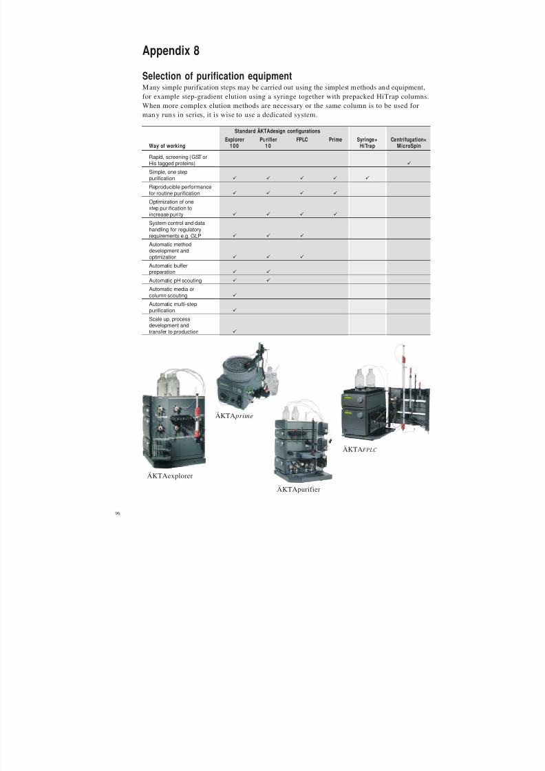
8/2/2019 The ant Protein Handbook
http://slidepdf.com/reader/full/the-ant-protein-handbook 98/112
96
Appendix 8
Selection of purification equipmentMany simple purification steps may be carried out using the simplest methods and equipment,
for example step-gradient elution using a syringe together with prepacked HiTrap columns.When more complex elution methods are necessary or the same column is to be used formany runs in series, it is wise to use a dedicated system.
ÄKTA prime
ÄKTAFPLC
ÄKTAexplorerÄKTApurifier
Standard ÄKTAdesign configurations
Explorer Purifier FPLC Prime Syringe+ Centrifugation+Way of working 100 1 0 HiTrap MicroSpin
Rapid, screening (GST orHis tagged proteins) "
Simple, one steppurification " " " " "
Reproducible performancefor routine purification " " " "
Optimization of onestep purification toincrease puri ty " " " "
System control and datahandling for regulatoryrequirements e.g. GLP " " "
Automatic methoddevelopment andoptimization " " "
Automatic bufferpreparation " "
Automatic pH scouting " "
Automatic media orcolumn scouting "
Automatic multi-steppurification "
Scale up, processdevelopment andtransfer to production "

8/2/2019 The ant Protein Handbook
http://slidepdf.com/reader/full/the-ant-protein-handbook 99/112
97
Appendix 9
Principles and standard conditions for purification techniques
Affinity Chromatography (AC)AC separates proteins on the basis of a reversible interaction between a protein (or group of proteins) and a specific ligand attached to a chromatographic matrix. The technique is idealfor a capture or intermediate step and can be used whenever a suitable ligand is availablefor the protein(s) of interest. AC offers high selectivity, hence high resolution, and usuallyhigh capacity (for the protein(s) of interest).
The target protein(s) is specifically and reversibly bound by a complementary bindingsubstance (ligand). The sample is applied under conditions that favour specific binding tothe ligand. Unbound material is washed away, and the bou nd target pro tein is recovered bychanging conditions to those favouring desorption. Desorption is performed specifically,
using a competitive ligand, or non-specifically, by changing the pH, ionic strength or polarity.Samples are concentrated during binding and protein is collected in purified, concentratedform. The key stages in a purification are shown in Figure 29. Affinity chromatography isalso used to remove specific contaminants, for examp le Benzamidine Sepharose 4 Fast Flowcan remove serine proteases.
Column Volumes [cv]
begin sample
applicationchange to
elution buffer
x cv 1-2 cv>1
cv1-2 cv 1-2 cv
equilibration gel regeneration
adsorption of
sample and
elution of
unbound material
wash
away
unbound
material
elute
bound
protein(s)
Absorbance
Fig. 29 . Typical affinity p urification.
Further information
Protein Purification Handbook
Affinity Chrom atography H andbook : Principles and M ethods
Chapters 3 and 4 in this handbook for the purification of GST and H is tagged proteins,respectively.
Ion Exchange (IEX)
IEX separa tes prot eins with differences in charge to give a very high resolution separat ionwith high sample loading capacity. The separation is based on the reversible interactionbetween a charged p rotein and an oppositely charged chromatographic medium. Proteinsbind as they are loaded onto a column. Conditions are then altered so that bound substancesare eluted differentially. Elution is usually performed by increasing salt concentration or
changing pH . Changes are made stepwise or with a continuous gradient. M ost commonly,samples are eluted with salt (NaCl), using a gradient elution (Figure 30). Target proteinsare concentrated during binding and collected in a purified, concentrated form.

8/2/2019 The ant Protein Handbook
http://slidepdf.com/reader/full/the-ant-protein-handbook 100/112
98
Fig. 30 . Typical IEX gradient elution.
Fig. 31 . Effect of pH on protein elution patterns.
The net surface charge of proteins varies according to the surrounding pH. When above its
isoelectric point (pI) a protein will bind to an anion exchanger, when below its pI a proteinwill behind to a cation exchanger. Typically IEX is used to bind the target molecule, but itcan also be used to bind impurities if required. IEX can be repeated at different pH valuesto separate several proteins that have distinctly different charge properties, as shown inFigure 31.
Method development (in priority order)
1. Select optimum ion exchanger using small columns as in the HiTrap IEX Selection Kit to save time and sample.
2. Scout for optimum pH. Begin 0.5–1 pH unit away from the isoelectric point of the target protein if known.
3. Select the steepest gradient to give acceptable resolution at the selected pH.
4. Select the highest flow rate that maintains resolution and minimizes separation time. Check recommendedflow rates for the specific medium.
[NaCl]
Column volumes [cv]0
1 M1-4 cv
equilibration re-equilibrationgradient
elution
sample
application
unbound molecules elute
before gradient begins
wash
high salt wash
tightly bound molecules
elute in high salt wash
2 cv2 cv
10-20 cv
Selectivity
pH of mobile phase
V V V
Cation
Anion
pH 0
+
-
V
V V V V
Abs Abs Abs Abs
Surfacenetcharge
Abs Abs Abs Abs
To reduce separation times and buffer consumption, transfer to a step elution after methodoptimization as shown in Figure 32. It is often possible to increase sample loading whenusing step elution.

8/2/2019 The ant Protein Handbook
http://slidepdf.com/reader/full/the-ant-protein-handbook 101/112
99
Fig. 32. Step elution.
Further information
Protein Purification Handbook
Ion Ex change Chromatography Handbook : Principles and M ethods
Hydrophobic Interaction Chromatography (HIC)
HIC separates proteins with differences in hydrophobicity. The technique is ideal for thecapture or intermediate steps in a purification. Separation is based on the reversibleinteraction between a prot ein and the hydrophobic surface of a chroma tographic medium.This interaction is enhanced by high ionic strength buffer which makes HIC an ideal “nextstep” after precipitation with ammonium sulphate or elution in high salt during IEX.Samples in high ionic strength solution (e.g. 1.5 M ammonium sulphate) bind as they areloaded onto a column. Conditions are then altered so that the bound substances are eluted
differentially.
Elution is usually performed by decreases in salt concentration (Figure 33). Changes aremade stepwise or with a continuous decreasing salt gradient. Most commonly, samples areeluted with a decreasing gradient of ammonium sulphate. Target proteins are concentratedduring binding and collected in a purified, concentrated form. O ther elution proceduresinclude reducing eluent polarity (ethylene glycol gradient up to 50%), adding chaotropicspecies (urea, guanidine hydrochloride) or detergents, changing pH or temperature.
Fig. 33. Typical HIC gradient elution.
[ N a
C l ]
Column volumes [cv]
2 cv2 cv
1-2 cv
1-2 cv
equilibration re-equilibration
sample
injectionvolume
unboundmolecules
elute
tightly boundmolecules
elute
elution of
unwantedmaterial
elutionof target
molecule
2 cv
high salt wash
[ammonium
sulphate]
Column volumes [cv]
2 cv
0
1 M
2 cv
10-15 cv
unbound molecules elute
before gradient begins
equilibration re-equilibrationgradient
elution
sample
applicationsalt free wash
tightly bound molecules
elute in salt free conditions

8/2/2019 The ant Protein Handbook
http://slidepdf.com/reader/full/the-ant-protein-handbook 102/112
100
Method development (in priority order)
1. The hydrophobic behaviour of a protein is difficult to predict and binding conditions must be studiedcarefully. Use HiTrap HIC Selection Kit or RESOURCE HIC Test Kit to select the medium that givesoptimum binding and elution over the required range of salt concentration. For proteins with unknownhydrophobic properties begin with 0–100%B (0%B=1M ammonium sulphate).
2. Select the gradient that gives acceptable resolution.3. Select the highest flow rate that maintains resolution and minimizes separation time. Check recommended
flow rates for the specific medium.
4. If samples adsorb strongly to a medium then conditions that cause conformational changes, such as pH,temperature, chaotropic ions or organic solvents can be altered. Conformational changes caused by theseagents are specific to each protein. Use screening procedures to investigate the effects of these agents.Alternatively, change to a less hydrophobic medium.
To reduce separation times and buffer consumption, transfer to a step elution after methodoptimization, as shown in Figure 34. It is often possible to increase sample loading whenusing step elution.
Fig. 34 . Step elution.
Further information
Protein Purification Handbook
Hydrophobic Interaction Chromatography Handbook : Principles and Methods
Gel Fil tration (GF) Chromatography
GF separates proteins with differences in molecular size. The technique is ideal for the final
polishing steps in purification when sample volumes have been reduced (sample volumesignificant ly influences speed and resolution in gel filtrat ion). Samples are eluted isocratically(single buffer, no gradient, Figure 35). Buffer conditions are varied to suit the sample type orthe requirements for further purification, analysis or storage, since buffer composition doesnot directly affect resolution. Pro teins are collected in purified form in the chosen buffer.
Column volumes [cv]
2 cv
1-2 cv
equilibration re-equilibration
sample
injection
volume
unbound
molecules
elute
tightly bound
molecules
elute
elution of
unwanted
material
elution
of target
molecule
2 cv
salt free wash
[ammonium
sulphate]
1-2 cv

8/2/2019 The ant Protein Handbook
http://slidepdf.com/reader/full/the-ant-protein-handbook 103/112
101
Column Volumes (cv)
UV
absor
bance
sample
injection
volume
equilibration
high
molecular
weight
intermediate
molecular weight
low
molecular
weight
1 cv
Fig. 35. Typical GF elution.
Further information
Protein Purification Handbook
Gel Filtration Handbook: Principles and Methods
Reversed Phase Chromatography (RPC)
RPC separates proteins and peptides with differing hydrophobicity based on their reversibleinteraction w ith the hydrophobic surface of a chromato graphic medium. Samples bind asthey are loaded on to a column. Conditions are then altered so that the bound substancesare eluted differentially. Due to the nature of the reversed phase matrices, the binding isusually very strong and requires the use of organic solvents and other additives (ion pairingagents). Elution is usually performed by increases in organic solvent concentration, mostcommonly acetonitrile. Samples, which are concentrated during the binding and separation
process, are collected in a purified, concentrated form. The key stages in a separation areshown in Figure 36.
Fig. 36. Typical RPC grad ient elution.
RPC is often used in the final polishing of oligonucleotides and peptides and is ideal foranalytical separations, such as peptide mapping.
RPC is not recommended for protein purification if recovery of activity and return to a
correct tertiary structure are required, since many proteins are denatured in the presence of organ ic solvents.
Column Volumes [cv]
10-15 cv
2-4 cv
2 cv2-4 cv
wash out unbound molecules
before elution begins
0
100%
[CH
CN/0.1%TFA]
3
sample
applicationre-equilibration
column
equilibrationgradient
elution
clean after
gradient

8/2/2019 The ant Protein Handbook
http://slidepdf.com/reader/full/the-ant-protein-handbook 104/112
102
Method development
1. Select medium from screening results.
2. Select optimum gradient to give acceptable resolution. For unknown samples begin with 0–100% elution buffer.
3. Select highest flow rate which maintains resolution and minimizes separation time.
4. For large scale purification transfer to a step elution.
5. Samples that adsorb strongly to a medium are more easily eluted by changing to a less hydrophobic medium.
Further information
Protein Purification Handbook
Reversed Phase Chromatography Handbook: Principles and Methods
Expanded Bed Adsorp tion (EBA)
EBA is a single pass operation in which target proteins are purified from crude sample,without the need for separate clarification, concentra tion and initial purification to remove
particulate matter. Crude sample is applied to an expanded bed of STREAMLINE™adsorbent particles within a specifically designed STREAMLINE column. Target proteins arecaptured on the adsorbent. Cell debris, cells, par ticulate matter, whole cells, and contaminantspass through and target proteins are then eluted. Figure 37 shows a representat ion of thesteps involved in an EBA pur ification and Figure 38 shows a typical EBA elution.
Fig. 37 . Steps in an EBA purification process.
0.Sedimented
adsorbent
1.Equilibration
(expanded)
2.Sample appl.
(expanded)
3.Washing
(expanded)
4.Elution
(packed bed)
5.Regeneration
(packed bed)
Fig. 38 . Typical EBA elution.
Sample volumes
Begin
sample
application
adorbance
Volume
Begin
wash with
start buffer
Change to
elution
buffer
equilibration adsorption
of sample
and elutionof unbound
material
wash
away
unboundmaterial
column
wash
elute
bound
protein(s)
UV
absorbance

8/2/2019 The ant Protein Handbook
http://slidepdf.com/reader/full/the-ant-protein-handbook 105/112
103
Method development
1. Select suitable ligand to bind the target protein.
2. Scout for optimal binding and elution conditions using clarified material in a packed column(0.02–0.15 litres bed volume of media). Gradient elution may be used during scouting, but the goal is todevelop a step elution.
3. Optimize binding, elution, wash and cleaning-in-place procedures using unclarified sample in expandedmode at small scale (0.02–0.15 litres bed volume of media).
4. Begin scale up process at pilot scale (0.2–0.9 litres bed volume of media).
5. Full scale production (up to several hundred litres bed volume of media).
Further information
Protein Purification Handbook
Expanded Bed A dsorption Handb ook : Principles and M ethods

8/2/2019 The ant Protein Handbook
http://slidepdf.com/reader/full/the-ant-protein-handbook 106/112
104
Code No.
Gel Media Guide (electrophoresis) 18-1129-79
Protein Electrophoresis Technical Manual 80-6013-88
Protein Purification Handbook 18-1132-29
Protein and Peptide Purification Technique Selection 18-1128-63
Fast Desalting and Buffer Exchange of Proteins and Peptides 18-1128-62
Gel Filtration Handbook
Principles and Methods 18-1022-18
Ion Exchange Chromatography Handbook
Principles and Methods 18-1114-21
Chromatofocusing with Polybuffer and PBE 50-01-022PB
Hydrophobic Interaction Chromatography Handbook
Principles and Methods 18-1020-90
Affinity Chromatography Handbook
Principles and Methods 18-1022-29
Reversed Phase Chromatography HandbookPrinciples and Methods 18-1134-16
Expanded Bed Adsorption Handbook
Principles and Methods 18-1124-26
Antibody Purification Handbook 18-1037-46
Convenient Purification, HiTrap Column Guide 18-1129-81
ÄKTAdesign Brochure 18-1129-05
Protein Purification, Principles, High Resolution Methods and
Applications, J.C. Janson and L. Rydén, 1998, 2nd ed. Wiley VCH 18-1128-68
GST fusion proteins
GST Gene Fusion Manual 18-1123-20
Data File, GSTrap FF and Glutathione Sepharose 4 Fast Flow 18-1136-89
Miniposter, "Rapid Purification of GST-fusion proteins from
large sample volumes" 18-1139-51
Purification and on-column cleavage of GST-tagged proteins
Application Note, "Efficient, rapid protein purification and
on-column cleavage using GSTrap FF columns" 18-1146-70
(His)6
fusion proteins
Data File, HisTrap Kit 18-1212-00
Data File, HiTrap Chelating HP 1 ml and 5 ml 18-1134-78
Miniposter, "Purification of Poly(His)-tagged Recombinant
Proteins using HisTrap Kit" 18-1116-26
On-column refolding of (His)6 fusion proteinsApplication Note, “Rapid and efficient purification and refolding of
a (His)6-tagged recombinant protein produced in E. coli as inclusion bodies” 18-1134-37
Multi-step purification
Application Note, “Purification of a labile, oxygen sensitive enzyme for
crystallization and 3D structural determination” 18-1128-91
Application Note, “Purification of a recombinant phosphatase using pre-
programmed generic templates for different chromatographic techniques” 18-1142-32
Detection
Application Note, “ECL Western and ECL Plus Western blotting” 18-1139-13
Column Packing
Column Packing Video PAL 17-0893-01Column Packing Video NTSC 17-0894-01
Additional reading and reference material

8/2/2019 The ant Protein Handbook
http://slidepdf.com/reader/full/the-ant-protein-handbook 107/112
105
Ordering information
Product Quantity Code No.
GST fusion proteins
Protein Amplific ationPGEX- 4T-1 25 µg 27-4580-01PGEX- 4T-2 25 µg 27-4581-01PGEX- 4T-3 25 µg 27-4583-01PGEX- 5X-1 25 µg 27-4584-01PGEX- 5X-2 25 µg 27-4585-01PGEX- 5X-3 25 µg 27-4586-01PGEX- 6P-1 25 µg 27-4597-01
PGEX- 6P-2 25 µg 27-4598-01PGEX- 6P-3 25 µg 27-4599-01All vectors include E. coli B21
PurificationGST MicroSpin Purification Module 50 purifications 27-4570-03
MicroPlex 24 Vacuum 1 system 27-3567-01GSTrap FF 2 x 1 ml 17-5130-02
5 x 1 ml 17-5130-011 x 5 ml 17-5131-01
Glutathione Sepharose 4 Fast Flow 25 ml 17-5132-01100 ml 17-5132-02500 ml 17-5132-03
Glutathione Sepharose 4B 10 ml 17-0756-01100 ml 27-4574-01
DetectionGST Detection Module 50 reactions 27-4590-01GST 96 Well Detection Module 96 reactions 27-4592-01Anti-GST antibody 0.5 ml 27-4577-01
(His)6
fusion proteins
PurificationHis MicroSpin Purification Module 50 purifications 27-4770-01MicroPlex 24 Vacuum 1 system 27-3567-01HisTrap Kit HiTrap Chelating HP columns (3 x 1 ml), 17-1880-01
accessories, pre-made buffersfor up to 12 purifications
HiTrap Chelating HP 5 x 1 ml 17-0408-011 x 5 ml 17-0409-01
Chelating Sepharose Fast Flow 50 ml 17-0575-01500 ml 17-0575-025 l 17-0575-04
DetectionAnti-His antibody 170 µl 27-4710-01HRP conjugated (anti-mouse) IgG 1 ml NA931
Cleavage
EnzymesThrombin 500 units 27-0846-01
Factor Xa 400 units 27-0849-01PreScission Protease 500 units 27-0843-01
Removal of thrombin and Factor XaHiTrap Benzamidine FF (high sub) 2 x 1 ml 17-5143-02
5 x 1 ml 17-5143-011 x 5 ml 17-5144-01
Benzamidine Sepharose 4Fast Flow (high sub) 25 ml 17-5123-10

8/2/2019 The ant Protein Handbook
http://slidepdf.com/reader/full/the-ant-protein-handbook 108/112
106
Product Quantity Code No.
Companion Products
E. coli B21 1 vial 27-1542-01
Isopropyl b-D-thiogalactoside (IPTG) 1 g 27-3054-035 g 27-3054-0410 g 27-3054-05
Western BlottingHybond-P 10 sheets RPN2020FHybond-ECL 10 sheets RPN2020DECL Western BlottingDetection Reagents for 1000 cm2 RPN2109ECL Plus Western BlottingDetection System for 1000 cm2 RPN2132
HiTrap Columns: AffinityHiTrap rProtein A FF 2 x 1 ml 17-5079-02
5 x 1 ml 17-5079-011 x 5 ml 17-5080-01
HiTrap Protein A HP 2 x 1 ml 17-0402-035 x 1 ml 17-0402-011 x 5 ml 17-0403-01
HiTrap Protein G HP 2 x 1 ml 17-0404-035 x 1 ml 17-0404-011 x 5 ml 17-0405-01
MAbTrap Kit HiTrap Protein G HP (1 x 1 ml), 17-1128-01accessories, pre-made buffersfor 10 purifications
HiTrap IgY Purification HP 1 x 5 ml 17-5111-015 x 1 ml 17-5110-01
HiTrap Blue HP 5 x 1 ml 17-0412-011 x 5 ml 17-0413-01GSTrap FF 2 x 1 ml 17-5130-02
5 x 1 ml 17-5130-011 x 5 ml 17-5131-01
HiTrap Chelating HP 5 x 1 ml 17-0408-011 x 5 ml 17-0409-01
HisTrap Kit HiTrap Chelating HP (3 x 1 ml), 17-1880-01accessories, pre-made buffers forup to 12 purifications
HiTrap Streptavidin HP 5 x 1 ml 17-5112-01HiTrap Heparin HP 5 x 1 ml 17-0406-01
1 x 5 ml 17-0407-01
HiTrap Benzamidine FF (high sub) 2 x 1 ml 17-5143-025 x 1 ml 17-5143-011 x 5 ml 17-5144-01
HiTrap NHS-activated HP 5 x 1 ml 17-0716-011 x 5 ml 17-0717-01
HiTrap Columns: IEXHiTrap IEX Selection Kit 7 x 1 ml HiTrap Q XL, 1 ml 17-6002-33
HiTrap SP XL, 1 mlHiTrap ANX FF (high sub), 1 mlHiTrap DEAE FF, 1 mlHiTrap CM FF, 1 mlHiTrap Q FF, 1 mlHiTrap SP FF, 1 ml
HiTrap Q HP 5 x 1 ml 17-1153-01
5 x 5 ml 17-1154-01

8/2/2019 The ant Protein Handbook
http://slidepdf.com/reader/full/the-ant-protein-handbook 109/112
107
Product Quantity Code No.
HiTrap Columns: IEXHiTrap SP HP 5 x 1 ml 17-1151-01
5 x 5 ml 17-1152-01HiTrap Q XL 5 x 1 ml 17-5158-01
5 x 5 ml 17-5159-01HiTrap SP XL 5 x 1 ml 17-5160-01
5 x 5 ml 17-5161-01HiTrap ANX FF (high sub) 5 x 1 ml 17-5162-01
5 x 5 ml 17-5163-01HiTrap DEAE FF 5 x 1 ml 17-5055-01
5 x 5 ml 17-5154-01HiTrap CM FF 5 x 1 ml 17-5056-01
5 x 5 ml 17-5155-01HiTrap Q FF 5 x 1 ml 17-5053-01
5 x 5 ml 17-5156-01
HiTrap SP FF 5 x 1 ml 17-5054-015 x 5 ml 17-5157-01
HiTrap Columns: HICHiTrap HIC Selection Kit 5 x 1 ml HiTrap Butyl FF, 1 ml 17-1349-01
HiTrap Octyl FF, 1 mlHiTrap Phenyl FF (low sub), 1 mlHiTrap Phenyl FF (high sub), 1 mlHiTrap Phenyl HP, 1 ml
HiTrap Phenyl FF (high sub) 5 x 1 ml 17-1355-015 x 5 ml 17-5193-01
HiTrap Phenyl FF (low sub) 5 x 1 ml 17-1353-015 x 5 ml 17-5194-01
HiTrap Phenyl HP 5 x 1 ml 17-1351-01
5 x 5 ml 17-5195-01HiTrap Butyl FF 5 x 1 ml 17-1357-015 x 5 ml 17-5197-01
HiTrap Octyl FF 5 x 1 ml 17-1359-015 x 5 ml 17-5196-01
Desalting and Buffer ExchangeMicroSpin G-25 Columns 50 columns 27-5325-01
HiTrap Desalting 5 x 5 ml 17-1408-01HiPrep 26/10 Desalting 1 x 53 ml 17-5087-01

8/2/2019 The ant Protein Handbook
http://slidepdf.com/reader/full/the-ant-protein-handbook 110/112
108

8/2/2019 The ant Protein Handbook
http://slidepdf.com/reader/full/the-ant-protein-handbook 111/112
Ettan, H ybond, H iTrap, G STrap, Sephadex, Superdex, H isTrap, Sepharose, MAbTrap,
ÄKTA, FPLC, HiPrep, PhastSystem, PhastGel, SOURCE, RESOURCE, H iLoad, PreScission, ExcelGel,Multiphor, STREAMLINE, ECL, MicroPlex and Ultrospec are trademarks of Amersham Biosciences
or its subsidiaries.
Amersham is a trademark o f Nycomed Amersham plc
Multipipette is a trademark of Eppendorf-Netheler-Hinz GmbH
Tween is a trademark of ICI Americas Inc
MicroSpin is a trademark o f Lida M anufacturing Corp
Pefabloc is a registered trademark of Pentafam AG
Triton is a registered tra demark of Union Ca rbide Chemicals and Plastics Co
ABTS is a registered trademark of Boehringer Mannheim GmbHCoomassie is a trademark of ICI plc
Amicon is a t rademark of Millipore Inc.
Diaflow is a tra demark o f Cannon-Rub ber Limited
Licensing Inf ormation
All materials for research use on ly. N ot for diagnostic or therap eutic purposes.A license for commercial use of pGEX vectors must b e obtained from AMRAD Corp oration Ltd.,
17-27 Co tham R oad, Kew, Victoria 3101 , Australia.
PreScission Protease is licensed from the University of Singapore, 10 Kent Ridge Crescent, Singapore 0511.
PreScission Protease is produ ced under commercial license from AMRAD C orpora tion Ltd.,
17-27 Co tham R oad, Kew, Victoria 3101 , Australia.
All goods an d services are sold subject to the terms and conditions of sale of the company w ithin
the Amersham Biosciences group that supplies them.
A copy of these terms and cond itions is available on request.
© Amersham Biosciences 2001 – All rights reserved.
Amersham Biosciences Björkgatan 30, SE-751 84 Uppsala, Sweden
Amersham Biosciences UK Limited Amersham Place, Little Chalfont, Buckinghamshire HP7 9NA, England
Amersham Biosciences Inc 800 Centennial Avenue, PO Box 1327 , Piscataway, NJ 088 55, USAAmersham Biosciences Europe GmbH Mun zinger Strasse 9, D-79111 Freiburg, Germany
Amersham Biosciences K.K. Sanken Building, 3-25-1, H yakunincho, Shinjuku-ku, Tokyo 169-0073 , Japan

8/2/2019 The ant Protein Handbook
http://slidepdf.com/reader/full/the-ant-protein-handbook 112/112
P r o d u c t i o n : R A K
D e s i g n
A B