the annual report on prenatal diagnostic testing in
TRANSCRIPT

©Murdoch Children’s Research Institute, 2020
The Annual Report on Prenatal Diagnostic Testing in Victoria, 2019
Reproductive Epidemiology group
Genetics theme
Murdoch Children’s Research institute

The Annual report on Prenatal Diagnostic testing in Victoria, 2019 | 2
Contents
ABOUT THIS REPORT 4
TRENDS IN THE UPTAKE OF PRENATAL DIAGNOSTIC PROCEDURES 5
FIGURE 1. ANNUAL NUMBER OF PRENATAL DIAGNOSTIC TESTS AND UPTAKE AS A PERCENTAGE OF TOTAL BIRTHS (1976-2019) INDICATIONS FOR PRENATAL DIAGNOSTIC PROCEDURES 6
FIGURE 2. INDICATIONS FOR PRENATAL DIAGNOSIS AS A PERCENTAGE OF TOTAL TESTS
SINGLE GENE CONDITIONS 7
TABLE 1. ANNUAL TESTS FOR THE MOST COMMON SINGLE GENE CONDITIONS FIGURE 3. ANNUAL NUMBER OF SINGLE GENE CONDITIONS FOR WHICH PRENATAL DIAGNOSIS HAS BEEN PERFORMED
CHROMOSOME ANALYSIS 8
FIGURE 4. NUMBER OF PRENATAL SAMPLES BY ANALYSIS TYPE, 2011-2019 OVERALL DIAGNOSTIC YIELD 9
FIGURE 5. DIAGNOSTIC YIELD OF PRENATAL DIAGNOSTIC TESTS BY YEAR DIAGNOSTIC YIELD BY INDICATION 10
FIGURE 6. DIAGNOSTIC YIELD BY INDICATION FOR TESTING PRENATAL DIAGNOSIS OF CHROMOSOME CONDITIONS 2013-2019 11
TABLE 2. PRENATAL DIAGNOSIS RESULTS 2013-2019 PUBLICATIONS FROM THE VICTORIAN PRENATAL DIAGNOSIS DATA COLLECTION 12

The Annual report on Prenatal Diagnostic testing in Victoria, 2019 | 3
This report is produced by the Reproductive Epidemiology group in the Genetics theme at the MCRI. For
more information about us, go to our page at https://www.mcri.edu.au/reproductiveepidemiology
How to cite this report: Pynaker C, Loughry L, Hui L, Halliday J. Annual report on Prenatal Diagnosis in
Victoria 2019, The Victorian Prenatal Diagnosis Database, Murdoch Children's Research Institute 2020.
doi: 10.25374/MCRI.14347436.
Contact us
If you wish to be included in our distribution list, contact Cecilia Pynaker at [email protected]
For other enquiries, please contact A/Prof Lisa Hui at [email protected] or Prof Jane Halliday at
The Annual report on Prenatal Diagnostic testing in Victoria, 2019 | 4
About this report
This annual report from the Victorian Prenatal Diagnosis Database (VPDD) summarises the results of fetal
chromosome testing in Victoria during 2019. Victoria has approximately 76,111 confinements annually,
and a median maternal age of 32 years (Australian Bureau of Statistics; https://www.abs.gov.au/).
The VPDD has been collecting state-wide data on prenatal diagnostic procedures since 1976. We
acknowledge our long-standing collaborators - the Victorian Clinical Genetics Service (VCGS) and Monash
Medical Centre (current contributors), Melbourne Pathology and Australian Clinical Labs (former
contributors).
All amniocentesis and chorionic villus sampling (CVS) results performed prior to 25 weeks’ gestation on
women living in Victoria are included in the annual report. This gestational age limit was chosen to
capture diagnostic testing performed after routine screening for chromosome and fetal structural
conditions in first and second trimester.
The data fields collected for each woman include: maternal age and gestation at the time of testing, type
of diagnostic test, indication for testing, chromosome results, and pregnancy plurality. A single record is
created for twin pregnancies or women who required repeat testing in the same pregnancy.
Major chromosome conditions included: autosomal trisomies, autosomal monosomies, polyploidy, sex
chromosome aneuploidies, pathogenic copy number variants (CNVs), unbalanced rearrangements and high
level mosaicism.
Minor chromosome conditions included: CNVs of uncertain or unknown significance, long continuous
stretches of homozygosity (LCSH), confined placental mosaicism (CPM) and balanced rearrangements.
Diagnostic yield was defined as the percentage of women with a major fetal chromosome condition
confirmed on diagnostic testing as a proportion of total tests.
Technical notes Ethics approval for this study was provided by the Royal Children’s Hospital (RCH) Human Research Ethics
Committee (HREC) on 17 December 2020 (Ref. No. 31135) and Monash Health local governance
authorisation on 17 December 2020 (Ref. No. SSA/42279/RCHM-2020).
Funding: The following institutions have contributed salary support to the work of the VPDD: National
Health and Medical Research Council (NHMRC), Medical Research Future Fund (MRFF), Murdoch Children’s
Research Institute (MCRI) and the Victorian Department of Health.

The Annual report on Prenatal Diagnostic testing in Victoria, 2019 | 5
Trends in the uptake of prenatal diagnostic procedures
In 2019, 1614 women < 25 weeks’ gestation underwent a prenatal diagnostic procedure, representing 2.1%
of total births in Victoria. The steep decline in prenatal diagnostic procedures since the peak in 1998
(n=5300) appears to have plateaued since 2016.
Figure 1. Annual number of prenatal diagnostic tests and uptake as a percentage of total births (1976-2019)
0.0%
1.0%
2.0%
3.0%
4.0%
5.0%
6.0%
7.0%
8.0%
9.0%
10.0%
0
1000
2000
3000
4000
5000
6000
1976
1977
1978
1979
1980
1981
1982
1983
1984
1985
1986
1987
1988
1989
1990
1991
1992
1993
1994
1995
1996
1997
1998
1999
2000
2001
2002
2003
2004
2005
2006
2007
2008
2009
2010
2011
2012
2013
2014
2015
2016
2017
2018
2019
Num
ber
of p
rena
tal d
iagn
osti
c te
sts
Year
CVS AMNIO % Uptake

The Annual report on Prenatal Diagnostic testing in Victoria, 2019 | 6
Indications for prenatal diagnostic procedures
Indications for prenatal diagnosis are obtained from the clinical referral information. More than one
indication may be recorded. In 2019, 1628 indications were recorded for 1614 diagnostic procedures. The
three most common indications for prenatal diagnosis were ultrasound abnormality (41.9%), positive NIPT
result (21.6%) and testing for single gene conditions (11.1%).
Figure 2. Indications for prenatal diagnosis as a percentage of total tests
*Maternal age >36 years at estimated due date of delivery.
0%
5%
10%
15%
20%
25%
30%
35%
40%
45%
50%
2000 2001 2002 2003 2004 2005 2006 2007 2008 2009 2010 2011 2012 2013 2014 2015 2016 2017 2018 2019
Perc
enta
ge o
f pr
enat
al d
iagn
osti
c te
sts
Year
Advanced maternal age* Second trimester serum screening
Ultrasound anomaly Noninvasive prenatal testing
Combined first trimester screening Single gene condition
History of chromosome condition Other indication

The Annual report on Prenatal Diagnostic testing in Victoria, 2019 | 7
Single gene conditions
Although the number of prenatal diagnostic procedures for the most common single gene conditions has
remained relatively consistent over the past 6 years, the number of conditions for which testing is
provided has increased from 36 in 2014 to 82 in 2019.
Table 1. Annual tests for the most common single gene conditions
Single gene conditions 2014 2015 2016 2017 2018 2019
Fragile X 22 20 21 22 29 33
Thalassaemia 28 31 23 28 31 18
Cystic fibrosis 18 13 14 23 14 12
Spinal Muscular atrophy 5 6 6 5 5 8
Figure 3. Annual number of single gene conditions for which prenatal diagnosis has been performed
0
10
20
30
40
50
60
70
80
90
100
1993
1994
1995
1996
1997
1998
1999
2000
2001
2002
2003
2004
2005
2006
2007
2008
2009
2010
2011
2012
2013
2014
2015
2016
2017
2018
2019
Num
ber
of s
ingl
e ge
ne c
ondi
tion
s
Year

The Annual report on Prenatal Diagnostic testing in Victoria, 2019 | 8
Chromosome analysis
In 2019, the vast majority of samples were analysed with chromosomal microarray (87%), regardless of the
indication for testing.
Figure 4. Number of prenatal samples by analysis type, 2011-2019
0
500
1000
1500
2000
2500
3000
3500
4000
2011 2012 2013 2014 2015 2016 2017 2018 2019
Num
ber
of s
ampl
es a
naly
sed
Year
Microarray Karyotype and/or FISH
The Annual report on Prenatal Diagnostic testing in Victoria, 2019 | 9
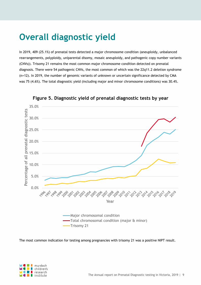
Overall diagnostic yield
In 2019, 409 (25.1%) of prenatal tests detected a major chromosome condition (aneuploidy, unbalanced
rearrangements, polyploidy, uniparental disomy, mosaic aneuploidy, and pathogenic copy number variants
(CNVs)). Trisomy 21 remains the most common major chromosome condition detected on prenatal
diagnosis. There were 54 pathogenic CNVs, the most common of which was the 22q11.2 deletion syndrome
(n=12). In 2019, the number of genomic variants of unknown or uncertain significance detected by CMA
was 75 (4.6%). The total diagnostic yield (including major and minor chromosome conditions) was 30.4%.
Figure 5. Diagnostic yield of prenatal diagnostic tests by year
The most common indication for testing among pregnancies with trisomy 21 was a positive NIPT result.
0.0%
5.0%
10.0%
15.0%
20.0%
25.0%
30.0%
35.0%
1996
1997
1998
1999
2000
2001
2002
2003
2004
2005
2006
2007
2008
2009
2010
2011
2012
2013
2014
2015
2016
2017
2018
2019
Perc
enta
ge o
f al
l pre
nata
l dia
gnos
tic
test
s
Year
Major chromosomal conditionTotal chromosomal condition (major & minor)Trisomy 21

The Annual report on Prenatal Diagnostic testing in Victoria, 2019 | 10
Diagnostic yield by indication
The diagnostic yield is the percentage of women with a major chromosome condition confirmed on
diagnostic testing as a proportion of total tests. Diagnostic yield varied according to clinical indication for
testing. In 2019, the yield was highest for women undergoing testing for a positive NIPT result (66%),
followed by ultrasound abnormality (29%), failed/inconclusive NIPT (27%) and positive first trimester
combined screening result (25%).
Figure 6. Diagnostic yield by indication for testing
^8/30 confirmed chromosome conditions from 9 inconclusive NIPT indications and 21 failed NIPT.
*Maternal age >36 years at estimated due date of delivery.
**Other indication included: testing for diagnostic confirmation following preimplantation genetic diagnosis, maternal request, and opportunistic chromosome testing following amniocentesis for other indications (congenital infection, twin-twin transfusion syndrome, selective reduction, therapeutic amnioreduction).
0.0 10.0 20.0 30.0 40.0 50.0 60.0 70.0
Other indication**
Single gene
Second trimester serum screening
Advanced maternal age*
History of chromosome condition
Combined first trimester screening
Failed/ inconclusive NIPT^
Ultrasound anomaly
Noninvasive prenatal testing
Diagnostic yield (%)
The Annual report on Prenatal Diagnostic testing in Victoria, 2019 | 11
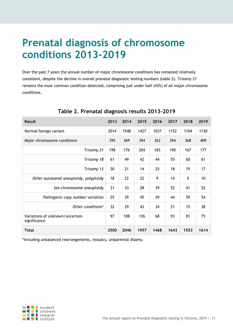
Prenatal diagnosis of chromosome conditions 2013-2019
Over the past 7 years the annual number of major chromosome conditions has remained relatively
consistent, despite the decline in overall prenatal diagnostic testing numbers (table 2). Trisomy 21
remains the most common condition detected, comprising just under half (43%) of all major chromosome
conditions.
Table 2. Prenatal diagnosis results 2013-2019
Result 2013 2014 2015 2016 2017 2018 2019
Normal/benign variant 2014 1548 1427 1037 1152 1104 1130
Major chromosome conditions 395 369 394 363 394 368 409
Trisomy 21 198 176 204 183 190 167 177
Trisomy 18 61 49 42 44 55 60 61
Trisomy 13 30 21 14 25 18 19 17
Other autosomal aneuploidy, polyploidy 18 22 22 9 14 5 10
Sex chromosome aneuploidy 31 33 28 39 52 41 52
Pathogenic copy number variation 25 39 45 29 44 59 54
Other conditions* 32 29 43 34 21 15 38
Variations of unknown/uncertain significance
97 108 126 68 93 81 75
Total 2500 2046 1957 1468 1643 1553 1614
*Including unbalanced rearrangements, mosaics, uniparental disomy.
The Annual report on Prenatal Diagnostic testing in Victoria, 2019 | 12
Publications from the Victorian Prenatal Diagnosis Data Collection
• Lindquist A, Hui L, Poulton A, Kluckow E, Hutchinson B, Pertile MD, Bonacquisto L, Gugasyan L, Kulkarni A, Harraway J, Howden A, McCoy R, Da Silva Costa F, Menezes M, Palma-Dias R, Nisbet D, Martin N, Bethune M, Poulakis Z, Halliday J. State-wide utilization and performance of traditional and cell-free DNA-based prenatal testing pathways: the Victorian Perinatal Record Linkage (PeRL) study. Ultrasound Obstet Gynecol. 2020 Aug;56(2):215-224. doi: 10.1002/uog.21899.
• Hui L, Poulton A, Kluckow E, Lindquist A, Hutchinson B, Pertile MD, Bonacquisto L, Gugasyan L, Kulkarni A, Harraway J, Howden A, McCoy R, Costa FDS, Menezes M, Palma-Dias R, Nisbet D, Martin N, Bethune M, Poulakis Z, Halliday J. A minimum estimate of the prevalence of 22q11 deletion syndrome and other chromosome abnormalities in a combined prenatal and postnatal cohort. Hum Reprod. 2020 Mar 27;35(3):694-704. doi: 10.1093/humrep/dez286.
• Kluckow E, Halliday J, Poulton A, Lindquist A, Hutchinson B, Bethune M, Bonacquisto L, Da Silva Costa F, Gugasyan L, Harraway J, Howden A, Kulkarni A, Martin N, McCoy R, Menezes M, Nisbet D, Palma-Dias R, Pertile MD, Poulakis Z, Hui L. Association between timing of diagnosis of trisomy 21, 18, and 13 and maternal socio-economic status in Victoria, Australia: A population-based cohort study from 2015 to 2016. Prenat Diagn. 2019 Dec;39(13):1254-1261. doi: 10.1002/pd.5577.
• Lostchuck E, Hui L. Should second-trimester hypoplastic nasal bone be sole indication for diagnostic testing with chromosomal microarray analysis? Ultrasound Obstet Gynecol. 2019 Jun;53(6):848-850. doi: 10.1002/uog.20141.
• Lostchuck E, Poulton A, Halliday J, Hui L. Population-based trends in invasive prenatal diagnosis for ultrasound-based indications: two decades of change from 1994 to 2016. Ultrasound Obstet Gynecol. 2019 Apr;53(4):503-511. doi: 10.1002/uog.19107.
• Howard-Bath A, Poulton A, Halliday J, Hui L. Population-based trends in the prenatal diagnosis of sex chromosome aneuploidy before and after non-invasive prenatal testing. Prenat Diagn. 2018 Dec;38(13):1062-1068. doi: 10.1002/pd.5363.
• Poulton A, Lewis S, Hui L, Halliday JL. Prenatal and preimplantation genetic diagnosis for single gene disorders: A population-based study from 1977 to 2016. Prenat Diagn. 2018 Nov;38(12):904-910. doi: 10.1002/pd.5352.
• Hui L, Norton M. What is the real "price" of more prenatal screening and fewer diagnostic procedures? Costs and trade-offs in the genomic era. Prenat Diagn. 2018 Mar;38(4):246-249. doi: 10.1002/pd.5228. Epub 2018 Feb 21.
• Hui L, Barclay J, Poulton A, Hutchinson B, Halliday JL. Prenatal diagnosis and socioeconomic status in the non-invasive prenatal testing era: A population-based study. Aust N Z J Obstet Gynaecol. 2018 Aug;58(4):404-410. doi: 10.1111/ajo.12778.
• Lindquist A, Poulton A, Halliday J, Hui L. Prenatal diagnostic testing and atypical chromosome abnormalities following combined first-trimester screening: implications for contingent models of non-invasive prenatal testing. Ultrasound Obstet Gynecol. 2018 Apr;51(4):487-492. doi: 10.1002/uog.18979.
• Hui L, Hutchinson B, Poulton A, Halliday J. Population-based impact of noninvasive prenatal screening on screening and diagnostic testing for fetal aneuploidy. Genet Med. 2017 Dec;19(12):1338-1345. doi: 10.1038/gim.2017.55.

The Annual report on Prenatal Diagnostic testing in Victoria, 2019 | 13
• Hui L, Muggli EE, Halliday JL. Population-based trends in prenatal screening and diagnosis for aneuploidy: a retrospective analysis of 38 years of state-wide data. BJOG. 2016 Jan;123(1):90-7. doi: 10.1111/1471-0528.13488.
• Susman MR, Amor DJ, Muggli E, Jaques AM, Halliday J. Using population-based data to predict the impact of introducing noninvasive prenatal diagnosis for Down syndrome. Genet Med. 2010 May;12(5):298-303. doi: 10.1097/GIM.0b013e3181d5d022.
• Jaques AM, Collins VR, Muggli EE, Amor DJ, Francis I, Sheffield LJ, Halliday JL. Uptake of prenatal diagnostic testing and the effectiveness of prenatal screening for Down syndrome. Prenat Diagn. 2010 Jun;30(6):522-30. doi: 10.1002/pd.2509.
• Muggli EE, Collins VR, Halliday JL. Mapping uptake of prenatal diagnosis for Down syndrome and other chromosome abnormalities across Victoria, Australia. Aust N Z J Obstet Gynaecol. 2006 Dec;46(6):492-500. doi: 10.1111/j.1479-828X.2006.00648.x.
• Muggli EE, McCloskey D, Halliday JL. Health behaviour modelling for prenatal diagnosis in Australia: a geodemographic framework for health service utilisation and policy development. BMC Health Serv Res. 2006 Sep 1;6:109. doi: 10.1186/1472-6963-6-109.
• Muggli EE, Halliday JL. Prenatal diagnostic testing and Down Syndrome in Victoria 1992--2002. Aust N Z J Public Health. 2004 Oct;28(5):465-70. doi: 10.1111/j.1467-842x.2004.tb00029.x.
• Collins VR, Webley C, Sheffield LJ, Halliday JL. Fetal outcome and maternal morbidity after early amniocentesis. Prenat Diagn. 1998 Aug;18(8):767-72.
• Halliday J, Lumley J, Watson L. Comparison of women who do and do not have amniocentesis or chorionic villus sampling. Lancet. 1995 Mar 18;345(8951):704-9. doi: 10.1016/s0140-6736(95)90872-2.
• Halliday JL, Watson LF, Lumley J, Danks DM, Sheffield LJ. New estimates of Down syndrome risks at chorionic villus sampling, amniocentesis, and livebirth in women of advanced maternal age from a uniquely defined population. Prenat Diagn. 1995 May;15(5):455-65. doi: 10.1002/pd.1970150509.
• Halliday JL, Lumley J, Sheffield LJ, Robinson HP, Renou P, Carlin JB. Importance of complete follow-up of spontaneous fetal loss after amniocentesis and chorion villus sampling [published correction appears in Lancet 1992 Nov 14;340(8829):1236]. Lancet. 1992;340(8824):886-890.