the alterations in the extracellular matrix of … alterations in the extracellular matrix of hearts...
TRANSCRIPT

The Alterations in the Extracellular Matrix of Hearts from Copper-Deficient Rats
Denis M. Medeiros and Laura Shiry
Department of Human Nutrition and Food Management, Ohio Agricultural and Research Development Center, The Ohio State University, Columbus
Abstract Morphological and biochemical aspects of the copper-deficient heart model are briefly reviewed in terms of intracellular and extracellular changes. The extracellular aspects of hearts from copper-deficient rats demonstrate fibrosis and thickened and fragmented basal laminae. In addition, heart valves from copper-deficient rats have altered morphological character and are thickened compared to valves from copper-adequate rats. The ultrastructural observations pertaining to the altered basal laminae are pursued in this study in terms of immunohistological staining against specific proteins that compose the structure. Specifically, we fed copper-adequate and deficient diets to rats for 5 weeks and processed the hearts for light microscopy and imrnuiiohistochemistry. From such observations it could be inferred that the hearts from copper-deficient rats had markedly increased staining for Type IV collagen and fibronectin, whereas the reverse for laminin was observed in lhat little staining was detected in the copper-deficient myocytes. Furthermore, there was increased binding of the antibody against the laminin receptor in the copper-deficient rats. The implications of the compromised basal laminae in the heart are discussed. Key words: copper, basal laminae, Type IV collagen, laminin, fibronectin, integrin.
Currently there is no Recommended Dietaiy Allowance for copper by the Food and Nutrition Board of the National Academy of Sciences. An estimated Safe and Adequate intake of 1.5 to 3.0 mgper day has been established [19]. The study of copper requirements and metabolism in humans is of interest, since many survey studies suggest an appreciable level of the American population often consume copper levels below this recommended amount. Whether such individuals are at risk for health consequences is debatable. A potential consequence of a lack of coppercould be its known deleterious effects upon cardiac pathology and function. This paper reviews this subject in terms of the role of copper in maintaining cardiac metabolism and function, and in particular focuses upon the roles copper exerts upon the extracellular matrix. We present immunohistochemistry findings that support previous biochemical studies from our lab reporting biochemical and ultrastructural alterations in matrix architecture.
Copper Deficiency Signs It is widely known that feeding a copper deficient
diet to animals results in characteristic deficiency signs. Brain, cardiac, blood and bone anomalies have historically been reported as signs of copper deficiency. Rats fed a copper-deficient diet for several weeks exhibit cardiovascular
Basic AppL Myol. 8 (2): 151-158, 1998
defects such as increase fragility of the connective tissue, ventricular aneurysms, hemothorax, pleural effusion, car-diac hypertrophy, aberrant electrocardiograms, hyper-cholcsterolemia, impaired immune response, lower hematocrit, impaired glucose tolerance, hypotension, dis-turbances in fatty acid profiles and norepinephrine meta-bolism as reviewed by us elsewhere [ 15 j. Among the signs of copper-deficiency that are of interest in this paper is cardiac hypertrophy. As suggested above there are numerous alterations of the cardiovascular system in the copper-deficient organism. Studies on both copper-deficient pigs and rats have revealed the pathological appearance of glycogen granules and lipid droplets present in hearts that are characteristics of oxygen deficit cardiomyopathy in other heart disease models. A hallmark appearance of hearts from copper-deficient animals includes a markedly increased area occupied by mitochondria (Figure 1). The mitochondria, however, are vacuolated where the usual parallel array of the cristae have become fragmented. One reason for this appearance may be due to the reported decreased levels of the nuclear encoded subunits of the copper containing enzyme, cytochrorne c oxidase. The catalytic portions of this enzyme are on subunits I and II, and are produced from the mitochondria! genome. It is unclear as to why copper is necessary for the appearance
-151-

Extracellular matrix in copper deficient hearts
Figure I. Transmission electron micrograph demonstrating the contrasts between the myocytes from hearts of copper -adquate (A) as opposed to copper-deficient (B) rats. Note that the copper-deficient myocardium has increased number of mitochondria that are also swollen and vacuolated. Increased number ofgiycogen granules are apparent in hearts from copper-deficient rats (arrow). M, mitochondria; my, myofibrils. Bar = 1µm
of the nuclear encoded subunits. Transcripts for these peptides are not decreased, nor is iheir synthesis. However, these peplides are a part of the inner mitochondria! mem-brane. A lack of these peptides probably explains the deterioration of the mitochondria. Details on these intra-cellular events are reported elsewhere [15, 18],
The hearts from copper deficient rats are significantly greater in mass compared to control rats. Their abdominal cavities often become filled with fluid, and the animals tire easily from a treadmill exercise regimen, both of which are signs of cardiac failure [6]. The type of hypertrophy in hearts from copper-deficient animals is concentric where the free walls become markedly thickened, as does the interventricular septum, and the ventricular lumen vol-umes are significantly decreased. Concentric hypertrophy is often associated with obesity, cigarette smoking, and aortic stenosis. A large increase in blood pressure can cause this pattern to predominate. The other type of hyper-trophy is termed eccentric hypertrophy, where the muscle perimeter walls are greatly enlarged, and chambers wid-ened. Volume overload conditions, such as what occurs with severe anemia is a situation where this condition may be observed. While copper deficient rats may become anemic or at least have decreased hematocrit levels, the emerging consensus is that this is not related to cardiac hypertrophy in this model. We have demonstrated that the hearts enlarge prior to appearance of anemia, and the degree of anemia is not connected with heart enlargement
[14]. Some of the largest hearts from copper deficient rats have been observed with normal hematocrit levels. Also, the pattern of hypertrophy is concentric, not eccentric, as would be expected with anemia.
Extracellular Matrix of the Heart in Copper-Deficiency One of the major defects in hearts from copper deficient
rats is the connective tissue, in particular the collagens, and to a lesser extent, elastin. Most of the collagen affecting the heart are Types I, HI, and IV. Types I and III collagen are encoded by the nucleus within the fibroblasts, whereas Type IV is nuclear encoded by both myocytes and fibroblasts. These collagen types exert specific roles upon cardiac function. Tensile strength and stiffness are attributed to Type I collagen. Type III collagen has a major role in maintaining the elasticity and compliance of the rnyo-cyle. Type IV collagen is non-fibrillar in structure. In the heart it is a component of basement membranes of both myocytes and capillaries. Lysyl oxidase is an enzyme that contains copper as a cofactor that deaminates selected lysinc and hydroxylysine residues. This promotes covalent crosslinking between collagen molecules, as reviewed by Eyre et al. [8] and Reiser et al. [20]. Elastin, another type of connective tissue, is often synthesized during gestation. In humans little, if any, synthesis of new elastin crosslinks occurs. Elastin content of animals are likely to have little reduplication following birth. Most elastin is laid down during gestation and the infant is born with sufficient
-152-

Extracellular matrix in copper deficient hearts
Figure 2. Light micrograph of tricitspid valve from rats fed copper-adequate (A) or deficient (B) diets for 6 weeks. The views are cross-sectional and were sampled from the area be/ween [he chordae and line of closure. Note the thickening, but less densely packed, connective tissue of the copper-deficient valve relative to the capper-adequate valve, a denotes the atria/ side of the valve, and v the ventricular side. C with the arrow shows collagen fibrils. Original slides were observed at 400 X.
elastin to last almost the entire life span [21]. This aspect is likely to have a significant impact upon cardiac valves and blood vessels. Tricuspid valves from copper deficient rats show increased thickness, loosely arranged connective tissue and a separation of the three layers (cell thick cndo-thelial layer, zona spongiosa layer adjacent 10 the endolhe-lial layer, and the zona fibrosa layer) that comprise them (Figure 2). The collagen fibers are dispersed in the ground substance and the zona fibrosa has increased thickness. This is in contrast to the controls in that the connective tissue of the valves appear dense and neatly packed [14]. Similar findings have been shown with copper deficient pigs [22]. While the collagen content of the myocardium and valves did not tend to differ by copper status, the degree of collagen crossiinking was decreased as assessed by the levels of hydroxylysylpyridinoline, in copper deficient pigs. The left ventricle showed reduced collagen crossiinking as did the cardiac bicuspid valves. Despite the decreased crossiinking in these valves, stress strain tests revealed no difference in valvular rigidity. One of our recent unpublished studies used echocardiography to study the influence of copper deficiency upon valve function in rats. Rats were fed either copper-adequate or copper- deficient diets from weaning for 5 weeks. Prior to placing them under the echo probe, cardiac sounds were determined with a standard clinical stethoscope. Of the 12 copper deficient rats in the experiment, all 12 had definite murmurs. Echocardiography confirmed the presence of valvular regurgitation in rals fed copper deficient diets. We repeated a study using pigs in which they were fed purified diets containing cither copper or no added copper [23]. The pigs were fed from 15 days of age until 8 weeks of age. Decreased body weight of copper-deficient pigs were noted, and ihey appeared more lethargic. One died from cardiovascular complications, and one died on the echo table from the anesthesia. With the remaining 5 controls
and 3 copper deficient pigs, we were able to note valvular regurgitation in the copper-deficient pigs,
Borg et al. [4] first described that the connective tissue weave of hearts from copper deficient rats was signifi-cantly altered and in greater disarray than controls. Borg et al, [4] and Dawson ct al. [7] both demonstrated that there was a shift in a greater proportion of Type III collagen to Type I collagen in hearts from copper deficient rats. Vad-lamudi et al. [22] reported a shift from Type I to Type III collagen in both ventricle and bicuspid valves of copper-deficient pigs. In addition to the collagen proteins, laminin and fibronectin should be considered, as well as the inte-grins, in this case the laminin receptor. Farquharson and Robins [9] went on to further report that proteins comprising the basement membrane are altered. It is expected that when Type I collagen is present, fibronectin expression and synthesis is increased to serve as a scaffold or lattice for the fibrillar proteins.
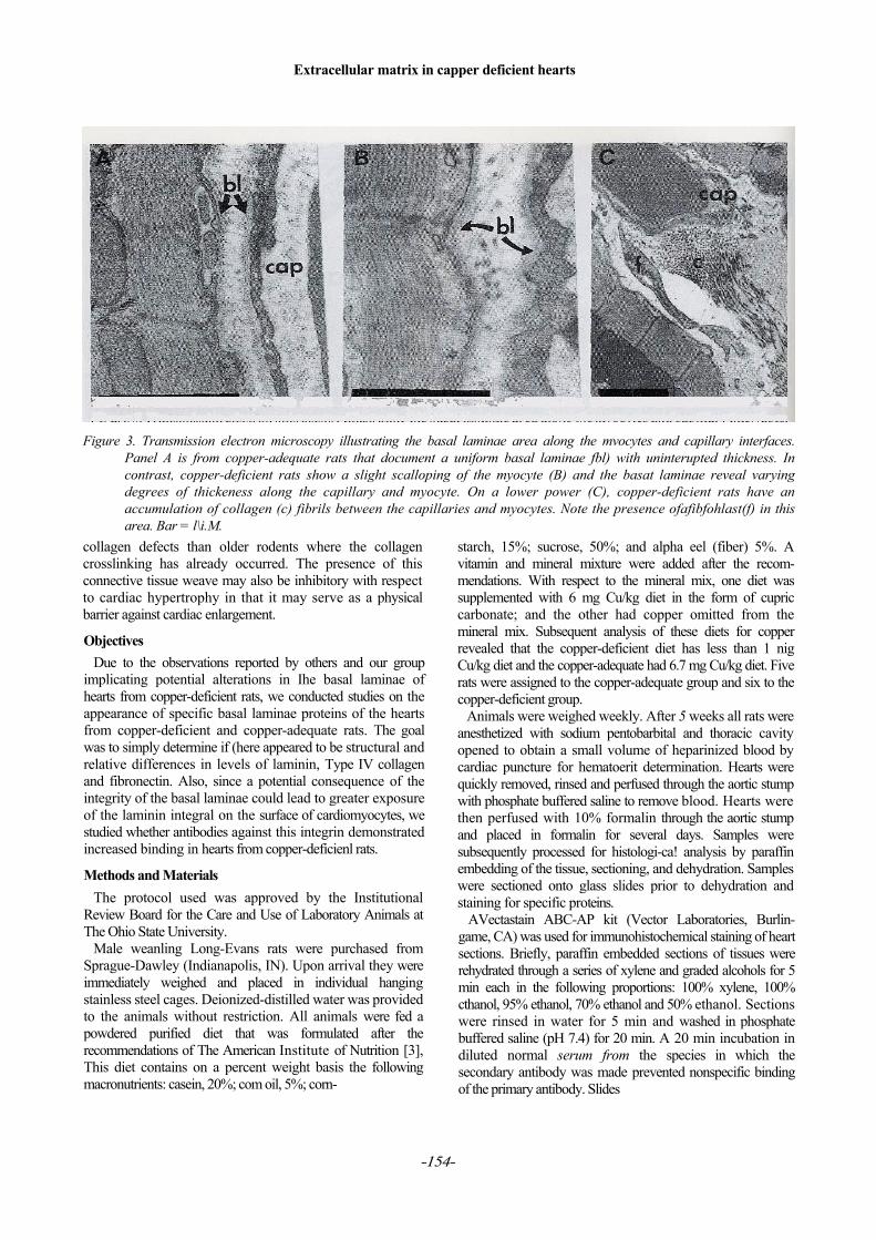
Several studies conducted by our group clearly demon-strated that collagen metabolism is significantly altered in copper deficiency. We have reported that fibrillar colla-gens tend to accumulate in the myocytal peripapillary regions of the heart. The presence of phagocytes are com-mon. Both the myocytes and blood vessels in the area appear to be equally affected (Figure 3) in thickening and disruption of the basal laminae. Older rats placed on diets deficient in copper when they are well past weaning do not tend to develop cardiac hypertrophy, but do present a greater accumulation of fibrillar collagen in the above mentioned region. Rats fed copper-deficient diets from weaning display some collagen, but not as much when compared to rats fed the copper-deficient diets starting as young adults. Younger rats fed copper deficient diet de-velop hypertrophy; older rats do not, or if they do, a much longer time period may be needed. Most of the collagen crossiinking occurs within the 2 to 3 weeks following weaning and these rats may be more susceptible to the
-153-
Extracellular matrix in capper deficient hearts
Figure 3. Transmission electron microscopy illustrating the basal laminae area along the mvocytes and capillary interfaces. Panel A is from copper-adequate rats that document a uniform basal laminae fbl) with uninterupted thickness. In contrast, copper-deficient rats show a slight scalloping of the myocyte (B) and the basat laminae reveal varying degrees of thickeness along the capillary and myocyte. On a lower power (C), copper-deficient rats have an accumulation of collagen (c) fibrils between the capillaries and myocytes. Note the presence ofafibfohlast(f) in this area. Bar = l\i.M.
collagen defects than older rodents where the collagen crosslinking has already occurred. The presence of this connective tissue weave may also be inhibitory with respect to cardiac hypertrophy in that it may serve as a physical barrier against cardiac enlargement.
Objectives Due to the observations reported by others and our group
implicating potential alterations in Ihe basal laminae of hearts from copper-deficient rats, we conducted studies on the appearance of specific basal laminae proteins of the hearts from copper-deficient and copper-adequate rats. The goal was to simply determine if (here appeared to be structural and relative differences in levels of laminin, Type IV collagen and fibronectin. Also, since a potential consequence of the integrity of the basal laminae could lead to greater exposure of the laminin integral on the surface of cardiomyocytes, we studied whether antibodies against this integrin demonstrated increased binding in hearts from copper-deficienl rats.
Methods and Materials The protocol used was approved by the Institutional
Review Board for the Care and Use of Laboratory Animals at The Ohio State University.
Male weanling Long-Evans rats were purchased from Sprague-Dawley (Indianapolis, IN). Upon arrival they were immediately weighed and placed in individual hanging stainless steel cages. Deionized-distilled water was provided to the animals without restriction. All animals were fed a powdered purified diet that was formulated after the recommendations of The American Institute of Nutrition [3], This diet contains on a percent weight basis the following macronutrients: casein, 20%; com oil, 5%; corn-
starch, 15%; sucrose, 50%; and alpha eel (fiber) 5%. A vitamin and mineral mixture were added after the recom-mendations. With respect to the mineral mix, one diet was supplemented with 6 mg Cu/kg diet in the form of cupric carbonate; and the other had copper omitted from the mineral mix. Subsequent analysis of these diets for copper revealed that the copper-deficient diet has less than 1 nig Cu/kg diet and the copper-adequate had 6.7 mg Cu/kg diet. Five rats were assigned to the copper-adequate group and six to the copper-deficient group.
Animals were weighed weekly. After 5 weeks all rats were anesthetized with sodium pentobarbital and thoracic cavity opened to obtain a small volume of heparinized blood by cardiac puncture for hematoerit determination. Hearts were quickly removed, rinsed and perfused through the aortic stump with phosphate buffered saline to remove blood. Hearts were then perfused with 10% formalin through the aortic stump and placed in formalin for several days. Samples were subsequently processed for histologi-ca! analysis by paraffin embedding of the tissue, sectioning, and dehydration. Samples were sectioned onto glass slides prior to dehydration and staining for specific proteins.
AVectastain ABC-AP kit (Vector Laboratories, Burlin-game, CA) was used for immunohistochemical staining of heart sections. Briefly, paraffin embedded sections of tissues were rehydrated through a series of xylene and graded alcohols for 5 min each in the following proportions: 100% xylene, 100% cthanol, 95% ethanol, 70% ethanol and 50% ethanol. Sections were rinsed in water for 5 min and washed in phosphate buffered saline (pH 7.4) for 20 min. A 20 min incubation in diluted normal serum from the species in which the secondary antibody was made prevented nonspecific binding of the primary antibody. Slides
-154-

Extracellular matrix in copper deficient hearts
were gently blotted of excess normal serum and incubated for 30 min with primary antiserum diluted in PBS. After a brief washing in buffer, sections were incubated for 30 min with diluted biotinylated antibody solution, and rinsed again in buffer. Sections were then incubated for 30 min with alkaline phosphatase reagent and rinsed in buffer. Sections were stained for approximately 15 minutes using the Alkaline Phosphatase Substrate solution in 100 mM Tris-HCI, pH 9.5 (Vector Laboratories). Sections were ihen rinsed in water and dehydrated through a series of alcohol and xylene concentrations. Cover slips were added for viewing under light microscopy. Control slides were made in which the primary antibody was omitted.
The antibodies used were: rabbit anti-iaminin or a6bl (Serotec Ltd, Oxford, UK), sheep anti-fibronectin (Serotec Ltd.), mouse anti-collagen. Type IV (Sigma Chemical Co., St. Louis, MO), mouse anti-laminin receptor (VLA-6, a6/bl) (Chemicon, Inc., Temecula, CA).
Photographs of selected fields were made for each animal specimen at 400X. In some instances, other magnifications were used. A plus-minus system was used to determine the relative level of staining intensity for each of the antigens probed. An impaired t-test was used to determine if statistical significance between selected vari-ables existed.
Results Rats fed the copper-deficient diets demonstrated some of
the usual signs of deficiency. In particular there was a significant increase in heart:body weight (p < 0.01) and an increase in absolute heart weight (p = 0.057). Hematocnt levels were significantly decreased (p < 0.01) in coppcr-
Figure 4. Typical results of light microscopy demonstrating the immunohisiochemical staining for Type IV collagen in
hearts from copper-adequate rats (A and C) as opposed to hearts from copper-deficient (B andD) rats. In the hearts from copper-deficient rats (B and D) a thickening of Type IVcollagen can be clearly observed. Arrows show where ihe collagen areas are located along the myocytes.
-155-

Extracellular matrix in topper deficient hearts
Figure 5. Fibronectin staining to heart tissue from copper-adequate (A) and copper-deficient (B) rats. Note the significant thickness in panel B (arrow) for fibronectin as compared Co panel A.
deficient rats. While there was a trend toward lower body weight in the topper-deficient group. Hie difference was not significanl (p > 0.05).
Immunoliislochemieal staining for specific basement membrane proteins demonstrated clear differences by dietary copper treatment that were consistent with biochemical measures (eg. eleclrophorcsis) previously reported by our group [13] and appeared consistent with our electron
microscopy observations reported earlier [5, 6]. Staining of heart sections for Type IV collagen and fibronectin demonstrated clear and consistent thickening of Ihe basement membrane surrounding the myocytes in ad of die copper-deficient rats; examined by lighl microscopy (Figures 4 and 5). In contrast, laminin levels were decreased in the myocytes from copper-deficient rats compared to the copper-adequate group (Figure 6). Since laminin levels
Figure 6. Staining for the laminin protein demonstrates presence in the hearts of copper-adequate rats (A, arrows), but
apparent lack in the hearts of copper-deficient rats (B). Staining against the laminin receptor shows a greaternumber of exposed receptors in the hearts from copper-deficient rats (D) compared to the hearts from copper-adequate rats (C). Also note the presence oflhe small dots throughout the photo on the a6bl or laminin receptor (D) demonstrating the receptor.
-156 -
Extracellular matrix in copper deficient hearts
appeared decreased in this study, and the basal laminae in previous studies conducted by out group suggested frag-mentation [5, 6] we hypothesized that there may be in-creased binding of antibodies against the a6bl or laminin receptor. As indicated in Figure 6, there appeared to be greater antibody binding in the hearts from copper-defi-cient rats. Using a scoring system of-2 to+ 2 for staining intensity of the signal, the copper-deficient group had a mean of 1.6 ± 0.25 as opposed to 0.4 ± 0.25 in the copper-adequate group (p< 0.01).
Discussion This research was conducted as a follow-up to the obser-
vations of Farquharson and Robins that reported altered basement membrane morphology in hearts of copper-de-ficient rats [9]. We noted from several of our studies that the basal laminae surrounding the myocytes and the blood vessels appeared thickened in some areas but thin in others [5, 6]. Discontinuity of the basal laminae was apparent in many of the copper deficient rat hearts. Besides offering a mechanical role or barrier for the myocytes and capi llaries, the basal laminae may also assist in cell division, orientation of cell position, and protection of cell receptors [2]. The laminin layer is adjacent to the sarcolemma of the capillary membrane and muscle sarcolemma. Type IV collagen is bound adjacent to the laminin, followed by the fibroneetin. The laminin is attached to the sarcolemma by a specific integrin. The fibroneetin is known to increase in mass whenever there is an increase synthesis of the fibrillar collagen, such as Type I. Fibroneetin is thought to serve as a scaffold in that the collagens may attach to as it develops and matures. That both fibroneetin and Type I collagen appeared thickened in hearts from copper-deficient rats from this study is therefore consistent with this morpho-logical arrangement. Using electrophoresis techniques in aprevious study [13] •
we were able to determine if changes relative to control hearts had occurred. SDS-P AGE results revealed a marked increase in fibroneetin levels, but a significant decrease in laminin levels [13], that is consistent with the findings reported here. Staining paraffin embedded slides with an-tibodies directed against the laminin integrin, showed dif-ferences as well. The enhanced staining obtained for the laminin integrin suggested these receptors were exposed for antibody binding, a situation considered abnormal. The long term significance of such a finding raises the question whether other receptors are exposed to the outside cellular environment where protease and phagocytes have access. Some pathology we observe in copper deficient heart disease could be related to changes in receptor population and function.
It is not totally clear as to why the basal laminae would be disrupted by copper-deficiency. Several possible expla-nation may explain the observations. Type IV collagen may have impaired cross-links due to potential decreased activity of the cupro-enzyme, lysyl oxidase. The unique Ihrce dimensional arrangement of this collagen may afford
stability to the basal laminae and consequent lack of crosslinking may make the architecture more unstable. The fibroneetin levels may increase due to the accumulation of Type 1 collagen within the intercellular spaces of copper-deficient hearts [5]. Alternatively, we have observed the presence of phagocytes along the myocytes from copper-deficient rats, especially in areas where the basal laminae demonstrated the greatest alterations. Secretion of pro-teases in the intercellular area by phagocylie cells may partly explain these observations. The overall significance of these alterations in the basal laminae upon the heart are not certain. Recently we observed that rats fed copper deficient diets developed cardiac hypertrophy prior to any changes in fibroneetin or laminin levels [12]. Decreased quantities of the nuclear encoded subunil peptides of cyto-chrome c oxidase appeared to coincide with the develop-ment of hypertrophy, as opposed to the changes in the proteins comprising the basal laminae. This suggests that damage to the basal laminae is not a significant stimulus in causing the hypertrophy in copper-deficiency. However, function of the inyocyte and heart such as electrical conduction through the myocardium as revealed by abnormal electrocardiograms could be compromised. The altered electrocardiograms reported by us [16] for copper-deficient rats could partly be explained by these results in addition to the heart enlargement. Subsequent studies on other tissues from copper-deficient animals that contain basement membranes may be of value in determining whether these observations are generalized or specific to the heart only. Histological studies have revealed thickening of the basement membranes of lung, kidney and spleen of copper-deficient rats [1,9-11], Furthermore, the overall implications of increased access to the laminin receptor likely implies other cell membrane components could be less protected and made more available to pro-teases and phagocytes. The impact upon cell surface receptors and other related issues remains to be determined,
Acknowledgements Supported by funds from the Ohio Agricultural and
Research Development Center, Project Number H-112, Manuscript Number 55-97.
Address correspondence to: Denis M. Medeiros, Department of Human Nutrition and
Food Management, Ohio Agricultural and Research De-velopment Center, 357 Campbell Hall, 1787 Neil Avenue, The Ohio State University, Columbus. OH 43210-1295, phone 614 292 5575, fax 614 292 7536, Email Medeiros. [email protected].
References [1] Akers TK, Saari .IT: Hypcrbaric hyperoxia exag-
gerates respiratory membrane defects in the cop-per-deficient rat lung. Bio! Tr El Res 1993; 38; 149-163.
-757-

Extracellular matrix in copper deficient hearts
[2] Alberts B, Bray P, Lewis J, Raff M. Roberts R., Watson JD: Molecular biology oflhe cell (3rd ed). Garland Publishing Inc, New York, 1994.
[3] American Institute of Nutrition: Report of the AIN ad hoc committee on standards for nutritional stud-ies. ./Mifr 1977; 107: 1340-1348.
[4] Borg TK, Klevay LM, Gay RE, Siegel R, Bergin ME: Alteration of the connective tissue network of striated muscle in copper deficient rats. J Mol Cell Cardiol\n$; 17: 1173-1183.
[5] Davidson JA, Medeiros DM, Hamlin RL: Cardiac ultra structure and electrophysiological abnormalities in postweanling copper-restricted and copper-repleted rats in the absence of hypertrophy. J Nutr 1992; 122: 1566-1575.
[6] Davidson J, Medeiros DM, Hamlin RL, Jenkins JE: Submaximal, aerobic exercise training exacerbates the cardiomyopathy of postweanling Cu-dep!eted rats. Biol Tr E! Res 1993; 38: 251-272.
[7] Dawson R, Milne G, Williams RB; Changes in the collagen of rat heart in copper-deficient-induced cardiac hypertrophy. Cardiovasc Res 1982; 16: 359-365.
[8] Eyre DR, Koob TT, von Ness KP: Quantitation and hydroxypyridinium cross-links in collagen by HPLC. Anal Biochem 1984; 137: 380-388.
[9] Farquharson C, Robins S: Immune localization of collagen types I, III, and IV, elastin and fibronectin within the heart of normal and copper-deficient rats./CompPathol 1991; 104: 245-255.
[10] Fell BF, Farmer LJ, Farquharson C, Bremnet I, Graca DS: Observations on the pancreas of cattle deficient in copper. J Comp Pathol 1985; 95: 573-590.
[11] Fell BF, Farquharson C, Riddoch GE: Kidney le-sions in copper-deficient rats. J Comp Physio! 1987; 97: 187-196.
[12] Jalili T, Medeiros DM, Prochaska L: Alterations in cardiac cytochrome c oxidase, but not in laminin and fibronectin, are observed within three weeks of copper restriction in rats: implications, for cardiac hypertrophy. Nutr Res 1997; 17: 493-504.
[13] Liao Z, Medeiros DM, McCune SA, Prochaska LJ: Cardiac levels of fibronectin, laminin, isomyosins, and cytochrome e oxidase of weanling rats are more vulnerable to copper deficiency than those of post-weanling rats. J Nutr Biochem 1995; 6: 385-391.
[14] Medeiros DM, Bagby D, Ovecka G, McCormick R: Myofibrillar, mitochondrial and valvular mor-phological alterations in cardiac hypertrophy among copper-deficient rats. J Nutr 1991; 121: 815-824.
[15] Medeiros DM, Davidson J, Jenkins JE: A unified perspective on copper def ic iency and cardiomyopathy. Proc Sac Exp Biol 1993; 203: 262-273.
[16] Medeiros DM, Liao Z, Hamlin R: Ultra structural and longitudinal measures of electrocardiographic activity and cardiac function in copper-deficient rats. J Nutr 1991; 121: 1026-1034.
[17] Medeiros DM, Shiry L, Lincoln, AJ, Prochaska L: Cardiac non-myofibrillar proteins in copper-defi-cient rats: amino acid sequencing and western blot-ting of altered proteins. Biol Trace El Res 1993; 36: 271-282.
[]8] Medeiros DM, Shiry L, Samelman T: Cardiac nu-clear encoded cytochrome c oxidase subunits are decreased with copper restriction but not from iron restriction: gene expression, protein synthesis and heat shock protein aspects. Comp Biochem Physiol 1997; 117A: 77-87.
[19] Recommended dietaiy allowances. Food and Nu-trition Board, Washington DC: National Academy of Sciences Press, 1989, pp 224-230.
[20] Reiser K, NcCormick RJ, Rucker RB: The enzy-matic and nonenzymatic cross-linking of collagen and elastin. FASEB J 1992; 6: 2439-2449.
[21] Shapiro SD, Endicott SK, Province MA, Pierce JA, Campbell EJ: Marked longevity of human lung parenchymal elastic fibers deduced from preval-ence of D-aspartate and nuclear weapons-related radiocarbon. J Clin Invest 1991; 87: 1828-1834.
[22] Vadlamudi RK, McCormick RJ, Medeiros DM, Vossoughi J, Failla ML: Copper deficiency alters collagen types and covalent crosslinking in swine myocardium and cardiac valves. Am J Physio! 1993; 264 (Heart Circ Physiol 33): H2154-H2161.
[23] Wildman REC, Medeiros DM, Hamlin RL, Stills H, Jones DA, Bonagura JD: Aspects of cardiomyopathy in copper-deficient pigs: electro-cardiography, echocardiography, and ultraslruclu-ral findings. Biol TraceElRes 1996; 55: 55-70.
-158 -