the acute and chronic effects of resveratrol on renal
TRANSCRIPT

Wayne State University
Wayne State University Dissertations
1-1-2015
The Acute And Chronic Effects Of Resveratrol OnRenal Function And Blood PressureKevin L. GordishWayne State University,
Follow this and additional works at: http://digitalcommons.wayne.edu/oa_dissertations
Part of the Physiology Commons
This Open Access Dissertation is brought to you for free and open access by DigitalCommons@WayneState. It has been accepted for inclusion inWayne State University Dissertations by an authorized administrator of DigitalCommons@WayneState.
Recommended CitationGordish, Kevin L., "The Acute And Chronic Effects Of Resveratrol On Renal Function And Blood Pressure" (2015). Wayne StateUniversity Dissertations. Paper 1335.

THE ACUTE AND CHRONIC EFFECTS OF RESVERATROL ON RENAL FUNCTION AND BLOOD PRESSURE
by
KEVIN L. GORDISH
DISSERTATION
Submitted to the Graduate School
of Wayne State University,
Detroit, Michigan
in partial fulfillment of the requirements
for the degree of
DOCTOR OF PHILOSOPHY
2015
MAJOR: PHYSIOLOGY
Approved by:
_____________________________________ Advisor Date
_____________________________________
_____________________________________
_____________________________________
_____________________________________

© COPYRIGHT BY
KEVIN L. GORDISH
2015
All Rights Reserved

ii
DEDICATION
To my loving departed parents I honor your memory with education and hard work,
To my wife Erin, a Hitchcock aficionado, for withstanding The Man Who Knew Too
Much, To Stuart “frogs” never frightened me away from school,
To RIW and my gaming cohorts, thank you for the fun distractions,
To my family and friends thank you for the understanding.

iii
ACKNOWLEDGEMENTS
My gratitude is forever extended to my advisor and friend, Dr. William
Beierwaltes. Your training and insight have been invaluable. Thank you for your tireless
investment in time and energy in me.
Thank you to the members of my dissertation committee (Dr. Joseph Dunbar, Dr.
Noreen Rossi, Dr. Todd Leff, and Dr. Douglas Yingst) for their support and scientific
acumen, D’Anna Potter for her technical assistance and levity, and the senior staff of
the Henry Ford Health System Division of Hypertension and Vascular Research. In
particular, I want to thank Dr. Pablo Ortiz, Dr. Pamela Harding, Dr. Surresh Palaniyandi,
and Dr. Oscar Carretero. Henry Ford Health System remains an unrivaled training
ground for inquisitive minds.
Lastly, thank you to my friend, Christine Cupps, who knows M&M’s really do melt
in your hand. Your administrative prowess is unmatched.

iv
PREFACE
In the 1940’s, the initial discovery of resveratrol by Japanese scientist, MJ
Takaota, in the roots of the white hellebore plant, garnered little interest from the
research community (164, 170). However, in the early 1990’s resveratrol was also
shown to be present in red wine from the skin of the grape. It was suggested
resveratrol might explain the lower incidence of cardiovascular events associated with
of red wine consumption (152). This observation sparked interest in resveratrol both
from the research community and the public. Interest in resveratrol was increased
when an association was made that there is a low incidence of heart disease and
obesity among the French society despite their relatively high-fat diet (138). In contrast,
a recent study disputes the cardiovascular benefit of resveratrol (148). Notwithstanding,
resveratrol was compelling to a growing field of researchers. Resveratrol is found in two
configurations (Figure 1). The trans-resveratrol isomer, the biologically active form, is
stable for an extended time when protected from light, but in the presence of light it can
undergo photoisomeric changes to the inactive cis-resveratrol isomer (173).
Figure 1: Chemical structure of resveratrol isomers: cis-resveratrol (left) and trans-resveratrol (right).
Resveratrol research crosses many disciplines ranging from anti-aging, cancer,
inflammation to diabetes and cardiovascular research. This expansive field of research
has yielded noteworthy results and controversy. Resveratrol was reported to activate

v
Sirt1 (22). Sirt1, known as NAD-dependent deacetylase sirtuin1, an enzyme found in
both in yeast and humans, is linked to increased longevity possibly under states of
caloric restriction (20). However, these results were drawn into question when it was
shown experimental artifacts of florescent tags may have led to falsely reported Sirt1
activation (20). Further other groups also report resveratrol has no effect on Sirt1
activity (50). It remains to be seen if the in vitro effects of Sirt1 activation is germane to
the biological effects of resveratrol in vivo.
Another area of resveratrol research is its potential effect to reduce inflammation.
The inflammatory response is mediated in part by prostaglandins which are metabolic
products of cyclooxygenase activity. Resveratrol has been shown to reduce
inflammation in a model of colitis in rat by reducing NF-kappa beta activity (Martin). NF-
kappa beta is found to be chronically active in many inflammatory diseases and is
considered to be pro-inflammatory (109). NF-kappa beta is known to regulate
interleukin-1 (IL-1), an inducer of pro-inflammatory cytokines (37). Resveratrol has
been shown to reduce IL-1activity possibly through inhibition of NF-kappa beta (149).
There is a growing association between inflammation and the development of
hypertension. IL-1 levels are increased in individuals with pulmonary hypertension and
IL-1 antagonists are shown to reduce pulmonary hypertension (30, 56).
Another area of resveratrol research is the effect of resveratrol treatment on
osteoporosis. Resveratrol treatment in ovariectomized rats improves bone density
(187). Further in ovariectomized rats, resveratrol treatment is shown to influence
changes in mRNAs that are associated with bone degradation. Guo et al. (57) has
shown resveratrol treatment diminishes osteoporosis by suppressing miR 338 3p in
ovariectomized rats.

vi
Despite these observations, it is apropos to address a major criticism underlying
all of resveratrol research. The in vivo biological effects of resveratrol may be severely
limited by low bioavailability. Although resveratrol is well absorbed and can readily
enter the cell by diffusion, resveratrol is not maintained in high levels within the blood
stream (4). Following absorption, under 4% of non-degraded resveratrol remains in the
circulation while the majority of resveratrol is degraded into two main metabolites:
resveratrol-3-sulfate and resveratrol-3-glucuronide (5, 105). The possible biological
effects of these resveratrol metabolites has not been well described. Potentially, the
resveratrol metabolites themselves could have biologic effects, especially after chronic
administration of resveratrol. However, the field is dominated by a plethora of
conflicting in vitro and in vivo studies with disparate results. This may in part be
explained that in vitro studies are able to provide resveratrol doses not normally found
during normal physiological conditions, in part due to the low resveratrol bioavailability.
It is likely the interplay between biologically active resveratrol and possible effects of the
metabolites will be the focus of future research.
Criticisms aside, resveratrol is fascinating to a renal physiologist for two reasons:
surprisingly the acute and long term effects of resveratrol on renal function in a normal
human or rat have not been characterized. Subsequently, resveratrol is known to
stimulate pathways which may influence changes in basic renal physiology. It is
unknown if resveratrol can modify changes in plasma renin activity (PRA), in vivo
alterations in renal nitric oxide, free radical formation, renal hemodynamics, or sodium
homeostasis. These are of particular interest because changes in these renal systems
may influence changes in overall renal function, sodium balance and blood pressure.
Resveratrol as a biological ‘effector’ is particularly interesting due to numerous

vii
reports that it increases nitric oxide (101, 185). Nitric oxide is produced by three
isoforms of nitric oxide synthase (NOS): the neuronal nNOS, the inducible iNOS, and
the endothelial (eNOS). Under physiological conditions, vascular nitric oxide is mainly
produced by eNOS. This enzyme is constitutively expressed in the endothelium, is
tonically active, and is activated by shear stress and by additional agonists (25). Nitric
oxide derived from all 3 NOS isoforms contributes to the overall regulation of kidney
function. Nitric oxide inhibition, in normal rats, has been shown to result in acute
hypertension and renal vasoconstriction (14).
Studies have demonstrated that treatment of cultured human endothelial cells
with resveratrol enhances the mRNA and protein expression of eNOS (177-178). An
extensive review by Li and Förstermann (101, 185) summarized pathways by which
resveratrol increased nitric oxide, including stimulation of eNOS enzymatic activity and
reducing oxidative stress. The potential effects of nitric oxide on blood pressure via
action in the kidney are multifactorial. Nitric oxide increases renal blood flow, enhances
glomerular filtration rate, regulates renin secretion, and inhibits sodium transport in the
nephron (46, 106, 145, 156). It is unknown whether resveratrol, acting through the
actions of increased nitric oxide, may influences changes in renal hemodynamics,
sodium homeostasis, and ultimately blood pressure.
Under normal physiological conditions, the kidneys (on average) receive 20-22%
of total cardiac output (83). This represents a large amount of blood when considering
that total kidney weight is relatively small when compared to total body weight. Thus,
relatively small changes in renal hemodynamics can have profound effects on renal
function. Nitric oxide inhibition in anesthetized rats significantly deceases blood flow to
visceral organs (kidney, intestine, and lung), but overall has minimal effect on blood flow

viii
to the brain, heart, or hindlimbs (153). In humans, nitric oxide synthase inhibition
deceases renal blood flow, glomerular filtration rate and urinary sodium excretion (12).
Agents that increase nitric oxide production may reduce renal vasoconstriction in
pathological states associated with reduced renal perfusion (171). In particular, the
renal vascular NO plays a role in balancing the effects of the endogenous
vasoconstrictor Angiotensin II (153, 156). These data suggest that the kidney and renal
hemodynamics are particularly sensitive to changes in nitric oxide. Moreover, changes
in nitric oxide can impact renal function including transport of electrolytes and plasma
renin activity.
Currently, there are no reports on the effect of resveratrol on plasma renin
activity. The renin-angiotensin system is an important regulator of sodium homeostasis,
extracellular fluid volume, systemic vascular resistance, and arterial blood pressure.
Renin is produced within and secreted from juxtaglomerular cells (JG cells) of the
afferent arteriole (41, 147). Control of renin secretion, the rate-limiting step in the
formation of angiotensin II, is therefore essential for blood pressure regulation. Renin
secretion is regulated by the second messenger cAMP (16). However, in contrast to
most secretory cells, renin secretion is inversely related to extracellular and intracellular
calcium concentrations (16). The inverse relation of calcium to renin is referred to as
the “calcium paradox.” Three classic factors promote the release of renin from JG cells:
activation of renal baroreceptor, the macula densa sensing decreased sodium chloride
delivery, and increased sympathetic nerve activity (64). Resveratrol is a selective
inhibitor of cyclooxygenase-1 (COX-1), but not COX-2 (78). The primary pathway for
the stimulation of renin via to the macula densa pathway is due to an upregulation of
COX-2 (63). PGE2 is known to stimulate renin secretion (80). While resveratrol does

ix
not inhibit COX-2, resveratrol has been shown to inhibit PGE2 production (32, 36). It is
unclear if resveratrol can modify plasma renin activity by influencing changes in renal
hemodynamics, distal sodium delivery, or prostaglandins. Further it unclear whether
resveratrol treatment, acting through changes in PRA and/or increasing vasodilation, is
responsible for its reported effects to lower blood pressure in animal models.
In multiple models of hypertension in the rat resveratrol has been shown to
reduce blood pressure. Bhatt et al (19) gave resveratrol (50 mg/L in drinking water) to
SHR and reported that SHR given resveratrol had lower systolic blood pressure than
non-treated SHR. Dolinsky et al (42) implanted mice with osmotic minipumps to deliver
Ang II and placed mice on a diet containing resveratrol. After two weeks of resveratrol
treatment, the Ang II plus resveratrol group had lower blood pressure when compared
to the Ang II infused alone. While these studies and other similar reports show a blood
pressure lowering effect of resveratrol (presumably due to enhanced nitric oxide
synthesis or decreased oxidative stress), there are no renal investigators studying the
influence of resveratrol on plasma renin activity, renal nitric oxide, sodium homeostasis,
or renal hemodynamics.
My overall hypothesis is that the antihypertensive effects of resveratrol are
mediated through increasing vasodilation, increasing NO synthesis, reducing oxidative
stress and sodium retention, all of which would diminish hypertension. We intend to
systematically investigate the acute and sustained effects of resveratrol on renal
physiology in the normal rat and the chronic effects in two models of hypertension.
Extrapolating from the existing literature, we anticipate resveratrol will increase renal
blood flow and decrease sodium retention due to a nitric oxide-mediated mechanism,
and gradually reverse increases in blood pressure.

x
A frequently asked question relates to how much resveratrol is found in red wine
and how does this compare to doses employed in experimental research? The amount
of resveratrol found in red wine varies greatly 0.1 to 14.3 mg/liter (9). The common
wine bottle holds 750 ml and using high-end estimates would contain as much as 10.7
mg of resveratrol. Over-the-counter resveratrol supplements range in doses of 100 to
1200 mg. Walle et al (176) found that human subjects who consumed 25 mg of 14C-
resveratrol (a supplement approximately equal to 2.5 bottles of red wine) showed
plasma levels of resveratrol peak at 491± 90 ng/ml within the 1st hour (176). While a
debate can be had on experimental resveratrol doses versus normal dietary
consumption of resveratrol, in our studies we are solely concerned with resveratrol-
induced changes in renal function. Thus, we choose a pharmacological approach,
using higher levels of resveratrol than found in red wine to exaggerate the possible
effects. This was intended to observe potential mechanisms that may be masked at
lower resveratrol levels. Further, we choose to use resveratrol alone to separate out
any confounding effects of alcohol.
To investigate the effects of resveratrol on renal physiology we will study: 1) the
acute effects of resveratrol on renal hemodynamics by administering a single bolus
resveratrol into systemic circulation in anesthetized rats, 2) the sustained effects of
resveratrol on renal hemodynamics and renal function during continued intravenous
resveratrol infusion, 3) effects of chronically ingested resveratrol on blood pressure and
sodium retention in a model of mild Ang II-induced increases in systemic blood
pressure, and finally 4) effects of chronically ingested resveratrol on blood pressure and
sodium retention in a salt-sensitive fructose-induced model of hypertension.
Each subsequent chapter in this dissertation is written in the format of a stand-

xi
alone manuscript. To the reader, please be advised there may be some recurrence of
themes, particularly in the introductions.

xii
TABLE OF CONTENTS
Dedication ii
Acknowledgements iii
Preface iv
List of Tables xiv
List of Figures xv
Chapter 1: Resveratrol induces acute endothelium-dependent renal vasodilation mediated through nitric oxide and reactive oxygen species scavenging 1
Abstract 1
Introduction 2
Materials and Methods 4
Results 9
Discussion 16
Chapter 2: Sustained resveratrol infusion increases natriuresis independent of renal vasodilation 23
Abstract 23
Introduction 24
Materials and Methods 25
Results 28
Discussion 32
Chapter 3: Chronic Resveratrol reverses a mild angiotensin II-induced pressor effect in a rat model 37
Abstract 37
Introduction 38
Materials and Methods 40
Results 44

xiii
Discussion 49
Chapter 4 Chronic resveratrol does not blunt fructose-induced salt sensitive increase in blood pressure in a rat model 55
Abstract 55
Introduction 56
Materials and Methods 57
Results 63
Discussion 74
Chapter 5: Conclusion and Perspectives 85
Conclusion 85
Perspectives 90
Appendix A Wayne State University IACUC Approval Letter 92
Appendix B Henry Ford Health System IACUC Approval Letter 93
Appendix C American Journal of Physiology Publishing Agreement 94
Appendix D Physiological Reviews Publishing Agreement 95
References 96
Abstract 119
Autobiographical Statement 121

xiv
LIST OF TABLES
Table 1: Time Control renal Hemodynamics and excretion with vehicle 32
Table 2: Body weight over 3 weeks 64
Table 3: Food Consumption over 3 weeks during changes in diet 66
Table 4: Sodium Consumption over 3 weeks during changes in diet 66
Table 5a: Daily Mean Water Consumption 67
Table 5b: Daily Mean Urine Excretion 67
Table 6: Daily Mean Urinary Sodium Excretion 68
Table 7: Cumulative Sodium Balance over 3 weeks 69
Table 8: Fecal Sodium Excretion as a percent of total sodium excretion 70

xv
LIST OF FIGURES
Figure 1: Chemical Structure of Resveratrol Isomers iv
Figure 2: Effect of a dose response by resveratrol on renal blood flow and renal vascular resistance 9
Figure 3: Effect of acute resveratrol on mean arterial pressure and heart rate 10
Figure 4: Effect of nitric oxide synthase inhibition with N-nitro-l-arginine methyl ester (L-NAME) on renal blood flow and renal vascular resistance on resveratrol-induced vasodilation 11
Figure 5: Effect of AT1 receptor inhibition with losartan on resveratrol-induced renal vasodilation 12
Figure 6: Effect of NOS inhibition with L-NAME after AT1 receptor inhibition with losartan on resveratrol-induced renal vasodilation and RVR 13
Figure 7: Effect of superoxide scavenging with tempol on resveratrol-induced renal vasodilation and renal vascular resistance 14
Figure 8: Effect of cyclooxygenase inhibition with indomethacin and NOS inhibition on resveratrol-induced renal vasodilation and renal vascular resistance 15
Figure 9: Mean arterial pressure with intravenous vehicle (30 min) followed by resveratrol infusion (30 min) in three different groups at three different doses 29
Figure 10: Renal blood flow in response to vehicle (30 min) followed by resveratrol infusion (30 min) 29
Figure 11: Renal vascular resistance in response to vehicle (30 min) followed by resveratrol infusion (30 min) 30
Figure 12: Glomerular Filtration Rate (GFR) during vehicle (30 min) followed by resveratrol infusion (30 min) 30
Figure 13: Urine Flow Rate in response to vehicle (30 min) followed by resveratrol infusion (30 min) 31
Figure 14: Urinary sodium excretion in response to vehicle (30 min) and resveratrol infusion (30 min) 32
Figure 15: Systolic blood pressure over four weeks 45
Figure 16: Delta change in systolic blood pressure at end of four weeks 46

xvi
Figure 17: Change in body weight measured over four weeks 46
Figure 18: Mean 24 hour urinary sodium excretion 47
Figure 19: Cumulative sodium balance 47
Figure 20: Plasma renin activity 48
Figure 21: Clearance of Creatinine 48
Figure 22: Urinary Excretion of Nitrate/Nitrite 49
Figure 23: Urinary Excretion of 8-Isoprostane 49
Figure 24: Systolic Blood Pressure over 3 weeks 64
Figure 25: Body Weight over 3 weeks 65
Figure 26: Cumulative Sodium Balance over 3 weeks 69
Figure 27: Urinary Excretion of NO2/NO3 70
Figure 28: Urinary Excretion of 8-Isoprostane 71
Figure 29: Plasma Renin Activity 71
Figure 30: Fasted Plasma Glucose during Terminal Blood Collection 72
Figure 31: Fasted Plasma Insulin Levels during Terminal Blood Collection 73
Figure 32: Glucose Tolerance Tests (3 mg/kg glucose bolus) in Control, 20% fructose, and 20% Fructose plus High Salt Groups 73

1
CHAPTER 1
RESVERATROL INDUCES ACUTE ENDOTHELIUM-DEPENDENT RENAL VASODILATION MEDIATED THROUGH NITRIC OXIDE AND REACTIVE OXYGEN
SPECIES SCAVENGING
(This Chapter contains previously published material. See Appendix C)
Abstract
Resveratrol is suggested to have beneficial cardiovascular and renoprotective
effects. Resveratrol increases endothelial nitric oxide synthase (eNOS) expression and
nitric oxide (NO) synthesis. We hypothesized resveratrol acts as an acute renal
vasodilator, mediated through increased NO production and scavenging of reactive
oxygen species (ROS). In anesthetized rats, we found a bolus injection of 5.0 mg/kg
body weight (bw) of resveratrol increased renal blood flow (RBF) by 8% [from 6.98±0.42
to 7.54±0.17 ml·min−1·gram of kidney weight−1 (gkw); n = 8; P < 0.002] and decreased
renal vascular resistance (RVR) by 18% from 15.00±1.65 to 12.32 ± 1.20 arbitrary
resistance units (ARU; P < 0.002). To test the participation of NO, we administered 5.0
mg/kg bw resveratrol before and after 10 mg/kg bw of the NOS inhibitor N-nitro-l-
arginine methyl ester (L-NAME). L-NAME reduced the increase in RBF to resveratrol
by 54% (from 0.59±0.05 to 0.27±0.06 ml·min−1·gkw−1; n = 10; P < 0.001). To test the
participation of ROS, we gave 5.0 mg/kg bw resveratrol before and after 1 mg/kg bw
tempol, a superoxide dismutase mimetic. Resveratrol increased RBF 7.6% (from
5.91±0.32 to 6.36±0.12 ml·min−1·gkw−1; n = 7; P < 0.001) and decreased RVR 19%
(from 18.83± 1.37 to 15.27±1.37 ARU). Tempol blocked resveratrol-induced increase in
RBF (from 0.45±0.12 to 0.10±0.05 ml·min−1·gkw−1; n = 7; P < 0.03) and the decrease in
RVR posttempol was 44% of the control response (3.56±0.34 vs. 1.57±0.21 ARU; n = 7;
P < 0.006). We also tested the role of endothelium-derived prostanoids. Two days of

2
10 mg/kg bw indomethacin pretreatment did not alter basal blood pressure or RBF.
Resveratrol-induced vasodilation remained unaffected. We conclude intravenous
resveratrol acts as an acute renal vasodilator, partially mediated by increased NO
production/NO bioavailability and superoxide scavenging but not by inducing
vasodilatory cyclooxygenase products.
Introduction
In addition to maintaining water balance and electrolyte levels, the kidney must
regulate renal vascular resistance (RVR) to maintain renal blood flow (RBF) and
glomerular filtration rate. RBF is maintained by vessels constantly adjusting diameter
and tone in response a variety of mechanisms including local shear stress, sympathetic,
endocrine, and paracrine factors (97). Nitric oxide (NO), a vasodilator, is tonically
produced in the vascular endothelium by the enzyme endothelial nitric oxide synthase
(eNOS) and synthesized from the amino acid substrate l-arginine (130). In
normotensive rats, NO inhibition results in acute hypertension and renal
vasoconstriction (10, 14). The vascular endothelium maintains the balance between
vasodilatation and vasoconstriction. In hypertension, renal failure, and diabetes
mellitus, diseases characterized by endothelial dysfunction, pharmacological agents
that increase nitric oxide production or reduce vasoconstriction may improve renal
perfusion (171).
Resveratrol (3,5,4′-trihydroxystilbene), a naturally occurring polyphenol found in
red wine and other dietary vegetation, is proposed to have cardioprotective effects as
well as anti-inflammatory, antioxidant, and anticancer properties (9). Resveratrol
increases eNOS expression and NO production (177,178). Resveratrol has been
shown to increase vascular relaxation in endothelial-intact aortic strips and N-nitro-l-

3
arginine methyl ester hydrochloride (L-NAME), a NOS inhibitor, was able to block this
effect (31). Bhatt et al. (19) reported resveratrol partially reversed the endothelial
dysfunction found in spontaneously hypertensive rats (SHRs). While SHRs had
significantly lower NO levels compared with control Wistar-Kyoto rats, resveratrol
treatment increased NO content and increased eNOS protein expression. They also
found vascular relaxation in phenylephrine-preconstricted mesenteric arterial rings was
significantly improved in SHR by resveratrol.
In addition to stimulating NO, resveratrol is itself a reported antioxidant and
scavenger of reactive oxygen species (ROS) (142, 99). Excessive production of ROS
contributes to the development of hypertension, and the antihypertensive effect of
(chronic) resveratrol may be in part through inhibition of ROS-producing NADPH and
xanthine oxidase and through direct actions as a free radical-scavenging antioxidant
(26, 88, 141). Resveratrol has also been shown to activate NAD(+)-dependent protein
deacetylase SIRT1 (70), which stimulates the mitochondrial free radical scavenging
enzyme superoxide dismutase (SOD2). Further, it has been shown that ROS are renal
vasoconstrictors (85, 96) and their production in renal afferent arterioles is induced by
increased luminal pressure (137). Tempol significantly attenuated this pressure-
induced renal vasoconstriction (137). Lai et al. (96) have also shown that superoxide
constricts the renal afferent arteriole.
Combined, these data suggest resveratrol may vasodilate by increasing the
synthesis of NO and decreasing the vasoconstrictor ROS. Although cardiac
hemodynamic effects of acute and chronic resveratrol have been studied, the effects of
resveratrol on renal hemodynamics still remain unclear (160). The only published report
of the effects of resveratrol on the renal artery are from Gojkovic-Bukarica et al (49),

4
which investigated the mechanism of resveratrol-induced vasorelaxation of the renal
artery in normal and diabetic rats using an in vitro mounted isometric tension
preparation.
Vasodilator arachidonic acid products such as prostaglandin E2 (PGE2) and PGI2
are renal vasodilators (24, 77, 81). Resveratrol has a complex relationship with
arachidonic acid metabolism. The anti-inflammatory actions of resveratrol are
suggested to be due to its chronic suppression of cyclooxygenase (COX) expression,
induction, activity, and prostanoid production (110). However, it has also been shown to
selectively inhibit the COX-1 isoform but not COX-2 (163). Similarly, prostaglandin H
synthase (PGHS), a primary enzyme in the biosynthesis of prostaglandins (65), is
differentially isoenzyme-specific for resveratrol. The constitutive PGHS-1 is inhibited by
resveratrol while PGHS-2 is activated by it (81). In vitro relaxation of porcine coronary
arteries has been characterized as partially NO-dependent but unaffected by COX
inhibition using indomethacin (102). In contrast, resveratrol has been shown to
enhance the antiaggregatory actions of PGE2 and PGI2 (184). Thus it is not really clear
if acute resveratrol-mediated renal vasodilation is in part a function of renal vascular
prostanoid production. One might build a case for or against such a role, but no data
exist in the literature for the renal response.
We hypothesized resveratrol would induce acute renal vasodilation and decrease
RVR, mediated through an increase in endothelium-derived NO and a reduction of
vasoconstrictive ROS including superoxide. We also tested the possibility that
resveratrol may act through stimulation of vasodilatory prostanoids.
Materials and Methods
Male Sprague-Dawley rats (Charles River, Wilmington MA) of 225–250 g body wt

5
(bw) were fasted overnight but allowed free access to drinking water. On the day of the
experiment, rats were anesthetized via intraperitoneal injection with thiobutabarbital
(125 mg/kg bw; Inactin, Sigma-Aldrich, St. Louis, MO). Rats were placed on a heated
surgical table to maintain constant body temperature (BrainTree Scientific, Braintree,
MA). A tracheotomy was performed using PE-240 tubing to allow free breathing of
room air. A femoral cut down was performed to cannulate the femoral artery and vein
with PE-50 catheters (Becton Dickinson, Franklin Lakes, NJ). The arterial catheter was
connected to an iWorx BP-102 probe with LabScribe2 software (iWorx, Dover, NH) for
simultaneous recording of mean arterial pressure (MAP) and heart rate (HR). Pressure
transducers were calibrated using a digital, mercury-free Traceable manometer (Fisher
Scientific, Pittsburg, PA). The femoral venous catheter was used for a 1-ml supplement
of 6% bovine serum albumin (Sigma-Aldrich), for a constant infusion physiologic saline
at a rate of 38 μl/min using a Genie Plus micro-pump (Kent Scientific, Torrington, CT),
and for bolus resveratrol administration. A mid-ventral abdominal incision was
performed, and the intestines were wrapped in moist gauze and moved to the side of
the peritoneal cavity to expose the left kidney. The renal artery and vein were carefully
isolated and the renal artery was fitted with a Doppler flow probe (Transonic, Ithaca,
NY) connected to a transit-time perivascular flow meter TS-420 model (Transonic) to
record RBF via the iWorx system. The rat was draped and allowed to stabilize for 30
min before the running of the experimental protocols.
All protocols and surgical procedures employed in this study were reviewed and
approved by Wayne State University and Henry Ford Health System Institutional Animal
Care and Use Committee and were performed in accordance with the Guide for the
Care and Use of Laboratory Animals endorsed by the American Physiological Society in

6
accordance with National Institutes of Health guidelines.
Protocol 1: measurement of renal hemodynamics in response to resveratrol
We hypothesized resveratrol would act as acute renal vasodilator. To test this,
we first ran a resveratrol dose response. Resveratrol (or vehicle) was given as an acute
bolus injection (300 μl over 30 s to minimize infusion artifacts) intravenously via the
femoral vein catheter. RBF, MAP, and HR were recorded. RVR was calculated by
dividing MAP by RBF in units of mmHg·ml·min−1·gram of kidney weight−1 (gkw)
hereafter referred to as arbitrary resistance units (ARU). Any small infusion artifact
found with the vehicle was subtracted from all paired responses to resveratrol in each
experiment. The resveratrol (Sigma-Aldrich) doses were prepared daily. Fifteen
milligrams of resveratrol (Sigma-Aldrich) were dissolved in DMSO and diluted with 0.9%
saline to 300 μl. Resveratrol is reported to be photosensitive (38). The resveratrol was
stored in light-protected Eppendorf tubes wrapped in aluminum foil and kept at 37°C
until experimental use. Resveratrol doses of 0 mg/kg (vehicle control), 0.5, 2.0, and 5.0
mg/kg bw were each administered over 30 s. Following each bolus, a recovery period
of 15 min was provided. At the conclusion of the protocol, animals were terminated by
barbiturate overdose and aortic transection. The left kidney was removed,
decapsulated, and weighed to allow for normalization of RBF per gram of kidney weight
(n = 8).
Protocol 2: resveratrol and NO-mediated renal vasodilation
To investigate the role of endothelium-derived NO in mediating resveratrol-
induced renal vasodilation, we used NOS inhibition via Nω-nitro-l-arginine methyl ester
hydrochloride (L-NAME; Sigma-Aldrich). We used a 5.0 mg/kg bw vasodilator dose of
resveratrol, as determined in Protocol 1. The same surgical procedures were

7
performed as above. Only the vehicle and 5.0 mg/kg resveratrol dose was administered
before and 10 min after 10 mg/kg bw L-NAME treatment. We have previously shown
that L-NAME at this dose produces complete and sustained inhibition of systemic and
renal endothelium-dependent vasodilation (153). L-NAME was administered
intravenously, via the femoral vein. RBF, MAP, and HR were recorded. (n = 10).
Protocol 3: resveratrol renal vasodilation with AT1 receptor blockade
To serve as a control for Protocol 4, we administered 5.0 mg/kg bw resveratrol
before and after delivering 10 mg/kg bw losartan (Cayman Chemicals, Ann Arbor, MI)
since two pharmacological treatments were used in Protocol 4 (losartan and L-NAME).
We measured RBF, MAP, HR, and RVR in response to resveratrol before and after
treatment with losartan (n = 6).
Protocol 4: resveratrol and NO-mediated renal vasodilation after eliminating the
L-NAME-induced changes in baseline hemodynamics
L-NAME given to an anesthetized rat produces significant renal vasoconstriction
and acute hypertension due to the unbridled effect of elevated angiotensin II in the
absence of NO (10). To minimize these hemodynamic effects of L-NAME in our
anesthetized preparation but still evaluate the role of NO, we administered the
angiotensin AT1 receptor blocker losartan (Cayman Chemicals) after our control period
but before L-NAME was given. This minimized the overall renal hemodynamic and
pressor effects of L-NAME treatment to change the baseline. The same surgical
procedures were performed as in Protocol 2 with the exception of treatment with 10
mg/kg bw losartan given 5 min before L-NAME administration. We measured RBF,
MAP, HR, and RVR in response to resveratrol before and after treatment with losartan
and L-NAME. (n = 11).

8
Protocol 5: resveratrol and scavenging of superoxide anion
We hypothesized resveratrol-induced renal vasodilation may be mediated in part
through NO scavenging of superoxide, reducing it’s inherit vasoconstriction. The ROS
superoxide anion reacts quickly with NO in the vasculature, producing peroxynitrite and
depleting NO. To investigate the role of superoxide scavenging properties of
resveratrol, we administered 5.0 mg/kg bw resveratrol before and after delivering 1.0
mg/kg bw tempol (Sigma-Aldrich), a superoxide dismutase mimetic. Thus if tempol
scavenged superoxide before the resveratrol bolus, resveratrol-induced renal
vasodilation should be diminished. (n = 7).
Protocol 6: resveratrol and vasodilatory prostanoids
To investigate whether resveratrol-induced vasodilation was mediated in part via
vasodilator prostaglandins, rats were pretreated with the nonselective COX inhibitor
indomethacin (Sigma-Aldrich); 4 mg/kg bw the day before the experiment, 4 mg/kg bw
again on the day of the experiment, and 2 mg/kg bw just before the surgery as
previously described (62). Indomethacin was dissolved in DMSO (Sigma-Aldrich).
Surgical procedures were performed as above using only vehicle and 5.0 mg/kg
resveratrol before and 10 min after 10 mg/kg L-NAME treatment in indomethacin-
pretreated animals. RBF, MAP, and HR were recorded. RVR was calculated. (n = 10).
Analysis
For dose responses (Protocol 1), the changes in RBF minus any vehicle infusion
artifact and the changes in RVR were analyzed using ANOVA for repeated measures
using a Bonferroni adjustment of the P value as a post hoc test for multiple
comparisons. All statistically significant responses are listed only as an adjusted P <
0.05 for Figure 2. For comparisons of the resveratrol response between untreated

9
controls and after treatment (Protocols 2–6), we used a paired Student's t-test with an α
acceptance at 0.05, and n values were chosen to provide a power of at least 0.8.
Results
Protocol 1: measurement of renal hemodynamics in response to resveratrol
A significant increase in RBF was observed with 5.0 mg/kg resveratrol (Figure 2).
At this dose RBF increased 8% from 6.98±0.42 to 7.54±0.17 ml·min−1·gkw−1 (n = 8;
P<0.05) and RVR decreased by 18% from 15.00 ± 1.65 to 12.32±1.20 ARU (P<0.05).
Neither MAP nor HR was changed in response to resveratrol (Figure 3). The increase
in RBF and decrease in RVR in response to resveratrol were transient responses, and
resveratrol-induced changes in RBF and RVR returned to preinjection values. Thus we
chose a bolus dose of 5.0 mg/kg bw for all subsequent protocols to investigate the
mechanism of vasodilation.
Figure 2: Effect of a dose response by resveratrol on renal blood flow (RBF) and renal vascular resistance (RVR); 5.0 mg/kg resveratrol increased RBF by 8% and decreased RVR by 18%. ARU, arbitrary resistance units; gkw, gram of kidney weight. *P<0.05, statistically significant responses.

10
Figure 3: Effect of acute resveratrol on mean arterial pressure (MAP) and heart rate (HR). Acute resveratrol had no effect on MAP or HR; bpm, beats/min.
Protocol 2: resveratrol and NO-mediated renal vasodilation.
As in Protocol 1, 5.0 mg/kg resveratrol increased RBF 8% from 7.09±0.44 to
7.68±0.46 ml·min−1·gkw−1 (P < 0.001). Likewise, RVR decreased 17% from 14.65±0.92
to 12.14±0.82 ARU (P < 0.001). MAP remained unchanged at 101±3 to 98±2 mmHg,
and HR was unaffected [327±16 beats/min (bpm)]. Following administration of L-
NAME, basal RBF decreased from 7.09±0.44 to 4.36±0.32 ml·min−1·gkw−1 (P < 0.0001),
RVR increased from 14.65±0.92 to 33.41±2.63 ARU (P < 0.0001), MAP increased from
101±3 to 139±6 mmHg (P < 0.0001), and HR decreased from 327±16 to 274±11 bpm (P
< 0.003). L-NAME significantly diminished but did not eliminate resveratrol-induced
renal vasodilation. Resveratrol-induced vasodilation increased RBF from 4.36±0.32 to

11
4.63±.32 ml·min−1·gkw−1 (P < 0.001), an absolute change of 0.27±0.06 ml·min−1·gkw−1,
which was only half the response seen before L-NAME (Figure 4). L-NAME-treated rats
had a significant decrease in RVR with resveratrol from 33.41±2.63 to 26.72±2.27 ARU.
After L-NAME administration, resveratrol did not affect HR, but it significantly decreased
MAP from 139±6 to 131±6 mmHg (P < 0.003). Overall, we found L-NAME reduced
resveratrol-induced vasodilation by 54% suggesting a significant component of
resveratrol-induced vasodilation is mediated in part by NO.
Figure 4: Effect of nitric oxide synthase (NOS) inhibition with N-nitro-l-arginine methyl ester (L-NAME) on RBF and RVR on resveratrol-induced vasodilation. L-NAME reduced the increase in RBF in response to resveratrol by 54% (P < 0.001) and significantly decreased the change in RVR by 20% (P < 0.004).
Protocol 3: resveratrol renal vasodilation with AT1 receptor blockade.
We expected resveratrol-induced renal vasodilation would decrease due to the
vasodilatory actions of losartan. 5.0 mg/kg resveratrol increased RBF 7.5% from
7.12±0.29 to 7.66±0.42 ml·min−1·gkw−1 (n = 6, P < 0.01) and decreased RVR 21% from
15.46±85 to 12.24±0.84 ARU (P < 0.001; Figure 5). Following administration of 10

12
mg/kg bw losartan, baseline RBF increased 16% from 7.12±0.29 to 8.27 ± 0.32
ml·min−1·gkw−1 (P < 0.0001) and RVR decreased 24% from 15.46±0.85 to 11.80±0.74
ARU (P < 0.0001), MAP decreased 12% from 109±4 to 97±4 mmHg (P < 0.0001), and
HR was unchanged from 312±19 to 309±23 bpm. Even after administration of losartan,
5.0 mg/kg resveratrol still significantly increased RBF 4.5% from 8.27±0.32 to 8.65±0.44
ml·min−1·gkw−1 (P < 0.049) and RVR decreased 14% from 11.80±0.74 to 10.10±0.90
ARU (P < 0.003).
Figure 5: Effect of AT1 receptor inhibition with losartan on resveratrol-induced renal vasodilation. With losartan treatment, resveratrol-induced vasodilation still increased RBF 4.5%.
Protocol 4: resveratrol and NO-mediated renal vasodilation after eliminating the
L-NAME-induced changes in baseline hemodynamics
Under control conditions, resveratrol increased RBF (Figure 6) from 6.58±0.36 to
6.91±0.39 ml·min−1·gkw−1 (P < 0.0001) and RVR decreased from 18.27±1.44 to
14.47±1.10 ARU (P < 0.0001). Losartan diminished the renal hemodynamic changes

13
induced by L-NAME treatment (seen in Protocol 2) by preventing the significant
decrease in RBF. Basal RBF was 6.58±0.36, and after both losartan and L-NAME, the
baseline was 6.16±0.53 ml·min−1·gkw−1. MAP was unchanged 123±4 mmHg, but HR
fell significantly from 304±12 to 263±14 bpm (P < 0.001). Similar to the results of
Protocol 2 with L-NAME alone, NO inhibition in the presence of losartan significantly
reduced the resveratrol-induced renal vasodilation from an increase in RBF of
0.33±0.05 to just 0.21±0.04 ml·min−1·gkw−1 (P < 0.02). RVR decreased from 21.29±
1.72 to 18.70±2.13 ARU (P < 0.03; Figure 6). MAP and HR were unchanged in
response to resveratrol. Overall, angiotensin receptor blockade prevented the
hemodynamic and pressor effects associated with NOS inhibition. Resveratrol-induced
renal vasodilation was significantly reduced by NOS inhibition, similar to the results of
Protocol 2.
Figure 6: Effect of NOS inhibition with L-NAME after AT1 receptor inhibition with losartan on resveratrol-induced renal vasodilation and RVR. Losartan and L-NAME reduced the resveratrol-induced increase in RBF by 36%.

14
Protocol 5: resveratrol and scavenging of superoxide anion
Resveratrol increased RBF 8% (from 5.91±0.32 to 6.36±0.12 ml·min−1·gkw−1; n =
7; P < 0.001) and decreased RVR 19% (from 18.83±1.37 to 15.27±1.37 ARU; Figure 7)
under control conditions. Tempol alone increased RBF 9% (from 5.91±0.32 to
6.43±0.28 ml·min−1·gkw−1; P < 0.005), decreased RVR 20% (18.83±1.37 to 15.01±0.77
ARU P < 0.006), decreased MAP 14 mmHg (from 109±2 to 95±3 n = 7 P < 0.001), and
decreased HR 15% (327±15 to 277±15 bpm; P < 0.003). Tempol treatment completely
blocked the resveratrol-induced increase in RBF (a decrease of 78% in vasodilation
from 0.45±0.12 to 0.10±0.05 ml·min−1·gkw−1; P < 0.03), and the decrease in RVR in
response to resveratrol was only 44% that of the control response (3.56±0.34 vs.
1.57±0.21 ARU; P < 0.006). Thus by scavenging superoxide, the resveratrol-induced
vasodilation was completely eliminated.
Figure 7: Effect of superoxide scavenging with tempol on resveratrol-induced renal vasodilation and RVR. Tempol eliminated resveratrol-induced vasodilation.

15
Protocol 6: resveratrol and vasodilatory prostanoids
In indomethacin-treated rats, a bolus of 5.0 mg/kg resveratrol resulted in a
significant increase in RBF (Figure 8) from 6.77±0.18 to 7.17 ± 0.23 ml·min−1·gkw−1 (P <
0.0004), similar to the change seen as in Protocol 2. Resveratrol also decreased RVR
from 15.08±0.50 to 12.53±0.49 ARU (P < 0.0001). In indomethacin-treated rats, L-
NAME increased MAP from 100±3 to 138±5 mmHg (P < 0.0001), decreased HR from
331±10 to 265±11 bpm (P < 0.001), and increased RVR from 15.08±0.50 to 31.77±3.81
ARU (P < 0.002). As in Protocol 2, L-NAME significantly blunted renal vasodilation
seen in response to resveratrol by 44%, from 0.50±0.09 to 0.28±0.06 ml·min−1·gkw−1 (n
= 10; P < 0.044). Resveratrol also decreased RVR from 31.77±3.81 to 25.62±2.51 ARU
(P < 0.007; Figure 8). In indomethacin-treated rats, resveratrol did not affect heart rate
but significantly decreased MAP from 100±3
to 95±3 mmHg (P < 0.001) and after L-
NAME decreased MAP from 138±5 to 131±6
mmHg (P < 0.003). Overall, COX inhibition
did not change the renal hemodynamic
response to resveratrol or its attenuation by
L-NAME.
Figure 8: Effect of cyclooxygenase inhibition with indomethacin and NOS inhibition on resveratrol-induced renal vasodilation and RVR. Similar to controls, indomethacin did not change resveratrol-induced vasodilation. With indomethacin treatment, L-NAME reduced resveratrol-induced vasodilation by 44% (P < 0.04).

16
Discussion
Very little is known about the renal hemodynamic effects of resveratrol. In this
study we present in vivo data to support our hypothesis that resveratrol is a renal
vasodilator. In particular the mechanisms by which resveratrol functions as a renal
vasodilator include 1) increased NO production and/or NO availability, and 2)
subsequent reduction of vasoconstrictor ROS, presumably superoxide. Resveratrol
treatment significantly increased RBF and with a concomitant decrease in RVR. Our
findings are consistent with the literature that resveratrol acutely increases NO
synthesis. Wallerath et al. (177, 179) demonstrated resveratrol increases eNOS
expression and NO production in human umbilical vein endothelial cells. Here we
demonstrate resveratrol is capable of inducing renal vasodilation in normal rats by an
endothelial dependent, NO-mediated pathway. However, only about half of the
resveratrol-induced renal vasodilation was blocked by competitive NOS inhibition. This
is similar to the in vitro results of Li et al. (102) using isolated porcine coronary arteries.
They found that either NOS inhibition or de-endothelization of the preparation only
partially reversed the resveratrol-induced vascular relaxation.
One concern of blocking NOS using L-NAME in an in vivo protocol is the
significant shift in the hemodynamic baselines. L-NAME given to an anesthetized rat
produces significant renal vasoconstriction and acute hypertension due to the unbridled
effect of elevated angiotensin II in the absence of NO (10). To serve as a control, in
Protocol 4, we solely treated with losartan because in Protocol 5 we treated with both
losartan and L-NAME to minimize hemodynamic changes. In Protocol 4, we found both
in the control and with losartan treatment, resveratrol elicited significant vasodilation,
albeit, the vasodilation magnitude was decreased with losartan treatment, as would be

17
expected. In Protocol 4, we controlled for the hemodynamic and pressor effects of L-
NAME. After the control period, we administered the AT1 antagonist losartan (47).
Previous work has shown losartan was able to diminish the decreased RBF due to L-
NAME (155). Under these conditions, we replicated our results of diminished
resveratrol-induced renal vasodilation after NOS inhibition with the inclusion of losartan
eliminating gross changes in the hemodynamic baseline induced by L-NAME.
Collectively, these data also support the results in Protocol 2 that resveratrol-induced
renal vasodilation is at least partially NO dependent, as evident during NOS inhibition.
Nitric oxide and superoxide interact and scavenge each other in a balance that
controls endothelial function (86). With approximately half of resveratrol-induced renal
vasodilation explained by an endothelial NO-dependent mechanism, we tested whether
this renal vasodilation is mediated through scavenging of ROS by reducing the
vasoconstrictor effects of superoxide. Besides the ROS scavenging of NO, resveratrol
has been shown to directly scavenge ROS (99, 162). Resveratrol treatment has been
shown to downregulate NOX4 expression (161). Since our protocol was an acute
experiment, expression is unlikely to be a significant factor. Our idea was that
resveratrol may be acting as an antioxidant, either through its stimulation of NO
synthesis, or independently acting as a ROS scavenger, or both. When we delivered
resveratrol after administration of the superoxide dismutase mimetic tempol, renal
vasodilation was completely blocked. This finding suggests resveratrol may increase
RBF through both increasing acute NO release as well as directly scavenging free
radicals. This could explain why tempol was more efficacious in blocking the
resveratrol-induced vasodilatory response than just NOS inhibition alone. Resveratrol-
induced renal vasodilation mediated by scavenging ROS is a novel finding under normal

18
basal conditions. In our protocol, we do not have direct measurements of NO synthesis
in the renal resistance vessels. Thus we do not really know if the resveratrol induces
increased NO synthesis or alternatively exerts a NOS-dependent vasodilation resulting
from a mechanism involving direct resveratrol scavenging of ROS, which in turn would
allow increased NO bioavailability without actually increasing NOS activity or NO
production (or both of these possibilities).
This scavenging and its effect on renal hemodynamics may have therapeutic
potential. Increased oxidative stress and reactive oxygen species and the subsequent
endothelial dysfunction are often found in hypertension (6) and other renal diseases
(45).
Following tempol treatment in our protocol, we witnessed an increase in RBF. It
should be noted that the involvement of ROS in regulation of renal hemodynamics
during resting basal conditions is a controversial topic. Conflicting studies debate the
role ROS contributes toward resting vasomotor tone in the kidney. Some studies
support ROS in having a tonic vasoconstricting effect as evident in knockout and
pharmacological inhibition experiments (61, 104, 107, 163). Conversely, different
studies demonstrate tempol does not alter renal hemodynamics in normotensive rats
(93, 143). Confounding this issue is different treatment dosages and RBF
measurements taken over different time periods make direct comparisons difficult.
One final closing thought concerning the interpretation of tempol protocol data
(Protocol 5) should be highlighted. It is straightforward to recognize when the
vasculature is in a vasodilated state, this will decrease the dilator response to
vasodilatory agent. In our data tempol increased basal RBF 9% and virtually abolished
resveratrol-induced vasodilation. However, in Protocol 4 we found losartan increased

19
basal RBF 16%, an amount greater than with tempol, and still resveratrol was able to
significantly vasodilate following losartan treatment. In both protocols, the
pharmacological treatments (losartan or tempol) increased basal RBF, but there were
divergent vasodilatory outcomes in response to resveratrol. Overall, this suggests the
reduced vasodilation in response to resveratrol during tempol treatment is not merely
due to dilated state of the vessel.
Previously, we have shown acute NOS inhibition unmasked renal
vasoconstriction with COX-2 inhibition, suggesting that the influence of COX-2-derived
vasodilator eicosanoids is exaggerated to maintain renal perfusion, compensating for
the acute loss of NO (15). However, in vitro fibroblast culture studies have shown
resveratrol may inhibit phospholipase A2 activity and PGE2 synthesis (119). In a cancer
line, resveratrol has been suggested to inhibit COX-2 (188). However, another study
has shown resveratrol increases COX mRNA levels (23). Since resveratrol-induced
renal vasodilation was not completely eliminated by NOS inhibition, we considered the
possible influence of prostanoids in resveratrol-induced renal vasodilation by pretreating
rats with indomethacin. However, our results suggest no involvement of prostanoids in
acute resveratrol-induced vasodilation. In vitro relaxation of porcine coronary arteries
has been characterized as partially NO dependent but similar to our data unaffected by
COX inhibition using indomethacin (102). In contrast, resveratrol has been shown to
enhance the anti-aggretory actions of PGE2 and PGI2 (184) suggesting some acute
interaction with the actions of prostaglandin. There are no existing data for an
interaction between resveratrol and vasodilator prostaglandins in the renal vasculature.
Overall these data generally support our findings that resveratrol-induced renal
vasodilation is not influenced by vasodilator prostanoids.

20
The acute renal vasodilation we observed in response to resveratrol
administration was a transient event. Peak vasodilation took place within 30 s following
the resveratrol bolus, and blood flow gradually returned to prebolus values within a few
of minutes. In human subjects treated with oral 14C-resveratrol (176), absorption of
resveratrol is ∼70%, and it has a plasma half-life of 9.2 ± 0.6h. Boocock et al (21) found
peak plasma levels of resveratrol at 539±384 ng/ml, in healthy human volunteers, 1.5 h
after consuming a 5-g dose of resveratrol. Our transient increase in RBF and decrease
in RVR may be explained in part due the metabolism, distribution and possibly excretion
of resveratrol.
We did not observe any significant changes in MAP with acute resveratrol
administration in normal rats under control conditions. However, in both L-NAME and
indomethacin plus L-NAME groups, we observed resveratrol significantly decreased
MAP. Other groups have reported that resveratrol may reduce blood pressure.
Thandapilly et al (169) reported grape powder in SHRs significantly decreased blood
pressure (200±4 vs.190±2 mmHg with treatment). In diabetic patients, resveratrol
supplements for 3 months (250 mg/day) lowered systolic pressure from 139±16 to
127±15 mmHg (18). Bhatt et al (19) showed that resveratrol treatment (5 mg/kg/day)
attenuated hypertension development in SHR as indicated by lower MAP in resveratrol-
treated SHR compared with control SHR (161±2 vs. 180±1 mmHg, respectively). These
are, however, chronic studies and do not reflect the immediate changes we see. The
explanation for why the treated animals responded but the controls did not remains
elusive, though the changes in MAP we report are relatively small.
Although the data we present support either increased NO production and/or NO
bioavailability, there may be a possible alternative which might explain NO-mediated

21
resveratrol induced renal vasodilation. Park et al (132) found resveratrol inhibits
phosphodiesterase (PDE) isoforms 1, 3, and 4. PDE-4 is expressed in endothelial cells,
and within the kidney several PDE isoforms are present including PDE-4 (122). In a
dog model, Tanahashi et al. (165) observed that intracranial arterial infusion of rolipram,
a selective phosphodiesterase IV inhibitor, increased renal blood flow. Dalaklioglu and
Ozbey (40) found, in isolated corpus cavernosum, resveratrol produced a concentration-
dependent relaxation that was significantly attenuated by removal of the endothelium, L-
NAME, or the soluble guanylyl cyclase inhibitor 1H-[1, 2,4]oxadiazolo[4,3-a]quinoxalin-
1-one (ODQ), suggesting a cGMP-mediated effect (40). As in our results, the COX
inhibitor indomethacin had no effect (40). El-Mowafy (44) found that in sheep coronary
arteries resveratrol increased cGMP formation threefold, and this was not abrogated by
the phosphodiesterase inhibitor IBMX. Further, resveratrol activated guanylyl cyclase in
the particulate, but not in the soluble membrane fraction. He proposed that resveratrol
increases cGMP in coronary arteries partially by activation of particulate guanylyl
cyclase (44). Thus resveratrol could act via its second messenger cGMP through a
direct effect on nitric oxide synthesis, through activation of particulate guanylyl cyclase,
through inhibition of phosphodiesterase(s), or through some combinations of all these
pathways. It remains to be shown if acute resveratrol increases renal vasodilation
through one or all of these actions.
Because our study is based on acute and transient responses to a bolus injection
of resveratrol, it does not lend itself to measuring any metric for NO or O2− levels in a
meaningful manner. Both these molecules are short-lived and highly reactive. Thus we
have not attempted to quantify such changes. Lastly, another possible limitation of this
study is that tempol increased RBF. The resultant vasodilation by tempol may have

22
masked the actions of vasodilatory actions of resveratrol, yet with losartan treatment
there was vasodilation and resveratrol still induced significant vasodilation.
In summary, our findings suggest acute resveratrol treatment influences renal
hemodynamics inducing a significant increase in RBF and decreased RVR. The
mechanism of renal vasodilation is partially mediated by either stimulating endothelial-
NO production and/or increasing NO availability and through a reduction of endogenous
reactive oxygen species; either due to the increased NO synthesis or a direct ROS
scavenging by resveratrol itself. Resveratrol-induced renal vasodilation is not
influenced by COX metabolism and vasodilatory prostanoids.

23
CHAPTER 2
SUSTAINED RESVERATROL INFUSION INCREASES NATRIURESIS INDEPENDENT OF RENAL VASODILATION
(This Chapter contains previously published material. See Appendix D)
Abstract
Resveratrol is reported to exert cardio‐renal protective effects in animal models
of pathology, yet the mechanisms underlying these effects are poorly understood.
Previously, we reported an i.v. bolus of resveratrol induces renal vasodilation by
increasing nitric oxide bioavailability and inhibiting reactive oxygen species. Thus, we
hypothesized a sustained infusion of resveratrol would also increase renal blood flow
(RBF), and additionally glomerular filtration rate (GFR). We infused vehicle for 30 min
followed by 30 min resveratrol at either: 0, 0.5, 1.0, 1.5 mg/min, and measured RBF,
renal vascular resistance (RVR), GFR, and urinary sodium excretion. At all three
doses, blood pressure and GFR remained unchanged. Control RBF was 7.69±0.84
mL/min/gkw and remained unchanged by 0.5 mg/min resveratrol (7.88±0.94
mL/min/gkw, n = 9), but urinary sodium excretion increased from 2.19±1.1 to 5.07±0.92
μmol/min/gkw (n = 7, P < 0.01). In separate experiments, 1.0 mg/min resveratrol
increased RBF by 17%, from 7.16±0.29 to 8.35±0.42 mL/min/gkw (P < 0.01, n = 10),
decreased RVR 16% from 13.63±0.65 to 11.36±0.75 ARU (P < 0.003) and increased
sodium excretion from 1.57±0.46 to 3.10±0.80 μmol/min/gkw (n = 7, P < 0.04). At the
1.5 mg/min dose, resveratrol increased RBF 12% from 6.76±0.57 to 7.58±0.60
mL/min/gkw (n = 8, P < 0.003), decreased RVR 15% (15.58±1.35 to 13.27±1.14 ARU, P
< 0.003) and increased sodium excretion (3.99±1.71 to 7.80±1.51 μmol/min/gkw, n = 8,
P < 0.04). We conclude that a constant infusion of resveratrol can induce significant
renal vasodilation while not altering GFR or blood pressure. Also, resveratrol infusion

24
produced significant natriuresis at all doses, suggesting it may have a direct effect on
renal tubular sodium handling independent of renal perfusion pressure or flow.
Introduction
Resveratrol (3,5,4′‐trihydroxystilbene), a polyphenol found in red wine and other
foods, is often attributed to providing cardioprotective effects through nitric oxide‐
mediated vascular relaxation and possible antioxidant properties (9). However, little is
known about its effects on the kidney. Recently, we reported an i.v. bolus of resveratrol
induces renal vasodilation by increasing nitric oxide (NO) availability and inhibiting
generation of reactive oxygen species (52).
NO has an important role in the control of renal function and long‐term regulation
of blood pressure (135). NO has multiple effects within the kidney including increasing
renal blood flow (RBF) (120), increasing glomerular filtration rate (GFR) (46), and
inhibiting thick ascending limb (TAL) sodium reabsorption (67). NO, a vasodilator, is
tonically produced in the vascular endothelium by the enzyme endothelial nitric oxide
synthase (eNOS) and synthesized from the amino acid substrate L‐arginine (131).
Inhibition of NOS results in acute hypertension and renal vasoconstriction (10,14).
Thus, the renal vascular endothelium maintains a tonic balance between vasodilatation
and vasoconstriction.
The existing literature supports an interactive involvement between resveratrol
and nitric oxide. Resveratrol increases eNOS expression and when incubated with
human umbilical vein endothelial cells can acutely increase NO synthesis (177, 178).
NO production is well‐established as an endothelial‐dependent vasodilator and N‐nitro‐
L‐arginine methyl ester hydrochloride (L‐NAME), a NOS inhibitor, reverses this effect
(31). At a molecular level, nanomolar concentrations of resveratrol phosphorylates

25
eNOS at serine 1177, thereby increasing eNOS activity (42). Consistent with our data
in the kidney (52), these data suggest resveratrol vasodilates through NO dependent
mechanisms.
Relatively few studies have investigated the effects of resveratrol on renal
hemodynamics and glomerular filtration, especially in normal rat. NO synthesis within
the kidney contributes toward basal tone, enhancing RBF, and GFR (95). In a rat model
of acute gentamicin‐induced kidney failure, 5 days of resveratrol treatment improved
diminished RBF and improved GFR (118). Similarly, in a rat model of sepsis‐induced
acute kidney injury, resveratrol treatment increased RBF and partially restored
diminished GFR (68). Also, resveratrol marginally improved glomerular filtration in an
acute model of renal failure induced by cisplatin (89). Combined, these data suggest
resveratrol, in various animal models of compromised renal function, may increase RBF
and partially restore GFR after an insult. However, the effects of resveratrol on GFR in
normal (uncompromised) rat kidneys remain unclear.
NO also plays a critical role in salt and water transport along the nephron (124).
NO enhances natriuresis through inhibition of sodium reabsorption along segments of
the nephron (113). eNOS generated NO inhibits sodium reabsorption in the thick
ascending limb (TAL) (127). However, it is not known if resveratrol influences NO
synthesis in these sites.
On the basis of these observations, we hypothesized a sustained resveratrol
infusion would increase RBF and also GFR. Furthermore, we hypothesized resveratrol,
independent of its hemodynamic effects, may increase sodium excretion.
Materials and Methods
All protocols and surgical procedures employed in this study were reviewed and

26
approved by Wayne State University and Henry Ford Health System Institutional Animal
Care and Use Committee (IACUC) and were performed in accordance with the Guide
for the Care and Use of Laboratory Animals endorsed by the American Physiological
Society in accordance with National Institutes of Health guidelines.
Male Sprague Dawley rats (Charles River, Wilmington, MA) of 300–400 g body
weight (b.w.) were fasted overnight, but allowed free access to drinking water. On the
day of the experiment, rats were anesthetized via intraperitoneal injection with
thiobutabarbital, 125 mg/kg b.w. (Inactin, Sigma Aldrich, St. Louis, MO). Rats were
placed on a heated surgical table to maintain constant body temperature (BrainTree
Scientific, Braintree, MA). A tracheotomy was performed using PE‐240 tubing to allow
free breathing of room air. A femoral cut down was performed to cannulate the femoral
artery and vein with PE‐50 catheters (Becton Dickinson, Franklin Lakes, NJ). The
arterial catheter was connected to an iWorx BP‐102 probe with LabScribe2 software
(iWorx, Dover, NH) for simultaneous recording of mean arterial pressure (MAP) and
also used for blood collection. Pressure transducers were calibrated using a digital,
mercury‐free Traceable manometer (Fisher Scientific, Pittsburg, PA). The femoral
venous catheter was used for a 1 mL postsurgical supplement of 6% bovine serum
albumin (BSA) (Sigma Aldrich) and for constant infusion of either vehicle or resveratrol
plus inulin. A midventral abdominal incision was performed and the intestines wrapped
in moist gauze and moved to the right side of the peritoneal cavity to expose the left
kidney. The left renal artery and vein were carefully isolated and the renal artery was
fitted with a Doppler flow probe (Transonic, Ithaca, NY) connected to a transit‐time
perivascular flow meter TS‐420 model (Transonic) and the iWorx data acquisition
system to record renal blood flow (RBF). The bladder was exposed through a

27
suprapubic incision and was cannulated with a 23‐gauge needle connected to PE‐50
tubing. It was secured with Vetbond (3M, St. Paul, MN). The rat was draped and
allowed to stabilize for 20–30 min prior to running the experimental protocols.
Protocol 1: Renal hemodynamics with resveratrol
We hypothesized sustained resveratrol would increase RBF and GFR. To test
the effects of resveratrol on renal hemodynamics, we employed three different doses
(0.5, 1.0, and 1.5 mg/min) using three separate groups of rats; one for each dose (n = 7,
10, and 7, respectively). Following surgical stabilization, we intravenously infused
vehicle at a rate of 80 μl/min for 30 min followed by a 30 min resveratrol infusion.
Recordings for the resveratrol infusion period began after readings stabilized between
the changing of the infusion syringes. Data presented for vehicle or resveratrol periods
are 30 min averages. FITC‐inulin was infused throughout the protocols. RBF and
mean arterial pressure (MAP) were recorded. Renal vascular resistance (RVR) was
calculated by dividing MAP by RBF in units of mmHg·mL/min·gram of kidney/weight
(gkw), hereafter referred to as arbitrary resistance units (ARU).
GFR was measured by the clearance of FITC‐inulin (154). Arterial blood was
sampled and urine was collected during both the vehicle and resveratrol infusions.
Plasma and urine inulin fluorescence was assessed with a Synergy H1 Microplate
Reader (BioTek, Winooski, VT) to calculate plasma and urine inulin concentrations.
Urinary sodium concentrations were measured with a Nova1 autoanalyzer (Nova
Biomedical, Waltham, MA). Urinary sodium excretion was calculated from urine
volume, collection time, and urine sodium concentration.
At the conclusion of the protocol, animals were terminated by barbiturate
overdose and aortic transection. The kidneys were removed, decapsulated, blotted,

28
and weighed.
The resveratrol (Cayman Chemical, Ann Arbor, MI) solution was prepared daily.
Resveratrol was dissolved in 4 mL of DMSO and diluted with 5.6 mL of 0.9% saline
(final concentration of DMSO was approximately 42%). The resveratrol solution was
wrapped in aluminum foil and kept at 37°C until it was used. Vehicle periods used the
saline plus DMSO cocktail without resveratrol.
Protocol 2: Renal hemodynamics with vehicle
We performed a set of vehicle infusion controls to serve as controls for Protocol
1. Vehicle, as in Protocol 1, contained 4 mL of DMSO and 5.6 mL 0.9% saline. We
infused vehicle at the same rate of 80 μl/min plus inulin, but vehicle infusion was
maintained throughout the entire protocol (without the addition of resveratrol as above).
As above, the time course for vehicle controls was similar to the experimental protocol.
Measurements were similar to those in Protocol 1.
Analysis
The resveratrol response was compared to matched vehicle periods using a
paired Student's t‐test with α acceptance at 0.05, and n values were chosen to provide
a power of at least 0.8.
Results
Renal hemodynamics with resveratrol
MAP was unchanged with 0.5 mg/min resveratrol (Figure 9). RBF was also not
changed by 0.5 mg/min resveratrol (7.69±0.84 vs. 7.88±0.94 mL/min/gkw, n = 9) (Figure
10), and thus RVR remained unchanged (Figure 11). In this group, GFR was 0.98 ± 13
mL/min/gkw (Figure 12) and was unchanged by the lower dose of resveratrol infusion.
However, urine flow rate was increased by 60% (19.37 ± 4.97 to 30.89 ± 2.81

29
μL/min/gkw, [Figure 13] P < 0.006), and urinary sodium excretion increased by 2.3‐fold,
from 2.19 ± 1.1 to 5.07 ± 0.92 μmol/min/gkw (Figure 14, n = 7, P < 0.01).
Figure 9: Mean arterial pressure (MAP) with intravenous vehicle (30 min) followed by resveratrol (Resv.) infusion (30 min) in three different groups (n = 7, 10, and 7, respectively) at three different doses. Resveratrol infusion had no effect on MAP.
Figure 10: Renal blood flow (RBF) in response to vehicle (30 min) followed by resveratrol (Resv.) infusion (30 min). The lower dose of 0.5 mg/min had no effect on RBF, but both higher doses increased RBF by 17% and 12%, respectively.
30
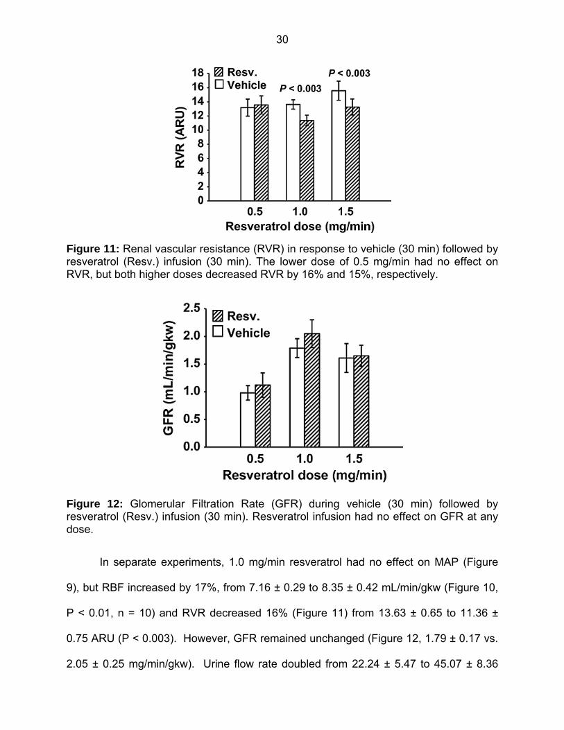
Figure 11: Renal vascular resistance (RVR) in response to vehicle (30 min) followed by resveratrol (Resv.) infusion (30 min). The lower dose of 0.5 mg/min had no effect on RVR, but both higher doses decreased RVR by 16% and 15%, respectively.
Figure 12: Glomerular Filtration Rate (GFR) during vehicle (30 min) followed by resveratrol (Resv.) infusion (30 min). Resveratrol infusion had no effect on GFR at any dose.
In separate experiments, 1.0 mg/min resveratrol had no effect on MAP (Figure
9), but RBF increased by 17%, from 7.16 ± 0.29 to 8.35 ± 0.42 mL/min/gkw (Figure 10,
P < 0.01, n = 10) and RVR decreased 16% (Figure 11) from 13.63 ± 0.65 to 11.36 ±
0.75 ARU (P < 0.003). However, GFR remained unchanged (Figure 12, 1.79 ± 0.17 vs.
2.05 ± 0.25 mg/min/gkw). Urine flow rate doubled from 22.24 ± 5.47 to 45.07 ± 8.36

31
μL/min/gkw (Figure 13, n = 7, P < 0.002), and sodium excretion increased from 1.57 ±
0.46 to 3.10 ± 0.80 μmol/min/gkw (Figure 14, n = 7, P < 0.04).
In rats given the higher 1.5 mg/min dose of resveratrol, again there was no effect
on MAP (Figure 9). Similar to the mid‐dose, resveratrol increased RBF 12% from 6.76
± 0.57 to 7.58 ± 0.60 mL/min/gkw (Figure 10, n = 8, P < 0.003) and thus RVR
decreased 15% (Figure 11) from 15.58 ± 1.35 to 13.27 ± 1.14 ARU (P < 0.003). As
before, GFR remained unchanged (Figure 12, 1.61 ± 0.26 vs. 1.65 ± 0.19 mL/min/gkw).
Urine flow rate increased 70% (Figure 13) from 33.01 ± 6.48 to 56.53 ± 6.56 μL/min/gkw
(n = 8, P < 0.005) and sodium excretion nearly doubled (Figure 14) from 3.99 ± 1.71 to
7.80 ± 1.51 μmol/min/gkw (n = 8, P < 0.04).
Protocol 2: Renal hemodynamics with vehicle
In vehicle time controls, (Table 1), MAP was unchanged and both RBF and RVR
remained constant. With the vehicle infusion, urine flow rate increased 20% from 42.83
± 5.36 to 53.08 ± 7.62 μL/min/gkw (P < 0.04), but sodium excretion remained
unchanged. Our vehicle including DMSO had no overall effect on MAP, RBF, or RVR.
Figure 13: Urine Flow Rate in response to vehicle (30 min) followed by resveratrol (Resv.) infusion (30 min). Resveratrol infusion rate increased urine flow in all three groups by 59%, 103%, and 71%, respectively.

32
Figure 14: Urinary sodium excretion in response to vehicle (30 min) and resveratrol (Resv.) infusion (30 min). All three doses of resveratrol infusion increased sodium excretion, by 132%, 97%, and 95%, respectively.
Table 1: Time Control renal Hemodynamics and excretion with vehicle.
Discussion
Very little is known about the effects of resveratrol on renal function, especially in
the normal state in the absence of renal pathology. We previously reported an i.v. bolus
of resveratrol produced an acute renal vasodilation that was mediated by increased NO
bioavailability and scavenging of reactive oxygen free radicals. This study expands
upon these observations, and addresses resveratrol‐induced changes in renal function
in response to sustained infusion. Notably, we still find significant renal vasodilation, but

33
surprisingly without changes in GFR. Additionally, we found resveratrol produced a
significant diuresis and natriuresis, and this can occur independent of hemodynamic
changes.
Resveratrol has been reported to lower blood pressure, but these observations
are primarily in animal models of hypertension (33, 42, 169) or diabetes with
hypertension as a comorbidity (140). Translational studies with human participants are
sparse and offer mixed results (135, 183). Chronic resveratrol supplementation (75
mg/day) for 6 weeks in mildly hypertensive obese human subjects did not lower blood
pressure (183) and another study using chronic resveratrol (500 mg 3×/day) for 4 weeks
also did not reduce blood pressure in obese men with prehypertension (135). In our
present acute protocol using different doses of sustained resveratrol infusion, our
normotensive rats did not have any change in blood pressure.
In our previous work (52), we addressed responsible mechanisms for renal
vasodilation. We found it was in part due to increased NO synthesis or availability and
also involved a reduction in reactive oxygen species. In this study, we infused
resveratrol hypothesizing it would increase RBF which would lead to increased GFR.
Interactions between vasoconstriction and vasodilation (including nitric oxide) in the
kidney plays a role in regulation of renal hemodynamics (11, 66, 153, 156). Our low
dose of 0.5 mg/min resveratrol did not increase RBF or decrease RVR. However, the
higher doses of resveratrol (1.0 and 1.5 mg/min) significantly increased RBF and
decreased RVR, consistent with our previous findings (52). The present results show
renal vasodilation in response to resveratrol is maintained during constant infusion at
the mid and high dose. However, there appears to be a maximal limit to resveratrol‐
induced renal vasodilation in a normotensive rat, as infusion at the mid and high dose

34
both reduced renal vascular resistance by 16% and 15%, respectively; similar to the
changes seen in response to an acute bolus (52). Resveratrol‐induced increases in
perfusion may be advantageous in compromised renal states when there is lower nitric
oxide levels and increased oxidative stress as is often the case in hypertension (60) and
other renal diseases (45).
Nonselective NO inhibition is found to increase RVR and decrease glomerular
filtration rate (12, 46). We hypothesized that resveratrol‐induced increases in RBF (due
to increased NO) would lead to increased GFR, but this was not supported by our data,
as all three resveratrol doses failed to increase GFR. It is not clear why the increased
RBF is uncoupled from GFR in these studies. A possible explanation may lay in that
while resveratrol reduced afferent arteriolar resistance and increased RBF, the efferent
arterioles (in the normal rat) may have also dilated to maintain glomerular filtration at a
constant rate. Thus, while resveratrol may rescue GFR in models of compromised renal
function (68, 89, 118), it had no effect in a normal, intact kidney.
The most novel finding of these studies was the significant resveratrol‐induced
diuresis and natriuresis, even in the absence of hemodynamics changes. This increase
in urine flow rate and urinary sodium excretion is also likely due to the effects of renal
NO. An extensive review by Li and Förstermann (99) on resveratrol and endothelial
function details numerous pathways in which resveratrol increases NO synthesis and
bioavailability. Perez‐Rojas et al (133) demonstrated (using NOS‐3 knock‐out mice)
how NO within the kidney promotes water and sodium excretion. Resveratrol may
possibly be acting as a natriuretic by stimulating NO production in the nephron as it
does in the endothelium (52).
NO is important regulator of thick ascending limb (TAL) sodium transport where

35
approximately 20–30% of filtered sodium is reabsorbed (54). NO has been shown to
inhibit sodium reabsorption within the TAL (123). Majid et al. (106) have shown when
NO is inhibited in dogs, renal blood flow, urine flow, and sodium excretion are
decreased, suggesting NO has both diuretic and natriuretic effects in vivo. Thus, NO
changes within the TAL are physiologically relevant. Increased shear stress in primary
culture of TAL cells has been shown to stimulate the production of NO by NOS 3 (28)
and increased renal luminal flow also stimulates NO production within in the TAL (27).
In our previous work (52), we demonstrated resveratrol increased renal vasodilation due
to increased NO synthesis or availability and through reductions in reactive oxygen
species including the possibility of superoxide formation. Changes in tubular flow‐
induced stretch and sodium chloride delivery stimulate superoxide production by
nicotinamide adenine dinucleotide phosphate (NADPH) oxidase in the TAL. Three
isoforms of NADPH oxidase are present within the TAL (111). When NADPH oxidase 4
(Nox4) was silenced superoxide production was decreased, suggesting Nox4 mediates
flow‐induced superoxide production in the TAL (69). Resveratrol, in addition to
increasing NO, has been shown to decrease NOX activity (35) and NOX4 expression
(161). Our whole animal studies do not allow us to pin‐point the tubular site of action of
the resveratrol‐induced natriuresis, but our results are consistent with the literature
suggesting NO acts within the TAL (69, 111). It remains unclear if the diuretic and
natriuretic effects of resveratrol are due to increased NO availability, reduced
superoxide production, or a combination of the above mechanisms as seen in the renal
vasculature (52). Overall, the data presented in our study are consistent with
resveratrol‐induced tubular NO synthesis promoting diuresis and natriuresis.
In our study, the resveratrol‐induced natriuretic effect was observed with all three

36
resveratrol doses. The increases in urinary sodium excretion were dissociated from
changes in blood flow. Separate from the effects of NO, resveratrol may affect renal
tubular sodium handling through direct inhibition of sodium transport. Weixel et al (180)
has shown that resveratrol inhibits Epithelial Sodium Channel (ENaC) in mouse cortical
collecting duct cells. It remains unknown if ENaC is being inhibited in vivo. However,
this may explain how resveratrol may contribute to lowering blood pressure in animal
models of hypertension, particularly when salt‐sensitive hypertension is present (42, 98,
115, 140).
In summary our findings suggest a constant infusion of resveratrol induced
significant renal vasodilation while not altering either GFR or blood pressure in normal
rats. Additionally, resveratrol infusion produced significant natriuresis at all doses,
independent of hemodynamic responses, suggesting it may have a direct effect on renal
tubular sodium handling independent of perfusion pressure, RBF, or changes in RVR.

37
CHAPTER 3
CHRONIC RESVERATROL REVERSES A MILD ANGIOTENSIN II-INDUCED PRESSOR EFFECT IN A RAT MODEL
Abstract
Resveratrol is reported to reduce blood pressure in animal models of
hypertension, but the mechanisms are unknown. We have shown resveratrol infusion
increases sodium excretion. We hypothesized chronic ingestion of resveratrol would
reduce Ang II-induced increases in blood pressure by decreasing sodium reabsorption
through a NO-dependent mechanism. We infused rats with vehicle or 80 μg Ang II/day
over 4 weeks. Vehicle or Ang II infused rats were individually housed, pair fed and
placed on a diet of normal chow or normal chow plus 146 mg resveratrol/day. Groups
included: 1) Control, 2) Resveratrol-fed., 3) Ang II-treated, and 4) Ang II plus resveratrol.
Systolic blood pressure was measured by tail cuff. During the 4th week rats were
placed in metabolic caging for urine collection. Ang II increased systolic blood pressure
in the first week by +14 ±5 mmHg (p<0.05) in group 3 and +10 ±3 mmHg (p<0.05) in the
group 4, respectively. Blood pressure was unchanged in groups 1 and 2. After 4
weeks, blood pressure remained elevated in group 3 rats with Ang II, (+9±3 mmHg,
p<0.05), but in group 4 blood pressure was no longer elevated (+2±2 mmHg). We
found no significant differences between groups in sodium excretion or cumulative
sodium balance (18.49 ±0.12, 17.75 ±0.16, 17.97±0.17, 18.46±0.18 µEq Na+/7 days in
groups 1-4 respectively). We conclude chronic resveratrol supplementation does not
blunt Ang II-increased blood pressure and resveratrol may have mild depressor effects,
but these do not seem to be due to natriuresis or enhanced kidney nitric oxide
synthesis.

38
Introduction
Resveratrol is a polyphenol found in some foods including the skins of grapes,
red wine, and peanuts. It is proposed to be responsible for the beneficial effects of red
wine consumption contributing possibly to reductions in oxidative stress and blood
pressure (9, 18). We were interested in whether a possible renal mechanism may
explain how resveratrol lowers blood pressure in a rat model. Resveratrol has been
reported to lower blood pressure primarily in animal models of hypertension (169).
Dolinsky et al. (42) showed that resveratrol was able to reduce increases in systolic
pressure in spontaneously hypertensive rats (SHR) and mice infused with Ang II via
osmotic minipumps. When resveratrol was removed, SHR blood pressure increased to
the same level of that to control SHR. The existing literature supports an interactive
involvement between resveratrol and nitric oxide (NO). Defects in NO production or
lack of NO have been implicated in hypertension (112, 113). Resveratrol has been
shown to increase NO synthesis in multiple tissues including human umbilical vein
endothelial cells and also in isolated preparations of thick ascending limb (50, 177, 178).
Consistent with this, our previous studies in the kidney show resveratrol acts as a renal
vasodilator (52). Renal vasodilation was achieved partly through a NO-dependent
mechanism and also through a reduction in reactive oxygen species and/or increasing
NO bioavailability (52).
Nitric Oxide plays a critical role in regulation of blood pressure along with salt and
water transport along the nephron (60, 124). NO enhances natriuresis through
inhibition of sodium reabsorption along segments of the nephron (113). Endothelial
nitric oxide synthase generated NO inhibits sodium reabsorption in the thick ascending
limb (127). Gonzelez-Vincente et al (50) have shown resveratrol increases NO
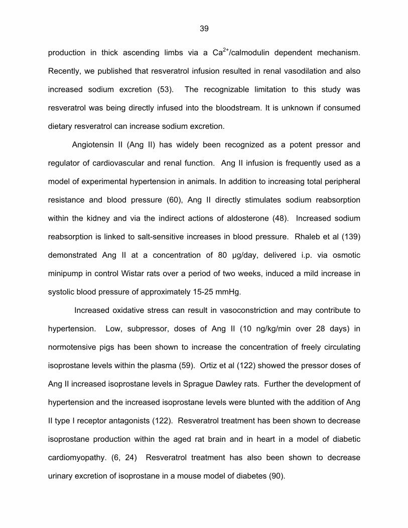
39
production in thick ascending limbs via a Ca2+/calmodulin dependent mechanism.
Recently, we published that resveratrol infusion resulted in renal vasodilation and also
increased sodium excretion (53). The recognizable limitation to this study was
resveratrol was being directly infused into the bloodstream. It is unknown if consumed
dietary resveratrol can increase sodium excretion.
Angiotensin II (Ang II) has widely been recognized as a potent pressor and
regulator of cardiovascular and renal function. Ang II infusion is frequently used as a
model of experimental hypertension in animals. In addition to increasing total peripheral
resistance and blood pressure (60), Ang II directly stimulates sodium reabsorption
within the kidney and via the indirect actions of aldosterone (48). Increased sodium
reabsorption is linked to salt-sensitive increases in blood pressure. Rhaleb et al (139)
demonstrated Ang II at a concentration of 80 µg/day, delivered i.p. via osmotic
minipump in control Wistar rats over a period of two weeks, induced a mild increase in
systolic blood pressure of approximately 15-25 mmHg.
Increased oxidative stress can result in vasoconstriction and may contribute to
hypertension. Low, subpressor, doses of Ang II (10 ng/kg/min over 28 days) in
normotensive pigs has been shown to increase the concentration of freely circulating
isoprostane levels within the plasma (59). Ortiz et al (122) showed the pressor doses of
Ang II increased isoprostane levels in Sprague Dawley rats. Further the development of
hypertension and the increased isoprostane levels were blunted with the addition of Ang
II type I receptor antagonists (122). Resveratrol treatment has been shown to decrease
isoprostane production within the aged rat brain and in heart in a model of diabetic
cardiomyopathy. (6, 24) Resveratrol treatment has also been shown to decrease
urinary excretion of isoprostane in a mouse model of diabetes (90).

40
On the basis of these observations, we hypothesized chronic ingestion of
resveratrol would reduce Ang II-induced increases in blood pressure by decreasing
sodium reabsorption through a NO-dependent mechanism.
Materials and Methods
Male Sprague Dawley Rats weighing 200 to 225 grams were housed in standard
caging. Rats were fed normal chow containing 0.4% sodium (Harlan Teklad, Madison,
WI) and allowed free access to distilled water. Prior to the beginning of the experiment,
rats were pre-trained on a non-invasive tail cuff plethsmography multi-channel system
(Kent Scientific, Torrington, CT) three times a week for two weeks to measure systolic
pressure. All procedures were approved by Wayne State University and Henry Ford
Health System Institutional Animal Care and Use Committee (IACUC) and adhered to
the guiding principles in the care and use of experimental animals in accordance with
the National Institute of Health (NIH) guidelines. Henry Ford Hospital operates an
AALAC-certified animal care facility.
Diet Assignment
After the training period, rats were moved to individually housed caging and
assigned to one of four groups: 1) control, consuming normal rat chow diet
supplemented with 3 grams dark chocolate frosting vehicle (Duncan Hines, Cherryhill,
NJ) (n=7); 2) normal rat chow supplemented with 3 grams chocolate frosting containing
146 mg of resveratrol (Cayman Chemical, Ann Arbor, MI) (n=8), 3) normal rat chow with
frosting vehicle and administered Ang II (80µg/day) (Sigma-Aldrich, St. Louis, MO) i.p.
via an osmotic minipumps (implantation methodology outlined below) (n=7), 4) Ang II
(80µg/day) i.p. supplemented with 146 mg resveratrol mixed into 3 grams of chocolate
frosting (n=8). Rats were pair-fed. Rat chow was rationed and increased over time to

41
accommodate for growth. Effort was taken to ensure food/caloric consumption was
equal between groups. Rats not infused with Ang II (Groups 1 and 2) were implanted
with osmotic minipumps containing vehicle solution (0.01 N acetic acid in sterile 0.9%
NaCl). Dark frosting was used as a vehicle for the resveratrol because the rats
consumed it all voraciously, the resveratrol otherwise has a bitter taste the rats do not
like, and the dark opaque color prevented photo-degradation of the resveratrol.
Implantation of Osmotic Minipumps
After two weeks of tail cuff training and at the beginning of dietary assignments,
Model 2002 osmotic minipumps (Alzet, Cupertino, CA) were primed overnight according
to the manufacturer’s instructions. Pumps were loaded with vehicle or Ang II (80µg/day)
dissolved in sterile 0.9% NaCl plus 0.01N acetic acid. Rats were anesthetized with 50
mg/kg body weight Nembutal (Ovation Pharmaceuticals, Deerfield, IL) via
intraperitoneal injection. The surgical procedure was performed using aseptic
techniques on a heating pad to maintain constant body temperature. The incision site
was shaved, cleaned with 70% isopropyl alcohol, and a topical antiseptic bactericide,
Betadine was applied (Purdue Products, Stamford, Ct). An incision was made between
the scapulae, and the minipumps were implanted subcutaneously. The incision site
was closed with surgical staples. Buprenex (2.5 mg/kg) was administered to reduce
pain (Reckitt Benckiser Healthcare, Parsippany, NJ). Rats were allowed to regain
consciousness and recover on the hot pad before being returned to their cages.
Sodium Balance Studies
Daily consumption of rat chow and frosting was monitored to measure sodium
intake. After three weeks on the diets/treatments, rats were moved from regular caging
to metabolic caging for the final 7 days and continued on their previously assigned diets.
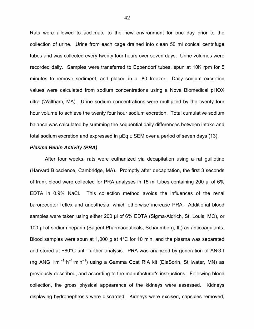
42
Rats were allowed to acclimate to the new environment for one day prior to the
collection of urine. Urine from each cage drained into clean 50 ml conical centrifuge
tubes and was collected every twenty four hours over seven days. Urine volumes were
recorded daily. Samples were transferred to Eppendorf tubes, spun at 10K rpm for 5
minutes to remove sediment, and placed in a -80 freezer. Daily sodium excretion
values were calculated from sodium concentrations using a Nova Biomedical pHOX
ultra (Waltham, MA). Urine sodium concentrations were multiplied by the twenty four
hour volume to achieve the twenty four hour sodium excretion. Total cumulative sodium
balance was calculated by summing the sequential daily differences between intake and
total sodium excretion and expressed in µEq ± SEM over a period of seven days (13).
Plasma Renin Activity (PRA)
After four weeks, rats were euthanized via decapitation using a rat guillotine
(Harvard Bioscience, Cambridge, MA). Promptly after decapitation, the first 3 seconds
of trunk blood were collected for PRA analyses in 15 ml tubes containing 200 μl of 6%
EDTA in 0.9% NaCl. This collection method avoids the influences of the renal
baroreceptor reflex and anesthesia, which otherwise increase PRA. Additional blood
samples were taken using either 200 μl of 6% EDTA (Sigma-Aldrich, St. Louis, MO), or
100 µl of sodium heparin (Sagent Pharmaceuticals, Schaumberg, IL) as anticoagulants.
Blood samples were spun at 1,000 g at 4°C for 10 min, and the plasma was separated
and stored at −80°C until further analysis. PRA was analyzed by generation of ANG I
(ng ANG I·ml−1·h−1·min−1) using a Gamma Coat RIA kit (DiaSorin, Stillwater, MN) as
previously described, and according to the manufacturer's instructions. Following blood
collection, the gross physical appearance of the kidneys were assessed. Kidneys
displaying hydronephrosis were discarded. Kidneys were excised, capsules removed,

43
blotted, and weighed.
Creatinine Clearance
Creatinine was measured in the 24-hour urine collections and in the plasma
during terminal sampling of trunk blood to accommodate the large volume of blood
needed without the confounding influence of anesthesia. Blood samples were taken
using either 200 μl of 6% EDTA in 0.9% NaCl (Sigma-Aldrich, St. Louis, MO) or 100 µl
of sodium heparin (Sagent Pharmaceuticals, Schaumberg, IL) as anticoagulants. Blood
samples were spun at 1,000 g at 4°C for 10 min, and the plasma was separated and
stored at −80°C until creatinine analysis. Creatinine clearance was calculated by
multiplying the concentration of urinary creatinine by the twenty four hour urine volume,
dividing by the plasma creatinine concentration, and then correcting to units of ml/min
and for kidney weight (kw). Creatinine clearance was calculated from the urine
collected during the last two days while the rats were in metabolic caging. Clearance
values were expressed as ml/min/gkw. Following blood collection, the gross physical
appearance of the kidneys were assessed. Renal hydronephrosis was an exclusion
criteria. Kidneys were excised, capsules removed, blotted, and weighed. (Groups 2 and
4 n=8, Groups 1 and 3 n=7)
Urinary Nitric Oxide Assessment
At Day 27, a 24 hour urinary excretion of nitric oxide metabolites (NO2/NO3) was
measured across groups while rats were housed in metabolic caging. 200 µl of Anti-
Anti, an antibiotic and antimycotic, was placed in the urine collection tubes prior to urine
collection to prevent degradation of the samples. NO concentration was measured
using a Nitrate/Nitrite Colorimetric Assay Kit (Cayman Chemical, Ann Arbor, MI) via the
Griess Reaction. NO excretion was calculation by multiplying the concentration by

44
twenty four hour urine volume. Units are expressed as µM/24 hr (n=3/group).
Urinary Isoprostane Assessment
At Day 26, urinary excretion of 8-Isoprostane, a marker of oxidative stress, was
measured across groups. 100 µl of 0.005, BHT, 3,5-Di-tert-4-butylhydroxytoluene was
added to urine collection tubes to prevent degradation of the samples. An 8-
Isoprostane EIA kit was used to measure urinary concentrations (Cayman Chemical,
Ann Arbor, MI). Urinary concentrations were multiplied by urine volumes to reach
isoprostane excretion values, expressed as pg/12 hr (n=3/group).
Statistics
When multiple comparisons between groups with one differing factor were
performed, one-way ANOVA with a Student-Newman-Keuls post hoc test was
performed. When comparing two groups, an unpaired student’s t-test was performed.
When comparing changes within a group, a paired student’s t-test was used. All data
are presented as mean ± SE. In all cases, p < 0.05 was considered statistically
significant.
Results
Blood Pressure
In this study, four groups of rats were fed a standard rat chow containing 0.4%
NaCl. The groups included: 1) Control group fed rat chow plus frosting vehicle, 2) a
group fed rat chow diet, but supplemented with 146 mg resveratrol in vehicle, 3) a group
fed rat chow diet, but administered Ang II (80µg/day) plus vehicle only, and 4) a group
fed rat chow diet supplemented with 146 mg resveratrol in vehicle. Blood pressure in
Group 1, the control group, was unchanged from 121±2 to 123±1 mmHg over the
course of four weeks (Figure 15). In Group 2, normal chow plus resveratrol, blood

45
pressure also remained unchanged; 122±2 to 126±5 mmHg over four weeks. In groups
3 and 4, blood pressure significantly and similarly increased in the first week following
the implantation of Ang II osmotic minipumps. In Group 3 (Ang II), blood pressure
increased from 118±3 to 132±4 mmHg p<0.04, whereas in Group 4 (Ang II plus
resveratrol) blood increased from 122±1 to 132±3 mmHg, p<0.01). However, by the
end of four weeks, blood pressure in Group 3 remained significantly elevated at 128±2
mmHg (Figure 16), while in Group 4 (Ang II plus resveratrol), the increase in blood
pressure was reversed between weeks 3 and 4. After 4 weeks, group 3 (Ang II) was
the only one in which blood pressure remained elevated (Figure 16).
Figure 15: The change in systolic blood pressure over four weeks. Systolic blood pressure remained unchanged in the control and resveratrol treated groups (Groups 1 and 2). Blood pressure initially increased with Ang II infusion (Groups 3 and 4), but between weeks 3 and 4, the increase in systolic blood pressure was reversed in the Ang II plus resveratrol group (Group 4). * p<0.05 vs. week 0 control. Left panel: delta blood pressure over four weeks. Right panel: actual blood systolic pressure

46
Figure 16: Change in systolic blood pressure at end of four weeks. Only Ang II treatment alone increased systolic blood pressure (p<0.025) after four weeks
Body Weight:
Effort was made to ensure the four groups of rats had similar caloric intake.
There were no significant differences in body weight between the four groups at the
beginning of the experiment: Groups 1) 243±5; 2) 248±8; 3) 243±7; and 4) 253±8 g.
Similarly, there were no differences in body weight after 4 weeks between the four
groups: 1) 360±7; 2) 338±10; 3) 345±10; and 4) 355±9 g, so that growth rate was similar
in all 4 groups (Figure 17).
Figure 17: The change in body weight measured over four weeks. Body weight in all four groups significantly increased over four weeks (p<0.05), but no differences in growth was seen between the four groups.
Sodium Excretion and Cumulative Sodium Balance
To examine whether resveratrol consumption can increase sodium excretion and
reduce positive cumulative sodium balance, we measured daily sodium intake,
excretion, and cumulative sodium balance during the final week. Between Day 20 and
Day 27 there were no differences between daily sodium excretion between groups: 1)
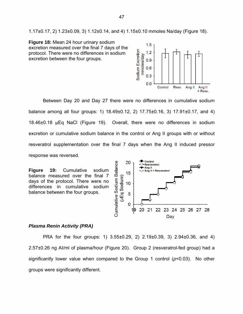
47
1.17±0.17, 2) 1.23±0.09, 3) 1.12±0.14, and 4) 1.15±0.10 mmoles Na/day (Figure 18).
Figure 18: Mean 24 hour urinary sodium excretion measured over the final 7 days of the protocol. There were no differences in sodium excretion between the four groups.
Between Day 20 and Day 27 there were no differences in cumulative sodium
balance among all four groups: 1) 18.49±0.12, 2) 17.75±0.16, 3) 17.91±0.17, and 4)
18.46±0.18 µEq NaCl (Figure 19). Overall, there were no differences in sodium
excretion or cumulative sodium balance in the control or Ang II groups with or without
resveratrol supplementation over the final 7 days when the Ang II induced pressor
response was reversed.
Figure 19: Cumulative sodium balance measured over the final 7 days of the protocol. There were no differences in cumulative sodium balance between the four groups.
Plasma Renin Activity (PRA)
PRA for the four groups: 1) 3.55±0.29, 2) 2.19±0.39, 3) 2.94±0.36, and 4)
2.57±0.26 ng AI/ml of plasma/hour (Figure 20). Group 2 (resveratrol-fed group) had a
significantly lower value when compared to the Group 1 control (p<0.03). No other
groups were significantly different.

48
Figure 20: Plasma Renin Activity: Resveratrol group along had lower PRA.
Clearance of Creatinine, Urinary Excretion of NO Metabolites (NO2/NO3) and 8-
Isoprostane
We measured clearance of creatinine, urinary excretion of NO metabolites
(NO2/NO3), and urinary excretion of 8-isoprostane at the conclusion of the study. There
were no differences in clearance of creatinine between the four groups: 1) 0.40±0.06
(n=7), 2) 0.38±0.04 (n=8), 3) 0.29±0.06 (n=7), and 4) 0.32±0.05 ml/min/gkw (Figure 21).
Figure 21: Clearance of Creatinine: There were no differences in clearance of creatinine between the four groups.
To test if sodium excretion was influenced by increased nitric oxide production,
we sampled urinary excretion of NO2/NO3 in the four groups and obtained values of: 1)
1631±207, 2) 1045±236, 3) 1490±161, and 4) 609±17 µM/day (Figure 22). Group 4
(Ang II plus resveratrol) total nitrate/nitrite excretion was decreased compared to Group

49
1 (p<0.05) and Group 3 (p<0.05, ANOVA adjusted for multiple comparisons).
Values for urinary excretion of 8-Isoprostane excretion were: 1) 9669±1969, 2)
11218±3004, 3) 7992±1376, and 4) 14516±2139 pg/12 hr (Figure 23). There were no
significant differences between the 4 groups.
Figure 22: Urinary Excretion of Nitrate/Nitrite. Resveratrol did not increase excretion of nitric oxide metabolites. NO2/NO3 excretion was decreased in Ang II plus resveratrol group.
Figure 23: Urinary Excretion of 8-Isoprostane. Resveratrol did not decrease renal oxidative stress, as no differences were observed between groups in urinary excretion of 8-Isoprostane.
Discussion
We hypothesized chronic ingestion of resveratrol (over 4 weeks) would reverse
Ang II increases in blood pressure by decreasing sodium reabsorption (and thus
cumulative sodium balance) through a NO-dependent mechanism. To test our
hypothesis, we specifically selected a mild dose of Ang II infusion (139) to allow for
possible resveratrol-induced reductions in blood pressure. We found resveratrol

50
treatment was unable to prevent Ang II-induced increases in blood pressure. However,
with extended resveratrol consumption, the Ang II-induced increases in blood pressure
were reversed.
Resveratrol has been reported to lower blood pressure (42, 169). Bhatt et al (19)
used resveratrol dissolved in drinking water (50 mg/L) in spontaneously hypertensive
rats (SHR) and reported that SHR given resveratrol had lower systolic blood pressure
than non-treated SHR (221±2 vs. 196±3 mmHg). Overall, the existing literature
referencing resveratrol and Ang II in whole animal studies is surprisingly sparse.
Polydatin, a resveratrol glucoside, was delivered in an Ang II-induced model of
hypertension by Zhang et al (186) who found the chronic infusion of Ang II significantly
increased systolic blood pressure from 120±3 to 162±7 mmHg with Ang II, and oral
administration of polydatin for five weeks did not decrease the pressor effect of Ang II.
Dolinsky et al (42) implanted mice with osmotic minipumps to deliver Ang II (1.4
mg/kg/day) and placed mice on a diet containing resveratrol (using approximately 320
mg/kg/day resveratrol). Following two weeks of resveratrol treatment, the Ang II plus
resveratrol group had lower blood pressure when compared to the Ang II infused alone.
They also showed that resveratrol-treated SHR maintained lower systolic blood
pressures than control SHR. However, even with resveratrol treatment, the SHR still
had systolic blood pressures above 160 mmHg (42). Inanaga et al (74), using C57/B6
mice, showed that resveratrol treatment blunted Ang II-induced increases in systolic
blood pressure. However, their systolic blood pressure values using tail cuff
measurements were unusually low.
During the course of our 4 week study, the control and normal chow plus
resveratrol groups (Groups 1 and 2) showed no increase in blood pressure. However,

51
both Ang II infused groups (Groups 3 and 4) saw similar, significant increases in systolic
blood pressure in the first week. Resveratrol treatment was unable to prevent the
pressor effects of mild Ang II infusion. Blood pressure remained elevated in our Ang II
group, but blood pressure in the Ang II plus resveratrol group returned to control levels
between weeks 3 and 4. Javkhedkar et al (79) using SHR and normotensive controls
treated for 9 weeks with resveratrol (50 mg/L) or vehicle in their drinking water. They
reported that SHR plus resveratrol group had lower mean arterial pressure during
weeks 7-12, but not to normotensive levels. These data corroborates our findings that
resveratrol treatment is unable to prevent the onset of hypertension, but there may be a
long-term, temporal effect of resveratrol reversing increased blood pressure after a few
weeks of treatment.
In our previous studies (52) we showed resveratrol acted as an acute renal
vasodilator working through increasing NO and decreasing reactive oxygen species.
We also found (53) that sub-vasodilatory doses of sustained resveratrol administration
could induce natriuresis without affecting glomerular filtration rate. This suggested a
direct tubular action of resveratrol (53). We wanted to test whether a dietary resveratrol
supplement would increase sodium excretion, ultimately leading to a decreased positive
sodium balance. Our approach of metabolic studies to measure sodium intake, sodium
excretion, and sodium balance in resveratrol-treated rats is a novel approach to
determine possible tubular sodium effects of resveratrol.
Resveratrol is rapidly degraded within the body and later cleared by the kidney.
Juan et al (84) demonstrated rats given an intravenous administration of 15 mg/kg
resveratrol distributed at the highest concentrations in kidney. It has been shown 100
µM of resveratrol can increase NO in thick ascending limb suspensions (50). NO is

52
important regulator of thick ascending limb sodium transport (54). NO has been shown
to inhibit sodium reabsorption within the thick ascending limb of the loop of Henle (126).
Majid et al (108) have shown when NO is inhibited in dogs, sodium excretion is
decreased. Their results suggest NO contributes towards diuresis and natriuresis in
vivo. We found resveratrol consumed in the diet did not increase sodium excretion in
Group 2 (normal rat chow plus resveratrol) nor did it increase sodium excretion in Group
4. No differences in urinary sodium excretion or cumulative sodium balance existed
between any of the groups. Contrary to our hypothesis, we conclude that resveratrol
had no effect in increasing sodium excretion or diminishing positive sodium balance.
Thus, the mechanism by which resveratrol lowers blood pressure does not appear to be
linked to sodium handling within the nephron. However, we did not measure sodium
excretion during the first weeks, though there were no differences in systolic pressure
during this period. Notably, we did find an Ang II-induced positive cumulative sodium
balance in our model, at least during the final week of treatment which would be
consistent with the lack of any effect of resveratrol on natriuresis.
Oxidative stress is determined by the balance between the generation of reactive
oxygen species and opposing antioxidant systems (91). The renal medulla is the
primary site of superoxide in the kidney, and also the location of thick ascending limbs
of juxtamedullary nephrons. In our previous work (52), we showed that resveratrol-
induced renal vasodilation was both nitric oxide dependent and due to a possible
reduction in reactive oxygen species and/or increase in nitric oxide bioavailability. To
test if resveratrol reduced sodium reabsorption through a nitric oxide or superoxide
dependent mechanism, we measured urinary excretion of nitric oxide metabolites and
urinary 8-isoprostane, a reliable index of renal oxidative stress.

53
Resveratrol treatment has been shown to lower 8-isoprostane levels in diabetic
rats (116). Bertelli et al (17) in a model of acute kidney failure induced by ischemia
showed rats pretreated with resveratrol raised urinary excretion of cGMP, a marker for
nitric oxide synthase activity. We did not find that resveratrol stimulated an increase in
urinary excretion of nitric oxide metabolites. Contrary to our expectations, resveratrol
treatment significantly decreased excretion of nitric oxide metabolites. This finding is
clearly contrary to our hypothesis and an obvious explanation is lacking. We also
measured urinary excretion of 8-isoprostane to test if resveratrol would potentially lower
oxidative stress. We saw no decrease in 8-isoprostane excretion with resveratrol
treatment. Thus, we conclude resveratrol treatment at this dose was not able to
stimulate measurable NO within the nephron or decrease oxidative stress. Our
measurements of these metrics was only done at the end of the protocol, but that is also
the period in which we observed a depressor effect of resveratrol.
To evaluate the effect of Ang II and resveratrol on renal function we measured
clearance of creatinine at the conclusion of the protocol. Ang II induced hypertension is
associated with decreased glomerular filtration rate (179). In our study, the clearance of
creatinine in the Ang II group (Group 3) was not significantly lower when compared to
the non-Ang II treated groups. There were no differences in clearance of creatinine
between the four groups after 4 weeks.
In summary, our findings suggest resveratrol is unable to prevent increases
blood pressure due to Ang II. Rigorous metabolic studies show that resveratrol
supplementation does not increase urinary sodium excretion, decrease cumulative
sodium balance, or diminish sodium-sensitive increases in blood pressure. However,
we did observe a reversal of the Ang II increase in blood pressure with chronic dietary

54
resveratrol treatment following 3-4 weeks of treatment. In view of our data, chronic
supplementation with resveratrol may have mild depressor effects, but these do not
seem to be due to natriuresis or chronically enhanced nitric oxide synthesis.

55
CHAPTER 4
CHRONIC RESVERATROL DOES NOT BLUNT FRUCTOSE-INDUCED SALT SENSITIVE INCREASE IN BLOOD PRESSURE IN A RAT MODEL
Abstract
Resveratrol is suggested to reduce blood pressure and oxidative stress in rodent
models of hypertension, but mechanisms are poorly understood. Previously, we have
shown resveratrol infusion induces natriuresis. We hypothesized chronic ingestion of
resveratrol would reduce fructose-induced salt sensitive increases in blood pressure by
decreasing sodium retention through a NO-dependent mechanism. Rats were placed in
metabolic caging for 3 weeks to measure sodium balance and groups included: 1)
Control, 2) 20% fructose and resveratrol, (146 mg/day) 3) 20% fructose and high salt
(4%NaCl in the food), and 4) 20% fructose, high salt, and resveratrol. We measured
blood pressure and urinary excretion of NO2/NO3 and 8-isoprostane. During week 1 all
rats received normal rat chow diet 20% fructose and resveratrol was introduced during
week 2, and high salt during week 3. Systolic blood pressure was unchanged in the
control group over three weeks as well as the 20% fructose-treated groups (Groups 2,
3, and 4) (121±1 to 125±1 mmHg). However, blood pressure significantly increased in
Group 3 (124±1 to 138±2 mmHg p<0.001), and Group 4 (126±2 to 142±3 mmHg
p<0.003) with high salt. Increases in blood pressure were coincident a significant
retention of dietary sodium. Resveratrol did not increase urinary excretion of sodium or
NO2/NO3. Fructose treatment, independent of blood pressure or resveratrol, increased
8-isoprostane excretion with 2 weeks (p<0.003). Overall, we conclude 20% fructose
coupled with high salt intake can induce elevated blood pressure in as little as 2 weeks.
Resveratrol did not blunt salt-sensitive increases in blood pressure nor did resveratrol

56
have an effect on sodium retention or reduce oxidative stress.
Introduction
Resveratrol is a polyphenol found in some foods including the skins of grapes,
red wine, and peanuts (9). It is purported to contribute to reductions in oxidative stress
and blood pressure in various models of hypertension (58, 169). Resveratrol has been
shown to increase nitric oxide (NO) synthesis in multiple tissues including human
umbilical vein endothelial cells and also in isolated preparations of renal thick ascending
limb (50, 178). Our previous studies (52) show resveratrol acts as a renal vasodilator.
Renal vasodilation was mediated through a NO-dependent mechanism, and also
through a reduction in reactive oxygen species and/or increasing NO bioavailability (52).
We also have shown (53) that resveratrol can induce natriuresis, independent of
changes in renal hemodynamics. NO enhances natriuresis through inhibition of sodium
reabsorption along multiple segments of the nephron (113). Because of the salt-
sensitivity in fructose-induced hypertension (29), we used this model to test our
hypothesis that resveratrol-stimulated natriuresis could blunt increases in blood
pressure. On the basis of these observations, overall we hypothesized that chronic
ingestion of resveratrol would reduce fructose-induced salt sensitive increases in blood
pressure by decreasing sodium reabsorption and overall cumulative sodium balance
through a NO-dependent mechanism. We were interested in whether a possible renal
mechanism may explain the effect of resveratrol lowering blood pressure in a salt-
sensitive model of hypertension.
Fructose is increasingly implicated as a causal factor in the development of
hypertension, diabetes, and obesity (73, 76, 168). However, the exact mechanism of
fructose-induced hypertension is not fully understood. High intake of fructose (60%)

57
can induce insulin resistance, hypertriglyceridemia, and changes in endothelial function
within two weeks (73). A strong association has been shown that Type 2 diabetes rates
are twenty percent higher in countries where fructose is consumed the most, compared
to disease rates in countries with low fructose consumption (51).
Within the kidney, a diet containing 60% fructose in the drinking water can induce
renal hypertrophy, afferent arteriolar thickening, glomerular hypertension, and
vasoconstriction (144). Fructose intake can easily overwhelm hepatic fructose
metabolism and eventually escapes into the systemic circulation and thus enters the
renal circulation (174). Fructose consumption may contribute to hypertension by
abnormally increasing sodium reabsorption within the intestine and kidney (158).
Fructose is shown to stimulate sodium hydrogen exchange 3 (NHE3) activity within the
proximal tubule (29). However, high experimental levels of fructose do not adequately
reflect average fructose intake within the American diet (82). A diet containing 20%
fructose is more akin to the upper end of the American diet as percentage of their total
caloric intake. Importantly, low doses of fructose (with the addition of salt) can induce
salt-sensitive increases in blood pressure with two weeks (29). Elevations in blood
pressure in this fructose model occur prior to the development of diabetes and
metabolic syndrome. Increases in plasma glucose and insulin have been reported with
10% and 20% fructose starting beyond 6 weeks of fructose treatment suggesting the
cumulative effects of enhanced dietary fructose on hypertension and eventually
metabolic syndrome are both concentration and time dependent (39).
Materials and Methods
Male Sprague Dawley Rats weighing 200 to 225 grams were housed in standard
caging. Rats were fed normal chow containing 0.4% sodium (Harlan Teklad, Madison,

58
WI) and allowed free access to distilled water. Prior to the beginning of the protocol,
rats were pre-trained on a non-invasive tail cuff plethsmography multi-channel system
(Kent Scientific, Torrington, CT) three times a week for two weeks to measure systolic
pressure. All procedures were approved by Wayne State University and Henry Ford
Health System Institutional Animal Care and Use Committee (IACUC) and adhered to
the guiding principles in the care and use of experimental animals in accordance with
the National Institute of Health (NIH) guidelines. Henry Ford Hospital operates an
AALAC-certified animal care facility.
Diet Assignment
After the blood pressure training period, rats were moved to metabolic caging
and allowed to acclimate to the new environment for one day. During week 1 (days 1 to
5) all rats were pair-fed normal rat chow containing 0.4% NaCl (Harlan Teklad, Madison,
WI) and allowed free access to distilled water. During week 2 (days 6-12) rats were
assigned to one of four dietary groups: 1) control, normal rat chow supplemented with 3
grams of fructose-free chocolate frosting vehicle (Duncan Hines, Cherryhill, NJ) with
access to distilled water (n=9); 2) 20% fructose (in the drinking water) (Sigma-Aldrich,
St. Louis, MO), normal rat chow supplemented with 3 grams chocolate frosting
containing 146 mg of resveratrol (Cayman Chemical, Ann Arbor, MI) (n=9), 3) 20%
fructose, normal rat chow supplemented with 3 grams chocolate frosting vehicle (n=9),
and 4) 20% fructose, normal rat chow supplemented with 3 grams chocolate frosting
containing 146 mg of resveratrol (n=9). During week 3 (days 13-19) groups 1 and 2
were maintained on the same diet regime as week 2. However, groups 3 and 4 were
switched from normal rat chow to a high salt diet (4% NaCl rat chow, Harlan Teklad,
Madison, WI).

59
Effort was taken to ensure food/caloric consumption was equal between groups
by pair feeding of rat chow. During the course of the 3 week protocol, rat chow was
rationed equally to all groups, and increased over time to accommodate for growth. Rat
chow was restricted across all four groups when a rat did not consume the daily rat
chow allotment in entirety. Dark frosting was used as a vehicle for the resveratrol
because the rats consumed it all voraciously, the resveratrol otherwise has a bitter taste
the rats do not like, and the dark opaque color prevented photo-degradation of the
resveratrol.
Sodium Balance Studies
Daily consumption of rat chow and frosting was recorded to measure sodium
intake. Urine from each cage drained into clean 50 ml conical centrifuge tubes and was
collected every twenty four hours over 19 days. Urine volumes were recorded daily.
Urine samples were transferred to Eppendorf tubes, spun at 10K rpm for 5 minutes to
remove sediment, and placed in a -80 freezer. Daily sodium excretion values were
calculated from 24 hour urine volumes and sodium concentrations measured by a Nova
Biomedical 1 electrolyte analyzer (Waltham, MA). Total cumulative sodium balance
was calculated by summing the sequential daily differences between intake and total
sodium excretion and expressed in µEq ± SEM over a period of 19 days (2). Volume of
consumed distilled water or 20% fructose was also recorded.
Fecal Sodium Excretion
Fecal samples were collected in a subset within the four groups (n=3) to measure
fecal sodium excretion as a percent of total sodium excretion (urinary and fecal sodium
excretion). Fecal samples were transferred to 50 ml conical centrifuge tubes and
placed in a -80 freezer until further analysis. Fecal samples were thawed to room

60
temperature and total fecal weight was recorded. One gram of feces was combined
with 4ml of distilled water, and homogenized with a Tissuemiser (Fisher Scientific,
Pittsburgh, PA). The fecal slurry was transferred to a 1.5 ml Eppendorf, spun down at
1,000 g at 4°C for 5 minutes, the supernatant pipetted into a new Eppendorf. Daily fecal
sodium excretions were calculated from sodium concentrations using a Nova
Biomedical 1 Electrolyte Analyzer (Waltham, MA) and total fecal mass to achieve a
twenty four hour fecal sodium excretion.
Urinary Nitric Oxide Assessment
At Day 19, a 24 hour urinary excretion of nitric oxide (NO) metabolites (NO2/NO3)
was measured as an index of renal NO production across groups while rats were
housed in metabolic caging. 200 µl of Anti-Anti, an antibiotic and antimycotic, (Life
Technologies, Grand Island, NY) was placed in the urine collection tubes prior to urine
collection to prevent growth of bacteria. NO2/NO3 concentration was measured using a
Nitrate/Nitrite Colorimetric Assay Kit (Cayman Chemical, Ann Arbor, MI) via the Griess
Reaction. NO2/NO3 excretion was calculation by multiplying the concentration by twenty
four hour urine volume. Units are expressed as µM/24 hr (n=9/group).
Urinary Isoprostane Assessment
At Day 19, a portion of the collected rat urine samples were subdivided to
measure urinary excretion of 8-Isoprostane, a marker of oxidative stress. 0.005%, BHT,
3,5-Di-tert-4-butylhydroxytoluene (Sigma-Aldrich, St. Louis, MO) was added to urine
collection tubes to prevent degradation of 8-Isoprostane. An 8-Isoprostane EIA kit was
used to measure urinary concentrations (Cayman Chemical, Ann Arbor, MI). Urinary
concentrations were multiplied by urine volumes to reach isoprostane excretion values,
expressed as pg/24 hr (n=7-9/group).
61
Plasma Renin Activity (PRA)
After three weeks, rats were euthanized via decapitation. Promptly after
decapitation, the first 3 seconds of trunk blood were collected for plasma renin activity
(PRA) analyses in 15 ml tubes containing 200 μl of 6% EDTA in 0.9% NaCl. This
collection method avoids the influences of the renal baroreceptor reflex and anesthesia
on PRA. Additional blood samples were collected using either 200 μl of 6% EDTA
(Sigma-Aldrich, St. Louis, MO) or 100 µl of sodium heparin (Sagent Pharmaceuticals,
Schaumberg, IL) as anticoagulants. Blood samples were spun at 1,000 g at 4°C for 10
min, and the plasma was separated and stored at −80°C until further analysis. PRA
was analyzed by generation of ANG I (ng ANG I·ml−1·h−1) using a Gamma Coat RIA kit
(DiaSorin, Stillwater, MN) as previously described and according to the manufacturer's
instructions. Following blood collection, the gross physical appearance of the kidneys
were assessed. Renal hydronephrosis was an exclusion criterion. Kidneys were
excised, decapsulated, blotted, and weighed.
Plasma glucose, insulin levels, glucose tolerance test (GTT), and Renal
Hypertrophy
To confirm that fructose-induced, salt-sensitive hypertension preceded diabetes
and metabolic syndrome, we measured plasma glucose and insulin levels at the
conclusion of the protocol. Following euthanasia, blood droplets were collected in the
four groups of rats to measure fasting plasma glucose levels. Plasma glucose was
measured with a True Result Blood Glucose Monitor (CVS, Woonsocket, RI). The
additional plasma samples (containing 6% EDTA) stored at -80 were thawed and insulin
levels were measured using a rat insulin EIA kit (Cayman Chemical, Ann Arbor, MI)
according to the manufacturer’s instructions.

62
In a separate protocol, glucose tolerance tests (GTT) were performed in three
groups of rats: 1) control 2) 20% fructose (20% fructose in water for two weeks), and 3)
20% fructose plus high salt (4%) rat chow fed over the final 7 days to demonstrate that
only 14 days of fructose-feeding was not (yet) initiating metabolic syndrome. Male
Sprague Dawley rats (Charles River, Wilmington MA) of 300 to 500 grams body weight
(b.w.) were fasted overnight but allowed free access to drinking water. On the day of
the experiment, rats were anesthetized via intraperitoneal injection with thiobutabarbital,
125 mg/kg b.w. (Inactin, Sigma Aldrich, St. Louis, MO). Rats were placed on a heated
surgical table to maintain constant body temperature (BrainTree Scientific, Braintree,
MA). A tracheotomy was performed using PE-240 tubing to allow free breathing of
room air. A femoral cut down was performed to cannulate the femoral artery with PE-50
catheters. Arterial blood was collected and plasma glucose measured with a True
Result Blood Glucose Monitor (CVS, Woonsocket, RI). Plasma glucose was measured
at baseline and following an intragastric bolus by gavage of glucose (3mg/kg bw),
sampling at 5, 10, 15, 30, 60, 120 minutes after gavage (n=3-5/group).
To quantify any renal hypertrophic response to fructose and high salt in the
presence or absence of resveratrol, we calculated total kidney weight/body weight
(mg/g) ratios in the four groups of rats at the end of the 3 week dietary protocol.
Confirmation of Resveratrol Activity
As a negative control for the activity of resveratrol, we performed a systemic
injection bolus of resveratrol (5 mg/kg) in an anesthetized rat to assess if it induced
renal vasodilation. Procedure for bolus injection is found in Chapter 1.
Statistics
When multiple comparisons between groups with one differing factor were

63
performed, one-way ANOVA with a Student-Newman-Keuls post hoc test was
performed. When comparing two groups, an unpaired student’s t-test was performed.
When comparing changes within a group, a paired student’s t-test was used. All data
are presented as mean ± SE. In all cases, p < 0.05 was considered statistically
significant.
Results
Blood Pressure
During the initial 5 days of normal diet, there were no significant changes in
systolic blood pressure (Figure 24). Systolic blood pressure in Group 1, the control
group, was unchanged from 121±2 to 124±3 mmHg from week 1 to week 2.
Collectively, in Groups 2, 3, and 4, with the addition of 20% fructose blood pressure
remain unchanged from week 1 to week 2 (121±1 to 125±1 mmHg) (Figure 24). From
week 2 to week 3, blood pressure remained unchanged in Group 1, the control group
(124±3 to 122±2 mmHg), as well as in Group 2 (20% fructose and resveratrol, 126±2 to
128±1 mmHg). However, in week 3, with the addition of high salt diet to Groups 3 and
4, blood pressure significantly increased (Figure 24). In Group 3 (20% fructose and
high salt), blood pressure increased from 124±1 to 138±2 mmHg (p<0.001), and in
Group 4 (20% fructose, high salt, and resveratrol) blood pressure also increased from
126±2 to 142±3 mmHg (p<0.003). Thus, resveratrol did not diminish the salt-sensitive
hypertension in the presence of fructose.

64
Figure 24: Systolic Blood Pressure over 3 weeks. High salt diet to Groups 3 and 4 significantly increase blood pressure in the presence of fructose. Resveratrol had no effect. Left panel: actual blood pressure over 3 weeks. Right panel: delta blood pressure from week 2 to week 3.
Body Weight
Effort was made to ensure the four groups of rats had similar caloric intake.
There were no significant differences in body weight between the four groups at the
beginning of the experiment (Table 2). By the end of three weeks, as expected, all four
groups of rats significantly increased in body weight. However, by the end of three
weeks, there were differences in growth rates between Group 1 compared to Groups 2,
3, and 4 (p<0.0001) (Figure 25). Pair-feeding of rat chow was maintained across all
four groups, but Group 1 lacked the 20% fructose in its water and its accompanying
calories.
Table 2: Body Weights (g)
Beginning End of Week 1 End of Week 2 End of Week 3
Control 323±8 332±9 351±6 353±8
F+R 337±10 342±11 382±10 418±9
F+HS 330±9 335±10 384±8 407±10
F+HS+R 332±7 341±9 381±8 402±9
F = fructose, HS = high salt, and R = resveratrol

65
Figure 25: Body Weight over 3 weeks. Fructose-fed rats grew significant greater rate.
Food Intake/Sodium Consumption
Rats were pair fed to ensure consumption of rat chow (and its sodium content)
was similar across groups. During week 1 through week 3, there were no differences in
chow consumption across all four groups (Table 3). However, with the addition of
fructose to the drinking water in the beginning of week 2, food consumption decreased
as fructose replaced calories from rat chow. Food consumption decreased on average
20% (p<0.0001) when 20% fructose was added the diet. Food consumption further
decreased on average 26% with the addition of high salt during week 3 in groups 3 and
4 (p<0.0001) (Table 3). The frosting vehicle was normally consumed daily in all groups
in entirety.
During the first two weeks of the protocol there were no differences in sodium
intake across all four groups, but sodium intake decreased as expected with decreased
rat chow consumption by some 13%. However, during week 3, with the addition of high
salt diet to only Groups 3 and 4, sodium intake significantly increased by nearly 6-fold.
(Table 4)

66
Table 3: Food Consumption/Day (g)
Week 1 Week 2 % Change p value Week 3 % Change p value
Control 23.1±0.5 19±0.5 -17 0.0001 14.5±0.5 -24 0.0001
F+R 23.0±0.5 17.8±0.5 -23 0.0001 14.2±0.4 -20 0.0001
F+HS 23.1±0.5 18.7±0.5 -19 0.0001 13.5±1.0 -28 0.0001
F+HS+R 22.8±0.5 18.2±0.5 -20 0.0001 13.2±1 -27 0.0001
Week 1: All rats on normal rat chow, Week 2: Introduction of fructose and resveratrol, and Week 3: Introduction of high salt. F = fructose, HS = high salt, and R = resveratrol. Represents food intake alone.
Table 4: Sodium Intake/Day(µEq NaCl)
Week 1 Week 2 % Change p value Week 3 % Change p value
Control 4.00±0.09 3.64±0.08 -9 0.0001 2.68±0.08 -26 0.0001
F+R 3.97±0.07 3.14±0.08 -21 0.0001 2.81±0.07 -11 0.0001
F+HS 4.00±0.08 3.57±0.09 -11 0.0001 23.71±1.33 +564 0.0001
F+HS+R 3.96±0.07 3.48±0.05 -12 0.0001 23.14±1.31 +565 0.0001
Week 1: All rats on normal rat chow, Week 2: Introduction of fructose and resveratrol, and Week 3: Introduction of high salt. F = fructose, HS = high salt, and R = resveratrol. Represents total sodium from food and frosting.
Water Consumption/Urine Excretion:
During Week 1, there were no differences in the amount of water consumed
across groups (Table 5a). In week 2, the control group 1 deceased water consumption
by 22% (p<0.003) while water consumption remained constant in all fructose-fed groups
2-4. During week 3, water consumption in the control group decreased marginally. In
group 2 water consumption increased by 10% (p<0.015), and with the addition of high
salt water intake substantially increased in group 3 (p< 0.002) and group 4 (p<0.005).
There were no differences in water intake with or without resveratrol supplementation
(Table 5a). There were no differences in urine volumes between groups during weeks 1
or 2. However, with the addition of high salt during week 3, as water consumption

67
increased, so also urine volumes increased in groups 3 (p<0.0003) and 4 (p<0.007).
(Table 5b)
Table 5a: Water Consumption (ml/24 hr)
Week 1 Week 2 % Change p value Week 3 % Change p value
Control 45±2 35±2 -22 0.003 33±2 -6 0.046
F+R 41±2 41±4 0 n.s. 45±4 10 0.015
F+HS 41±3 44±2 -7 n.s. 56±4 27 0.002
F+HS+R 38±1 38±2 0 n.s. 52±3 37 0.005
F = fructose, HS = high salt, and R = resveratrol
Table 5b: Daily Urine Excretion (ml/24 hr)
Week 1 Week 2 Week 3 p-value
Control 11.7±1.4 9.5±1.3 10.3±1.4 n.s.
F+R 11.9±1.3 10.1±1.9 14.6±2.3 n.s.
F+HS 13.4±2.2 13.3±1.5 21.5±2.4 0.001
F+HS+R 10.6±1.1 9.4±1.0 17.6±1.9 0.007
F = fructose, HS = high salt, and R = resveratrol
Sodium Excretion and Cumulative Sodium Balance:
To examine whether resveratrol treatment can increase sodium excretion and
reduce sodium retention, we measured sodium excretion and cumulative sodium
balance over all 19 days of the diet manipulations. During week 1, there were no
significant differences in mean urinary sodium excretion between the four groups while
on normal rat chow (Table 6). Mean urinary sodium excretion did not change in Group
1 (control) from week 1 to week 2. However, sodium excretion within Groups 2, 3, and
4, significantly decreased (29%, 15%, 27%, respectively) (p<0.05) with the addition to
20% fructose to their diets, as chow and sodium consumption decreased (Table 3).
During week 3, with the addition of the high salt diet, sodium excretion

68
significantly increased in both Groups 3 and Group 4 (p<0.001). Overall, there were no
differences in sodium excretion between Group 1 (control) and Group 2 (20% fructose
and resveratrol). Further, we found no differences in sodium excretion between Groups
3 and 4 during the high salt diet, with or without resveratrol supplementation.
Resveratrol had no measurable effect on sodium excretion in any experimental period.
Table 6: Mean Sodium Excretion (µEq/Day)
Week 1 Week 2 % change p-value Week 3 % change p-value
Control 1.35±0.1 1.31±0.09 -3 n.s. 1.13±0.07 -14 0.01
F+R 1.56±0.08 1.11±0.13 -29 0.02 1.20±0.10 +8 n.s.
F+HS 1.61±0.07 1.40±0.09 -15 0.02 5.63±0.26 +402 0.001
F+HS+R 1.38±0.10 1.10±0.10 -27 0.02 5.04±0.32 +458 0.001
Week 1: All rats on normal rat chow, Week 2: Introduction of fructose and resveratrol, and Week 3: Introduction of high salt. F = fructose, HS = high salt, and R = resveratrol. N.S.= not significant. Represents total sodium from food and frosting.
There were no differences in cumulative sodium balance between groups over
the first week (Table 7). Further, by the end of week 2 there were no differences
between Group 1 (control) and the fructose-treated groups, with or without resveratrol
supplementation. However, by the end of the week 3, there were significant differences
in cumulative sodium balance (Figure 26). The cumulative sodium balance in both
fructose plus high salt groups (with or without resveratrol) increased significantly, 3.8-
fold greater than the normal sodium diet groups (1 and 2), suggesting increased sodium
retention. However, resveratrol treatment had no effect in reducing cumulative sodium
balance. The increase in positive sodium balance representing sodium retention during
week 3 in the 20% fructose and high salt groups with or with resveratrol (Groups 3 and
4), corresponds directly to the increases in blood pressure (Figure 24).

69
Table 7: Cumulative Sodium Balance (µEq NaCl):
Day 1 Day 5 Day 7 Day 19 p-value
Control 2.59±0.19 13.23±0.79 29.32±1.37 41.45±2.03
F+R 2.27±0.13 12.10±0.62 28.16±1.54 39.52±2.31
F+HS 2.37±0.16 11.97±0.51 26.88±1.39 153.26±8.46 p<0.001 vs. control
F+HS+R 2.52±0.11 12.89±0.64 29.57±1.41 156.25±8.75 p<0.001 vs. control
F = fructose, HS = high salt, and R = resveratrol
Figure 26: Sodium Balance over 3 weeks. The cumulative sodium balance in both fructose plus high salt groups (with or without resveratrol) increased significantly suggesting increased sodium retention. Resveratrol treatment had no effect in reducing cumulative sodium balance.
Fecal Sodium Excretion
To examine the effects of normal diet, 20% fructose, and high salt diet with and
without resveratrol on fecal sodium excretion as a percent of total sodium excretion, we
measured fecal sodium over the course of 3 weeks (Table 8). There were no
differences in fecal sodium excretion across all groups during week 1 with normal rat
chow as a percent of total sodium excretion. Again, during week 2, the addition of 20%
fructose with or without resveratrol supplementation produced no differences in percent
of fecal sodium excretion as a percent of total sodium excretion. During week 3, when
high salt was added to the diet (in Groups 3 and 4), fecal sodium excretion decreased
as a percent of total sodium excretion from 3.3±0.4 and 5.1±2.2% to 0.4±0.2 and
0.4±0.1% in Groups 3 and 4, respectively (p<0.001). Overall, fecal sodium excretion
across groups was consistent with or without resveratrol supplementation and

70
represented a trivial, but consistent amount of total sodium excretion. Interestingly, with
the addition of high salt diet, excess sodium intake did not pass through the digestive
system to increase fecal sodium excretion.
Table 8: Fecal Sodium Excretion as a Percent of Total Sodium Excretion (%)
Week 1 Week 2 Week 3
Control 3.8±0.3 4.0±0.3 2.5±0.3
F+R 3.4±0.5 4.2±0.4 2.7±0.2
F+HS 4.0±0.6 3.3±0.4 0.4±0.2
F+HS+R 3.8±0.8 5.1±2.2 0.4±0.1 F = fructose, HS = high salt, and R = resveratrol
Urinary Excretion of NO metabolites (NO2/NO3) and 8-Isoprostane
We measured urinary excretion of NO metabolites (NO2/NO3) and urinary
excretion of 8-isoprostane on the concluding day of the study. To test if sodium
excretion was influenced by resveratrol-stimulated nitric oxide production we sampled
urinary excretion of NO2/NO3 in the four groups. NO2/NO3 excretion in the four groups
were: 1) 1579±126, 2) 1452±175, 3) 2375±242 and 4) 1902±242 µM/24 hr (Figure 27).
Collectively, the high salt groups (Group 3 and 4) had significantly higher nitrate/nitrite
excretion (p<0.04) than normal salt groups 1 and 2. Resveratrol had no effect on
nitrate/nitrite excretion, either with a normal salt diet or in groups with a high salt diet.
Figure 27: Urinary Excretion of NO2/NO3.
Urinary excretion of NO2/NO3 was significantly higher in fructose and high salt groups. Resveratrol did not increase NO2/NO3 excretion.

71
To evaluate if resveratrol reduced oxidative stress, we measured urinary
excretion of 8-Isoprostane. 8-Isoprostane was significantly higher in all fructose-treated
groups compared to control: 1) 16381±1303, 2) 32176±3894, 3) 29387±2848, and 4)
33063±4305 pg/24 hr (p<0.003) (Figure 28). Resveratrol supplementation had no effect
on fructose-induced oxidative stress in the presence of fructose or fructose and high
salt.
Figure 28: Urinary Excretion of 8-Isoprostane. 8-Isoprostane excretion increased in all fructose-treated groups. Resveratrol did not decease oxidative stress.
Plasma Renin Activity (PRA)
Plasma renin activity (PRA) for the four groups were: 1) 3.27±0.58, 2) 3.48±0.45,
3) 2.05±0.27 and 4) 2.66±0.43 ng AI/ml/hour. PRA was suppressed in both high salt
diet groups (groups 3 and 4) vs. normal salt groups 1 and 2 (p<0.026). There were no
differences in PRA between the resveratrol-treated and non-treated groups (Figure 29).
Figure 29: Plasma Renin Activity: High Salt diet decreased PRA. Resveratrol had no effect.

72
Plasma Glucose, Insulin, Glucose Tolerance Test and Renal Hypertrophy
Plasma glucose was measured at the conclusion of the study to evaluate if the
onset of hypertension preceded the development of diabetes (Figure 30). Plasma
glucose levels at the end of week 3 were: 1) 64±2, 2) 72±2, 3) 74±2, and 4) 68±1 mg/dl.
Plasma levels were significantly higher in the three fructose treated groups, yet
remained in the normal glycemic range of a normal fasted rat (p<0.002). Plasma
glucose levels were unaffected by resveratrol treatment.
Figure 30: Fasted Plasma Glucose: Plasma glucose were elevated in all fructose-fed groups. Resveratrol had no effect on plasma glucose.
Plasma insulin was measured to assess whether the development of fructose-
induced salt sensitive hypertension was independent from the onset of insulin
resistance (Figure 31). Plasma insulin levels in the four groups were: 1) 1.09±0.18, 2)
2.18±0.43, 3) 2.39±0.37, and 4) 1.94±0.19 ng/ml. There were no differences in insulin
levels among the fructose-treated groups (Groups 2, 3, and 4). However, insulin levels
were higher in fructose-treated group as compared to Group 1, control (p<0.005).
Resveratrol treatment had no effect on plasma insulin levels (Figure 31).
Glucose tolerance tests were run rats on a control diet, after 2 weeks of either
20% fructose-feeding or 20% fructose plus high salt to assess the possible development
of insulin resistance or diabetes. While the fructose-fed rats had a higher baseline

73
plasma glucose (Figure 32), all three groups increased to a similar blood glucose levels,
then returned to their baseline in a similar fashion.
Figure 31: Fasted Plasma Insulin: Insulin levels were elevated in all fructose-fed groups. Resveratrol had no effect on insulin levels.
Figure 32: Glucose Tolerance Tests in Control, 20% Fructose (treated for 2 weeks), and 20% Fructose plus high salt.
To assess possible renal hypertrophy, total kidney weight/body weight ratios
were calculated and these values for the four groups were: 1) 6.89, 2) 6.29, 3) 6.60, and
4) 6.55. The ratios were not different between the four groups suggesting no renal
hypertrophy.
Negative Control: Bolus Injection of Resveratrol
We performed a bolus intravenous injection of resveratrol as we had previously
performed (see chapter 1) is to confirm resveratrol is biologically active and still induced

74
renal vasodilation. Since we observed no effect of resveratrol on systolic blood
pressure (Figure 24), we wanted to confirm it was still biologically active. We found a
resveratrol bolus increased renal blood flow from 8.32 ml/min/gkw to 9.22 ml/min/gkw
and decreased renal vascular resistance from 13.10 ARU to 10.85 ARU. Post-injection
renal hemodynamics returned to baseline. This suggests the resveratrol was still
biologically active.
Discussion
The prevailing literature suggests that resveratrol, acting through the kidney, may
induce natriuresis and this may play a role in lowering blood pressure. However, this
idea has not been studied. The kidney plays an essential role in blood pressure
regulation by controlling short-term and long-term NaCl reabsorption and water balance.
The known mechanisms of resveratrol act to increase nitric oxide and decrease
superoxide production suggest that resveratrol may work to decrease sodium
reabsorption. Resveratrol has been shown to increase endothelial nitric oxide synthase
expression and increase nitric oxide synthesis (177, 178, 185). Resveratrol is found to
reduce superoxide production through NADPH (nicotinamide adenine dinucleotide
phosphate-oxidase) oxidase inhibition (58). Reductions in superoxide and oxidative
stress can increase nitric oxide availability. Relevant to our fructose-induced salt-
sensitive model, fructose is shown to increase Angiotensin II (Ang II) in fructose-fed
rats. Ang II stimulates NADPH oxidase through its actions on AT1 receptors and
increases superoxide (166). NADPH oxidase inhibition in aortic strips in fructose-fed
rats has been shown to reduce superoxide production (150). Superoxide within the
kidney has been shown to stimulate sodium absorption within the thick ascending limb
(157). Conversely, NO inhibits sodium reabsorption in rat thick ascending limb (126).

75
Lastly, superoxide is increased during nitric oxide deficiency and this may contribute
toward the development of salt-sensitive hypertension (94)
Collectively, these data support our hypothesis that chronic ingestion of
resveratrol would reduce fructose-induced salt sensitive increases in blood pressure
through decreasing sodium reabsorption through a nitric oxide-dependent mechanism.
In this study, we are the first group to use resveratrol treatment to measure sodium
homeostasis using metabolic balance studies. Data from our study suggests: 1) 20%
fructose in the presence of high salt intake is associated with positive sodium balance
concomitant with significant increases in blood pressure. Strikingly, changes in blood
pressure occurred within only two weeks of initiating 20% fructose treatment. 2)
Resveratrol treatment was ineffective in ameliorating fructose-induced salt retention, nor
did it blunt the salt-sensitive increases in blood pressure.
Resveratrol research crosses many disciplines, for purposes in this paper, we
limit the scope of this discussion exclusively to the effects of resveratrol on the
cardiovascular and renal systems. Within the literature there is currently a large gap of
knowledge of the effect of resveratrol on normal renal physiology. However, there are
an abundance of studies showing resveratrol lowering blood pressure in Ang II models
of hypertension and spontaneously hypertensive rat (SHR). Thandapilly et al (169)
treated SHR with grape powder (600mg/day) for ten weeks. At the end of 10 weeks,
the grape powder-treated SHR had significantly lower blood pressure. Dolinsky et al
(42) treated 10 week old SHR with resveratrol (4g resveratrol/kg rat chow) for 5 weeks
and then removed resveratrol treatment in the final 2 weeks. Blood pressure was
monitored via implanted telemetry. The SHR-resveratrol group initially had lower blood
pressure, but with the removal of resveratrol from the diet, blood pressure steadily

76
increased back to the level of the non-treated SHR (42). These experiments are difficult
to make direct comparisons with our study because they used SHR, the varying lengths
of their experiments eclipse our protocol.
We wanted to study the effect of resveratrol in a fructose-induced salt sensitive
model. Hwang et al. (73) made the original observation that fructose intake can induce
hypertension and insulin resistance. They demonstrated 66% fructose over 2 weeks
could result in a 22 mmHg increase in blood pressure. A review of the literature
indicates the development of hypertension due to fructose is both time and
concentration-dependent (19). Thus, dietary intake of 40-60% fructose can induce
hypertension in rats more rapidly than 10-20% fructose (29, 39, 73). Further, lower
percentages of enhanced dietary fructose intake (10-20%) can still induce hypertension,
but the onset of hypertension requires a longer fructose treatment time (2). The
prevailing literature suggests 20% fructose has little to no effect on blood pressure
within 1 week and more often requires 4-12 weeks to increase blood pressure (19, 29).
Within our study, in agreement with the literature, we find that 20% enhanced fructose
intake over 2 weeks has no effect on blood pressure. However, blood pressure did
increase with the addition of high salt in rats pretreated over 1 week with 20% fructose,
with or without the presence of resveratrol. Overall, these data support salt-sensitive
increases in blood pressure within our fructose model, and the time required for the
onset of increased blood pressure is shortened with the presence of high salt in 20%
fructose-treated rats.
The few reports of resveratrol supplementation in fructose-treated models in the
existing literature are not equivalent counterparts to our study. We specifically use a
mild level of added dietary fructose (20%), and we have a limited protocol using only 2

77
weeks of enhanced fructose-feeding. Extended fructose treatment can result in
increases in blood pressure that are comorbid with the onset of diabetes and metabolic
syndrome. These studies (discussed below) use significantly higher fructose intake (up
to 3 times our dosage) or the length of their protocols were 2-5 times the length of our
study. Leibowitz et al (98) treated rats with 60% fructose over 5 weeks with resveratrol
of varying doses (200, 400 and 800 mg/kg/day) in the final 2 weeks (98). 60% fructose
increased systolic blood pressure approximately 25 mmHg in all groups after 3 weeks.
In the resveratrol-supplemented groups (by week 5) blood pressures were lower than
fructose alone group. However, even with resveratrol treatment, blood pressures were
still significantly elevated in the presence of 60% fructose (≈150 mmHg). These blood
pressure values are significantly higher than the increases in blood pressure in our
study.
In experiments with fructose levels more analogous to our fructose dosage, Akar
et al. (3) treated rats with 20% fructose over ten weeks in the presence or absence of
resveratrol (50 mg/L) and found 20% fructose induced significant increases in systolic
blood pressure and resveratrol treatment decreased elevated blood pressure. The
increases in blood pressure they report due to 20% fructose seen over 10 weeks are
unusually high with systolic values in the 160 mmHg range, 40 mmHg above control
group blood pressure readings. Miatello et al (115) treated rats with 10% fructose over
6 weeks with and without resveratrol (10 mg/kg/day). After 2 weeks, the 10% fructose
group had very mild increases in systolic blood pressure and by the end of 6 weeks
blood pressure was increased by approximately 16 mmHg in the 10% fructose alone
group. By the end of 6 weeks, blood pressure in the resveratrol-treated group was
significantly lower as compared to fructose alone.

78
Cheng et al (33) fed 10% fructose to rats for 4 weeks with the introduction of
resveratrol treatment (10 mg/kg/day) at week 2. They show resveratrol reduced
fructose-induced increases in blood pressure. However, their results are highly atypical
when compared with the current literature. The increases in blood pressure with 10%
fructose after 1 week were exceptionally high, inconsistent with all the literature using
10 or 20% fructose supplementation. At 4 weeks their blood pressure data in the 10%
fructose group were also much higher than expected. This is in distinct contrast to
prevailing literature which shows 20% fructose having little or no effect on blood
pressure during 1 week. 10% fructose has been shown to require 4-12 weeks to induce
increases blood pressure (1, 39). Overall, it is difficult to make direct comparisons to
our study with the existing resveratrol-fructose studies because fructose intake is higher
and varying time courses which may carry the rat into a stage of insulin resistance-
associated hypertension and metabolic syndrome.
It was our aim to investigate resveratrol-stimulated changes in sodium excretion
and balance during fructose-induced salt-sensitive changes in blood pressure prior to
the development of diabetes and metabolic syndrome seen with longer durations or
higher levels of fructose supplementation. At the conclusion of our study we measured
plasma glucose levels in fasted animals. We found 20% fructose in all fructose-treated
groups, regardless of high salt or resveratrol treatment mildly elevated plasma glucose
levels. However, plasma glucose levels were still within normal glycemic range for a
non-diabetic rat. The mild elevation of plasma glucose suggests that there is a nearly
sufficient pancreatic compensation. Plasma insulin levels were also elevated in the
fructose-fed rats, but still within a normal range, and further resveratrol had no effect
insulin levels.

79
We also performed glucose tolerance tests in three groups of rats: control, 20%
fructose treated (2 weeks), and 20% fructose and high salt. The glucose tolerance tests
in the fructose-fed groups had higher baseline plasma glucose levels, but in response to
a glucose bolus, had similar curve profiles as compared to the control group.
Collectively, these data suggest some metabolic changes could be in the initial stages,
but our rats are not diabetic.
Oudot et al (129) showed that insulin resistance associated with fructose intake
is linked to increases in proximal tubular and glomerular area also known as renal
hypertrophy. In our study we found that there were no differences in kidney to body
weight ratios, suggesting within our 2 week time course there was no renal hypertrophy
associated with fructose consumption. With all of these factors taken together, it
appears the elevations in blood pressure within our model are occurring prior to the
development of diabetes and metabolic syndrome.
Blood pressure in all four groups did not increase during week 1 while receiving a
normal rat chow diet, nor did blood pressure increase with the addition of 20% fructose
in the following week. In our study, blood pressure in response to 20% enhanced
fructose-feeding was similar to Abdulla et al (1). In the next week, blood pressure
significantly increased in groups 3 and 4 (20% fructose), only with the addition of the
high salt diet with and without resveratrol. Resveratrol treatment was unable to prevent
the salt-sensitive increases in blood pressure. It is unknown if resveratrol-induced
reductions in blood pressure require extended treatment. However, it was the succinct
aim of this study to measure possible resveratrol-stimulated changes in sodium balance
during the salt-driven initial increase in blood pressure.
The literature suggests resveratrol may increase nitric oxide generation and

80
reduce oxidative stress (58, 177, 178, 185). Balancing sodium reabsorption is important
in regulation of long term blood pressure, and explains why thiazide-type diuretics are
often a first-line antihypertensive therapy (103). Increased nitric oxide or decreased
superoxide in the renal thick ascending limbs is shown to inhibit sodium reabsorption
(126, 157). If this is the case, resveratrol acting within the nephron may reduce the
propensity to develop salt-sensitive hypertension as found in our fructose model.
Metabolic studies show that increased positive sodium balance is associated with
the onset of hypertension in spontaneously hypertensive rats (12). Moreover, defects in
sodium reabsorption are linked with salt-sensitive hypertension (13, 118). We found
resveratrol had no effect in reducing cumulative sodium balance even with the addition
of high salt to our fructose-induced salt-sensitive model. Interestingly, independent of
resveratrol, the increases in blood pressure occurred during the increases in cumulative
sodium balance in the presence of fructose when high salt was added. The data gained
from our metabolic studies is the first to report a positive sodium balance linking
increases in blood pressure to salt in the presence of 20% fructose.
With the introduction of fructose (Group 2, 3, and 4) we observed significant
decreases in urinary sodium excretion; changes absent in the control group. The
decreases in urinary sodium excretion also coincided with decreases in food
consumption due to (sodium-free) fructose replacing calories within the diet. The
decreases in sodium excretion suggest fructose may increase sodium reabsorption.
When high salt was added to the diet, as expected we saw significant increases in
sodium excretion. Divergent from our hypothesis, we saw no differences in urinary
sodium excretion with or without resveratrol treatment in our high salt groups (Groups 3
and 4). We measured fecal sodium excretion in a third of the rats to assess whether the

81
high salt diet would disproportionately be excreted within the stool versus the kidney.
We found that during normal diet with or without 20% fructose, fecal sodium excretion
was relatively low and consistent across groups (3-5%) and when high salt was added
to the diet the majority of sodium was excreted through urine and not the stool. Overall,
we found resveratrol treatment, independent of fructose or high salt, had no effect on
increasing sodium excretion.
There is substantial evidence that oxidative stress and nitric oxide deficiency
contributes towards hypertension (182). A sodium-restricted diet in fructose fed rats
has been shown to reduce oxidative stress and inflammation independent of blood
pressure (129). Urinary excretion of nitric oxide has been shown to increase in a
fructose fed rats (129). Additionally, high salt diets have been shown to increase
urinary nitric oxide excretion (151, 181). This may be a compensatory response to
facilitate increased sodium excretion to help maintain normal blood pressure. To what
degree both fructose and high salt contribute towards increased nitric oxide excretion in
the same in vivo model is unknown.
To test if resveratrol reduced sodium reabsorption through a nitric oxide or
superoxide dependent mechanism, we measured urinary excretion of nitric oxide
metabolites (NO2/NO3) and 8-isoprostane. In our study, we observed NO2/NO3
excretion was significantly increased in our high salt groups (Groups 3 and 4). Contrary
to our hypothesis, our dose of resveratrol treatment had no effect on nitric oxide
excretion. The explanation remains elusive, but it may be in part that since both
fructose and high salt increased nitric oxide production, it exists at such a sufficiently
high level that resveratrol would have little to no effect.
Pharmacokinetic studies in humans treated with oral 14C-resveratrol shows

82
resveratrol is well absorbed (70%+), but low circulating levels of non-degraded
resveratrol suggest rapid degradation and low availability (176). Juan et al (84)
demonstrated high concentrations of resveratrol in at kidneys following infusion.
Resveratrol treatment in isolated thick ascending has been shown to increase nitric
oxide bioavailability (50). Majid et al (108) have shown, when nitric oxide is inhibited in
dogs, sodium excretion is decreased suggesting NO contributes towards diuresis and
natriuresis in vivo. Bertelli et al (17), in a model of acute kidney failure induced by
ischemia, showed rats pretreated with resveratrol raised urinary excretion of cGMP, a
marker for renal nitric oxide synthase activity.
We also measured urinary excretion of 8-isoprostane to test if resveratrol would
lower oxidative stress. Resveratrol, within multiple tissues, has been shown to
decrease 8-isoprostane levels, and also decrease urinary isoprostane excretion in a
mouse model of diabetes (34, 90, 116). Within the existing literature there are several
reports of 60% fructose feeding from 6 to 12 weeks, as well as 10% fructose
supplementation over 12 weeks, increasing 8-isoprostane excretion (43, 72, 129, 160).
However, the fructose levels in these experiments are either really high or fed over 6-
times the length of our experiment. In our study, we report significantly higher urinary 8-
isoprostane excretion within all fructose-treated groups (Groups 2, 3, and 4), but
resveratrol had no effect on 8-isoprostane excretion, presumably not decreasing
oxidative stress. Notably, we observed that 20% fructose, even without high salt or
increases in blood pressure, increased oxidative stress in as little as 2 weeks.
To evaluate the effect of 20% and high salt diet, with or without resveratrol on
PRA, we measured PRA at the conclusion of our study. Angiotensin II type 1 receptor
antagonists have been shown to reduce fructose-induced increases in systolic blood

83
pressure suggesting the renin-angiotensin system plays a role in the development of
fructose-induced hypertension (71, 75, 87, 114). We found that PRA was suppressed in
our fructose-treated high salt groups as compared to non-high salt groups. Increased
sodium delivery within the nephron is known to have an inhibitory effect of plasma renin
activity (7). Overall, resveratrol treatment had no effect on PRA in either normal salt or
high salt diet rats.
In summary, our findings suggest 2 weeks of dietary resveratrol treatment at 146
mg/day is ineffective in preventing increases in blood pressure due to fructose-induced
salt sensitivity. This may be a function of limited resveratrol exposure; either due to an
insufficient dose or the lack of sufficient duration of the treatment. Alternatively,
resveratrol may just not work as an antihypertensive in this model of hypertension.
Notably, the increases in blood pressure with 20% fructose were only observed with the
addition of high salt (15-16 mmHg increase in Groups 3 and 4) and this coincided with a
large increase in sodium retention. The increase in blood pressure was produced within
only 2 weeks preceding the onset of hypertension typically associated with 20%
fructose alone (1, 29). The elevated blood pressure also preceded the onset of
diabetes and metabolic syndrome seen with fructose-enhanced diets over longer
durations or higher levels of fructose consumption. There were no indications of renal
hypertrophy, hyperglycemia, shifts in the glucose tolerance tests, or overt
hyperinsulinemia within our 2 week 20% fructose model. Digestion of resveratrol in
pair-fed metabolic studies did not increase sodium excretion, nor did it decrease
cumulative sodium balance. Resveratrol did not diminish oxidative stress due to
fructose. Overall, our data suggests that high fructose consumption coupled with high
salt taken can have deleterious effects, increasing blood pressure and oxidative stress.

84
Increases in urinary nitric oxide excretion, due to fructose and high salt were not able to
compensate for increased sodium load, even with resveratrol added to the diet.

85
CHAPTER 5
CONCLUSION AND PERSPECTIVES
Conclusion
We systematically investigated the effects of resveratrol on renal physiology
using the following approaches: 1) the acute effects of resveratrol on renal
hemodynamics by administering a single bolus resveratrol into systemic circulation in
anesthetized rats, 2) the sustained effects of resveratrol on renal hemodynamics and
renal function during continuous intravenous resveratrol infusion, 3) the effects of
chronically ingested resveratrol on blood pressure and sodium retention in a model of
mild Ang II-induced increases in systemic blood pressure, and finally 4) the effects of
chronically ingested resveratrol on blood pressure and sodium retention in a salt-
sensitive fructose-induced model of hypertension.
An acute bolus of resveratrol delivered into systemic circulation, independent of
changes in blood pressure, influenced changes in renal hemodynamics. Resveratrol
increased renal blood flow and decreased renal vascular resistance. Renal vasodilation
was contingent on the resveratrol dose administered. No effect was seen at lower
doses. Renal vasodilation was mediated through resveratrol-stimulated nitric oxide and
through a reduction in reactive oxygen species. Although resveratrol is reported to
inhibit cyclooxygenase (78), vasodilation was not influenced by cyclooxygenase
metabolism and vasodilatory prostanoids.
After our initial findings that resveratrol-induced renal vasodilation, we examined
the effects of continuous intravenous resveratrol infusion. We found resveratrol infusion
induced significant renal vasodilation while not altering either blood pressure or
glomerular filtration rate. Unexpectedly, we found that resveratrol infusion also

86
produced significant natriuresis, independent of renal hemodynamic responses,
suggesting it may have a direct effect on renal tubular sodium handling independent of
perfusion pressure.
Data from the resveratrol-induced natriuresis experiment led us to test if
resveratrol-induced natriuresis could blunt increases in blood pressure if resveratrol was
consumed within the diet. The hypothesis was tested in two models: Ang II and
fructose-induced salt sensitive hypertension. Resveratrol treatment was ineffective in
preventing increases blood pressure in response to Ang II. Contrary to the original
hypothesis, metabolic studies revealed that resveratrol had no measurable effect on net
sodium handling within the nephron. Also, plasma renin activity was unaffected by
resveratrol. Ultimately, we observed a reversal of the Ang II increase in blood pressure
with chronic resveratrol treatment following 3 weeks of treatment, but this was due to an
undetermined mechanism.
Further, we observed somewhat similar results with resveratrol in a model of
fructose-induced salt sensitivity. Resveratrol, consumed through the diet, was
ineffective in preventing increases in blood pressure due to fructose-induced salt
sensitivity. The increase in blood pressure also preceded the onset of diabetes and
metabolic syndrome seen with fructose-enhanced diets over longer durations or higher
levels of fructose consumption. However, rats may have been considered pre-diabetic
due to minor elevations in plasma glucose and insulin. Additionally, the increase in
blood pressure was concomitant in the fructose treated groups only with the addition of
high salt to the diet. Again, resveratrol ingestion did not measurably increase sodium
excretion, nor did it decrease cumulative sodium balance. Fructose, independent of
high salt or increases in blood pressure, increased oxidative stress and resveratrol had

87
no effect on decreasing oxidative stress as measured through urinary excretion of 8-
isoprostane.
Taken all together the resveratrol data generated both from acute experiments
and long term chronic studies reveal resveratrol stimulated biological effects are
inherently dependent of its method of delivery. In our acute studies nitric oxide-
dependent renal vasodilation following a resveratrol bolus was a transient event. With
intravenous resveratrol infusion we observed preserved increased renal blood flow and
decreased renal vascular resistance. Other agents can decrease renal vascular
resistance in a more long action fashion. For instance, in two-kidney-one clip rats
during nitric oxide inhibition, a losartan bolus (AT1 receptors antagonist) has a long-
lasting effect (past 40 minutes). In the non-clipped kidney, losartan can prevent large
increases in renal vascular resistance during nitric oxide inhibition (153). Contrasting
losartan to resveratrol suggests while both can reduce renal vascular resistance, the
resveratrol effects are relatively short-lived.
The acute effects of resveratrol delivered by systemic injection juxtapose the
chronic effects of resveratrol we observed in the Ang II and fructose-induced salt-
sensitive models of elevated blood pressure. In the Ang II study, resveratrol did not
blunt even mild Ang II-induced increases in blood pressure. It was only following 3
weeks of resveratrol consumption that we observed resveratrol reducing blood
pressure. The effect of resveratrol lowering blood pressure was not due decreased
sodium retention. A potential criticism of the Ang II study is that we did not perform
metabolic studies prior to giving Ang II in osmotic minipumps. Also, it is possible that
we could have missed changes in sodium handling and the rats could have adapted
prior our metabolic measurements. However, blood pressure decreased in the final

88
weeks of the experiment in our resveratrol-treated group when we were measuring
sodium balance. Conversely it could be argued that resveratrol-induced decreases in
blood pressure require more extended periods of resveratrol treatment. Nevertheless,
during the period we measured, resveratrol had no effect on sodium excretion or
sodium balance.
Because of the possibility of missing changes in sodium retention during the Ang
II experiment, we consciously decided to expand metabolic collections during the
fructose-induced salt-sensitive model. We measured sodium excretion and cumulative
sodium balance during the transition from a normal diet to experimental diets of
fructose, high salt, and resveratrol. This would avoid the potential time-associated
limitation of the previous study. Again, Resveratrol it did not blunt salt-sensitive
increases it blood pressure. However, unlike the Ang II experiments in which we
observed resveratrol eventually lowering elevated blood pressure, we saw no such
effect in the fructose study. Our fructose study was two weeks shorter than the Ang II
study. This raises the possibility that resveratrol may require extended treatment or
resveratrol with its poor bioavailability simply has no effect in this model. Additionally,
the chronic depressor effects could also have been due to a build-up of metabolites of
resveratrol which might have additional biological activity especially over long durations.
The issue of bioavailability must enter into discussion. It might explain the
seemingly diverging biological effects of resveratrol in our experiments: resveratrol
entered directly into systemic circulation as opposed to dietary consumption. Poor
bioavailability of unchanged resveratrol (4% or less; 4) within the blood stream may
explain the striking contrast between the positive effects of renal vasodilation and
natriuresis with resveratrol infusion while no natriuresis was observed when resveratrol

89
was taken through the diet. Resveratrol may reside in such a low concentration that no
effect can be seen through normal dietary consumption.
A frequent criticism of resveratrol is that it is found in low concentration in wine
and further upon consumption it has poor bioavailability. This criticism may carry
weight. Despite total wine consumption increasing on per capita basis within the U.S.
during the past two decades (U.S. Wine Institute), heart disease remains a leading
cause of death for men and women within the United States (92, 143). Correlative
studies do not seem to support resveratrol reducing cardiovascular disease risk. The
results of the InCHIANTI Study (148), a longitudinal study of elderly individuals living in
two Italian towns, were followed over a long term basis from 1998 to 2009 with follow up
evaluations every three years. Participants were separated into quartiles based from
total urinary resveratrol metabolite concentrations. The InCHIANTI study found no
correlation between total urinary resveratrol metabolites and prevalence of cancer,
cardiovascular disease, markers of inflammation, and cause of mortality. An editorial in
Pharmacological Research by editor-in-chief, Francesco Visioli adeptly summarizes the
failures of resveratrol research (175). Paraphrasing from “The resveratrol fiasco,”
resveratrol has shown no proven human activity. He suggests that the use of non-
physiological concentrations and cell culture experiments do not yield translatable
clinical outcomes (175). In perhaps a definitive review of past resveratrol clinical trials,
Tang et al (166), reports no resounding evidence that resveratrol lowers blood pressure
in humans.
To that end, resveratrol supplementation may not act as an effective
antihypertensive. The “resveratrol paradox” frequently mentioned in the literature may
not be a paradox. The “resveratrol paradox” (a coined term designed to elicit intrigue)

90
describes an observation within French society. There are improved cardiovascular
outcomes related to their wine consumption, despite French consumption of a high-fat
diet. However, in the past twenty years wine consumption within the United States has
increased and yet the U.S. is still plagued by high rates of hypertension and
cardiovascular disease. Additionally, exogenous antioxidant supplements have not
been proven to lower blood pressure in humans. The recent availability of resveratrol
as dietary supplements at levels that are orders of magnitude higher than what wine
provides may help end the resveratrol debate. Overall, the current literature suggests
that resveratrol may exert a physiological effect in controlled animal models. It is clear
from our data that resveratrol has distinct physiological actions on nitric oxide
production, free radical formation, natriuresis, and sustained hypertension. However,
the data does not yet suggest efficacy in prevention or diminishment of hypertension in
humans.
My view of resveratrol is pragmatic. The lack of evidence of resveratrol having
utility in humans is very forceful argument (166). While resveratrol consumption is not
harmful to health, it is not a convincing antihypertensive treatment for humans. It is a
fact that not all animal research may translate directly to humans. However, our
observations showing consumption of fructose coupled salt intake abruptly increasing
blood pressure is highly disturbing. In a broad sense and in retrospective, the fructose
salt-sensitive model appears to offer greater translational opportunities for translational
research in human hypertension over resveratrol.
Perspectives
It is rather trite, but the endeavor of research to answer questions has inevitably
led to more unanswered questions. Although, resveratrol treatment in the Ang II and

91
fructose-induced salt sensitive models mostly generated negative data, the observation
of continuous resveratrol infusion inducing natriuresis offers a prospect to further delve
into the mechanisms of action of resveratrol.
Park et al. (7) have shown that resveratrol is a non-selective phosphodiesterase
(PDE) inhibitor capable of inhibiting PDE-4. PDE-4 activity is found to be increased in
Dahl salt-sensitive rats (8). Tanahashi et al demonstrated in anesthetized dogs that
intrarenal arterial infusion of rolipram, a selective PDE4 inhibitor, increased renal blood
flow, glomerular filtration rate, urine flow rate, and urinary Na+ excretion. Their results of
PDE4 inhibition are strikingly similar to the resveratrol data. In future studies, to further
test the mechanism of resveratrol and its role in PDE4 inhibition in vivo, one could
infuse with rolipram followed by Resveratrol.
To that end, resveratrol research and the scientific method to me is reminiscent
of the Socratic Method, a form of inquiry between parties based on asking and
answering questions. When experimental data is seemingly contradictory, experimental
data forces the necessity of asking new questions and arriving at suitable conclusions.
In this regard my training was not designed merely for the goal of performing technical
work, but in preparation to learn how to be critical of my data and to ask questions.

92
APPENDICES
Appendix A – Wayne State University IACUC Approval Letter

93
Appendix B – Henry Ford Health System IACUC Approval Letter

94
Appendix C – American Journal of Physiology Publishing Agreement

95
Appendix D – Physiological Reports Publishing Agreement
(yellow highlighted text)

96
REFERENCES
1. Abdulla MH, Sattar MA, Johns EJ, Abdullah NA, Hye Khan MA, Rathore HA.
High-fructose feeding impacts on the adrenergic control of renal haemodynamics
in the rat. Br J Nutr. 107(2):218-28, 2012.
2. Abdulla MH, Sattar MA, Johns EJ. The Relation between Fructose-Induced
Metabolic Syndrome and Altered Renal Haemodynamic and Excretory Function
in the Rat. Int J Nephrol. 934659, 2011.
3. Akar F, Uludağ O, Aydın A, Aytekin YA, Elbeg S, Tuzcu M, Sahin K. High-
fructose corn syrup causes vascular dysfunction associated with metabolic
disturbance in rats: protective effect of resveratrol. Food Chem Toxicol.
50(6):2135-41, 2012.
4. Amri A, Chaumeil JC, Sfar S, Charrueau C. Administration of resveratrol: What
formulation solutions to bioavailability limitations? J Control Release. 158(2):182-
93, 2012.
5. Andlauer W, Kolb J, Siebert K, Fürst P. Assessment of resveratrol bioavailability
in the perfused small intestine of the rat. Drugs Exp Clin Res. 26(2):47-55, 2000.
6. Araujo M, Wilcox C. Oxidative stress in hypertension: role of the kidney. Antioxid
Redox Signal. 20: 74–101, 2013.
7. Atchison DK, Beierwaltes WH. The influence of extracellular and intracellular
calcium on the secretion of renin. Pflugers Arch. 465(1):59-69, 2013.
8. Bagul PK, Middela H, Matapally S, Padiya R, Bastia T, Madhusudana K, Reddy
BR, Chakravarty S, Banerjee SK. Attenuation of insulin resistance, metabolic
syndrome and hepatic oxidative stress by resveratrol in fructose-fed rats.

97
Pharmacol Res. 66(3):260-268, 2012.
9. Baur JA, Sinclair DA. Therapeutic potential of resveratrol: the in vivo evidence.
Nat Rev Drug Discov. 6: 493-506, 2006.
10. Baylis C, Harton P, and Engels K. Endothelium derived relaxing factor controls
renal hemodynamics in normal rat kidney. J Am Soc Nephrol. 6: 875-881,1990.
11. Baylis C, Harvey J, Engels K. Acute nitric oxide blockade amplifies the renal
vasoconstrictor actions of angiotensin II. J Am Soc Nephrol. 5:211-214, 1994.
12. Bech JN, Nielsen CB, Pedersen EB. Effects of systemic NO synthesis inhibition
on RPF, GFR, UNa, and vasoactive hormones in healthy humans. Am J Physiol.
270(5 Pt 2):F845-51, 1996.
13. Beierwaltes WH, Arendshorst WJ, Klemmer PJ. Electrolyte and water balance in
young spontaneously hypertensive rats. Hypertension. 4(6):908-15, 1982.
14. Beierwaltes WH, Sigmon DH, and Carretero OA. Endothelium modulates renal
blood flow but not autoregulation. Am J Physiol Renal Physiol. 262: F943-F949,
1992.
15. Beierwaltes WH. Cyclooxygenase-2 products compensate for inhibition of nitric
oxide regulation of renal perfusion. Am J Physiol Renal Physiol. 283: F68–F72,
2002.
16. Beierwaltes WH. The role of calcium in the regulation of renin secretion. Am J
Physiol Renal Physiol. 298(1):F1-F11, 2010.
17. Bertelli AA, Migliori M, Panichi V, Origlia N, Filippi C, Das DK, Giovannini L.
Resveratrol, a component of wine and grapes, in the prevention of kidney
disease. Ann N Y Acad Sci. 957:230-238, 2002.
18. Bhatt JK, Thomas S, Nanjan MJ. Resveratrol supplementation improves

98
glycemic control in type 2 diabetes mellitus. Nutr Res. 32(7):537-541, 2012.
19. Bhatt SR, Lokhandwala MF, Banday AA. Resveratrol prevents endothelial nitric
oxide synthase uncoupling and attenuates development of hypertension in
spontaneously hypertensive rats. Eur J Pharmacol. 667(1-3): 258-64, 2011.
20. Bitterman JL, Chung JH. Metabolic effects of resveratrol: addressing the
controversies. Cell Mol Life Sci. 72(8):1473-88, 2015.
21. Boocock DJ, Faust GE, Patel KR, Schinas AM, Brown VA, Ducharme MP, Booth
TD, Crowell JA, Perloff M, Gescher AJ, Steward WP, Brenner DE. Phase I dose
escalation pharmacokinetic study in healthy volunteers of resveratrol, a potential
cancer chemopreventive agent. Cancer Epidemiol Biomarkers Prev. 6: 1246–
1252, 2007.
22. Borra MT, Smith BC, Denu JM. Mechanism of human SIRT1 activation by
resveratrol. J Biol Chem. 280(17):17187-95, 2005.
23. Botden IP, Oeseburg H, Durik M, Leijten FP, Van Vark-Van Der Zee LC,
Musterd-Bhaggoe UM, Garrelds IM, Seynhaeve AL, Langendonk JG, Sijbrands
EJ, Danser AH, Roks AJ. Red wine extract protects against oxidative-stress-
induced endothelial senescence. Clin Sci (Lond) 123: 499–507, 2012.
24. Breyer MD, Breyer RM. G protein-coupled prostanoids receptors and the kidney.
Annu Rev Physiol. 63: 579–605, 2001.
25. Buga GM, Gold ME, Fukuto JM, Ignarro LJ. Shear stress-induced release of
nitric oxide from endothelial cells grown on beads. Hypertension. 17(2):187-93.
1991.
26. Burkitt MJ, Duncan J. Effects of trans-resveratrol on copper-dependent hydroxyl-
radical formation and DNA damage: evidence for hydroxyl-radical scavenging

99
and a novel, glutathione-sparing mechanism of action. Arch Biochem Biophys.
381: 253–263, 2000.
27. Cabral PD, Hong NJ, Garvin JL. ATP mediates flow-induced NO production in
thick ascending limbs. Am J Physiol Renal Physiol. 303(2):F194-200, 2012.
28. Cabral PD, Hong NJ, Garvin JL. Shear stress increases nitric oxide production in
thick ascending limbs. Am J Physiol Renal Physiol. 299(5):F1185-1192, 2010.
29. Cabral PD, Hong NJ, Hye Khan MA, Ortiz PA, Beierwaltes WH, Imig JD, Garvin
JL. Fructose stimulates Na/H exchange activity and sensitizes the proximal
tubule to angiotensin II. Hypertension. 63(3):e68-73, 2014.
30. Chada M, Nögel S, Schmidt AM, Rückel A, Bosselmann S, Walther J,
Papadopoulos T, von der Hardt K, Dötsch J, Rascher W, Kandler MA. Anakinra
(IL-1R antagonist) lowers pulmonary artery pressure in a neonatal surfactant
depleted piglet model. Pediatr Pulmonol. 43(9):851-857, 2008.
31. Chen CK, Pace-Asciak CR Vasorelaxing activity of resveratrol and quercetin in
isolated rat aorta. Gen Pharmacol. 27: 363–366, 1996.
32. Chen X, Lu J, An M, Ma Z, Zong H, Yang J. Anti-inflammatory effect of
resveratrol on adjuvant arthritis rats with abnormal immunological function via the
reduction of cyclooxygenase-2 and prostaglandin E2, Mol Med Rep. 9(6):2592-
2598, 2014.
33. Cheng PW, Ho WY, Su YT, Lu PJ, Chen BZ, Cheng WH, Lu WH, Sun GC, Yeh
TC, Hsiao M, Tseng CJ. Resveratrol decreases fructose-induced oxidative
stress, mediated by NADPH oxidase via an AMPK-dependent mechanism. Br J
Pharmacol. 171(11):2739-50, 2014.
34. Chiavaroli A, Brunetti L, Orlando G, Recinella L, Ferrante C, Leone S, Di Michele

100
P, Di Nisio C, Vacca M. Resveratrol inhibits isoprostane production in young and
aged rat brain. J Biol Regul Homeost Agents. 24(4):441-446, 2010.
35. Chow SE, Hshu YC, Wang JS, and Chen JK. Resveratrol attenuates oxLDL-
stimulated NADPH oxidase activity and protects endothelial cells from oxidative
functional damages. J Appl Physiol. 102(4):1520-1527, 2007.
36. Cianciulli A, Calvello R, Cavallo P, Dragone T, Carofiglio V, Panaro MA.
Modulation of NF-κB activation by resveratrol in LPS treated human intestinal
cells results in downregulation of PGE2 production and COX-2 expression.
Toxicol In Vitro. 26(7):1122-8, 2012.
37. Cogswell JP, Godlevski MM, Wisely GB, Clay WC, Leesnitzer LM, Ways JP,
Gray JG. NF-kappa B regulates IL-1 beta transcription through a consensus NF-
kappa B binding site and a nonconsensus CRE-like site. J Immunol. 153(2):712-
723, 1994.
38. Corduneanu O, Patricia Janeiro P, Brett AM. On the electrochemical oxidation of
resveratrol. Electroanalysis. 18: 757–762, 2006.
39. Dai S, McNeill JH. Fructose-induced hypertension in rats is concentration- and
duration-dependent. J Pharmacol Toxicol Methods. 33(2):101-107, 1995.
40. Dalaklioglu S, Ozbey G. The potent relaxant effect of resveratrol in rat corpus
cavernosum and its underlying mechanisms. Int J Impot Res 25: 188–193, 2013.
41. Davis JO, Freeman RH. Mechanisms regulating renin release. Physiol Rev. 1-44,
1976.
42. Dolinsky VW, Chakrabarti S, Pereira TJ, Oka T, Levasseur J, Beker D, Zordoky
BN, Morton JS, Nagendran J, Lopaschuk GD, Davidge ST, Dyck JR. Resveratrol
prevents hypertension and cardiac hypertrophy in hypertensive rats and mice.

101
Biochim Biophys Acta. 1832(10):1723-33, 2013.
43. El-Bassossy HM, Shaltout HA. Allopurinol alleviates hypertension and proteinuria
in high fructose, high salt and high fat induced model of metabolic syndrome.
Transl Res. 165(5):621-30, 2015.
44. El-Mowafy AM. Resveratrol activates membrane-bound guanylyl cyclase in
coronary arterial smooth muscle: a novel signaling mechanism in support of
coronary protection. Biochem Biophys Res Commun. 15: 1218–1224, 2012
45. Forbes JM, Coughlan MT, Cooper ME. Oxidative stress as a major culprit in
kidney disease in diabetes. Diabetes. 57: 1446–1454, 2008.
46. Gabbai FB, Blantz RC. Role of nitric oxide in renal hemodynamics. Semin
Nephrol. 19: 242–250, 1999.
47. Gandhi SK, Ryder DH, Brown NJ. Losartan blocks aldosterone and renal
vascular responses to angiotensin II in humans. Hypertension. 28: 961–966,
1996.
48. Garvin JL. Angiotensin stimulates glucose and fluid absorption by rat proximal
straight tubules. J Am Soc Nephrol. 1(3):272-277, 1990.
49. Gojkovic-Bukarica L, Kanjuh V, Novakovic R, Protic D, Cvejic J, Atanackovic M.
The differential effect of resveratrol on the renal artery of normal and diabetic
rats. BMC Pharmacol Toxicol 13, Suppl 1: A48, 2012.
50. Gonzalez-Vicente A, Cabral PD, Garvin JL. Resveratrol increases nitric oxide
production in the rat thick ascending limb via Ca2+/calmodulin. PLoS One.
14;9(10):e110487, 2014.
51. Goran MI, Ulijaszek SJ, Ventura EE. High fructose corn syrup and diabetes
prevalence: a global perspective. Glob Public Health. 8(1):55-64, 2013.

102
52. Gordish KL and Beierwaltes WH. Resveratrol induces acute endothelium renal
vasodilation mediated through nitric oxide and reactive oxygen species
scavenging. Am J Renal Physiol. 306(5):F542-F550, 2014.
53. Gordish KL and Beierwaltes WH. Sustained resveratrol infusion increases
natriuresis independent of renal vasodilation. Physiol Rep. 2(9):e12144, 2014.
54. Greger R and Velázquez H. The cortical thick ascending limb and early distal
convoluted tubule in the urinary concentrating mechanism. Kidney Int. 31(2):590-
596, 1987.
55. Griendling KK, Murphy TJ, Alexander RW. Molecular Biology of the renin-
angiotensin system. Circulation. 87: 1816-1828, 1993.
56. Groth A, Vrugt B, Brock M, Speich R, Ulrich S, Huber LC. Inflammatory cytokines
in pulmonary hypertension. Respir Res.16;15:47, 2014.
57. Guo DW, Han YX, Cong L, Liang D, Tu GJ. Resveratrol prevents osteoporosis in
ovariectomized rats by regulating microRNA-338-3p. Mol Med Rep. 12(2):2098-
106, 2015.
58. Guo S, Yao Q, Ke Z, Chen H, Wu J, Liu C. Resveratrol attenuates high glucose-
induced oxidative stress and cardiomyocyte apoptosis through AMPK. Mol Cell
Endocrinol. 412:85-94, 2015.
59. Haas JA, Krier JD, Bolterman RJ, Juncos LA, Romero JC. Low-dose angiotensin
II increases free isoprostane levels in plasma. Hypertension. 34(4 Pt 2):983-986,
1999.
60. Hall JE. Control of sodium excretion by angiotensin II: intrarenal mechanisms
and blood pressure regulation. Am J Physiol. 250(6 Pt 2):R960-R972, 1986.
61. Haque MZ, Majid DS. Assessment of renal functional phenotype in mice lacking

103
gp91PHOX subunit of NAD(P)H oxidase. Hypertension. 43: 335–340, 2004.
62. Harding P, Carretero OA, Beierwaltes WH. Chronic cyclooxygenase-2 inhibition
blunts low sodium-stimulated renin without changing renal haemodynamics. J
Hypertens. 18: 1107–1113, 2000.
63. Harris RC. Cyclooxygenase-2 in the kidney. J Am Soc Nephrol. 11: 2387–2394,
2000.
64. Harrison-Bernard LM. The renal renin-angiotensin system. Adv Physiol Educ.
33(4):270-4, 2009.
65. Hata AN, Breyer RM. Pharmacology and signaling of prostaglandin receptors:
multiple roles in inflammation and immune modulation. Pharmacol Ther. 103:
147–166, 2004.
66. Herrera M, Garvin JL. Recent advances in the regulation of nitric oxide in the
kidney. Hypertension. 45(6):1062-1067, 2005.
67. Herrera M, Ortiz PA, Garvin JL. Regulation of thick ascending limb transport: role
of nitric oxide. Am J Physiol Renal Physiol. 290(6):F1279-1284, 2006.
68. Holthoff JH, Wang Z, Seely KA, Gokden N, Mayeux PR. Resveratrol improves
renal microcirculation, protects the tubular epithelium, and prolongs survival in a
mouse model of sepsis-induced acute kidney injury. Kidney Int. 81(4):370-8,
2012.
69. Hong NJ and Garvin JL. NADPH oxidase 4 mediates flow-induced superoxide
production in thick ascending limbs. Am J Physiol Renal Physiol. 303(8):F1151-
1156, 2012.
70. Hosoda R, Kuno A, Hori YS, Ohtani K, Wakamiya N, Oohiro A, Hamada H, Horio
Y. Differential cell-protective function of two resveratrol (trans-3,5,4′-

104
trihydroxystilbene) glycosides against oxidative stress. J Pharmacol Exp Ther.
344: 124–132, 2013.
71. Hsieh PS, Tai YH, Loh CH, Shih KC, Cheng WT, Chu CH. Functional interaction
of AT1 and AT2 receptors in fructose-induced insulin resistance and
hypertension in rats Metabolism. 54(2):157-64, 2005.
72. Hsieh PS, Tsai HC, Kuo CH, Chan JY, Shyu JF, Cheng WT, Liu TT. Selective
COX2 inhibition improves whole body and muscular insulin resistance in
fructose-fed rats. Eur J Clin Invest. 38(11):812-9, 2008.
73. Hwang IS, Ho H, Hoffman BB, Reaven GM. Fructose-induced insulin resistance
and hypertension in rats. Hypertension. 10(5):512-6, 1987.
74. Inanaga K, Ichiki T, Matsuura H, Miyazaki R, Hashimoto T, Takeda K, Sunagawa
K. Resveratrol attenuates angiotensin II-induced interleukin-6 expression and
perivascular fibrosis. Hypertens Res. 32(6):466-471, 2009.
75. Iyer SN, Katovich MJ. Effect of chronic losartan potassium treatment on fructose-
induced hypertension. Life Sci. 55(7):PL139-44, 1994.
76. J Schulze MB, Manson JE, Ludwig DS, Colditz GA, Stampfer MJ, Willett WC, Hu
FB. Sugar-sweetened beverages, weight gain, and incidence of type 2 diabetes
in young and middle-aged women. JAMA. 292(8):927-34, 2004.
77. Jackson EK, Heidemann HT, Branch RA, Gerkens JF. Low dose intrarenal
infusion of PGE2, PGI2, and 6-keto-PGE1 vasodilate the in vivo rat kidney. Circ
Res. 51: 67–72, 1982.
78. Jang M, Cai L, Udeani GO, Slowing KV, Thomas CF, Beecher CW, Fong HH,
Farnsworth NR, Kinghorn AD, Mehta RG, Moon RC, Pezzuto JM. Cancer
chemopreventive activity of resveratrol, a natural product derived from grapes.

105
Science. 275(5297):218-20, 1997.
79. Javkhedkar AA, Quiroz Y, Rodriguez-Iturbe B, Vaziri ND, Lokhandwala MF,
Banday AA. Resveratrol restored Nrf2 function, reduced renal inflammation, and
mitigated hypertension in spontaneously hypertensive rats. Am J Physiol Regul
Integr Comp Physiol. 308(10):R840-R846, 2015.
80. Jensen BL, Schmid C, Kurtz A. Prostaglandins stimulate renin secretion and
renin mRNA in mouse renal juxtaglomerular cells. Am J Physiol. 271(3 Pt
2):F659-669, 1996.
81. Johnson JL, Maddipati KR. Paradoxical effects of resveratrol on the two
prostaglandin synthases. Prostaglandins Other Lipid Mediat. 56: 131–143, 1998.
82. Johnson RJ, Segal MS, Sautin Y, Nakagawa T, Feig DI, Kang DH, Gersch MS,
Benner S, Sánchez-Lozada LG.. Potential role of sugar (fructose) in the epidemic
of hypertension, obesity and the metabolic syndrome, diabetes, kidney disease,
and cardiovascular disease. Am J Clin Nutr. 86(4):899-906, 2007.
83. Johnson, RL. Essential Medical Physiology. Academic Press. 246. 2003.
84. Juan ME, Maijó M, Planas JM. Quantification of trans-resveratrol and its
metabolites in rat plasma and tissues by HPLC. J Pharm Biomed Anal.
20;51(2):391-398, 2010.
85. Just A, Whitten C, Arendshorst W. Reactive oxygen species participate in acute
renal vasoconstrictor responses induced by ETA and ETB receptors. Am J
Physiol Renal Physiol. 294: F719–F728, 2008
86. Kalinowski MD, Dobrucki LW, Brovkovych MS, Malinski V, Tadeusz M. Increased
nitric oxide bioavailability in endothelial cells contributes to the pleiotropic effect.
Circulation. 105: 933–938, 2002.

106
87. Kamide K, Rakugi H, Higaki J, Okamura A, Nagai M, Moriguchi K, Ohishi M,
Satoh N, Tuck ML, Ogihara T. The renin-angiotensin and adrenergic nervous
system in cardiac hypertrophy in fructose-fed rats. Am J Hypertens. 15(1 Pt
1):66-71, 2002.
88. Karlssol J, Emgrad M, Burkitt MJ. Trans-resveratrol protects embryonic
mesencephalic cells from tert-butyl hydroperoxide: electron paramagnetic
resonance spin trapping evidence for a radical scavenging mechanism. J
Neurochem. 75: 141–150, 2000.
89. Kim DH, Jung YJ, Lee JE, Lee AS, Kang KP, Lee S, Park SK, Han MK, Lee SY,
Ramkumar KM, Sung MJ, Kim W. SIRT1 activationby resveratrol ameliorates
cisplatin-induced renal injury through deacetylation of p53. Am J Physiol Renal
Physiol. 301(2):F427-435, 2011.
90. Kim MY, Lim JH, Youn HH, Hong YA, Yang KS, Park HS, Chung S, Ko SH, Shin
SJ, Choi BS, Kim HW, Kim YS, Lee JH, Chang YS, Park CW. Resveratrol
prevents renal lipotoxicity and inhibits mesangial cell glucotoxicity in a manner
dependent on the AMPK-SIRT1-PGC1α axis in db/db mice. Diabetologia.
56(1):204-217, 2013.
91. Kitiyakara C, Chabrashvili T, Chen Y, Blau J, Karber A, Aslam S, Welch WJ,
Wilcox CS. Salt intake, oxidative stress, and renal expression of NADPH oxidase
and superoxide dismutase. J Am Soc Nephrol. 14(11):2775-2782, 2003.
92. Kochanek KD, Xu J, Murphy SL, et al. Deaths: Final Data for 2009. National vital
statistics reports; National Center for Health Statistics. 60(3). 2012.
93. Kopkan L, Castillo A, Navar L, Majid D. Enhanced superoxide generation
modulates renal function in ANG II-induced hypertensive rats. Am J Physiol

107
Renal Physiol. 290: F80–F86, 2006.
94. Kopkan L, Hess A, Husková Z, Cervenka L, Navar LG, Majid DS. High-salt intake
enhances superoxide activity in eNOS knockout mice leading to the development
of salt sensitivity. Am J Physiol Renal Physiol. 299(3):F656-63, 2010.
95. Lahera V, Salom MG, Miranda-Guardiola F, Moncada S, Romero JC. Effects of
NG-nitro-l-arginine methyl ester on renal function and blood pressure. Am J
Physiol Renal Fluid Electrolyte Physiol 261: F1033–F1037, 1991.
96. Lai EY, Wellstein A, Welch WJ, Wilcox CS. Superoxide modulates myogenic
contractions of mouse afferent arterioles. Hypertension. 58: 650–656, 2011.
97. Lamas S, Rodriguez-Puyol D. Endothelium and the kidney endothelial control of
vasomotor tone: the kidney perspective. Semin Nephrol. 32: 156–166, 2012.
98. Leibowitz A, Faltin Z, Perl A, Eshdat Y, Hagay Y, Peleg E, Grossman E. Red
grape berry-cultured cells reduce blood pressure in rats with metabolic-like
syndrome. Eur J Nutr. 53(3):973-80, 2014.
99. Leonard SS, Xia C, Jiang BH, Stinefelt B, Klandorf H, Harris GK, Shi X.
Resveratrol scavenges reactive oxygen species and effects radical-induced
cellular responses. Biochem Biophys Res Commun. 309: 1017–1026, 2003.
100. Li H, Förstermann U. Resveratrol: a multifunctional compound improving
endothelial function. Cardiovasc Drugs Ther. 23(6):425-429, 2009.
101. Li H, Xia N, Förstermann U. Cardiovascular effects and molecular targets of
resveratrol. Nitric Oxide. 26(2):102-10, 2012.
102. Li HF, Tian ZF, Qui XQ, Wu JX, Zhang P, Jia ZJ. A study of mechanisms
involved in vasodilation induced by resveratrol in isolated porcine coronary
artery. Physiol Res. 55: 365–372, 2006.
108
103. Li S, Kelly SC, Petrasko M.. Implementing effective hypertensive management--
review of the 2014 high blood pressure management guidelines. S D Med. Spec
No:33-5, 2015.
104. Lopez B, Salom MG, Arregui B, Valero F, Fenoy FJ. Role of superoxide in
modulating the renal effects of angiotensin II. Hypertension. 42: 1150–1156,
2003.
105. Maier-Salamon A, Hagenauer B, Wirth M, Gabor F, Szekeres T, Jäger
W.Increased transport of resveratrol across monolayers of the human intestinal
Caco-2 cells is mediated by inhibition and saturation of metabolites. Pharm Res.
23(9):2107-15, 2006.
106. Majid DS, Navar LG. Nitric oxide in the control of renal hemodynamics and
excretory function. Am J Hypertens. 14(6 Pt 2):74S-82S, 2001.
107. Majid DS, Nishiyama A. Nitric oxide blockade enhances renal responses to
superoxide dismutase inhibition in dogs. Hypertension 39: 293–297, 2002.
108. Majid DS, Williams A, Kadowitz PJ, Navar LG. Renal responses to intra-arterial
administration of nitric oxide donor in dogs. Hypertension. 22(4):535-541, 1993.
109. Martín AR, Villegas I, Sánchez-Hidalgo M, de la Lastra CA. The effects of
resveratrol, a phytoalexin derived from red wines, on chronic inflammation
induced in an experimentally induced colitis model. Br J Pharmacol. 147(8):873-
85, 2006.
110. Martinez J, Moreno JJ. Effect of resveratrol, a natural polyphenolic compound, on
reactive oxygen species and prostaglandin production. Biochem Pharmacol. 59:
865–870, 2000.
111. Massey KJ, Hong NJ, Garvin JL. Angiotensin II stimulates superoxide production

109
in the thick ascending limb by activating NOX4. Am J Physiol Cell Physiol.
303(7):C781-789, 2012.
112. Mattson DL, Lu S, Nakanishi K, Papanek PE, Cowley AW Jr. Effect of chronic
renal medullary nitric oxide inhibition on blood pressure. Am J Physiol. 266(5 Pt
2):H1918-H1926, 1994.
113. Mattson DL, Roman RJ, Cowley AW Jr. Role of nitric oxide in renal papillary
blood flow and sodium excretion. Hypertension. 19(6 Pt 2):766-769, 1992.
114. Miatello R, Risler N, Castro C, Cruzado M, González S, Zumino AP. Chronic
administration of losartan reverses cardiovascular changes in hypertensive
fructose-fed rats. Cell Mol Biol. 49(6):945-52, 2003.
115. Miatello R, Vázquez M, Renna N, Cruzado M, Zumino AP, Risler N. Chronic
administration of resveratrol prevents biochemical cardiovascular changes in
fructose-fed rats. Am J Hypertens. 18(6):864-70, 2005.
116. Mohammadshahi M, Haidari F, Soufi FG. Chronic resveratrol administration
improves diabetic cardiomyopathy in part by reducing oxidative stress. Cardiol J.
21(1):39-46, 2014.
117. Möhring J, Möhring B, Näumann H-J, Philippi A, Homsy E, Orth H, Dauda G,
Kazda S. Salt and water balance and renin activity in renal hypertension of rats.
Am J Physiol. 228(6):1847-55, 1975.
118. Morales AI, Buitrago JM, Santiago JM, Fernández-Tagarro M, López-Novoa JM,
Pérez-Barriocanal F. Protective effect of trans-resveratrol on gentamicin-induced
nephrotoxicity. Antioxid Redox Signal. 4(6):893-898, 2002.
119. Moreno JJ. Resveratrol modulates arachidonic acid release, prostaglandin
synthesis, and 3T6 fibroblast growth. J Pharmacol Exp Ther. 294: 333–338,

110
2000.
120. Navar LG, Inscho EW, Majid SA, Imig JD, Harrison-Bernard LM, Mitchell KD.
Paracrine regulation of the renal microcirculation. Physiol Rev. 76:425–536,
1996.
121. Netherton SJ, Maurice DH. Vascular endothelial cell cyclic nucleotide
phosphodiesterases and regulated cell migration: implications in angiogenesis.
Mol Pharmacol. 67: 263–272, 2005.
122. Ortiz MC, Sanabria E, Manriquez MC, Romero JC, Juncos LA. Role of endothelin
and isoprostanes in slow pressor responses to angiotensin II. Hypertension. 37(2
Pt 2):505-510, 2001.
123. Ortiz PA and Garvin JL. NO Inhibits NaCl absorption by rat thick ascending limb
through activation of cGMP-stimulated phosphodiesterase. Hypertension. 37(2 Pt
2):467-471, 2001.
124. Ortiz PA and Garvin JL. Role of Nitric oxide in the regulation of nephron
transport. Am J Renal Physiol . 282(5):F777-F784, 2002.
125. Ortiz PA, Garvin JL. Role of nitric oxide in the regulation of nephron transport.
Am J Physiol Renal Physiol. 282: F777–F784, 2002.
126. Ortiz PA, Garvin JL. NO Inhibits NaCl absorption by rat thick ascending limb
through activation of cGMP-stimulated phosphodiesterase. Hypertension.37(2 Pt
2):467-71, 2001.
127. Ortiz PA, Hong NJ, Wang D, Garvin JL. Gene transfer of eNOS to the thick
ascending limb of eNOS-KO mice restores the effects of l-arginine on NaCl
absorption. Hypertension. 42: 674–679, 2003.
128. Oudot A, Behr-Roussel D, Compagnie S, Caisey S, Le Coz O, Gorny D,

111
Alexandre L, Giuliano F. Endothelial dysfunction in insulin-resistant rats is
associated with oxidative stress and COX pathway dysregulation. Physiol Res.
58(4):499-509, 2009.
129. Oudot C, Lajoix AD, Jover B, Rugale C. Dietary sodium restriction prevents
kidney damage in high fructose-fed rats. Kidney Int. 83(4):674-83, 2013.
130. Palmer RM, Ashton DS, Moncada Palmer S. Vascular endothelial cells
synthesize nitric oxide from l-arginine. Nature 333: 664–666, 1988.
131. Palmer RM, Ashton DS, Moncada S. Palmer. Vascular endothelial cells
synthesize nitric oxide from L-arginine. Nature. 333: 664-666, 1988.
132. Park SJ, Ahmad F, Philp A, Baar K, Williams T, Luo H, Ke H, Rehmann H,
Taussig R, Brown AL, Kim MK, Beaven MA, Burgin AB, Manganiello V, Chung
JH. Resveratrol ameliorates aging-related metabolic phenotypes by inhibiting
cAMP phosphodiesterases. Cell. 148(3):421-33, 2012.
133. Perez-Rojas JM, Kassem KM, Beierwaltes WH, Garvin JL, Herrera M. Nitric
oxide produced by endothelial nitric oxide synthase promotes diuresis.Am J
Physiol Regul Integr Comp Physiol. 298(4):R1050-1055, 2010.
134. Persson PB. Nitric oxide in the kidney. Am J Physiol Regul Integr Comp Physiol.
283: R1005–R1007, 2002.
135. Poulsen MM, Vestergaard PF, Clasen BF, Radko Y, Christensen LP, Stødkilde-
Jørgensen H, Møller N, Jessen N, Pedersen SB, Jørgensen JO. High-dose
resveratrol supplementation in obese men: an investigator-initiated, randomized,
placebo-controlled clinical trial of substrate metabolism, insulin sensitivity, and
body composition. Diabetes. 62(4):1186-1195, 2013.
136. Rebello T, Hodges RE, Smith JL. Short-term effects of various sugars on

112
antinatriuresis and blood pressure changes in normotensive young men. Am J
Clin Nutr. 38(1):84-94, 1983.
137. Ren Y, D'Ambrosio MA, Liu R, Pagano PJ, Garvin JL, Carretero OA. Enhanced
myogenic response in the afferent arteriole of spontaneously hypertensive rats.
Am J Physiol Heart Circ Physiol 298: H1769–H1775, 2010.
138. Renaud S, de Lorgeril M. Wine, alcohol, platelets, and the French paradox for
coronary heart disease. Lancet. 339:1523-1526, 1992.
139. Rhaleb NE, Yang XP, Nanba M, Shesely EG, Carretero OA. Effect of Chronic
Blockade of the Kallikrein-Kinin System on the Development of Hypertension in
Rats. Hypertension. 37(1):121-128, 2001.
140. Rivera L, Moron R, Zarzuelo A, Galisteo M. Long-term resveratrol administration
reduces metabolic disturbances and lowers blood pressure in obese Zucker rats.
Biochem Pharmacol. 77:1053–1063, 2009.
141. Rodrigo R, Gil D, Miranda-Merchak A, Kalantzidis G. Antihypertensive role of
polyphenols. Adv Clin Chem 58: 225–254, 2012.
142. Rodrigo R, Miranda A, Vergara L. Modulation of endogenous antioxidant system
by wine polyphenols in human disease. Clin Chim Acta 412: 410–424, 2011.
143. Roger VL, Go AS, Lloyd-Jones DM, Benjamin EJ, Berry JD, Borden WB, et al.
Heart disease and stroke statistics—2012 update: a report from the American
Heart Association. Circulation. 125(1):e2–220, 2012.
144. Sánchez-Lozada LG, Tapia E, Jiménez A, Bautista P, Cristóbal M, Nepomuceno
T, Soto V, Avila-Casado C, Nakagawa T, Johnson RJ, Herrera-Acosta J, Franco
M. Fructose-induced metabolic syndrome is associated with glomerular
hypertension and renal microvascular damage in rats. Am J Physiol Renal
113
Physiol. 292(1):F423-9, 2007.
145. Sayago CM, Beierwaltes WH Nitric oxide synthase and cGMP-mediated
stimulation of renin secretion. Am J Physiol Regul Integr Comp Physiol.
281(4):R1146-51, 2001.
146. Schnackenberg CG, Welch WJ, Wilcox CS. Normalization of blood pressure and
renal vascular resistance in SHR with a membrane-permeable superoxide
dismutase mimetic: role of nitric oxide. Hypertension. 32: 59–64, 1998.
147. Schweda F, Kurtz A. Regulation of renin release by local and systemic factors.
Rev Physiol Biochem Pharmacol. 161:1-44, 2011.
148. Semba RD, Ferrucci L, Bartali B, Urpí-Sarda M, Zamora-Ros R, Sun K,
Cherubini A, Bandinelli S, Andres-Lacueva C. . Resveratrol levels and all-cause
mortality in older community-dwelling adults. JAMA Intern Med. 174(7):1077-84,
2014.
149. Shakibaei M, Csaki C, Nebrich S, Mobasheri A. Resveratrol suppresses
interleukin-1beta-induced inflammatory signaling and apoptosis in human
articular chondrocytes: potential for use as a novel nutraceutical for the treatment
of osteoarthritis. Biochem Pharmacol. 76(11):1426-39, 2008.
150. Shinozaki K, Ayajiki K, Nishio Y, Sugaya T, Kashiwagi A, Okamura T. Evidence
for a causal role of the renin-angiotensin system in vascular dysfunction
associated with insulin resistance. Hypertension. 43:255–262, 2004.
151. Shultz PJ, Tolins JP. Adaptation to increased dietary salt intake in the rat. Role of
endogenous nitric oxide. J Clin Invest. 91(2):642-50, 1993.
152. Siemann EH, Creasy LL. Concentration of the phytoalexin resveratrol in wine.
Am. J. Eno. Vitic. 43, 49–52 1992.

114
153. Sigmon DH, Beierwaltes WH. Angiotensin II: nitric oxide interaction and the
distribution of blood flow. Am J Physiol Regul Integr Comp Physiol 265: R1276–
R1283, 1993.
154. Sigmon DH, Beierwaltes WH. Influence of nitric oxide derived from neuronal nitric
oxide synthase on glomerular filtration. Gen Pharmacol. 34(2):95-100, 2000.
155. Sigmon DH, Beierwaltes WH. Renal nitric oxide and angiotensin II interaction in
renovascular hypertension. Hypertension. 22(2):237-42, 1993.
156. Sigmon DH, Carretero OA, Beierwaltes WH. Plasma renin activity and the renal
response to nitric oxide synthesis inhibition. J Am Soc Nephrol. 3(6):1288-94,
1992.
157. Silva GB, Ortiz PA, Hong NJ, Garvin JL. Superoxide stimulates NaCl absorption
in the thick ascending limb via activation of protein kinase C. Hypertension.
48(3):467-72, 2006.
158. Singh AK, Amlal H, Haas PJ, Dringenberg U, Fussell S, Barone SL, Engelhardt
R, Zuo J, Seidler U, Soleimani M. Fructose-induced hypertension: essential role
of chloride and fructose absorbing transporters PAT1 and Glut5. Kidney Int.
74(4):438-47, 2008.
159. Song D, Hutchings S, Pang CC. Chronic N-acetylcysteine prevents fructose-
induced insulin resistance and hypertension in rats. Eur J Pharmacol. 508(1-
3):205-10, 2005.
160. Spaak J, Merlocco AC, Soleas GJ, Tomlinson G, Morris BL, Picton P, Notarius
CF, Chan CT, Floras JS. Dose-related effects of red wine and alcohol on
hemodynamics, sympathetic nerve activity, and arterial diameter. Am J Physiol
Heart Circ Physiol 294: H605–H612, 2008.

115
161. Spanier G, Xu H, Xia N, Tobias S, Deng S, Wojnowski L, Forstermann U, Li H.
Resveratrol reduces endothelial oxidative stress by modulating the gene
expression of superoxide dismutase 1 (SOD1), glutathione peroxidase 1 (GPx1)
and NADPH oxidase subunit (Nox4). J Physiol Pharmacol 60, Suppl 4:111–116,
2009.
162. Subramanian M, Balasubramanian P, Garver H, Northcott C, Zhao H, Haywood
JR, Fink GD, MohanKumar SM, MohanKumar PS. Chronic estradiol-17β
exposure increases superoxide production in the rostral ventrolateral medulla
and causes hypertension: reversal by resveratrol. Am J Physiol Regul Integr
Comp Physiol 300: R1560–R1666, 2011.
163. Szewczuk LM, Forti L, Stivala LA, Penning TM. Resveratrol is a peroxidase-
mediated inactivator of COX-1 but not COX-2; a mechanistic approach to the
design of COX-1 selective agents. J Biol Chem 279: 22727–22737, 2004.
164. Takaoka, MJ Synthesis of Resveratrol and its Derivatives. 8, The Japan
Academy, Proceedings of the Imperial Academy, Vol. 16, pp. 406-407, 1940.
165. Tanahashi M, Hara S, Yoshida M, Suzuki-Kusaba M, Hisa H, Satoh S. Effects of
rolipram and cilostamide on renal functions and cyclic AMP release in
anesthetized dogs. J Pharmacol Exp Ther. 289(3):1533-8, 1999.
166. Tang PC, Ng YF, Ho S, Gyda M, Chan SW. Resveratrol and cardiovascular
health--promising therapeutic or hopeless illusion? Pharmacol Res. 90:88-115,
2014.
167. Tappy L, Lê KA. Metabolic effects of fructose and the worldwide increase in
obesity. Physiol Rev. 90(1):23-46, 2010.
168. Tawar U, Kotlo K, Jain S, Shukla S, Setty S, Danziger RS. Renal

116
phosphodiesterase 4B is activated in the Dahl salt-sensitive rat. Hypertension.
51(3):762-6, 2008.
169. Thandapilly SJ, LeMaistre JL, Louis XL, Anderson CM, Netticadan T, Anderson
HD. Vascular and cardiac effects of grape powder in the spontaneously
hypertensive rat. Am J Hypertens 25: 1070–1076, 2012.
170. Timmers S, Auwerx J, Schrauwen P. The journey of resveratrol from yeast to
human. Aging. 4(3): 146–158, 2012.
171. Tousoulis D, Kampoli AM, Tentolouris C, Papageorgiou N, Stefanadis C. The
role of nitric oxide on endothelial function. Curr Vasc Pharmacol 10: 4–18, 2012.
172. Tran LT, MacLeod KM, McNeill JH. Endothelin-1 modulates angiotensin II in the
development of hypertension in fructose-fed rats. Mol Cell Biochem. 325:89–97,
2009.
173. Trela BC, Waterhouse AL. Resveratrol: Isomeric molar absorptivities and
stability. J. Agric. Food Chem. 44: 1253–1257, 1996.
174. Vijayalakshmi Varma, László G. Boros, Greg T. Nolen, Ching-Wei Chang, Martin
Wabitsch, Richard D. Beger, Jim Kaput. Metabolic fate of fructose in human
adipocytes: a targeted 13C tracer fate association study. Metabolomics. 11(3):
529–544, 2015.
175. Visioli F. The resveratrol fiasco. Pharmacol Res. 90:87, 2014.
176. Walle T. Bioavailability of resveratrol. Ann NY Acad Sci. 1215: 9–15, 2011.
177. Wallerath T, Deckert G, Ternes T, Anderson H, Li H, Witte K, Förstermann U.
Resveratrol, a polyphenolic phytoalexin present in red wine, enhances
expression and activity of endothelial nitric oxide synthase.
Circulation.10613:1652-1658, 2002.

117
178. Wallerath T, Poleo D, Li H, Förstermann U. Red wine increases the expression of
human endothelial nitric oxide synthase: a mechanism that may contribute to its
beneficial cardiovascular effects. J Am Coll Cardiol. 41(3):471-478, 2003.
179. Wang CT, Zou LX, Navar LG. Renal responses to AT1 blockade in angiotensin
II-induced hypertensive rats. J Am Soc Nephrol. 8(4):535-542, 1997.
180. Weixel KM, Marciszyn A, Alzamora R, Li H, Fischer O, Edinger RS, Hallows KR,
Johnson JP. Resveratrol inhibits the epithelial sodium channel via
phopshoinositides and AMP-activated protein kinase in kidney collecting duct
cells. PLoS One.;8(10), 2013.
181. Wilcox CS, Deng X, Welch WJ. NO generation and action during changes in salt
intake: roles of nNOS and macula densa. Am J Physiol. 274(6 Pt 2):R1588-93,
1998.
182. Wilcox CS. Oxidative stress and nitric oxide deficiency in the kidney: a critical link
to hypertension? Am J Physiol Regul Integr Comp Physiol. 289(4):R913-35,
2005.
183. Wong RH, Berry NM, Coates AM, Buckley JD, Bryan J, Kunz I, Howe PR.
Chronic resveratrol consumption improves brachial flow-mediated dilatation in
healthy obese adults. J Hypertens. 31(9):1819-27, 2013
184. Wu CC, Wu CI, Wang WY, Wu YC. Low concentrations of resveratrol potentiate
the antiplatelet effect of prostaglandins. Planta Med. 73: 439–443, 2007.
185. Xia N, Förstermann U, Li H. Resveratrol and endothelial nitric oxide. Molecules.
19(10):16102-21, 2014.
186. Zhang Q, Tan Y, Zhang N, Yao F. Polydatin prevents angiotensin II-induced
cardiac hypertrophy and myocardial superoxide generation. Exp Biol Med. 0:1-

118
10, 2014.
187. Zhao H, Li X, Li N1, Liu T, Liu J, Li Z, Xiao H, Li J. Long-term resveratrol
treatment prevents ovariectomy-induced osteopenia in rats without hyperplastic
effects on the uterus. Br J Nutr. 111(5):836-46, 2014.
188. Zykova TA, Zhu F, Zhai X, Ma WY, Ermakova SP, Lee KW, Bode AM, Dong Z.
Resveratrol directly targets COX-2 to inhibit carcinogenesis. Mol Carcinog. 47:
797–805, 2008.

119
ABSTRACT
THE ACUTE AND CHRONIC EFFECTS OF RESVERATROL ON RENAL FUNCTION AND BLOOD PRESSURE
by
KEVIN L. GORDISH
August 2015
Advisor: Dr. William Beierwaltes
Major: Physiology
Degree: Doctor of Philosophy
We investigated the acute and chronic effects of resveratrol on renal function and
blood pressure. We hypothesized that resveratrol would act as a renal vasodilator
through a nitric oxide-dependent mechanism. In our acute studies, we found an
intravenous bolus of resveratrol influenced changes in renal hemodynamics by
increasing renal blood flow and decreasing renal vascular resistance. The mechanism
of renal vasodilation was nitric oxide dependent and through a reduction of endogenous
reactive oxygen species. Resveratrol-induced renal vasodilation was not influenced by
COX metabolism and vasodilatory prostanoids. We found with continuous intravenous
resveratrol infusion induced significant renal vasodilation via a nitric-oxide dependent
mechanism while not altering either glomerular filtration rate or blood pressure in normal
rats. Resveratrol infusion produced significant natriuresis at all doses, independent of
hemodynamic responses, suggesting it may have a direct effect on renal tubular sodium
handling independent of perfusion pressure, renal blood flow, or changes in renal
vascular resistance. We hypothesized chronic ingestion of resveratrol would reduce
Ang II-induced and fructose-induced salt-sensitive increases in blood pressure by

120
decreasing sodium reabsorption through a NO-dependent mechanism. In both models
of elevated blood pressure, we found resveratrol does not increase sodium excretion of
decrease sodium retention. Resveratrol treatment did not blunt initial increases in blood
pressure in either the Ang II or fructose-induced salt sensitive model. However,
following 3 weeks of treatment the increases in blood pressure began to reverse on the
Ang II model, but not the fructose-salt sensitive model (during 2 weeks of treatment).
Resveratrol did not decrease oxidative stress, as measured by urinary excretion of 8-
isoprostane, in either model. Notable, short term exposure to fructose (2 weeks),
independent of blood pressure or salt, increased oxidative stress. It is well-defined from
our data that resveratrol has distinct physiological actions on nitric oxide production,
free radical formation, natriuresis, and sustained hypertension. However, the data does
not yet show efficacy in prevention of hypertension.

121
AUTOBIOGRAPHICAL STATEMENT
Kevin L. Gordish
Education 2015 Ph.D. in Physiology, Wayne State University, School of Medicine
2003 B.A., Biology, Wayne State University
Peer-reviewed Publications:
1. Atchison DK, Westrick E, Szandzik DL, Gordish KL, Beierwaltes WH. Parathyroid hormone-related protein stimulates plasma renin activity via its anorexic effects on sodium chloride intake. Am J Physiol Endocrinol Metab. 303(4):E457-63, 2012.
2. Gordish KL and Beierwaltes, WH. Resveratrol induces acute endothelium-dependent renal vasodilation mediated through nitric oxide and reactive oxygen species scavenging. AJP Renal, 2014.
3. Gordish KL and Beierwaltes WH. Sustained Resveratrol Infusion increases natriuresis independent of renal vasodilation and without changing Glomerular Filtration Rate. Physiological Reports, 2014
4. Rossi, NF, Gordish KL, Beierwaltes WH, Ortiz PA. Dietary Fructose Increases Renal Sympathetic Nerve Activity in Response to High Salt Diet in Awake Freely Moving Rats. ASN, 2015
5. Gordish KL, Beierwaltes WH. Chronic Resveratrol reverses a mild angiotensin II-induced pressor effect in a rat model. Amer. J Physiology. 2015. (in review)
Invited Oral Presentations: 1. Gordish, KL and Beierwaltes, WH. Resveratrol induces acute endothelium- dependent
renal vasodilation. Henry Ford Hospital Division of Hypertension and Vascular Research, 2013.
2. Gordish, KL and Beierwaltes WH. Sustained Resveratrol Infusion increases Renal Blood Flow without changing Glomerular Filtration Rate in Normal Rat. 2014.
3. Gordish, KL and Beierwaltes WH. Effects of Resveratrol on Sodium Balance and Blood Pressure in Angiotension II infused rats. September 2014.
4. Gordish, KL and Beierwaltes. Presented a progress update on ongoing work. 2015
Selected Poster Presentations:
1. Gordish, KL and Beierwaltes, WH. Resveratrol induces acute endothelium- dependent renal vasodilation. Experimental Biology, 2013.
2. Gordish, KL and Beierwaltes, WH. Resveratrol induces acute endothelium- dependent renal vasodilation. Wayne State University and University of Michigan Symposium, 2013.
3. Gordish, KL and Beierwaltes WH. Sustained Resveratrol Infusion increases Renal Blood Flow without changing Glomerular Filtration Rate in Normal Rat. Experimental Biology, 2014.
4. Gordish, KL and Beierwaltes WH. Sustained Resveratrol Infusion increases Renal Blood Flow without changing Glomerular Filtration Rate in Normal Rat. Michigan APS Annual Meeting, 2014.
5. Gordish KL, and Beierwaltes. Chronic Resveratrol reverses a mild angiotensin II-induced pressor effect in a rat model. Experimental Biology, 2015.
6. Gordish KL, and Beierwaltes. Resveratrol and Angiotensin work. Henry Ford Research Day, 2015.