th2 profile in asymptomatic taenia solium human neurocysticercosis
TRANSCRIPT

Original article
TH2 profile in asymptomatic Taenia solium human neurocysticercosis
Anahí Chavarría a, Beatrice Roger b, Gladis Fragoso a, Graciela Tapia c, Agnes Fleury a,d,e,Michel Dumas e, Alain Dessein b, Carlos Larralde a,f, Edda Sciutto a,*
a Departamento de Inmunología, Instituto de Investigaciones Biomédicas, Universidad Nacional Autónoma de México, UNAM,AP70228, México D.F. 04510, Mexico
b Immunologie et Génétique des Maladies Parasitaires, INSERM, Unité 399, Faculté de Médicine, Marseille,27, Bd Jean Moulin, Marseille 13385 cedex 5, France
c Departamento de Genética y Bioestadística, Facultad de Medicina Veterinaria y Zootecnia, Universidad Nacional Autónoma de México, UNAM,AP70228, México D.F. 04510, Mexico
d Instituto Nacional de Neurología y Neurocirugía, Insurgentes Sur 3877, México D.F. 14269, Mexicoe Institut d’Epidemiologie Neurologique et de Neurologie Tropical, Université de Limoges, 2, rue du Dr. Marcland, Limoges 87025, France
f Centro Internacional de la Ciencia, Universidad Nacional Autónoma de México, UNAM, Av. Universidad S/N, Col. Chamilpa,Cuernavaca 62210, Morelos, Mexico
Received 12 May 2003; accepted 7 July 2003
Abstract
Neurocysticercosis (NC), a parasitic disease caused by Taenia solium, may be either asymptomatic or have mild to severe symptoms dueto several factors. In this study, the immunological factors that underlie NC pleomorphism were studied. Ten of the 132 inhabitants of a ruralcommunity in Mexico (Tepez) had a computerized tomography (CT) scan compatible with calcified NC, and all were asymptomatic. Theirimmunological profiles were compared with those of 122 CT scan negative (non-NC) subjects from the same village. NC was associated witha TH2 response (IgG4, IL-4, IL-5, IL-13). Subjects from Tepez had higher levels of specific antibodies (IgG1, IgG2, IgG4, IgE) and specificcell proliferation than subjects from an area with low exposure (Ensenada). This suggests that non-NC subjects from Tepez had been exposedto T. solium and resisted infection in the brain. Distinct immunological profiles in equally exposed individuals differing in outcome of infectionsupport the hypothesis of host-related factors in resistance to and pathogenesis of NC. This is the first study reporting the immunologicalprofile associated with the asymptomatic form of NC.
© 2003 Editions scientifiques et médicales Elsevier SAS. All rights reserved.
Keywords: Taenia solium; Neurocysticercosis; Asymptomatic; Immunological profiles; TH2 response
1. Introduction
Neurocysticercosis (NC) is a disease caused by the estab-lishment of Taenia solium larvae (cysticercus) in the centralnervous system (CNS) of the host, be it man or pig. NC is amajor human health problem in developing or poor areas ofLatin America, Asia and Africa, and it is spreading world-wide due to increased migration of NC cases and of tape-worm carriers [1–3], putting new populations at risk andcausing significant public health costs. In Mexico, NC is thefirst cause of adult epilepsy onset [4,5]; it is also the cause of11% of the neurological consultations [6], of 25% of thecraniotomies [7] and is found in 2–3% of large necropsy
series [8]. It is also notable that many cases of NC (approxi-mately 50%) are asymptomatic or clinically silent [8]. Inaddition, there is wide variation in the clinical and pathologi-cal pictures of symptomatic NC, as well as in the parasite’smacro- and microscopic appearance, some being alive,whilst others are observed at different levels of disintegrationor are even totally substituted by calcium deposits [9]. Thecourse of the symptomatic disease, the immunological andinflammatory responses of the host to the parasite, as well asthe effectiveness of treatment, are also quite variable [10].
We propose that the extreme diversity of NC presentationis due not only to exposure and parasite factors [11] but alsoto host factors, where immunity, genes and gender might beinvolved [12]. The clinical variants of NC would constitutethe susceptible phenotype, which has been sub-classified intosilent-NC, symptomatic, mild NC and symptomatic, severe
* Corresponding author. Tel.: +52-555-622-3152; fax: +52-555-622-3369.E-mail address: [email protected] (E. Sciutto).
Microbes and Infection 5 (2003) 1109–1115
www.elsevier.com/locate/micinf
© 2003 Editions scientifiques et médicales Elsevier SAS. All rights reserved.doi:10.1016/S1286-4579(03)00206-5

NC, according to symptoms and state of the host–parasiterelationship. The resistant phenotype is proposed to be com-posed of those individuals exposed but with no CNS infec-tion. Should the hypothesis be correct, each phenotype andits subtypes should show distinct immunological profiles, asdefined by T. solium antigen-stimulated expression of TH1and TH2 immunological components with known inflamma-tory activity. The density of each phenotype is expected tovary according to the sampled population: the symptomaticphenotypes will be found primarily in patients attendingneurological institutions, whereas the silent-NC phenotypesand the resistant phenotype will have to be searched for inapparently healthy people living in areas of high exposure,distinguished from each other by computerized tomography(CT) scan and/or resonance imaging. Here we report on theimmunological profiles of the 10 silent-NC cases found in aprevious CT scan epidemiological survey [11] of peopleliving in a highly exposed village in Mexico—Tepetzezintla(Tepez) state of Puebla, of the 122 non-NC individuals alsoliving in Tepez, and of 28 individuals from an area with lowexposure (Ensenada, Baja California). There are significantdifferences among their immunological profiles, a findingthat strengthens the notion of the involvement of significanthost-related immunological factors in susceptibility to andpathogenesis of human NC.
2. Materials and methods
2.1. Study subjects
This study was performed in subjects from the communityof Tepetzezintla (Tepez), state of Puebla. This communitywas selected because of the inadequate sanitary and socio-economic conditions, which promote [11] a high transmis-sion of T. solium cysticercosis [13]. A previous report deter-mined an NC prevalence of 9.1% by head CT scan; all caseswere asymptomatic and had calcified lesions. There was nocorrelation between NC and any of the measured exposurefactors (i.e. characteristics of living quarters, hygienic, socio-economic and dietary data). A head CT scan without contrastwas applied to a sample of 132 residents randomly selectedin the Hospital General de Puebla. All participants volun-teered to enter the study, donated a blood sample and gaveinformed consent. None of the participants received antihel-minthic treatment, steroids or anticonvulsive therapy duringthe study, when not medically indicated. The study lastedfrom August 2000 to July 2001.
In addition, to determine the immunological profile ofpeople living in the low-exposure area, blood samples from11 women and 17 men (12–62 years old), resident inEnsenada, Baja California, were collected and assayed as forthe Tepez group. The Ensenada group was considered unex-posed, based on a questionnaire concerning exposure factors[11], on the absence of rustic pig breeding and on the lowpositive serology for cysticercosis [14]. Informed consent forthis study was obtained from all participants or their guard-
ians. The study was approved by the ethical committee of theInstituto de Investigaciones Biomédicas of the UniversidadNacional Autónoma de México.
2.2. Antigen preparation
Whole T. solium cysticerci were obtained from skeletalmuscle of one infected pork from central Mexico, washedwith phosphate-buffered saline solution, homogenized, andcentrifuged at 25 000 × g for 45 min at 4 °C. The solubleantigens in the supernatant were recovered; calcium wasprecipitated with ammonium oxalate 0.3 M and ammoniumhydroxide 1:10 and centrifuged at 25 000 × g for a further40 min at 4 °C. The supernatant was recovered and filteredwith 0.22 mm under sterile conditions, quantified with themethod of Lowry, and frozen at –20 °C until used as a wholeantigen fraction (TsAg).
2.3. Immunological profile
The following set of features was selected as a first ap-proach to define the immunological profile (antibody levels,in vitro cell proliferation in response to antigen stimulation)and with TH1 (INF-c, TNF-a) and TH2 (IL-4, IL-5, IL-10,IL-13) cytokines.
2.3.1. Lymphocyte proliferationTen to twenty milliliters of peripheral venous blood from
each participant was drawn into a tube containing EDTA.The blood was then diluted 1:2 with RPMI medium 1640(Gibco BRL, Grand Island, NY) and layered over Ficoll-Hypaque (Amersham Life Science, Little Chalfont, UK).The peripheral blood mononuclear cells (PBMCs) were col-lected after 30 min of 400 × g centrifugation at room tem-perature. Diluted plasma was recovered and frozen at –80 °Cuntil used. The PBMCs were washed three times with RPMI,suspended in RPMI-1640 (Gibco BRL) supplemented with10% human serum AB (donated from the blood bank ofCentro Médico Siglo XXI, México), 2 mM L-glutamine,100 U/ml penicillin and 100 µg/ml streptomycin, 1% non-essential amino acids and 1% pyruvate (Gibco BRL). ThePBMCs were stimulated with concanavalin A (0.5 µg perwell; Sigma, St. Louis, MO), TsAg (10 µg per well), incu-bated at 37 °C and 5% CO2 humidified atmosphere in 96-well flat-bottom culture plates (Costar, Cambridge, MA) at acell concentration of 1 × 10⁵ cells per 200 µl of final volumeper well. After 6 days, the cells were pulsed with 1 µCi ofmethyl-[3H]-thymidine (Amersham Life Science) for a fur-ther 18 h, and PBMCs were harvested onto glass filter papers.The amount of incorporated label was measured by countingin a 1205-b spectrometer (Wallac).
2.3.2. Cytokine productionPBMCs were plated at a cell concentration of 2.5 × 10⁶
cells per ml per well in 12-well cluster plates (Costar),incubated at 37 °C and 5% CO2 humidified atmosphere.Cells were stimulated with TsAg (10 µg/ml). After 48 (IL-10,
1110 A. Chavarría et al. / Microbes and Infection 5 (2003) 1109–1115

TNF-a) and 120 h (for the other cytokines) of incubation, theculture supernatants were harvested and stored at –80 °C forcytokine quantification.
2.3.3. Cytokine titrationSandwich ELISAs were performed with 96-well, flat-
bottom microtiter plates (Nunc-Immuno Plate Maxisorp,Rosekilde, Denmark). Microplates were coated for 18 h at4 °C with the capture antibody (BD Pharmingen, San Diego,CA for IL-4, IL-5, IL-10, IL-13, INF-c, R&D Duo-Set,Abingdon, UK, for TNF-a), washed three times with PBS–Tween-20 (0.05%), blocked for 30 min at room temperaturewith PBS–BSA 2%, and washed three times. Plates wereincubated for a further 18 h with the antibody standards andsupernatants diluted 1:2 with PBS–Tween-20 (0.05%)–BSA0.5% at 4 °C, washed three times, and incubated with thedetection antibody (BD Pharmingen for IL-4, IL-5, IL-10,IL-13, INF-c, R&D Duo-Set for TNF-a) for 2 h at roomtemperature. Bound detection antibodies were detected using1:10 000 diluted streptavidine–phosphatase conjugate (BDPharmingen) and p-nitrophenylphosphate (Sigma) as sub-strate. Optical density (OD) reading was performed at 30 and60 min of incubation at 405 nm. Assay sensitivity was4.69 pg/ml for IL-4, 9.38 pg/ml for IL-5, 3.91 pg/ml forIL-10, 6.25 pg/ml for IL-13, 9.38 pg/ml for INF-c and15.63 pg/ml for TNF-a.
2.3.4. Total IgG antibody detection by ELISATotal IgG (IgGt) titers were measured in plasma by indi-
rect ELISA using T. solium cyst fluid as described before[15]. Plates were incubated overnight at 4 °C with T. soliumcyst fluid (1 µg per well) at a final volume of 100 µl per well.Then, wells were washed, incubated with the 1:50 dilutedplasma for 1 h at 37 °C, washed, incubated with rabbitanti-human IgG alkaline phosphatase conjugate (ZymedLaboratories, San Francisco, CA) for 1 h at 37 °C, washedand incubated with 100 µl of substrate (p-nitro-phenylphosphate, Sigma) for 10 min at 37 °C. Plates wereread at 405 nm. All assays were performed in duplicate.
2.3.5. IgG subclasses and IgE antibody detection byELISA
Plasma antibody titers were measured by indirect ELISA.T. solium cyst fluid (1 µg per well) at a final volume of 100 µlper well was incubated overnight at 4 °C. The wells werewashed, incubated with the 1:50 diluted plasma for 1 h at37 °C. Bound immunoglobulins were developed using rabbitanti-human biotin-labeled IgG1, IgG2, IgG3, IgG4 or IgE(Zymed Laboratories) and streptavidin alkaline phosphatase(Zymed Laboratories). p-Nitrophenylphosphate (Sigma) wasused as substrate. Plates were read at 405 nm. All assays wereperformed in duplicate.
2.4. Statistical analysis
Data were processed in Excel 7.0 (Microsoft) and Spss10.0 for Windows. The Mann–Whitney non-parametric
U-test was used to identify the differences in the immuno-logical response between groups. The silent-NC phenotypecan depend on several co-variables, some of these co-variables can be confounded with the effect of others, andtheir effect on the phenotype must be tested simultaneously(multivariate analysis). Logistic regression was performedconsidering age, gender, cytokines and immunoglobulinstested. Descending stepwise-regression analysis was per-formed on the risk of developing silent-NC with the follow-ing variables: IL-4, IL-5, and IFN-c were treated as qualita-tive variables with two classes defined by the mediancytokine level value. This was necessary, since a number ofcytokine titration values were below detection levels. IL-13and IgG4 levels were treated as quantitative variables. Vari-ables IL-10, TNF-a, IgG1, IgG2, IgG3, IgE, IgGt thatyielded P values >0.2 in the univariate analysis were notincorporated to the regression analysis.
3. Results
3.1. CT scan results
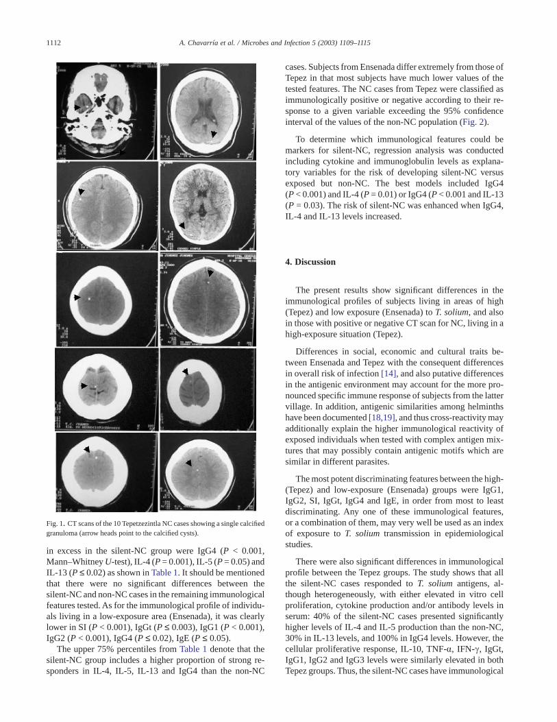
Ninety-two females and 40 males, with ages ranging from3 to 79 years, participated in the CT scan study. Individualswere classified as silent-NC or non-NC according to whethertheir CT scan images showed lesions compatible with NC.Ten cases of silent-NC (one man and nine women) werefound with a single calcified granuloma (Fig. 1). The remain-ing 122 subjects showed CT scans negative for NC (four hadanatomical abnormalities and 118 had normal brain images).The non-NC group probably included subjects who wereexposed and did not develop an established infection, otherswith an asymptomatic infection now resolved without calci-fication, and some who were not exposed. All individualswith silent-NC were examined by two neurologists andfound to be apparently healthy and neurologically asymp-tomatic.
Calcifications found in the CT scan were considered to beof cysticercal origin based on the high prevalence of neuro-cysticercosis in Mexico [16], the disproportionately highprevalence of neurocysticercosis relative to tuberculousgranulomas in large necropsy series of medical institutions[8,9], the characteristic image of calcified cysticerci (ap-proximately 1 cm diameter, round, uniformly hyperdense,sharply bordered lesion, with small or no sign of surroundinginflammation, that does not resemble cerebral or meningealtuberculoma at all [17], and the extremely low prevalence ofcerebral hydatid disease of humans in Mexico [8].
3.2. Immunological profiles
The levels of PBMC proliferation after TsAg stimulationand cytokine (IL-4, IL-5, IL-10, IL-13, INF-c and TNF-a)production in the supernatants, as well as specific antibodies(IgGt, IgG1, IgG2, IgG3, IgG4 and IgE) are shown inTable 1. The immunological features most frequently found
1111A. Chavarría et al. / Microbes and Infection 5 (2003) 1109–1115
in excess in the silent-NC group were IgG4 (P < 0.001,Mann–Whitney U-test), IL-4 (P = 0.001), IL-5 (P = 0.05) andIL-13 (P ≤ 0.02) as shown in Table 1. It should be mentionedthat there were no significant differences between thesilent-NC and non-NC cases in the remaining immunologicalfeatures tested. As for the immunological profile of individu-als living in a low-exposure area (Ensenada), it was clearlylower in SI (P < 0.001), IgGt (P ≤ 0.003), IgG1 (P < 0.001),IgG2 (P < 0.001), IgG4 (P ≤ 0.02), IgE (P ≤ 0.05).
The upper 75% percentiles from Table 1 denote that thesilent-NC group includes a higher proportion of strong re-sponders in IL-4, IL-5, IL-13 and IgG4 than the non-NC
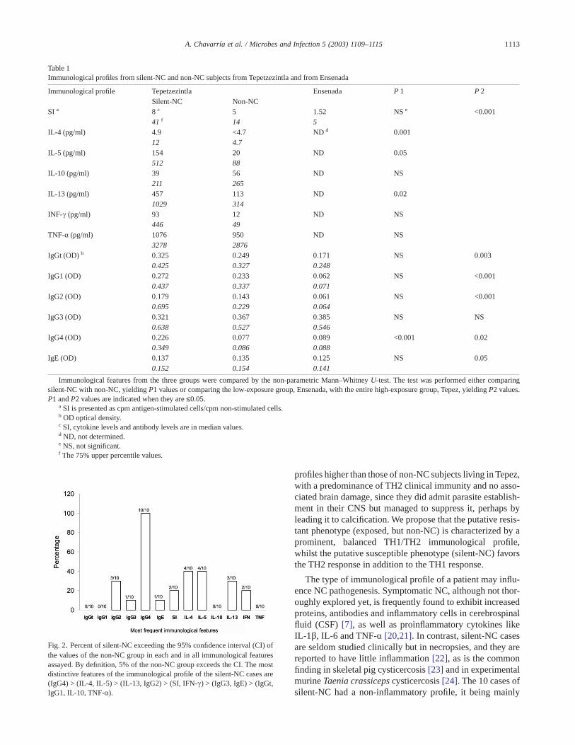
cases. Subjects from Ensenada differ extremely from those ofTepez in that most subjects have much lower values of thetested features. The NC cases from Tepez were classified asimmunologically positive or negative according to their re-sponse to a given variable exceeding the 95% confidenceinterval of the values of the non-NC population (Fig. 2).
To determine which immunological features could bemarkers for silent-NC, regression analysis was conductedincluding cytokine and immunoglobulin levels as explana-tory variables for the risk of developing silent-NC versusexposed but non-NC. The best models included IgG4(P < 0.001) and IL-4 (P = 0.01) or IgG4 (P < 0.001 and IL-13(P = 0.03). The risk of silent-NC was enhanced when IgG4,IL-4 and IL-13 levels increased.
4. Discussion
The present results show significant differences in theimmunological profiles of subjects living in areas of high(Tepez) and low exposure (Ensenada) to T. solium, and alsoin those with positive or negative CT scan for NC, living in ahigh-exposure situation (Tepez).
Differences in social, economic and cultural traits be-tween Ensenada and Tepez with the consequent differencesin overall risk of infection [14], and also putative differencesin the antigenic environment may account for the more pro-nounced specific immune response of subjects from the lattervillage. In addition, antigenic similarities among helminthshave been documented [18,19], and thus cross-reactivity mayadditionally explain the higher immunological reactivity ofexposed individuals when tested with complex antigen mix-tures that may possibly contain antigenic motifs which aresimilar in different parasites.
The most potent discriminating features between the high-(Tepez) and low-exposure (Ensenada) groups were IgG1,IgG2, SI, IgGt, IgG4 and IgE, in order from most to leastdiscriminating. Any one of these immunological features,or a combination of them, may very well be used as an indexof exposure to T. solium transmission in epidemiologicalstudies.
There were also significant differences in immunologicalprofile between the Tepez groups. The study shows that allthe silent-NC cases responded to T. solium antigens, al-though heterogeneously, with either elevated in vitro cellproliferation, cytokine production and/or antibody levels inserum: 40% of the silent-NC cases presented significantlyhigher levels of IL-4 and IL-5 production than the non-NC,30% in IL-13 levels, and 100% in IgG4 levels. However, thecellular proliferative response, IL-10, TNF-a, IFN-c, IgGt,IgG1, IgG2 and IgG3 levels were similarly elevated in bothTepez groups. Thus, the silent-NC cases have immunological
Fig. 1. CT scans of the 10 Tepetzezintla NC cases showing a single calcifiedgranuloma (arrow heads point to the calcified cysts).
1112 A. Chavarría et al. / Microbes and Infection 5 (2003) 1109–1115
profiles higher than those of non-NC subjects living in Tepez,with a predominance of TH2 clinical immunity and no asso-ciated brain damage, since they did admit parasite establish-ment in their CNS but managed to suppress it, perhaps byleading it to calcification. We propose that the putative resis-tant phenotype (exposed, but non-NC) is characterized by aprominent, balanced TH1/TH2 immunological profile,whilst the putative susceptible phenotype (silent-NC) favorsthe TH2 response in addition to the TH1 response.
The type of immunological profile of a patient may influ-ence NC pathogenesis. Symptomatic NC, although not thor-oughly explored yet, is frequently found to exhibit increasedproteins, antibodies and inflammatory cells in cerebrospinalfluid (CSF) [7], as well as proinflammatory cytokines likeIL-1b, IL-6 and TNF-a [20,21]. In contrast, silent-NC casesare seldom studied clinically but in necropsies, and they arereported to have little inflammation [22], as is the commonfinding in skeletal pig cysticercosis [23] and in experimentalmurine Taenia crassiceps cysticercosis [24]. The 10 cases ofsilent-NC had a non-inflammatory profile, it being mainly
Table 1Immunological profiles from silent-NC and non-NC subjects from Tepetzezintla and from Ensenada
Immunological profile Tepetzezintla Ensenada P 1 P 2Silent-NC Non-NC
SI a 8 c 5 1.52 NS e <0.00141 f 14 5
IL-4 (pg/ml) 4.9 <4.7 ND d 0.00112 4.7
IL-5 (pg/ml) 154 20 ND 0.05512 88
IL-10 (pg/ml) 39 56 ND NS211 265
IL-13 (pg/ml) 457 113 ND 0.021029 314
INF-c (pg/ml) 93 12 ND NS446 49
TNF-a (pg/ml) 1076 950 ND NS3278 2876
IgGt (OD) b 0.325 0.249 0.171 NS 0.0030.425 0.327 0.248
IgG1 (OD) 0.272 0.233 0.062 NS <0.0010.437 0.337 0.071
IgG2 (OD) 0.179 0.143 0.061 NS <0.0010.695 0.229 0.064
IgG3 (OD) 0.321 0.367 0.385 NS NS0.638 0.527 0.546
IgG4 (OD) 0.226 0.077 0.089 <0.001 0.020.349 0.086 0.088
IgE (OD) 0.137 0.135 0.125 NS 0.050.152 0.154 0.141
Immunological features from the three groups were compared by the non-parametric Mann–Whitney U-test. The test was performed either comparingsilent-NC with non-NC, yielding P1 values or comparing the low-exposure group, Ensenada, with the entire high-exposure group, Tepez, yielding P2 values.P1 and P2 values are indicated when they are ≤0.05.
a SI is presented as cpm antigen-stimulated cells/cpm non-stimulated cells.b OD optical density.c SI, cytokine levels and antibody levels are in median values.d ND, not determined.e NS, not significant.f The 75% upper percentile values.
Fig. 2. Percent of silent-NC exceeding the 95% confidence interval (CI) ofthe values of the non-NC group in each and in all immunological featuresassayed. By definition, 5% of the non-NC group exceeds the CI. The mostdistinctive features of the immunological profile of the silent-NC cases are(IgG4) > (IL-4, IL-5) > (IL-13, IgG2) > (SI, IFN-c) > (IgG3, IgE) > (IgGt,IgG1, IL-10, TNF-a).
1113A. Chavarría et al. / Microbes and Infection 5 (2003) 1109–1115

composed of IL-5, IL-13 and IL-4 production and by thepresence of IgG4 in serum.
If prominent TH2 profiles in infected subjects underlielow or no symptoms at all in NC, one could speculate thatIL-4 downregulation of the inflammatory response is in-volved, as it is in schistosomiasis granulomas [25,26], inexperimental cysticercosis by T. crassiceps [27], and in braingranulomas in symptomatic NC [28]. Also, IL-4 can inducethe switch of IgG4 and IgE [29] and thus explain the higherlevel of IgG4 in the NC group. The role of IL-5 and IL-13 inparasitic disease seems to be important in the elimination ofthe parasite [30,31]; here we show the presence of IL-5 in cellsupernatants of the silent-NC group, as shown previously insymptomatic NC in serum and CSF [30,32]. Thus, one wayto eliminate the cysticercus without creating too much in-flammatory response in the brain would be to induce highIL-4, IL-13 and IL-5 peaking-out of a generally more alertimmune system but not particularly high in TH1 activity andinflammatory cytokines. IgG4 and IgE are both considered tobe TH2 immunoglobulins in humans [29]. IgE has beenassociated with resistance to parasitic disease as in schisto-somiasis [33,34], whilst IgG4 has been proposed as an IgE-competing immunoglobulin [34], because high levels ofIgG4 in serum are associated with susceptibility, as proposedfor Schistosoma mansoni and haematobium infections[33,35]. Among highly exposed individuals from Tepez,IgG4 was increased only in those with silent-NC, a sugges-tion of a profile of susceptibility to the benign form of theCNS infection. It is tempting to speculate that resistance toexposure may be related to their low IgG4 responsiveness.The non-NC Tepez group had levels of IgE indistinguishablefrom those of the silent-NC, but both differed from thelow-exposure Ensenada group. This result points to IgE as anadditional marker for parasite exposure.
Consequently, for NC we would propose that the immu-nological profile combining high TH1 with peaking TH2features associates with a noiseless course of the infectionand with its resolution in the silent-NC cases, and predict thatthe opposite is behind the immunoinflammatory responseand clinical manifestations of the symptomatic NC cases[36,37]. The study of more NC cases, in all their forms, isnecessary to formally establish their specific immunoinflam-matory profiles.
Acknowledgements
We thank Constantino Beltrán, Teresa Gómez, DemetriaMeza and Israel Alvarez for technical support, and MercedesBaca and Marisela Hernández for administrative assistance.Isabel Pérez Montfort corrected the English version of themanuscript.
This study was funded by Dirección General de Asuntosde Personal Académico (IN220999), Universidad NacionalAutónoma de México; CONACyT (G25955 and 31378M),México; ECOS-ANUIES (M99S03); Howard Hughes Medi-cal Institute (grant 55000643), the British Council, the
French Embassy and the French Programme for Fundamen-tal Research in Microbiology, Parasitology and InfectiousDiseases.
References
[1] A.C. White Jr, Neurocysticercosis: updates on epidemiology, patho-genesis, diagnosis, and management, Annu. Rev. Med. 51 (2000)187–206.
[2] P.M. Schantz, A.C. Moore, J.L. Muñoz, B.J. Hartman, J.A. Schaefer,A.M. Aron, D. Persaud, E. Sarti, M. Wilson, A. Flisser, Neurocysticer-cosis in an orthodox Jewish community in NewYork city, New Engl. J.Med. 327 (1992) 692–695.
[3] W.X. Shandera, A.C. White, J.C. Chen, P. Diaz, R. Armstrong, Neu-rocysticercosis in Houston, Texas. A report of 112 cases, Medicine 73(1994) 37–52.
[4] M.T. Medina, E. Rosas, F. Rubio-Donnadieu, J. Sotelo, Neurocys-ticercosis as the main cause of late-onset epilepsy in Mexico, Arch.Intern. Med. 150 (1990) 325–327.
[5] O.H. Del Brutto, Prognostic factors for seizure recurrence after with-drawal of antiepileptic drugs in patients with neurocysticercosis, Neu-rology 44 (1994) 1706–1709.
[6] V. Vázquez, J. Sotelo, The course of seizures after treatment forcerebral cysticercosis, New Engl. J. Med. 327 (1992) 696–701.
[7] J. Sotelo, V. Guerrero, F. Rubio, Neurocysticercosis: a new classifica-tion based on active and inactive forms. A study of 753 cases, Arch.Intern. Med. 145 (1985) 442–445.
[8] J. Villagrán, J.E. Olvera, Cisticercosis humana: Estudio clínicopatológico de 481 casos de autopsia, Patología 26 (1988) 149–156.
[9] M.T. Rabiela, A. Rivas, J. Rodríguez, S. Castillo, F. Cancino, in:A. Flisser, K. Willms, J.P. Laclette, C. Larralde, C. Ridaura, F. Beltrán(Eds.), Cysticercosis: Present Stage of Knowledge and Perspectives,Academic Press, New York, 1982, pp. 179–200.
[10] J. Sotelo, O.H. Del Brutto, Brain cysticercosis, Arch. Med. Res. 31(2000) 3–14.
[11] A. Fleury, T. Gomez, I. Alvarez, D. Meza, M. Huerta, A. Chavarria,R.A. Carrillo Mezo, C. Lloyd, A. Dessein, P.M. Preux, M. Dumas,C. Larralde, E. Sciutto, G. Fragoso, High prevalence of calcified silentneurocysticercosis in a rural village of Mexico, Neuroepidemiology22 (2003) 139–145.
[12] E. Sciutto, G. Fragoso, A. Fleury, J.P. Laclette, J. Sotelo, A. Aluja,L. Vargas, C. Larralde, Taenia solium disease in humans and pigs: anancient parasitosis disease rooted in developing countries and emerg-ing as a major health problem of global dimensions, Microbes Infect.15 (2000) 1875–1890.
[13] M. Huerta, A.S. de Aluja, G. Fragoso, A. Toledo, N. Villalobos,M. Hernandez, G. Gevorkian, G. Acero, A. Diaz, I. Alvarez, R. Avila,C. Beltran, G. Garcia, J.J. Martinez, C. Larralde, E. Sciutto, Syntheticpeptide vaccine against Taenia solium pig cysticercosis: successfulvaccination in a controlled field trial in rural Mexico, Vaccine 12(2001) 262–266.
[14] C. Larralde, A. Padilla, M. Hernández, T. Govezensky, E. Sciutto,G. Gutierrez, R. Tapia-Conyer, B. Salvatierra, J. Sepulveda, Seroepi-demiology of cysticercosis in Mexico, Salud Pública Mex. 34 (1992)197–210.
[15] C. Larralde, J.P. Laclette, C.S. Owen, I. Madrazo, M. Sandoval,R. Bojalil, E. Sciutto, L. Contreras, J. Arzate, M.L. Diaz, Reliableserology of Taenia solium cysticercosis with antigens from cystvesicular fluid: ELISA and hemagglutination tests, Am. J. Trop. Med.Hyg. 35 (1986) 965–973.
[16] O.H. Del Brutto, J. Sotelo, G.C. Roman, in: O.H. Del Brutto, J. Sotelo,G.C. Roman (Eds.), Neurocysticercosis. A Clinical Handbook, Swets& Zeitlinger, Lisse, 1998, pp. 73–94.
[17] J. Rodriguez-Carbajal, B. Boleaga-Duran, J. Dorfsman, The role ofcomputed tomography (CT) in the diagnosis of neurocysticercosis,Child’s Nerv. Syst. 4 (1987) 199–202.
1114 A. Chavarría et al. / Microbes and Infection 5 (2003) 1109–1115

[18] C. Larralde, R.M. Montoya, E. Sciutto, M.L. Diaz, T. Govezensky,E. Coltorti, Deciphering western blots of tapeworm antigens (Taeniasolium, Echinococcus granulosus, and Taenia crassiceps) reactingwith sera from neurocysticercosis and hydatid disease patients, Am. J.Trop. Med. Hyg. 40 (1989) 282–290.
[19] L. Benitez, L.J. Harrison, R.M. Parkhouse, L.M. Gonzalez,B. Gottstein, T. Garate, Sequence and immunogenicity of the Taeniasaginata homologue of the major surface antigen of Echinococcusspp, Parasitol. Res. 84 (1998) 426–431.
[20] L. Ostrosky-Zeichner, E. Garcia-Mendoza, C. Rios, J. Sotelo,Humoral and cellular immune response within the subarachnoid spaceof patients with neurocysticercosis, Arch. Med. Res. 27 (1996)513–517.
[21] F. Aguilar-Rebolledo, R. Cedillo-Rivera, P. Llaguno-Violante,J. Torres-López, O. Muñoz-Hernández, J.A. Enciso-Moreno, Interleu-kin levels in cerebrospinal fluid from children with neurocysticerco-sis, Am. J. Trop. Med. Hyg. 64 (2001) 35–40.
[22] A. Escobar, in: E. Palacios, J. Rodriguez-Carbajal, J.M. Taveras(Eds.), Cysticercosis of the Central Nervous System, Thomas Spring-field, USA, 1983, pp. 27–54.
[23] A. Aluja, D. Gonzalez, C. Rodriguez, A. Flisser, Histological descrip-tion of tomographic images of T. solium cysticerci in pig brains, Clin.Imaging 13 (1989) 292–298.
[24] A. Padilla, T. Govezensky, E. Sciutto, L.F. Jimenez-Garcia, M.E. Gon-sebatt, P. Ramirez, C. Larralde, Kinetics and characterization of cel-lular responses in the peritoneal cavity of mice infected with Taeniacrassiceps, J. Parasitol. 87 (2001) 591–599.
[25] E. Pearce, P. Caspar, J. Grzynch, F. Lewis, A. Sher, Downregulation ofTH1 cytokine production accompanies induction of TH2 responses bya parasitic helminth, Schistosoma mansoni, J. Exp. Med. 173 (1991)159–166.
[26] T.A. Wynn, A.W. Cheever, Cytokine regulation of granuloma forma-tion in shistosomiasis, Curr. Opin. Immunol. 119 (1995) 193–201.
[27] P. Robinson, R. Atmar, D. Lewis, C. White, Granuloma cytokines inmurine cysticercosis, Infect. Immun. 65 (1997) 2925–2931.
[28] B. Restrepo, J.Alvarez, J. Castaño, L.F.Arias, M. Restrepo, J. Trujillo,C.H. Colegial, J.M. Teale, Brain granulomas in neurocysticercosispatients are associated with a TH1 and TH2 profile, Infect. Immun. 69(2001) 4554–4560.
[29] M. Lundgren, U. Persson, P. Larsson, C. Magnusson, E. Smith, Inter-leukin 4 synthesis of IgE and IgG4 in human B cells, Eur. J. Immunol.19 (1989) 1311–1315.
[30] C. Evans, H. Garcia, A. Hartnell, R.H. Gilman, P.J. Jose, M. Martinez,D.G. Remick, T.J. Williams, J.S. Friedland, Elevated concentrationsof eotaxine and interleukin-5 in human neurocysticercosis, Infect.Immun. 66 (1998) 4522–4525.
[31] F. Finkelmann, T. Wynn, D.D. Donaldson, J. Urban, The role of IL13in helminth-induced inflammation and protective immunity againstnematode infections, Curr. Opin. Immunol. 11 (1999) 420–426.
[32] V. Rodrigues, F.A. de-Mello, E.P. Magalhães, S.B.F. Ribeiro,J.O. Marquez, Interleukine-5 and interleukin-10 are major cytokinesin cerebralspinal fluid from patients with active neurocysticercosis,Braz. J. Med. Biol. Res. 33 (2000) 1059–1063.
[33] C.E. Demeure, P. Rihet, L. Abel, M. Ouattara, A. Bourgois,A. Dessein, Resistance to Schistosoma mansoni in humans: influenceof IgE/IgG4 balance and IgG2 in immunity to reinfection after che-motherapy, J. Infect. Dis. 168 (1993) 1000–1008.
[34] P. Rihet, C. Demeure, A. Dessein, A. Bourgois, Strong serum inhibi-tion of specific IgE correlated to competing IgG4, revealed by a newmethodology in subjects from a S. mansoni endemic area, Eur. J.Immunol. 22 (1992) 2063–2070.
[35] P. Hagan, U.J. Blumenthal, D. Dunn, A.J. Simpson, H.A. Wilkins,Human IgE, IgG4 and resistance to reinfection with Schistosomahaematobium, Nature 349 (1991) 243–245.
[36] J. Singh, S. Kaur, G. Bhatti, I.M.S. Sawhney, N.K. Ganguly,R.C. Mahajan, N. Malla, Cellular immune response in human neuro-cysticercosis, Parasitol. Res. 86 (2000) 500–503.
[37] B.I. Restrepo, P. Llaguno, M.A. Sandoval, J.A. Enciso, J.M. Teale,Analysis of immune lesions in neurocysticercosis patients: centralnervous system response to helminth appears TH1–like instead ofTH2, J. Neuroimmunol. 89 (1998) 64–72.
1115A. Chavarría et al. / Microbes and Infection 5 (2003) 1109–1115