th indian fertility society-fertivision 2011 ... - ivf-hub.net · recently a large survey was...
TRANSCRIPT

Supplement to Volume 2, No. 2, December 2011 Journal of Reproductive and Stem Cell Biotechnology (JRSCB)
Proceedings of the 7th Annual Conference of the Indian Fertility Society-FERTIVISION 2011
16-18 December 2011 New Delhi, India
[http://www.fertivision2011-ifs.org/home]
[Copyright for the above Proceedings is held by THE INDIAN FERTILITY SOCIETY.
No part of this Proceedings may be reproduced or transmitted in any form without the prior permission of the owners of the Copyright]
Country Editor for the JRSCB: Dr Rajvi Mehta, PhD
Journal of Reproductive and Stem Cell Biotechnology Volume 2 Number 2 2011 (Supplement) [Online ISSN No. 2180-1398]
3
IFS Oration - Can we do something for Poor Responders?Kuldeep JainPresident, IFS, Director, KJIVF & Laparoscopy Centre, Delhi
Poor ovarian response, defined as failure of the development of sufficient number of mature follicle to proceed to oocyte retrival or yielding only a few oocyte following gonodotropin stimulation in women undergoing in vitro fertilization treatment, occurs in 9-24% of women.
Poor ovarian response is likely to be an increasing problem with women delaying child bearing & presenting later in their reproductive life.
These patients have impaired fertilization rates, lower embryo quality & decreased pregnancy rates. Cycle cancellation rates are very high leading to emotional distress for the couple or service providers. Managing poor responder remains a great challenge for the clinicians.
Various treatment regimens and interventions have been proposed in an effort to improve ovarian response & IVF outcome in this group of patients however none of regimens can be recommended to all poor responders & no single protocol is considered superior to other.
There are few concerns need to be considered in regard to poor responders like definition of poor response is not standardized yet. Recently “Bologna criteria” were suggested by ESHRE to define poor responders making two out of three criteria must to label a patient poor responder.
Another issue is defining / identifying the ultimate goal. Is it increasing number of follicle / oocyte, implantation rate or pregnancy rates?
Careful prediction, thorough assessment of ovarian reserve & careful review of past response goes a long way in deciding the best protocol for individual patient.
Cause of poor response may be many fold like low reserve, impending ovarian failure, advanced age, high BMI, previous ovarian surgery & inappropriate stimulation protocol. Predicting poor response is another controversial issue as there is no single marker which can be used with confidence. AFC, AMH & advanced age & High FSH are most used & informative markers to predict poor response however, if used singly, may not give satisfactory prediction. Stimulation itself may be considered only good marker in most of the cases.
Recently a large survey was conducted including 196 centre from 45 countries & 1,24,700 IVF cycle focusing on the trends in different parts of world & current strategies being used for poor responders.
Newer strategies like use of DHEA, AACEP protocol & GRS-PREP protocol have been suggested to improve the outcome in poor responders however lack of large controlled trials & comparative data limits the final conclusions & usefulness of these newer strategies.
To conclude, Poor Responders are a challenge to the IVF specialists & need to be investigated for the cause. Age is an established indicator of poor response. There are in sufficient evidence to support the routine use of any particular intervention either for pituitary down regulation, ovarian stimulation or adjuvant therapy like DHEA, pretreatment with OCP or newer protocol like AACEP or GRS-PREP protocol. More robust data from good quality RCTs with relevant outcome is needed.

4
Self-Operated Endovaginal Telemonitoring (SOET): A Step Towards More Patient-Centered Art?
Jan Gerris, Petra De SutterCentre for Reproductive Medicine, University Hospital Ghent, De Pintelaan 185 – 9000 Gent, Belgium
The need for serial vaginal sonographies to monitor ovarian stimulation for artificial reproductive technology (ART) treatments remains a major practical and organizational drawback both for patients and health care providers. We explore the possibility to have patients and/or their partners make their own vaginal sonographies at home. To make this happen, a portable, easy-to-use, home-applied vaginal probe for recording relevant images has to be developed, as well as appropriate software to transfer images using modern communication technology to the centre, to analyse the recordings and to send a swift structured response, comprising dosing advice and next-step instructions. A simplification of the uncontested need to perform these sonographies, even if applicable to just a selected proportion of IVF patients, could fit in the general tendency to make IVF more patient-centered and -friendly, to implement telemedicine and to increase patient empowerment by supervised active participation to their treatment.
The advantages of such a patient-empowering and environment-friendly technology are explored in this paper, aiming at opening up a debate on whether patients themselves would, could and should achieve a further substantial simplification of ART without loss off quality while strongly curbing costs.
The problem to be debatedAlthough initial attempts at obtaining a pregnancy by in vitro fertilization and embryo transfer (IVF-ET) were conducted in a natural cycle (Edwards et al., 1980), it was soon recognized that ovarian stimulation using gonadotropins was needed in order to obtain more oocytes and increase the chance for success by morphology-based embryo selection. The need to monitor ovarian stimulation has for long been based on serial sonographic assessments of follicular growth (Kemeter and Feichtinger, 1989) and to a lesser degree on serial measurements of serum estradiol. These became less important since sonography, after shifting from the abdominal to the vaginal route, soon became the most important monitoring tool as well as the preferred route for oocyte aspiration. The relevance of estradiol measurements is limited to cases of threatening ovarian hyperstimulation syndrome (OHSS) whereas serial sonography as the sole method of monitoring follicular growth has become routine and cannot be dispensed with, both for optimal timing of HCG administration and for early detection of OHSS.
Patient-friendly ART gently creeps into our clinics (Pennings and Ombelet, 2007) and includes self-administration of gonadotropins, newer gonadotropin formulations, milder stimulation protocols (Baart et al., 2007), reduction of multiple pregnancies by judicious single embryo transfer (Gerris et al., 2002), a decrease of the risk for ovarian hyperstimulation syndrome, more efficacious embryo freezing programs (Tiitinen et al., 2004) and less treatment-induced stress. The embryo utilization rate has improved. Less multiples are born for an equal or even enhanced pregnancy chance per attempt. Milder protocols result both in less side-effects and substantial savings for patients or third-party payers. Natural or minimally modified natural cycles as well as in vitro maturation also helped to avoid or minimize gonadotropin use (Pelinck et al., 2006). Further simplifications in the pipeline comprise the use of long-acting injectable gonadotropin preparations allowing to reduce the number of injections and the total dose of gonadotropins, and oral gonadotropins.
Notwithstanding all these helpful developments, one major inconvenience still exists, i.e. the need for women to visit the IVF clinic or their gynaecologist to perform serial vaginal sonographies. No hard data exist regarding the average number of pre-oocyte-retrieval sonographic visits but a good guess would be four to five per attempt. The total annual number of IVF/ICSI attempts amounts to ~one million and is probably still rising. It is not exaggerated to add another one million of non-IVF/ICSI stimulations. This implies between eight and ten million annual sonograms for pre-ovulatory ART-monitoring only or, for ~300 working days per year, a daily trek to the IVF clinic for 90,000 women! Huge distances in large countries, whether they belong to the first, the second or
5
the third world, where sonography is not available within a reasonable distance from homes, decrease access to treatment for many couples who could benefit from it. This is the case in large developed societies e.g. the USA, Australia, Canada and other giant countries, but also in many countries in the developing world where assisted reproductive services are scarce though on the rise. The two major impediments to wide access to ART overtly identified are cost of treatment and legal limitations. Recent attention has been given to so-called “IVF in low-resource settings” to be implemented in third-world countries with poor access to treatment because of financial constraints. It is a noble intention of making subfertility a global issue because of its severe social consequences in all human societies. High costs and legal obstacles indeed may hamper treatment. But we must recognize that limited access to ART treatment exists almost everywhere. This follows from the wide variation in the number of ART cycles per number of inhabitants between different countries (e.g. Nyboe Andersen et al., 2008), ranging from >2,000 cycles/million inhabitants to just a few hundred cycles/million inhabitants as in the US or a mere 20/million inhabitants in India. High access rates are usually observed in small countries, e.g. Denmark, Israel and Belgium. Low access is observed in larger countries, where sheer distance and/or the time needed to cover it, severely limit treatment utilization, even in high-resource settings. Whether it be the Australian out-back, the American mid-west, the Russian steppes, the deep interior of rural India or China, or the more remote areas of many other countries, distance and/or the time needed to reach expert sonography plays as preponderant role in making ART a feasible option for many patients who need it.
An idea that may help solve the problemTo solve this problem, we suggest the idea to have patients have their sonographies perform themselves, after instruction by a midwife, a nurse-practitioner or an experienced sonography technician, using a small, re-usable, safe, cheap and easy to use customized device, allowing registration of real time images under direct visual inspection of the patient or her partner. Images can be sent with proper identification over the internet to the centre where specialized personnel and technology receives, stores, analyzes and interprets the images followed by swift appropriate action, i.e. dosage adaptation or next-step decisions. These responses could be semi-automated by using specific new software. They should comprise proper instructions regarding the dose of gonadotropins to be self-injected during the following day(s), the timing for a subsequent sonogram, the precise timing of HCG injection and of oocyte retrieval. If extended to intrauterine insemination or freeze/thaw cycles after IVF, even in a natural cycle or after oral ovulation induction, planning of insemination or embryo replacement can be communicated. If needed, these self-operated endovaginal sonographies (SOETs) are performed with a backup of same-day “live” (if needed) or next-day “real-time” sonography in the centre. The idea could work for IVF/ICSI cycles but it could be extended to non-IVF ovarian stimulation protocols using oral or intramuscular drugs.
Several questions and considerations need to be addressed, a.o. (re)organisational aspects, protection of privacy (who has access to the data?), legal (who is responsible for errors resulting in complications?) and financial aspects (who will pay the interpretation, storage, communication etc. given the loss of direct income linked to individual sonographic procedures?). We will address these secondary aspects after considering the present experience in IVF.
It is not the idea to replace all vaginal sonography in the context of IVF, let alone all gynaecological sonography, by SOET. “Real-time” sonography will remain a very important tool in IVF. During a transitional phase of acceptance of SOET, and even if accepted, SOET will not replace sonography in complicated cases, when follicular growth is abnormally slow or quick, when abnormal images (cysts, hydrosalpinges, unexpected tumours, endometriosis, etc.) are observed, or on simple request from anxious patients. It is conceived in the first place to alleviate an abnormal amount of time-consuming, routine work and to allow patients living far from the centre to make less demanding sacrifices.

6
Follicular DynamicsUmesh Jindal
Whenever any gynaecologist asks me regarding starting an IVF center, I ask only two questions. How many gonadotrophin stimulations cycles are you doing per month? Have you developed masterly skills in follicular sonography? In other words the IVF laboratory is dependent upon an optimal number of good quality oocytes, whcih can only be ensured by proper understanding of follicular dynamics.
A baseline scan is done before starting stimulation to confirm down regulation in agonist protocols. In addition an antral follicle count is done to predict the type of response and depending upon that the starting dose is decided. A mid- stimulation scan on day 4-8 is done to assess response and adjust the dose accordingly. Additional serum E2 levels may be done. The dose of gonadorophin is adjusted and antagonist is added in non down regulated cycles. A decision for hCG is taken after assessing the maturity of the follicles with ultrasound which occurs after 8-12 days of stimulation.
The ovarian response has a very wide range from poor, reduced, adequate, plentiful, hyper to almost explosive. The response depends primarily on the ovarian reserve. The stimulation related parameters such as agonist or antagonist protocol, the starting and step up or down dose and the clinical judgment of the clinicians is assessing response and maturity decide the ultimate quality and quantity of the oocyte harvest as well as complications like OHSS.
A poor responder is generally an older women with less than 4-5 antral follicle count in both ovaries, low AMH and FSH on the higher side of normal. Despite an apparent normal spontaneous ovulatory cycle these women perform poorly with any protocol. Cancellation is almost guaranteed. A couple of follicles which may develop may end up in empty follicles or poor quality oocytes.
A reduced responder is one borderline case who may have fair success rate albeit lower. These are young women with decreased ovarian reserve due to previous ovarian surgery, tuberculosis, impending premature menopause, or older women just before the final exhaustion of the ovaries. These cycles may be continued depending upon the development of follicles, age of the women and a desire to “take a chance” even with decreased pregnancy and high abortion rate.
A normal responder is an ideal case with AFC between 4-16 (in both ovaries), will develop 6-12 follicles. A sub category of normal responder i.e. young age women with AFC of 10-20 (in both ovaries), but not polycystic. Both these categories develop adequate and good number of follicles containing oocytes of excellent quality and high implantation rate. There is no risk of OHSS. However this category may end up into reduced or hyper response if the starting dose is not appropriate or non judicious step up or step down of gonadotrophins is done.
The third category is that of hyperresponder, typically a PCOD case with more than 24 AFC in both ovaries. Many of these women have more than 30-40 AFC in each ovary and have a potential to develop almost an explosive course. Now the antagonist regimen with a GnRH-a trigger is the standard protocol for these women. A very low starting dose (75-150 IU), proper monitoring and Gn-RH-a trigger has reduced the risk of OHSS significantly in these women without compromising the success rate.
On day 5 a step up or step down of dose may be required. This may improve the cycle outcome however the best cycles are those in which no change of Gonadotrophin dose is required.
An E2 assessment in poor responder and hyper responder does aid in the clinical management of these women in adjusting the dose and predicting the outcome as well as OHSS.
Success of IVF depends upon oocytes. An excellent clinical and ultrasound acumen is required to ensure a good oocyte harvest.
7
Advances in FSH and LH in Controlled Ovarian StimulationKD NayarChief Consultant, Akanksha IVF Centre, Mata Chanan Devi Hospital, New Delhi-110 058
The pituitary gonadotropins, FSH and LH have an important role so that the ovary can perform its two essential functions - ovulation and formation of steroid hormones. They have distinct but complementary function in ensuring optimal oocyte maturation and ovulation.
FSH and LH in ovarian stimulation: the two cell theory Initiation of folliculogenesis occurs independent of gonadotropins. Secretion of FSH and LH must happen in an orderly fashion for final development of a mature oocyte. In the menstrual cycle, recruitment of multiple immature follicles occurs in response to FSH. Selection of the dominant follicle occurs which has the lowest FSH threshold. In the midfollicular phase (day7-9), dominant follicle emerges (≥ 10mm) acquires LH receptors and is uniquely responsive to both FSH and LH, and synthesises estradiol. LH stimulates androgen production from theca cells, to be transformed into estrogen by FSH-stimulated aromatase activity in the granulosa cells (two-cell, two gonadotropin model). So FSH is essential for triggering antral follicle formation with some LH essential in the pre-antral stage (follicle size <10mm) to stimulate androgen secretion from theca cells.
LH therapeutic windowIt is known that, a threshold of LH is required below which oocyte maturation will not occur (lessons from hypogonadotropic hypogonadism patients). Also, high concentrations have detrimental effect on follicles-follicle atresia, premature luteinization and poor quality oocytes (LH ceiling). The optimal concentration is around ≥ 1.2IU/l and ≤ 5IU/l1 (therapeutic window).
Advances in FSH and LH preparationsMajor advances in technology have brought the field of gonadotropin therapy a very long way since the era of animal, human pituitary and urinary derived hormones. In 1958, Carl Gemzell extracted gonadotropins from human pituitary glands which was successfully used for OI in many centres. But, it had two main drawbacks of limited reservoir and danger of CJD.
In 1962, it was followed by introduction of HMG (Menotropins). Urofollitropins or FSH (low LH activity, < 1%)) were developed next. In mid 1990’s highly purified urinary FSH preparations (FSH-HP) (< 0.1% LH, no contaminating urinary proteins) was prepared. Quality control was impossible as urine donors were not traceable although no infectivity has ever been reported in four decades of use.
In 1992, recombinant gonadotropins produced by genetic engineering technology, became available for clinical use. Important advantages were- lack of prion transmission, traceability, high purity and specific activity (accounting for >99% of the preparation’s protein content, allowing SC administration), unlimited supply with batch to batch consistency, and no contamination from other gonadotropins. Adverse immune reactions were also minimised. In 1993 first clinical report was published about recombinant LH which is now available for clinical use in form of vials with syringe delivering 75 IU, in a specific subgroup of patients.
In 2000, a urinary derived product re-entered the market – highly purified Menotropins ( HP-HMG) containing equal amount of FSH and LH, ratio of 1:1 where the LH activity consists of added hCG.
In 2007, long acting Corifollitropin alfa, a synthetic recombinant follicle-stimulating hormone (r-FSH) molecule containing a hybrid beta subunit, which provides a plasma half-life of 65-70 hours while maintaining its pharmocodynamic activity was introduced. A single injection of corifollitropin alfa can replace daily FSH injections for the first week of ovarian stimulation for in vitro fertilization. The optimal corifollitropin dose has been calculated to be 100 µg for women with a body weight ≤ 60 kg and 150 µg for women with a body weight >60 kg, respectively. Combination of corifollitropin with daily gonadotropin-releasing hormone antagonist
8
injections starting on stimulation day 5 seems to yield similar or significantly higher numbers of oocytes and good quality embryos, as well as similar ongoing pregnancy rates compared with women stimulated with daily r-FSH injections.
Future - Presently a product for effective oral administration is in development.
Comparision of Old vs NewUrinary versus Recombinant – safetyIn more than 30 years of use of urinary products not a single case of infection by prions or slow virus has been reported. The theoretical immunogenic potential of contamination in urinary products has been expressed in the occasional local allergic reaction with IM injection. S/C injection of recombinant products always produces more local odema and skin reaction. Uneven biological potency is also shown by recombinant products, an ampoule labelled to contain 75 IU may range in true activity from 50 to 120 IU FSH2. It is clearly shown several times that unit for unit recombinant FSH is more potent than urinary FSH and hence slightly lower doses are needed.
Urinary versus Recombinant – efficacyUrinary FSH vs rFSH: In IUI cycles, a meta-analysis showed rFSH was associated with higher per cycle PR than HP-FSH, when used at the same dose, whereas the PR were similar when the dose of r-FSH was 50% lower3
In IVF cycles, a randomized trial showed no difference between urinary HP FSH vs. rFSH as regards ovulation and pregnancy rates, miscarriage, hyperstimulation or multiple pregnancy rates4. Meta-analysis of randomized controlled trials comparing urinary and recombinant FSH for ovulation induction in women with PCOS has also confirmed these findings5. The only difference is the increased unit to unit potency of r FSH.
HP HMG vs. r-FSH: In ART cycles using agonist protocol, a similar or higher pregnancy rate has been reported with the use of HP HMG6. In antagonist cycles, similar ongoing pregnancy rates have been reported, although more number of oocytes were obtained using rFSH.
Exogenous LH supplementation—Consensus and recommendationsThe use of LH for supplementation in ART is controversial.Strongest predictive factor for the need of exogenous LH is in women who have a poor response to ovarian stimulation (1) Poor response in previous cycle defined as less than 4 oocytes retrieved, r-FSH dose needed more than 3000 IU per completed stimulated cycle or estradiol less than 800 pg/ml. (2) Sub optimal ovarian response with sub optimal follicular progression in a current cycle by day 6-8, defined as no follicle more than 10 mm by day 6, estradiol is less than 200 pg/ml by day 6, poor progression or slowing of follicular growth i.e. 1-2 mm progression per day slowing to less than 2mm in 3 days7.(3) Patient with marker suggestive of sub optimum ovarian response i.e. woman more than 35 years of age, adjuvant r-LH starting either on day 1 of stimulation or day 6-8 may be beneficial.
Further research is needed for patient at risk of poor ovarian response based on the following bio markers (1) AFC less than 6 in both ovaries (2) AMH less than 1.5 ng/ml (3) LH polymorphism.
A meta-analysis8 presented in ASRM 2011, reported, addition of rLH in poor responders undergoing ovarian stimulation for IVF using rFSH and GnRH analogues does not seem to increase the probability of clinical pregnancy. However, there was a wide rate difference and the confidence interval; further studies are needed for solid conclusions to be drawn.
r-LH vs. HMG: r-LH is analogous to endogenous LH, with high purity, precision of dosing and consistency. HMG may have hCG which is added for LH activity. Human chorionic gonadotropin may lead to LH receptor internalization leading to tolerance or lack of effect.
ConclusionUrinary and recombinant gonadotropins, for ovarian stimulation for IVF, are probably equally safe and effective. However, recombinant gonadotropins have the advantage of being more convenient to use and better batch to batch consistency. The higher cost for recombinant products limits their worldwide use in IVF. Conflicting data exist regarding the benefit of adding LH to FSH in the stimulation regimens.
9
Despite the significant advances, it is prudent to remember that gonadotropin therapy is costly and has significant risks, and should therefore be used only by clinicians having the requisite training and experience.
References1. O’Dea L, O’Brien F, Currie K, Hemsey G., Follicular development induced by recombinant luteinizing hormone(LH)
and follicle stimulating hormone(FSH) in anovulatory women with LH and FSH deficiency: evidence of threshold effect. Curr Med Res Opin 2008; 24:2785-2793.
2. Driegberg R, et al., Quantification of follicle stimulating hormone (follitropin alfa): is invivo bioassay still relevant in the recombinant age? Curr Med Res Opin 2003;19:41-46.
3. Matorras R, Osuna C, Exposito A, Crisol L, Pijoan JI., Recombinant FSH versus highly purified FSH in intrauterine insemination: systematic review and metaanalysis. Fertil Steril 2011; 95(6):1937- 1942.e3.
4. Yarali H, et al., Urinary follicle stimulating hormone versus recombinant FSH in clomiphene citrate resistant,normogonadotropic,chronic anovulation : a prospective randomized study. Fertil Steril 1999;72:276-28.
5. Bayram N, et al., Recombinant FSH versus urinary gonadotropins or recombinant FSH for ovulation in subfertility associated with polycystic ovary syndrome.The Cochrane Library 2002; issue 1.oxford: update software.
6. Bosch E, Vidal C, Labarta E, Simon C, Remohi J., Highly purified hMG versus recombinant FSH in ovarian hyperstimulation with GnRH antagonists—a randomized study. Hum Reprod 2008;23 (10): 2346-2351.
7. Pezzuto A, Ferrari B, Coppola F, Nardelli GB., LH supplementation in down-regulated women undergoing assisted reproduction with base line low serum LH levels. Gynecol Endocrinol 2010; 26,118 -124.
8. Venetis CA, Kolibianakis EM, Bosdou J, Toulis K, Goulis DG, Tarlatzis BC., Addition of recombinant LH in poor responders undergoing Ovarian stimulation with recombinant FSH and GnRH analogues for in vitro fertilization:, Thessaloniki, Greece, ASRM 2011 Abstracts.

10
Oocyte Mitochondrial Inheritance and Emerging Technologies “A child with three parents?”
Sohani VermaIncharge IVF, Sr Consultant and Academic Coordinator, Dept. of Obstetrics & Gynaecology, Indraprastha Apollo Hospitals, New Delhi
Despite enormous advances in ART, the pregnancy rates remain low. Further, there are several medical disorders- both early and late onset, for which a clear inheritance is not identified. Apart from the well known aneupliody disorders transmitted through nuclear DNA, there is a growing consensus of opinion that subtle cytoplasmic defects involving mitochondrial dysfunctions may be the critical determinant of human embryo development and later development of certain disorders. Based on recent research studies several proactive clinical therapies are currently being explored. The presentation is aimed to provide an overview of the current understanding of the role of oocyte mitochondria and emerging assisted reproductive technologies based on this knowledge.
Oocyte MitochondriaA mitochondrion is a membrane –enclosed organelle found in most eukaryotic cells. They vary in number and location according to cell type. The oocyte has the largest number of mitochondria and mitochondrial DNA (mt DNA) copies (2X10⁵ copies). This is much more than cells with high energy requirements such as muscles cell and neurons which contain several thousand copies. Mitochondria generate energy for the cells by an oxidative phosphorylation pathway (OXPHOS), that is by converting ADP into ATP. They are essential for the survival and referred as the ‘powerhouse’ of the cell. A spectacular growth of the oocyte takes place during follicular maturation. The energy for the metabolic requirements of the oocyte is provided solely by mitochondria. During the recruitment of the follicles there is a marked increased in the mitochondrial mass – from 6000 mt DNA to an average of 200,000. Each mitochondrion contains about 2 to 10 mt DNA molecules.
Properties of Mitochondrial GemoneThe mt DNA is a double- stranded circular DNA containing 16,569 base pairs that are organized in nucleotides. These unlike nuclear DNA(n DNA) do not contain histones or introns, making the mt DNA more vulnerable to mutations and deletions.
Mt DNA codes for 13 essential subunits of the respiratory chain complexes that provide the main ATP supply of the cell.
Human mt DNA are derived exclusively from maternally inherited DNA (Giles et al 1980) which go on to control embryonic metabolism and apoptotic events. There are only approximately 100- 1000 mitrocondria in each sperm. These paternal mitochondria are also eliminated after fertilization and before the 4- cell stage (Kaneda et al 1995), probably by a mechanism involving ubiquitination (Sutovski et al 1999)
While chromosomal inheritance follows normal Mendelian law, the mitochondrial inheritance is non- Mendelian.
Clinical ImplicationsI. Fertilization and embryo development• The structural, spatial or genetic dysfunctions that affect the capacity of mitochondria to produce ATP, could
have pleiotropic affects on spindle organization and chromosomal separation, timing of the cell cycle and morphodynamic process such as compaction, cavitation and blastocyst hatching.
• Several studies (Reynier et al 2001, Heng – Kien Au et al 2005, Justin St John et al 2006) have reported lower mt DNA copy numbers in cohorts suffering from fertilization failure or compromised embryo development.
• It has been found that some cases of IVF/ICSI failure can be due to defective mitochondrial biogenesis.II. Ovarian Insufficiency• Significant lower mt DNA copy number (100000 ± 99000, P < 0.0001) are found in oocytes from women
with ovarian insufficiency as compared to 256000 ± 213000 in women with a normal ovarian profile (May

11
–Panloup et al 2005)• The authors concluded that ovarian insufficiency and the associated poor oocyte quality are specifically
linked with impaired mitochondrial biogenesis.III. Oocytes and Aging• Aging and age - related pathalogies are frequently associated with loss of mitochondrial function mainly
due to accumulation of mt DNA mutations and deletions. It also appears that oxidative phosphorylation and ATP production in the follicle is impaired in older women. These changes might lead both to chromosomal nondisjunction and to arrested embryo development.
IV. Oxidative Stressmt DNA is exposed to oxidative damage by Reactive oxygen species (ROS), a hypothesis supported by the physical location of it near the inner membrane where ROS are generated. A vicious cycle is thought to occur – oxidative stress leads to mt DNA mutations, which can lead to enzynatic abnormalities and further oxidative stress.V. Inheritance of mitochondrial diseases• Mitochondrial diseases are caused by disorder of oxidative phosphorylation and may result from either
nDNA or mt DNA mutation or dysfunction.• The proportion of individuals affected with mitochondrial disease is estimated to be I in 5000. (Schaefer et
al 2004). As many as I in 200 persons may at birth carry one of a number of identified pathogenic mt DNA mutations (Elliott HR et al 2008)
• Mitochondrial diseases preferentially affect brain and muscules. These are progressive, severely debilitating and after fatal. Treatment till date is merely palliative and not curative.(A) Diseases caused by mutation in mt DNA
• Kearns- Sayre Syndrome• MELAS Syndrome • Leber’s hereditary optic neuropathy• Myoclonic epilepsy with ragged red fibers (MERRF)• Pearson’s Syndrome
(B) Diseases due to defects in nuclear genes leading to dysfunction of mitochondrial proteins• Friedreich’s ataxia • hereditary spastic paraplegia• Wilson’s disease
(C) Mitochondrial dysfunction (linked with cellular damages causing oxidative stress)• Schizophrenia, bipolar disorder, dementia, Alzheimer’s disease, epilepsy, stroke, cardiovascular
disease, retinitis pigmentosa and diabetes mellitus
Emerging TechnologiesI. To detect mt DNA mutations• PGD • Prenatal diagnosis using maternal serum cell –free fetal DNA testing
II. To prevent inheritance of defective mt DNA and improve pregnancy rates in ART• (A) Ooplasm / Cytoplasmic Transfer (OT/CT)• (B) Nuclear Transfer (NT)• (C) Spindle transfer • (D) Pronuclear Transfer
III. Use of mitochondrial nutrientsEthical Issues – “A child with three parents?”Use of donor egg cytoplasm for mitochondrial donation implies concerns about genetic parenthood. Whether the mitochondrial donor should be thought as a “transplant” donor or a “genetic parent” still remains arguable.
Conclusion• Oocyteplaysacrucialrole innormalembryodevelopment.Apart fromthemeioticeuploidyoftheoocyte,
cytoplasmic maturation (involving mitochondrial biogenesis and mt DNA replication) is also essential for complete developmental competence of embryo.
• Severalnewresearchstudiesandscientificadvancesarebeingmadetoexploretherapeuticpotentialsintermsof improving ART success rates as well as preventing otherwise non curable severly debilitating mitochondrial diseases. Apart from strong ethical issues surrounding these therapies, further clinical trials are needed to establish them in routine clinical practice.

12
Astrology, Spirituality and InfertilitySN BasuVice-President, Indian Fertility Society, Chairperson, Department of Obstetrics, Gynaecology and Infertility, Jaipur Golden Hospital, Delhi 110085
Astrology is defined in the Chambers Dictionary as: “...the study of the movements of the stars and planets and their influence on people’s lives”.
It is viewed with scepticism by most people. However, there is no doubt that the sun, moon, and planets of our solar system have an effect on the human body and the planet Earth. Everyday we rise with the sun when our bodies release hormones that awaken us in the morning. In polar regions where there is darkness for six months and the sun never sets for half the year the menstrual cycle of the women is affected accordingly.
Many serious astrologers would dismiss as “nonsense” the popular sun sign astrology” which is responsible for the plethora of daily horoscope columns, zodiac sign personality profiles, and the annual tabloid predictions given by astrologers each year, but it is equally true that many people ignorantly equate the entire science of astrology with these dubious things.
Prayer and spirituality can have a profound effect on the mind and not only provide hope and strength but can also affect the hormonal mileu of the human body.
We have attempted to look at the evidence available on the relationship between astrology and spirituality with infertility in order to understand it better.
13
Comparison of Pregnancy Outcomes Following Transfer of D2/D3 Embryos Versus Sequential Transfers
Kamala Selvaraj, Priya SelvarajFertility Research Centre, GG Hospital, 6E, Nungambakkam High Road, Chennai - 600 034
AIMTo compare pregnancy outcomes following transfer of D2/D3 embryos versus sequential transfer (D2/D3 + BT)
Material and MethodsThe retrospective study was conducted on 3112 patients who underwent embryo transfers at Fertility research centre from the period of September 2008 – September 2011. The Patients were randomized into two groups namely, Group A – (n = 1814) who had transfer of D2/D3 embryos and Group B – (n = 1230) who had transfer of D2/D3 + BT on D5/6/7. The patients study group comprises of both self stimulations and donor programmes. A mention is also being made on pure BT transfer (n = 68), although this is not comparable.
ResultsThe clinical pregnancy rate in group A and group B were 33.24% and 47.23% respectively. The fetal loss rates between the two groups were 19.56% and 23.92% respectively. The delivery rate per transfer was 59.36% and 50.25% respectively. The multiple gestation rates in group A and group B were 30.30% and 50.68% respectively. The incidence of heterotropic pregnancy in sequential transfer was 1.03%.
ConclusionsOur study showed higher pregnancy rates in group B when compared to D2/D3 embryo transfers. No significant difference was found between these two groups in terms of early fetal loss and carry home baby rates. So, sequential transfer can be applied globally as first line treatment in patients undergoing ART while Blastocyst transfers can be restricted to selected cases, making D2/D3 transfers feasible for those who have minimal embryos available.
14
Management of Missed Missed AbortionDhiraj GadaDirector, Gada Life ART Center, President, ISAR, Member, Board of Directors, IFFS Member, Scientific Committee, IFFS Chairman, LOC, IFFS India 2016
Missed Missed abortion is a condition when the embryo or foetus has died but a miscarriage has not yet occurred. Normally when conceptus dies inside uterus, placenta stops production of chorionic gonadotrophins and progesterone and stops stimulation of corpus luteum. Myometrial relaxing effect of progesterone goes away and uterine contractions set in, expelling conceptus out from uterus. In ART practice progesterone is given routinely to take care of luteal phase defect. We monitor these pregnancies by repeated ultrasound examination and diagnose missed abortion earlier, when she is asymptomatic. The success achieved by ART, in asymptomatic case of missed abortion, it is neither accepted by us, nor is it digested by the patient and her relatives and process of pregnancy termination is not taken up. Rather the patient is more vigoursly treated with progesterone and chorionic gonadotrophins, aspirin and other endometrial blood flow improving regimes with a ray of hope that miracle will happen.
In 35 years of practice we made two observations while terminating a missed abortion1. Suction evacuation of these cases, quite a few cases all the products of conceptus could not be sucked out and
some adherent tissue need vigorous curettage damaging endometrial linning.2. The incidence this finding was higher in cases who waited long after diagnosis of missed abortion or who failed
to abort with medical termination.
On searching literature, I came across an article “Pathology of Placenta in Intrauterine death and missed abortion” by Emmrich P, 1992, from Germany which quotes “With IU death – foetal vessels in terminal villi collapse followed by substantial proliferation of connective tissue in peripheral villi and proliferation of syncytiotrophoblast. Stromal fibrosis occurs with activation of mesanchyma in terminal villi. With time lapse there is complete regression with totally fibrosed villi, increased intravillous deposition of fibrin, morphologically equivalent to missed abortion.”
With this fibrosis, degenerate villi gets adherent in decidual tissue and do not come out on termination of pregnancy causing damage to basal layers and stem cells of endometrium.
Our large study has shown clearly that once missed abortion is diagnosed by Trans Vaginal sonography, there is no place to treat the case with progesterone or chorionic gonadotrophin. The delay in terminating this pregnancy either by medical method or surgical evacuation leads to increased incidence of
1. Incomplete evacuation2. Intrauterine synaechia3. Subsequent spontaneous abortion4. Secondary sub fertility
Carry Home message of this large study involving 6832 patients and 10,339 miseed abortion is very clear “A stich in time seves nine.”

15
Trouble Shooting in ART Lab – Fighting against Murphy’s LawDilip PatilBiomedical Engineer, CEO, Trivector Scientific International, Mumbai
BackgroundTrouble shooting and Total Quality Management (TQM) are the most important aspects of day to day life of an IVF personnel. This presentation discusses issues and possible solutions to array of problems in an IVF Clinic.
DiscussionMurphy’s Law: “If something can go wrong, it will” holds true for IVF labs too. From power failure to failed fertilization the IVF labs have to face many unforeseen problems on daily basis. Fantastic results in one batch and then next batch goes blank. Sometimes you experience gradual decline in success rates over a period. It is difficult to pin-point the exact reason for failure. There are numerous variables which contribute to this process of making babies and normally not one but multiple factors are responsible for failure.
Patient’s intrinsic factors, quality of gametes, hormonal stimulation, ovum pick-up and embryo transfer techniques, culture media (cold-chain, correct Ph, osmolality), temperatures of heating devices and incubators, ambient air quality, quality of gases, contaminations, accidental mix of gametes, operational accidents, power failures, loss of data, staff related problems like hygiene, skill variation, attitude, egos and absenteeism, these issues may create havoc in an IVF centre.
Following aspects of trouble shooting need to be considered: Parameters used to evaluate reduction in performance, checklist of issues to evaluate clinical procedures and the laboratory protocols, investigation of disposables, drugs, media, instrumentation, air handling, environmental factors in and around the clinic, evaluation of staff, and adhering to quality control standards.
ConclusionScientific and systemic approach towards quality, attention to details, preventive maintenance of equipment, having stand-by equipment, routine calibration and quality check of the systems, computerized data base maintenance (back-up) and spirit of team-work would certainly minimize the effect of above Murphy’s law and benefit all the stake holders in IVF clinics.
16
IVF Culture Media: Selection and recent advancesJayant G MehtaScientific Director/Consultant, Embryologist Barking, Havering and Redbridge NHS University, Hospital, UK Queen’s Hospital, Romford, UK
IVF culture media and embryo culture systems used for in-vitro culture of embryos have evolved continuously over the years. Simple media have been replaced by more complex culture media that support the pre-embryo up to more advanced stages of development. Human embryos may be cultured in simple salt solutions with added energy substrates, complex tissue culture media, or sequential media. It is generally agreed that despite significant changes in composition, there appears to be minor differences between media in their ability to support the development of human embryos up to the cleavage stage. It is not until the embryos are cultured beyond the cleavage stage that differences in media composition significantly affect quality and viability.
Sequential media have attracted widespread attention over recent years, as they take into account the changes in embryo physiology and nutritional requirements that occur during prolonged in-vitro culture Sequential media have also undergone development, with optimization of amino acid, vitamin, and ionic composition, and more recently with the addition of macromolecules. Addition of macromolecules such as hyaluronan to culture media was shown to enhance blastocyst development and cryosurvival rates of bovine embryos. Nevertheless, there is still no consensus regarding the necessity of sequential media, as similar results have been obtained with a one-medium formulation that supports all stages of the preimplantation period.
Newer culture media take into account the environment to which the embryos are exposed in vivo and also by studying the physiology and metabolism of the embryo in culture to determine what causes intracellular stress. Intracellular stress can be induced by inappropriate media formulations. Amino acids in sequential or complex media may release ammonium ions into the culture system and cause retardation of embryonic and fetal growth. To prevent ammonium-induced embryonic damage, the culture media should be renewed every 48 h and glutamine, the major source of ammonium, should be replaced by alanyl-glutamine. It should be noted, however, that recently there has been resurgence in the use of one medium formulation to support all stages of preimplantation embryo development and system to generate formulations based on the response of in vitro cultured mouse embryos.
The safety of inclusion of serum as a source of albumin in the culture systems has also been questioned mainly based on animal data. The use of serum and particularly human serum in culture media has been associated with abnormal ultra structure of the mitochondria, abnormal energy metabolism, premature blastocele development, and abnormally large offspring. Although there are no data showing that these abnormalities result from human assisted reproduction, it is advisable to avoid the addition of serum to embryo culture media.
Recently, protein free media (PFM) - one media formulation for all stages, has been developed and clinical trials have reported development of morphologically normal embryos with similar implantation rates when compared with sequential media. Could this be the start of new media formulation?
17
Comparative Genomic Hybridisation- is there a Future?Shailaja NairDeputy Clinical Director, IVF, The London Women’s Clinic, UK
Embryo selection in conventional IVF is currently based on morphological criteria including cleavage rates and fragmentation. However morphologic grading offers limited information on embryo viability. Excellent grade embryos do not often implant and it is not unusual for lower grade embryos to lead to healthy pregnancies.
It is well established that one of the commonest cause of failure to implant is the presence of numerical chromosomal abnormalities (aneuploidy). Aneuploidy increases with maternal age with more than 50% of all day 3 embryos after 38 years being aneuploid.
Chromosomal screening was suggested to select euploid embryos for replacement which in theory should lead to increase in implantation rate, lower the risk of miscarriage and Down’s syndrome and also allow fewer embryos to be replaced and hence reduce risk of multiple pregnancy.
Comparative genomic hybridisation (CGH) has made it possible to analyse all 46 chromosomes of an embryonic cell. Briefly biopsied cells are lysed and their DNA amplified using polymerase chain reaction. Nucleotides are then labelled with green flurochrome. DNA from normal chromosome is labelled with red flurochrome and the 2 DNAs applied to normal chromosomes on a microscopic slide. Red and green nucleotides hybridise to each chromosome producing fluroscence i.e green: red ratio proportional to the number of copies of chromosomes in the embryo cell sample. Chromosomes with green: red above 1.12 are considered to be present in excess e.g trisomy and those with ratio less than 0.8 considered to be lost e.g monosomy.
However studies have not consistently shown an improvement in outcome with CGH adding a further 50% increase in cost to a standard IVF-ET cycle
Possible reasons for this could be poor embryo biopsy technique, inadequate cytogenetic methods and incomplete understanding of important aspects of embryo biology such as chromosomal mosaicism.
Chromosomal mosaicism is common and 20-60% of both cleavage and blastocyst stage embryos are reported to be composed of a mixture of normal and aneuploid cells. Sampling more than one cell of cleavage stage embryo is likely to lower implantation due to trauma to the embryo.
Biopsy of blastocyst trophodectoderm was proposed as an alternative since it allows more cells to be studied and should improve accuracy. However first trimester chorion villus biopsy has revealed that perfectly normal pregnancies may have significant karyotype abnormalities confined to the trophodectoderm and early placenta.
In conclusion, determination of karyotypic normality represents a promising breakthrough in our attempts to optimise IVF outcomes although the benefits remain unclear. Establishment of a large multi centre randomised trial is crucial before these technologies can be incorporated into standard ART protocols at the present time.
18
Genomics Versus Metabolomics in Embryo Selection for Elective Single Embryo Transfer
Jayant G MehtaScientific Director/Consultant, Embryologist Barking, Havering and Redbridge NHS University Hospital, UK Queen’s Hospital, Romford, Essex
IntroductionElective single-embryo transfer (eSET) is the most effective strategy to reduce multiple pregnancies. In principle, embryos could be selected for transfer based on data obtained from the ‘omics’ technology - genomic, transcriptomic, proteomic and/or metabolomic levels. Genomic is an invasive procedure, usually requiring analysis of biopsied cellular material, while metabolomic is a non-invasive analysis of spent embryo culture medium. In absence of routinely applicable techniques or analytical devices available, IVF clinics worldwide continue to utilize embryo scoring systems which are noninvasive and are solely based on morphological criteria. including pronuclear zygote morphology, early cleavage, symmetry of blastomeres, fragmentation pattern, number of blastomeres, presence of multinucleation in blastomeres, and cleavage rate as assessed by light microscopy. Can we depend on ‘omics’ data to select a single implantable embryo?
GenomicsAs human embryo development occurs through a process that encompasses reprogramming, sequential cleavage divisions and mitotic chromosome segregation and embryonic genome activation. Chromosomal abnormalities may arise during germ cell and/or pre-implantation embryo development, and are a major cause of spontaneous miscarriage or birth defects.
Until recently, fluorescent in situ hybridization (FISH) of blastomeres, biopsied at the cleavage stage, was thought to be the best method for screening human preimplantation embryos for numerical chromosome abnormalities. However, recent randomized studies have all shown that PGS using cleavage-stage biopsy and FISH does not improve the live birth rate compared with a control group, and some of these studies have shown harm or had to be terminated prematurely. The American Society of Reproductive Medicine, the British Fertility Society and the European Society of Human Reproduction and Embryology (ESHRE) have all concluded that PGS, as it is currently practiced, does not improve the live birth rates in patients with an advanced maternal age, recurrent implantation failure or recurrent pregnancy loss. In 2008, the ESHRE PGS Task Force sought proposals for a molecular test which had to be applicable on the first and second PB (PB1and PB2) (and zygotes for confirmation) and able to identify whole chromosome aneuploidy reliably within 12 h, allowing fresh embryo transfers. The study demonstrated that chromosome aneuploidy of the oocyte can reliably and timely be predicted by array CGH analysis of both PBs and improved the pregnancy rates.
MetabolomicsMetabolomics is a novel technology that allows non-invasive analyses of constituents of medium taken up, or released, by the embryo in embryo culture media. The range of constituents which can be assayed is wide, and historically has included uptake of energy substrates such as glucose and pyruvate, and formation of lactate and ammonium as by-products of metabolism. The release of human leukocyte antigen- G into medium has also been correlated with implantation success. Therefore, metabolic profiling can provide an instantaneous snapshot of the physiology of that cell or, in the case of assisted reproduction, the whole embryo. HPLC analysis of the amino acid composition of the culture medium in which the embryo develops has provided strong evidence that a number of amino acids are predictive of pregnancy and live birth.
Differences between viable and non-viable embryos have also been detected in the culture media using both Raman and near-infrared (NIR) spectroscopy. Interestingly, when human embryos of similar morphology are examined using the same NIR spectral profile, their ViaTest-E scores vary remarkably in relation to morphology, indicating that the metabolome of embryos that look similar differ significantly. Since embryo metabolism is

19
thought to be a critical determinant of viability there is much potential in a metabolomics approach. In addition, metabolic fingerprinting of the follicular fluid in which the oocyte develops may predict embryonic viability. Thus, it is reasonable to assume that a viable human embryo will possess a unique metabolic fingerprint, and that this secretome will be expressed in culture medium as a metabolic footprint.
ConclusionIn consideration of risk associated with the development of embryo selection criteria, in terms of loss or damage to embryos, non-invasive assays should be clearly favored. It is proposed that metabolic fingerprinting of the embryonic secretome may offer significant benefits over genomic assessment in selection of implantable embryo. Array CGH offers wider scope for genomic assessments.

20
Ovarian Reserve Tests- How reliable and useful?Gouri DeviDirector, Ridge IVF Centre, Gouri Hospitals Ltd Delhi
With the postponement of childbearing age, related fertility decline is on the rise. Female age is the most important factor to influence the reproductive outcome. From the age of 30 the decline starts and by mid 40s conception is impossible unless donar oocytes (from a younger donar) are used. Because of variation of female fertility within a certain age category, the need was felt for tests which better identified cases. How best to estimate ovarian reserve clinically remains controversial.
Ovarian reserve is currently defined as number and quality of the follicles left in the ovary at any given time. Prediction of poor response does not clearly alter treatment. Low reserve will hardly lead to refusal of treatment.
Ovarian Reserve Tests (ORT) should be used in specific conditions• Womenmorethan40yrsofage• Ifthereisonlyoneovary• Previousovariansurgery• PoorresponsetoFSH• Previousexposuretochemotherapyorradiation• Unexplainedinfertility. (Fert. Ster 2006; Repro Biol Endo 2009)
The Various Tests for OR:PASSIVE ASSESSMENT: Measurement ofa. S.FSH (Follicular stimulating hormone)b. S.E2 (Estradiol)c. S Progesterone leveld. S.AMH (Anti mullerian Hormone)e. S. Inhibin Bf. Ultrasound techniqes: Antral follicular count Ovarian Volume Ovarian blood flow
Dynamic TestsClomiphene citrate challenge test (CCCT)Exogenous FSH ovarian response test (EFFORT)Gonadotropin agonist stimulation test (GAST)Anatomical: Ovarian biopsy
Though these tests have been tried, most have been tried in infertile population. Its application to general population is untested. So although these tests may be useful for counselling before ART treatment, abnormal tests do not preclude the possibility of pregnancy.
Take HomeSystemic & meta analysis conclude that ORT known todate have only very modest predictive properties, hence to be used with caution for relevant clinical use. (Brockman F J et al: 2009).
None of the currently employed test of ovarian reserve can predict pregnancy after Assisted conception. (Domingues et al: curr.opinion in Obs & gyne: 2010)
21
Evidence Based Safety: Principles, Paradigms and Practice do we have the data and is it affordable?
Jan Gerris, Petra De SutterCentre for Reproductive Medicine, University Hospital Ghent, De Pintelaan 185 – 9000 Gent, Belgium
PrinciplesSafety is a state of continuous technical, human and organizational proficiency resulting in the absence of accidents. To obtain safety, we can introduce the principle of zero-tolerance which aims at the total absence of an undesired phenomenon. Or can we, in (reproductive) medicine, use “relative” zero-tolerance accepting a similar incidence of unwanted phenomena as after natural conception? Or allowing comparison with the non-100% safety obtained in other industries?
ParadigmsThree sectors where safety is of the utmost importance are scrutinized: car traffic, air traffic and nuclear energy. In all three, zero-tolerance for deaths and accidents is strongly advocated but not obtained. A significant decrease in car accidents has been obtained, at the cost of many government actions, police interventions and countless driver fines. Air traffic invests a lot in safety yet counts >500 deaths annually due to accidents. Even space travel has its deaths due to preventable incidents as became painfully clear when the Columbia Accident Investigating Board decided that a simple closing ring foam determined the space shuttle’s fate. A deeply rooted total safety culture prevails in the nuclear sector, especially after Tjernobyl. Yet, today, the Fukushima nuclear plant is leaking in the aftermath of an unseen earthquake and tsunami in Japan. Yet no voice is heard pleading for the abolition of road or air traffic, nor of nuclear energy. When comparing with such high-safety areas of life, where does safety stand in ART?
PracticeThere exist several levels, of increasing complexity, of practice: from personal ethics and morals, over shared values and agreed-upon procedures and goals of individual centres for reproductive medicine, to regional, national and supranational bodies, e.g. ESHRE and ASRM. Increasingly, our work is governed by external conditions and expectations with respect to safety, sometimes expressed in untenable laws. The major safety issues with respect to ART are: multiple pregnancies (MP), ovarian hyperstimulation syndrome (OHSS), bleeding and infection caused by oocyte pick up (OPU), congenital and cytogenetic abnormalities, oncological effects, psychosocial effects and laboratory errors. For several of these, data are available, in national or international registries e.g. the EIM (ESHRE) or SART (ASRM). We analyzed the incidence of twin and triplet pregnancies recorded by the EIM as a typical example of the importance of such large data bases. The evolution of the proportion of single, double, triple and (more than) quadruple embryo transfers (ET) parallels the concomitant decrease in the proportion of twin and triplet deliveries, although the cumulative pregnancy rate per OPU and per ET for IVF/ICSI steadily rose. This serves as an example of increasing quality of care: more singletons, less multiple pregnancy-related complications. The final goal, to be determined, has not yet been reached. Similar conclusions can be drawn for some other risks. Large databases are however like big oil tankers: once their direction is slowly taken, they do not quickly change course. For MPs, the course taken is the course wanted. For some risks, we do not know. We need the dashboard of an agile speedboat with the right key performance indicators (KPI’s) to examine, in pioneering projects (clinical studies), which clinical practices are safe and which need correction. The higher the safety level desired, the higher the cost to obtain that level. It is likely that ICT-based monitoring systems, e.g. with respect to ovarian stimulation (prevention of OHSS) or number-of-embryos-to-transfer algorithms, will play an increasing role in safety maintenance and replace “wet finger” decisions. People will always want children, as they want to drive and fly and need energy, even at some risk. A judicious trade-off between willingness to pay (whoever pays) and risk acceptance has to be obtained.
22
Heavy Load Embryo Transfer in IVF/ICSI: A structured reflection and a suggested study protocol
Gerris J, Visschers B, De Sutter P, Ottoy L, Dhont MCentre for Reproductive Medicine, Dept. of Ob-Gyn, Ghent University Hospital, Ghent, Belgium
BackgroundSingle Embryo Transfer (SET) has slowly become the standard of care for those patients for whom it was intended. This strategy has proven to lower the incidence of multiple pregnancies and improve the outcome of IVF in a relatively large but specific group of patients. Typically, patients in whom SET has to be considered are the relatively young women (e.g. < 38 years, but no strict age limit) in their first two attempts at IVF or ICSI, with a good embryo yield and preferably with other causes of infertility than tubal. This is the so-called ‘twin prone group’ of patients. Recommendations have been given (e.g. in the USA; SART) or legal regulations have been decreed (e.g. in Belgium, BELRAP) to perform SET in specific subgroups of patients. There is general agreement that SET has a role to play in the reduction of the very incidence of twin and higher-order multiple pregnancies.
One of the mechanism by which SET has made its entry into good medical practice, is the creation of a feeling of failure when a twin pregnancy does occur. Many patients now are aware of the risks of premature delivery typical for multiple gestations. The climate is one of promoting and accepting a limited number of embryo to transfer.
However, whereas SET ought to be the strategy of choice in ‘twin prone’ women in their first and/or second attempt at IVF/ICSI, it is not a good choice in patients at the other side of the spectrum. These ‘never pregnant prone’ patients have perhaps been suffering from overzealousness to keep numbers of embryos transferred too low. Typically, these women are near or over 40 years of age, have poor ovarian response in IVF and/or had several attempts of IVF treatment without success. Once they get pregnant the abortion rate is high and multiple pregnancies are relatively rare.
It may seem paradoxical to fervently propagate SET on the one hand, and yet consider the transfer of many embryos on the other, but it isn’t. Both groups represent patients with a fertility problem, but they should be considered separately because of a different biomedical background. For both, there is a rationale for the strategy. In the ‘never pregnant prone’-group, the rationale is to transfer more embryos in the hope that we can single out a good one from many bad ones; in the good prognosis group, the challenge is to single out the best one from several good ones.
Genetics and aneuploidy screening?Many authors have approached this challenge from a genetic point of view. They reason from the observation that there is a higher percentage of aneuploid embryos in women of advanced reproductive age. The question is whether it is possible to select chromosomally normal embryos and replace these while avoiding to transfer abnormal ones, thereby improving the pregnancy rate. This approach is theoretically of use in older women, women with repeated failure of implantation and women with repeated (early) miscarriage (Munné et al., 2006). Although open studies have been enthusiast of good results after aneuploidy screening for women of higher reproductive age (Munné et al. 2002; Verlinsky et al., 2004), a randomized controlled trial showed that there is no advantage of aneuploidy screening (Staessen et al., 2004). In the screened group both the pregnancy rate and the miscarriage rate were similar as in the non-screened group. Quite expectedly, the implantation rate rose, but that was due to the elimination of transfers in case no euploid embryos were available. These and other results show that aneuploidy screening is not a method to improve results of IVF in the older age group and not even an appropriate method of miscarriage prevention. It is probable that this technique will remain in business for some time but other approaches should be explored to remedy the poor chances of the older patient.
One hypothesis is that by transferring a maximum number of (fresh) embryos, we can increase the pregnancy and live birth rates while maintaining the multiple pregnancy rate within reasonable limits. We define “reasonable
23
limits” for the risk of a twin or a multiple gestation as similar or inferior to the chance for a twin or multiple gestation in young patients (where it now hovers around 10% in countries that are implementing SET on a large scale). The underlying idea is that a large cohort (heavy load) of embryos implies a higher random chance to contain at least one genetically normal and competent embryo.
This is not to say that aneuploidy screening has seen its last days. It is just not efficacious right now to improve the pregnancy rate in the older age group. Perhaps extending the technique to all chromosomes or using alternative techniques e.g. complete genome hybridization (CGH) may prove efficacious in the future.
Furthermore, genetics is important but it is not all that counts. Other aspects of oocyte aging, e.g. mitochondrial function, will also have to be taken into account. Mitochondrial numbers in oocytes dwindle with age and so does the competence of this unique cell to provide the energy needed for its incredible functions (Van Blerkom et al., 2000) such as creating an aster figure from the centriole, splitting the DNA chains, pulling them over the aster figure and arrange for recombination between chromatin pairs. No wonder this process fails in cells that cannot provide the DNA trains with sufficient energy to move this long caravan of nucleotides along the cell’s rail system.
For all these reasons, the transfer of a high number or even a maximum number (= all available) embryos in a fresh cycle, regains status as a rational way to increase the chance of pregnancy is this group of women. Clinical research has to find out whether the chance of pregnancy is indeed increased by transferring many embryos over just two or three and whether the effect is at the price of an acceptable multiple pregnancy rate.
To define extremes is relatively easy, but the majority of patients are somewhere in the middle of the prognostic spectrum and that is what makes the implementation of (e)SET so difficult. Balancing SET on the one side with HLT on the other calls for a structured approach of when, how and in whom to consider HLT.
What are the options in patients with repeated implantation failure?Patients who have had three unsuccessful attempts ate IVF/ICSI can be given several options, few or none of which are based on evidence:
1. Patients at this point in their treatment career need a good explanation on statistics. Even at a pregnancy rate of 30% per attempt, it is easy to calculate that only 66% of patients are pregnant after three attempts; just continuing the treatment will continue to result in further pregnancies;
2. Additional examinations to exclude rare but significant pathologies, e.g. karyotyping both partners (to find balanced translocations), hysteroscopy (to find myomata, polyps, congenital anomalies or endometrial disease), genetic screening for MTHFR (methyltetrahydrofolate reductase) mutations, etc.
3. Zona pellucida assisted hatching, aneuploidy screening, additional treatment with salicylic acid, addition of embryo glue to the transfer medium all have been given a place in the management of the “difficult case”;
4. Increasing the number of embryos to transfer.
Published data on IVF with large numbers of embryos transferredWe must first of all make the distinction between heavy load transfer of good quality embryos in good prognosis patients (as is the routine in many centres all over the world, and where it should be replaced by judicious SET) (better called MET or multiple embryo transfer ) and true heavy load transfer in patients with a poor prognosis. What we are interested in here are reports on the relationship between the number of embryos transferred and outcome in women of >40 years of age.
When reviewing the literature on the concept of heavy load embryo transfer, we found four retrospective studies.
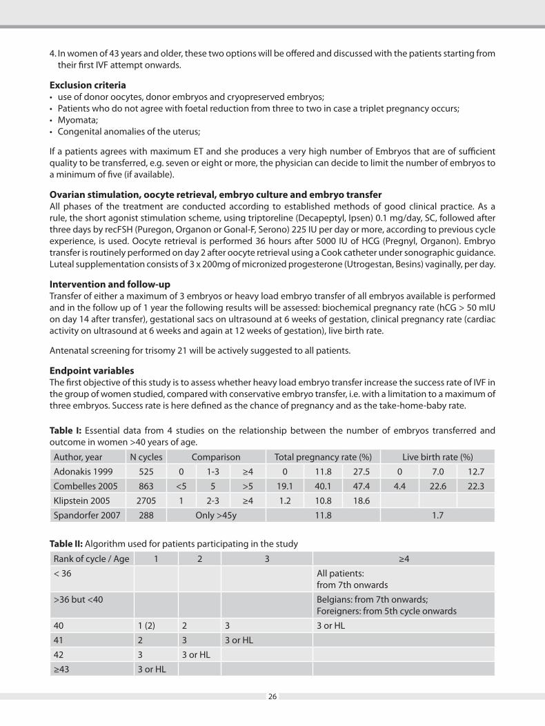
An early retrospective Belgian study compared the pregnancy rates in a total of 525 patients aged 40 years and older: in 112 cycles, no embryo for transfer was available; in 271 cycles, 1-3 embryos (mean 2.1±1) were transferred and in 142 cycles, 4 or more embryos (mean 4.7±1) were transferred. The percentage of total pregnancies rose from 11.8% to 27.5% (p<0.0001) and the clinical pregnancy rate increased from 10% to 20.4% (p<0.005) between the last two groups (Adonakis et al., 1997). The delivery rate showed no statistically significant difference (7% vs. 12.7%; NS). Similarly, the difference in twin pregnancy rate (11.1% vs. 17.2%) between both groups was not statistically significant as was the incidence of spontaneous abortion (25.9% vs. 34.5%).
A Bostonian retrospective study of 2.705 ART cycles in women of ≥40 years of age showed that the pregnancy
24
rate and live birth rate increased with increasing number of embryos transferred (Klipstein et al., 2005). The overall live birth rate per cycle start was 9.7%, with a cumulative live birth rate of 28.4% at age 40 and 0 by age 46. When studying the higher order multiple pregnancy rates, only 0.44% per cycle started resulted in triplets or quadruplets. However, in the women of 40 and 41 years old, the incidence of higher order multiple pregnancy rates was found to be 8% (n=12) after transferring 3 or more embryos. Of these, 6 patients had embryos cryopreserved. No higher order multiple pregnancy rates were found in women aged 42 and older. Interestingly, the outcome of the first attempt did not predict the outcome of subsequent cycles.
Another Bostonian retrospective study analysed 863 ART cycles in women >40 years of age to determine the optimum number of embryos to be transferred in this group (Combelles et al., 2005). They compared the transfer of <5, 5 and >5 embryos and found that on transferring five or more embryos the total pregnancy rate (19.1%, 40.1% and 47.4%) and live birth rate (4.3%, 22.6% and 22.3%) significantly increased. There was no difference in live birth rate when comparing the groups of patients who received 5 embryos versus more than 5 embryos. Miscarriage rates were 42.7%, 14% and 25.6% for the <5, 5 and >5 embryos groups, respectively. Total early pregnancy loss was a staggering 80% for the <5 embryos group, 47.4% for the 5 embryos group and 59.6% for the >5 embryos group. The authors concluded that the optimum number of embryos to transfer in women above 40 years is 5. Twin pregnancies occurred in almost 40% of the patients who received 5 embryos.
Recently, a fourth retrospective study was conducted in 288 consecutive cycles in women of 45 years of age and older (Spandorfer et al., 2007). The results of this study are that no pregnancy was obtained in women aged 46 years and older regardless of how many embryos were transferred. Out of a total of 288 women of 45 years of age, who asked for IVF, 11.8% obtained a positive pregnancy test and 1.7% (five individuals) delivered a live baby. The mean number of embryos transferred was 2.9±1.8. Pregnancies occurred only in the group of women with an ovarian response of >5 oocytes.
The main data from these four papers are shown in Table I. In conclusion, it appears that the idea of obtaining a higher pregnancy rate in the older group of women by augmenting the number of embryos transferred has shown that a correlation exists between a high number of embryos transferred (e.g. 5 or more) and outcome. Data indicating a positive effect of transferring ≥5 embryos, seem to suggest that this is the case in the <45 years of age group only.
Aim of the present studyWe want to prospectively compare the outcome of a transfer of a maximum of three embryos versus a transfer of the maximum available number of embryos in women of poor prognosis.
We must first formulate two definitions: what is a poor prognosis patient in the context of this study and what is a very high number of embryos?
Definition of the poor prognosis patientThe most important a priori prognostic factor for IVF/ICSI patients is the age of the female partner and, to a lesser degree, the non-tubal character of the infertility. Another important determinant which cannot be known a priori is the rank of the treatment attempt. Because live birth is the true endpoint of infertility treatment, we will consider for this study an attempt resulting in a non-ongoing pregnancy as a failed attempt. Hence, when counting the rank of attempt, we take into account all cycles not resulting in the birth of a child.
Some women have a poor prognosis from their first attempt onwards. A 44 year old woman has a very feeble chance to conceive from the very beginning; in contrast a 36 year old woman has an excellent a priori prognosis, but if she fails to conceive after 6 transfers of good embryos, her prognosis becomes much reduced.
There are not enough data on heavy load transfer to support strict criteria of age or of rank of attempt to categorize a patient as poor prognosis (i.e. a candidate for heavy load transfer). The clinical reality consists of a gliding scale of relative indications where age of the female and rank of the attempts play the most important role, but other factors are equally important or compelling.
Definition of a very high number of embryos (HLT)Similarly, the definition of a heavy load transfer is not unequivocal.
25
HLT could be anything from 3 embryos up to all embryos available. To make the selection bias as low as possible, we constructed the algorithm in Table II, which intends to guarantee always the same approach in the different groups of age.
For a 35 year old woman in a fifth attempt, three embryos is not even permitted by the Belgian law; in a 43 year old woman, three embryos in a first attempt is permitted. Hence, a first restriction to the definition of HLT is what the law permits. Limitations are especially clear in the <36 years of age group (for all clarity, this is the group up to 35 years and 364v days), less so in the >36<40 years of age group and not present at all in the >40 years of age group. It is not clear whether the law explicitly forbid the transfer of more than two embryos in the group <36 years of age from the 7th cycle onwards. Yes, according to a very legalistic interpretation of the law; no, according to a more lenient interpretation.
According to Belgian legislation, there is no restriction in the number of embryos to be transferred in women of 40 years and older. In general practice, sometimes we already transfer 3 embryos in this group of patients.
Definition of study groups Whatever the definition used, the aim is to create two groups that are sufficiently different to allow for the possibility of a significant difference, hence for a clinically useful conclusion.
We shall compare two strategies, both coined heavy load transfer. One is to transfer the three best embryos (if three are available) (group A), the other is to transfer an unlimited number of embryos with a minimum of five – based on the literature (Combelles et al., 2005; Spandorfer et al., 2007) - that are of sufficient quality to be either transferred or frozen (group B). Some patients in group A will not have three embryos whereas some patients in group B may have six or more embryos, and some perhaps just one or three. But it can be anticipated that the average number of embryos transferred in group B will be larger than in group A. Patients in group B are allowed to have more than 5 embryos, up to all embryos available, if they wish so, but the number cannot be intended to be less than 5 (if available).
Design of the studyThe ideal methodology for a clinical study is a prospective randomized design. However, randomization between a maximum of three and an undefined maximum could be very difficult to achieve. Some patients, even if their prognosis is very poor indeed, do not want more than three embryos, whereas others will agree to have more. In any case, patients always have to agree in writing with selective foetal reduction in case a triplet pregnancy arises. If they do not agree, no more than two embryos are transferred and they can not participate in the study. Because randomization would create ethical dilemma’s that can not be overcome, we decided on a prospective comparison by patient’s (couple’s) choice and physician’s agreement.
The following inclusion- and exclusion criteria will be used.
Inclusion criteriaOnly women in whom the law does not limit the number of embryos to transfer can be included in the study. Although literature data are available only for women of >40 years of age, we suggest to consider heavy load in some categories of women of the <40 years of age group as well. We suggest to include the following categories of patients:
1. For women <36 years of age, the strictly legal interpretation is adhered to, i.e. up to sixth attempt, no more than two embryos can be transferred. From the 7th cycle onwards, the law can be interpreted as not explicitly forbidding the transfer of more than two embryos. Hence, all patients of <36 years of age can participate to the study;
2. In women between 36-39 years of age, participation to the study will be suggested to patients from their seventh attempt onwards. For foreign patients, in a fourth cycle, three embryos can be transferred and participation to the study is possible from the 5th cycles onwards;
3. All women of 40-42 years old (i.e. up to 42 years and 364 days on the day of transfer) enrolled in our IVF/ICSI program, who are to receive their second, third, fourth (or subsequent n-th higher order) attempt or their very last IVF cycle (Table II). The patients will be explained the two possibilities of treatment. The first option is to transfer a maximum of 3 embryos, regardless of how many embryos are available (group A). The second option is to transfer all the embryos available with no maximum (group B) but with a minimum of 5.
26
4. In women of 43 years and older, these two options will be offered and discussed with the patients starting from their first IVF attempt onwards.
Exclusion criteria• useofdonoroocytes,donorembryosandcryopreservedembryos;• Patientswhodonotagreewithfoetalreductionfromthreetotwoincaseatripletpregnancyoccurs;• Myomata;• Congenitalanomaliesoftheuterus;
If a patients agrees with maximum ET and she produces a very high number of Embryos that are of sufficient quality to be transferred, e.g. seven or eight or more, the physician can decide to limit the number of embryos to a minimum of five (if available).
Ovarian stimulation, oocyte retrieval, embryo culture and embryo transferAll phases of the treatment are conducted according to established methods of good clinical practice. As a rule, the short agonist stimulation scheme, using triptoreline (Decapeptyl, Ipsen) 0.1 mg/day, SC, followed after three days by recFSH (Puregon, Organon or Gonal-F, Serono) 225 IU per day or more, according to previous cycle experience, is used. Oocyte retrieval is performed 36 hours after 5000 IU of HCG (Pregnyl, Organon). Embryo transfer is routinely performed on day 2 after oocyte retrieval using a Cook catheter under sonographic guidance. Luteal supplementation consists of 3 x 200mg of micronized progesterone (Utrogestan, Besins) vaginally, per day.
Intervention and follow-upTransfer of either a maximum of 3 embryos or heavy load embryo transfer of all embryos available is performed and in the follow up of 1 year the following results will be assessed: biochemical pregnancy rate (hCG > 50 mIU on day 14 after transfer), gestational sacs on ultrasound at 6 weeks of gestation, clinical pregnancy rate (cardiac activity on ultrasound at 6 weeks and again at 12 weeks of gestation), live birth rate.
Antenatal screening for trisomy 21 will be actively suggested to all patients.
Endpoint variablesThe first objective of this study is to assess whether heavy load embryo transfer increase the success rate of IVF in the group of women studied, compared with conservative embryo transfer, i.e. with a limitation to a maximum of three embryos. Success rate is here defined as the chance of pregnancy and as the take-home-baby rate.
Table I: Essential data from 4 studies on the relationship between the number of embryos transferred and outcome in women >40 years of age.
Author, year N cycles Comparison Total pregnancy rate (%) Live birth rate (%)
Adonakis 1999 525 0 1-3 ≥4 0 11.8 27.5 0 7.0 12.7
Combelles 2005 863 <5 5 >5 19.1 40.1 47.4 4.4 22.6 22.3
Klipstein 2005 2705 1 2-3 ≥4 1.2 10.8 18.6
Spandorfer 2007 288 Only >45y 11.8 1.7
Table II: Algorithm used for patients participating in the study
Rank of cycle / Age 1 2 3 ≥4
< 36 All patients: from 7th onwards
>36 but <40 Belgians: from 7th onwards;Foreigners: from 5th cycle onwards
40 1 (2) 2 3 3 or HL
41 2 3 3 or HL
42 3 3 or HL
≥43 3 or HL
27
Secondarily, we shall compare the incidence of multiple pregnancies (and consequently the poor obstetrical outcome) between both groups.
Primary outcome variables• %positiveHCG• %clinicalpregnancies(>12weekswithcardiacactivity)• %livebornchildren Secondary outcome variables• %totaltwins(firstsonogram)• %ongoingtwins• %liveborntwins• %vanishingtwins• %OHSS(withhospitalisation)
Power calculation and number of patients to treatOpen study, first evaluation after one year of intake.
Ethical committeeBecause no other interventions are performed than those belonging to standard IVF treatment, the agreement of the EC will not be sought.
References1. Adonakis G, Camus M, Joris H, Vandervorst M, Van Steirteghem A and Devroey P (1997) The role of the
number of replaced embryos on intracytoplasmic sperm injection outcome in women over the age of 40. Hum Reprod 12, 2542-2545.
2. ASRM Practice Committee (2004) Guidelines on the number of embryos transferred. Fertil Steril; 82 (suppl): S1-S2.
3. BELRAP (2007) Report of the College of Physicians for Artificial Reproductive Therapy. Annual Report 2004. www.belrap.be.
4. Combelles CMH, Orasanu B, Ginsburg E and Racowsky C (2005) Optimum number of embryos to transfer in women more than 40 years of age undergoing treatment with assisted reproductive technologies. Fertil Steril 84; 1637-1642.
5. Klipstein S, Regan M, Ryley DA, Goldman MB, Alper MM and Reindollar RH (2005) One last chance for pregnancy: a review of 2,705 in vitro fertilization cycles initiated in women age 40 years and above. Fertil Steril 84; 435-445.
6. Munné S, Cohen J and Sable D (2002) Preimplantation genetic diagnosis for advanced maternal age and other indications. Fertil Steril 78; 234-236.
7. Munné S, Fischer J, Warner A, Chen S, Zouves C, Cohen J and Referring Centers PGD Group (2006) Preimplantation genetic diagnosis significantly reduces pregnancy loss in infertile couples: a multiccenter study. Fertil Steril 85; 326-332.
8. Spandorfer SD, Bendikson K, Dragisic K, Schattman G, Davis OK and Rosenwaks Z (2007) Outcome of in vitro fertilization in women 45 years and older who use autologous oocytes. Fertil Steril 87; 74-76.
9. Van Blerkom J, Davis P, Alexander S. Differential mitochondrial distribution in human pronuclear embryos leads to disproportionate inheritance between blastomeres: relationship to microtubular organization, ATP content and competence. Hum Reprod 2000;15: 2621-2633.
10. Verlinsky Y, Cohen J, Munné S, Gianaroli L, Simpson JL, Ferraretti AP and Kuliev A (2004) Over a decade of experience with preimplantation diagnosis: A multicentre report. Fertil Steril; 82: 292-294.
28
Recurrent Implantation FailurePetra De SutterUniversity Hospital Gent, Belgium
Recurrent implantation failure (RIF) is classically defined as the failure to achieve a pregnancy following 2-6 IVF cycles in which more than 10 high-grade embryos were transferred to the uterus. Some authors prefer to redefine RIF as the failure of 3 cycles in which reasonably good embryos were transferred. It is clear that the age of the woman is an important variable and that RIF should be defined as the situation wherein the odds of underlying pathology is larger than the role of hazard in explaining IVF failure. The younger the woman, the earlier RIF should be suspected, the older the woman, the less likely underlying pathology is at play. In literature, assumed etiologies for RIF are decreased endometrial receptivity, embryonic defects (caused by sperm or oocyte defects) and multifactorial problems. Uterine cavity abnormalities can be ruled out in RIF patients by careful ultrasound imaging hysteroscopy or MRI. The impact of intramural lesions without cavity distortion or fibroids less than 4 cm on RIF remains controversial. Hysteroscopic correction of cavity pathology is recommended in RIF patients, especially for polyps, submucous fibroids or Asherman syndrome. Thin endometrium may be a cause for RIF, although this remains controversial and is difficult to treat. Some studies have shown that in RIF patients adhesive molecules or cytokines are expressed in an aberrant way, but the significance of this finding is unclear. Regarding immunological pathology or genetic or acquired thrombophilia, controversy is even bigger. Some studies have suggested association of antiphospholipid or other autoantibodies or disturbances in NK cells and RIF, and it has been shown that the prevalence of multiple thrombophilic gene mutations among RIF patients was higher than among fertile patients. Among thrombophilia, Factor V Leiden, prothrombin mutation 20210G>A and MTHFR 677C>T mutations have been pointed to as possible causes for RIF. In a retrospective analysis in 266 couples, our group has found that Factor V Leiden mutations were less frequent, Factor II significantly more frequent and MTHFR 677C>T mutations not different in RIF patients compared to control populations. Empirically, F V and F II mutated patients are usually treated with aspirin or heparin and MTHFR-homozygotics with aspirine 80mg/d + folic acid 4mg/d, but this is not evidence-based. Subclinical thyroid dysfunction should be corrected. Genetic abnormalities in embryos are well known to be a reason for implantation failure and the incidence of chromosome translocations is increased in RIF patients. Karyotyping of RIF patients may be advised, although the evidence of the success of subsequent preimplantation genetic diagnosis may be weak. Many of these couples can be helped effectively by gamete donation. Severe endometriosis, hydrosalpinges or suboptimal ovarian stimulation have also been considered to be responsible for RIF and should be taken care of. In some cases of unexplained RIF, endometrial stimulation or injury may cause a pseudodecidual reaction that enhances implantation. Some authors have observed increased pregnancy rates following a (even normal) hysteroscopy or obtained good results with repeated endometrial biopsies. In RIF patients, assisted hatching seems to increase the pregnancy rates, as shown in a recent Cochrane update. Preimplantation genetic screening, however, does not seem to be efficient, at least using the current genetic diagnostic techniques. In some cases, good results have been obtained using zygote intra-Fallopian transfer, embryo co-culture, blastocyst transfer or cytoplasmic transfer, but many of these procedures are unproved. Of great importance is the embryo transfer technique and special attention should be given to this part of the treatment in RIF patients. Finally, psychotherapy reduces anxiety and depression and possibly enhances conception success, but proof of efficacy is lacking. In conclusion, RIF is a clinically challenging problem and there is very little evidence about the importance of and possible treatment of certain conditions deemed to be responsible for RIF. Counselling these patients is of utmost importance, as well as timely discussing limits and alternatives.
29
Fertilisation Failure and Artificial Oocyte Activation Petra De SutterUniversity Hospital Gent, Belgium
Oocyte activation by sperm consists of a trigger followed by calcium oscillations. The trigger is a first Ca2+ rise followed by a series of secondary Ca2+ rises. This trigger is normally caused by sperm binding on the oocyte membrane, and starts from the site of sperm penetration. The oscillator results from the release of a sperm factor, called phospholipase C ζ (PLCζ), which is a not species-specific protein of 70kDa. In clinical ICSI, activation failure may result from the fact that the sperm cannot initiate activation, or from the fact that the oocyte cannot respond properly to the PLCζ -induced activation mechanism, or finally from technical failure (not enough permeabilization of the sperm membrane during ICSI whereby PLCζ is not released). In a study by Yoon et al. (2008) it has been shown that in human sperm showing failed fertilisation after ICSI, no or an abnormal Ca pattern is observed. Some of these patients show an absence of PLCζ, and our group (Heytens et al. (2009)) has confirmed these results but also demonstrated a possible mutation in PLCζ in one patient suffering from fertilisation failure, having normal sperm morphology but no activation potential. About the importance of the oocyte in activation failure, not much is known! There are animal studies investigating the role of kinases (eg. MAPK, MPF), phosphorylation status, ATP and IP3R in the oocyte activation mechanism, but almost no studies in human oocytes (due to lack of availability). From a clinical perspective, the important question is how to treat activation failure. There are different artificial activation agents that have been used in mouse and sometimes also in human, such as electrical pulses, ethanol, Ca-ionophore (A23187, ionomycin), inhibitors of protein synthesis (puromycin, DMAP), - all leading to one prolonged Calcium peak - , or thimerosal, strontium ions, electrical pulses, or recombinant PLCζ, leading to Calcium oscillations. In our centre, Ca-ionophore has been used successfully in the human. In case of patients with failed or low fertilisation after ICSI, we usually first propose a MOAT (mouse oocyte activation test) in order to distinguish between sperm and oocyte borne activation deficiency. In this test patient sperm is injected in mouse oocytes and activation rates are measured. If the mouse oocytes do not activate and these seems to be a sperm factor involved, we perform genetic screening of PLCζ and treat by assisted oocyte activation by Ca-ionophore. Possibly in the future, PLCζ cRNA may offer an alternative and would be a more physiological stimulus than ionophore. In case the MOAT is normal, we suspect an oocyte factor and although we also have obtained pregnancies in these cases, the pregnancy rates are lower in this group. As of today, we have obtained around 25 healthy children after artificial oocyte activation, and there seem to be no risks attached to the procedure. However, we caution that this technique remains highly experimental, should only be performed in strict indications and that the children should be carefully followed-up.
30
Avoiding Mix -Ups in IVF LabAmrit K SwainEmbryologist, IVF Unit, Apollo Hospital, Delhi
Several incidents were reported recently in media on mishaps in IVF programs. These included instances when eggs were inseminated with the wrong sperm, embryos belonging to one couple were transferred into another woman and frozen embryos were improperly labeled and/or could not be accounted for. Such incidents are devastating not only for those directly involved, but are also a source of much anxiety for every IVF patient and fertility clinics.
The Human Fertilisation and Embryology Authority (HFEA) already requires a system of “double witnessing” in which staff check and document each other’s work to reduce the risk of mistakes. Some clinics already use a system of technological checks, involving bar codes, electronic sensors and alarms. The tagging system works by sounding an alarm if samples are put in the wrong place, for example if egg and sperm samples from different couples are put too close together.
RI Witness™ uses Radio Frequency Identification (RFID) technology to monitor all critical work carried out in the laboratory, creating a complete record of each stage of a patient’s cycle. RI Witness™ is watching, tracking and recording all of the time and locks the patient’s identity to the sperm, eggs and embryos at every stage of the treatment. The system also actively prevents patient samples coming into proximity with those of other patients.
There is scope of mix up from the egg retrieval, semen collection, cumulus oocyte complex handling, denudation,spem processing, invitro insemination, embryo culture, embryo transfer, sperm freezing, embryo freezing etc. The advanced techniques like bar coding and electronic sensors assist to avoid mix up in ivf lab. Every laboratory has their standard practice to deal the mix up issues.
The consequences of mix up is devastating for the patients, IVF Lab, the hospital but the good way to deal with is that it is a human error and can be controlled in IVF labs. Assembling the right team is the most important and fundamental part of running a good laboratory. There should be healthy communication between all those involved in executing the procedures.
The current practice followed in different laboratories and further steps to strengthen the areas where near misses can happen will be elaborated in this lecture.
31
Treatment of Infertility is Less More?Siladitya BhattacharyaUniversity of Aberdeen
The treatment of infertility has traditionally been guided by the principles of etiological management where a cause is identified prior to treatment. This approach can be problematic in unexplained infertility where tests of ovulation, semen analyses and tubal evaluation fail to reveal any obvious cause for lack of conception and the evidence base underpinning the effectiveness and cost effectiveness of common treatments is limited. The likelihood of pregnancy is influenced by duration of infertility as well as age and parity of the female partner and there is a role of expectant managent in couples with a good prognosis. Clomifene citrate and intra uterine insemination (IUI) in a natural cycle are unlikely to offer any significant benefits over expectant management but are more expensive. Stimulated IUI is more effective than unstimulated IUI but is associated with a risk of multiple pregnancy. IVF is now accepted as an effective treatment for couples with unresolved prolonged infertility. Data from randomised trials suggest that it is more effective than stimulated IUI in unexplained infertility, and as effective as intra-cytoplasmic sperm injection (ICSI) in non-male factor infertility.
Where IVF is appropriate it is important to recognise the potential danger of iatrogenic multiple pregnancies and attempt to minimise the risk of multiples by means of elective single embryo transfer. Given the paucity of longterm outcome data on children conceived through assisted reproduction it may be prudent to limit it use to cases whether it is indicated and to avoid the routine use of additional invasive and expensive techniques.
32
Recent Advances in Ovulation Controlled Ovarian Stimulation in PCOSShweta Mittal GuptaConsultant, Centre of IVF, Sir Ganga Ram Hospital, New Delhi
Subfertility due to anovulation (PCOS) is a common problem in women. First-line oral treatment for ovulation induction still remains anti-oestrogens like clomiphne citrate. Alternative and adjunctive treatments have been used such as tamoxifen, dexamethasone, and bromocriptine.
As a result of the disappointing results of clomiphene citrate treatment, several authors have advocated aromatase inhibitors as new ovulation-inducing agents. The most widely used aromatase inhibitor is letrozole. Compared to clomiphene citrate, its use is associated with thicker endometrium and a trend towards higher pregnancy rates. Letrozole works by inhibiting aromatization, leading to a decrease in estrogen production. As a result, FSH secretion increases, stimulating the development of ovarian follicles. In addition, androgens that are normally converted to estrogens accumulate in the ovary and these androgens increase follicular sensitivity to FSH. As the ovaries from women with PCOS produce high amounts of estrogen, the effects of aromatase inhibitors in these women are more pronounced .The optimal dose advocates is 5mg daily for 5 days. When letrozole is combined with gonadotropin, it leads to lower gonadotropin requirements with pregnancy rates comparable to gonadotropin treatment alone. Another promising aromatase inhibitor is anastrazole.
As first-step therapy for anovulation, no consistent evi¬dence suggests that metformin is better than clomiphene citrate regarding cumulative ovulation, pregnancy or live-birth rates. Metformin (2.0–2.5 g per day) is recently advocated for women with PCOS showing altered OGTT. An oral glucose tolerance test should be performed in all patients with PCOS initially and every 2 years thereafter
Dexamethazone has also been used in conjunction with clomiphene citrate. It improves ovarian function directly by suppressing the action of estradiol on the pituitary gland or indirectly by increasing serum growth hormone and serum insulin-like growth factor. Elnashar et al evaluated the use of dexamethazone among 80 women with clomiphene citrate-resistant. A combination of clomiphene citrate 100mg daily from day 3 to 7 and dexamethazone 2mg daily from day 3 to 12 were administered. The combined group had a significantly higher number of mature follicles than the clomiphene citrate group (1.25_0.67 vs. 0.15_0.04) as well as thicker endometrium (8.8 vs. 7.0mm). Compared to the clomiphene citrate group, the clomiphene citrate/dexamethazone group had higher ovulation rates (15 vs.75%; P<0.001) and pregnancy rates (5 vs. 40%; P<0.05). However evidence in favor of these interventions is flawed due to the lack of evidence on live births.
Gonadotropins are more effective for ovulation induction and are associated with higher pregnancy rates than clomiphene citrate, but they are expensive and carry a higher risk of ovarian hyperstimulation syndrome and multiple gestations. Recent evidence suggests that luteinizing hormone activity in human menopausal gonadotropin modifies follicular development so that fewer intermediate-sized follicles develop. Compared to the use of follicular stimulating hormone only, human menopausal gonadotropin is associated with less ovarian hyperstimulation.
A rsearch was conducted using the Cochrane Menstrual Disorders and Subfertility Group Trials Register (May 2009), CENTRAL (The Cochrane Library 2009, Issue 2), MEDLINE (1966 to May 2009), and EMBASE (1980 to May 2009) for identification of relevant randomized controlled trials (RCTs). The United Kingdom National Institute for Clinical Excellence (NICE) guidelines and the references of relevant reviews and RCTs were searched.
Clomiphene was effective in increasing pregnancy rate compared to placebo (OR 5.8, 95% CI 1.6 to 21.5) as was clomiphene plus dexamethasone treatment (OR 9.46, 95% CI 5.1 to 17.7) compared to clomiphene alone. No evidence of a difference in effect was found between clomiphene versus tamoxifen or clomiphene in conjunction with human chorionic gonadotrophin (hCG) versus clomiphene alone. A significant improvement in the pregnancy rate was reported for clomiphene plus combined oral contraceptives versus clomiphene alone. No evidence of a difference in effect on pregnancy rate was found with any of the other comparisons.

33
Stress and infertility: Effect and ManagementRajvi H MehtaScience Director, Trivector Embryology Support Academy
Stress is now considered as a universal culprit for all kinds of physiological, mental and emotional disorders. Several studies in the past few decades have clearly implicated that stress influences reproductive function in both men and women. Stress is defined as the body’s reaction to a change that requires a physical, mental or emotional response or adjustment. Change is possibly the only constant in life! And, most individuals have an intrinsic ability to cope with the constant physical, mental or emotional changes that one is constantly exposed to. Whenever one is no longer able to cope with these changes then it leads to strain which is commonly perceived as ‘stress’.
The cause for such strain could be a variety of factors such as financial, social, work related or relationships. This perceived stress expresses detrimental effects on the physiological systems including the reproductive system. The psycho affects the neuro which in turn acts of the endocrine system… a system which governs reproduction. Stress can lead to hormonal abnormalities reflecting in compromised gametes or endometrium and subsequently infertility. It is not essential that stress can be the sole cause of infertility but could be an ‘add-on’ cause.
Infertility in turn causes its own stress making it into a vicious circle. The assisted reproduction technologies (ART) with the unpredictability of each state of treatment adds on to the stress. The management of stress is nothing but altering the perception of stress and utilizing a variety of coping mechanisms to break away from the vicious cycle of stress- infertility – Stress – ART – stress.
The presentation would discuss the details on the impact of stress and the variety of coping mechanism available.

34
Quality Management in IVF LaboratoryRajvi H MehtaScience Director, Trivector Embryology Support Academy
Despite all the technological advances and a better understanding of early human development – the success of Assisted Reproduction Technology (ART) in terms of take-home baby rate remains around 30-40% only. There are number variables in the technology of ART which either keep the success rates inconsistent or low. One of these variables is the quality of the laboratory.
Managing the quality of the laboratory includes maintenance and calibration of the equipment, optimizing the environment in the laboratory as well within the incubators; assessing the microbial sterility and keeping a check of processes with the Standard Operating Protocols (SOPs).
This presentation will detail the different aspects of quality maintenance of the ART laboratory.

35
Free CommunicationsORAL / VIDEO
PRESENTATIONS
40
[03]
To Evaluate the Effect of N-Acetyl Cysteine (NAC) Therapy on Insulin Resistance, Clinical, Hormonal or Metabolic Parameters in Infertile Patients of PCOS
Pikee SaxenaLady Hardinge Medical College and Smt. SSKH, New Delhi-110 001
Objective: To evaluate the efficacy of N Acetyl Cysteine (NAC) therapy on insulin resistance (IR), clinical, hormonal and metabolic profile of infertile women with polycystic ovary syndrome. Methods: This prospective observational study was carried out in a tertiary care infertility clinic. Twenty women with PCOS were enrolled to receive NAC 600 mg 8 hourly for a period of six months. IR along with clinical, hormonal and metabolic parameters was compared before and after six months of NAC therapy. Results: NAC therapy improved the regularity of periods (p< 0.001), rate of ovulation and resulted in reduction of total testosterone (p< 0.001), IR (p= 0.001) and cholesterol (p < 0.05). No significant change was observed in the levels of FSH, LH, Prolactin and TSH. Conclusions: NAC is an effective insulin sensitizer and also reduces testosterone and cholesterol levels in women with PCOS. It is safe and is well tolerated.
[04]
Clinico-biochemical and Ultrasonologic Characteristics of Young Women with Polycystic Ovary Syndrome-An observational study
SS Trivedi, Sharda PatraDeptt. of Obst & Gynae, Lady Hardinge Medical College & Smt. S.K. Hospital, New Delhi, India
The present observational study was proposed to screen girls to study the prevalence, the clinico-biochemical and sonologic characteristics of those with PCOS. Materials and Method: 230 girls (18-23yrs of age) were screened and those with abnormal menstruation suggestive of ovulation disorder underwent clinical examination including BMI and waist to hip ratio, biochemical evaluation consisting of serum Free Testosterone, LH / FSH ratio, DHEAS Fasting glucose, Serum insulin, Triglycerides & HDL-cholesterol and pelvic ultrasound for ovarian assessment was done. Out of 30 girls with menstrual disorder, 18 had PCOS (18/230). Results: The prevalence of PCOS among these girls was 7.8%. 7/18 (38.8%) had oligomenorrhea, clinical /biochemical evidence of hyperandrogenism and polycystic ovaries, 8 (44.4%) had polycystic ovaries with oligo/amenorrhea and 3 (16.6%) had oligomenorrhea and hyperandrogenism. The BMI and Waist to Hip ratio revealed, 16 (87%) were lean (BMI<30) compared to only 2 girls (13%) who were obese (BMI>30) (p=0.001). The Waist to Hip ratio was <0.85 in 83.3% girls and >0.85 in 16.7%. Hirsutism and acne was present in 13% and 22% of girls. 5 out of 18 (28%) had fasting blood sugar (FBS) >100mg/dl and fasting Insulin >25 mu/l though majority were euglysemic. Abnormal serum lipids were present in 2 girls (8%) suggestive of metabolic syndrome. On comparing the factors in the lean and obese girls polycystic ovaries and high testosterone levels were comparatively more in the lean group (p=0.001). Conclusion: This study demonstrated that young Indian women with PCOS share many of the same clinical and metabolic abnormalities as the adult population Increased awareness and the need for assessment of co morbidities, such as dyslipidemia and impaired glucose tolerance by a simple screening approach can allow for earlier detection and management especially by early life style modification and treatment if required.
[05]
Clinical Spectrum of PCOS with Special Reference to Biochemical Parametars and Serum Insulin Levels
Aruna Verma1, Abhilasha Gupta2, Vidushi3
1Assistant Prof, 2Prof & Hod, 3Resident, Llrm Medical College Meerut, Up
Introduction: Anovulation is the hallmark of PCOS & is the cause of all endocrinolagic abnormalities & Polycystic ovaries. Various biochemical parameters (LH, FSH, PRL, TSH, F.Insulin, IR, GTT) are disturbed with PCOS. There is
41
no specific pathophsiologic defect in PCOS. Aims & Objective: To study the clinical spectrum & biochemical parameters of PCOS especially serum insulin levels & insulin resistance. Material & Methods: This prospective observational study was carried out on 36 diagnosed PCOS patiets attending the gynae-OPD of SVBP hospital Meerut. Detailed history taking & examination followed by investigations like TVS, LH, FSH, PRL, TSH, fasting & 2hr PP sugar levels, F.insulin levels were done. Results and Conclusions: Hyperinsulinemia, Hyperandrogeinsn and IR are seen in both obese & nonobese POCS subjects. 42.86 % of Hyperinsulinemic PCOS subjects had clinical hyperandrogeinsm while none of normoinsulinimic subjects had such signs. Thus it is concluded that Hyperinsulinemia is a necessary factor for the development of hyperandrogeinsn. Key Words: PCOS, Hyperinsulinemia, Hyperandrogenism, Insulin-resistance.
[06]
Role of Hysteroscopy in ARTAnju VermaDr Verma Fertility & Test Tube Baby Centre, Gwalior
The place of routine hysteroscopy in management of infertile women without prior diagnosis or doubtful intrauterine pathologies is still a matter of debate as it is an invasive procedure. Despite advances in field of Assisted Reproductive Techniques, Implantation Rate per embryo transfer still remains low. Two key factors responsible for success of procedure are quality of embryos and receptivity of endometrium. Structural abnormalities of uterine endometrial cavity can adversely affect the reproductive outcome by interfering with implantation and causing spontaneous abortions. Aim of Study: To evaluate the role of routine Hysteroscopy prior to IVF and to find out the causes of implantation failure in uterine cavity. Material & Method: It is a retrospective study of 200 infertile women, who attended our IVF centre in Gwalior from April 2008 to April 2011 and registered for In-Vitro Fertilization procedure. Result: Out of 200 women, 120 women were those, who underwent first IVF cycle and remaining 80 women were entered in repeat IVF cycle. Patients were distributed in groups depending upon the age of female partner, duration of infertility, cause of infertility and number of previous IVF attempts. Out of 200 women undergone hysteroscopy procedures 130 had normal findings and 70 patients had intrauterine pathologies of various types like polyp, adhesions, septa, cervical stenosis, T- shaped uterus etc. Conclusion: Hysteroscopy is a good diagnostic and therapeutic tool in patients undergoing IVF for treatment of infertility. Intrauterine pathologies and structural abnormalities that may be responsible for IVF failure can be detected and treated to improve pregnancy rates. This would also save the patients additional cost of IVF cycles, where cause of the failures is because of intrauterine pathologies.
Session F2: Surgical Procedures (Oral + Videos)Hall - B (Silver Oaks Hall) • Time: 13:00 - 13:45 hr
[07]
Operative Laparoscopy – A boon for enhancing fertilityMeenakshi B Chauhan, Smiti Nanda, Vani Malhotra, Anju BalaDepartment of Obstetrics & Gynacology, Pt B D Sharma PGIMS, Rohtak
Objective: To evaluate the laproscopic findings and intervention done in infertile women and its role in enhancing fertility. Methods: A retrospective study was carried out on the women who had undergone operative laparoscopy for infertility over a three year period in the department of OBGYN at a tertiary care centre in Haryana. The demographic profile of women, type of infertility, findings at laparoscopy and operative procedures performed during laparoscopy were analyzed. The pregnancy rates, number of abortions, ectopic pregnancies and number of live births were studied. Results: The data was analyzed and will be presented. Conclusion: Laparoscopy is an important tool in evaluation and treatment of fertility.
42
[08]
Hysteroscopic Management of Adenomyotic Myometrial Cysts Prior to IVF
Priya Bhave ChittawarAssociate Professor and Head, Reproductive Medicine Unit, Sri Aurobindo Institute of Medical Sciences, Indore
Objective: To describe a novel method of treatment of myometrial cysts close to endometrial cavity by hysteroscopy prior to undertaking ivf cycles. Methods: 50% of the women with adenomyosis have myometrial cysts on ultrasound1. Such cases have been managed by loop resection of the adenomyotic tissue within the cyst in patients not seeking fertility2. This treatment is not feasible for infertile patients with adenomyosis. Fertility outcome in patients with adenomyosis is compromised because of defective Endometrial Myometrial Interface (EMI) Zone contractility. Here we describe a novel minimally invasive techinique of dealing with myometrial cysts with hysteroscopic surgery to restore the anatomy and function of the EMI and optimize the outcome. Women presenting with infertility and planned for IVF who showed evidence of Adenomyosis on transvaginal sonography with myometrial cysts confluent to endometrial cavity were submitted to hysteroscopic drainage under anesthesia using Bettochi operative hysteroscope and scissors. Intraoperatively transabdominal ultrasound was used to localize the myometrial cysts and endomerium over it was excised to drain the collected altered blood. Three such cases will be described with their recorded surgical videos. There was a successful IVF outcome in two of the three cases where IVF was undertaken subsequently. Conclusion: Hysteroscopic drainage of the adenomyoic myometrial cysts is a novel, feasible and safe treatment option in women with adenomyosis presenting with myometrial cysts on transvaginal ultrasound and infertility.
1. Reinhold, C., Atri, M., Mehio, A., Zakarian, R., Aldis, A.E. and Bret, P.M. (1995) Diffuse uterine adenomyosis: morphologic criteria and diagnostic accuracy of endovaginal sonography. Radiology, 197, 609±614
2. Large intramyometrial cystic adenomyosis: a hysteroscopic approach with bipolar resectoscope: case report. Giana M, Montella F, Surico D, Vigone A, Bozzola C, Ruspa G. Eur J Gynaecol Oncol. 2005;26(4):462-3.
[09]
Endoscopic Myomectomy any Contra Indication!Punita BhardwajMax Hospital Saket, New Delhi
Location, size and numbers of myomas are no longer any contraindication to endoscopic removal of myomas. Cases with myomas intending child bearing opting for endoscopic myomectomies were taken into account. Diluted vasopressin was used primarily for hemostasis. Amount and dilution would vary depending on size, numbers and consistency of the fibroid. The incisions transverse, vertical, oblique or multiple would be decided by the location, number and size of the myoma. Suturing in single, multiple layers would be decided by the depth of the myometrial invasion by the fibroid. Results indicate that laparoscopic myomectomy is a useful technique with no contraindication in expert hands. Myomectomy indeed is a challenging procedure as it involves the reconstruction of an organ that can undergo remarkable structural changes, as it does in pregnancy. Risk of uterine rupture after laparoscopic myomectomy is low and should not be a contraindication for this operation if pregnancy is desired provided myomectomy is truly justified.
[10]
Video Presentations of Endoscopic Surgical Procedures to Enhance Fertility
Gupta Rajni, Gupta Neena
Introduction: Endoscopic surgery has developed quickly and is preferred over laparotomy for most surgeries on female reproductive tract. Though IVF has emerged as the main treatment of infertility for tubal diseases but surgery has a definite place with good results.

43
This video presentation includes:1. Video of hysteroscopic removal of submucus fibroid removal2. Video of hysteroscopic foreign body removal from uterine cavity3. Video of hysteroscopic bilateral tubal cannulation4. Video of laparoscopic ovarian cyst removal5. Video of laparoscopic adhesiolysis in endometriosis6. Video of laparoscopic ovarian drilling7. Video of laparoscopic myomectomy8. Video of laparoscopic salpingostomy in ectopic tubal pregnancy
[11]
Success Rate of Single Egg Donor in Multiple Recipient with Single Embryo Transfer
Shashi SinghDr. Singh Test Tube Baby Centre, Near Circuit House, Civil Lines, Meerut, UP
Objective: The study is done to evaluate success rate when oocyte are taken from single egg donor and used in many recipients and one embryo is transferred. Method: 52 egg recipients included in this study, age of oocyte recipient was between 30 to 45 years, and cause of infertility was menopause, premature ovarian failure etc. And 9 egg donors used. Antagonist protocol used for stimulation .Single embryo was transferred in each. Result: 5 pregnancy observed among 52 cases. Conclussion: When eggs of a single donor used and single embryo is transferred in many recipient low success is observed.
[12]
Degeneration of Oocytes After ICSI and its Association with Implantation Rate
Amrit K Swain, Sushma P Sinha, Sohani VermaIndraprastha Apollo Hospital, New Delhi
Objective: To determine the association of degeneration of oocytes in ICSI procedure with implantation rate. Method: The present study was carried out in 436 numbers of metaphase II oocytes from 56 human subjects with an age group between 23 to 42 years. These were undergone intra cytoplasmic sperm injection (ICSI). Following oocyte retrieval, it was further exposed to 80 IU/ml hyaluronidase for denudation. The spermatozoa were immobilized in Poly vinyl pyrolidone. The intracytoplasmic sperm injection carried out using eppendorf microinjection pipettes. The no of oocytes fertilized, abnormally fertilized, unfertilized and degenerated were noted after 14-18 hrs of ICSI. The fertilized oocytes further cultured and embryo transfer done on Day 2/3. The level of serum human chorionic Gonadotropin (HCG) was analyzed for detecting implantation. The subjects were grouped into Group I, as successful category with HCG value ≥ 400 and rest as Group II, unsuccessful category. The percentage of oocytes degenerated during ICSI procedure of both the groups were calculated. Further overall degeneration rate off all the oocytes were calculated using the ratio of the number of oocyte degenerated with the total number of metaphase II oocytes in this study. Result: The degeneration rate in case of Group I, successful category were observed as 18.1 % and the degeneration rate in case of Group II, unsuccessful category were observed as 16.7 %. The overall degeneration rate was found to be 16.9 % and the implantation rate was found to be 37 % as observed in the above subjects. This present study therefore concludes that the degeneration rate did not appear to be associated with the implantation rate.
Session F3: IVF / ICSIHall-C (Silver Oaks Hall) • Time: 13:00- 13:45 hr
44
[14]
Successful Conception in Patients Undergoing Blastocyst (Day 5) Versus Day 2-3 Embryo Transfer: A retrospective cohort study
Vandana Bhatia, Sonia MalikSouthend Fertility and IVF Centre, Vasant Vihar, New Delhi
Objective: Previous studies have shown that blastocyst transfer has a better implantation rate as it is thought to be more competent than the cleaving embryo’s. We aimed to study whether there is any difference in rates of pregnancy between a day 2-3 embryo transfer versus day 5 ( blastocyst) transfer in our patient cohort. Methods: This is a single centre retrospective cohort study. Patients undergoing IVF between January 2010 to July 2011 were analysed. A total of 100 patients was taken and two equal groups were identified based on either a day 2-3 embryo transfer (group 1) or a blastocyst (day 5) transfer (group 2). Data was collected for demographics, protocol used, total dose of gonadotrophins required, total number of stimulation days, oocyte number fertilised, number and grade of embroys/blastocyst formed and confirmed pregnancy. Primary outcome was defined as an ultrasound confirmed intrauterine pregnancy. In failed cycles an endometrial biopsy was done to assess the endometrium. Results: Among a total of 100 patients undergoing IVF, 50 patients underwent day 2-3 embryo transfer and another 50 day five blastocyst transfer. Age, dose of gonadotrophin required, days of stimulation, oocyte number fertilised were comparable between the two groups. There was no statistically significant difference among pregnancy rates between the two groups; 27 (54%) women in group 1 had a confirmed pregnancy versus 19 (38%) in group 2, p=0.15 (Fisher’s exact test). Conclusion: There is no significant difference in the two methods of IVF for rates of pregnancy achieved. Factors other than the timing of transfer may be responsible for differences observed in pregnancy rates in the previous studies.
[15]
A Study of Recipient Related Predictors of Success in Oocyte Donation Programme
Pooja Garg, Manish Banker, Pravin PatelPulse Women’s Hospital and Nova Pulse IVF Clinics, Ahmedabad
Introduction Oocyte donation is an invaluable therapy for couples with impending or complete ovarian failure. In addition, oocyte donation affords a scientific opportunity to study the unique biologic participation of the
[13]
Comparison of IVF/ICSI Outcomes in Patients Receiving Recombinant LH (r-LH) Versus Human Menopausal Gonadotropin (hMG) Supplementation
Usha D B, Sathya Balasubramanyam, Thangam VarmaFellow, Institute of Reproductive Medicine and Women’s Health, Madras Medical Mission, Chennai, Tamil Nadu, India
Objectives: To compare the outcome of recombinant LH (r-LH) and Human Menopausal Gonadotropin (hMG) supplementation in women undergoing IVF/ICSI with r-FSH in the GnRH-a protocol. Methods: It was a retrospective analysis of the case records of 90 consecutive women who underwent non donor IVF/ICSI cycle with GnRH agonist. All women received recombinant FSH on day 2/3 of programming cycle. When the level of LH was <0.5mIU/ml during any phase of stimulation, then addition of LH either as rec LH or HMG is given along with rec FSH. Results: The number of oocytes collected, the number of oocytes in metaphase II and fertilization rate were similar in both groups. In addition, the mean number of embryos produced per cycle and the mean number of frozen embryos per cycle were similar in both groups. The cost of gonadotrophin is similar in both groups. Ongoing pregnancy rate at week 10–12 was 20.4% after rFSH + HMG and 29.2% after rFSH + rLH. Conclusions: Supplementing rFSH with rLH when compared with HMG showed increased ongoing pregnancy rates in the general population.
45
uterus in the process of human embryo implantation. Aim: The current study aims to identify recipient variables and cycle factors that may have a significant impact on pregnancy outcome in order to optimize results of an oocyte donation program. Material & Methods: This is a prospective study of all recipients (n = 270) resulting in embryo transfer as a result of oocyte donation, occurring from 1st March 2010 through 31st march 2011. settings: Private tertiary care IVF Clinic. Results: The clinical pregnancy and embryo implantation rates of the oocyte recipient group were 95 of 270(35.18%) and 150 of 780 (19.23%), respectively, resulting in 48 (50.52%) singleton, 39 (41.05%) twin, and 8(8.42%) triplet gestations. Of the 95 pregnancies, 85 eventuated in ongoing pregnancies (31.48%). A significant decline in the ongoing pregnancy and implantation rate was noted in recipients above 40yr of age, (P = .001). Clinical pregnancy rate decreased and miscarriage rate increased above 40 but was not statistically significant. Endometrial Thickness and Indication for oocyte donation are not useful in predicting the pregnancy outcome. An increase in clinical, ongoing pregnancy and implantation rate with Day three embryo transfer was demonstrated. Previous myomectomy and endometriosis did not affect pregnancy outcome. Embryo quality and laboratory component was constantly same in all cycles. Conclusion: Uterine ageing may result in poor reproductive outcome in women with advanced age. Possible explanation may be reduced uterine perfusion or decreased estrogen/ progesterone receptors on endometrial cells and increase in collagen content, but none of these mechanisms has been proven. Minimum Endometrial Thickness of >8 mm is considered ideal before transfer. Transfer of two selected embryos on day 3 yields a favorable pregnancy outcome. Further studies are needed to investigate prognostic factors such as oocyte donor heterogeneity, embryo aneuploidy rates, male factor infertility, and role of Doppler study in endometrial receptivity that could have a relevant influence on cycle outcome.
46
[16]
Vaginal Sildenafil Citrate for Promoting Endometrial Development in Stimulated Cycles
Pratik Tambe, S KrishnakumarNirmiti Fertility and IVF
Objective: To examine whether vaginally administered sildenafil citrate can be used to improve endometrial thickness in stimulated treatment cycles. Design: Prospective observational study. Setting: Two private infertility clinics in suburban Mumbai. Patients: Patients with a thin endometrium (endometrial thickness <8 mm). Interventions: Sildenafil citrate 100 mg/day administered vaginally for one week starting from Day 5/6 of the treatment cycle. Main Outcome Measures: Endometrial thickness as assessed by transvaginal ultrasound. Results: Sildenafil citrate improved endometrial thickness in x out of y patients (%) (Results awaited). Conclusion: The present study shows vaginally administered sildenafil citrate is effective in promoting endometrial development (as per current results).
[17]
Pregnancy Outcomes After Assisted Reproductive TechniquesNymphaea Walecha, M Gouri DeviRidge IVF Centre
Objectives: To review the effect of assisted reproductive technology (ART)on perinatal outcomes, to provide guidelines to optimize obstetrical management and counselling of women using ART. Methods: A retrospective comparative study of 100 patients, 50 undergoing ART and 50 natural conceptions.
Major outcomes considered were:• Incidenceofobstetricalandperinatalcomplications• Incidenceofmultiplegestations• Rateofcongenitalabnormalities,chromosomalabnormalities
Results: A total of 124 children were born from 100 pregnancies. Incidence of multiple pregnancies and therefore maternal complications related to them were higher after ART. Even in singleton pregnancies conceived by assisted reproductive technologies, the risk of prematurity and newborns small for gestational age is increased. There is no increase in rate of miscarriage though Caesarean delivery rates were higher after ART. Apgar scores, growth restriction and pregnancy complications were comparable in both groups. Conclusions: Maternal complications are mainly related to multi-foetal gestations and Caesarean delivery. Multi-foetal gestations remain the principal cause of adverse perinatal outcomes after ART. Singleton ART pregnancies have an increased risk of preterm delivery and low birth weight at term. Non-lethal congenital malformation rates are not increased following ART.
[18]
Effectiveness of Paracervical Block with 2% Lignocaine for Pain Relief in Women Undergoing Hysterosalpingography (HSG) for Evaluation of Infertility
Shikha Jain, Dattaprasad Inamdar, Shweta Mittal, Abha MajumdarCentre of IVF & Human Reproduction, Sir Ganga Ram Hospital, New Delhi - 110 060
Objectives: This randomized, controlled study aimed to evaluate the potential benefit, in terms of pain relief, of the paracervical block with 2% Lignocaine in women undergoing hysterosalpingography (HSG). Methods: One
Day II: Sunday, 18th December, 2011Session F4: Miscellaneous
Hall - A (Stein Auditorium) • Time: 08:00 - 09:00 am
47
[19]
Transabdominal Multifetal Pregnancy Reduction (MFPR) at an Early Gestation Offers Better Results
Megha Gupta1, Reeti Sahni2, Sohani Verma3
1Post Graduate Student (Dept. of OBG), 2Senior Consultant (Dept. of Radiodiagnosis), 3Senior Consultant (Dept. of OBG) & Incharge IVF Unit, Indraprastha Apollo Hospitals, New Delhi
Aim: High order multiple pregnancies (HOMP) - a well known complication of ovarian stimulation and assisted reproductive technology, are characterized by increase in maternal complications and perinatal mortality and morbidity. There are numerous reports of MFPR using different approaches, (transvaginal or transabdominal) and gestation periods. Better results have been reported with transabdominal approach, usually performed after 10 weeks gestation than transvaginal approach performed at an early gestation (7 to 10 weeks). We have been performing MFPR procedure using transabdominal approach from 7 weeks gestation onwards. The present study shows their follow up and final outcome. Methods: This is a retrospective study carried out in our hospital which is a private tertiary care centre with IVF and high risk obstetric care units. A total of 66 cases of MFPR were performed. Majority of them were referred from outside hospitals. Out of these total cases 40 were triplets, 20 quadruplets, 4 quantiplets, 1 septuplet and 1 octuplet. Each of these HOMP was reduced to a twin pregnancy. Patients were accepted from 7 weeks gestation onwards and after detailed counseling and written consent MFPR was performed. Under local anesthesia KCl and 0.9% normal saline were injected transabdominally under ultrasound guidance to achieve cessation of fetal cardiac activity. Results: Out of the 66 cases there was loss of total 2 pregnancies, 39 patients delivered so far while 25 are still on going uneventful successful pregnancies. There was no pregnancy loss when the procedure was performed at less than 9 weeks of gestation irrespective of the starting number of fetuses. No congenital malformations were found. Besides this there was better patient acceptance. The details will be presented. Conclusion: With modern high resolution USG machines and necessary skills, transabdominal MFPR can be performed even at an early gestation of 7 weeks onwards. Our results show that this approach offers better results both in terms of clinical outcome and patient acceptance.
hundred four patients undergoing hysterosalpingography (HSG) as part of infertility evaluation were randomized into 2 groups: Group I receiving Paracervical block with 2% Lignocaine at the time of HSG (53) and Group II not receiving paracervical block (51), in this prospective controlled study. 10 mg Hyoscine oral tablet was given to all the patients 30 minutes before the procedure. Pain perception related to the HSG procedure was scored on a scale of 0 to 10 as analysed by the patient during and immediately after HSG. In addition, the patients were asked to retrospectively grade the pain during the entire procedure during the follow-up in outpatient department the next day. Results: There were 53 patients (50.96%) in the Group I (Paracervical block), and 51 patients (49.03%) in the Group II (Control). Mean pain score, during and immediately after HSG, in Group I was 4.9 and in Group II, it was 5.1.When comparing the mean pain scores in two groups, paracervical block with 2% Lignocaine at the time of HSG was not significantly associated with less pain than the control group; 4.9 ± 2.7 versus 5.1 ± 2.8, respectively (P = 0.35). Retrospective analysis of pain during the OPD follow-up was likewise not significantly different; 4.3 ± 1.9 versus 4.8 ± 2.1, respectively (P = 0.18), in the two groups of the patients. Conclusions: There is no significant benefit in terms of pain relief with paracervical block with 2% Lignocaine in women undergoing hysterosalpingography (HSG), during or immediately after HSG.
[20]
Evaluation of Nerve Fibres in the Eutopic Endometrium as a Potential Diagnostic Marker for Endometriosis
Garima Yadav, Gita Radhakrishnan, Navjeevan Singh, AG RadhikaUniversity College of Medical Sciences and Guru Teg Bahadur Hospital, Delhi
Objectives: To detect the presence of nerve fibres by immunohistochemical staining method using anti PGP9.5 antibody (a specific pan-neuronal marker) in the eutopic and ectopic endometrium of patients with endometriosis and to find the correlation between the density of nerve fibres in endometrial biopsy specimen with intensity
48
[21]
Serum Estradiol as a Predictor of Success of in Vitro FertilizationPrerna Gupta, Suneeta Mittal, Neena Malhotra, Neeta Singh
Aim: To assess the role of total serum estradiol on the day of injection HCG, estradiol per mature follicle and estradiol per oocytes retrieved on clinical pregnancy rate and oocyte/embryo quality in assisted reproduction. Material and Methods: A retrospective review of three hundred and forty two in vitro fertilization cycle with normal ovarian reserve who underwent long GnRH agonist protocol were included. Outcomes assessed are number of oocytes retrieved(OR), number of mature oocytes (MO), number of oocytes fertilized(FO), fertilization rate(FR), number of embryos cleaved(EC), cleavage rate(CR), number of Grade I embryos(E), number of cryo-preserved embryos(CPE) and clinical pregnancy rate(CPR). The Estradiol/follicle ratio (E2/fol) was defined as estradiol level per mature follicle >14mmin diameter. Estradiol/ oocyte (E2/O) ratio was defined as estradiol level per oocytes retrieved. These two ratios were categorized by 25th percentile into four groups. Results: A positive correlation was seen between E2/fol and oocytes retrieved (r=.334, pvalue=.0001),no. of mature oocytes (r=.335,pvalue= .0001), no. of oocytes fertilized (r=.222,pvalue=.002) and number of cryopreserved embryos(r-.289,pvalue=.0001). Increased clinical pregnancy rate (CPR) was seen in Group C (E2/fol= 200-299.99) compared to Group A, B&D (pvalue =.033). With E2/O ratio negative correlation was seen between E2/O and oocytes retrieved (r=-.281, p value =.002), mature oocytes (r=-.296, p value=.008), oocytes fertilized ( r=-.220, p value=.003), embryos cleaved (r=-.211,p value=.004),Grade 1 embryo (r=-.216,p value=.001), cryo-preserved embryos(r=-.206, p value=.005). No difference in fertilization rate, cleavage rate or clinical pregnancy rate was seen. No difference was seen in clinical pregnancy rate with total serum estradiol.Conclusion: In conclusion serum estradiol is an important determinate of IVF success. While total serum estradiol does not exert any positive or negative influence on IVF outcome, estradiol per mature follicle and retrieved oocytes does have an impact. Pregnancy rate is better when E2/fol is between 200-299.99 pg/ml. Also increasing serum E2/fol positively correlates with better oocytes and embryo quality. In contrast E2/ O negatively correlates with oocytes and embryo quality parameters.
of pain. Method: Patients presenting with pelvic pain and/or infertility were allotted a VAS score based on their pain intensity and were subjected to premenstrual endometrial biopsy with laparoscopy. 30 patients diagnosed as endometriosis formed the study group and 30 patients with no histological evidence of endometriosis formed the control group. To detect nerve fibres endometrial biopsy tissue of all the 60 patients along with peritoneal biopsy of confirmed cases of endometriosis were stained using anti PGP 9.5 antibody by immunohistochemical method. Results: Neural elements were observed in the eutopic endometrium of 24/30 cases (80%) in the study group and in none of the controls (p<0.01) whereas in ectopic endometrium (peritoneal biopsy) neural elements were seen in all the cases. Though, the nerve fibres in the endometrial tissue were heterogeneously distributed and were sparse in number with an observed density of <1 nerve fibre/ mm², their presence positively correlated with the intensity of pain (p<0.001). Thus, detection of nerve fibres in the eutopic endometrium could predict endometriosis with a sensitivity of 80%, specificity and PPV of 100% and a NPV of 83.3%. Conclusion: Diagnosing endometriosis by a simple procedure of endometrial biopsy appears to be an exciting proposition as in the present study, eutopic endometrium exhibited positivity for nerve fibres with anti-PGP 9.5 in 24 out of 30 cases of proven endometriosis, though the density of nerve fibres was low as compared to that reported in a few studies.
[22]
Effectiveness of Combination of LMW Heparin and Aspirin in Improving the Pregnancy Outcome in Cases of Unexplained RPL
Soma Singh, Sunita Sharma, BN ChakravartyInstitute of Reproductive Medicine, DD Block 18/5/1, Sector-I, Salt lake City, Kolkata
Objectives: Prospective study to evaluate the role of low molecular weight (LMW) Heparin on live birth rate in subsequent pregnancies among women with unexplained recurrent pregnancy losses(RPL). Methods: Women
Session F5: MiscellaneousHall - B (Silver Oaks Hall) • Time: 08:00 - 09:00 am
49
[23]
Role of Aspirin in IVF: Systematic reviewSweta Gupta1, Sushma Ved2
1Consultant IVF & Reproductive Medicine, 2Embryologist, IVF, Moolchand Medcity Hospital, New Delhi
Objectives: Is it evidence based to use Aspirin in unselected IVF treatment? Introduction: Aspirin has been used in IVF treatment as it was considered to increase the blood flow to uterus and ovaries. But recent Cochrane review did not show any evidence for this. Therefore we did systematic review to find out whether it is evidence based to use aspirin routinely for all cases of IVF treatment. Method: Literature search was done through pubmed, Key words used: Aspirin and IVF. Inclusion Criteria: Aspirin in unselected population undergoing IVF/ICSI, studies from 1999 to 2011(Last 12 years), human. Exclusion Criteria: Another intervention along with aspirin, Autoimmune or Anti phospholipid factors, repeated miscarriages or implantation failure, non-English, Review articles. Result: 37 studies found, 6 studies were selected for systematic review. Rests were excluded in the first stage by reading abstracts and in the second stage exclusion was done by reading articles. Each trial was studied closely by studying the characteristics including the trail characteristics (method of randomization, blinding, number of patients, inclusion and exclusion criteria, timing, duration), partipicipant characteristics (age, duration, cause of sub fertility), treatment characteristics (aspirin- dose, duration, timing, control) and outcomes (live birth, clinical pregnancy rate, miscarriage rate). We found that majority of studies (4) did not show any improvement, only 2 studies showed some improvement. 2 studies in which aspirin was used in poor responders also did not show any benefit. There is no convincing evidence for beneficial use of Aspirin in IVF. Conclusions: At present the evidence does not rationalize the use of aspirin in routine unselected IVF treatment. There is a need for meta-analysis of large number of randomized trials before we recommend routine use of aspirin in IVF.
[24]
Evaluation of Predictive Factors for Pregnancy in IUI Cycles Athar Sufia1, Swain Amrit K2, Verma Sohani3
1Registrar, Department of Obstetrics and Gynecology, 2Consultant Embryologist , IVF Unit, 3Incharge IVF Unit, Senior Consultant and Academic Coordinator, Department of Obstetrics and Gynecology, Indraprastha Apollo Hospital, New Delhi
Objective: To identify prognostic factors affecting outcome in IUI cycles. Material & Methods: This study was done retrospectively on 200 patients who had undergone IUI cycles at private infertility care center. These patients had ovulation induction with different drug regimes and IUI was performed in peri-ovulatory phase after 38-42 hours of injection hCG or if spontaneous rupture has already occurred. t-Test for Independent Samples was used to assess correlation between prognostic factors and outcome of IUI. Results: Overall pregnancy rate was 20% per cycle and live birth rate was 19% per cycle. Out of these 38 were singleton, one was a triplet and two were twin pregnancies. Two patients had spontaneous early miscarriage. Age factor, number of follicles on the day of IUI, endometrial thickness on the day of injection HCG and TMSC were identified as statistically significant in determining IUI outcome. Best rates were observed in patients with 20-25 years of age and a decreasing trend was seen with increasing age. However better pregnancy rates were observed in patients with secondary infertility, Day 2 FSH AND E2 levels 3-6 mIU/ml and 30-50 pg/ml respectively, endometrial thickness of 9-11 mm on the day of giving injection hCG, two follicles >17 mm size on the day of IUI and if IUI was done after rupture of dominant follicle. Conclusion: Age, number of follicles, endometrial thickness and TMSC are most important factors (p <0.05) in determining success rate in IUI, others may improve pregnancy rate but are not statistically significant.
with past history of ≥ 2 spontaneous abortions occurring between 6 to 12 weeks, with normal BMI and age ≤ 35 years were investigated to rule out the known causes RPL. Altogether 32 women with unexplained RPL were divided into study group (n=19) and control group (n=13). Patients in study group received 2500IU of LMW heparin (dalteparin) subcutaneously daily after established fetal cardiac activity by USG, which was continued up to 14 weeks with close follow-up. Patients in control group did not receive any antithrombotic therapy. Results: Both the groups were comparable in terms of age and no. of previous abortions. In the study group, 15 out of 19 patients (78.94%) had term deliveries, 3(15.79%) had preterm deliveries and only 1(5.26%) patient had miscarriage. In the control group, 7 patients out of 13(53.8%) had term deliveries, 2(15.38%) had miscarriages while 4 patients had IUFD. Conclusion: Antithrombotic treatment in form of heparin is very effective in improving the viable delivery rate in women with unexplained RPL.

50
[25]
Prevalence of Factor V Leiden Mutation in Recurrent Miscarriages: Observations from a North Indian teaching hospital
Renu Singh1, Sujata1, Sarita Agrawal2, Mandakini Pradhan3, Reena Pal1, Indu Tandon1
1Department of Obstetrics & Gynaecology, 2CSMMU, Lucknow, Department of Medical Genetics, 3Maternal & Reproductive Health, SGPGIMS, Lucknow
Objective: To determine the prevalence of Factor V Leiden mutation, a common inherited defect of coagulation in women with recurrent miscarriages. Methods: It was a case control study done in tertiary care teaching institutions of North India.83 consecutive women referred for evaluation of recurrent unexplained miscarriages were taken as cases. 100 women with no history of miscarriage and having ≥2 normal children were taken as controls. In all, genomic DNA was extracted from whole blood followed by PCR using the known primers to amplify exon 10 of the factor V gene, which contains the G >A mutation at nucleotide position 1691. Genotype, allele frequency, odds ratio and 95% CI of FVL gene in both groups were calculated. The prevalence of Factor V Leiden mutation was compared between the two groups using chi square test. A p value of <0.05 was taken as significant. Results: The median age of cases and controls was 25 years. Of the 83 cases, 38 had two and 45 had ≥3 fetal losses. The number of miscarriages ranged from 2 to 6. The prevalence of FVL mutation was 10.8% in cases and 3% in the controls. The mutant allele (A) frequency was 5.4% & 1.5% in cases & controls respectively. Women with GA heterozygous genotype were at 3.9 times higher risk for spontaneous miscarriage whereas women with abnormal A allele were at 3.7 times higher risk for spontaneous miscarriage. Conclusions: The Leiden mutation is significantly associated with recurrent unexplained miscarriages in Indian population.
[26]
Role of Estradiol Valerate in Clomiphene Citrate Induced Intra Uterine Insemination (IUI) Cycles
Shweta, A TempeMaulana Azad Medical College
Objective: To compare pregnancy rates and endometrial thickness on the day of trigger in clomiphene citrate induced IUI cycles with or without estradiol valerate. Methodology: Patients undergoing ovulation induction with IUI for indications like anovulation, PCOS, mild male factor, minimal tubal factor, endometriosis in last 6 months(april-september 2010) were randomised into two groups. GROUP A: CLOMIPHENE CITRATE(100 mg x 5 days DAY3-DAY7) (n=34). GROUP B: CC + ESTRADIOL VALERATE (2 mg TDS starting from DAY 8 onwards for 14 days post ovulation) (n=26). Ultrasound monitoring was done every alternate day for follicular size, number and endometrial thickness. When dominant follicle was 18-20mm, trigger for ovulation( Hcg 10000 IU) was given intra muscularly. IUI was done 34-36 hours after Hcg administration. B Hcg levels were evaluated after 2 weeks to confirm pregnancy. Results: The patients clinical characteristics were comparable in two groups. The pregnancy rate in group A is 20% and in group B IS 15%. The average endometrial thickness in group A is 7.7mm and in group B IS 7.5 mm. Conclusions: There was no statistically significant difference in thwo groups. The study cohort in the present study is small and needs to be validated in larger groups.
[27]
High Implantation Rate and Pregnancy Rate with Antagonist Protocol Using Luperolide Acetate Trigger in Donor-Egg IVF Cycles
Ashim KumarFertility and Surgical Associates of California
Objectives: The objective of the study was to assess the viability of the antagonist protocol with leuprolide acetate trigger for egg-donor IVF cycles in comparison to the conventional hCG trigger. Methods: This was a retrospective observational evaluation. Data was retrospectively collected from all fresh IVF cycles using donor-
51
eggs for the first eight months of the year by one physician at a private fertility clinic. Results: A total of 28 fresh, donor-egg IVF cycles (14 using Lupron trigger and 14 using hCG trigger) were performed starting from January 1, 2011 until August 31, 2011. The average age of the egg donors was 26 years (range 21-41 years). An antagonist protocol was used in all cycles. The ovarian stimulation lasted an average of 10.3 days (range 9-14 days) and used an average of 2946 units (range 1375-4950 units). The average peak estradiol level was 4151 pg/mL (range 1552-8163 pg/mL); in a cycle with an peak estradiol level higher than 4,000 pg/mL, leuprolide acetate and low-dose hCG (less than or equal to 2,000 units) were used to trigger ovulation; hCG only was used in cycles with lower peak estradiol levels or if the donor had previously done well with hCG trigger (3,333 to 10,000 units). An average of 28 eggs were retrieved per cycle. Fertilization rates were 67% with IVF (total of 787 eggs, this includes eggs that were judged to be immature by the embryologists) and 79% with ICSI (total of 489 eggs). A mean of 1.7 embryos were transferred. The implantation rate (IR) and ongoing pregnancy rate (oPR) for the two groups combined was 67% and 79%, respectively. The pregnancy rate was the same for the two groups. The IR was 76% for the Lupron group and 58% for the hCG group. Conclusion: An antagonist protocol using a Lupron trigger achieves a high implantation rate (76%) and pregnancy rate (79%) in IVF cycles using egg-donors. The difference in implantation rates noted between the two groups may be biased by the criterion used for the use of the Lupron trigger.
[28]
Does Pelvic Tuberculosis Affect the Ovarian Reserve and the Oocyte Yield in Patients Undergoing In-Vitro Fertilisation?
Dahiya Priya1, Nayar K D2, Gupta Priti3
1Consultant, 2Chief Consultant, 3Consultant, Akanksha IVF Centre, Mata Chanan Devi Hospital, New Delhi-110 058
Objective: To evaluate the effect of pelvic tuberculosis on ovarian reserve and to see the difference in oocyte yield in the patients undergoing ovarian stimulation in in-vitro fertilisation who have/had pelvic tuberculosis and those who didn’t have. Design: Prospective study from January 2010 to March 2011. Material & Methods: Seventy eight patients undergoing IVF for various reasons were assessed. Women were diagnosed as having pelvic tuberculosis if they had a combination of positive mantoux (PPD), raised ESR and PCR for Mycobacterium Tuberculosis DNA positive in an endometrial aspirate or laparoscopic evidence of pelvic tuberculosis and had taken anti-tubercular treatment or are taking treatment for the same. Mean basal AMH values and the number of oocytes retrieved in women undergoing ovarian stimulation during IVF was calculated for women having evidence of pelvic tuberculosis and those who did not have. To assess the difference between the two groups, t–test was applied. Results: There is no difference in the basal AMH values and the number of oocytes retrieved in those who are diagnosed to have pelvic tuberculosis and those who do not have any evidence of tuberculosis. However, the pregnancy rate is less in patients diagnosed as tuberculosis (with one woman having an ectopic pregnancy) than in those who have no evidence of the disease.[table 1]
Table 1: Mean AMH, mean no. of oocytes retrieved and pregnancy rate in women with and without evidence of pelvic tuberculosis
No. of observations Mean AMH Mean no. of oocytes retrieved
Pregnancy rate (%) (no. of women pregnant)
Evidence of TB 19 3.19±2.32 5.842±3.59 15.7(3)
No evidence of TB 59 3.004±2.801 5.406±3.73 30.59(18)
F value(p-value) 1.45(0.3984) 1.08(0.8921) 1.49(0.3538)
Conclusions: Pelvic tuberculosis does not appear to affect the ovarian reserve and the oocyte yield in women undergoing IVF. Since the disease primarily affects the tubes and the endometrium, as anticipated, the pregnancy rate is lower in those affected with this infection. However, the numbers in this study are small and larger studies are needed to confirm this finding.
Session F6: Genital Koch’s and EndometriumHall-A (Stein Auditorium) • Time: 13:00 - 13:45 hr
52
[29]
Treatment of Sub-clinical Endometrial Tuberculosis Improves Infertility Outcome
Umesh N Jindal, Verma A, Bala YJindal IVF and Sant Memorial Nursing Home, Chandigarh
Objectives: Female genital tuberculosis (GTB) is a common cause of infertility in India. Endometrial biopsy and polymerase chain reactions for mycobacterium tuberculosis (MTB PCR) positivity in the absence of tubal damage raises the possibility of a false positive test or the detection of sub-clinical or latent disease. This study was undertaken to assess whether early treatment of GTB based on TB PCR positivity improves infertility outcome. Material and Methods: This was an intervention study conducted at an IVF center in northern India. Of 443 women 169 (38.15%) women were found to have positive MTB-PCR (Group I) while 274 (61.85%) had negative MTB-PCR (Group II). All the MTB-PCR positive women were administered standard six month anti-tubercular chemotherapy. Results: There were no statistical differences in the two groups in over all pregnancy rate i.e. 101(59.8%) vs 167(60.9%). In group I, 48(92.3%) spontaneous conceptions occurred within first 12 months i.e. during ATT or within 6 months of treatment completion, while in the group II, occurrence of spontaneous conceptions was distributed more evenly in relation to time i.e 36 (53.7%) in less than 12 months as compared to 31; 46.3% after first year (p<.001). Conclusion: Early detection and treatment of sub-clinical GTB by endometrial MTB PCR improves chances of spontaneous conception in infertile women with no demonstrable evidence of tubal or endometrial involvement.
[30]
Comparison of Different Methods of Diagnosis of Endometrial Tuberculosis in Infertile Women
Geetika Goel, Ritu Khatuja, Gita Radhakrishnan, Sarla Agarwal, Rachna Agarwal, Iqbal KaurUniversity College of Medical Sciences (UCMS) and Guru Teg Bahadur Hospital, Delhi
Objectives: To compare different methods i.e. histopathology (HPE), AFB smear, LJ culture, Bactec and PCR for diagnosing endometrial tuberculosis in infertile women. Methods: A retrospective study of 550 women undergoing premenstrual endometrial sampling for the evaluation of infertility from January to October 2011. All the samples were subjected to HPE, AFB smear, LJ culture and wherever feasible (n=90) Bactec and PCR were done. The data was collected and then analyzed. Results: Of the 550 patients, endometrial sampling from 49 women was inadequate for opinion. In the remaining 501, 90 patients underwent all the tests including Bactec and PCR. Thirteen HPE samples were positive for tuberculosis, 6 had LJ culture positive, smear positive in one patient, PCR in 20 and Bactec positive in 8 patients. Conclusion: Even though bacterial culture always remains the gold standard in the diagnosis of tuberculosis, HPE should also be taken into account in the paucibacillary cases. Earlier studies have shown that PCR is quite (78.5%) sensitive in the diagnosis of extrapulmonary tuberculosis, however only PCR positivity should not be considered an indication for starting treatment. These patients should be subjected to thorough laparoscopic examination and targeted biopsies before treatment.
References1. Kulshrestha V, Kriplani A, Agarwal N, Singh UB, Rana T. Genital tuberculosis among infertile women and fertility
outcome after antitubercular therapy. Inter J Gynec & Obs 2011;113(3), 229-2342. Crochet JR, Hawkins KC, Holland DP, Copland SD. Diagnosis of pelvic tuberculosis in a patient with tubal
infertility. Fertil Steril 2011, 95, 289
53
[31]
Changes in Endometrial Thickness and Sub-Endometrial Doppler Blood Flow After Hysteroscopic Adhesiolysis in Women with Asherman’s Syndrome
Anupama Bahadur, Neena Malhotra, Mani Kalaivani, Suneeta MittalAll India Institute of Medical Sciences, New Delhi, India
Objectives: Asherman’s syndrome or intra-uterine adhesions are abnormal fibrous bands connecting tissue surfaces in abnormal sites. Hysteroscopy is the gold standard in diagnosis and adhesiolysis under direct vision has promising results. This study investigated if the endometrial thickness (ET) and Doppler flows in the sub-endometrium, a surrogate marker of endometrial receptivity improved after hysteroscopic adhesiolysis. Methods: In a prospective study, forty women with Asherman’s syndrome confirmed on hysteroscopy out of 194 women with infertility were recruited. ET was measured on day 2 and day 21 of the cycle prior to adhesiolysis as were the flow wave forms [resistive index (RI), peak systolic velocity (PSV), SD ratio] in the spiral arteries with pulsed Doppler on transvaginal sonography. Patients underwent adhesiolysis in subsequent cycle and were followed after 3 months for ET and Doppler velocimetry. Results: The mean age and duration of infertility was 29.2 years and 6.9 years respectively. Twenty six women (65%) had moderate to severe adhesions and 14 (35%) had mild adhesions. There was a significant improvement in endometrial thickness post adhesiolysis on day 2 and day 21. The ET and RI on day 2 before (-0.21, p=0.19) and after adhesiolysis (-0.14, p=0.47), showed a clinical correlation but was not statistically significant. The correlation between ET and RI on day 21 before (-0.08, p=0.64) and after adhesiolysis (-0.10, p=0.61) was also not statistically significant. There were 5 (12.5%) pregnancies in the 6 month follow-up of these women. Conclusions: Although a significant improvement in endometrial thickness was seen, the Doppler waveforms of spiral artery do not confirm improvement after hysteroscopic adhesiolysis.
[32]
Assessment of Endometrial Receptivity by Transvaginal Color Doppler and Three –dimensional Power Doppler Ultrasonography in Infertile and Fertile Female
Vaishali Jain, Vinita Das, Anjoo Agarwal, Amita Pandey, PK SrivastavChattrapati Shahuji Maharaj Medical University (CSMMU), Lucknow
Objective:1) To assess endometrial receptivity during implantation window by Transvaginal Color Doppler and 3D Power Doppler Ultrasonography [endometrial volume, uterine artery pulsatility index (PI) and sub endometrial vascularization flow(VFI )]. 2) To correlate these markers with conception. Method: A case control study was done in the infertility unit of department of Obst. & Gynec. CSMMU Lucknow. Infertile (n=63) and Fertile (n=18) women were recruited in the study. These women had baseline TVS on day 2 of menses, ovulation was confirmed by USG, on post ovulation day 7 TVS color & power Doppler study was done. All the infertile women were subjected to routine IUI treatment in the subsequent cycles and followed upto three months for conception. Results: Mean age of infertile & fertile female was 26.7 ±4.08 and 28.4±1.7 respectively. 15.8% (10/63) infertile women conceived during follow up. Fertile women showed higher subendometrial VFI than the nonpregnant infertile women (p value 0.0001). In contrast neither VFI nor uterine PI scores differ between nonpregnant and pregnant infertile women .Only Endometrial thickness (p value 0.03) correlated with pregnancy. Conclusion: 3D power Doppler marker (VFI) was useful for evaluating endometrial receptivity in infertile female. However it is not good a predictor for conception in infertile female.
54
[33]
Correlation between Anti-Mullerian Hormone and Antral Follicle Count in Different Age Groups of Indian Females
Khanijao Suyesha1, Sinha Sushma2
1DNB, Department of Obstetrics and Gynaecology, 2Senior Consultant, Obstetrics and Gynaecology, Indraprastha Apollo Hospital, New Delhi
Aim: The aim of the prospective study were to investigate the correlation between anti-Müllerian hormone (AMH) and antral follicle count in ovulatory and non ovulatory patients. Material and Method: The study was carried out in the Department of Obstetrics and Gynecology, Indraprastha Apollo Hospitals, Delhi. It is a prospective observational study design. Results: There were 50 participants in this study. Blood sample for measurement of plasma AMH level was taken and a transvaginal ultrasound scan to determine the AFC (follicles measuring 2-5 mm in diameter) was performed. Information about age, menstrual cycle was taken using case report form. The main outcome measures were plasma AMH concentrations and total number of small antral follicles (AFC). Patients were grouped in age and correlated with plasma AMH and antral follicle count.In age group 20-30 years(n=16) median value of AMH was 6.3ng/dl and of AFC was 12, in 31-35 years(n=17) median AMH was 2.5 ng/dl and median AFC was 7, in 36-40 years(n=12) median AMH was 2.25 ng/dl and median AFC was 5, in > 40 years(n=6) median AMH was 1.45 ng/dl and median AFC was 4. Median of plasma AMH in PCOS patients uptil the age of 45 yrs(n=13) was 6.54 ng/dl and in non PCOS patients(n=37) was 2.1 ng/dl. Median plasma AMH of PCOS (n=12) patients uptil the age of 35 yrs was 8.13ng/dl. Median of AFC in PCOS patients(n=13) uptil the age 45 yrs was 15 and in non PCOS (n=37)patients median was 4 (including patients of perimenopausal age group). The median of AFC of PCOS patients uptil age 35 yrs patients (n=12) was 15. Conclusion: The results of the studies described above indicate that serum AMH level can be used as a marker for the number of growing follicles. Besides being a marker for a diminishing follicle pool in infertile patients, serum AMH level can also serve as a marker of ovarian reserve and as well as in polycystic ovarian syndrome (PCOS), in which the antral follicle pool is enlarged. Hence, there can be a direct correlation between AMH level and the severity of PCOS which can anticipate ovarian hyperstimulation syndrome.
[34]
Is AFC a Better Marker than AMH and FSH in Predicting the Number of Oocytes Retrieved in Women Undergoing Ovarian Stimulation for in Vitro Fertilization?
Gupta Priti1, Nayar KD2, Dahiya Priya3
1MD, DNB, MRCOG, Consultant, 2MD, DGO, DIP OBST (IRELAND), Chief Consultant, 3M.D.,D.N.B, Consultant, Akanksha IVF Centre, Mata Chanan Devi Hospital, New Delhi-110 058
Objective: To compare the three predictors of ovarian reserve – AFC, AMH and FSH and determine the best marker for predicting ovarian response in women undergoing ovarian stimulation for in-vitro fertilization. Design: Retrospective study from Jan 2010-Feb 2011. Material and Methods: 80 patients undergoing IVF for various reasons from Jan2010-Mar2011 were analyzed. Their AMH, AFC, Day-3 FSH values were observed and categorised into AMH <2, 2-6.8 and >6.8ng/ml. AFC was divided into <5, 5-8 and >8. Day-3 FSH levels were divided into >10 and <10 IU/l. The results were first independently and then together correlated with the number of oocytes retrieved. They were also correlated with the serum E2 level on the day of trigger and the ongoing pregnancy rate. Univariate analysis was done. Regression analysis was done to assess the impact of the three variables together on the number of oocytes collected and the ongoing pregnancy rate. Results were analyzed using the SAS system. Results: In 80 patients, AFC had the best correlation to the no. of oocytes collected(r= 0.87912, p<0.0001). AMH followed AFC but the correlation was weak(r=0.5621, p<0.0204). FSH was negatively correlated to the no. of oocytes retrieved (r=-0.37274, p=0.0008). AFC also correlated best with the E2 levels on the day of trigger. None of the three variables however predicted the pregnancy outcome.
Session F7: AMH & Male InfertilityHall - B (Silver Oaks Hall) • Time: 13:00 - 13:54 hr
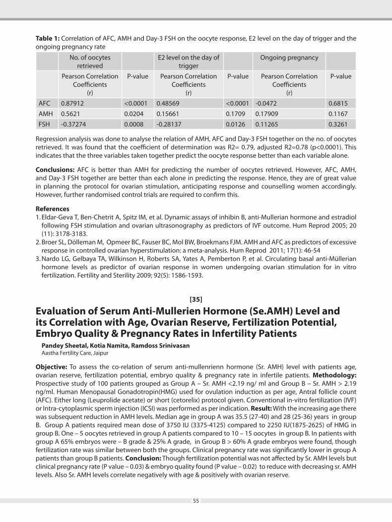
55
Table 1: Correlation of AFC, AMH and Day-3 FSH on the oocyte response, E2 level on the day of trigger and the ongoing pregnancy rate
No. of oocytes retrieved
E2 level on the day of trigger
Ongoing pregnancy
Pearson Correlation Coefficients
(r)
P-value Pearson Correlation Coefficients
(r)
P-value Pearson Correlation Coefficients
(r)
P-value
AFC 0.87912 <0.0001 0.48569 <0.0001 -0.0472 0.6815
AMH 0.5621 0.0204 0.15661 0.1709 0.17909 0.1167
FSH -0.37274 0.0008 -0.28137 0.0126 0.11265 0.3261
Regression analysis was done to analyse the relation of AMH, AFC and Day-3 FSH together on the no. of oocytes retrieved. It was found that the coefficient of determination was R2= 0.79, adjusted R2=0.78 (p<0.0001). This indicates that the three variables taken together predict the oocyte response better than each variable alone.
Conclusions: AFC is better than AMH for predicting the number of oocytes retrieved. However, AFC, AMH, and Day-3 FSH together are better than each alone in predicting the response. Hence, they are of great value in planning the protocol for ovarian stimulation, anticipating response and counselling women accordingly. However, further randomised control trials are required to confirm this.
References1. Eldar-Geva T, Ben-Chetrit A, Spitz IM, et al. Dynamic assays of inhibin B, anti-Mullerian hormone and estradiol
following FSH stimulation and ovarian ultrasonography as predictors of IVF outcome. Hum Reprod 2005; 20 (11): 3178-3183.
2. Broer SL, Dólleman M, Opmeer BC, Fauser BC, Mol BW, Broekmans FJM. AMH and AFC as predictors of excessive response in controlled ovarian hyperstimulation: a meta-analysis. Hum Reprod 2011; 17(1): 46-54
3. Nardo LG, Gelbaya TA, Wilkinson H, Roberts SA, Yates A, Pemberton P, et al. Circulating basal anti-Müllerian hormone levels as predictor of ovarian response in women undergoing ovarian stimulation for in vitro fertilization. Fertility and Sterility 2009; 92(5): 1586-1593.
[35]
Evaluation of Serum Anti-Mullerien Hormone (Se.AMH) Level and its Correlation with Age, Ovarian Reserve, Fertilization Potential, Embryo Quality & Pregnancy Rates in Infertility Patients
Pandey Sheetal, Kotia Namita, Ramdoss SrinivasanAastha Fertility Care, Jaipur
Objective: To assess the co-relation of serum anti-mullenrienn hormone (Sr. AMH) level with patients age, ovarian reserve, fertilization potential, embryo quality & pregnancy rate in infertile patients. Methodology: Prospective study of 100 patients grouped as Group A – Sr. AMH <2.19 ng/ ml and Group B – Sr. AMH > 2.19 ng/ml. Human Menopausal Gonadotropin(HMG) used for ovulation induction as per age, Antral follicle count (AFC). Either long (Leuprolide acetate) or short (cetorelix) protocol given. Conventional in-vitro fertilization (IVF) or Intra-cytoplasmic sperm injection (ICSI) was performed as per indication. Result: With the increasing age there was subsequent reduction in AMH levels. Median age in group A was 35.5 (27-40) and 28 (25-36) years in group B. Group A patients required mean dose of 3750 IU (3375-4125) compared to 2250 IU(1875-2625) of HMG in group B. One – 5 oocytes retrieved in group A patients compared to 10 – 15 oocytes in group B. In patients with group A 65% embryos were – B grade & 25% A grade, in Group B > 60% A grade embryos were found, though fertilization rate was similar between both the groups. Clinical pregnancy rate was significantly lower in group A patients than group B patients. Conclusion: Though fertilization potential was not affected by Sr. AMH levels but clinical pregnancy rate (P value – 0.03) & embryo quality found (P value – 0.02) to reduce with decreasing sr. AMH levels. Also Sr. AMH levels correlate negatively with age & positively with ovarian reserve.
56
[36]
Comparison of Anti-Mullerian Hormone in Relation to the Antral Follicle Count in the Assessment of Ovarian Reserve
Sonu Balhara, Ila GuptaArtemis Health Sciences, Gurgoan, Haryana
Objective: To correlate whether Anti-Mullerian hormone (AMH) is more efficacious than antral follicle count (AFC) in the assessment of ovarian reserve and to find out which of the two is a cost effective predictor? Methods: In 100 subjects basal level of AMH was done irrespective of the day of the cycle. The study was conducted over a period of 6 months and patients in the age group of 25 to 44 years were observed. The AFC was done on second or the third day of the menstrual cycle. The two were then correlated dividing the AMH level into three groups from ≤ 1, 1-4 and ≥ 4 ng/ml and the AFC from ≤ 5, 5-10 and ≥10. Results: Out of the 100 women observed, 84 were found to have excellent correlation between the AMH and AFC, while in 16 patients the AFC and AMH values did not match. Conclusion: AMH and AFC are comparable predictors of the ovarian reserve and both serve well in predicting the ovarian reserve except in a few patients where the two did not correlate. AFC was found to be more dependable and cost effective than the AMH level.
[37]
Incidence of Y Chromosome Microdeletions in Blood and Sperm DNA of Infertile Males of Assam
R Mahanta1, PNB Chaudhury1, GU Ahmed2, IK Bhattacharyya1, P Sharma3, A Gogoi1
1Cotton College, Guwahati, Assam, India, 2Gauhati University, Guwahati, Assam, India, 3Pratiksha Hospitals, Guwahati, Assam, India
Background: Infertility is a major reproductive health problem. Male infertility problems tend to aggregate in the families and a decline of ~50% in the sperm concentration has occurred in the past 50 years. The frequency of male infertility due to Y-chromosome microdeletions is 13-18% in the human population. Aim: The aim of this study was to determine the frequency of Y chromosome microdeletions in Blood and Sperm DNA of Infertile males of Assam. Material and Methods: A total of 600 male patients were analyzed, of which 500 were infertile patients and 100 were fertile controls. Among the patients 100 were azoospermic, 182 were oligozoospermic and 218 were asthenozoospermic. Both peripheral blood and semen samples were collected from the patients and genomic DNA was isolated for microdeletion study and polymerase chain reaction was carried out with 7 genes: PRY-2, RBMY, BPY-2, XKRY, CDY-1, CSPG4LY, and DAZ and 3 STS markers: sY-84, sY-254 and sY-127. Results: We observed 77(15.4%) microdeletions in blood samples and 94(18.8%) microdeletions in semen samples. Percentage of microdeletion observed for individual genes and STS markers in blood samples are as follows:- PRY-2: 18.18%, CDY-1: 14.28%, CSPG4LY : 6.49%, XKRY : 11.68%, RBMY : 3.89%, DAZ : 3.89%, BPY-2: 2.59%, sY84 : 25.9%, sY127 : 2.59% and sY254 : 10.38%. Percentage of microdeletion observed for individual genes and STS markers in semen samples are as follows:- PRY-2 : 17.02%, CDY-1: 13.82%, CSPG4LY: 6.38%, XKRY : 11.70%, RBMY : 3.19%, DAZ : 4.25% , BPY-2 : 5.31%, sY84: 24.46%, sY127: 3.19% and sY254 : 10.63%. Conclusion: In our study we found that the percentage of microdeletion was higher in semen samples as compared to blood samples.
[38]
Role of Trans Rectal Ultrasound Scanning (TRUS) in the Diagnosis of male infertility
Sharath Kumar C*, Najafi M, Malini SS*Director and Chief Fertility Surgeon, Mediwavw IVF and Fertility Research Hospital, Sayyaji Rao Road, Mysore 570 001
Infertility is a common problem, with male infertility detectable in approximately 50% of involuntarily childless couples. Transrectal and scrotal ultrasound are the imaging techniques of choice, allowing detailed examination of the male reproductive system. No cause for subfertility is detected (idiopathic infertility) in the majority of
57
patients. Detection of the cause of infertility is important to allow planning of the most appropriate techniques for achieving conception. High frequency ultrasound scans with transrectal transducers provide accurate, cost-effective and non-invasive means of assessing the integrity of the distal seminal tract. Thus, in infertile men with low semen values for whom retrograde ejaculation has been ruled out, endorectal sonography may clarify the anatomical abnormalities responsible for infertility. TRUS is a safe and accurate method for evaluating the distal male reproductive tract that helps identify patients with potentially correctable causes of infertility. The use of ultrasonography has become an important component in the evaluation and treatment of male reproductive tract disorders. From the use of color flow Doppler ultrasonography for the assessment of varicoceles to transrectal ultrasonography combined with seminal vesiculography for the evaluation of ejaculatory duct obstruction, ultrasonography has practical clinical applications. In this paper, the authors review the indications and use of TRUS in the assessment and treatment of the infertile male. Keywords: Male infertility; TRUS; Subfertility.
[39]
A Comparative Study of GnRH Antagonist and GnRH Agonist in PCO Patients Undergoing Controlled Ovarian Stimulation in IVF-ICSI Cycles
Singh MonicaBhopal Test Tube Baby Centre, E-1/13A, Arera Colony, Bhopal-462 016
Background: Polycystic ovarian syndrome (PCOS) patients are prone to ovarian hyperstimulation syndrome (OHSS). Controlled Ovarian Stimulation in PCO patients can be achieved using GnRH Antagonists or GnRH agonists. Objective: The study aimed at comparing the effects of GnRH agonists and antagonists in PCOS patients in IVF-ICSI cycles. Material and Methods: A total of 164 PCOS patients under 35 years of age were studied. All patients received low-dose OCP before starting treatment. Subsequently, they were randomly divided into two groups. The agonist group underwent standard long GnRH analogue protocol. The antagonist group received 0.25mg antagonist injections upto the day of hCG injection, using a flexible start approach. Follicular development was monitored by vaginal ultrasound and serum estradiol levels. Results: There was no significant difference in the gonadotropin usage between the two groups. However, significant variation was found in the number of days of stimulation, serum estradiol level on hCG day, incidence of OHSS, number of Metaphase II oocytes, and clinical pregnancy rates between the two groups. Conclusion: Usage of GnRH antagonist offers the advantage of a shorter duration of stimulation in PCO subjects in IVF-ICSI cycles, as compared to an agonist protocol. Additionally, the number of immature oocytes is less and more M-II oocytes are recovered with antagonists. We also found the approach to be more patient friendly, with a reduction in OHSS and a better clinical pregnancy rate.
[40]
Effect of Pretreatment with Oral Contraceptives and Antagonistic Cycle on Pregnancy Rates
Rajul Tyagi
Aim: Aim of the study was to compare the pregnancy rates of patients taking oral contraceptive pills in the previous cycle and patients not taking O.C.P. Methods: In our study group of 20 cases, 12 patients were taking OCP and 8 patients were not taking OCP. in the previous cycle. Observation: We found that patients taking oral contraceptives pills in previous cycle did not perform as well as the the patients not taking O.C.P..O.C.P.s are commonly used in previous cycle before starting gonadotrophin stimulation to programe the cycle. Results & Conclusion: Probably the gestagen component of O.C.P. could exert negative impact on endometrial receptivity
Session F8: IVF / ICSIHall-C (Silver Oaks Hall) • Time: 13:00 - 13:45 hr
58
[41]
GnRH Agonist and GnRH Antagonist in Intracytoplasmic Injection Cycles
Shweta Kaul Jha, Thankam Varma, Kundevi Shankar, Satya Balasubramanian, Betty Thomas
Objective: To compare GnRH agonist versus Antagonist and its effect on ART outcome. Method: Retrospective study of 105 cycles at a tertiary teaching institute from 2010 to 2011. Result: 105 COH cycles followed by ICSI (agonist=54 and antagonist=51) were analysed. The end points studied included the number of oocytes recovered, number of mature (MII) oocytes, fertilization, morphology based embryo quality, pregnancy rate, quantity and cost of gonadotrophin. The average age of female subjects was 30.5 years. Major infertility factors were tubal blockage, male factor and endometriosis altogether comprising 68%. GnRH agonist and antagonist cycle parameters were comparable in except more amount of gonadotrophin was used with more resultant costs (both p<0.002 and <0.009) in antagonistic regime. Antagonist regime produce more good quality embryos (p=0.0005), a statistically significant difference. A clinical pregnancy rate per embryo transfer of 31.5% in agonist and 35.3% in antagonist regime was achieved respectively and ongoing pregnancy rate of 20.4% and 11.8% respectively,but statistically insignificant(p =0.164). Conclusion: GnRH antagonist protocol produced a COH response, embryonic development and pregnancy rates at par to GnRH agonist regime. Moreover GnRH antagonist protocol required a shorter stimulation period plus fewer complications but required more dose and resulted costelier in our study. Hence GnRH antagonist regime provided means for a friendlier and convenient protocol for our patients but costelier.
[42]
How Many Times IVF can be AttemptedAnirudh SinghDr Singh Test Tube Baby Centre, Meerut
Objective: Purpose of this study was to see the number of attempt of IVF to get pregnancy. Method: 6 patient were included in this study. 3 cases dropped the attempt in between. All cases were investigated thoroughly and ultrasound were done to see any uterine pathology. Out of three two were stimulated and in on embryo donation were done. Result: In one patient pregnancy occurred in 8th attempt. In one pregnancy occurred in 6th attempt. In one pregnancy occurred in 5th attempt. Conclusion: Previously it was thought that if a lady doesnot become pregnant in three attempt of IVF than it was advisable to stop the attempt. But Research done in Western Australia by Louise Stewart and colleague researchers estimate that upping the number of IVF cycles would boost success rates at least up until five attempts.
[43]
To Study the Advantages of IMSI (Intracytoplasmic Morphologically Selected Sperm Injection) Over Conventional ICSI (Intracytoplasmic Sperm Injection) in Men with Moderate and Severe Oligospermia
Sarabpreet Singh, Ila GuptaArtemis Health Institute
Objectives: Advantages of IMSI was evaluated in terms of: (1) Fertilization rate, (2) Embryo quality with respect to morphology, (3) Pregnancy rate and 4. abortion rate. Methods: IMSI was performed using OCTAX cytoscreen
in subsequentcycle. Low endogenous LH level after O.C.P. pretreatment might impair oocyte competence. But still reason for reduction of pregnancy rates in O.C.P. pretreatment is not very clear and additional research is needed to determine a reliable and efficacious way of scheduling GNRH antagonist ovarian stimulation cycle by pretreatment with sex steroids.

59
dry IMSI which can achieve magnification upto 7200X. It was done in patients with moderate and severe oligospermia [n = 21 including 13 patients of prior ICSI failures] with DNA fragmentation index higher than 21% using Halosperm kit. Sperm selection was based on based on MSOME criteria [motile sperm organellar morphology examination]. Results: compared to conventional ICSI: (1) Fertilization rate was similar. (2) Embryo quality improved in terms of morphology and blastocyst formation. (3) Pregnancy rate was higher. (4) 5 patients of prior ICSI failures conceived. (5) No abortions have been reported. Conclusions: In the present study: (A) With the use of IMSI, sperms with nuclear vacuoles could be identified. (B) IMSI was shown to be beneficial for patients with high DNA fragmentation and prior ICSI failures. (C) However, more patients need to be recruited in this ongoing study to comment on the significante levels.
63
[01]
Follicle Loss After Laparoscopic Stripping of Ovarian Endometriotic Cysts
Amar R, Sushma D, Anuradha Rao*, Ashwini V, Akhila Vasudev, Rajeshwari Bhat, Rajesh Bhaktha, Pratap KumarDivision of Reproductive Medicine, Department of Obstetrics and Gynecology, Department of Pathology, Kasturba Medical College, Manipal University, Manipal, Karnataka - 576 104
AimTo estimate whether size of endometriotic cyst is related to ovarian parenchyma excised along with cyst wall.
Material and MethodsProspective study of 56 women attending University teaching hospital who underwent laparoscopic endometriotic cystectomy from August 2010 to August 2011. On preoperative transvaginal sonography largest transverse diameter of cyst was measured. Ovarian cystectomy was done by stripping technique, cyst wall sent for histopathological evaluation and were categorised into 2 groups based on semi quantitative scale of 0-4 grades (0, complete absence of follicles; 1, primordial follicles only; 2, primordial and primary follicles; 3, primordial, primary and some secondary follicles; 4, follicles seen in normal ovaries). Group 1 and 2 showed grade 0, 1, 2 and 3, 4 in the cyst wall respectively.
ResultsMean age of patients was 31.4 years, duration of infertility was 4.1 years and cyst diameter measured 6.3 cms. 73.2% (n=41) were in group 1 and 26.8% (n=15) were in group 2, mean cyst diameter being 4.3 cms and 3.9 cms respectively. There was no significant statistical correlation between preoperative cyst diameter and ovarian parenchyma removed (p=0.35). 93.3% (n=14) of group 2, and only 65.8% (n=27) of group 1 were found to have moderate to severe endometriosis, indicating there is correlation between disease severity and loss of ovarian tissue (p= 0.04)
ConclusionEndometriotic cystectomy when performed with accurate surgical technique leads to no significant ovarian tissue removal. However disease severity significantly determines the loss of normal ovarian parenchyma
Key WordsEndometriosis, Laparosocpic cystectomy, Histopathology, Ovarian tissue.
[02]
Successful In-Vitro-Fertilization with Ovum Donation in Three Sisters with Premature Ovarian Failure
Sonwant Kaur, Umesh N JindalJindal IVF and Sant Memorial Nursing Home, Chandigarh
ObjectivesTo report the outcome of IVF with ovum donation in a unique family of three sisters with premature ovarian failure.
Material and MethodsThree sisters who were diagnosed with hypergonadotropic hypogonadism below 30 years of age underwent a total of seven fresh In-vitro-fertilization and one frozen embryo transfer with ovum donation in our hospital from 2005-2010. The possible causes of familial premature ovarian failure are discussed. Problems related to social and marital issues as well as arrangement of donors is discussed.
ResultsA total of eight embryo transfers were done. Two twin and one singleton pregnancy resulted in five live births. In addition there was one preterm delivery resulting in neo-natal death and two abortions.
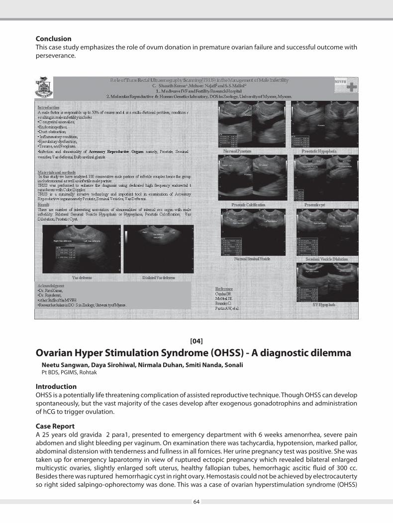
64
[04]
Ovarian Hyper Stimulation Syndrome (OHSS) - A diagnostic dilemmaNeetu Sangwan, Daya Sirohiwal, Nirmala Duhan, Smiti Nanda, SonaliPt BDS, PGIMS, Rohtak
IntroductionOHSS is a potentially life threatening complication of assisted reproductive technique. Though OHSS can develop spontaneously, but the vast majority of the cases develop after exogenous gonadotrophins and administration of hCG to trigger ovulation.
Case ReportA 25 years old gravida 2 para1, presented to emergency department with 6 weeks amenorrhea, severe pain abdomen and slight bleeding per vaginum. On examination there was tachycardia, hypotension, marked pallor, abdominal distension with tenderness and fullness in all fornices. Her urine pregnancy test was positive. She was taken up for emergency laparotomy in view of ruptured ectopic pregnancy which revealed bilateral enlarged multicystic ovaries, slightly enlarged soft uterus, healthy fallopian tubes, hemorrhagic ascitic fluid of 300 cc. Besides there was ruptured hemorrhagic cyst in right ovary. Hemostasis could not be achieved by electrocauterty so right sided salpingo-ophorectomy was done. This was a case of ovarian hyperstimulation syndrome (OHSS)
ConclusionThis case study emphasizes the role of ovum donation in premature ovarian failure and successful outcome with perseverance.
65
which was misdiagnosed to be ruptured ectopic. Retrospectively, it was found that she had received injection gonadotropins for ovulation induction.
ConclusionGynecologist must be familiar with the varied presentation of OHSS. It can present as surgical emergency in two situations- ovarian torsion and actively bleeding cyst rupture.
[05]
Effect of Low Dose Mifepristone on the Uterine Leiomyoma in Women in Reproductive Age Group
Varun Neha, Kumar Ashok, Prasad SudhaDepartment of Obstetrics and Gyaecology, Maulana Azad Medical College, Lok Nayak Hospital, New Delhi
Aims and ObjectiveTo study the effect of low dose mifepristone on the leiomyoma volume reduction, endocrinal profile and endometrium in symptomatic leiomyoma.
MethodSingle blind placebo controlled randomized clinical trial. Sixty two women with symptomatic uterine leiomyoma were recruited and randomized to receive 10 mg mifepristone (group 1) and placebo (group 2) daily for 3 months. Endometrial biopsy and hormonal profile was done before beginning therapy. Leiomyoma related symptoms and leiomyoma volume were assessed at baseline and every month for three months. Endometrial biopsy and hormonal profile repeated at the end of therapy. Primary outcome was reduction in uterine leiomyoma volume.
Result(s)There was a significant percentual decrease (P = 0.000) in the total leiomyoma volume in the mifepristone-treated group, -58.36±8.92% (mean ±S.D), compared with the control group values, 0%. Mifepristone treatment significantly reduced the bleeding days (P = 0.000) by causing amenorrhoea in all the patients (100%). Hormonal levels remained unchanged. Endometrial biopsies showed cystic glandular hyperplasia in 8 (25%) cases in mifepristone treated group without any premalignant changes. One patient in mifepristone group (infertility group) conceived after 8 months of therapy.
ConclusionLow dose mifepristone is efficacious in reducing leiomyoma volume and leiomyoma related symptoms of menorrhagia and dysmenorrhoea and offer an effective medical treatment option for women with uterine leiomyoma.
KeywordsMifepristone, Leiomyoma volume, Endometrial biopsy.
[06]
Modalities for Prevention of Severe OHSSRutvij Dalal
IntroductionOHSS is one of the commonest and most dreadful complications arising out of ART. Conventional protocols employing superovulation strategies aim to generate many follicles/oocytes thereby generating maximum embryos per retrieval cycle. However, this is accompanied by OHSS of varied severity. Various regimens have been used in the past to ensure the complete prevention or at least reduction in the severity of OHSS. What follows is a review of 50 of our patients who were falling in the high-risk category for whom we successfully employed an efficient suppression modality.
66
[07]
IVF Outcome in Primary and Secondary AmenorrhoeaGupta Anupam, Jindal UNJindal IVF and Sant Memorial Nursing Home, Chandigarh
ObjectiveTo compare the outcome of in-vitro fertilization (IVF) in cases of primary and secondary amenorrhoea in women < 40 years of age
MethodA retrospective case study of 11women with primary amenorrhoea and 20 women with secondary amenorrhoea < 40 years over a period of 4 years was done. Cases of amenorrhoea due to polycystic ovarian disease and Assherman’s syndrome were excluded. The cases were subjected to IVF with self or donor oocytes according to the cause of amenorrhoea.The etiology of amenorrhoea, association with other diseases and IVF outcome in these two groups was compared
ResultOf 11 cases of primary amenorrhoea, 6 had hypogonadotropic hypogonadism, 1 had Mullerian agenesis and 4 had gonadal dysgenesis (primary ovarian failure). Of 20 cases of secondary amenorrhoea < 40 years- 18 had hypergonadotropic hypogonadism and 2 had surgical menopause.Secondary amenorrhoea cases also had other associated causes of infertility like tubal factor(3), endometriosis (1) post-surgical adhesions(3) while the primary amenorrhoea cases did not have any such associated cause. Also endometrial biopsy for tuberculosis polymerase chain reaction was positive in 7 cases of secondary amenorrhoea and these were subjected to anti-tubercular treatment before starting IVF whereas in no patient of primary amenorrhoea was endometrial biopsy positive. Primary amenorrhoea cases underwent total of 19 IVF cycles, of which 12 cycles(63.1%) resulted in pregnancy .Of these, 9 pregnancies (75%)had live birth and 3 ended in abortion. Secondary amenorrhoea cases underwent total of 22 cycles, of which 10 cycles (45.4%) were positive and all of them(100%) resulted in live births
ConclusionIVF outcome in cases of primary and secondary amenorrhoea is very good.However the implantation rate in primary amenorrhoea is better, probably due to early presentation and non-association with other causes of infertility.
ObjectiveTo evaluate and analyse the suppression strategy for patients likely to develop severe OHSS.
Material and MethodsProspective study done at Lilavati Hospital, Mumbai from the month of April to October 2011.
ResultsOut of 50 patients analysed in the high risk category, 47 were successfully suppressed and OHSS prevented, 2 developed moderate OHSS and one patient developed severe OHSS and required repeated ascitic tapping and pleural effusion tapping.
ConclusionThe suppression strategy used at our center is a safe, patient-friendly, convenient and efficient modality for successful prevention of severe grade Ovarian Hyper-stimulation syndrome.
67
[08]
Endometrial Hyperplasia in Infertile Women with Anovulatory Infertility
Shalini Gainder, Lakhbir Dhaliwal, K R Gupta, Sumittu SahdevAssistant Professor, Deptt. of Obst. and Gynaecology, PGIMER, Chandigarh
A total of 3078 infertile women booked in the infertility clinic from year 2009 -2011 were evaluated and investigated for the cause of infertility.
As a routine protocol during evaluation endometrial biopsies were performed in all women for ruling out tuberculosis and sample was forwarded for histopathology. When Group analysis was done, it was found that 12% infertile women had PCOS of the total of 14% women with anovulatory infertility. Endometrial evaluation of these women showed that most had proliferative endometrium, 4% women had simple hyperplasia, one woman was found to have complex hyperplasia without atypia, complex hyperplasia with atypia was found in a single woman which on performing hysteroscopy guided biopsy proved to be well differentiated adenocarcinoma in association with complex hyperplasia with atypia.
All women with simple hyperplasia were treated simultaneously with ovulation induction and progesterone therapy in the luteal phase, woman with complex hyperplasia without atypia was treated with medroxy progesterone acetate 21 days therapy for 3 months and subsequently ovulation induction with intrauterine insemination was done, following which conception occurred and she delivered alive born baby. Woman with carcinoma endometrium was given high dose progesterone therapy along with Mirena for 3 months, subsequent biopsy showed persistence of carcinoma. She was offered conservative management versus complete surgery. She underwent complete radical surgery for carcinoma endometrium.
ConclusionEndometrial biopsy must be considered in women with PCOS coming for infertility treatment especially when there is clinical or sonological suspicion.
[09]
A Randomized Comparison of Antagonist Versus Agonist Protocol for Controlled Ovarian Hyperstimulation
Khandelwal N, Devi Gouri M, Sharma MeetaGouri Hospital and Ridge IVF, New Delhi
The aim of our study was to compare the GnRH antagonist and GnRH agonist flare up protocol for controlled ovarian hypestimulation in ivf cycles.
MethodsA total of 94 women randomly received COH using either GnRH agonist or GnRH antagonist. The primary endpoint was the mean number of oocytes retrieved per cycle. The secondary objectives were the mean number of mature oocytes, pregnancy and implantation rates.
ResultsThe mean number of oocytes retrieved in the agonist group was 10.96 compared to 7.42 in antagonist group, p value being significant. The mean number of mature oocytes was 8.89 in agonist group and 5.86 in antagonist group making the difference statistically significant. Pregnancy and implantation rates were also higher in agonist group. Pregnancy rates in agonist group was 48.93 and was 29.79 in antagonist group. Implantation rates were 31.91 versus 17.02 respectively.
ConclusionAgonist protocol yields better results in IVF cycles thereby increasing the success rate.
68
[10]
Ectopic Pregnancy an Obstetrical DilemmaDaya SirohiwalSenior Professor, Department of Obste & Gynae, 25/8FM, Medical Campus, PGIMS Rohtak, Haryana 124 001
Ectopic pregnancy is one of the most enduring dilemmas of modern medicine. Any woman of child bearing age is at potential risk. After an ectopic pregnancy only 50% of women will conceive of which 30-50% will have live child birth subsequently. The recurrent ectopic pregnancy rate has been reported between 12-18%. The maternal mortality in 2/3rd patients of ectopic pregnancy are due to delay in diagnosis and instituting management.
The diagnosis of ectopic pregnancy should be constantly kept in mind as a potential threat to any female in reproductive age group. Accuracy of initial clinical evaluation is less than 50%. The physician is thus depends upon various invasive and non invasive investigative diagnostic modalities like urine for pregnancy test, serum beta hCG, serum progesterone, trans-abdominal sonography, trans-vaginal sonography, laparoscopy to confirm or exclude the ectopic pregnancy.
We studied 56 patients admitted in Gynae at PGIMS, Rohtak, with high index of suspicion harboring ectopic pregnancy on the basis of suggestive history, physical examination and associated high risk factors. On TVS we observed that solitary abnormalities were uncommon. Majority of patients were showing multiple features; like adnexal masses in 90%; gestational sac 50%; embryo 25%; embryo with cardiac activity 13%; fluid in POD 79%; endometrial thickness 31% and pseudo-gestation sac was nil. Finally we made diagnosis of ectopic pregnancy 48, corpusluteal cysts 3, PID 3 and ruptured /torsion ovarian cyst in2 patients.
On laparoscopy and surgical exploration of these 48 ectopic pregnancies we found mostly were tubal (43), followed by cornual (2) and one each of abdominal, ovarian and rudimentary horn pregnancy. Out of these patients 25 had intact and 23 ruptured ectopic pregnancies. To conclude TVS was found to be statistically useful modality for diagnosis of ectopic pregnancy.
[11]
Luteinizing Hormone and ReproductionRajul Tyagi
Luteinizing hormone (LH) is a critical component of menstrual! Cycle physiology and of medications used for the treatment of infertility. The presence of LH receptors on theca and granulosa cells allows LH activity to stimulate steroidogenesis and to modulate folliculogenesis during ovulation induction. Both LH and human chorionic gonadotropin (hCG) interact with the LH receptor and can exert these actions. Medications containing LH activity (LH, hCG or a combination of them) as well as FSH appear to be more efficient than FSH alone in ovulation induction or controlled ovarian stimulation. Finally, the selective use of LH activity alone in the late stages of ovarian stimulation is capable of maintaining follicle development and maturation despite declining FSH levels. This approach may prove to be highly successful to limit the occurrence of severe complications such as the ovarian hyperstimulation syndrome.
[12]
How a Clinician can Improve ImplantationRajul Tyagi
Implantation is a complicated process in which a series of events occur involving both the embryo and the endometrium. Sometimes transferring of high quality embryos, implantation rates remain relatively low. In this article the clinical strategies employed to increase the change of implantation are reviewed. Embryo transfer technique is a critical step in assisted reproductive technology cycles. Recent studies have shown significant
69
[13]
Depicting the Flow Chart of Protocol Adopted in Procedure of in Vitro Fertilization and Embryo Transfer(IVF-ET) in Nutshell
Swati Singh1, BK Taneja2
1JR, 2Professor, MMIMSR, Mullana, Ambala
We have made a flow chart of Protocol followed in IVF and embryo transfer.
The aim is to make a simple flow chart which can be used to understand the basic steps if In Vitro Fertilization in nutshell, beginning from down regulation using GNRh agonists till embryo transfer. The protocol for controlled ovarian hyper stimulation is shown in poster attached. The next step is of monitoring ovarian response followed by hCG injection if lead follicle greater than 17-18 mm diameter. This is followed by oocyte retrieval, fertilization and embryo transfer. All of the above mentioned steps are shown in enclosed poster.
improvements in clinical pregnancy rates resulting from careful embryo transfer technique, appropriate catheter type and placing for embryo transfer. Increasingly, adjuvant pharmaceutical therapies are also being applied with the aim of improving embryo implantation. However, the evidence for their efficacy and safety is limited. Recent evidence suggests that adoption of milder ovarian stimulation regimens may provide a more effective clinical approach to improving implantation, since beneficial effects have been shown for both endometrial receptivity and embryo quality.
[14]
Study of Semen Parameters Over the Last Decade – 2000 – 2011Ved PrakashSouthend Fertility & IVF Centre, 1st Floor, Holy Angels Hospital, Basant Lok, Vasant Vihar, New Delhi-110 057
BackgroundDuring the last 20 years, at least in developed countries, decrease in fertility rates has been observed. Several reports have appeared in literature on the reported decline in semen quality globally. In view of the concerns expressed in the literature, we carried out a retrospective study on the data available with us to find out whether or not similar fall in semen quality can be observed in our studies as well.
MethodThe preliminary results of the analysis of the data obtained from 2000-2011 is presented. A total of 800 semen samples were evaluated according to the WHO guidelines for semen analysis. The data based on four seminal parameters: volume, sperm count, motility and morphology of the sperms is reported in this communication.
ResultsFor comparison, men were grouped in two groups, one carried out before 2000-n=400(GroupI) and the other after 2005 -n=400(GroupII). The data shows a significant decrease in volume 19.46 %(GroupI-59.73%& GroupII-40.27%). No significant changes were seen in sperm count -1.79 %(GI- 50.89% & GII-49.10%), motility –4.57%(GI-47.71% & GII-52.28%)and normal sperm morphology -2.5%(GI-48.75% & GII-51.25%) over a period of time.
ConclusionNo significant changes were seen in the semen parameters except volume.
70
[16]
Hyperprolactinemia, Hypothyroidism and Their Correlation in Infertile Women
Ananna Purakayastha1, Veena Aseeja2
1M.M.I.M.S.R, Mullana, Ambala, Haryana, 2Babbar Heart Care Centre, 89, Govt. College Road, Gurdaspur, Punjab
BackgroundInfertility affects large number of couples both globally and in India. though not a physically debilitating disease, it may outrageously jeopardise the couple’s psychological harmony and perilously shatter their marital stability.
ObjectiveTo find out the incidence of hyperprolactinemia and hypothyroidism in infertile women (both primary and secondary infertility) and to determine their correlation.
MethodOne hundred women coming to the department of Obstetrics and Gynecology of MMIMSR Mullana with complaints of infertility were included in this study. The exclusion criteria were male factor infertility, and amongst the female factor any congenital anomaly of the female genital tract and any obvious organic lesions of the female genital tract.
Results52% percent women had primary infertility and 48% percent of women had secondary infertility. Hyperprolactinemia was detected in 42% of patients. 22% of the patients also had hypothyroidism. The incidence of hypothyroidism in hyperprolactinemia was 26.19%. Anovulatory cycles were detected in 29% of the patients. Galactorrhea was present in 5% of women. There is a positive correlation between hyperprolactinemia and hypothyroidism in infertile women.
[15]
Outcome of Tubal Surgeries in InfertilityRicha Baharani, Pragya Dheerawani, Sangeeta ShrivastavaJHRC, Jabalpur (M.P.)
ObjectiveTo determine outcome of tubal surgeries in cases of infertility over a period of 5 years (2006-10)
MethodA retrospective analysis of 50 cases of infertility who underwent tubal surgeries at our hospital from Jan. 2006 to Jan. 2010 was done. Type of surgeries performed were aimed at the conservation & salvaging of tubes whenever possible.
ResultsIn this study 50 women underwent tubal surgery. Hydrosalpinx was found in 16 (32%) cases. Peritubal adhesions were present in 18 (36%) cases, cornual block in 12 (24%) of cases. Mid segment block was found in 4 (8%) of cases. Surgical access was through laparoscopy in 92% of cases. Only in 8% of cases laparotomy was done.
52% of patients were followed up for more than 6 months. While rest were lost to follow up after 6 months. Over all intrauterine pregnancy with full term live birth was found in 14% of cases. Tubal pregnancy was found in 1 case (2%) spontaneous abortion occurred in 2 cases (4%).
ConclusionWhenever indicated tubal surgery should be done. It plays a very important role in the management of infertility. Success rate depends on severity of preexisting tubal disease and some other factors. Proper selection of cases is crucial to find the best results. Laparoscopic tubal surgery has surpassed laparotomic tubal surgery with comparable success rates. Laparoscopic tubal surgery is less traumatic procedure and gives us benefits of laparoscopy over laparotomy.
71
[17]
Comparative Evaluation of PCR Test Alone or PCR Test with AFB Culture and Histopathological Examination in Diagnosis of Genital Tuberculosis
Rakhee MalhotraAAS Infertility and Maternity Centre, 128-A/19, Near Civil Hospital, Rohtak-124 001, Haryana
IntroductionTuberculosis is involved in 5–16% of cases of infertility among Indian women. An absolute diagnosis cannot be made from characteristic features in hysterosalpingogram or laparoscopy. Due to the paucibacillary nature of GTB, diagnosis by mycobacterial culture and histopathological examination have limitations and low detection rate. PCR is a rapid, sensitive and specific molecular biological method for detecting mycobacterial DNA.
Patients and MethodsAfter written informed consent from each patient, the prospective, randomized study was done to evaluate the diagnostic competence of PCR alone vis-a-vis PCR test with AFB culture and histopathological examination in diagnosis of genital tuberculosis was evaluated. From 2005-2010, fifty patients, aged 20–40 years, presenting to the AAS Infertility Centre, Rohtak with suspected diagnosis of genital tuberculosis were studied. Randomization was done with computer generated randomization table. In group-I (n=25) PCR test alone and in group-II (n=25) PCR test with AFB culture and histopathological examination was done respectively to confirm the diagnosis of genital tuberculosis.
ResultsAFB smear was positive in 2 patients (8%), culture in 3 patients (12%), HPE in 3 patients (12%) and PCR was positive in 18 (64%) of patients. On the basis of diagnostic criteria GTB was suspected in 26 of the 50 cases. The concordance of results between the clinical criteria and specific diagnostic tests were analysed and the culture and HPE showed mild agreement with the clinical criteria, whereas PCR showed a moderate agreement. False positive PCR in one patient was ruled out by multiple areas of sampling.
ConclusionConventional methods of diagnosis namely, HPE, AFB smear and culture have low sensitivity. PCR has been found to be useful in diagnosing early disease as well as confirming diagnosis in clinically suspected cases. False negative PCR can be an important limitation.
ConclusionThere is a greater propensity for thyroid disorder in infertile women than the fertile ones. There is also a higher prevalence of hyperprolactinemia in infertile patients.
[18]
Rudimentary Horn: A rare cause of chronic pelvic painShruti Gupta, Vaishali Sharma, Sunesh KumarAll India Institute of Medical Sciences, 9/53, Hostel no. 9, AIIMS, Ansari Nagar, New Delhi -110 029
IntroductionUterine anomalies often present as a cause of reproductive failure. The incidence of uterine anomalies has never been established, as there is no estimate of its occurrence in women with a normal reproductive performance. A double uterus with obstruction in one cavity may also present later on as a cause of chronic pelvic pain and dysmenorrhoea in parous women.
CaseA 35 year old parous woman with 3 children came to the E.R with excruciating pain in left lower abdomen. She admitted to similar episodes for the past 10 years. She had normal spontaneous vaginal deliveries including the
72
[19]
Fitz Hugh Curtis Syndrome – Pathognomic of Salpingitis in Infertile Patients
Shalini Chawla Khanna, Sudha PrasadIVF & Reproductive Biology Centre, Maulana Azad Medical College, New Delhi
BackgroundFitz Hugh Curtis Syndrome(Perihepatic “violin –string adhesions”) is an extrapelvic manifestation of pelvic inflammatory disease and increased incidence is reported in infertile patients. Its not a new syndrome but most often we neglect it on laparoscopy.
AimTo assess the prevalence of FHCS and its correlation with tubal status in infertile patients on laparoscopy.
DesignProspective Observational Study.
Study PeriodJanuary 2010-July 2011.
InstitutionIVF & Reproductive Biology Centre, Maulana Azad Medical College, New Delhi.
Material & MethodsThis prospective observational study was carried out on 200 consecutive women undergoing laparoscopy for tubal evaluation. After visualizing the pelvis and the chromopertubation test, the upper abdomen was examined by directing the laparoscope cranially and liver surface was inspected for evidence of any adhesions. The normal healthy patent tubes were categorized as Group I (n=99) where as abnormally dilated, tortuous, beaded, blocked tubes constituted Group II (n=101) and the prevalence of perihepatic adhesions were noted for both and analysed statistically.
ResultsThe patient’s clinical characteristics like mean age, duration and type of infertility was similar in both the groups and not statistically different. The overall prevalence of FHCS was 36.5% in infertile patients. The perihepatic adhesions were statistically significant high in group II (82.2%) vs (17.8%) in group I, p<0.001. The sensitivity, specificity, positive predictive value, negative predictive value of perihepatic adhesions in predicting salpingitis is 59.4%, 86.9%, 82.2%, 67.7% respectively.
last one which was at home. Her abdomen was soft on examination with deep tenderness in left iliac fossa. On pelvic examination there was a normal sized uterus with a smooth 4x5 cm tender, left adnexal mass. On Ultrasound we found the same hypoechoeic mass with central echogenic stripe. A tentative diagnosis of acute abdomen with left adnexal mass… a remote possibility of rudimentary horn was also kept. Laparoscopy found unicornuate uterus with a non functional non communicating rudimentary horn on the left side. There was no haematometra and no evidence of endometriosis. The rudimentary horn was excised and sent for histopathological examination.
DiscussionUnicornuate uterus comprises 10% of all Mullerian anomalies. It has the worst obstetric outcome with estimated live birth rate of only 40%. It is also one anomaly which is commonly associated with urinary tract abnormalities (up to 40%). A unicornuate uterus with a rudimentary horn that is non functional is a rare cause of chronic pelvic pain. It might still lead to dyspareunia but this history was not forthcoming in this patient.
ConclusionIt can be said that to predict the patient with poor reproductive outcome/ pain symptoms is a delicate predicament.
73
ConclusionThis study suggests that FHCS is a marker of salpingitis and its visualization should be a part of diagnostic evaluation in infertile patients.
[20]
Correlation of Body Mass Index with Outcome of in Vitro Fertilization in a Developing Country
Prerna Gupta, Neeta Singh, Neena Malhotra, Suneeta MittalAll India Institute of Medical Sciences, New Delhi
Aims & ObjectiveTo correlate ovarian response to stimulation and IVF outcome according to the women’s body mass index (BMI).
Material & MethodsWe retrospectively reviewed all patients who underwent IVF cycle in our institution from January 2008 to October 2010. Three hundred and twenty eight patients underwent 342 in vitro fertilization cycles or ICSI and were divided into four subgroups according to BMI; underweight, BMI <18.5; normal weight, 18.5–24.9; overweight, 25.0–29.9; and obese, >30.0.
ResultsWe found that with increasing BMI negative co-relation was seen with clinical pregnancy rate (p value=0.40). Increasing doses of gonadotropins were required as patients BMI increased (p value=0.45). In the present study no difference was seen in the number of oocyte retrieved but a decreased fertilization and cleavage rate was seen with decreased number of cryo-preserved embryos with increasing BMI. This study shows that poorer oocyte quality is seen with increasing BMI which results in reduced clinical pregnancy rate. In our study no deleterious effect of low BMI was seen on IVF outcome and clinical pregnancy rate.
ConclusionFemale obesity impairs IVF outcome potentially by impairing oocyte quality but does not affect ovarian response to stimulation.
[21]
Evaluation of Impact of GnRH Agonist as Adjuvant in Luteal Phase Support on IVF Outcome
Dattaprasad Inamdar, M Kochhar, Shweta Mittal, Neeti Tiwari, Ruma Satwik, Shikha Jain, Abha MajumdarSir Ganga Ram Hospital, New Delhi - 110 060
ObjectivesThis prospective, randomized study evaluates whether daily doses of GnRH agonist (Inj. Lupride 1mg SC) for three days, administered 6 days after IVF/ICSI increases ongoing pregnancy rates following embryo transfer in cycles stimulated with the long GnRH agonist protocol.
MethodsFour hundred and twenty six women undergoing embryo transfer following controlled ovarian stimulation with a long GnRH agonist protocol were included. In addition to routine luteal phase support with progesterone, women were randomized to receive three 1 mg doses of Inj Lupride 6 days after IVF/ICSI. Randomization was done on the day of embryo transfer according to a computer generated randomization table. Pregnancy rate was the primary outcome measure. The study was powered to detect a 13% absolute increase from an assumed 33% pregnancy rate in the control group, with an alpha error level of 0.05 and a beta error level of 0.2.

74
ResultsThere were 73 (34.27%) pregnancies in the GnRH agonist group, and 72 (33.96%) in the control group (absolute difference 0.31%) (Interim analysis). Implantation, clinical pregnancy and multiple pregnancy rates were likewise similar in the GnRH agonist and control groups.
ConclusionsThree 1 mg doses of Lupride administration 6 days after IVF/ICSI following ovarian stimulation with the long GnRH agonist protocol does not seem to result in an increase in pregnancy rates.