test of desert rose (adenium arabicum - free...
TRANSCRIPT
http://www.jofamericanscience.org ) 9(102014;Journal of American Science
67
Test of Desert Rose (Adenium Arabicum Forssk) Leaves on Blood Lipid Profile in Male Rats
Abdulbasit I. Al-Sieni
Biochemistry Department, Faculty of Science, King Abdulaziz University, Jeddah, Saudi Arabia [email protected]
Abstract: Desert rose (Adenium arabicum) leaves powder was supplemented orally in the diet to hypercholesterolemic male rats for 8 weeks to test its effect on blood lipid profile. The tested animals (180-200 gm) were divided into three groups (6 rats per each); the first group is untreated control group was fed by a basal diet, the second group was fed on 2% cholesterol in the diet to induce hypercholesterolemia (positive control group), and the third group was fed on 2% cholesterol and treated with 500 mg/kg body weight rose leaf powder for 8 weeks. The positive control group showed a significant increase in lipid profile parameters, liver enzymes, and kidney function parameters. Furthermore, the heart and testes showed histopathological alteration compared with that of the negative control group. Treating the hypercholesterlemic rats with desert rose leaves improved the biochemical blood tests and the histopathology of the studied organs. It can be concluded that desert rose leaves have antilipidemic activity and ameliorated the lipid profile of blood in hypercholesterlemic male rats. Moreover, an improvement in liver and kidney functions and histopathology of the studied organs was also noticed in hypercholesterlemic male rats of the desert rose leaves treated group. [Abdulbasit I. Al-Sieni. Test of Desert Rose (Adenium Arabicum Forssk) Leaves on Blood Lipid Profile in Male Rats. J Am Sci 2014;10(9):67-74]. (ISSN: 1545-1003). http://www.jofamericanscience.org. 9 Key words: hypercholesterolemic, rats, desert rose, histopathology. 1. Introduction
Adenium arabicum Forssk (desert rose, giant desert rose, impala lily, Adnah) belongs to the family Apocyanaceae. It is an endemic, rare, on the mountains of southern region of Saudi Arabia (Rahman et al., 2004). It is poisonous and toxic and has some medicinal properties; the sap and bark are used in bones dislocations, painful joints, wounds and skin infections (Mossa et al., 1987 and Shahina, 1994).
It is used for bonsai and cultivated for its leaves, growth form and flowering characteristics. It is characterized by caudex formation (swollen roots and stem bases) with a very fat and squat swollen trunk and stem bases. It has pretty pink flowers and interesting growth form, and it grows up to 4m in natural habitat and much smaller in the cultivated forms.
The increase of lipid parameters has been shown to be a strong risk factor for coronary heart diseases in many populations (Makni et al., 2008). Hypercholesterolemia is an indication and risk factor for early atherosclerosis prior to the appearance of over atherosclerotic changes in the vascular wall (Bentley et al., 2002). Moreover, it induces vascular functional changes that may lead to local ischemia and vascular remodeling. Amundsen et al. (2002) stated that clinical trials lane show that lowering lipids reduces the morbidity and mortality associated with cardiovascular complication.
Other studies showed that desert rose can be used as a treatment for trypanosomiasis in animals (Atawodi et al., 2002). Desert rose was also used as poison fishing in tropical Africa (Neuwinger, 2004). It was traditionally used against snakebites (Molander et al., 2012). In addition, desert rose
leaves have also an antibacterial activity of (Magassouba et al., 2007). Moreover, various plant products have been accredited with anti-influenza virus activity, such as a cardiotonic glycoside from Adenium obesum (Kiyohara et al., 2012).
The objective of the current study was testing the efficiency of desert rose leaves on the lipid profile in hypercholesterolemic male rat. 2. Materials and Methods The desert rose leaves
Fresh leaves of desert rose were collected from a local desert rose tree identified as Adenium arabicum Forssk. These leaves were washed, air dried, milled by mixer and then mixed to the diet in a ratio of 500 mg/kg body weight. Basal lipid rich diet
The basal diet consisted of the following: 16% casein, 10% corn oil, 4% N.N cellulose, 4% salt Mixture, 1% vitamin mixture, 0.2% choline chloride, 0.2% DL.methionine and 64.5% corn starch. Animals and housing conditions
The male Albino Wister rats “Rattas rattas” weighing 180-200g were obtained from Faculty of Pharmacy, King Abdulaziz University, Jeddah, Saudi Arabia. Rats were housed six per polycarbonate cage. Cages, bedding, and glass water bottles (equipped with stainless steel sipper tubes) were replaced twice per week. The stainless steel feed containers were changed once per week. Experiment design
The rats were kept at room temperature (25 ± 5ºC) with a natural lighting cycle (12 hours), fed a standard basal diet and kept under observation for 2-weeks before the start of the experiment to exclude any undercurrent infection. The test
http://www.jofamericanscience.org ) 9(102014;Journal of American Science
68
animals were then divided randomly into three groups as follows: first group is untreated (control) group, fed normal diet; the second group, fed 2% cholesterol (Rathod et al., 2011) in diet to induce hypercholesterolemia (positive control group), and the third group, fed 2% cholesterol (to induce hypercholesterolemia) and treated with 500 mg/kg body weight rose leaf powder for 8 weeks. The experiment period was 8 weeks to induce hypercholesterolemia (Ahmed, 2001). At the end of the experiment, animals fasted 14-16 hours after their last feeding and blood samples were collected from the heart of pre-anaesthetized rats (anaesthetized by Dimethyl-ether). Blood was collected in plain tubes for chemistry analyses. Blood serum was obtained by centrifugation at 1000 rpm for 10 min at room temperature, and then stored at -20oC until analysis was performed. Dissection and blood collection
At the end of the experiment and after collection of blood, anaesthetized animals were scarified by cervical dislocation. The abdomen was opened and the organs were rapidly dissected out and weighed. The testis and the heart were saved in saline for histopathological investigations. The following biochemical analyses were achieved i- Serum lipids:
Serum total cholesterol (S.TC), serum triglyceride (S.TG), serum high density lipoprotein cholesterol, low density lipoprotein cholesterol (S.LDLc) and serum very low density lipoproteins cholesterol (VLDLc) were estimated using Spinreact kit (Spain) according to the instruction of the supplier. ii- Liver enzymes:
Serum alanine aminotransferase (ALT), serum aspartate transaminase (AST) and serum alkaline phosphatase (ALP) were estimated using Human Kit (Germany) according to the instruction of the supplier. iii- Kidney function tests:
Serum uric acid was estimated using spinreact kit (Spain), serum creatinine and serum urea were
estimated using biomerieux Kit (France) according to the instruction of the supplier. Organ weight and relative organ weight: heart, right testis and left testis were weighed after dissection and the relative organ weight was calculated by dividing the organ weight on the total body weight of each rat and then multiplied by 100. Histopathological investigations
The heart and one testis were washed in sterile saline and fixed in 10% neutral formalin for histopathological studies. Organs were then dehydrated in gradual ethanol (50-99%), cleared in xylene, and embedded in paraffin. Sections were prepared and then stained with hematoxylin and eosin (H&S) dye for microscopic investigation (Drury et al., 1976). The stained sections were examined and photographed under a light microscope. Statistical analysis
All data were analyzed using the SPSS (Statistical Program for Sociology Scientists) Statistics Version 17.0 for computing the mean values, the standard errors (SE) and test of significance (t-test). 3. Results 1. Lipid profile
As shown, table (1), the effect of desert rose treatment on serum lipids in hypercholesterolemic rats for 8 weeks, the mean value of serum total cholesterol (S.TC) of the positive control group was very high significantly (at P<0.001) higher than that of the negative control group (276.33±5.23, and 158.00±4.05 mg% respectively). As well as, the mean value of desert rose leaves treated group was lower than that of the positive control group and higher than that of the negative control group (231.8±5.30, 276.33±5.23 and 158.00±4.05 mg% respectively). Differences were very highly significant at (P<0.001), when compared with the mean value of the negative and positive control groups.
Table 1: Effect of treating hypercholesterolemic rats with desert rose leaves powder for 8 weeks on serum lipids.
Parameters
Treatments Statistics
G1 -ve Control
G2 +ve Control
G3 Desert rose leaves
S.TC (mg %)
Mean±SE 158.00±4.05 276.33±5.23 231.8±5.30 T. test -15.31*** 4.60***
S.T.G (mg/dl)
Mean±SE 126.17±4.73 203.17±3.65 180.83±1.72 T. test -9.37*** 5.26***
S.HDLc (mg/dl)
Mean±SE 39.33±1.58 40.00±1.54 44.00±1.77 T. test -.24 (NS) -1.35 (NS)
S.LDLc (mg/dl)
Mean±SE 93.10±5.06 195.97±5.82 149.23±7.20 T. test -10.96*** -10.96*** 3.77**
S.VLDLc (mg/dl)
Mean±SE 25.43±0.87 40.36±0.62 36.16±0.34 T. test -14.32*** -14.32*** 5.14***
Significant differences with controls calculated by paired sample t-test; NS: Nonsignificant, **P<0.01*** P<0.001. G1: Control-ve: Normal rats fed on basal diet. G2: Control +ve: Rats supplemented with 2% cholesterol and fed on basal diet. G3: Rats treated with 2% cholesterol and fed on desert rose leaves.
The mean value of S.TG of the positive control was very high significant at (P<0.001), when compared to the negative control
(203.17±3.65, and 126.17±4.73 mg/dl, respectively). While, treating hypercholesterolemic rat with desert rose leaves lowered the S.TG

http://www.jofamericanscience.org ) 9(102014;Journal of American Science
69
compared with the positive and negative control groups (180.83±1.72, 203.17±3.65, and 126.17±4.73 mg/dl, respectively).
The same table shows that, the mean value of serum S. HDLc of positive control group was non significantly higher than that of the negative control (40.00±1.54 and 39.33±1.58 mg/dl, respectively). Similarly, the mean value of the desert rose leaves group was non significantly higher than that of the positive and the negative control groups (44.00±1.77, 40.00±1.54 and 39.33±1.58 mg/dl, respectively).
The mean value of serum S.LDLc of the positive control group was higher than the negative control and the differences were very high significant at (P<0.001), when compared with the negative control group (195.97±5.82 and 93.10±5.06 mg/dl, respectively). Whereas, treating hypercholesterolemic rats with desert rose leaves group reduced S.LDLc compared to the positive control group, although being significantly higher (at P<0.01) than that of the negative control group (149.23±7.20 and 195.97±5.82 and 93.10±5.06 mg/dl, respectively).
The mean value of serum VLDLc in the positive control group was higher than that of the negative control group. Differences were very high significant at (P<0.001), when compared with that of the negative control group (40.36±0.62 and 25.43±0.87 mg/dl, respectively). The desert rose
leaves treated group showed reduced mean value of VLDLc compared with that of the positive control group in spite of being higher than that of the negative control group (36.16±0.34, 40.36±0.62 and 25.43±0.87 mg/dl respectively). The differences were very high significant at (P<0.001), when compared with the mean values of the negative and positive control groups. 2. Liver enzymes
As illustrated in table (2), the mean differences value of serum alanine aminotransferase (ALT) of positive control was very high significant at (P<0.001), when compared to that of the desert rose leaves treated group and the negative control group (64.96±2.35, 34.90±3.14 and 34.58±3.24, U/l, respectively). The desert rose leaves treated group showed very high significant reduction in serum ALT, compared with the positive control group (34.90±3.14, and 64.96±2.35 U/l, respectively).
The mean value of serum aspartate transaminase (AST) in the positive control group was very high significantly (at P<0.001) higher than that of the negative control group (81.26±3.81 and 33.13±2.09 U/l, respectively), and the mean serum AST value of desert rose leaves treated group was very high significantly (at P<0.001) lower than that of the positive control group (30.91±2.58 and 81.26±3.81U/l, respectively).
Table (2): Effect of treating hypercholesterolemic rats with desert rose leaves powder for 8 weeks on serum liver enzymes.
Parameters Treatments Statistics
G1 -ve Control
G2 +ve Control
G4 Desert rose leaves
ALT U/l)(
Mean±SE 34.58±3.24 64.96±2.35 34.90±3.14 T. test -6.56*** 7.12 ***
AST U/l)(
Mean±SE 33.13±2.09 81.26±3.81 30.91±2.58 T. test -11.21*** 9.42***
ALP U/l)(
Mean±SE 83.66±3.56 89.83±6.14 88.00±4.42 T. test -0.86 (NS) -0.86(NS) 0.20 (NS)
Significant differences with controls calculated by paired sample t-test; NS: Nonsignificant, *** P<0.001. G1: Control-ve: Normal rats fed on basal diet. G2: Control +ve: Rats supplemented with 2% cholesterol and fed on basal diet. G3: Rats treated with 2% cholesterol and fed on desert rose leaves.
The mean value of ALP in the positive control group was non significantly increased compared with that of the negative control group (89.83±6.14 and 83.66±3.56 U/l, respectively), while the mean value of ALP in the desert rose leaves treated group was non significantly lower than that of the positive control group (88.00±4.42 and 89.83±6.14 U/l, respectively). 3. Kidney functions
As shown in table (3), the mean value of serum uric acid in the positive control group was non significantly lower than that of the negative control (2.50±0.32 and 2.85±0.25 mg/dl,
respectively), and the mean value of the uric acid in the desert rose leaves treated group was non significantly lower than that of the positive and negative control groups (2.26±0.12, 2.50±0.32 and 2.85±0.25mg/dl, respectively).
The mean value of serum creatinine in the positive control group was significantly (at P<0.05) higher than that of the negative control group (0.58±0.01 and 0.50±0.02 umol/l, respectively), and the mean value of creatinine in the desert rose leaves treated group was non significantly lower than that of the positive control group (0.56±0.02, and 0.58±0.01 umol/l, respectively).
http://www.jofamericanscience.org ) 9(102014;Journal of American Science
70
Table (3): Effect of treating hypercholesterolemic rats with desert rose leaves powder for 8 weeks on kidney functions. Parameters Treatments
Statistics G1
-ve Control G2
+ve Control G3
Desert rose leaves Uric acid mg/dl Mean±SE 2.85±0.25 2.50±0.32 2.26±0.12
T. test 0.74 (NS) 0.67 (NS) Creatinine umol/l Mean±SE 0.50±0.02 0.58±0.01 0.56±.02
T. test -2.30* 0.50 (NS) Urea umol/l Mean±SE 20.33±0.48 24.48±0.82 22.58±1.32
T. test -3.39** 1.04 (NS) Significant differences with controls calculated by paired sample t-test; NS: Non significant, *P<0.05 **P<0.01. G1: Control-ve: Normal rats fed on basal diet.G2: Control +ve: Rats supplemented with 2% cholesterol and fed on basal diet. G3: Rats treated with 2% cholesterol and fed on desert rose leaves.
The mean value of serum urea in the positive control group was high significantly (at P<0.01) higher than that of the negative control group (24.48±0.82 and 20.33±0.48 umol/l, respectively). On the other hand, the mean value of desert rose leaves treated group was non significantly lower than that of the positive control group (22.58±1.32 and 24.48±0.82 umol/l, respectively). 4. Organ weight:
Data recorded in Table (4) illustrate, the effect of desert rose supplementation for 8 weeks on
organ weight (heart, right testis and left testis) in hypercholesterolemic rats. The mean values of weight of all organs (heart, right testis and left testis) in the positive control group were non significantly higher than the negative control, whereas the mean values of these organs in the desert rose leaves treated group were non-significantly lower than that of the positive control group.
Table (4): Effect of treating hypercholesterolemic rats with desert rose leaves powder for 8 weeks on organ weights.
Organ Weight (g) Treatments Statistics
G1 -ve Control
G2 +ve Control
G4 Desert rose leaves
Heart Mean±SE 0.58±.06 0.63±0.07 0.63±0.03 T. test -0.50 (NS) 0.00 (NS)
Right testis Mean±SE 1.13±0.07 1.36±0.04 1.16±0.07 T. test -2.44 (NS) 2.33 (NS)
Left testis Mean±SE 1.16±0.05 1.30±0.03 1.25±0.06 T. test -1.86 (NS) 0.65 (NS)
Significant differences with controls calculated by paired sample t-test; NS: Nonsignificant, *P<0.05 G1: Control-ve: Normal rats fed on basal diet. G2: Control +ve: Rats supplemented with 2% cholesterol and fed on basal diet. G3: Rats treated with 2% cholesterol and fed on desert rose leaves. Relative organ weight
Table (5) illustrates the effect of desert rose supplementation for 8 weeks on relative organ (heart, right testis and left testis) weight in hypercholesterolemic rats. As shown, the mean
value of relative weight for all organs (heart, right testis and left testis) in the positive control and the desert rose treated group were non significantly higher than that of the negative control group.
Table (5): Effect of treating hypercholesterolemic rats with desert rose leaves powder for 8 weeks on relative organ weight.
Relative Organ Weight %
Treatments Statistics
G1 -ve Control
G2 +ve Control
G3 Desert rose leaves
Heart % Mean±SE 0.24±0.02 0.27±0.02 0.28±0.01 T. test -0.63 (NS) -0.76 (NS)
Right testes % Mean±SE 0.48±0.04 0.57±0.02 0.55±0.04 T. test -1.70 (NS) 0.31 (NS)
Left testes % Mean±SE 0.48±0.04 0.53±0.01 0.59±0.02 T. test -0.97 (NS) -1.82 (NS)
Significant differences with controls calculated by paired sample t-test; NS: Non significant. G1: Control-ve: Normal rats fed on basal diet. G2: Control +ve: Rats treated with cholesterol and fed on basal diet. G3: Rats treated with Cholesterol and fed on desert rose leaves. Histopathological investigations Histopathology of heart
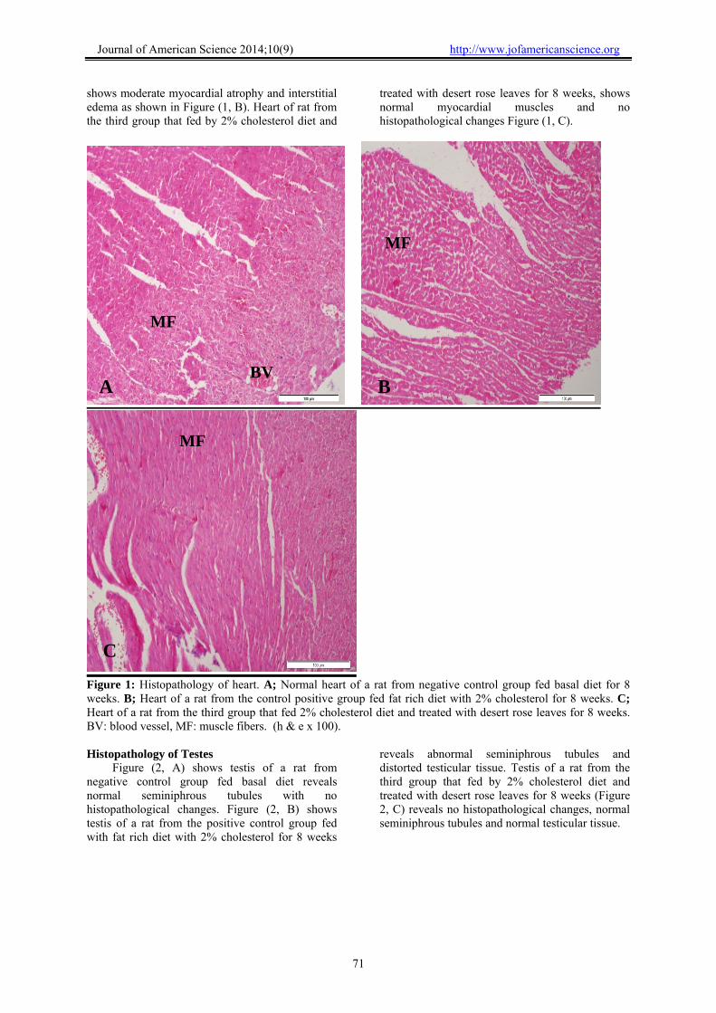
Histopathology of heart of normal rat from negative control group fed basal diet for 8 weeks
reveals normal cardiac tissues with no histopathological changes as shown in Figure (1, A). Heart of the positive control group which fed with fat rich diet with 2% cholesterol for 8 weeks
http://www.jofamericanscience.org ) 9(102014;Journal of American Science
71
shows moderate myocardial atrophy and interstitial edema as shown in Figure (1, B). Heart of rat from the third group that fed by 2% cholesterol diet and
treated with desert rose leaves for 8 weeks, shows normal myocardial muscles and no histopathological changes Figure (1, C).
Figure 1: Histopathology of heart. A; Normal heart of a rat from negative control group fed basal diet for 8 weeks. B; Heart of a rat from the control positive group fed fat rich diet with 2% cholesterol for 8 weeks. C; Heart of a rat from the third group that fed 2% cholesterol diet and treated with desert rose leaves for 8 weeks. BV: blood vessel, MF: muscle fibers. (h & e x 100). Histopathology of Testes
Figure (2, A) shows testis of a rat from negative control group fed basal diet reveals normal seminiphrous tubules with no histopathological changes. Figure (2, B) shows testis of a rat from the positive control group fed with fat rich diet with 2% cholesterol for 8 weeks
reveals abnormal seminiphrous tubules and distorted testicular tissue. Testis of a rat from the third group that fed by 2% cholesterol diet and treated with desert rose leaves for 8 weeks (Figure 2, C) reveals no histopathological changes, normal seminiphrous tubules and normal testicular tissue.
MF
MF
MF
BV A B
C

http://www.jofamericanscience.org ) 9(102014;Journal of American Science
72
Figure 2: Histology of Testes. A; Testis of a rat from negative control group fed basal diet 8 weeks. B; Testis of a rat from positive control group fed fat rich diet with 2% cholesterol for 8 weeks. C; Testis of a rat from the third group that fed 2% cholesterol diet and treated with desert rose leaves for 8 weeks. Spermatogonia (arrow), ST: seminiferous tubules. (h & e x 100). 4. Discussion
This study describes a new in vivo investigation to test the effect of desert rose leaves (Adenium arabicum Forssk.) supplementation on lipid profile in male rats. No previous studies were recorded on this plant material in treatments of diseases except some local people who use desert rose seeds as cardiac drug with sever side effect. Hypercholesterolemia often occurs in conjunction with other metabolic risk factors including glucose intolerance, obesity, diabetes and metabolic syndromes and oxidation of the lipid core of low-density lipoproteins leads to a change in the lipoprotein conformation (Rathod et al., 2011).
Feeding rats with 2% cholesterol increased the serum total cholesterol in the positive control group (Onody et al., 2003 and El Rabey et al., 2013). Treating hypercholesterolemic rats with desert rose leaves (500 mg/kg body weight in the diet for 8 weeks) has significantly reduced the serum lipid profile.
In addition, liver function parameters (serum alanine aminotransferase, serum aspartate transaminase and serum alkaline phosphatase) were also significantly affected under induced hyper lipidemic condition in male rats. This result is consistent with that of El Rabey et al. (2013). However, treating hypercholesterolemic rats with
ST
ST
ST
A B
C
http://www.jofamericanscience.org ) 9(102014;Journal of American Science
73
desert rose leaves has greatly improved the liver function enzymes under study.
Kidney functions parameters; serum uric acid was not affected with hypercholesterolemia, whereas creatinine and urea were significantly affected in the positive control group. This result is consistent with other studies demonstrated a relationship between kidney disease and increased cholesterol in the diet (Trevisan et al., 2006 and El Rabey et al., 2013). Treating hypercholesterolemic rats with desert rose leaves for 8 weeks has greatly improved the kidney function parameters.
Biological parameters;, organ weight (heart, right testis and left testis) and relative organ weight were not affected either by cholesterol supplementation or desert rose leaves treatment.
The histopathological investigations of heart and testis showed histopathological alteration as a result of 2% cholesterol feeding in the positive control group. This result is consistent with other studies showed a correlation between hypercholesterolemia and histological changes in these organs under study (Altunkaynak et al., 2008, Ouvrier et al., 2011 and El Rabey et al., 2013), whereas an improvement in microscopic examination of tissues of the desert rose leaves fed group of the tested organs was noticed. Conclusion:
The current study presented the role of desert rose leave powder supplementation in treating hyperlipidemia induced by supplementing male albino rat with 2% cholesterol in the diet. The use of desert rose leaves, reach to an improvement in lipid profile, improvement of liver and kidney function, and improvement in tissues of heart and testis was encountered for the hyperlipidemia. Acknowledgement
The project was funded by the Deanship of Scientific Research (DSR), King Abdulaziz University, Jeddah, under grant No. 1434/130/345. The authors, therefore, acknowledge with thanks DSR technical and financial support. References Ahmed O.M. (2001). Histopathological and
biochemical evaluation of Liver and kidney lesions in streptozotocin diabetic rats treated with glimepiride and various plant extracts. J. Union Arab Biol., 164: 585-625.
Altunkaynak M.E., Ozbek E., Altunkaynak B.Z., Can I., Unal D. and Unal B. (2008). The effects of high fat diet on the renal structure and morphometric parametric of kidneys in rats. Journal of anatomy, 212: 845-852.
Amundsen A.L., Ose L., Nenseter M.S. and Natnios F.Y. (2002). Plant sterol ester-enriched spread lowers plasma total and LDL cholesterol
in children with familial hypercholesterolemia. Am. J. Clin. Nutr., 76: 338-344.
Atawodi S.E., Ameh D.A., Ibrahim S. et al. (2002). Short communication Indigenous knowledge system for treatment of trypanosomiasis in Kaduna state of Nigeria. Journal of Ethnopharmacology, 79: 279–282.
Bentley M.D., Rodriguez-Porcel M., Lerman A., Hershman-Sarafov M. and Romero J.C., et al. (2002). Enhanced renal cortical vascularization in experimental hypercholesterolemia. Kidney Int., 61: 1056-1063.
Drury R.A., Wallington E.A. and Cancerson R. (1976). Carlton’s histopathological techniques, 4th edn. Oxford University Press, Oxford, 1976.
El Rabey H.A., Al-Seeni M.N. and Amer H.M. (2013). Efficiency of Barley Bran and Oat Bran in Ameliorating Blood Lipid Profile and the Adverse Histological Changes in Hypercholesterolemic Male Rats. BioMed Research International, Article ID 263594, 10 pages.
Kiyohara H., Ichino C., Kawamura Y., Nagai T., Sato N., Yamada H., Salama M.M. and Abdel-Sattar E. (2012). In vitro anti-influenza virus activity of a cardiotonic glycoside from Adenium obesum (Forssk.). Phytomedicine, 19:111-114.
Magassouba F.B., Diallo A., Kouyat´ M. et al. (2007). Ethnobotanical survey and antibacterial activity of some plants used in Guinean traditional medicine. Journal of Ethnopharmacology, 114: 44–53.
Makni, M., Fetoui H., Gargouri N. K., Garoui E. M., Jaber H., Makni J., Boudawara T. and Zeghal N. (2008). Hypolipidemic and hepatoprotective effects of flax and pumpkin seed mixture rich in 3-‰د and 6- ‰د fatty acids in hypercholesterolemic rats. Food and Chemical Toxicology, 46: 3714-3720.
Molander M., Saslis-Lagoudakis C.H., Jager A.K. and Rønsted N. (2012). Cross-cultural comparison of medicinal floras used against snakebites. Journal of Ethnopharmacology, 139: 863– 872.
Mossa JS, Al-Yahya MA, Al-Meshal IA. Medicinal plants of Saudi Arabia. Riyadh: King Saud University Press, 1987.
Neuwinger H.D. (2004). Plants used for poison fishing in tropical Africa. Toxicon 44: 417–430.
Onody, A, Csonka C, Giricz Z, Ferdinandy P. (2003). Hyperlipidemia induced by a cholesterol-rich diet leads to enhanced peroxynitrite formation in rat hearts. Cardiovasc Res. 1, 58(3):663-70.
Ouvrier A., Alves G., Damon-Soubeyrand C., Marceau G., Cadet R., Janny L., Brugnon F., Kocer A., Pommier A. and Lobaccaro J.M.A. (2011). Dietary cholesterol-induced post-testicular infertility. PloS one, 6 (11): 26966.
http://www.jofamericanscience.org ) 9(102014;Journal of American Science
74
Rahman M.A., Jaber S. Mossa, Mansour S. Al-Said, Mohammed A. Al-Yahya (2004). Medicinal plant diversity in the flora of Saudi Arabia 1: a report on seven plant families. Fitoterapia, 75 (2004) 149–161.
Rathod D., Dodiya H. and Goswami S. (2011). Effect of Nicorandil: A Potassium Channel Opener against Experimentally-induced
Hyperlipidemia. International Journal of Pharmacology, 7 (6): 690-696.
Shahina A.Z. (1994). Handbook of Arabian medicinal plants. Boca Raton, Florida—London—Tokyo: CRC Press, 1994.
Trevisan R., Dodesini A.R. and Lepore G. (2006). Lipids and renal disease. Journal of the American Society of Nephrology, 17:S145-S147.
6/13/2014