tesis final smm sophie 260817hera.ugr.es/tesisugr/28031180.pdf · 2018. 1. 16. · garantizamos, al...
TRANSCRIPT


Universidad de Granada
Programa de doctorado en Biología Fundamental y de Sistemas
Response of the bacterial communities in oxygen limited environments to the presence of polycyclic
aromatic hydrocarbons (PAHs).
TESIS DOCTORAL
Sophie Marie Martirani Von Abercron
Granada, 2017

Universidad de Granada EEZ-CSIC
Programa de doctorado en Biología Fundamental y de Sistemas
Response of the bacterial communities in oxygen limited environments to the presence of polycyclic
aromatic hydrocarbons (PAHs).
Tesis doctoral presentada por la Licenciada en Ciencias Ambientales Sophie Marie Martirani Von Abercron
para optar al Título de Doctora
Fdo.: Sophie Marie Martirani Von Abercron
VoBo de la directora de Tesis
Dra. Silvia Marqués Martín Investigadora Científica CSIC,
Estación Experimental del Zaidín

Editor: Universidad de Granada. Tesis Doctorales Autora: Sophie Marie Martirani Von AbercronISBN: 978-84-9163-694-6 URI: http://hdl.handle.net/10481/48911

El doctorando / The doctoral candidate [ Sophie Marie Martirani Von Abercron ] y los directores de la tesis / and the thesis supervisor/s: [ Silvia Marques Martin ]
Garantizamos, al firmar esta tesis doctoral, que el trabajo ha sido realizado por el doctorando bajo la dirección de los directores de la tesis y hasta donde nuestro conocimiento alcanza, en la realización del trabajo, se han respetado los derechos de otros autores a ser citados, cuando se han utilizado sus resultados o publicaciones.
/
Guarantee, by signing this doctoral thesis, that the work has been done by the doctoral candidate under the direction of the thesis supervisor/s and, as far as our knowledge reaches, in the performance of the work, the rights of other authors to be cited (when their results or publications have been used) have been respected.
Lugar y fecha / Place and date:
Granada, 14 de Junio de 2017
Director/es de la Tesis / Thesis supervisor/s; Doctorando / Doctoral candidate:
Firma / Signed Firma / Signed


Esta tesis doctoral ha sido realizada en el Grupo de Microbiología Ambiental y Biodegradación perteneciente al Departamento de Protección Ambiental de la Estación Experimental del Zaidín (CSIC), gracias a una beca JAE Predoc del CSIC concedida a Sophie Marie Martirani Von Abercron, cuya Investigación ha sido financiada por los proyectos BIO-2011-23615 del MICINN y P08-CVI03591 de la Junta de Andalucía.
Parte de los resultados de este trabajo han sido presentados en los siguientes congresos y publicaciones:
Publicaciones
Acosta-González A, Martirani-Von Abercron SM, Rosselló-Mora R, Wittich RM & Marqués S. The effect of oil spills on the bacterial diversity and catabolic function in coastal sediments: a case study on the Prestige oil spill. Env Sci Pollut Res, 22:15200-15214 (2015). DOI: 10.1007/s11356-015-4458-y
Martirani-Von Abercron SM, Pacheco D, Benito-Santano P, Marín P and Marqués S. Polycyclic Aromatic Hydrocarbon-Induced Changes in Bacterial Community Structure under Anoxic Nitrate Reducing Conditions. Front Microbiol 7:1775 (2016). DOI:10.3389/fmicb.2016.01775. Martirani-Von Abercron SM, Marín P, Solsona-ferraz M, Castañeda-Cataña MA and Marqués S. Naphthalene biodegradation under oxygen limiting conditions: changes in the bacterial community and relevance of biofilm formation capacity. Microb Biotechnol, 2017 DOI: 10.1111/1751-7915.12842
Congresos
Martirani Von Abercron SM, Acosta González A, Rosselló-Mora R, Rodríguez-Garcia I and S Marqués. Anaerobic PAHs degradation: diversity of naphthalene degrading bacteria from marine polluted sediments. FEMS, 4th Congress of European Microbiologists, Geneva, Suiza, 26-30 de junio de 2011.
Martirani Von Abercron SM, Acosta González A, Rosselló-Mora R and S Marqués. Microbial diversity in polluted sediment focusing on nitrate-reducing communities. Environment workshops 2011, Metagenomics and Environmental Microbiology, UNIA, Baeza, Spain. 3-5 October 2011
Rama-Garda R, Pacheco D, Martirani Von Abercron SM, Marín P, Acosta-González A & Marqués S. Occurrence and diversity of the novel oxidative pathway involved in the anaerobic degradation of dihydroxylated aromatic compounds. FEMS, 5th Congress of European Microbiologists, Leipzig, Germany, July 21-25, 2013.
Martirani Von Abercron SM, Marín, P, Solsona-Ferraz, M and Marqués, S. Characterization of a naphthalene-degrading multi-species biofilm isolated from oil-contaminated ground water. FEMS, 6th Congress of European Microbiologists, Maastricht, the Netherlands, June 7-11, 2015.
Martirani Von Abercron SM, Marqués S and Wittich RM. Towards the anaerobic-aerobic bacterial mineralization of chlorendic acid. FEMS, 6th Congress of European Microbiologists, Maastricht, the Netherlands, June 7-11, 2015.
Marqués S, Martirani Von Abercron SM, Marín P, Solsona M y Castañeda M. Biodegradación de hidrocarburos aromáticos policíclicos en condiciones de limitación de oxígeno. VI Congreso De Microbiología Industrial Y Biotecnología Microbiana (CMIBM). León (España) 12-14 de septiembre de 2016.
Martirani von Abercron SM, Marín P, Solsona M, Castañeda M and Marqués S. Production of bacterial added-value polymers from polycyclic aromatic hydrocarbons (PAHs). BioRemid 2017, International Meeting on New Strategies in Bioremediation Processes, supported by the Group of Biodegradation, Bioremediation and Biodeterioration (BBB) of the Spanish Society of Microbiology (SEM), Granada, Spain, 9-10 March 2017.


A mi queridísima y especial familia Giuliana, Charles, Giovanna, Martin, Elena y Zia Maria
A mí ‘familia personal’, mis amores Gaetano, Jonathan y Christian


No te deseo un regalo cualquiera, te deseo aquello que la mayoría no tiene,
te deseo tiempo, para reír y divertirte. Si lo usas adecuadamente, podrás obtener lo que quieras.
Te deseo tiempo para tu quehacer y tu pensar,
no sólo para ti mismo, sino también para dedicárselo a los demás.
Te deseo tiempo, no para apurarte y andar con prisas,
sino para que siempre estés contento.
Te deseo tiempo, no sólo para que transcurra, sino para que se quede: tiempo para asombrarte y tiempo para tener confianza,
y no sólo para que lo veas en el reloj.
Te deseo tiempo para tener esperanza otra vez, y para amar; no tiene sentido añorar.
Te deseo tiempo para que te encuentres contigo mismo.
Para vivir cada día, cada hora, cada minuto, como un regalo.
También te deseo tiempo para perdonar y aceptar.
Te deseo de corazón que tengas tiempo, tiempo para la vida, y para tu vida.
Elli Michler


Agradecimientos
Esta aventura empezó hace algunos años, cuando decidí seguir con la investigación después de la carrera. Aunque no muchos entendían porqué quería seguir investigando, porqué quería seguir siendo un ‘estudiante’, y no encontrar un trabajo ‘normal’ en mi ciudad, en mi país, yo deseaba seguir descubriendo, seguir aprendiendo, seguir buscando respuestas, seguir mi pasión. Al final no sé cuántas respuestas concretas he encontrado, muy pocas probablemente, más bien tengo muchas más preguntas e incertidumbres. Pero sí he crecido como persona, y ¡mi corazón se ha enriquecido gracias a todas las personas que he encontrado por el camino!
La primera fue Silvia…un sábado durante las vacaciones de navidad recibió mi CV y el lunes me contestó con algunas preguntas y luego: ¡venga vamos a probar las becas! Me diste tu confianza en seguida junto con un proyecto que yo veía mucho más grande que mis posibilidades, porque no sabía nada, tenía que aprender mucho, partiendo del idioma. He hecho muchos errores, pero tú me dejaste hacerlos, me dejaste pensar con mi cabeza, sin decirme nunca que tenía que hacer. A veces me has dicho, también un poco enfadada: ‘¡Sophie tienes que pensar!’. Alguna vez me pregunté: ‘¿porque no me dice que tengo que hacer y punto?’, porque han sido muchos los momentos que me sentía perdida…hubiera sido más fácil ¡pero no lo correcto! Eres una persona muy justa y siempre dices lo que piensas, y esto lo hace todo más fácil, aunque también más duro, porque lo más difícil es aceptar y mejorar lo que somos. Tu nos enseñas a reflexionar con nuestras cabezas, a hacer nuestros errores, nos dejas caer así que podamos levantarnos más fuertes...y tú estás ahí desde fuera, y cuando acudimos a ti por ayuda siempre nos la das...también desde lejos, cuando me he tenido que ir por trabajo y por mi familia has estado ahí dándome caña y ánimo y sobre todo toda la ayuda que he necesitado. ¡¡Gracias!!
Luego he entrado en el laboratorio 316 donde he tenido la suerte de encontrar una ¡FAMILIA! El primero que he conocido fue Alejo en Sevilla. ¡Alejo te he querido y te quiero muchísimo! Has sido mi referencia por mucho tiempo…no sé cuantas preguntas te echo, y tú siempre has tenido la paciencia de contestarme y de enseñarme todo lo que sabías. ¡Con tu Sofí y Mija me habéis acogido en vuestra casa como una hermana! Águeda tu amistad, tus risas y alegría todavía me llenan el corazón, has estado ahí en mis momentos de bajón dándome animo con tu positividad. Moha…el hombre HPLC…que decirte…has sido mi amigo, compañero, hermano, me has acompañado en todos momentos en lab: ayudándome en el trabajo, discutiendo juntos de los bichos anaerobios y del naftaleno, haciéndome partir de las risas, con tus billetitos de buenos días en todos lados y tus ’Ispecciónes’…jajajaja…¡no sabes cuánto te hemos echado de menos en el lab! ¡Eres una de las personas más lista que conozco, espero verte pronto!
Y luego están ellos…lo que han estado conmigo desde el primer día hasta el último, y siguen en mi vida, en mi corazón cada día: ¡Patri y Pacheco! No creo que unas pocas líneas puedan expresar lo que tengo en mi corazón. Me habéis acogido juntos con Antonio y Mónica en vuestras casas como una más de vuestra familia. ¡Os Quiero con locura! Patri he tardado en conocerte, eres reservada. Nuestra amistad ha ido intensificándose poco a poco, hasta que entraste en mi corazón y yo te di el mío, muchas veces un poco roto para que me lo arreglaras. Me siento privilegiada al haberte conocido, tu generosidad e inteligencia me sorprenden cada día. ¡No he visto a nadie trabajar como tú en lab! Eres mi amiga, hermana y consejera. Muchas veces me pregunto ¿que haría Patri? Sobre todo cuando no sé que hacer con mis niños :)
Pachequito mío, hemos empezado juntos esta aventura y seguimos en ello. Has estado conmigo desde el primer día, cuando no conocía a nadie ni sabía el idioma ni nada y me hiciste sentir como en casa

abriéndome la tuya. En seguida me quisiste, aunque decías que no querías a nadie. :D Eres mi hermano, mi sabio hermano mayor, tus palabras y tus consejos los tengo bien guardados. Me has dicho siempre lo que piensas sin reservas, y también nos hemos peleado a veces, ¿pero acaso no es esto lo que hacen las personas que se quieren? Patri y Pacheco gracias por cuidar de mí incluso en la distancia.
No puedo olvidarme de todos los ‘pollitos’ que han pasado en el lab. Ruth, me has acompañado en mi primer año, juntas hemos empezado los experimentos de anaerobios. Ramon, no olvidaré la ayuda que me diste cuando estaba con mi barriguita, y sobre todo las tartas de chocolate que hiciste para mis antojos :) ¿¡¡¿ah y recuerdas de los NMP y DGGE?!!? Mi Patri chica: ¡gracias por tu ayuda sin fin y tu cariño! Eres una trabajadora única. Alberto te quiero muchísimo, me acuerdo todavía cuando llorabas como un niño chico y un poco borracho en mi boda porque te ibas del lab. ¡Eres especial! Marta, tu fuerza, tu sonrisa y determinación a pesar de las pruebas que la vida te ha dado me siguen asombrando, ¡eres muy valiente!
Quiero agradecer también a todo el grupo de degradación de tóxicos, y el resto de la EEZ. Si me olvido de algunos ¡perdonadme!
A Miriam gracias por tu amistad, tu cariño, tus abrazos, tu sonrisa, tu fuerza, tu apoyo en todo momento, por nuestras charlas sobre la vida, muchas veces necesitaba otro punto de vista. ¡Te echo de menos y te quiero! A Patri B. por abrirme las puertas de tu casa y acogerme, por tu amistad, tu afecto, tu dulzura y por compartir tantos momentos conmigo. ¡Te he extrañado mucho cuándo te fuiste a Londres! A Carlos por tu amistad desde el primer momento, por tu disponibilidad a ayudarme siempre y por tu animo tan gentil. A Ali por tu sonrisa contagiosa en todos momentos, y sobre todo por tu sonrisa, optimismo y energía en los momentos más difíciles. A Joaquín… has llegado más tarde, pero hemos compartido tantos momentos. Gracias por tu amistad, tu cariño, tu alegría y por las palabras bonitas que me has dedicado. A Cristi y Josemi por nuestros desayunos llenos de risas que me han recargado cada día y por vuestra disposición a ayudar. A Jesús por ayudarme y por enseñarme tantas cosas de ciencia y no. A Oscar por tu energía, fuerza y sonrisa siempre en la cara. A Laura por tu amistad y por escucharme muchas veces. A Silvia por estar siempre disponible y por hacerme reír tanto. A María por tu simpatía y cariño, eres muy fuerte y paciente como no he visto nunca. A Miguel que he conocido en los últimos meses y que me ha regalado tanta alegría.
A Zulema, Saray, Marta, Vero, Mati, Lázaro, Alicia, Álvaro, Andrés, Jose, Nené, Marta, Sara, Bertrand, Sandy, Ana María, Adela por todos los momentos que he tenido la suerte de compartir con vosotros, cada uno me ha dejado su huella.
A M. Angustias por tener el cariño de una mamá y por hacer cada día más fácil y ligero.
A los jefes: Juan Luis, no he visto jefe más listo y carismático. A pesar de tu poco tiempo siempre has tenido unas palabras para dedicarnos, de ciencia o de apoyo. Estrella, tu cariño y atenciones me han acompañado desde mi primer día en Granada. Regina, gracias por tu tiempo y consejos sobre los anaerobios y sus rutas y por preocuparte por mí. Pieter, gracias por ser siempre disponible a enseñarme todo lo que sabes sobre la piro y el QIIME y por darme animo cuando lo he necesitado. Echaré de menos nuestras charlas en el servidor :) Marian, eres un ejemplo de mujer, madre e investigadora. Gracias por darme fuerza y apoyo. Tino, gracias por tus consejos y confianza. Ana gracias por tu ayuda y sugerencias. Maribel y Manolo gracias por vuestras sugerencias y por compartir vuestro conocimiento.

A Bea por tu amistad y afecto. Por escucharme y apoyarme todas las veces que lo he necesitado.
A Pao, la persona más dulce que he conocido nunca. Eres encantadora y me has regalado tu cariño y afecto desde el primer momento que nos conocimos. No pierdas nunca esta sonrisa tierna que te hace una persona maravillosa.
A Rafa mil gracias por estar siempre disponible a explicarme con paciencia una y otra vez y con una sonrisa todo sobre el GC-MS, para darme tantas ideas diferentes y para dedicarme tanto tiempo. Haces tu trabajo con tanta dedición e ilusión.
A Javi y César por ser siempre disponibles con todas mis dudas y problemas informáticos.
A mi familia ‘napolitana’ en Granada: Chiara y Salvatore. Chiara, mí Amiga, mi alma gemela, ya son 16 años de amistad pura y sincera, contigo no necesito decir una palabra, porque tienes la capacidad de leerme dentro. Sin ti nunca habría venido a Granada, y haberte tenido a mi lado ha sido un privilegio. Salvatore se dice ‘quien encuentra un amigo encuentra un tesoro’. Pues contigo ¡ya soy la mujer más rica del mundo! Has estado y estas siempre, y digo siempre ahí todas las veces que hemos necesitado ayuda. Las veces me ha hecho reír de corazón son infinitas. Estar lejos de la familia, sobre todo con un niño y trabajando, es duro, pero vosotros lo habéis hecho mucho más llevadero. ¡Chicos os quiero con toda mi alma y no sabéis cuanto os echo de menos!
A mi familia tan querida y tan especial. A mi mamá por ser la persona más valiente que he conocido, por ser un modelo que seguir, por no rendirte nunca, por darme tu amor incondicionado siempre, por transformarme en la mujer que soy, por confiar siempre en mí, por dejarme soñar mis sueños, por apoyarme en todo momento…¡¡Gracias!! A mi hermano, la persona más generosa y lista que conozco, gracias por quererme a tu lado, por amarme tanto, por confiar ciegamente en mí, por hacerme ver el mundo desde otra perspectiva, por hacerme reflexionar tanto y por estimularme siempre a mejorar…eres mi orgullo, mi fuerza… contigo a mi lado siempre me sentiré segura y protegida…¡te quiero y te cuidaré siempre! A Giovanna, Martin y Elena por vuestro amor tan infinito, por la alegría y por toda la felicidad que me regaláis cada vez que os veo. ¡Ha sido muy difícil estar lejos de vosotros! A mi tía María, eres una madre y una confidente, la persona más buena del mundo. No hay día sin ti, sin tu voz, sin tu dulce sonrisa impresa en mi mente…estarás siempre en mi corazón, en un lugar especial y seguro.
A mis amores, mis hombres, mi todo…Gaetano son 13 años que estas a mi lado, en cualquier lugar del mundo tu estas ahí…primero en Nápoles, y luego en Australia, en Suecia, en Granada, en Albania, en Ibiza y dondequiera sé que tu estarás. Tu prioridad ha sido siempre amarme y crecer juntos, por esto viniste a Granada donde hemos construido nuestro hogar, donde hemos iniciado nuestra familia con el don más grande que la vida nos podía regalar: Jonathan. Gracias por ayudarme tanto a seguir adelante con la tesis, porque si este año he parido dos veces es también gracias a ti. Gracias por darme fuerza cada día, gracias por aceptarme por lo que soy con todas mis debilidades, gracias por alejar de mi la tentación de rendirme, gracias por ser la constante en mi vida, gracias por tu comprensión, gracias por hacer mi vida aún más maravillosa. Y a ti, mi Principito, este año han sido demasiadas las veces que te he tenido que decir que no, sea por el trabajo, por la tesis o por el embarazo…pero tú estabas siempre con una sonrisa diciéndome que estabas enamorado de mí, que la cosa más bella soy yo, que lo más bonito es darme abracitos y besitos. Gracias por enseñarme tanto cada día, por demostrarme que realmente el amor puede ser infinito y que una sonrisa basta para ser feliz. Y por último a ti, mi pequeño gordito, que me has acompañado más que nadie en la escritura de la tesis, y que ya sabes muchas cosas sobre el naftaleno, gracias por recordarme con tus pataditas que la vida es un milagro y hay que cuidarlo.


i
Contents
FIGURE INDEX .............................................................................................................................................................. IV TABLE INDEX................................................................................................................................................................ IV LIST OF ABBREVIATIONS ................................................................................................................................................. VII RESUMEN .................................................................................................................................................................... IX SUMMARY ................................................................................................................................................................... XI
I. GENERAL INTRODUCTION .......................................................................................................... 1
1. THE ENVIRONMENTAL QUESTION ............................................................................................................................ 3 2. POLYCYCLIC AROMATIC HYDROCARBONS ................................................................................................................... 4
2.1. General properties ................................................................................................................................. 4 2.2. Physicochemical properties ................................................................................................................... 5 2.3. Toxicity .................................................................................................................................................. 6
3. PAHS IN THE ENVIRONMENT .................................................................................................................................. 7 4. BIOREMEDIATION OF PAHS ................................................................................................................................... 9
4.1. Novel bioremediation strategies ......................................................................................................... 11 5. PAH DEGRADATION BY BACTERIA .......................................................................................................................... 12
5.1. Aerobic PAH degradation .................................................................................................................... 13 5.2. Anaerobic PAH degradation ................................................................................................................ 18 5.2.1. Anaerobic naphthalene degrading microorganisms. .......................................................................... 21 5.2.2. Anaerobic naphthalene biodegradation mechanisms ......................................................................... 22
6. MICROBIAL DIVERSITY ......................................................................................................................................... 25 7. MICROBIAL COMMUNITY ANALYSIS ........................................................................................................................ 27
7.1. Culture dependent methods ................................................................................................................ 27 7.2. Culture independent methods ............................................................................................................. 28
8. REFERENCES ..................................................................................................................................................... 31
II. OBJECTIVES ............................................................................................................................. 41
III. GENERAL METHODS ................................................................................................................ 45
1. CULTURE CONDITIONS, ENRICHMENT AND ISOLATION PROCEDURES .............................................................................. 47 2. MOST PROBABLE NUMBER ENUMERATION OF DENITRIFYING BACTERIA ......................................................................... 48 3. CHEMICAL ANALYSIS OF HYDROCARBON CONTENT .................................................................................................... 48 4. TOTAL DNA EXTRACTION, PCR AMPLIFICATION AND PYROSEQUENCING LIBRARY CONSTRUCTION ...................................... 49 5. 16S RRNA GENE AMPLIFICATION .......................................................................................................................... 50 6. DATA ANALYSIS ................................................................................................................................................. 50 7. REFERENCES ..................................................................................................................................................... 51
IV. RESULTS AND DISCUSSION ...................................................................................................... 53
CHAPTER 1 ...................................................................................................................................... 55
POLYCYCLIC AROMATIC HYDROCARBON-INDUCED CHANGES IN BACTERIAL COMMUNITY STRUCTURE UNDER ANOXIC NITRATE REDUCING CONDITIONS. ........................................................................... 55
1.1. INTRODUCTION ............................................................................................................................................. 59 1.2. MATERIALS AND METHODS ............................................................................................................................. 61
1.2.1. Sample collection and experimental design ........................................................................................ 61 1.2.2. Culture conditions, enrichment and isolation procedures ................................................................... 61 1.2.3. Most probable number enumeration of denitrifying bacteria ............................................................ 63 1.2.4. Chemical analysis ................................................................................................................................ 64 1.2.5. Total DNA extraction, PCR amplification and pyrosequencing library construction ........................... 65 1.2.6. Functional gene amplification ............................................................................................................. 65 1.2.7. 16S rRNA gene amplification ............................................................................................................... 66 1.2.8. Data Analysis ....................................................................................................................................... 66 1.2.9. Nucleotide sequence accession numbers ............................................................................................ 67
1.3. RESULTS AND DISCUSSION ............................................................................................................................... 68 1.3.1. Presence of a nitrate-reducing, PAH-degrading bacterial community in environmental samples ...... 68 1.3.2. Bacterial community structure in the selected environments ............................................................. 70

ii
1.3.3. General effect of PAHs on the bacterial community structure ............................................................ 74 1.3.4. Effect of PAHs on specific bacterial populations ................................................................................. 77 1.3.5. Isolation of nitrate reducing bacteria from PAH enrichments ............................................................. 81 1.3.6. Aromatic anaerobic degradation genes .............................................................................................. 82
1.4. CONCLUDING REMARKS .................................................................................................................................. 83 1.5. REFERENCES ................................................................................................................................................. 84 1.6. SUPPLEMENTARY MATERIAL ............................................................................................................................ 89
1.6.1. Supplementary tables .......................................................................................................................... 89 1.6.2. Supplementary figures ........................................................................................................................ 97
CHAPTER 2 .................................................................................................................................... 103
MICROBIAL COMMUNITIES IN FUENTE DE PIEDRA ATHALASSOHALINE LAGOON SEDIMENTS: IMPACT OF THE CONTAMINATION WITH NAPHTHALENE. ............................................................................ 103
2.1. INTRODUCTION ........................................................................................................................................... 107 2.2. MATERIALS AND METHODS ........................................................................................................................... 109
2.2.1. Site description and Sampling ........................................................................................................... 109 2.2.2. Microcosm experiment ...................................................................................................................... 109 2.2.3. Culture conditions and anaerobic enrichment .................................................................................. 111 2.2.4. Total DNA extraction ......................................................................................................................... 112 2.2.5. Illumina sequencing and Data Analysis ............................................................................................. 114
2.3. RESULTS AND DISCUSSION ............................................................................................................................ 115 2.3.1. Effect of naphthalene on bacterial community structure and diversity in the microcosm. ............... 115 2.3.2. Microcosms bacterial community structure ...................................................................................... 117 2.3.3. Effect of microcosm incubation on bacterial communities ............................................................... 119 2.3.4. Bacterial community evolution in naphthalene enrichments ............................................................ 122 2.3.5. Patterns of microbial diversity in the anaerobic enrichments ........................................................... 125 2.3.5.1. First sampling campaign: enrichments with PAHs dissolved in HMN. .......................................... 125 2.3.5.2. Second sampling campaign: enrichment with naphthalene provided as crystals. ....................... 127
2.4. CONCLUDING REMARKS ................................................................................................................................ 130 2.5. REFERENCES ............................................................................................................................................... 131 2.6. SUPPLEMENTARY MATERIAL .......................................................................................................................... 136
CHAPTER 3: ................................................................................................................................... 137
NAPHTHALENE BIODEGRADATION UNDER OXYGEN LIMITING CONDITIONS: COMMUNITY DYNAMICS AND THE RELEVANCE OF BIOFILM FORMATION CAPACITY .............................................................. 137
3.1. INTRODUCTION ........................................................................................................................................... 141 3.2. EXPERIMENTAL PROCEDURES ........................................................................................................................ 143
3.2.1. Sampling and site description. .......................................................................................................... 143 3.2.2. Most Probable Number enumeration of bacteria. ............................................................................ 143 3.2.3. Culture conditions, enrichment, and isolation procedures. ............................................................... 144 3.2.4. Measurement of oxygen concentration in the culture bottles. ......................................................... 145 3.2.5. Synthetic microbial community. ........................................................................................................ 146 3.2.6. Chemical analysis. ............................................................................................................................. 146 3.2.7. Total DNA extraction, 16S rRNA gene 454-Pyrosequencing and data analysis. ................................ 146 3.2.8. Full-length 16S rRNA gene amplification and clone library construction. ......................................... 147 3.2.9. Functional gene amplification. .......................................................................................................... 147 3.2.10. RT-PCR assays. .............................................................................................................................. 148 3.2.11. Biofilm quantification in multi-well plates (crystal violet assay). ................................................. 148 3.2.12. Scanning electron microscopy (SEM). ........................................................................................... 149 3.2.13. Nucleotide sequence accession numbers. .................................................................................... 149
3.3. RESULTS AND DISCUSSION ............................................................................................................................. 150 3.3.1. Betaproteobacteria are dominant in a hydrocarbon polluted aquifer .............................................. 150 3.3.2. Changes in the community structure under strictly anoxic denitrifying conditions. ......................... 154 3.3.3. Microaerophilic conditions strongly select for two dominant strains. .............................................. 156 3.3.4. A pathway with high affinity for oxygen is favoured under microaerophilic conditions. .................. 160 3.3.5. Evolution of a synthetic community under microaerophilic conditions ............................................. 164

iii
3.3.6. Aerobic growth allows higher bacterial diversity .............................................................................. 164 3.3.7. Starkeya novella strain N1B is responsible for biofilm formation and naphthalene degradation. ... 165
3.4. CONCLUDING REMARKS ................................................................................................................................ 166 3.5. REFERENCES ............................................................................................................................................... 167 3.6. SUPPLEMENTARY MATERIAL .......................................................................................................................... 171
3.6.1. Supplementary tables ........................................................................................................................ 171 3.6.2. Supplementary figures ...................................................................................................................... 175
V. GENERAL DISCUSSION ........................................................................................................... 179
1. ANAEROBIC NAPHTHALENE DEGRADATION: THE PROBLEM. ....................................................................................... 182 2. CHANGES IN THE MICROBIAL COMMUNITIES: EFFECT OF THE CULTURE CONDITIONS. ...................................................... 184 3. CHANGES IN THE MICROBIAL COMMUNITIES: EFFECT OF PAHS. ................................................................................. 186 4. NAPHTHALENE-DEGRADING COMMUNITIES UNDER MICROAEROPHILIC CONDITIONS. ..................................................... 188 5. REFERENCES ................................................................................................................................................... 189
VI. CONCLUSIONES ..................................................................................................................... 193

iv
Figure index
GENERAL INTRODUCTION
FIGURE 1. STRUCTURES AND NOMENCLATURES OF THE 16 PAHS ON THE EPA PRIORITY POLLUTANT LIST .........................5 FIGURE 2. GENE ORGANIZATION OF THE NAPHTHALENE DEGRADATION PATHWAY IN NAH7 PLASMID OF PSEUDOMONAS
PUTIDA G7 ............................................................................................................................................ 14 FIGURE 3. PROPOSED CATABOLIC PATHWAYS OF NAPHTHALENE BY AEROBIC BACTERIA ............................................... 15 FIGURE 4. PATHWAY OF ANAEROBIC TOLUENE AND BENZOATE METABOLISM TO BENZOYL-COA IN THAUERA AROMATICA 20 FIGURE 5. GENE PRODUCTS INVOLVED IN THE BENZOYL-COA DEGRADATION PATHWAY IN FACULTATIVE AND OBLIGATE
ANAEROBES ........................................................................................................................................... 21 FIGURE 6. PROPOSED PATHWAYS FOR ANAEROBIC NAPHTHALENE AND 2-METHYLNAPHTHALENE DEGRADATION IN THE
ENRICHMENT CULTURE N47. .................................................................................................................... 24 FIGURE 7. 16S RIBOSOMAL RNA SECONDARY STRUCTURE MODEL AND ITS VARIABLE REGIONS. ................................... 29
RESULTS AND DISCUSSION
Chapter 1
FIGURE 1. 1. MPN ENUMERATION IN THE ENVIRONMENTAL SAMPLES OF NITRATE REDUCING BACTERIA ABLE TO GROW ON DIFFERENT PAHS. ................................................................................................................................... 69
FIGURE 1. 2. CUMULATIVE PLOT OF BACTERIAL PHYLA DETECTED IN THE INITIAL ENVIRONMENTAL SAMPLES ................... 72 FIGURE 1. 3. CUMULATIVE PLOT OF BACTERIAL PHYLA DETECTED IN THE DIFFERENT ENRICHMENTS. .............................. 75 FIGURE 1. 4. PRINCIPAL COORDINATE ANALYSIS (PCOA) AND UNWEIGHTED PAIR GROUP METHOD WITH ARITHMETIC
AVERAGE (UPGMA IN THE INITIAL ENVIRONMENTAL SAMPLES AND THE ENRICHMENTS OBTAINED FROM THEM ..... 76 FIGURE 1. 5. EFFECT OF PAHS ON THE RELATIVE ABUNDANCE OF THE MOST RELEVANT GROUPS IN THE COMPOST PILE (CP)
SAMPLE ................................................................................................................................................ 77
Chapter 2
FIGURE 2. 1. CORES USED IN THIS WORK FOR THE MICROCOSM EXPERIMENT, ENRICHMENT CULTURES AND ILLUMINA-SEQUENCING AT THE START OF THE EXPERIMENT ........................................................................................ 110
FIGURE 2. 2. SCHEME OF EXPERIMENTAL SET-UP ............................................................................................... 113 FIGURE 2. 3. BOXPLOTS OF THE OBSERVED OTUS PER RANDOMIZED SAMPLE IN THE CONTROL AND NAPHTHALENE CORES AT
DIFFERENT INCUBATION TIMES ................................................................................................................ 116 FIGURE 2. 4. ANALYSIS OF MICROCOSM MICROBIAL COMMUNITIES WITH WEIGHTED AND UNWEIGHTED UNIFRAC
DISTANCE.. .......................................................................................................................................... 117 FIGURE 2. 5. RELATIVE ABUNDANCE OF BACTERIAL PHYLA OR CLASS (ONLY FOR PROTEOBACTERIA) IN THE FUENTE DE PIEDRA
SEDIMENT MICROCOSMS ....................................................................................................................... 119 FIGURE 2. 6. EFFECT OF MICROCOSM INCUBATION ON SPECIFIC BACTERIAL POPULATION AT ORDER LEVEL. ................... 120 FIGURE 2. 7. MICROBIAL COMMUNITY ANALYSIS OF THE SEDIMENT SAMPLES AND ENRICHMENT CULTURES USING WEIGHTED
AND UNWEIGHTED UNIFRAC DISTANCE .................................................................................................... 124 FIGURE 2. 8. RELATIVE ABUNDANCE OF BACTERIAL PHYLA OR CLASS (ONLY FOR PROTEOBACTERIA) IN THE FUENTE DE PIEDRA
ENRICHMENTS.. ................................................................................................................................... 126

v
Chapter 3
FIGURE 3. 1. AQUIFER SAMPLE SHOWING A VISIBLE BIOFILM GROWING AT THE INTERFACE BETWEEN THE OIL AND THE WATER LAYERS. .................................................................................................................................... 143
FIGURE 3. 2. OXYGEN CONCENTRATION IN THE MICROAEROPHILIC CULTURE BOTTLES USED IN THIS STUDY ................... 145 FIGURE 3. 3. ALIPHATIC HYDROCARBON DISTRIBUTION (%) IN THE INITIAL CONTAMINATED AQUIFER SAMPLE ............... 151 FIGURE 3. 4. MPN ENUMERATION IN THE AQUIFER INITIAL SAMPLE OF NITRATE REDUCING AND AEROBIC BACTERIA ABLE TO
GROW ON DIFFERENT PAHS AS CARBON SOURCE. ...................................................................................... 152 FIGURE 3. 5. CUMULATIVE PLOT OF BACTERIAL PHYLA DETECTED IN THE INITIAL ENVIRONMENTAL SAMPLES AND IN THE
ANAEROBIC ENRICHMENTS AMENDED WITH NAPHTHALENE (NAP), 2-METHYLNAPHTHALENE (2MN) AND HEPTAMETHYLNONANE (HMN). ............................................................................................................. 155
FIGURE 3. 6. BACTERIAL BIOFILM DEVELOPED AROUND THE NAPHTHALENE CRYSTAL.: ENRICHMENT CULTURE BOTTLE AND SCANNING ELECTRON MICROSCOPY (SEM) MICROGRAPH ........................................................................... 157
FIGURE 3. 7. CUMULATIVE PLOT OF BACTERIAL TAXONS PRESENT AT MORE THAN 1% OF THE COMMUNITY DETECTED IN THE MICROAEROPHILIC, AEROBIC AND SYNTHETIC MICROAEROPHILIC ENRICHMENTS AMENDED WITH CRYSTALS OF NAPHTHALENE AS CARBON SOURCE.. ....................................................................................................... 158
FIGURE 3. 8. NEIGHBOUR-JOINING TREE OF THE 16S RRNA GENE OF THE BACTERIAL STRAINS ISOLATED IN THIS STUDY AND THEIR CLOSEST RELATIVES IN THE DATABASES ............................................................................................ 159
FIGURE 3. 9. PHYLOGENY OF THE ALPHA SUBUNIT OXYGENASE COMPONENT OF HYDROXYLATING NAPHTHALENE DIOXYGENASE AND GENTISATE 1,2-DIOXYGENASE PARTIAL AMINO ACID SEQUENCE RETRIEVED FROM SUBCULTURE 5 AND DIFFERENT ISOLATES OBTAINED IN THIS STUDY. ................................................................................... 163
FIGURE 3. 10. RT-PCR ANALYSIS OF TOTAL RNA EXTRACTED FROM SUBCULTURE 15 .............................................. 163 FIGURE 3. 11. BIOFILM FORMATION AT THE AIR/LIQUID INTERPHASE BY DIFFERENT ISOLATES IN LB OR LB SUPPLEMENTED
WITH A NAPHTHALENE CRYSTAL .............................................................................................................. 165

vi
Table index
GENERAL INTRODUCTION
TABLE 1. PHYSICOCHEMICAL AND CANCEROGENIC PROPERTIES OF 16 PAHS ENLISTED AS PRIORITY POLLUTANTS BY US EPA ..............................................................................................................................................................6
TABLE 2. POSSIBLE FATE OF PAHS IN THE ENVIRONMENT .........................................................................................8 TABLE 3. PROKARYOTIC ABUNDANCE DETERMINED BY TOTAL GENOMIC DIVERSITY IN PROKARYOTIC COMMUNITIES
CALCULATED FROM THE REASSOCIATION RATE OF DNA ISOLATED FROM THE COMMUNITY. ................................. 26
RESULTS AND DISCUSSION
Chapter 1
TABLE 1. 1. CHARACTERIZATION OF THE SAMPLES USED IN THIS STUDY. ................................................................... 62 TABLE 1. 2. COMPARISON OF OTU NUMBER, DIVERSITY, EVENNESS INDICES AND COVERAGE FOR THE DIFFERENT SAMPLES.
........................................................................................................................................................... 71 TABLE 1. 3. RELATIVE ABUNDANCE OF ACIDOBACTERIA IN THE INITIAL SAMPLES AND ENRICHMENT CULTURES. ............... 78
Chapter 2
TABLE 2. 1. PROFILE DESCRIPTION OF THE CORES COLLECTED IN THE FIRST CAMPAIGN. ............................................. 110 TABLE 2. 2. GRAVIMETRIC WATER CONTENT IN THE DIFFERENT CORES LAYERS (P1 TO P4) AND IN THE MIXED LAYER (P1 TO
P4). ................................................................................................................................................... 111 TABLE 2. 3. COMPARISON OF OTU NUMBER, DIVERSITY, EVENNESS INDICES AND COVERAGE FOR THE DIFFERENT CORES
SAMPLES. ............................................................................................................................................ 115 TABLE 2. 4. COMPARISON OF OTU NUMBER, DIVERSITY, EVENNESS INDICES AND COVERAGE FOR THE ENRICHMENT CULTURE
OF THE FIRST AND SECOND SAMPLING EVENT. ............................................................................................ 122
Chapter 3
TABLE 3. 1. AROMATIC HYDROCARBON COMPOSITION AND ABUNDANCE IN THE CONTAMINATED AQUIFER INITIAL SAMPLES. ......................................................................................................................................................... 150
TABLE 3. 2. CHARACTERIZATION OF THE INITIAL POLLUTED AQUIFER SAMPLE. ......................................................... 150 TABLE 3. 3. COMPARISON OF OTU NUMBER, DIVERSITY, EVENNESS INDICES AND COVERAGE FOR THE DIFFERENT SAMPLES.
......................................................................................................................................................... 153 TABLE 3. 4. STRAINS ISOLATED FROM THE MICROAEROPHILIC ENRICHMENT CULTURES. ............................................. 161

vii
List of abbreviations
2MN 2-methylnaphthalene 2NA 2-naphtoic acid Ace Acetate ANT Anthracene AS Activated sludge Bss Benzylsuccinate synthase CODEHOP Consensus degenerate hybrid oligonucleotide primers CP Composting pile DHNCR 5,6-dihydro-2-naphthyl-CoA reductase EEA European environment agency EPA Environmental protection agency EPS Extracellular polymeric substances FdP Fuente de Piedra FISH Fluorescence in situ hybridization GMOs Genetically modified organisms HMN 2,2,4,4,6,8,8-heptamethylnonane HMW High molecular weight LB Luria-Bertani medium LMW Low molecular weight MID Multiplex identifier MOPS 3-(N-morpholino)propanesulfonic acid MPN Most probable number MS Marine sediment sample NCR 2-naphthoyl-CoA reductase Nms Naphthyl-2-methyl succinate synthase NRB Nitrate reducing bacteria OM Organic matter OUT Operational taxonomic unit OYE Old yellow enzymes PAHs Polycyclic aromatic hydrocarbons PCoA Principal coordinate analysis PCR Polymerase chain reaction QIIME quantitative insights into microbial ecology RPCal Rice-paddy in Calasparra RPS Rice-Paddy Soil RPW Rice-Paddy Water RT-PCR Reverse transcription polymerase chain reaction SEM Scanning electron microscopy SMM Seawater minimal medium SRB Sulphate reducing bacteria TCA Tricarboxylic acid cycle THNCR 5,6,7,8-tetrahydro-2-naphthoyl-CoA UPGMA Unweighted pair group method with arithmetic mean

viii

Resumen
ix
Resumen
El uso masivo del petróleo como fuente de energía y como materia prima ha generado serios problemas de contaminación ambiental. Entre los principales contaminantes ambientales, los compuestos aromáticos generan gran preocupación porque son relativamente persistentes en el medio ambiente debido a la alta estabilidad termodinámica del anillo de benceno. El benceno y muchos hidrocarburos aromáticos policíclicos (HAPs) presentan efectos tóxicos, carcinogénicos, mutagénicos y teratogénicos y su eliminación del medio ambiente es importante para proteger el entorno y la salud humana. Una medida que ha tenido un éxito significativo es el empleo de microorganismos para la eliminación de contaminantes con la aplicación de técnicas de biorremediación. Los procesos aerobios de degradación de HAPs se han caracterizado exhaustivamente durante los últimos años; sin embargo, en muchos ambientes naturales la concentración de oxígeno es limitante y, en estas condiciones, los procesos activos son esencialmente anaerobios. La degradación anaeróbica bacteriana de los compuestos monoaromáticos se ha caracterizado en profundidad; no obstante, la degradación de los hidrocarburos aromáticos policíclicos (HAPs) como el naftaleno sólo ha comenzado a ser comprendida en las bacterias reductoras de sulfato, y se sabe muy poco sobre la degradación anaeróbica de los HAPs en bacterias nitrato reductoras. Partiendo de una serie de ambientes que habían sufrido diferentes grados de contaminación por hidrocarburos, utilizamos la técnica del número más probable (NMP) para detectar y cuantificar la presencia de comunidades bacterianas capaces de degradar varios HAPs usando el nitrato como aceptor de electrones. Con el NMP se detectó la presencia de una comunidad nitrato reductora capaz de degradar naftaleno, 2-metilnaftaleno (2MN), y antraceno en algunos de los sitios muestreados. Además, con el fin de aislar cepas capaces de degradar los HAPs en condiciones desnitrificantes, establecimos una serie de cultivos de enriquecimiento con nitrato como aceptor terminal de electrones y HAP como única fuente de carbono y seguimos los cambios en las comunidades bacterianas a lo largo del proceso. Los resultados evidenciaron cambios atribuibles al régimen de respiración de nitrato impuesto, que en varias muestras se exacerbó en presencia de los HAPs. La presencia de naftaleno o 2MN enriqueció a la comunidad en grupos de organismos no cultivados y mal caracterizados, y notablemente en el grupo no cultivado de Acidobacteria iii1-8, que en algunos casos era sólo un componente menor de las muestras iniciales. Otros taxa seleccionados por HAPs en estas condiciones incluyeron Bacilli, que fueron enriquecidos en los cultivos con naftaleno. Varias cepas nitrato reductoras han sido aisladas en medio solido con naftaleno y 2MN como única fuente de carbono, aunque el fenotipo no pudo reproducirse en cultivos líquidos. El análisis de los genes funcionales de degradación anaeróbica de HAPs en las muestras originales y los cultivos de enriquecimiento no revelaron la presencia de secuencias del gen nmsA para la degradación anaerobia de 2MN, pero confirmaron la presencia de genes de bssA relacionados con la degradación anaeróbica de tolueno. En conjunto, estos resultados sugieren que la degradación de las HAPs por las bacterias reductoras de nitrato puede requerir la contribución de diferentes cepas, en condiciones de cultivo que todavía necesitan ser definidas.
Los resultados de los NMP de una de las muestras analizadas, los sedimentos anóxicos de Fuente de Piedra mostraron una comunidad importante de nitrato reductores degradadores de naftaleno y, en menor parte, de 2MN. Quisimos entonces tomar muestras para ver la respuesta de la microbiota autóctona de los sedimentos de la laguna ante la presencia de naftaleno, y además

Resumen
x
iniciar cultivos de sulfato reductores, al ser los sedimentos ricos en sulfatos, para enriquecer degradadores potenciales de HAPs. Las comunidades bacterianas y sus cambios se analizaron mediante secuenciación masiva Illumina MiSeq. Proteobacteria, Bacteroidetes, Chloroflexi, Gemmatimonadetes y Firmicutes fueron los grupos más abundantes de todos los microcosmos y también los más frecuentemente encontrados en ambientes hipersalinos. Después de 4 y 12 meses de incubación con naftaleno, las comunidades bacterianas de los microcosmos no sufrieron cambios relevantes. El factor estructurante de las comunidades no fue la presencia del contaminante sino el tiempo de incubación de los cores. Sin embargo, se observaron cambios significativos en los enriquecimientos. En particular en el medio mínimo salino diseñado para sulfato reductores hubo un aumento significativo de la familia de las Desulfobulbaceae (Deltaproteobacteria), previamente identificadas como degradadoras anaerobias de tolueno y naftaleno. En los cultivos preparados con el agua filtrada de Fuente de Piedra los Firmicutes fueron el taxa más enriquecido con el dominio del genero Dethiosulfatibacter, un grupo de bacteria probablemente relacionado con la degradación de HAPs. El uso de condiciones diferentes de cultivo ha permitido seleccionar poblaciones diversas, y en particular, el empleo de un medio natural se ha revelado una opción valida para enriquecer microorganismos procedentes de un ambiente extremo como es el de Fuente de Piedra. En cualquier caso, serían convenientes ensayos adicionales para determinar la actividad degradadora de las poblaciones seleccionadas, y detectar los posibles genes implicados en la ruta.
Además de condiciones estrictamente anaerobias para nitrato y sulfato reductores, quisimos testar condiciones de microaerofilia para aislar cepas degradadoras de HAPs. Estas condiciones son la que más se acercan, en muchos casos, a las condiciones ambientales reales, donde hay gradientes de concentración de oxígeno. Establecimos cultivos de enriquecimiento a partir de una muestra de acuífero contaminado con petróleo, utilizando condiciones anóxicas o microaerofílicas y HAPs como única fuente de carbono. A pesar de la presencia de una comunidad significativa de bacterias nitrato reductoras detectada con el NMP, la comunidad inicial, dominada por Betaproteobacteria, era incapaz de degradar la HAPs en condiciones anóxicas estrictas, aunque se observó un claro cambio en la estructura de la comunidad hacia un aumento en la Alphaproteobacteria (Sphingomonadaceae), Actinobacteria y un grupo no cultivable de Acidobacteria en los enriquecimientos. Por otro lado, el crecimiento en condiciones microaerofılicas con naftaleno como fuente de carbono evidenció el desarrollo de una estructura de biofilm alrededor del cristal de naftaleno. Después de varios pases, el 97% de la comunidad bacteriana y del biofilm estaba compuesto por solo dos géneros: Variovorax spp. (54%, Betaproteobacteria) y Starkeya sp. (43%, Xanthobacteraceae). Las dos cepas eran capaces de crecer con naftaleno, aunque sólo Starkeya tenía la habilidad de reproducir la estructura del biofilm alrededor del cristal de naftaleno. También se identificó la ruta para la degradación del naftaleno, que incluía dioxigenasas con alta afinidad por el oxígeno y que mostraba una identidad del 99% con el grupo de dbd de Xanthobacter polyaromaticivorans para la degradación de HAPs. Dada la presencia en nuestros primeros pases de los enriquecimientos de Xanthobacter polyaromaticivorans probablemente la ruta de degradación del naftaleno haya sido adquirida por Starkeya por transferencia horizontal. Por otro lado, la capacidad de formar biofilm de Starkeya ha favorecido la cepa en el cultivo, proporcionando una estructura compleja para situarse a una distancia apropiada de la fuente de carbono tóxico y crear un ambiente favorable al crecimiento.

Resumen
xi
En conjunto, hemos estudiado las respuestas de las comunidades bacterianas a los HAPs en las condiciones limitantes de oxígeno más frecuentemente encontradas en la naturaleza (nitrato reductoras, sulfatoreductoras y microaerofilicas) pero menos investigadas tanto por las dificultades de cultivos de estas bacterias como por los largos tiempos de crecimiento que requieren estos microorganismos. No hemos logrado aislar cepas anaerobias estrictas capaces de degradar HAPs, pero sí de individualizar poblaciones bacterianas presentes en la naturaleza y probablemente involucradas en la eliminación de contaminantes en el medio ambiente. También hay bacterias no cultivables (v.g. Acidobacteria, iii1-8) que se han enriquecido, de las cuales todavía desconocemos el papel ecológico, pero que podrían ser importantes en la biodegradación anaerobia. En condiciones de microaerofilia, sí hemos logrado cepas degradadoras, y además capaces de formar biofilm a partir de HAPs. El crecimiento en forma de biofilm es probablemente una manera de crecer que las bacterias adoptan en condiciones de estrés y que le ayuda a hacer frente a los factores adversos. Esto posiblemente nos indique que las condiciones de cultivo que imponemos en el laboratorio son demasiado restrictivas y se alejan de la variabilidad que hay en la naturaleza.
Summary
Polycyclic aromatic hydrocarbons (PAHs) are chemicals of particular environmental concern because of their stability, persistence in the environment and resistance to degradation. Many of them are known to be toxic to various organisms and dangerous for health. They are frequently released into the environment either from natural sources (e.g. hydrocarbon seeps) or as consequence of industrial activities such as the massive transport or the synthesis of added value chemicals. Several strategies are currently available for the remediation of aromatics contaminated sites. Among them, bacterial bioremediation stands out because of its low cost and little physical alteration of the environment, and seems especially suitable for low molecular weight aromatic compounds. The aerobic aromatic metabolism in bacteria has been studied for decades and numerous bacterial strains able to degrade aromatics in the presence of oxygen have been isolated and characterized. However, oxygen is not always available in contaminated sites, such as marine sediments, or as in initially oxic sites turned anoxic due to the high oxygen demand for aerobic degradation processes. Although bacterial anaerobic degradation of mono-aromatic compounds has been characterized in depth, the degradation PAHs such as naphthalene has only started to be understood in sulphate-reducing bacteria, and little is known about the anaerobic degradation of PAHs in nitrate-reducing bacteria. Starting from a series of environments, which had suffered different degrees of hydrocarbon pollution, we used most probable number (MPN) enumeration to detect and quantify the presence of bacterial communities able to degrade several PAHs using nitrate as electron acceptor. We detected the presence of a substantial nitrate-reducing community able to degrade naphthalene, 2-methylnaphthalene (2MN) and anthracene in some of the sites. With the aim of isolating strains able to degrade PAHs under denitrifying conditions, we set up a series of enrichment cultures with nitrate as terminal electron acceptor and PAHs as the only carbon source and followed the changes in the bacterial communities throughout the process. Results evidenced changes attributable to the imposed nitrate respiration regime, which in several samples were exacerbated

Resumen
xii
in the presence of the PAHs. The presence of naphthalene or 2MN enriched the community in groups of uncultured and poorly characterized organisms, and notably in the Acidobacteria uncultured group iii1-8, which in some cases was only a minor component of the initial samples. Other phylotypes selected by PAHs in these conditions included Bacilli, which were enriched in naphthalene enrichments. Several nitrate-reducing strains showing the capacity to grow on PAHs could be isolated on solid media, although the phenotype could not be reproduced in liquid cultures. Analysis of known PAH anaerobic degradation genes in the original samples and enrichment cultures did not reveal the presence of PAH-related nmsA-like sequences but confirmed the presence of bssA-like genes related to anaerobic toluene degradation. Altogether, our results suggest that PAH degradation by nitrate-reducing bacteria may require the contribution of different strains, under culture conditions that still need to be defined.
The MPN results of one of the Fuente de Piedra lagoon samples evidenced an important community of nitrate-reducing bacteria able to degrade naphthalene and to a lesser extent 2MN. We decided to collect new samples to follow the response of the sediment bacterial community to the presence of naphthalene. Since these sediments were rich in sulphate, we also to initiated enrichment cultures of sulphate-reducing bacteria to enrich potential PAHs degraders. The bacterial communities and the changes produced during enrichment were analyzed using Illumina MiSeq. Proteobacteria, Bacteroidetes, Chloroflexi, Gemmatimonadetes y and Firmicutes were the most abundant groups in all the microcosms, as normally found in hypersaline environments. After 4 and 12 months incubation with naphthalene, the changes in the bacterial community were not significant. However, significant changes were observed during enrichment. In particular, the use of mineral salt medium designed for the selection of sulphate-reducing bacteria produced a significant increase in the Desulfobulbaceae family (Deltaproteobacteria), previously identified as toluene and naphthalene degraders. In the cultures prepared with filtered water from the lagoon, Firmicutes was the most enriched taxon, where the genus Dethiosulfatibacter, a bacterial group probably related to PAH degradation, was dominant. The use of different culturing procedures has allowed the enrichment of different communities. In particular, the use of a natural medium, such as lagoon water, as proved to be a valid approach to enrich the community in organisms originating from extreme habitats such as the Fuente de Piedra lagoon. Further assays will be required to assess the degrading capacity of the selected communities, and to detect the genes involved in naphthalene degradation.
Besides strict anoxic conditions, we also wanted to test microaerophilic conditions to isolate PAHs degrading strains. These conditions are closer to the real situation in environments polluted with hydrocarbons, where oxygen gradients are generally established. We set up a series of enrichment cultures starting from samples from a hydrocarbon polluted aquifer, using PAHs as sole carbon source. Despite the presence of a significant population of nitrate-reducing bacteria, the initial community dominated by Betaproteobacteria was incapable of PAH degradation under strict anoxic conditions, although a clear shift in the community towards an increase in Alphaproteobacteria (Sphingomonadaceae), Actinobacteria and a group of uncultured Acidobacteria was observed. In contrast, growth under microaerophilic conditions with naphthalene as carbon source evidenced the development of a biofilm structure around the naphthalene crystal. After several enrichment steps, 97% of the community in the biofilm was exclusively composed of two strains: Variovorax spp. (54%, Betaproteobacteria) and Starkeya

Resumen
xiii
sp. (43%, Xanthobacteraceae). The two strains were able to grow on naphthalene, although only Starkeya was capable of reproducing the biofilm structure around the naphthalene crystal. Its pathway for naphthalene degradation was identified, which included as essential steps dioxygenases with high affinity for oxygen showing 99% identity with Xanthobacter polyaromaticivorans dbd cluster for PAH degradation. Our results suggest that the biofilm formation capacity of Starkeya favoured horizontal gene acquisition of the pathway while providing a structure to allocate its cells at an appropriated distance from the toxic carbon source.
Altogether, we have analyzed the response of the bacterial communities to the presence of PAHs in oxygen limiting conditions, which are the conditions more frequently found in real polluted environments (sulphate-reducing, nitrate-reducing and microaerophilic), but which have been less investigated both because of the difficulty to cultivate these organisms and their slow growth rates. In this study, we have been unable to isolate strict anaerobes able to degrade PAHs, although we have identified certain groups potentially involved in the elimination of pollutants in the environment. We also were able to enrich the cultures in previously uncultured groups (i.e. Acidobacteria, iii1-8), which could play a relevant role in biodegradation. Under microaerophilic conditions we were able to select the most efficient degraders, which in addition were able to produce a biofilm structure from PAHs. Biofilm structure are possibly a response of the bacteria to the presence of stressors, which would help them to face these conditions. This might be an indication that the culture conditions imposed in our experimental set-ups are too restrictive to recover the diversity present in natural samples.

Resumen
xiv

Resumen
1
I. GENERAL INTRODUCTION

Resumen
2

General Introduction
3
1. The environmental question
Human activities have had an important impact on the environment, especially during the last
two centuries with the increase of industrial activities. Nowadays there is a general awareness
about the harmful effects of certain products and sub-products from industrial processes, and their
release and disposal into the environment should be controlled or even prevented.
The oil industry is one of the most important industrial activities of our era. In fact, oil is our
main source of energy, both industrial and domestic, accounting for the largest share in total
primary energy supply with 36.1%, followed by natural gas (26%) and coal (18%) in 2015 (IEA,
2016a). Every day about 96 million barrels of oil and liquid fuels are produced and consumed
(IEA, 2016b), and the global demand is constantly growing. About 15% of the oil is used as raw
material for the synthesis of other compounds, including plastics (polyalkenes, polystyrenes and
polyvinyl chloride), pharmaceuticals, solvents, fertilizers, pesticides and synthetic fragrances. The
remaining heaviest and least valuable fractions of oil, called asphaltenes, are used as constituents
of pavement.
The massive use of oil as a source of energy and as raw material has generated serious
environmental pollution problems. It is estimated that there are more than 250.000 contaminated
sites in Europe, among which 20% would include aromatic compounds, especially polyaromatic
hydrocarbons (PAHs), as contaminants (EEA, 2007).
Aromatic compounds are toxic and potentially carcinogenic and have been classified as
dangerous for health and environment by the EC (Pedersen and Falck, 1997). The European
Environment Agency (EEA) recommends increasing the countries’ efforts devoted to remediation
policies, both in the short term, by implementing direct remediation protocols, and in the long
term, by stimulating research on remediation strategies. Several strategies are currently available
for the remediation of aromatics contaminated sites. Among them, bacterial bioremediation stands
out because of its low cost and little physical alteration of the environment. Bioremediation uses
the metabolic versatility of microorganisms to degrade hazardous contaminants. A viable recovery
technology requires microorganisms able of fast adaptation and of efficient use of pollutants in a
reasonable time interval (Seo et al., 2009). Bacteria have developed a striking adaptive capacity
to degrade natural and synthetic aromatics to CO2 and water.
Initially, research on the field focussed towards the engineering of recombinant bacterial
strains improved in their degradation competence, especially against extremely recalcitrant
compounds. However, the limited efficiency of this approach in natural environments (Cases and
de Lorenzo, 2005), and especially the EC restrictive policies on the use of genetically modified
organisms (GMOs) has encouraged the development of strategies based on the use of natural

General Introduction
4
autochthonous bacterial populations, a process known as natural attenuation. Natural attenuation
can be improved either through the addition of specific nutrients to promote microbial
communities’ growth and activity (biostimulation), or through the inoculation of previously
selected and enriched autochthonous microorganisms (bioaugmentation) (El Fantroussi and
Agathos, 2005). Many factors influence the use of pollutants as substrates or their cometabolism
by the autochthonous microbial communities. Therefore, understanding the molecular
mechanisms underlying the capacity of these bacteria to degrade aromatic compounds is essential
for the development and improvement of bioremediation tools (Ramos et al., 2011).
2. Polycyclic aromatic hydrocarbons
2.1. General properties
Polycyclic aromatic hydrocarbons (PAHs) are non-polar organic compounds composed by
two or more fused benzene rings in linear, angular or clustered arrangements. By definition, they
only contain carbon and hydrogen atoms in their structure, although nitrogen, sulphur and oxygen
atoms may substitute some carbons of the benzene ring to form heterocyclic aromatic compound.
PAHs are classified according to the number of rings, the type of ring and the atom composition.
The low molecular weight (LMW) PAHs contain two or three aromatic rings and the high
molecular weight (HMW) ones more than three.
In 1976 the Environmental Protection Agency (EPA) of the United States has recorded 16
PAHs as priority pollutants based on their occurrence and persistence in the environment and their
toxicity (Keith and Telliard, 1979) (Figure 1). They have been used by regulatory authorities to
identify contamination sites and to specify monitoring guidelines. Despite PAHs are a group of
compounds that consists of more than one hundred individual homologues and isomers, only the
16 EPA PAHs have been routinely investigated in many environmental situations in the last 40
years, and nowadays there is a debate on the completeness of the list. In fact, the currently state of
knowledge is very different from that of the 80s and questions are being asked whether the
presence of some of these compounds in the list is still useful or whether others might be included
(Keith, 2015). For example, there are PAHs of considerably higher toxicity than that of the priority
PAHs in environmental samples such as alkylated PAHs, higher molecular weight PAHs and
substituted PACs (amino-PAHs and cyano-PAHs). However, more studies are needed about the
toxicity, presence in the environment and chemical analysis of these compounds (Andersson and
Achten, 2015).

General Introduction
5
Figure 1. Structures and nomenclatures of the 16 PAHs on the EPA priority pollutant list. Taken from
(Ghosal et al., 2016).
2.2. Physicochemical properties
PAHs have a flat structure, are crystalline solid, from colourless to white or pale yellow-green
colour in appearance. Physicochemical data of PAHs are used to predict their distribution and
effect in the different environmental compartment (air, water, soil and sediment). Aqueous
solubility, octanol-water partition coefficient and vapour pressure are their most important
physicochemical properties (Table 1). The physical and chemical characteristics vary with both
the number of aromatic rings and the pattern of ring linkage (de Maagd et al., 1998). The aqueous
solubility of PAHs is very low and decreases for each additional ring (Masih et al., 2010). The
hydrophobicity is expressed as the n-octanol/water partition coefficient (Kow) and increases with
the molecular weight. Their hydrophobic nature and very low water solubility make them very
persistent in the environment. The persistence of PAHs is also due to the presence of dense π
electrons on both sides of the ring structure that makes them more resistant to nucleophilic attack
(Haritash and Kaushik, 2009). Most PAHs are soluble in non-polar organic solvents. The vapour
pressure of PAHs is low; therefore, most of them are not volatile. An exception are the PAHs with
two aromatic rings, like naphthalenes, which show a tendency to volatilize.

General Introduction
6
Table 1. Physicochemical and cancerogenic properties of 16 PAHs enlisted as priority pollutants by US EPA (Ghosal et al., 2016; Gupte et al., 2016).
PAHs Molecular
formula
Molecular weight
(g/mol)
Solubility
(mg/L) Kow
Vapor pressure
(Pa) EPAa
Naphthalene C10H8 128.17 31 3.37 11.86 C
Acenaphthene C12H10 154.21 3.8 3.92 0.50 D
Acenaphthylene C12H8 152.2 16.1 4.00 3.86 D
Anthracene C14H10 178.23 0.045 4.54 3.40x10⁻³ D
Phenanthrene C14H10 178.23 1.1 4.57 9.07x10⁻² D
Fluorene C13H10 166.22 1.9 4.18 0.432 D
Fluoranthene C16H10 202.26 0.26 5.22 1.08x10⁻³ D
Benzo[a]anthracene C18H12 228.29 0.011 5.91 2.05x10⁻⁵ B2
Chrysene C18H12 228.29 0.0015 5.91 1.04x10⁻⁶ B2
Pyrene C16H10 202.26 0.132 5.18 5.67x10⁻⁴ D
Benzo[a]pyrene C20H12 252.32 0.0038 5.91 6.52x10⁻⁷ B2
Benzo[b]fluoranthene C20H12 252.32 0.0015 5.80 1.07x10⁻⁵ B2
Benzo[k]fluoranthene C20H12 252.32 0.0008 6.00 1.28x10⁻⁸ B2
Dibenzo[a,h]anthracene C22H14 278.35 0.0005 6.75 2.80x10⁻⁹ B2
Benzo[g,h,i]perylene C22H12 276.34 0.00026 6.50 1.33x10⁻⁸ D
Indenol[1,2,3-cd]pyrene C22H12 276.34 0.062 6.50 1.87x10⁻⁸ B2 aEPA carcinogenic classification: A, human carcinogenic; B1 and B2: probable human carcinogenic; C, possible human carcinogenic; D, not classifiable as to human carcinogenicity; E, evidence of non-carcinogenicity for humans.
2.3. Toxicity
The toxicity of PAHs was first recognized in the 18th century by the physician John Hill, who
documented a high incidence of nasal cancer in tobacco snuff consumers, and then by the surgeon
Percival Pott that reported a high rate of scrotal skin cancer in chimney sweeps (Cerniglia, 1984).
The impact of PAHs on human health depends mainly on time exposure, concentration, and
subjective factors (i.e. health status and age). Generally, the LMW PAHs are considered acutely
toxic, causing short-term effect like skin irritation and inflammation, whilst HMW PAHs are
largely considered as genotoxic (Cerniglia, 1992). Genotoxicity plays an important role in the
carcinogenicity process. PAHs can induce carcinogenesis by the enzymatic activation of the PAH
into metabolites, the covalent binding of the PAH metabolites to DNA, RNA and proteins and the
induction of mutations as a result of the formation of PAH-DNA adducts (Marston et al., 2001).
In addition, some PAH-derived products are more toxic than their parent PAHs and can lead to
critical cellular effects (Schnitz et al., 1993). Naphthalene, chrysene, benz(a)anthracene,
benzo(a)pyrene, benzo(b)fluoranthene, benzo(k)fluoranthene, dibenz(ah)-anthracene, and
indeno(1,2,3-cd)pyrene are some of the PAHs classified as probable human carcinogens by the
EPA and the International Agency for Research on Cancer (Abdel-Shafy and Mansour, 2016).

General Introduction
7
PAHs can induce immune suppression, although the mechanisms involved are still not clear.
Burchiel and Luster (2001) concluded that most of the immunotoxic effects of PAHs are correlated
with aromatic hydrocarbon receptor binding and the formation of reactive metabolites that exert
genotoxicity and/or produce oxidative stress. PAHs can also act as a teratogen agent. Embryotoxic
effects of benzo(a)anthracene, benzo(a)pyrene, and naphthalene have been described in animal
(Kristensen et al., 1995; Wassenberg and Di Giulio, 2004). Moreover, it has been established that
exposure to PAHs can have adverse effects during pregnancy and childhood (Perera et al., 2005;
Edwards et al., 2010).
Living organisms can undergo additional toxicity mechanisms such as phototoxicity and
nonpolar narcosis. Phototoxicity is based on the property of some PAHs (i.e. anthracene, pyrene,
chrysene, benzo(a)pyrene) to adsorb UV light. The photo-excited PAH molecules transfer the UV
energy to an oxygen molecule, creating an oxygen radical that can disrupt cell membranes via lipid
peroxidation. This phenomenon has been described in freshwater and marine environment
(McDonald and Chapman, 2002). However, the ecological relevance of PAH phototoxicity
remains questionable, and environmental management decisions should be based on more factors.
Narcosis is the prevalent mechanism of PAH toxicity in sediments which results in the alteration
of cell membrane function and structure. This toxicity is inversely correlated to the octanol-water
partition coefficient (Kow) that determines the accumulation of toxicant in the biological
membranes (Vaes et al., 1998). Naphthalene, fluorene and phenanthrene are some of the PAHs
that can exert narcosis effect.
3. PAHs in the environment
The PAHs of natural origin are produced by petrogenic, pyrogenic and biogenic processes.
Petrogenic PAHs are produced during crude oil maturation and similar processes, and are often
composed of alkyl-substituted PAHs. Pyrogenic PAHs are formed from incomplete combustion
of organic matter such as fossil fuels, wood and coal and are principally composed of unsubstituted
PAHs. Biogenetic aromatics are the result of biotransformation processes mainly derived from
plants (i.e. aromatic amino acids, lignin compounds, etc.) and bacteria (Seo et al., 2009).
PAHs are released into the environment either from natural or anthropogenic sources. Natural
sources include volcano eruptions, burning of vegetation in forests and bush fires, plant and
bacterial reactions and thermal and geological reactions associated with fossil fuel (Suess, 1976).
However, the main sources of aromatic in the environment are anthropogenic and mostly originate
from petroleum refining industries, combustion of fossil fuels, coal gasification and liquefaction

General Introduction
8
processes, accidental oil spills, transport activities and waste incineration, and from wood-
treatment processes and wood-preservative production (Cerniglia, 1992).
Natural and anthropogenic sources of PAHs, in combination with global transport phenomena,
result in their worldwide distribution (Ghosal et al., 2016). Their ubiquity together with their
marked stability, persistence and resistance to degradation make them of particular environmental
concern. Additionally, their physical-chemical properties such as lipophilic nature and
hydrophobicity leads to their bioconcentration, bioaccumulation and bioamplification, reaching
relevant toxic concentrations in living organisms (Jones and de Voogt, 1999).
The possible fates of PAHs in the environment are volatilization, photooxidation, chemical
oxidation, bioaccumulation, adsorption to soil particles, leaching and microbial degradation
(Cerniglia, 1992) (Table 2). The importance of these processes varies depending on the
environment compartment.
Table 2. Possible fate of PAHs in the environment (Gupte et al., 2016).
Process Consequence Factors Transfer (processes that relocate PAHs without altering their structure)
Volatilization Loss of PAHs due to evaporation from soil, plant, or aquatic ecosystems.
Vapor pressure, wind speed, temperature.
Absorption Uptake of PAHs by plant roots or animal ingestion. Polycyclic aromatic hydrocarbons usually do no transfer into aboveground biomass from soil.
Cell membrane transport, contact time, susceptibility, plant species.
Leaching Translocation of PAHs either laterally or downward through soils.
Water content, macropores, soil texture, clay and organic matter content, rainfall intensity, irrigation.
Erosion Movement of PAHs by water or wind action. Rainfall, wind speed, size of clay and organic matter particles with adsorbed PAHs on them.
Degradation (processes that alter the PAH structure) Biological Degradation of PAHs by microorganisms,
biodegradation and cometabolism. Environmental factors (pH, moisture, temperature, oxygen), nutrient status organic matter content, PAH bioavailability, microbial community present, molecular weight of the PAHs (LMW or HMW).
Chemical Alteration of PAHs by chemical processes such as photochemical (i.e. UV light) and oxidation-reduction reactions.
High and low pH, structure of PAHs, intensity and duration of sunlight, exposure to sunlight, and same factors as for microbial degradation.
Sequestration (processes that relocate PAHs into long-term storage without altering structure) Adsorption Removal of PAHs from bioavailable pools through
interaction with soils and sediments. Clay and organic matter content, clay type, moisture.
Diffusion Diffusion of PAHs into soil micropores where it is unavailable for microbial degradation.
Hydrophobic nature of micropores and PAHs.

General Introduction
9
The atmosphere is the first dispersion vehicle of PAHs. PAHs are emitted to the atmosphere
primarily from the burning of biomass, including both biofuel combustion and wildfires. Then
they can be transported in a vapor phase or adsorbed onto particulate matter and be deposited into
the different environmental compartment by dry or wet deposition processes (Zhang and Tao,
2009). In soil, abiotic reactions such as photooxidation and chemical oxidation account for loss of
two- and three-ring PAH compounds. Naphthalene and methylnaphthalenes are partially lost by
volatilization; for the remaining compounds, volatilization is negligible (Park et al., 1990).
Furthermore, aromatic compounds can be sequestered in the soil matrix. Aquatic systems also
receive PAHs from industrial, urban, and sanitary effluents as well as roadway runoff. Because of
their non-polar structures and low solubility, most of them are incorporated into sediments, and
the lower molecular weight PAHs such as naphthalene and alkyl-derivatives can be partially
dissolved in water (Abdel-Shafy and Mansour, 2016). Some PAHs can be photo-oxidized and
volatilized. However, despite PAHs may undergo adsorption, volatilization, photolysis, and
chemical degradation in the different environments, microbial degradation is the major
degradation process (Cerniglia, 1992).
4. Bioremediation of PAHs
In the last years several physical, chemical, thermal and biological technologies have been
developed to clean-up PAH polluted sites. Unfortunately, many of these physicochemical-thermal
techniques, such as incineration, thermal conduction, and chemical oxidation have a high
economical cost and can have potentially destructive ecological consequences (Kuppusamy et al.,
2017). Thus, there is considerable interest in the use of biological system to remediate
contaminated environments. Biological remediation procedures have been developed for some
hazardous organic pollutants and have shown to be successful, in terms of pollutant elimination
efficiency, economic feasibility, and eco-friendly approach.
Bioremediation relies on biological mechanisms to reduce (degrade, detoxify, mineralize, or
transform) the concentration of pollutants to an innocuous state. In this process microorganisms
play a fundamental role for their innate ability to degrade many types of pollutants (Azubuike et
al., 2016). Indeed, many bacterial, fungal, and algal strains have been shown to degrade a wide
variety of PAHs (Ghosal et al., 2016). Bioremediation of PAH contaminated sites includes both
ex-situ (i.e bioreactors, biopiles) and in-situ (i.e biostimulation, bioaugmentation, composting,
phytoremediation, biosparging) treatment options. Ex-situ techniques involve excavation and
transport of contaminants from the polluted sites to the treatment site. In-situ bioremediation

General Introduction
10
allows treating polluted sites at the place of pollution, avoiding any excavation and transport of
contaminants, which implies little or no disturbance of the habitat.
As mentioned before there are two major approaches to enhance in-situ microbial
communities’ degradation activities in contaminated sites: biostimulation and bioaugmentation.
Biostimulation processes stimulate the autochthonous microorganisms to use the pollutants as a
carbon source providing oxygen, nutrients, co-susbstrates or surfactants to the indigenous
community. Bioaugmentation methods introduce exogenous microorganisms with degradative
capabilities into the contaminated environment. This process might proceed without any form of
stimulation (Straube et al., 2003). Moreover, instead of treatments with a single microorganism,
the decontamination could be carried out by a microbial consortium. In fact, it has been reported
that microbial consortia may degrade target pollutants more efficiently than a single strain, and
give better results for the remediation of a contaminated site (Silva-Castro et al., 2012). This is
probably due to the higher genetic and metabolic diversity associated with microbial consortia as
compared to pure cultures. The effectiveness of biostimulation and bioaugmentation have been
observed in many PAH contaminated sites, and the efficiency of both approaches have been
compared. In general, better performances are obtained using a combination of both methods,
improving the remediation results for both LMW and HMW PAHs. This has been shown in the
recovery of refinery waste waters using a PAH-degrading Rhodococcus ruber isolate (Sun et al.,
2012) and in landfarming experiments with the aid of a biosurfactant-producing Pseudomonas
aeruginosa strain (Straube et al., 2003), among many other examples. However, the convenience
of the addition of microbes to natural environments is still a matter of debate. Competition between
endogenous and exogenous microbial populations, the risk of introducing pathogenic organisms
into an ecosystem, and the possibility that the inoculated microorganisms may not survive are
aspects to consider when a bioaumentation method is being applied.
Ex- or in-situ bioremediation are usually evaluated on the basis of the cost of treatment, depth
and degree of pollution, type of pollutant, geographical location, and geology of the polluted site
(Frutos et al., 2012). Ex-situ bioremediation techniques may be used to treat a wide range of
pollutants in a controlled manner and generally is considered to be more expensive due to the
excavation and transportation costs. In-situ bioremediation does not include costs of excavation
and transportation, and is generally considered economically favourable. However, in some cases
the costs due to the on-site installation equipment can be higher. Therefore, the costs of
remediation should not be the major aspect determining the bioremediation technique to be applied
to any polluted site. In fact, additional factors such as those mentioned above should be taken into
account and lead to the decision on the most suitable and efficient method to effectively treat

General Introduction
11
polluted sites. Furthermore, the application of combined remediation techniques in a polluted site
has been proven to increase the decontamination efficiency, reducing the weakness of individual
methods (Cassidy et al., 2015; Martínez-Pascual et al., 2015).
4.1. Novel bioremediation strategies
In addition to the studied and documented PAHs remediation methods mentioned above, there
are several promising sustainable systems and technologies in full development, like mixed cell
culture systems, biosurfactant flushing, transgenic approaches, nanoremediation and electrode-
based biodegradation.
Mixed cell culture systems are consortia of different organisms, usually bacteria, algae and
fungi, which are used for environmental remediation. They are developed to overcome the possible
limitation arising when only one type of microorganism is used in a contaminated site. It has been
observed, for example, that bacteria-algae consortia are more efficient than bacterial consortia in
the degradation of PAHs. Microalgae can produce a variety of compounds (i.e. nucleic acids,
lipids, proteins, excretion and products) and oxygen that improve the degradation of contaminants
(Kirkwood et al., 2006).
The use of GMOs was a promising approach in the bioremediation field. In fact, with the
advances in protein and genetic engineering and molecular biological techniques it was possible
to include in a single host the desirable enzymes and degradation pathways, to increase the
substrate specificity and to improve the degradation rate (Timmis and Pieper, 1999). However, as
mentioned previously, the fitness and performance of these system was limited under natural
conditions (Cases and de Lorenzo, 2005). Also, the use of genes encoding biosynthetic pathways
for biosurfactants or conferring resistance to environmental stress could improve the efficiency of
the degradation of pollutants (Dua et al., 2002). However, before the releasing of any GMO, it is
critical to ensure its stability, and also the stability of any recombinant plasmid introduced in the
bacteria. Furthermore, the current legal limitations to the use of GMOs in natural ecosystems, due
to the limited predictability of the side effect of these organisms in the environment, is the major
burden to their use in bioremediation (Ang et al., 2005).
Althoug it cannot be considered a bioremediation strategy senso stricto, nanoremediation has
emerged as a new promising technology in contaminated site remediation. It involves the use of
nanomaterial with two major types of strategies: adsorptive and reactive remediation. The
adsorptive remediation removes contaminant by sequestration on nanoparticles (i.e. dendritic
material), while the reactive remediation (i.e. photooxidation) transforms the pollutant into
innocuous compounds. These remediation techniques could be applied ex-situ and in-situ

General Introduction
12
(Tratnyek and Johnson, 2006). However, the use of in-situ nanotechnologies is controversial
because of the unknown effect that this technology might have to human or ecological health. In
fact, to date, few studies have investigated the toxicological and environmental effects of direct
and indirect exposure to nanomaterials and no clear guidelines exist to quantify these effects
(Colvin, 2003). For this reason, research seeks on the development of ‘eco-friendly’ nanomaterial,
for the future environmental nanotechnologies applications. Some examples that could be used in
PAH removal are nanofertilizer (for biostimulation and bioaugmentation), nanominerals (for
biostimulation) or green synthesized nanooxidizers (PAH oxidation) (Kuppusamy et al., 2017).
Electrode-based bioremediation applies the capacity of many bacterial strains to use
conductive graphite electrodes as electron acceptor, either by direct contact or through indirect
mechanisms, for the degradation of contaminants, thus overcoming the limitation of electron
acceptors (Zhang et al., 2010; Daghio et al., 2017). The effectiveness of this methodology for the
remediation of polluted sites is being evaluated for an increasing number of pollutants and
conditions, with promising results (Domínguez-Garay et al., 2016).
Currently, we can say that a standard bioremediation method does not exist, and for each
polluted site a strategy should be designed based on the environmental conditions, the nature of
the contaminant and the presence of the suitable degrading microorganisms. Also, the
simultaneous application of different remediation methods could lead to a more efficient
decontamination. However, despite the great progress and knowledge about the physiology,
ecology, biochemistry of PAHs degrading microorganisms and catabolism of organic pollutants
more research is required to have a more comprehensive understanding of PAHs contaminated
sites. Moreover, still very little is known about the microrganisms, genes and catabolic pathway
of PAHs degradation in high salinity environments or in anaerobic environments.
5. PAH degradation by bacteria
Bacteria have been evolving since more than three billion years and have developed strategies
to obtain energy from nearly every compound available in nature. The abundance of
microorganisms, together with their great genetic diversity and the ability for horizontal gene
transfer allows them to evolve quickly and to adapt to many different environmental conditions,
even to extreme environments. The presence of PAHs in the biosphere since the primitive earth
explains the metabolic versatility and the evolution of microbial catabolic pathways to utilize these
compounds as energy and carbon sources (Diaz, 2004). Microorganisms play, in fact, a crucial
role in the recycling of carbon from the aromatic ring. Various bacteria have been found to degrade

General Introduction
13
PAHs, and aerobic degradation of naphthalene and phenanthrene has been most widely studied for
decades (Davies and Evans, 1964; Kiyohara et al., 1976; Foght and Westlake, 1988; Churchill et
al., 1999; Kim et al., 2003; Pumphrey and Madsen, 2007).
Aromatic compounds have limited chemical reactivity and two key steps are involved in their
degradation pathway: the destabilization of the resonance energy that stabilizes the ring structure,
and its subsequent cleavage (Fuchs et al., 2011). This is also true for the degradation of polycyclic
aromatic hydrocarbons (PAHs). The aerobic and anaerobic degradation of these compounds, and
the methods that bacteria use in the corresponding PAHs degradation pathways, differ
fundamentally. There are two major strategies to degrade PAHs depending on the availability of
oxygen. These strategies are based on different ring activation mechanisms that consist of either
oxidation to produce dihydroxylated derivatives under aerobic conditions, or reduction of the
aromatic ring under anoxic conditions. An additional strategy is functional in some organisms
under low oxygen concentrations: oxidation to a non-aromatic ring-epoxide, which suffers
rearrangement followed by hydrolytic cleavage (Fuchs et al., 2011). In the aerobic PAH
catabolism pathways, the first activation step involves direct incorporation of oxygen by the
activity of multicomponent dioxygenases. The key enzymes are ring-hydroxylating dioxygenases
(RHDs) belonging to the Rieske-type non-heme iron oxygenase family (Pieper et al., 2004), and
the key central intermediates are dihydroxylated PAHs. In anaerobic conditions two main
mechanisms were initially proposed for PAH molecule activation: i) ring carboxylation and ii)
ring methylation followed by addition to fumarate. However, methylation as initial step in PAH
degradation has been discarded and currently only the carboxylation pathway, in which the key
enzyme is a carboxylase belonging to the UbiD-like proteins family (Rabus et al., 2016), is
considered (see below).
5.1. Aerobic PAH degradation
Principally, microbial communities prefer aerobic conditions for the degradation of PAHs
because oxygen provide a higher amount of energy to the cells (Field et al., 1995). The
biodegradation of aromatic compounds carried out by aerobic microorganisms is well understood
and numerous bacterial strains able to degrade PAHs have been isolated and characterized (Korda
et al., 1997; Gibson and Parales, 2000; Diaz, 2004; Cao et al., 2009). A wide phylogenetic
diversity of bacteria, such as species of the genus Alcaligenes, Acinetobacter, Pseudomonas,
Rhodococcus, and Nocardia, is known to aerobically degrade PAHs. Most of them are isolated
from contaminated soil or sediments (Cao et al., 2009). Pseudomonas species and closely related

General Introduction
14
organisms have been the most extensively studied due to their ability to degrade many different
aromatic compounds, from benzene to benzo(pyrene) (Balashova et al., 1999; Wackett, 2003).
The enzymatic pathways for the degradation of a variety of PAHs have been elucidated, the
genes characterized, their regulation, distribution and diversity have been analysed, so that the
current cumulative knowledge allows a thorough and deep view of the processes involved
(Harayama et al., 1992; Gibson and Parales, 2000; Diaz, 2004). Naphthalene has often been used
as a model compound to investigate the ability of bacteria to degrade PAHs because it is the
simplest and most soluble PAH. Naphthalene degrading bacteria are ubiquitous in nature and there
are enormous numbers of reports documenting the bacterial degradation of naphthalene including
the elucidation of the biochemical pathways, enzymatic mechanisms, and genetic regulations
(Cerniglia, 1992; Peng et al., 2008). Therefore, information of the bacterial degradation of
naphthalene has been used to understand and predict pathways in the degradation of three- or more
ring PAHs. Furthermore, the bacterial naphthalene dioxygenase system is particularly useful for
the oxidation of bi- and tri-cyclic PAH substrates, such as phenanthrene and anthracene. The
bacterial degradation of naphthalene has been well characterized for the catabolic enzyme system
encoded by the plasmid NAH7 of Pseudomonas putida G7. The plasmid NAH7 has two operons
that contain the structural genes for naphthalene degradation (nah): one of them contains the genes
for the so-called upper pathway encoding the enzymes involved in the conversion of naphthalene
to salicylate, and the other operon contains the genes for the lower pathway encoding the enzymes
involved in the conversion of salicylate to pyruvate and acetaldehyde (Simon et al., 1993)
(Figure2). The nahR coding gene for the LysR family regulator that controls the expression of the
pathway constitutes a third operon (Wagner and Haider, 2012).
Figure 2. Gene organization of the naphthalene degradation pathway in NAH7 plasmid of Pseudomonas putida G7. The genes for the upper pathway are shown in yellow, those for the lower pathway in blue, and the nahR regulator in red. A receptor involved in naphthalene chemotaxis is shown in pale blue. Adapted from (Yeates et al., 2003).

General Introduction
15
In the upper pathway, naphthalene degradation is initiated by the activation of one of the
aromatic rings by a naphthalene dioxygenase to form cis-1,2-dihidroxy-1,2-dihydronaphthalene,
which is further transformed to 1,2-dihydroxy-naphthalene by the activity of a cis-dihydrodiol
dehydrogenase (Figure 3). The resulting activated dihydroxynaphthalene is cleaved in meta
position by a 1,2-dihydroxynaphthalene dioxygenase to produce 2-hydroxy-2H-chromene-2-
Figure 3. Proposed catabolic pathways of naphthalene by aerobic bacteria (Peng et al., 2008). The genes involved in the different steps are written in blue.

General Introduction
16
carboxylic acid, which is successively transformed to trans-o-hydroxy-benzylidenepyruvic acid,
salicylaldehyde and finally salicylic acid. These steps constitute the upper pathway. In the so-
called lower pathway, salicylic acid is further metabolized through either catechol or gentisate to
tricarboxylic acid (TCA) cycle intermediates (Cerniglia, 1992; Gibson and Parales, 2000; Wagner
and Haider, 2012) (Figure 3).
The multicomponent naphthalene dioxygenase is the key step in naphthalene degradation and
includes an NADH oxidoreductase, a ferredoxin and an oxygenase component, which contains the
active site and is composed of large and small subunits in an 33 organization (Peng et al., 2008).
In the different naphthalene-degrading Pseudomonas strains, sequences of genes encoding the
upper pathway enzymes are organized as operons and are very conserved (more than 90%
identical) (Peng et al., 2008; Wagner and Haider, 2012). Although the paradigm of PAH
degradation is based upon Pseudomonas pathways, an increasing number of non-pseudomonads
PAH degraders are described. The discovery in different strains of distantly-related dioxygenase
suggests a higher diversity than initially thought (Larkin et al., 2005). However, it is interesting to
note that despite a significant sequence divergence from classical Pseudomonas sequences, there
seems to be a substantial structural conservation of subunit arrangement (Ahn et al., 1999).
Ralstonia sp. U2, originally classified as a Pseudomonas sp., has the naphthalene upper pathway
genes similar to the classical nah genes of Pseudomonas strains placed in the same order (Zhou et
al., 2001). Also, Comamonas testeroni strain GZ42 has a similar genetic pathway to that of
Ralstonia sp. U2, although more divergent from the typical nah genes (Goyal and Zylstra, 1997).
Different strains have also been reported to carry naphthalene degradation pathways, such as
Burkholderia sp. RP007, Alcaligenes faecalis AFK2 and Nocardioides sp. KP7 (Kiyohara et al.,
1982; Iwabuchi and Harayama, 1997; Laurie and Lloyd-Jones, 1999), although the sequence
similarity and gene organization of the pathways may greatly vary between strains. The genus
Sphingomonas, Gram-negative bacteria within the Alphaproteobacteria, includes a number of
strains able to degrade a wide range of natural and xenobiotic compounds including PAHs like
naphthalene, phenanthrene, and anthracene, using common pathways found in other Gram-
negative bacteria. It has been shown that their degradative capabilities rely on the large plasmids
present in these bacteria. The catabolic versatility of this genus does not seem to depend on the
significant differences in degradative pathways with other bacteria, but on the unusual catabolic
gene organization, and possibly on the presence of high-affinity uptake systems. The degradative
genes are not organized in regulated operons, and this ‘flexible’ gene organization seems to be
responsible of their quick and efficient adaptation to novel compounds (Basta et al., 2005). The
Gram-positive Rhodococcus strains are another very diverse group of the order Actinomycetales

General Introduction
17
that possesses the ability to degrade many recalcitrant and toxic organic compounds. Rhodococcus
species are common in many environmental niches from soils to industrial waste water (Gürtler et
al., 2004). The ability of rhodococci to degrade numerous organic compounds depends largely on
their tolerance to solvent and toxic substrate, and on their large genome that hide an enormous
gene diversity. For example, many oxygenases, including at least six ring-hydroxylating
dioxygenases and ten cytochromes P450, are encoded in the 9.7 Mb genome of Rhodococcus sp.
strain RHA1 (Larkin et al., 2005).
Phenanthrene, a PAH containing three condensed rings, is often used as a model substrate for
studies on the metabolism of carcinogenic PAHs. The enzymes involved in the conversion of
naphthalene to salicylate can also degrade phenanthrene via deoxygenation at the 3,4 position, to
finally render 1-hydroxy-2-naphtoate. This intermediate is further channelled through two possible
pathways, according to the host: i) by hydroxylation to 1,2-dihydroxynaphthalene, which would
then enter the regular naphthalene degradation pathway, and ii) by direct ring cleavage by a
dioxygenase and further transformation to o-phthalate, which is then metabolized via
protocatechuate (Wagner and Haider, 2012). Many Gram-positive and Gram-negative bacteria
have been isolated that utilize phenanthrene as sole source of carbon and energy (Peng et al., 2008;
Seo et al., 2009; Mallick et al., 2011). We mention some: a Staphylococcus sp. strain with a novel
phenanthrene degradation pathway was isolated from petroleum-contaminated soil. The genes
involved in phenanthrene assimilation were found to be plasmid-encoded, probably acquired by
horizontal transfer (Mallick et al., 2007). Ghosal et al. (2010) documented mineralization of
phenanthrene by Ochrobactrum sp. strain PWTJD, isolated from municipal waste-contaminated
soil. It was the first report describing an Ochrobactrum species with PAH biodegradation abilities
and the involvement of the meta-cleavage pathway of 2-hydroxy-1-naphthoic acid in phenanthrene
assimilation in a Gram-negative species. Novosphingobium sp. HS2a isolated from soil was
described as a good phenanthrene and naphthalene degrader (Rodriguez-Conde et al., 2016). Other
bacterial strains able to catabolize phenanthrene are member of the genera Acidovorax,
Arthrobacter, Brevibacterium, Burkholderia, Comamonas, Mycobacterium, and Pseudomonas
(Seo et al., 2009).
Other PAHs like anthracene, fluorene, acenaphthene and acenaphthylene are also present in
high amounts in PAH-contaminated environments, and many bacterial species can mineralize
these compounds. Among these the Actinobacteria Mycobacterium sp. strain LB501T was isolated
from enrichments of PAH-contaminated soil and sludge samples, where PAHs were adsorbed on
hydrophobic membranes. LB501T grows primarily as a biofilm on solid anthracene, probably a

General Introduction
18
response to optimize the bioavailability of the substrate (Bastiaens et al., 2000). Generally, the
anthracene degradation pathway proceeds through 2,3-dihydroxynaphthalene and further through
the naphthalene degradation pathway to form salicylate. Nevertheless, the transient accumulation
of o-phthalic acid during the Mycobacterium sp. cultivation on anthracene suggests the metabolism
goes through o-phthalic acid and protocatechuic acid (van Herwijnen et al., 2003). Many
Mycobacterium species have been reported to degrade also other PAHs such as pyrene,
fluoranthene, benz[a]anthracene, and benzo[a]pyrene (Heitkamp and Cerniglia, 1988; Schneider
et al., 1996; López et al., 2006). The degradation of acenaphthene and acenaphthylene by an
Acinetobacter sp. was first reported by Ghosal et al. (2013). The strain was isolated from municipal
waste-contaminated soil. The characterization of metabolites suggested the hydroxylation of
acenaphthene to 1-acenaphthenol, which was subsequently transformed to catechol before entering
into the TCA cycle.
In addition to the cited strains, many other species are involved in LMW and HMW PAHs
aerobic biodegradation. Some examples are Bacillus, Cycloclasticus and Marinobacter (Peng et
al., 2008; Seo et al., 2009). However, the degradation of HMW PAHs and its regulatory
mechanisms still need further investigations.
5.2. Anaerobic PAH degradation
While PAH oxidation by aerobic bacteria has been extensively studied for years, the anaerobic
catabolism of aromatic compounds has only recently attracted the general interest and still needs
a deeper understanding (Widdel and Rabus, 2001; Jones et al., 2008; Meckenstock and Mouttaki,
2011). The rate of PAH anaerobic degradation was initially considered very slow and its ecological
importance in most environments and biodegradation strategies non-significant. However, the
availability of oxygen is often limited in natural environments. In many polluted sites, oxygen can
be rapidly consumed in the aerobic biodegradation processes, resulting in a decreasing oxygen
concentration gradient, so that reduction of other electron acceptors becomes energetically
favourable (Rivett et al., 2008a). A similar pattern is observed in habitats that are under permanent
anoxic conditions, such as flooded sediments in marine and fresh-water environments (Brune et
al., 2000). First, microorganisms consume the oxygen that penetrates the first
millimetres/centimetres of the sediments (Braker et al., 2001). Once the oxygen consumption
exceeds its supply, anoxic conditions are established. Below the zone of oxygen-influence, anoxic
processes are stimulated: nitrate, manganese, iron and sulphate are then sequentially used for
anaerobic respiration, if available (Sørensen, 1978; Canfield et al., 1993). The theoretical yield of

General Introduction
19
energy obtained from the oxidation of organic matter using each of the electron acceptors is the
driving force for microbes to use a specific metabolism when oxygen is absent.
Thus, anoxic conditions dominate in many natural habitats and contaminated sites, where
anaerobes, using alternative final electron acceptors, metabolize organic compound that would
otherwise persist in nature (Foght, 2008; Fuchs et al., 2011). Field studies indicate that in many
cases contaminant degradation under natural conditions is dominated by anaerobic processes
(Meckenstock et al., 2004b). In sites where anaerobic processes are established, biostimulation
approaches maintaining the intrinsic anoxic conditions could enhance the active and acclimated
natural bacterial population and promote efficient contaminant degradation (Coates and Anderson,
2000).
Although the anaerobic degradation of aromatic compounds in bacteria has been extensively
studied in the past decades, our current knowledge on the biochemistry, genetics and physiology
of the anaerobic pathways is essentially centred on the degradation of model monoaromatics such
as benzoate, toluene and substituted derivatives in bacterial strains with different respiratory
metabolisms (e.g. iron reducing, denitrifying, sulfate reducing bacteria, etc.), and of the model
PAHs naphthalene and 2-methylnaphthalene (2MN) in sulphate-reducing bacteria (SRB).
Progresses in the field are generally linked to the availability of bacterial isolates able to carry out
anaerobic biodegradation of specific compounds (Dolfing et al., 1990; Lovley and Lonergan,
1990; Evans et al., 1991; Rabus et al., 1993; Fries et al., 1994; Beller et al., 1996; Galushko et al.,
1999a; Meckenstock et al., 2000a). Besides the overall conservation of the main known central
pathways, different strategies may be employed depending on the electron acceptors used and the
metabolic efficiency of the organism (Boll et al., 2014). As for aerobic degradation, activation of
a broad range of aromatic compounds converges into a few major central metabolites, which are
further dearomatized and channelled to the central cell metabolism (Heider and Fuchs, 1997;
Schink et al., 2000; Foght, 2008; Carmona et al., 2009).

General Introduction
20
Figure 4. Pathway of anaerobic toluene and benzoate metabolism to benzoyl-CoA in Thauera aromatica. The enzymes involved are indicated by their gene names: (1) benzylsuccinate synthase, BssABC; (2) succinyl-CoA: benzylsuccinate CoA-transferase, BbsEF; (3) benzylsuccinyl-CoA dehydrogenase, BbsG; (4) phenylitaconyl-CoA hydratase, BbsH; (5) 3-hydroxyacyl-CoA dehydrogenase, BbsCD; (6) benzoylsuccinyl-CoA thiolase, BbsB; (7) succinate dehydrogenase, Sdh. BclA, benzoate-CoA ligase. Modified from (Leuthner and Heider, 2000).
The most common of the central intermediates used by bacteria in the absence of oxygen is
benzoyl-CoA, in which the carboxy-thioester group acts as an electron-withdrawing substituent
that facilitates reduction of the aromatic ring (Fuchs et al., 2011). Under anoxic conditions,
compounds such as those belonging to the BTEX group, benzoate, phenol, cresols or phenylacetate
are transformed through different peripheral pathway to benzoyl-CoA. Figure 4 shows as example
the metabolism of toluene to benzoyl-CoA in Thauera aromatica. The essential step is carried out
by the glycyl radical enzyme benzylsuccinate synthase (bssABC genes). Benzyl-succinate synthase
catalyses the addition of the methyl group of toluene to the double bond of fumarate to produce
benzyl-succinate, which is then further metabolized to benzoyl-CoA through a pathway similar to
-oxidation. Benzoate is directly activated to benzoyl-CoA by the activity of an AMP forming
benzoate-CoA ligase (Schühle et al., 2003). In most denitrifying organisms, the central
intermediate benzoyl-CoA is dearomatized by an ATP-dependent (type I) benzoyl-CoA reductase,
while in low energy yielding fermenting, sulphate-reducing and most iron-reducing bacteria, ring
reduction is carried out by an ATP-independent (type II) benzoyl-CoA reductase through a
recently proposed electron bifurcation mechanism (Kung et al., 2010; Buckel and Thauer, 2013;
Boll et al., 2014) (Figure 5). In both cases the resulting product 1,5-dienoyl-CoA suffers a further
reduction and ring opening through a series of reactions similar to a modified β-oxidation (Heider,
2007).

General Introduction
21
Figure 5. Gene products involved in the benzoyl-CoA degradation pathway in facultative and obligate anaerobes. Bcr, benzoyl-CoA reductase (ATP-dependent); Bam, benzoic acid metabolism. Modified from (Heintz et al., 2009).
5.2.1. Anaerobic naphthalene degrading microorganisms.
Anaerobic naphthalene degradation under nitrate-reducing conditions was first reported by
Mihelcic and Luthy (1988). Rockne et al. (2000) reported the isolation of three naphthalene
degrading NRB, but the activity of these strains could never be confirmed (Meckenstock and
Mouttaki, 2011). Despite a number of studies documenting naphthalene degradation with nitrate
as electron acceptor (Rockne and Strand, 2001; Eriksson et al., 2003b; Dou et al., 2009, Acosta-
González et al., 2013b), to date there is no isolate available able to degrade naphthalene using
nitrate as terminal electron acceptor (Meckenstock et al., 2016).
Naphthalene degradation was later described with other electron acceptors, including sulphate
(Coates et al., 1996a; Bedessem et al., 1997; Zhang and Young, 1997; Galushko et al., 1999a;
Meckenstock et al., 2000a), ferric iron (Coates et al., 1996b; Anderson and Lovley, 1999;
Kleemann and Meckenstock, 2011), and manganese (Langenhoff et al., 1996). Zhang and Young
(1997) described the first consortium, isolated from heavily contaminated marine sediments,
capable of mineralizing naphthalene under sulphate-reducing conditions. Furthermore, this was
the first report providing evidence for carboxylation as initial activation reaction in naphthalene
anaerobic degradation. Isolation of pure cultures of naphthalene degrading anaerobes from marine
sediment has only been possible for two sulphate-reducing marine Delsulfobacteriaceae, the
NaphS2 and NaphS3 strains (Galushko et al., 1999a; Musat et al., 2009), although only strain

General Introduction
22
NaphS2 has been characterized in depth. In addition, a bacterial consortium, enriched from a
contaminated aquifer and composed mainly of the Desulfobacterium strain N47 and a
Spirochaetes as partner organism has been stably maintained, although the naphthalene degrading
partner N47 strain was unable to grow on its own on naphthalene (Meckenstock et al., 2000a;
Selesi et al., 2010)
Although ferric iron is an important and highly abundant electron acceptor in anoxic
environments, few report documented PAH degradation with this electron acceptor. Anderson and
Lovley (1999) first observed anaerobic oxidation of benzene and naphthalene to CO2 in sediments
collected from a petroleum-contaminated aquifer under Fe(III)-reducing conditions. Later, an
anaerobic naphthalene-degrading culture (N49) was enriched from an aquifer contaminated with
tar oil with ferric iron as electron acceptor (Kleemann and Meckenstock, 2011). The culture was
mainly composed of bacteria belonging to the family Peptococcaceae within the phylum
Firmicutes. In this enrichment culture, production of 2-naphthoic acid was observed, suggesting
carboxylation as the initial activation reaction of naphthalene mineralization. Langenhoff et al.
(1996) studied the degradation of naphthalene in anaerobic sediment columns supplemented with
different electron acceptors, among which manganese. They observed that naphthalene was only
partly transformed when nitrate or manganese were used as electron acceptor, but most of the
added naphthalene disappeared under sulphate-reducing conditions, indicating a favourable redox
condition for sulphate-reducing naphthalene degradation.
Naphthalene degradation under methanogenic conditions was also reported, although in some
cases the naphthalene biodegradation was only partial (Christensen et al., 2004; Chang et al., 2006;
Dolfing et al., 2009; Maillacheruvu and Pathan, 2009). Christensen et al. (2004) observed
naphthalene removal from sewage sludge enrichment cultures under methanogenic conditions, in
the presence of hydrogen utilizing methanogens. Cultures were a mixture of bacteria, oxidizing
the naphthalene, and archaea, generally identified as Methanobacteriales, converting the hydrogen
produced by oxidation to methane. Later, Dolfing et al. (2009) evaluated the thermodynamic
constraints on methanogenic PAH degradation, demonstrating that PAH-based methanogenesis is
an exergonic reaction and therefore energetically favourable.
5.2.2. Anaerobic naphthalene biodegradation mechanisms
The extensive knowledge on anaerobic degradation of substituted aromatics summarized
above is much scanter when we consider non-substituted aromatics such as benzene and
naphthalene. The special interest of these compounds relies on the difficulty to activate the non-
substituted aromatic ring in the absence of oxygen (Meckenstock and Mouttaki, 2011). Several
lines of evidence suggest that in both cases activation proceeds through direct carboxylation of the

General Introduction
23
aromatic ring to render benzoic or naphthoic acid, respectively (Zhang and Young, 1997; Musat
et al., 2009; Abu Laban et al., 2010; DiDonato et al., 2010; Selesi et al., 2010; Luo et al., 2014)
(Figure 6). Candidate genes for a naphthalene carboxylase have been identified in the
metagenomes of the N47 enrichment and in NaphS2 genome (DiDonato et al., 2010; Bergmann
et al., 2011). Further activation of these compounds would involve a specific CoA ligase, as
already shown for benzoic acid, followed by reductive opening of the aromatic ring (Meckenstock
and Mouttaki, 2011). Specifically, naphthalene is degraded through the corresponding 2-naphtoil-
CoA intermediate. Further dearomatization of 2-naphtoil-CoA appears to proceed sequentially for
the two aromatic rings by means of two different sets of enzymes (DiDonato et al., 2010; Selesi
et al., 2010; Meckenstock and Mouttaki, 2011; Boll et al., 2014). In N47 enrichment a 2-
naphthoyl-CoA reductase (NCR) belonging to a new class of dearomatizing arylcarboxyl-CoA
reductases showing similarity to the family of “old yellow enzymes” (OYE), was initially thought
to catalyse the four-electron reduction of the non-activated ring of 2-naphtoyl-CoA to render
5,6,7,8-tetrahydro-2-naphthoyl-CoA (THNCoA) (Eberlein et al., 2013a). However, further
analysis demonstrated that two specific reductases belonging to the OYE family were involved in
dearomatization of naphtoyl-CoA: one was the previously mentioned NCR, producing 5,6-
DHNCoA, which was further transformed by a 5,6-DHNCoA reductase (DHNCR) to THNCoA
(Estelmann et al., 2015). In the naphthalene degrading sulphate-reducing strain NaphS2, an ncr
gene with homology with this reductase was found close to the gene for a naphtoyl-CoA ligase
responsible for the previous step in the degradation pathway. The next step in the pathway is the
ATP-dependent reduction of 5,6,7,8-tetrahydro-2-naphthoyl-CoA to a hexahydro-2-NCoA
compound by a class I (ATP-dependent) arylcarboxyl-CoA reductase (Figure 6) (Eberlein et al.,
2013b). It is worth noting that most of the genes for naphthalene degradation identified in one of
the strains had its homologue in the other strain (DiDonato et al., 2010).
In the case of naphthalene, it should be noted that all anaerobic naphthalene-degrading
cultures isolated so far were also able to degrade 2-methylnaphthalene (2MN), which was initially
taken as an indication that degradation could proceed via methylation (Annweiler et al., 2000).
However, this hypothesis, as well as the hydroxylation pathway, has been discarded (Meckenstock
et al., 2016). Degradation of 2MN would follow a pathway similar to anaerobic degradation of
toluene: activation through the addition of the methyl group to fumarate through a naphthyl-2-
methyl succinate synthase (nmsABC genes), followed by -oxidation-like steps to finally render

General Introduction
24
naphtoyl-CoA, which then suffers the sequential ring reduction steps (DiDonato et al., 2010;
Meckenstock et al., 2016) (Figure 6).
Figure 6. Proposed pathways for anaerobic naphthalene and 2-methylnaphthalene degradation in the enrichment culture N47. NmsABC = Naphthyl-2-methyl-succinate synthase; BnsEF = naphthyl-2-methyl-succinate-CoA transferase; BnsG = naphthyl- 2-methyl-succinyl-CoA dehydrogenase; BnsH = naphthyl-2- methylene-succinyl-CoA hydratase; BnsCD = naphthyl-2-hydroxymethyl- succinyl-CoA dehydrogenase; BnsAB = naphthyl- 2-oxomethyl-succinyl-CoA thiolase; NCR (N47_G38220) = 2-naphthoyl-CoA reductase; 5,6-DHNCR (N47_G38210) = 5,6-dihydro-2-naphthyl-CoA reductase; 5,6,7,8-THNCR = 5,6,7,8-tetrahydro-2-naphthoyl-CoA reductase. Reactions which have been identified and the activity measured in N47 cells grown with naphthalene or methylnaphthalene, respectively, are marked with an asterisk (Meckenstock et al., 2016).

General Introduction
25
6. Microbial diversity
Prokaryotic life on earth exists since at least 3.8 billion years ago, approximately 750 million
years after the Earth was formed. So, they dominated life prior to the appearance of eukaryotes, 1
to 1.5 billion years later, and this is probably the reason why the prokaryotic evolution led to its
great diversity (Cooper, 2000). Also, prokaryotes are ubiquitous organism and have adapted to
almost any environmental condition, even the most extreme one. They are a key component of the
biosphere. It has been estimated that about one-half of the “living protoplasm” on earth is microbial
(Kluyver and van Niel, 2014). The number of prokaryotes cells was initially estimated to be 4-6 ×
1030 and their biomass to be comparable to the total carbon found in plants (Whitman et al., 1998).
However, these figures have recently been revised and reduced by almost one order of magnitude
(Kallmeyer et al., 2012). The enormous abundance, the ubiquity and the genetic diversity of
prokaryotic microorganisms make them play crucial roles in the geochemical cycles that process
the major elements of life, including C, N, and S (Falkowski et al., 2008).
Despite the great interest to evaluate the magnitude of prokaryotic diversity, to date there are
no definite estimates of the total number of prokaryotic species on earth (Whitman, 2009). Up to
a few years ago the estimates were based on cultivable organisms, but only an average of 1% of
the microbes can be cultured in the laboratory (Whitfield, 2005), so these efforts had led to a great
underestimation of microbial diversity. Nowadays, with the development of the molecular
techniques and massive sequencing possibilities, scientists are trying to get a more accurate
answer. Torsvik et al., (2002) calculated the prokaryotic diversity with the DNA reassociation
method. This technique uses the reassociation of denatured DNA to measure the genome size and
complexity of prokaryotes and to estimate the total community genetic diversity. This method has
revealed a great diversity in high organic content soil and pristine sediment. In contrast, aquatic
and extreme environments seem to host much less prokaryotic genetic complexity (Table 3).
However, recent analysis of the environmental 16S rRNA sequences revealed that hypersaline
ecosystems have a diversity comparable to that of soil (Lozupone and Knight, 2007). Other authors
using different approaches have estimated the number of microbial species in the range from 106
to 109 (Dykhuizen, 1998; Curtis et al., 2002), but other authors consider this number to be
overestimated (Finlay, 2002; Pedrós-Alió, 2006). Then, despite a growing knowledge of the
magnitude of prokaryote diversity, a precise estimation is not available yet. This is probably due
to the lack of a unique concept of prokaryote species (Rossello-Mora and Amann, 2001; Tamames
et al., 2010). Also, the methods to study microbial communities’ diversity allow a better
knowledge of the abundant taxa, while the rare species are often underestimated (Lynch and
Neufeld, 2015). However, in the last few years new sequencing technologies are allowing a deeper

General Introduction
26
Table 3. Prokaryotic abundance determined by total genomic diversity in prokaryotic communities calculated from the reassociation rate of DNA isolated from the community. Community genome complexity is described as numbers of base pairs (bp). Genome equivalents are given relative to the Escherichia coli genome (4.1x10⁶ bp) (Torsvik et al., 2002).
DNA source Abundance (cells x cm-3)
Community genome complexity (pb)
Genome equivalents
Forest soil 4.8x10⁹ 2.5x10¹⁰ 6000
Forest soil, cultivated prokaryotes 1.4x10⁷ 1.4x10⁸ 35
Pasture soil 1.8x10¹⁰ (1.5x10¹⁰)-(3.5x10¹⁰) 3500-8800
Arable soil (5.7x10⁸)-(1.4x10⁹) 140-350
Pristine marine sediment 3.1x10⁹ 4.8x10¹⁰ 11400
Marine fish-farm sediment 7.7x10⁹ 2x10⁸ 50
Salt-crystallizing pond, 22% salinity 6.0x10⁷ 2.9x10⁷ 7
sampling and a more complete analysis of the different species in a given ecosystem, and probably
in the next few years we will be able to have a good approximation to overall diversity values
(Yarza et al., 2014).
Diversity is one of the emerging properties of the study of community ecology. Two main
characteristics can be determined in the microbial ecology exploration: the taxon richness and the
relative abundance of different taxa in the community (Konopka, 2009). Bacterial population are
normally governed by the few most abundant taxa, also defined as the core taxa. These species are
responsible for carbon and energy flow in the ecosystem. On the other hand, there are the low
abundant species that are considered as genetic seed bank of the habitat (Pedrós-Alió, 2006). The
rare taxa seem to contribute significantly to the nitrogen and sulphur cycling. Although the role of
the ‘rare biosphere’ is not well understood, there is a growing attention in knowing its ecological
function and the reason why the ecosystem supports so many species in low numbers. Probably
they constitute the immense genetic and functional diversity reserve of the ecosystem, that can be
activated when environmental conditions change, in agreement with the view ‘everything is
everywhere; the environment selects’ (Whitfield, 2005; Lynch and Neufeld, 2015). If fact, some
studies are supporting the idea that environmental factors (i.e. temperature, pH, salinity, etc.) are
the driving forces of the microbial diversity pattern (Lozupone and Knight, 2007; Tamames et al.,
2010; Jeffries et al., 2011).
However, though the scientific community still has the debate about microbial biogeography
and diversity open, they agree about the importance to understand how microbes contribute to the
ecological and biogeochemical functioning of our ecosystems and how they respond to stresses
such as climate change and contamination (Torsvik et al., 2002; Whitfield, 2005; Falkowski et al.,

General Introduction
27
2008). Microorganisms represent near half of the biomass of our planet and we still know little of
the prokaryotic diversity on the biosphere, which reveals the largely unexplored genetic pool of
the microbial world (Diaz, 2004). In addition, they are considered a crucial component of our
efforts to restore degraded ecosystem. In fact, as discussed above, the enormous versatility and
genetic diversity of microorganisms offer interesting strategies to reduce environmental pollution
(Whitfield, 2005).
7. Microbial community analysis
A systematic analysis of microbial communities may help towards the understanding of how
microorganism diversity can be managed for bioremediation purposes or for biotechnological
applications. Both, the exploration of the microbial diversity and the genetic content of the
communities will lead to this purpose. After an environmental stress factor in a specific habitat,
like contamination by PAHs, the initial community is disturbed. Microbial communities’ structure,
diversity and dynamics are important to identify the key population and their probable interaction
to be able to enhance microbial activities during biodegradation. There is not a single method to
study the shifts of microbial communities, and a combination of different approaches can give a
more comprehensive overview. Generally, culture-independent and-dependent methods for
microbial community analysis have been used simultaneously to describe bacterial communities.
7.1. Culture dependent methods
Culture-dependent approaches rely on the cultivation of prokaryotes. In this group, we find
the classic isolation and culture methods in solid and liquid media and the most probable number
(MPN) techniques. Culture-based methods have been extremely useful to have a first view of the
microbial communities’ diversity and to understand the physiological potential of isolated
organisms, although only about 0.1%-1% of the total bacteria could be enumerated or isolated
(Staley and Konopka, 1985; Amann et al., 1995). Those techniques have, in fact limits and
restrictions. For example, differences of several orders of magnitude have been observed between
direct microscopic and viable-cell counts of bacteria for oligotrophic to mesotrophic aquatic
ecosystems. Staley and Konopka (1985) called this phenomenon ‘the great plate count anomaly’.
Conventional culture media could not satisfy specific requirements (nutrients, pH conditions,
incubation temperatures or levels of oxygen) for the growth of a bacterium (Vartoukian et al.,
2010). Most culture media used to date are rich in nutrients and promote the growth of faster-
growing bacteria at the expense of slow-growing species, some of which can only thrive in

General Introduction
28
nutrient-poor environments (Connon and Giovannoni, 2002). Also, some bacteria could not grow
in monoculture, because of their need of bacterial interactions and signalling molecules. Due to
this documented disparity between cultivatable and in-situ diversity, it is often difficult to assess
the significance of cultured members in resident microbial communities. Some studies, for
example, have employed culture-independent molecular methods to show that cultivated
microorganisms often may represent very minor components of the microbial community (Ward
et al., 1990; Suzuki et al., 1997). Despite the potential biases of the culture-dependent methods, it
is only through pure cultures that we can fully characterize bacteria. Therefore, it is important to
use both culture-dependent and -independent approaches to understand the composition and
function of natural microbial communities and to uncover the unknown diversity in the various
ecosystems.
7.2. Culture independent methods
Culture-independent approaches are based on molecular methods to study microbes within
their environments. These techniques give a more representative view of whole communities and
are not subject to the limits of pure-culture techniques. Among them we find methods based on
polymerase chain reaction (PCR), fluorescent in situ hybridization (FISH) and metagenomic and
proteomic analysis. We briefly described some. Many communities’ analyses studies use PCR
techniques to amplify the small subunit (SSU) of ribosomal 16S RNA (16S rRNA) or highly
conserved functional genes. The 16S rRNA genes has been widely used as molecular marker to
phylogenetically characterize microbial communities (Woese and Fox, 1977). The SSU rRNA has
both highly conserved and species-dependent variable regions (Figure 7), which evolved at
different time rates and can be used to unravel old phylogenetic relationships (domains or phyla)
and more recent evolutionary relationships between closely related organisms (genera or species)
(Yarza et al., 2014). The differently conserved regions of 16 rRNAs provide target sites for specific
probes that are a powerful tool to study microbes and their taxonomic relationships. Conserved
regions in the 16S rRNA sequence can be useful for the design of universal primers, for sequence
alignment and for the search of homologous regions in different bacteria sequences, while the
variable regions can be used for species-specific primers or hybridization probes (Van de Peer et
al., 1996). The shift from Sanger sequencing to the massive sequencing technologies (the so-called
next generation sequencing) have opened new opportunities in microbial community analysis.
There is a growing number of environmental 16S rRNA gene sequences available at ‘low cost’
that can be used to study the unexplored biodiversity and the ecological characteristics of either
whole communities or individual microbial taxa (Sogin et al., 2006; Caporaso et al., 2011).

General Introduction
29
Perhaps, in the next years, we will be able to detect most of the existing taxa using the high-
throughput sequencing techniques. However, we should consider the information limits that SSU
rRNA gene shorter sequence lengths provide. In fact, only near full-length 16s rRNA gene can
give accurate richness estimations and taxa classifications (Yarza et al., 2014).
An important tool to study the microbial communities’ genetic potential is the functional gene
analysis. Commonly, there is a positive correlation between the abundance of biodegradation
genes and natural attenuation and bioremediation processes. Catabolic genes from aerobic and
anaerobic PAHs degradation pathways can be used as genetic biomarker to investigate microbial
activity both in field and in vitro studies. Functional genes can be detected by PCR with
degenerated primer designed based on the homology of the amino acid sequences of relevant and
conserved proteins in the degradation pathways. Generally, the target genes of oligonucleotides
Figure 7. 16S ribosomal RNA secondary structure model and its variable regions. In red, fragment R1 including regions V1 and V2; in orange, fragment R2 including region V3; in yellow, fragment R3 including region V4; in green, fragment R4 including regions V5 and V6; in blue, fragment R5 including regions V7 and V8; and in purple, fragment R6 including region V9. Taken from (Yarza et al., 2014).

General Introduction
30
are those involved in the first activation step of a determined degradation pathway. For the aerobic
aromatic degradation pathway, many primers have been designed targeting the α-subunit of
aromatic dioxygenases (Iwai et al., 2011). Among these, the naphthalene dioxygenase genes (nah)
are of particular interest as a marker for PAH degradation because they are highly conserved and
the enzyme encoded by this gene could also mediate degradation of phenanthrene, anthracene,
dibenzothiophene, fluorine, and methylated naphthalenes (Ahn et al., 1999). For the PAH
anaerobic degradation pathway, there is much less information available. Genetic biomarkers
targeting genes encoding benzylsuccinate synthase-like enzymes (bssA), which are known to
catalyse anaerobic biodegradation of many mono-aromatic substrates, such as toluene, o-, m-, and
p-xylene and ethylbenezene, have been used to determine the potential hydrocarbon
biodegradation in contaminated sites (Winderl et al., 2007; Acosta-González et al., 2013a).
Recently, another fumarate-adding enzyme, naphthyl-2-methyl succinate synthase (nmsA)
involved in the first step of the degradation of 2MN by SRB (Selesi et al., 2010), has been
established as specific functional marker genes for anaerobic hydrocarbon degraders and different
sets of primers have been designed (von Netzer et al., 2013). Morris et al. (2014) were the first to
design degenerate oligonucleotide probes specifically targeting the gene encoding 2-naphthoyl-
CoA reductase (ncr), responsible for the dearomatization of the nonsubstituted ring of the central
intermediate in PAH degradation 2-naphthoyl-CoA (Eberlein et al., 2013a) (Figure 6). However,
the paucity of anaerobic PAH degradation gene sequences makes difficult the development of
universal functional marker genes. In addition, it has to be considered that the genes associated
with aromatic removal can be present but not expressed. So, there is an increasing interest in
quantifying the mRNA of key catabolic genes via real-time PCR (Debruyn and Sayler, 2009).
Fluorescence in situ hybridization (FISH), is another useful technique that provides a rapid
phylogenetic identification and quantification of microorganisms in natural environment (Amann
et al., 2001). Intact fixed cells are hybridized with fluorescently labelled probes that target 16S
rRNA. Recently FISH method has been used to target also functional genes (Wagner and Haider,
2012). Even though FISH is a powerful technique in which PCR biases are avoided, this method
can have limits. In fact, application of FISH probes requires optimization of the hybridization
conditions to avoid non-specific results, the probe targeting a taxonomic group could be non-
specific, and cells can have limited permeability to the specific probes (Yeates et al., 2003).

General Introduction
31
8. References
Abdel-Shafy, H.I., and Mansour, M.S.M. (2016). A review on polycyclic aromatic hydrocarbons: Source, environmental impact, effect on human health and remediation. Egypt J Petrol 25: 107-123.
Abu Laban, N., Selesi, D., Rattei, T., Tischler, P., and Meckenstock, R.U. (2010). Identification of enzymes involved in anaerobic benzene degradation by a strictly anaerobic iron-reducing enrichment culture. Environ Microbiol 12: 2783-2796.
Acosta-González, A., Rosselló-Mora, R., and Marqués, S. (2013a). Diversity of benzylsuccinate synthase-like (bssA) genes in hydrocarbon-polluted marine sediments suggests substrate-dependent clustering. Appl Environ Microbiol 79: 3667-3676.
Acosta-González, A., Rosselló-Mora, R., and Marqués, S. (2013b). Characterization of the anaerobic microbial community in oil-polluted subtidal sediments: aromatic biodegradation potential after the Prestige oil spill. Environ Microbiol 15, 77-92.
Ahn, Y., Sanseverino, J., and Sayler, G.S. (1999). Analyses of polycyclic aromatic hydrocarbon-degrading bacteria isolated from contaminated soils. Biodegradation 10: 149-157.
Amann, R., Fuchs, B.M., and Behrens, S. (2001). The identification of microorganisms by fluorescence in situ hybridisation. Curr Opin Biotechnol 12: 231-236.
Amann, R.I., Ludwig, W., and Schleifer, K.H. (1995). Phylogenetic identification and in situ detection of individual microbial cells without cultivation. Microbiol Rev 59: 143-169.
Anderson, R.T., and Lovley, D.R. (1999). Naphthalene and Benzene Degradation under Fe(III)-Reducing Conditions in Petroleum-Contaminated Aquifers. Bioremediation J 3: 121-135.
Andersson, J.T., and Achten, C. (2015). Time to Say Goodbye to the 16 EPA PAHs? Toward an Up-to-Date Use of PACs for Environmental Purposes. Polycycl Aromat Compd 35: 330-354.
Ang, E.L., Zhao, H., and Obbard, J.P. (2005). Recent advances in the bioremediation of persistent organic pollutants via biomolecular engineering. Enzyme Microb Technol 37: 487-496.
Annweiler, E., Richnow, H.H., Antranikian, G., Hebenbrock, S., Garms, C., Franke, S., Francke, W., and Michaelis, W. (2000). Naphthalene degradation and incorporation of naphthalene-derived carbon into biomass by the thermophile Bacillus thermoleovorans. Appl Environ Microbiol 66: 518-523.
Azubuike, C.C., Chikere, C.B., and Okpokwasili, G.C. (2016). Bioremediation techniques–classification based on site of application: principles, advantages, limitations and prospects. World J Microbiol Biotechnol 32: 180.
Balashova, N.V., Kosheleva, I.A., Golovchenko, N.P., and Boronin, A.M. (1999). Phenanthrene metabolism by Pseudomonas and Burkholderia strains. Process Biochem 35: 291-296.
Basta, T., Buerger, S., and Stolz, A. (2005). Structural and replicative diversity of large plasmids from sphingomonads that degrade polycyclic aromatic compounds and xenobiotics. Microbiology 151: 2025-2037.
Bastiaens, L., Springael, D., Wattiau, P., Harms, H., Dewachter, R., Verachtert, H., and Diels, L. (2000). Isolation of Adherent Polycyclic Aromatic Hydrocarbon (PAH)-Degrading Bacteria Using PAH-Sorbing Carriers. Appl Environ Microbiol 66: 1834-1843.
Bedessem, M.E., Swoboda-Colberg, N.G., and Colberg, P.J.S. (1997). Naphthalene mineralization coupled to sulfate reduction in aquifer-derived enrichments. FEMS Microbiol Lett 152: 213-218.
Beller, H.R., Spormann, A.M., Sharma, P.K., Cole, J.R., and Reinhard, M. (1996). Isolation and characterization of a novel toluene-degrading, sulfate-reducing bacterium. Appl Environ Microbiol 62: 1188-1196.
Bergmann, F.D., Selesi, D., and Meckenstock, R.U. (2011). Identification of new enzymes potentially involved in anaerobic naphthalene degradation by the sulfate-reducing enrichment culture N47. Arch Microbiol 193: 241-250.
Boll, M., Loffler, C., Morris, B.E., and Kung, J.W. (2014). Anaerobic degradation of homocyclic aromatic compounds via arylcarboxyl-coenzyme A esters: organisms, strategies and key enzymes. Environ Microbiol 16: 612-627.
Braker, G., Ayala-Del-Rio, H.L., Devol, A.H., Fesefeldt, A., and Tiedje, J.M. (2001). Community structure of denitrifiers, bacteria, and archaea along redox gradients in Pacific Northwest marine sediments by terminal restriction fragment length polymorphism analysis of amplified nitrite reductase (nirS) and 16S rRNA genes. Appl Environ Microbiol 67: 1893-1901.

General Introduction
32
Brune, A., Frenzel, P., and Cypionka, H. (2000). Life at the oxic-anoxic interface: microbial activities and adaptations. FEMS Microbiol Rev 24: 691-710.
Buckel, W., and Thauer, R.K. (2013). Energy conservation via electron bifurcating ferredoxin reduction and proton/Na(+) translocating ferredoxin oxidation. Biochim Biophys Acta 1827: 94-113.
Burchiel, S.W., and Luster, M.I. (2001). Signaling by Environmental Polycyclic Aromatic Hydrocarbons in Human Lymphocytes. Clin Immunol 98: 2-10.
Canfield, D.E., Jorgensen, B.B., Fossing, H., Glud, R., Gundersen, J., Ramsing, N.B., Thamdrup, B., Hansen, J.W., Nielsen, L.P., and Hall, P.O. (1993). Pathways of organic carbon oxidation in three continental margin sediments. Mar Geol 113: 27-40.
Cao, B., Nagarajan, K., and Loh, K.C. (2009). Biodegradation of aromatic compounds: current status and opportunities for biomolecular approaches. Appl Microbiol Biotechnol 85: 207-228.
Caporaso, J.G., Lauber, C.L., Walters, W.A., Berg-Lyons, D., Lozupone, C.A., Turnbaugh, P.J., Fierer, N., and Knight, R. (2011). Global patterns of 16S rRNA diversity at a depth of millions of sequences per sample. Proc Natl Acad Sci USA 108: 4516-4522.
Carmona, M., Zamarro, M.T., Blázquez, B., Durante-Rodríguez, G., Juárez, J.F., Valderrama, J.A., Barragán, M.J., García, J.L., and Díaz, E. (2009). Anaerobic catabolism of aromatic compounds: a genetic and genomic view. Microbiol Mol Biol Rev 73: 71-133.
Cases, I., and De Lorenzo, V. (2005). Genetically modified organisms for the environment: stories of success and failure and what we have learned from them. Int Microbiol 8: 213-222.
Cassidy, D.P., Srivastava, V.J., Dombrowski, F.J., and Lingle, J.W. (2015). Combining in situ chemical oxidation, stabilization, and anaerobic bioremediation in a single application to reduce contaminant mass and leachability in soil. J Hazard Mater 297: 347-355.
Cerniglia, C.E. (1984). Microbial metabolism of polycyclic aromatic hydrocarbons. Adv Appl Microbiol 30: 31-71.
Cerniglia, C.E. (1992). Biodegradation of polycyclic aromatic hydrocarbons. Biodegradation 3: 351-368. Chang, W., Um, Y., and Holoman, T.R. (2006). Polycyclic aromatic hydrocarbon (PAH) degradation
coupled to methanogenesis. Biotechnol Lett 28: 425-430. Christensen, N., Batstone, D.J., He, Z., Angelidaki, I., and Schmidt, J.E. (2004). Removal of polycyclic
aromatic hydrocarbons (PAHs) from sewage sludge by anaerobic degradation. Water Sci Technol 50: 237-244.
Churchill, S.A., Harper, J.P., and Churchill, P.F. (1999). Isolation and Characterization of a Mycobacterium Species Capable of Degrading Three- and Four-Ring Aromatic and Aliphatic Hydrocarbons. Appl Environ Microbiol 65: 549-552.
Coates, J.D., and Anderson, R.T. (2000). Emerging techniques for anaerobic bioremediation of contaminated environments. Trends Biotechnol 18: 408-412.
Coates, J.D., Anderson, R.T., and Lovley, D.R. (1996a). Oxidation of Polycyclic Aromatic Hydrocarbons under Sulfate-Reducing Conditions. Appl Environ Microbiol 62: 1099-1101.
Coates, J.D., Anderson, R.T., Woodward, J.C., Phillips, E.J.P., and Lovley, D.R. (1996b). Anaerobic Hydrocarbon Degradation in Petroleum-Contaminated Harbor Sediments under Sulfate-Reducing and Artificially Imposed Iron-Reducing Conditions. Environ Sci Technol 30: 2784-2789.
Colvin, V.L. (2003). The potential environmental impact of engineered nanomaterials. Nat Biotech 21: 1166-1170.
Connon, S.A., and Giovannoni, S.J. (2002). High-Throughput Methods for Culturing Microorganisms in Very-Low-Nutrient Media Yield Diverse New Marine Isolates. Appl Environ Microbiol 68: 3878-3885.
Cooper, G. (2000). The Cell: A Molecular Approach. Curtis, T.P., Sloan, W.T., and Scannell, J.W. (2002). Estimating prokaryotic diversity and its limits. Proc
Natl Acad Sci USA 99: 10494-10499. Daghio, M., Aulenta, F., Vaiopoulou, E., Franzetti, A., Arends, J.B.A., Sherry, A., Suárez-Suárez, A., Head,
I.M., Bestetti, G., and Rabaey, K. (2017). Electrobioremediation of oil spills. Water Res 114: 351-370.
Davies, J.I., and Evans, W.C. (1964). Oxidative metabolism of naphthalene by soil pseudomonads. The ring-fission mechanism. Biochem J 91: 251-261.
De Maagd, P.G.-J., Ten Hulscher, D.T.E.M., Van Den Heuvel, H., Opperhuizen, A., and Sijm, D.T.H.M. (1998). Physicochemical properties of polycyclic aromatic hydrocarbons: Aqueous solubilities, n-

General Introduction
33
octanol/water partition coefficients, and Henry's law constants. Environ Toxicol Chem 17: 251-257.
Debruyn, J.M., and Sayler, G.S. (2009). Microbial community structure and biodegradation activity of particle-associated bacteria in a coal tar contaminated creek. Environ Sci Technol 43: 3047-3053.
Diaz, E. (2004). Bacterial degradation of aromatic pollutants: a paradigm of metabolic versatility. Int Microbiol 7: 173-180.
Didonato, R.J., Jr., Young, N.D., Butler, J.E., Chin, K.J., Hixson, K.K., Mouser, P., Lipton, M.S., Deboy, R., and Methe, B.A. (2010). Genome sequence of the deltaproteobacterial strain NaphS2 and analysis of differential gene expression during anaerobic growth on naphthalene. PLoS One 5: e14072.
Dolfing, J., Xu, A., Gray, N.D., Larter, S.R., and Head, I.M. (2009). The thermodynamic landscape of methanogenic PAH degradation. Microb Biotechnol 2: 566-574.
Dolfing, J., Zeyer, J., Binder-Eicher, P., and Schwarzenbach, R.P. (1990). Isolation and characterization of a bacterium that mineralizes toluene in the absence of molecular oxygen. Arch Microbiol 154: 336-341.
Domínguez-Garay, A., Boltes, K., and Esteve-Núñez, A. (2016). Cleaning-up atrazine-polluted soil by using Microbial Electroremediating Cells. Chemosphere 161: 365-371.
Dou, J., Liu, X., and Ding, A. (2009). Anaerobic degradation of naphthalene by the mixed bacteria under nitrate reducing conditions. J Hazard Mater 165: 325-331.
Dua, M., Singh, A., Sethunathan, N., and Johri, A.K. (2002). Biotechnology and bioremediation: successes and limitations. Appl Microbiol Biotechnol 59: 143-152.
Dykhuizen, D.E. (1998). Santa Rosalia revisited: why are there so many species of bacteria? Antonie Van Leeuwenhoek 73: 25-33.
Eberlein, C., Estelmann, S., Seifert, J., Von Bergen, M., Muller, M., Meckenstock, R.U., and Boll, M. (2013a). Identification and characterization of 2-naphthoyl-coenzyme A reductase, the prototype of a novel class of dearomatizing reductases. Mol Microbiol 88: 1032-1039.
Eberlein, C., Johannes, J., Mouttaki, H., Sadeghi, M., Golding, B.T., Boll, M., and Meckenstock, R.U. (2013b). ATP-dependent/-independent enzymatic ring reductions involved in the anaerobic catabolism of naphthalene. Environ Microbiol 15: 1832-1841.
Edwards, S.C., Jedrychowski, W., Butscher, M., Camann, D., Kieltyka, A., Mroz, E., Flak, E., Li, Z., Wang, S., Rauh, V., and Perera, F. (2010). Prenatal exposure to airborne polycyclic aromatic hydrocarbons and children's intelligence at 5 years of age in a prospective cohort study in Poland. Environ Health Perspect 118: 1326-1331.
EEA (2007). "Progress in management of contaminated sites (CSI 015) - Assessment published Aug 2007," in http://www.eea.europa.eu/data-and-maps/indicators/progress-in-management-of-contaminated-sites/progress-in-management-of-contaminated-1.).
El Fantroussi, S., and Agathos, S.N. (2005). Is bioaugmentation a feasible strategy for pollutant removal and site remediation? Curr Opin Microbiol 8: 268-275.
Eriksson, M., Sodersten, E., Yu, Z., Dalhammar, G., and Mohn, W.W. (2003). Degradation of Polycyclic Aromatic Hydrocarbons at Low Temperature under Aerobic and Nitrate-Reducing Conditions in Enrichment Cultures from Northern Soils. Appl Environ Microbiol 69: 275-284.
Estelmann, S., Blank, I., Feldmann, A., and Boll, M. (2015). Two distinct old yellow enzymes are involved in naphthyl ring reduction during anaerobic naphthalene degradation. Mol Microbiol 95: 162-172.
Evans, P.J., Mang, D.T., Kim, K.S., and Young, L.Y. (1991). Anaerobic degradation of toluene by a denitrifying bacterium. Appl Environ Microbiol 57: 1139-1145.
Falkowski, P.G., Fenchel, T., and Delong, E.F. (2008). The microbial engines that drive Earth's biogeochemical cycles. Science 320: 1034-1039.
Field, J.A., Stams, A.J.M., Kato, M., and Schraa, G. (1995). Enhanced biodegradation of aromatic pollutants in cocultures of anaerobic and aerobic bacterial consortia. Antonie Van Leeuwenhoek 67: 47-77.
Finlay, B.J. (2002). Global Dispersal of Free-Living Microbial Eukaryote Species. Science 296: 1061-1063. Foght, J. (2008). Anaerobic biodegradation of aromatic hydrocarbons: Pathways and prospects. J Mol
Microbiol Biotech 15: 93-120. Foght, J.M., and Westlake, D.W. (1988). Degradation of polycyclic aromatic hydrocarbons and aromatic
heterocycles by a Pseudomonas species. Can J Microbiol 34: 1135-1141.

General Introduction
34
Fries, M.R., Zhou, J., Chee-Sanford, J., and Tiedje, J.M. (1994). Isolation, characterization, and distribution of denitrifying toluene degraders from a variety of habitats. Appl Environ Microbiol 60: 2802-2810.
Frutos, F.J., Pérez, R., Escolano, O., Rubio, A., Gimeno, A., Fernández, M.D., Carbonell, G., Perucha, C., and Laguna, J. (2012). Remediation trials for hydrocarbon-contaminated sludge from a soil washing process: evaluation of bioremediation technologies. J Hazard Mater 199-200: 262-271.
Fuchs, G., Boll, M., and Heider, J. (2011). Microbial degradation of aromatic compounds - from one strategy to four. Nat Rev Microbiol 9: 803-816.
Galushko, A., Minz, D., Schink, B., and Widdel, F. (1999). Anaerobic degradation of naphthalene by a pure culture of a novel type of marine sulphate-reducing bacterium. Environ Microbiol 1: 415-420.
Ghosal, D., Chakraborty, J., Khara, P., and Dutta, T.K. (2010). Degradation of phenanthrene via meta-cleavage of 2-hydroxy-1-naphthoic acid by Ochrobactrum sp. strain PWTJD. FEMS Microbiol Lett 313: 103-110.
Ghosal, D., Dutta, A., Chakraborty, J., Basu, S., and Dutta, T.K. (2013). Characterization of the metabolic pathway involved in assimilation of acenaphthene in Acinetobacter sp. strain AGAT-W. Res Microbiol 164: 155-163.
Ghosal, D., Ghosh, S., Dutta, T.K., and Ahn, Y. (2016). Current State of Knowledge in Microbial Degradation of Polycyclic Aromatic Hydrocarbons (PAHs): A Review. Front Microbiol 7: 1369.
Gibson, D.T., and Parales, R.E. (2000). Aromatic hydrocarbon dioxygenases in environmental biotechnology. Curr Opin Biotechnol 11: 236-243.
Goyal, A.K., and Zylstra, G.J. (1997). Genetics of naphthalene and phenanthrene degradation by Comamonas testosteroni. J Ind Microbiol Biotechnol 19: 401-407.
Gupte, A., Tripathi, A., Patel, H., Rudakiya, D., and Gupte, S. (2016). Bioremediation of Polycyclic Aromatic Hydrocarbon (PAHs): A Perspective. The Open Biotechnology Journal 10: 363-378.
Gürtler, V., Mayall, B.C., and Seviour, R. (2004). Can whole genome analysis refine the taxonomy of the genus Rhodococcus? FEMS Microbiol Rev 28: 377-403.
Harayama, S., Kok, M., and Neidle, E.L. (1992). Functional and evolutionary relationships among diverse oxygenases. Annu Rev Microbiol 46: 565-601.
Haritash, A.K., and Kaushik, C.P. (2009). Biodegradation aspects of Polycyclic Aromatic Hydrocarbons (PAHs): A review. J Hazard Mater 169: 1-15.
Heider, J. (2007). Adding handles to unhandy substrates: anaerobic hydrocarbon activation mechanisms. Curr Opin Chem Biol 11: 188-194.
Heider, J., and Fuchs, G. (1997). Anaerobic metabolism of aromatic compounds. Eur J Biochem 243: 577-596.
Heintz, D., Gallien, S., Wischgoll, S., Ullmann, A.K., Schaeffer, C., Kretzschmar, A.K., Van Dorsselaer, A., and Boll, M. (2009). Differential membrane proteome analysis reveals novel proteins involved in the degradation of aromatic compounds in Geobacter metallireducens. Mol Cell Proteomics 8: 2159-2169.
Heitkamp, M.A., and Cerniglia, C.E. (1988). Mineralization of polycyclic aromatic hydrocarbons by a bacterium isolated from sediment below an oil field. Appl Environ Microbiol 54: 1612-1614.
IEA (2016). "Key world energy statistics 2116". International Energy Agency. IEA (2016). "Oil Market Report". International Energy Agency. Iwabuchi, T., and Harayama, S. (1997). Biochemical and genetic characterization of 2-
carboxybenzaldehyde dehydrogenase, an enzyme involved in phenanthrene degradation by Nocardioides sp. strain KP7. J Bacteriol 179: 6488-6494.
Iwai, S., Johnson, T.A., Chai, B., Hashsham, S.A., and Tiedje, J.M. (2011). Comparison of the Specificities and Efficacies of Primers for Aromatic Dioxygenase Gene Analysis of Environmental Samples. Appl Environ Microbiol 77: 3551-3557.
Jeffries, T.C., Seymour, J.R., Gilbert, J.A., Dinsdale, E.A., Newton, K., Leterme, S.S., Roudnew, B., Smith, R.J., Seuront, L., and Mitchell, J.G. (2011). Substrate type determines metagenomic profiles from diverse chemical habitats. PLoS One 6: e25173.
Jones, D.M., Head, I.M., Gray, N.D., Adams, J.J., Rowan, A.K., Aitken, C.M., Bennett, B., Huang, H., Brown, A., Bowler, B.F.J., Oldenburg, T., Erdmann, M., and Larter, S.R. (2008). Crude-oil biodegradation via methanogenesis in subsurface petroleum reservoirs. Nature 451: 176-180.
Jones, K.C., and De Voogt, P. (1999). Persistent organic pollutants (POPs): state of the science. Environ Pollut 100: 209-221.

General Introduction
35
Kallmeyer, J., Pockalny, R., Adhikari, R.R., Smith, D.C., and D’hondt, S. (2012). Global distribution of microbial abundance and biomass in subseafloor sediment. Proc Natl Acad Sci USA 109: 16213-16216.
Keith, L.H. (2015). The Source of U.S. EPA's Sixteen PAH Priority Pollutants. Polycyc Aromatic Compounds 35: 147-160.
Keith, L.H., and Telliard, W.A. (1979). Priority pollutants I- A perspective view. Environ Sci Technol 13: 416-423.
Kim, T.J., Lee, E.Y., Kim, Y.J., Cho, K.-S., and Ryu, H.W. (2003). Degradation of polyaromatic hydrocarbons by Burkholderia cepacia 2A-12. World J Microbiol Biotechnol 19: 411-417.
Kirkwood, A.E., Nalewajko, C., and Fulthorpe, R.R. (2006). The Effects of Cyanobacterial Exudates on Bacterial Growth and Biodegradation of Organic Contaminants. Microb Ecol 51: 4-12.
Kiyohara, H., Nagao, K., Kouno, K., and Yano, K. (1982). Phenanthrene-degrading phenotype of Alcaligenes faecalis AFK2. Appl Environ Microbiol 43: 458-461.
Kiyohara, H., Nagao, L., and Nomi, R. (1976). Degradation of Phenanthrene through o-Phthalate by an Aeromonas sp. Agric Biol Chem 40: 1075-1082.
Kleemann, R., and Meckenstock, R.U. (2011). Anaerobic naphthalene degradation by Gram-positive, iron-reducing bacteria. FEMS Microbiol Ecol 78: 488-496.
Kluyver, A.J., and Van Niel, C.B. (2014). The Microbe's Contribution to Biology. Konopka, A. (2009). What is microbial community ecology? ISME J 3: 1223-1230. Korda, A., Santas, P., Tenente, A., and Santas, R. (1997). Petroleum hydrocarbon bioremediation: sampling
and analytical techniques, in situ treatments and commercial microorganisms currently used. Appl Microbiol Biotechnol 48: 677-686.
Kristensen, P., Eilertsen, E., Einarsdottir, E., Haugen, A., Skaug, V., and Ovreba, S. (1995). Fertility in mice after prenatal exposure to benzo[a]pyrene and inorganic lead. Environ Health Perspect 103: 588-590.
Kung, J.W., Baumann, S., Von Bergen, M., Muller, M., Hagedoorn, P.L., Hagen, W.R., and Boll, M. (2010). Reversible biological Birch reduction at an extremely low redox potential. J Am Chem Soc 132: 9850-9856.
Kuppusamy, S., Thavamani, P., Venkateswarlu, K., Lee, Y.B., Naidu, R., and Megharaj, M. (2017). Remediation approaches for polycyclic aromatic hydrocarbons (PAHs) contaminated soils: Technological constraints, emerging trends and future directions. Chemosphere 168: 944-968.
Langenhoff, A.a.M., Zehnder, A.J.B., and Schraa, G. (1996). Behaviour of toluene, benzene and naphthalene under anaerobic conditions in sediment columns. Biodegradation 7: 267-274.
Larkin, M.J., Kulakov, L.A., and Allen, C.C. (2005). Biodegradation and Rhodococcus -masters of catabolic versatility. Curr Opin Biotechnol 16: 282-290.
Laurie, A.D., and Lloyd-Jones, G. (1999). The phn genes of Burkholderia sp. strain RP007 constitute a divergent gene cluster for polycyclic aromatic hydrocarbon catabolism. J Bacteriol 181: 531-540.
Leuthner, B., and Heider, J. (2000). Anaerobic toluene catabolism of Thauera aromatica: the bbs operon codes for enzymes of beta oxidation of the intermediate benzylsuccinate. J Bacteriol 182: 272-277.
López, Z., Vila, J., Minguillón, C., and Grifoll, M. (2006). Metabolism of fluoranthene by Mycobacterium sp. strain AP1. Appl Microbiol Biotechnol 70: 747-756.
Lovley, D.R., and Lonergan, D.J. (1990). Anaerobic Oxidation of Toluene, Phenol, and p-Cresol by the Dissimilatory Iron-Reducing Organism, GS-15. Appl Environ Microbiol 56: 1858-1864.
Lozupone, C.A., and Knight, R. (2007). Global patterns in bacterial diversity. Proc Natl Acad Sci USA 104: 11436-11440.
Luo, F., Gitiafroz, R., Devine, C.E., Gong, Y., Hug, L.A., Raskin, L., and Edwards, E.A. (2014). Metatranscriptome of an anaerobic benzene-degrading, nitrate-reducing enrichment culture reveals involvement of carboxylation in benzene ring activation. Appl Environ Microbiol 80: 4095-4107.
Lynch, M.D.J., and Neufeld, J.D. (2015). Ecology and exploration of the rare biosphere. Nat Rev Micro 13: 217-229.
Maillacheruvu, K.Y., and Pathan, I.A. (2009). Biodegradation of naphthalene, phenanthrene, and pyrene under anaerobic conditions. J Environ Sci Health A Tox Hazard Subst Environ Eng 44: 1315-1326.
Mallick, S., Chakraborty, J., and Dutta, T.K. (2011). Role of oxygenases in guiding diverse metabolic pathways in the bacterial degradation of low-molecular-weight polycyclic aromatic hydrocarbons: a review. Crit Rev Microbiol 37: 64-90.

General Introduction
36
Mallick, S., Chatterjee, S., and Dutta, T.K. (2007). A novel degradation pathway in the assimilation of phenanthrene by Staphylococcus sp. strain PN/Y via meta-cleavage of 2-hydroxy-1-naphthoic acid: formation of trans-2,3-dioxo-5-(2'-hydroxyphenyl)-pent-4-enoic acid. Microbiology 153: 2104-2115.
Marston, C.P., Pereira, C., Ferguson, J., Fischer, K., Hedstrom, O., Dashwood, W.-M., and Baird, W.M. (2001). Effect of a complex environmental mixture from coal tar containing polycyclic aromatic hydrocarbons (PAH) on the tumor initiation, PAH–DNA binding and metabolic activation of carcinogenic PAH in mouse epidermis. Carcinogenesis 22: 1077-1086.
Martínez-Pascual, E., Grotenhuis, T., Solanas, A.M., and Viñas, M. (2015). Coupling chemical oxidation and biostimulation: Effects on the natural attenuation capacity and resilience of the native microbial community in alkylbenzene-polluted soil. J Hazard Mater 300: 135-143.
Masih, J., Masih, A., Kulshrestha, A., Singhvi, R., and Taneja, A. (2010). Characteristics of polycyclic aromatic hydrocarbons in indoor and outdoor atmosphere in the North central part of India. J Hazard Mater 177: 190-198.
Mcdonald, B.G., and Chapman, P.M. (2002). PAH phototoxicity-an ecologically irrelevant phenomenon? Mar Pollut Bull 44: 1321-1326.
Meckenstock, R.U., Annweiler, E., Michaelis, W., Richnow, H.H., and Schink, B. (2000). Anaerobic naphthalene degradation by a sulfate-reducing enrichment culture. Appl Environ Microbiol 66: 2743-2747.
Meckenstock, R.U., Boll, M., Mouttaki, H., Koelschbach, J.S., Cunha Tarouco, P., Weyrauch, P., Dong, X., and Himmelberg, A.M. (2016). Anaerobic Degradation of Benzene and Polycyclic Aromatic Hydrocarbons. J Mol Microbiol Biotechnol 26: 92-118.
Meckenstock, R.U., Morasch, B., Griebler, C., and Richnow, H.H. (2004). Stable isotope fractionation analysis as a tool to monitor biodegradation in contaminated acquifers. J Contam Hydrol 75: 215-255.
Meckenstock, R.U., and Mouttaki, H. (2011). Anaerobic degradation of non-substituted aromatic hydrocarbons. Curr Opin Biotechnol 22: 406-414.
Mihelcic, J.R., and Luthy, R.G. (1988). Degradation of polycyclic aromatic hydrocarbon compounds under various redox conditions in soil-water systems. Appl Environ Microbiol 54: 1182-1187.
Morris, B.E., Gissibl, A., Kummel, S., Richnow, H.H., and Boll, M. (2014). A PCR-based assay for the detection of anaerobic naphthalene degradation. FEMS Microbiol Lett 354: 55-59.
Musat, F., Galushko, A., Jacob, J., Widdel, F., Kube, M., Reinhardt, R., Wilkes, H., Schink, B., and Rabus, R. (2009). Anaerobic degradation of naphthalene and 2-methylnaphthalene by strains of marine sulfate-reducing bacteria. Environ Microbiol 11: 209-219.
Park, K.S., Sims, R.C., Dupont, R.R., Doucette, W.J., and Matthews, J.E. (1990). Fate of PAH compounds in two soil types: Influence of volatilization, abiotic loss and biological activity. Environ Toxicol Chem 9: 187-195.
Pedersen, F., and Falck, J. (1997). "Environmental Hazard Classification - Classification of selected substances as dangerous for the environment; NORDBAS2 software program. TemaNord. (Copenhagen, Denmark: Nordic Council of Ministers.), pp. 101.
Pedrós-Alió, C. (2006). Marine microbial diversity: can it be determined? Trends Microbiol 14: 257-263. Peng, R.H., Xiong, A.S., Xue, Y., Fu, X.Y., Gao, F., Zhao, W., Tian, Y.S., and Yao, Q.H. (2008). Microbial
biodegradation of polyaromatic hydrocarbons. FEMS Microbiol Rev 32: 927-955. Perera, F., Tang, D., Whyatt, R., Lederman, S.A., and Jedrychowski, W. (2005). DNA damage from
polycyclic aromatic hydrocarbons measured by benzo[a]pyrene-DNA adducts in mothers and newborns from Northern Manhattan, the World Trade Center Area, Poland, and China. Cancer Epidemiol Biomarkers Prev 14: 709-714.
Pieper, D.H., Martins Dos Santos, V.A., and Golyshin, P.N. (2004). Genomic and mechanistic insights into the biodegradation of organic pollutants. Curr Opin Biotechnol 15: 215-224.
Pumphrey, G.M., and Madsen, E.L. (2007). Naphthalene metabolism and growth inhibition by naphthalene in Polaromonas naphthalenivorans strain CJ2. Microbiology 153: 3730-3738.
Rabus, R., Boll, M., Heider, J., Meckenstock, R.U., Buckel, W., Einsle, O., Ermler, U., Golding, B.T., Gunsalus, R.P., Kroneck, P.M., Kruger, M., Lueders, T., Martins, B.M., Musat, F., Richnow, H.H., Schink, B., Seifert, J., Szaleniec, M., Treude, T., Ullmann, G.M., Vogt, C., Von Bergen, M., and Wilkes, H. (2016). Anaerobic Microbial Degradation of Hydrocarbons: From Enzymatic Reactions to the Environment. J Mol Microbiol Biotechnol 26: 5-28.

General Introduction
37
Rabus, R., Nordhaus, R., Ludwig, W., and Widdel, F. (1993). Complete oxidation of toluene under strictly anoxic conditions by a new sulfate-reducing bacterium. Appl Environ Microbiol 59: 1444-1451.
Ramos, J.L., Marqués, S., Van Dillewijn, P., Espinosa-Urgel, M., Segura, A., Duque, E., Krell, T., Ramos-González, M.I., Bursakov, S., Roca, A., Solano, J., Fernández, M., Niqui, J.L., Pizarro-Tobias, P., and Wittich, R.M. (2011). Laboratory research aimed at closing the gaps in microbial bioremediation. Trends Biotechnol 29: 641-647.
Rivett, M.O., Buss, S.R., Morgan, P., Smith, J.W., and Bemment, C.D. (2008). Nitrate attenuation in groundwater: a review of biogeochemical controlling processes. Water Res 42: 4215-4232.
Rockne, K.J., Chee-Sanford, J.C., Sanford, R.A., Hedlund, B.P., Staley, J.T., and Strand, S.E. (2000). Anaerobic Naphthalene Degradation by Microbial Pure Cultures under Nitrate-Reducing Conditions. Appl Environ Microbiol 66: 1595-1601.
Rockne, K.J., and Strand, S.E. (2001). Anaerobic biodegradation of naphthalene, phenanthrene, and biphenyl by a denitrifying enrichment culture. Water Res 35: 291-299.
biphenyl by a denitrifying enrichment culture. Water Res 35, 291-299. Rodriguez-Conde, S., Molina, L., Gonzalez, P., Garcia-Puente, A., and Segura, A. (2016). Degradation of
phenanthrene by Novosphingobium sp. HS2a improved plant growth in PAHs-contaminated environments. Appl Microbiol Biotechnol 100, 10627-10636.
Rossello-Mora, R., and Amann, R. (2001). The species concept for prokaryotes. FEMS Microbiol Rev 25, 39-67.
Schink, B., Philipp, B., and Muller, J. (2000). Anaerobic degradation of phenolic compounds. Naturwissenschaften 87: 12-23.
Schneider, J., Grosser, R., Jayasimhulu, K., Xue, W., and Warshawsky, D. (1996). Degradation of pyrene, benz[a]anthracene, and benzo[a]pyrene by Mycobacterium sp. strain RJGII-135, isolated from a former coal gasification site. Appl Environ Microbiol 62: 13-19.
Schnitz, A.R., Squibb, K.S., and O'connor, J.M. (1993). Time-varying conjugation of 7,12-dimethylbenz[a]anthracene metabolites in rainbow trout (Oncorhynchus mykiss). Toxicol Appl Pharmacol 121: 58-70.
Schühle, K., Gescher, J., Feil, U., Paul, M., Jahn, M., Schägger, H., and Fuchs, G. (2003). Benzoate-Coenzyme A Ligase from Thauera aromatica: an Enzyme Acting in Anaerobic and Aerobic Pathways. J Bacteriol 185: 4920-4929.
Selesi, D., Jehmlich, N., Von Bergen, M., Schmidt, F., Rattei, T., Tischler, P., Lueders, T., and Meckenstock, R.U. (2010). Combined genomic and proteomic approaches identify gene clusters involved in anaerobic 2-methylnaphthalene degradation in the sulfate-reducing enrichment culture N47. J Bacteriol 192: 295-306.
Seo, J.-S., Keum, Y.-S., and Li, Q.X. (2009). Bacterial Degradation of Aromatic Compounds. Int J Env Res Public Health 6: 278-309.
Silva-Castro, G.A., Uad, I., Gónzalez-López, J., Fandiño, C.G., Toledo, F.L., and Calvo, C. (2012). Application of selected microbial consortia combined with inorganic and oleophilic fertilizers to recuperate oil-polluted soil using land farming technology. Clean Technol. Envir. 14: 719-726.
Simon, M.J., Osslund, T.D., Saunders, R., Ensley, B.D., Suggs, S., Harcourt, A., Suen, W.C., Cruden, D.L., Gibson, D.T., and Zylstra, G.J. (1993). Sequences of genes encoding naphthalene dioxygenase in Pseudomonas putida strains G7 and NCIB 9816-4. Gene 127: 31-37.
Sogin, M.L., Morrison, H.G., Huber, J.A., Mark Welch, D., Huse, S.M., Neal, P.R., Arrieta, J.M., and Herndl, G.J. (2006). Microbial diversity in the deep sea and the underexplored "rare biosphere". Proc Natl Acad Sci USA 103: 12115-12120.
Sørensen, J. (1978). Denitrification Rates in a Marine Sediment as Measured by the Acetylene Inhibition Technique. Appl Environ Microbiol 36: 139-143.
Staley, J.T., and Konopka, A. (1985). Measurement of in situ activities of nonphotosynthetic microorganisms in aquatic and terrestrial habitats. Annu Rev Microbiol 39: 321-346.
Straube, W.L., Nestler, C.C., Hansen, L.D., Ringleberg, D., Pritchard, P.H., and Jones-Meehan, J. (2003). Remediation of Polyaromatic Hydrocarbons (PAHs) through Landfarming with Biostimulation and Bioaugmentation. Acta Biotechnol 23: 179-196.
Suess, M.J. (1976). The environmental load and cycle of polycyclic aromatic hydrocarbons. Sci Total Environ 6: 239-250.

General Introduction
38
Sun, G.-D., Xu, Y., Jin, J.-H., Zhong, Z.-P., Liu, Y., Luo, M., and Liu, Z.-P. (2012). Pilot scale ex-situ bioremediation of heavily PAHs-contaminated soil by indigenous microorganisms and bioaugmentation by a PAHs-degrading and bioemulsifier-producing strain. J Hazard Mater 233–234: 72-78.
Suzuki, M.T., Rappé, M.S., Haimberger, Z.W., Winfield, H., Adair, N., Ströbel, J., and Giovannoni, S.J. (1997). Bacterial diversity among small-subunit rRNA gene clones and cellular isolates from the same seawater sample. Appl Environ Microbiol 63: 983-989.
Tamames, J., Abellan, J.J., Pignatelli, M., Camacho, A., and Moya, A. (2010). Environmental distribution of prokaryotic taxa. BMC Microbiol 10: 85.
Timmis, K.N., and Pieper, D.H. (1999). Bacteria designed for bioremediation. Trends Biotechnol 17: 201-204.
Torsvik, V., Ovreas, L., and Thingstad, T.F. (2002). Prokaryotic diversity--magnitude, dynamics, and controlling factors. Science 296: 1064-1066.
Tratnyek, P.G., and Johnson, R.L. (2006). Nanotechnologies for environmental cleanup. Nano Today 1: 44-48.
Vaes, W.H.J., Ramos, E.U., Verhaar, H.J.M., and Hermens, J.L.M. (1998). Acute toxicity of nonpolar versus polar narcosis: Is there a difference? Environ Toxicol Chem 17: 1380-1384.
Van De Peer, Y., Chapelle, S., and De Wachter, R. (1996). A quantitative map of nucleotide substitution rates in bacterial rRNA. Nucleic Acids Res 24: 3381-3391.
Van Herwijnen, R., Springael, D., Slot, P., Govers, H.a.J., and Parsons, J.R. (2003). Degradation of Anthracene by Mycobacterium sp Strain LB501T Proceeds via a Novel Pathway, through o-Phthalic Acid. Appl Environ Microbiol 69: 186-190.
Vartoukian, S.R., Palmer, R.M., and Wade, W.G. (2010). Strategies for culture of 'unculturable' bacteria. FEMS Microbiol Lett 309: 1-7.
Von Netzer, F., Pilloni, G., Kleindienst, S., Krüger, M., Knittel, K., Gründger, F., and Lueders, T. (2013). Enhanced Gene Detection Assays for Fumarate-Adding Enzymes Allow Uncovering of Anaerobic Hydrocarbon Degraders in Terrestrial and Marine Systems. Appl Environ Microbiol 79: 543-552.
Wackett, L.P. (2003). Pseudomonas putida--a versatile biocatalyst. Nat Biotechnol 21: 136-138. Wagner, M., and Haider, S. (2012). New trends in fluorescence in situ hybridization for identification and
functional analyses of microbes. Curr Opin Biotechnol 23: 96-102. Ward, D.M., Weller, R., and Bateson, M.M. (1990). 16S rRNA sequences reveal numerous uncultured
microorganisms in a natural community. Nature 345: 63-65. Wassenberg, D.M., and Di Giulio, R.T. (2004). Synergistic Embryotoxicity of Polycyclic Aromatic
Hydrocarbon Aryl Hydrocarbon Receptor Agonists with Cytochrome P4501A Inhibitors in Fundulus heteroclitus. Environ Health Perspect 112: 1658-1664.
Whitfield, J. (2005). Boigeography: Is everything everywhere? Science 310: 960-961. Whitman, W.B. (2009). The Modern Concept of the Procaryote. J Bacteriol 191: 2000-2005. Whitman, W.B., Coleman, D.C., and Wiebe, W.J. (1998). Prokaryotes: The unseen majority. Proc Natl
Acad Sci USA 95: 6578-6583. Widdel, F., and Rabus, R. (2001). Anaerobic biodegradation of saturated and aromatic hydrocarbons. Curr
Opin Biotechnol 12: 259-276. Winderl, C., Schaefer, S., and Lueders, T. (2007). Detection of anaerobic toluene and hydrocarbon
degraders in contaminated aquifers using benzylsuccinate synthase (bssA) genes as a functional marker. Environ Microbiol 9: 1035-1046.
Woese, C.R., and Fox, G.E. (1977). Phylogenetic structure of the prokaryotic domain: the primary kingdoms. Proc Natl Acad Sci USA 74: 5088-5090.
Yarza, P., Yilmaz, P., Pruesse, E., Glockner, F.O., Ludwig, W., Schleifer, K.-H., Whitman, W.B., Euzeby, J., Amann, R., and Rossello-Mora, R. (2014). Uniting the classification of cultured and uncultured bacteria and archaea using 16S rRNA gene sequences. Nat Rev Micro 12: 635-645.
Yeates, C., Saunders, A.M., Crocetti, G.R., and Blackall, L.L. (2003). Limitations of the widely used GAM42a and BET42a probes targeting bacteria in the Gammaproteobacteria radiation. Microbiology 149: 1239-1247.
Zhang, T., Gannon, S.M., Nevin, K.P., Franks, A.E., and Lovley, D.R. (2010). Stimulating the anaerobic degradation of aromatic hydrocarbons in contaminated sediments by providing an electrode as the electron acceptor. Environ Microbiol 12: 1011-1020.

General Introduction
39
Zhang, X., and Young, L.Y. (1997). Carboxylation as an initial reaction in the anaerobic metabolism of naphthalene and phenanthrene by sulfidogenic consortia. Appl Environ Microbiol 63: 4759-4764.
Zhang, Y., and Tao, S. (2009). Global atmospheric emission inventory of polycyclic aromatic hydrocarbons (PAHs) for 2004. Atmos Environ 43: 812-819.
Zhou, N.Y., Fuenmayor, S.L., and Williams, P.A. (2001). nag genes of Ralstonia (formerly Pseudomonas) sp. strain U2 encoding enzymes for gentisate catabolism. J Bacteriol 183: 700-708.

40

41
II. OBJECTIVES

42

Objectives
43
Polycyclic aromatic hydrocarbons (PAHs) are among the most prevalent organic pollutants in
the environment. While the aerobic degradation of PAHs has been intensively studied, the
anaerobic pathways using alternative electron acceptors are much less investigated. The main aim
of this work was to study the microbial communities’ response to the presence of PAHs in various
environments with different degrees of hydrocarbon pollution and in oxygen limited condition.
We used culture-dependent and culture-independent methods to characterize the bacterial
communities and to enrich microbial populations able to thrive in the presence of PAHs under
oxygen limiting conditions, with the following specific objectives:
1. To investigate the effects that incubation with PAHs under nitrate-reducing condition
produced on the natural microbial communities’ and to identify potential microorganisms
involved in PAHs degradation.
2. To study the in-situ response of the indigenous microbial community from hypersaline
sediments of Fuente de Piedra lagoon (Málaga, Spain) to PAH contamination and to enrich
sulphate-reducing population possibly involved in PAHs degradation.
3. To investigate the shift of microbial population from a hydrocarbon polluted aquifer
exposed to PAHs under different oxygen limiting conditions: anaerobic nitrate-reducing
and microaerophilic conditions.
The results are presented as three independent chapters addressing the three objectives, each
including specific sections: Introduction, Material and methods, and Results and Discussion.

44

45
III. GENERAL METHODS

46

General methods
47
We describe here the general methods that are common to all the chapters, which include the
cultivation of nitrate reducing bacteria, the most probable number enumeration of culturable
communities, the amplification and analysis of 16S rRNA libraries and the chemical analysis of
hydrocarbon compounds. Notwithstanding, each chapter describes some specific modifications to
these general methods.
1. Culture conditions, enrichment and isolation procedures
For the cultivation of nitrate-reducing bacteria (NRB) 25 g of samples were transferred to 120
ml serum bottles containing 75 ml of non-reduced Widdel mineral medium, modified according
to (Darley et al., 2007): the medium was buffered with 30 mM 3-(N-morpholino)propanesulfonic
acid (MOPS) instead of bicarbonate, prepared in a nitrogen atmosphere and sealed with Teflon-
lined 1 cm thick stoppers. The basal freshwater medium used for the soil and sludge samples
contained 1.0 g NaC1 and 0.4 g MgCl·6H2O per liter, whilst the marine medium used for the
marine and lagoon sediments contained 20.0 g NaC1 and 3.0 g MgCl2 per liter. Techniques for the
preparation of media and for the cultivation of NRB under anoxic conditions were as follows:
briefly, after autoclaving, the medium was cooled in a sterile nitrogen atmosphere and
supplemented with 5 mM NaNO3 as electron acceptor, 1 mM Na2SO4 as the sulphur source, 1 ml
of a vitamin solution, 1 ml of the trace element solution SL10, 1 ml selenite-tungstate solution and
30 mM MOPS buffer, pH 7.2 (Darley et al., 2007), and dispensed in 120 ml serum bottles flushed
with sterile nitrogen gas. Naphthalene and 2-methylnaphthalene (2MN) were dissolved at 20 g l-1
in sterile anoxic 2,2,4,4,6,8,8-heptamethylnonane (HMN) that served as an inert carrier phase to
reduce the PAH concentration in the water phase. The resulting suspension (2 ml) was then added
to the serum bottles as an overlay using a nitrogen‐flushed syringe. A control bottle without added
aromatic compounds in the HMN phase was prepared for each sample. The cultures were
incubated in the dark at 28°C without shaking. To determine growth, the cultures were monitored
periodically for nitrite produced from nitrate respiration (Snell and Snell, 1949), or for nitrate
consumption by ion chromatography. After three months of incubation, the medium above
soil/sediment particles and the organic layer were removed and replaced by fresh mineral medium
and the corresponding carbon source in HMN. This operation was repeated twice every three
months, and after an additional two-month incubation samples were taken for pyrosequencing.
Further transfers (every 6 months) were made by inoculating 10% (v/v) of the cultures into fresh
medium. At several stages, bacterial strains were isolated under anoxic conditions, either using
serial agar dilutions (agar shakes) prepared in a nitrogen atmosphere (Widdel and Bak, 1992) and
overlaid with aromatic hydrocarbons in HMN, or in standard agar Petri dishes prepared and

General methods
48
incubated in a nitrogen atmosphere with naphthalene provided in the vapour phase as crystals on
the Petri dish cover placed up-side-down.
2. Most probable number enumeration of denitrifying bacteria
Nitrate reducing PAH-degrading bacterial populations were enumerated using the most-
probable number (MPN) assay in 10-fold serial dilutions. The medium was prepared and dispensed
as described above. Naphthalene, 2-methylnaphthalene, naphtoic acid and anthracene were added
to the tubes from stock diethyl ether solutions to reach a final concentration of 0.025‰, 0.028‰,
0.035‰ and 0.035‰, respectively, and the solvent was evaporated before dispensing the medium.
Acetate was used as a positive control to a final concentration of 5 mM, and controls with no added
carbon source were included. The minimal medium containing 5 mM sodium nitrate as the electron
acceptor was purged with sterile nitrogen gas and 9 ml were poured into oxygen-free tubes. Growth
of NRB was measured as the nitrite produced from nitrate respiration as described above. For each
sample, triplicate tubes were inoculated with 1 ml of the corresponding dilution in 10-fold serial
dilutions. The tubes were incubated at 28°C in the dark and growth was checked for 6 months. The
data are the result of three replicate per sample within the 95% confidence interval.
3. Chemical analysis of hydrocarbon content
For aliphatic and aromatic hydrocarbon compounds determination, we used duplicate 5 g
aliquots of soil/sediment samples. Prior to the extraction 100 ppb of the Mix 37 (manufactured by
Dr. Ehrenstorfer) and 15 ppm of the 5-alpha-cholestane (Aldrich) were added to each sample as
internal standards. A volume of 10 ml of a hexane/acetone mixture (2:1) was added to the samples,
and the mixture was shacked and sonicated twice during 5 min. Next 20 ml of saturated NaCl were
added to the suspensions. The supernatants were collected and passed through a BOND ELUT
TPH 500 mg Na2SO4 column (Varian) previously conditioned with 3 ml of hexane to separate the
aliphatic and the aromatic fractions. The aliphatic fraction was first collected and the BOND
ELUTE TPH columns were dried completely. The aromatic fraction was next eluted with 6.5 ml
of dichloromethane. The extracts were concentrated by evaporation under a gentle nitrogen flow
and the hydrocarbons were determined at the Scientific Instrumentation Service of the EEZ
(CSIC), Granada, Spain, in a Varian-450 Gas Cromatographer (GC) equipped with the TG-5SilMS
column (30 m x 0.25 mm x 0.25 μm) and a CTCGCpal autosampler. Data were acquired and
processed using Varian MS Workstation 6.9.1. Helium was used as carrier gas at a flow rate of 1
ml/min. The injected volume was 1 μl (splitless mode). The oven temperature started at 50°C and
was raised at 15°C/min up to 150°C. A new ramp of 6°C/min was started up to 300°C, when the

General methods
49
temperature was held for 10 min. Injector and detector temperatures were set at 300°C and 290°C
respectively. Compound identification was performed using the NIST08 library included in the
MS Workstation software 6.9.1 and the information provided by the standards injected under the
same conditions. The standards used in the chromatography were the alkane mixture DRH-008S-
R2 y DRH-001S, the PAH mixture Mix 37 y Mix 9 (manufactured by Dr. Ehrenstorfer), 1-
Chlorooctadecane and 5-alpha-cholestane, and the pure compounds naphthalene, 1-
methylnaphthalene, 2MN, 1-4-5-trimethylnaphthalene, 1-4-6-7-tetramethylnaphthalene,
dibenzothiophene, phenanthrene and anthracene.
4. Total DNA extraction, PCR amplification and pyrosequencing library
construction
DNA extraction was done following a modified version of the SDS-based method developed
by (Zhou et al., 1996) except that in the third extraction three freeze-thaw cycles were included
before the incubation at 65°C. Supernatants from the three extractions were combined and
extracted first with an equal volume of phenol-chloroform-isoamylalcohol (25:24:1, v/v) and then
with chloroform-isoamylalcohol (24:1, v/v), and precipitated overnight at room temperature with
0.6 volume of isopropanol. Nucleic acids were recovered by centrifugation (7000 rcf/45 min),
washed with cold 70% ethanol, resuspended in 100 μl of sterile deionized water and stored
at -20°C. Nucleic acid quantity and purity were determined with a NanoDrop ND-1000
Spectrophotometer (NanoDrop Technologies). A multiplex pyrosequencing amplicon approach
was used for the characterization of the bacterial communities. The PCR amplifications of the
hypervariable V1–V3 region of the 16S rRNA gene were carried out using the bacterial universal
primers 6F and 532R containing 5′ tags with multiplex identifier and sequencing adapters. PCR
amplifications were performed in 50 μl reactions containing 1x PCR Buffer (Biorad), 200 μM
dNTPs (Roche), 0.5 μM of each primer (Roche), 1U of iProof™ High-Fidelity DNA Polymerase
(Biorad) and 20 ng of target DNA. The PCR program consisted of an initial denaturation step at
98°C for 30 s, followed by 25 cycles at 98°C for 10 s, 50°C for 20 s, and 72°C for 30 s, with a
final extension at 72°C for 5 min. The PCR fragments were purified using the PCR Purification
Kit (Qiagen) and checked on 1.5% agarose gel. Amplicon products with adapters and barcodes
were quantified using QubitTM fluorometer (Invitrogen), pooled at an equimolar ratio and
sequenced using a 454 Titanium amplicon sequencing kit and a Genome Sequencer FLX 454 at
either Citius (University of Seville) or Macrogen (Korea).

General methods
50
5. 16S rRNA gene amplification
PCR amplification of the 16S rRNA gene to identify the isolated strains was performed with
the bacterial universal primers GM3F and GM4R (Muyzer et al., 1995). PCR reactions were
carried out in 50 μl reactions as previously described (Acosta-González et al., 2013). The amplified
fragments were cloned in pGEM-T (Promega) according to the manufacturer instructions. Positive
clones were checked by PCR with pUC/M13F and pUC/M13R primers and were Sanger-
sequenced as above. The generated chromatograms were analyzed and edited with the Chromas
(Technelysium) and DNA Baser (Heracle Biosoft) softwares for quality checking, vector trimming
and sequence assembly. Phylogenetic analysis was done with the ARB package (Ludwig et al.,
2004) using the online SINA alignment service and Silva database version SSU Ref 119.
6. Data Analysis
The 454 bacterial 16S sequences were analyzed using the Quantitative Insights Into Microbial
Ecology (QIIME) v. 1.7.0 pipeline (Caporaso et al., 2010). A total of 32 samples were analysed.
We first performed the sample demultiplexing, primer removal and quality-filtering. Briefly,
sequences with lengths <150 bp, ambiguous bases >0, homopolymers >6, primer mismatches, and
average quality scores <50 were removed. Reads were checked for chimeras using the
ChimeraSlayer algorithm (Haas et al., 2011). All chimeras and singletons were removed before
further analysis. The clustering method was used to assign similar sequences to operational
taxonomic units (OTUs) at a 97% similarity threshold. A representative sequence from each OTU
was annotated with PyNAST (Caporaso et al., 2010). The taxonomic assignment of OTUs was
performed using the RDP Classifier. A variable number of sequences were obtained per sample;
to avoid sampling size effects, the number of reads was normalized to 1,500 for each sample.
Otherwise the non-rarefied OTU table was used. Alpha and Beta diversity analyses, rarefaction
curves, Chao1 richness estimator, Shannon diversity index and Good’s sample coverage were
calculated using QIIME v. 1.7.0 pipeline. Principal coordinate analysis (PCoA) was performed
using the subsampled data to detect microbial community differences on the basis of weighted and
unweighted UniFrac distance metrics (Lozupone and Knight, 2005). Jackknife resampling was
used to assess the stability of the PCoA analysis. Hierarchical clustering was conducted to group
the communities of different samples using the unweighted pair group method with arithmetic
mean (UPGMA). Differences in the relative abundance among treatments were calculated by two-
sided t-tests. To construct the phylogenetic trees of iii1-8 sequences we used the Neighbour-
Joining method. Bootstrap test (1000 replicates) was used to calculate the percentage of replicate

General methods
51
trees in which the associated taxa clustered together. The evolutionary analyses were conducted in
MEGA5 (Tamura et al., 2011).
7. References
Acosta-González, A., Rossello-Mora, R., and Marqués, S. (2013). Diversity of benzylsuccinate synthase-like (bssA) genes in hydrocarbon-polluted marine sediments suggests substrate-dependent clustering. Appl. Environ. Microbiol. 79: 3667-3676.
Caporaso, J.G., Kuczynski, J., Stombaugh, J., Bittinger, K., Bushman, F.D., Costello, E.K., Fierer, N., Pena, A.G., Goodrich, J.K., Gordon, J.I., Huttley, G.A., Kelley, S.T., Knights, D., Koenig, J.E., Ley, R.E., Lozupone, C.A., Mcdonald, D., Muegge, B.D., Pirrung, M., Reeder, J., Sevinsky, J.R., Turnbaugh, P.J., Walters, W.A., Widmann, J., Yatsunenko, T., Zaneveld, J., and Knight, R. (2010). QIIME allows analysis of high-throughput community sequencing data. Nat methods 7: 335-336.
Darley, P.I., Hellstern, J.A., Medina-Bellver, J.I., Marques, S., Schink, B., and Philipp, B. (2007). Heterologous expression and identification of the genes involved in anaerobic degradation of 1,3-dihydroxybenzene (resorcinol) in Azoarcus anaerobius. J. Bacteriol. 189: 3824-3833.
Haas, B.J., Gevers, D., Earl, A.M., Feldgarden, M., Ward, D.V., Giannoukos, G., Ciulla, D., Tabbaa, D., Highlander, S.K., Sodergren, E., Methe, B., Desantis, T.Z., Human Microbiome, C., Petrosino, J.F., Knight, R., and Birren, B.W. (2011). Chimeric 16S rRNA sequence formation and detection in Sanger and 454-pyrosequenced PCR amplicons. Genome Res. 21: 494-504.
Lozupone, C., and Knight, R. (2005). UniFrac: a new phylogenetic method for comparing microbial communities. Appl. Environ. Microbiol. 71: 8228-8235.
Ludwig, W., Strunk, O., Westram, R., Richter, L., Meier, H., Yadhukumar, Buchner, A., Lai, T., Steppi, S., Jobb, G., Forster, W., Brettske, I., Gerber, S., Ginhart, A.W., Gross, O., Grumann, S., Hermann, S., Jost, R., Konig, A., Liss, T., Lussmann, R., May, M., Nonhoff, B., Reichel, B., Strehlow, R., Stamatakis, A., Stuckmann, N., Vilbig, A., Lenke, M., Ludwig, T., Bode, A., and Schleifer, K.H. (2004). ARB: a software environment for sequence data. Nucleic Acids Res. 32: 1363-1371.
Muyzer, G., Teske, A., Wirsen, C.O., and Jannasch, H.W. (1995). Phylogenetic relationships of Thiomicrospira species and their identification in deep-sea hydrothermal vent samples by denaturing gradient gel electrophoresis of 16S rDNA fragments. Arch. Microbiol. 164: 165-172.
Schumacher, B. (2002). Methods for the determination of total organic carbon (toc) in soil and sediments US EPA las vegas. US EPA.
Snell, F.D., and Snell, C.T. (1949). Colorimetric Methods of Analysis. New York: D. Van Nostrand Reinhold, Co.
Tamura, K., Peterson, D., Peterson, N., Stecher, G., Nei, M., and Kumar, S. (2011). MEGA5: molecular evolutionary genetics analysis using maximum likelihood, evolutionary distance, and maximum parsimony methods. Mol Biol.Evol 28, :2731-2739.
Widdel, F., and Bak, F. (1992). "Gram-Negative Mesophilic Sulfate-Reducing Bacteria," in The Prokaryotes, eds. A. Balows, H. Trüper, M. Dworkin, W. Harder & K.-H. Schleifer. Springer New York), 3352-3378.
Zhou, J., Bruns, M.A., and Tiedje, J.M. (1996). DNA recovery from soils of diverse composition. App. Environ Microbiol 62: 316-322.

52

53
IV. RESULTS AND DISCUSSION

54

55
CHAPTER 1
Polycyclic aromatic hydrocarbon-induced changes in bacterial community structure under anoxic nitrate reducing
conditions.
This chapter was published as:
Martirani-Von Abercron SM, Pacheco D, Benito-Santano P, Marín P and Marqués S. Polycyclic Aromatic Hydrocarbon-Induced Changes in Bacterial Community Structure under Anoxic Nitrate Reducing Conditions. Front Microbiol, 2016 7:1775 , DOI:10.3389/fmicb.2016.01775

56

57
If we knew what it was we were doing, it would not be called research,
would it?
Albert Einstein

58

Chapter 1
59
1.1. Introduction
Polycyclic aromatic hydrocarbons (PAHs) are chemicals of particular environmental concern
because of their stability, persistence in the environment and resistance to degradation. Many of
them are known to be toxic to various organisms and dangerous for health. They are frequently
released into the environment either from natural sources (e.g. hydrocarbon seeps) or as
consequence of industrial activities such as the massive transport or synthesis of added value
chemicals. For most low molecular weight PAHs, microbial biodegradation is the primary process
leading to their complete mineralization. The biodegradation of PAHs by aerobic bacteria has been
studied extensively and is well understood (Fuchs et al., 2011). In aerobic environments, oxygen
is not only the terminal electron acceptor for bacterial respiration, but is also an essential co-
substrate for the activation and cleavage of the aromatic ring. Enzymatic steps invariably involve
mono-oxygenases and dioxygenases as key activities for the molecular oxygen-dependent initial
attack of the aromatic ring (Doyle et al., 2008). However, the availability of oxygen is often limited
in natural environments. In many polluted sites, oxygen can be rapidly consumed in the aerobic
biodegradation processes, resulting in a decreasing oxygen concentration gradient whereupon
reduction of other electron acceptors becomes energetically favourable. Sediments are a good
example of such a situation, where initially microorganisms consume the oxygen that penetrates
the first millimetres/centimetres of the sediments. Once the oxygen consumption exceeds its
supply, anoxic conditions are established. Below the zone of oxygen-influence, anoxic processes
are stimulated: nitrate, manganese, iron and sulphate are then sequentially used for anaerobic
respiration, if available (Sorensen, 1978; Canfield et al., 1993). A similar pattern is observed in
habitats that are under permanent anoxic conditions, such as flooded sediments in marine and
fresh-water environments. Bioremediation of PAHs in these conditions becomes challenging. Our
current knowledge of the biochemistry of anaerobic degradation of PAHs is restricted to the
simplest molecules naphthalene and 2-methylnaphthalene (2MN) in sulphate-reducing bacteria
(SRB). To date only two examples of naphthalene degrading SRBs have been described and
isolated, one of which is a two-strain consortium (N47) that could not be obtained as a pure isolate
(Selesi et al., 2010; DiDonato et al., 2010). In SRB, naphthalene is activated through carboxylation
(Zhang and Young, 1997), and the resulting naphtoic acid is further converted to its coenzyme A
(CoA) thioester 2-naphtoil-CoA. Further dearomatization of 2-naphtoil-CoA proceeds
sequentially for the two aromatic rings: first a 2-naphthoyl CoA reductase, encoded by the ncr
gene, and then a 5,6-dihydro-2-naphthoyl-CoA reductase, catalyze two sequential 2-electron
reduction steps, followed by the final reduction of the resulting 5,6,7,8-tetrahydronaphthoyl-CoA
to hexahydro-2-naphthoyl-CoA (DiDonato et al., 2010; Boll et al., 2014; Estelmann et al., 2015).

Chapter 1
60
On the other hand, anaerobic degradation of 2MN by SRB requires initial activation through
addition of its methyl group to fumarate to render naphthyl-2-methyl-succinic acid, a reaction
carried out by a naphthyl-2-methylsuccinate synthase encoded by the nmsA gene (Selesi et al.,
2010). Naphthyl-2-methyl-succinic acid is further transformed to 2-naphthoil-CoA. To date, these
pathways have been biochemically and genetically verified in only one SRB isolate (DiDonato et
al., 2010) and in a sulphate-reducing enrichment (Selesi et al., 2010). Only recently SRB
microcosms with naphthalene as carbon source could link the enrichment of Desulfobacterium
strains related to N47 with naphthalene degradation, although the strains were not isolated
(Kummel et al., 2015). Anaerobic degradation of PAHs with nitrate as electron acceptor has been
repeatedly observed in environmental samples and microcosm experiments (al-Bashir et al., 1990;
Eriksson et al., 2003; Uribe-Jongbloed and Bishop, 2007; Acosta-González et al., 2013a), but little
is known about the degradation mechanism in nitrate-reducing bacteria (NRB). Initial reports of
naphthalene-degrading isolates closely related to Pseudomonas stutzeri and Vibrio pelagius
(Röckne et al., 2000) could not be reproduced. Despite efforts by different groups to isolate NRB
able to degrade PAHs under anoxic conditions, to date no bacterial strain or consortium able to
consistently degrade naphthalene using nitrate as terminal electron acceptor has been described.
With the aim of assessing nitrate reduction-dependent PAH degradation in the environment
and identifying potential organisms involved in the process, we selected a series of environmental
sites that had been exposed to different degrees of hydrocarbon pollution and presented transient
or permanent anoxic conditions. Most probable number estimation evidenced the presence of an
initial PAH degrading NRB community. Starting from this material, we initiated enrichment
cultures under nitrate-reducing conditions with naphthalene and its methylated derivative 2MN as
the added carbon source. We used culture-dependent and molecular techniques to investigate the
effects that incubation with PAHs produced on the natural microbial communities’ present in the
selected environments. We anticipated that exposure to PAHs would result in an altered microbial
community structure reflecting both the toxicity of the added aromatic and the opportunity for the
biodegradation of a new carbon source under the imposed respiration metabolism. We found that
shifting to nitrate as terminal electron acceptor strongly affected the structure of the bacterial
community. The presence of naphthalene further disturbed microbial communities and produced
a general increase in poorly characterized and uncultured groups.

Chapter 1
61
1.2. Materials and methods
1.2.1. Sample collection and experimental design
Samples were collected from 5 different sampling sites (Table 1. 11). Two rice-paddy soil
samples with different water content were taken in November 2011 from surface soil (0–15 cm
depth) in a rice paddy located in Las Cabezas de San Juan (Sevilla, Spain) (37°01'42N 5°58'49W),
close to some fuel oil leaks. One was obtained from the more aqueous top layer (upper 5 cm)
(RPW, Rice-Paddy Water) and the other one from the bottom layer (approximately 5-10 cm) (RPS,
Rice-Paddy Soil). A third rice-paddy sample (RPCal) was collected in December 2011 from the
surface (0–15 cm) of a rice-paddy in Calasparra (Murcia, Spain) (38°14'44N 1°41'32W). The
activated sludge (AS) and composting pile (CP) samples were collected in July 2010 from the
CEPSA oil refinery in La Rábida, Huelva (37°34'15N 0°55'30W). The marine sediment sample
(MS) was collected by scuba divers at 5-6 m below the water surface from Figueiras beach
(Atlantic Islands, Spain) in June 2005 (42°13'31N 8°53'59W) using 5 cm diameter cores inserted
in the sediment. The sediment column between 2 and 35 cm was mixed and used in this study.
Anoxic black sediments (0-10 cm in depth) from the athalassohaline lagoon of Fuente de Piedra
(FdP) (Málaga, Spain) were collected close to the shore (50 cm below water surface) in March
2012 (37°05'07N 4°47'05W).
1.2.2. Culture conditions, enrichment and isolation procedures
For the cultivation of nitrate-reducing bacteria (NRB) 25 g of samples were transferred to 120
ml serum bottles containing 75 ml of non-reduced Widdel mineral medium, modified according
to Darley et al. (2007): the medium was buffered with 30 mM 3-(N-morpholino)propanesulfonic
acid (MOPS) instead of bicarbonate, prepared in a nitrogen atmosphere and sealed with Teflon-
lined 1 cm thick stoppers. The basal freshwater medium used for the soil and sludge samples
contained 1.0 g NaC1 and 0.4 g MgCl·6H2O per liter, whilst the marine medium used for the
marine and lagoon sediments contained 20.0 g NaC1 and 3.0 g MgCl2 per liter. The cultures were
set up as follows: briefly, after autoclaving, the medium was cooled in a sterile nitrogen
atmosphere by flowing pure nitrogen gas through it for at least 20 min, and supplemented with 5
mM NaNO3 as electron acceptor, 1 mM Na2SO4 as the sulphur source, 1 ml of a vitamin solution,
1 ml of the trace element solution SL10, 1 ml selenite-tungstate solution and 30 mM MOPS buffer,
pH 7.2 (Darley et al., 2007), and dispensed in 120 ml serum bottles flushed with sterile nitrogen
gas. Naphthalene and 2-methylnaphthalene (2MN) were dissolved at 20 g l-1 in sterile anoxic
2,2,4,4,6,8,8-heptamethylnonane (HMN) that served as an inert carrier phase to reduce the PAH

Chapter 1
62
Table 1. 1. Characterization of the samples used in this study.
aGravimetric, % of dry weight. bThe sample duplicate was lost cThe detailed hydrocarbon composition of the samples can be found in supplementary tables S3a and S3b
Sample Name
Sample type Description Location Coordinates Watera content (%)
Organic matter (%)
Nitrate (μM)
Sulphate (mM)
Total hydrocarbonsc
(ug/kg) Aliphatic Aromatics
RPCal Rice pad Soil Calasparra (Murcia)
38°14'44N 1°41'32W
18.43±0.23 2.50±0.13 416.65±5.2 0.04±0.004 2990 857.0
RPW Rice pad Overlaying muddy water
Sevilla 37°01'42N 5°58'49W
74.85b 3.37b 14.96±5.1 0.32±0.004 3990 521.0
RPS Rice pad Soil Sevilla 37°01'42N 5°58'49W
5.6±0.0039 4.14±0.08 3.00±0.7 0.09±0.009 3890 460.0
AS Activated sludge Mud Huelva 37°34'15N 0°55'30W
1975.04±390 63.59±0.14 12.65±1.4 0.64±0.02 45150 888.5
CP Compost pile Soil Huelva 37°34'15N 0°55'30W
0.68±0.06 0.94±0.03 501.72±8.9 2.37±0.20 1760 745.1
FdP Athalassohaline lagoon
Sediment Fuente de Piedra (Málaga)
37°05'07N 4°47'05W
107.58±6.39 5.87±0.59 0 26.48±3.15 26400 117.0
MS Marine sediments Prestige oil spill-affected sandy sediments
(Pontevedra) 42°13'31N 8°53'59W
24.74±0.22 0.58±0.02 117.79±4.7 3.15±0.14 1740 95.0

Chapter 1
63
concentration in the water phase. The resulting suspension (2 ml) was then added to the serum
bottles as an overlay using a nitrogen‐flushed syringe. A control bottle without added aromatic
compounds in the HMN phase was prepared for each sample. Then the appropriate amount of
inoculum was added and the cultures were sealed with Teflon-lined 1 cm thick stoppers fixed with
aluminum crimp seals. The cultures were incubated in the dark at 28°C without shaking. To
determine growth, the cultures were monitored periodically for nitrite produced from nitrate
respiration (Snell and Snell, 1949), or for nitrate consumption by ion chromatography. After three
months of incubation, the medium above soil/sediment particles and the organic layer were
removed and replaced by fresh mineral medium and the corresponding carbon source in HMN.
This operation was repeated twice every three months, and after an additional two-month
incubation samples were taken for pyrosequencing. Further transfers (every 6 months) were made
by inoculating 10% (v/v) of the cultures into fresh medium. At several stages, bacterial strains
were isolated under anoxic conditions, either using serial agar dilutions (agar shakes) prepared in
a nitrogen atmosphere (Widdel and Bak, 1992) and overlaid with aromatic hydrocarbons in HMN,
or in standard agar Petri dishes prepared and incubated in a nitrogen atmosphere with naphthalene
provided in the vapour phase as crystals on the Petri dish cover placed up-side-down.
1.2.3. Most probable number enumeration of denitrifying bacteria
Nitrate-reducing PAH-degrading bacterial populations were enumerated using the most-
probable number (MPN) assay in 10-fold serial dilutions. The medium was prepared and dispensed
as described above. Naphthalene, 2-methylnaphthalene, naphtoic acid and anthracene were added
to the tubes from stock diethyl ether solutions to reach a final concentration of 0.025‰, 0.028‰,
0.035‰ and 0.035‰, respectively, and the solvent was evaporated before dispensing the medium.
Acetate was used as a positive control to a final concentration of 5 mM, and controls with no added
carbon source were included. The minimal medium containing 5 mM sodium nitrate as the electron
acceptor was purged with sterile nitrogen gas and 9 ml were poured into oxygen-free tubes. Growth
of NRB was measured as the nitrite produced from nitrate respiration as described above. For each
sample, triplicate tubes were inoculated with 1 ml of the corresponding dilution in 10-fold serial
dilutions. The tubes were incubated at 28°C in the dark and growth was checked during 6 months.
The data are the result of three replicate per sample within the 95% confidence interval.

Chapter 1
64
1.2.4. Chemical analysis
All determinations were carried out in duplicate just before starting the enrichments. Soil
moisture was determined as follows: samples (5 g) were dried to constant mass in an oven at
105°C. The difference in mass before and after the drying process was used to determine the dry
matter and the water content. The loss-on-ignition method was used to determine the organic
matter content (Schumacher, 2002). The concentration of soil-available nitrate and sulphate was
measured by ion chromatography (IC) using a Metrohm-761 Compact Ion Chromatograph with a
Metrosep A Supp 4-250 column with chemical suppression (50 mM H2SO4). The eluent was an
aqueous sodium carbonate/bicarbonate solution prepared by mixing NaHCO3 and NaCO3 to a final
concentration of 1.7 mM and 1.8 mM, respectively. Samples (1 g) were extracted with 5 ml of
deionized water by mechanical agitation for 16 h and then analyzed by IC. The detection limit for
ions was 0.5 ppm. The detection limit for nitrate determination with this method was 0.5 ppm, and
the associated error was 5%. For aliphatic and aromatic hydrocarbon compounds determination
we used duplicate 5 g aliquots of soil/sediment samples. Prior to the extraction 100 ppb of the Mix
37 (manufactured by Dr. Ehrenstorfer) and 15 ppm of the 5-alpha-cholestane (Aldrich) were added
to each sample as internal standards. A volume of 10 ml of a hexane/acetone mixture (2:1) was
added to the samples, and the mixture was shacked and sonicated twice during 5 min. Next 20 ml
of saturated NaCl were added to the suspensions. The supernatants were collected and passed
through a BOND ELUT TPH 500 mg Na2SO4 column (Varian) previously conditioned with 3 ml
of hexane to separate the aliphatic and the aromatic fractions. The aliphatic fraction was first
collected and the BOND ELUTE TPH columns were dried completely. The aromatic fraction was
next eluted with 6.5 ml of dichloromethane. The extracts were concentrated by evaporation under
a gentle nitrogen flow and the hydrocarbons were determined at the Scientific Instrumentation
Service of the EEZ (CSIC), Granada, Spain, in a Varian 450-GC GC gas chromatographer system
coupled to a Varian 240-MS ion-trap mass spectrometer with MS Workstation software, equipped
with the TG-5SilMS column (30 m x 0.25 mm x 0.25 μm) and a CTCGCpal autosampler. Data
were acquired and processed using Varian MS Workstation 6.9.1. Helium was used as carrier gas
at a flow rate of 1 ml/min. The injected volume was 1 μl (splitless mode). The oven temperature
started at 50°C and was raised at 15°C/min up to 150°C. A new ramp of 6°C/min was started up
to 300°C, when the temperature was held for 10 min. Injector and detector temperatures were set
at 300°C and 290°C respectively. Compound identification was performed using the NIST08
library included in the MS Workstation software 6.9.1 and the information provided by the
standards injected under the same conditions. The standards used in the chromatography were the
alkane mixture DRH-008S-R2 y DRH-001S, the PAH mixture Mix 37 y Mix 9 (manufactured by

Chapter 1
65
Dr. Ehrenstorfer), 1-Chlorooctadecane and 5-alpha-cholestane, and the pure compounds
naphthalene, 1-methylnaphthalene, 2MN, 1-4-5-trimethylnaphthalene, 1-4-6-7-
tetramethylnaphthalene, dibenzothiophene, phenanthrene and anthracene. The average PAH
extraction efficiency obtained with our extraction method was 64 (±18) %.
1.2.5. Total DNA extraction, PCR amplification and pyrosequencing library
construction
DNA extraction was done following a modified version of the SDS-based method developed
by Zhou et al. (1996) except that in the third extraction three freeze-thaw cycles were included
before the incubation at 65°C. Supernatants from the three extractions were combined and
extracted first with an equal volume of phenol-chloroform-isoamylalcohol (25:24:1, v/v) and then
with chloroform-isoamylalcohol (24:1, v/v), and precipitated overnight at room temperature with
0.6 volume of isopropanol. Nucleic acids were recovered by centrifugation (7000 rcf/45 min),
washed with cold 70% ethanol, resuspended in 100 μl of sterile deionized water and stored
at -20°C. Nucleic acid quantity and purity were determined with a NanoDrop ND-1000
Spectrophotometer (NanoDrop Technologies). A multiplex pyrosequencing amplicon approach
was used for the characterization of the bacterial communities. The PCR amplifications of the
hypervariable V1–V3 region of the 16S rRNA gene were carried out using the bacterial universal
primers 6F and 532R containing 5′ tags with multiplex identifier and sequencing adapters (Tables
S1a and S1b). PCR amplifications were performed in 50 μl reactions containing 1x PCR Buffer
(Biorad), 200 μM dNTPs (Roche), 0.5 μM of each primer (Roche), 1U of iProof™ High-Fidelity
DNA Polymerase (Biorad) and 20 ng of target DNA. The PCR program consisted of an initial
denaturation step at 98°C for 30 s, followed by 25 cycles at 98°C for 10 s, 50°C for 20 s, and 72°C
for 30 s, with a final extension at 72°C for 5 min. The PCR fragments were purified using the PCR
Purification Kit (Qiagen) and checked on 1.5% agarose gel. Amplicon products with adapters and
barcodes were quantified using QubitTM fluorometer (Invitrogen), pooled at an equimolar ratio and
sequenced using a 454 Titanium amplicon sequencing kit and a Genome Sequencer FLX 454 at
either Citius (University of Seville) or Macrogen (Korea).
1.2.6. Functional gene amplification
Gene fragments of bssA, ncr, and nmsA were amplified by PCR using available primer sets
(Table S2). For bssA gene amplification we used 7772f and 8546r primers (Winderl et al., 2007)
and cycling conditions as described by Acosta-González et al., 2013b. Thauera aromatica K172

Chapter 1
66
DNA was used as positive control. The nmsA gene was amplified using different primer sets
(7768f, 7363f, 7374f and 8543r; von Netzer et al., 2013) and the following cycling conditions: 1
min of initial denaturation (98°C), 30–35 amplification cycles of (10 s at 98°C, 20 s at 58°C, 30 s
at 72°C) and 5 min of final extension (72°C). PCR conditions for the ncr gene amplification
comprised initial denaturation at 98°C for 1 min, followed by 30 amplification cycles (20 s at
98°C, 20 s at 55°C, 15 s at 72°C), and a final extension step at 72°C for 5 min. Genomic DNA
from NaphS2 strain was used as a positive control. All PCR reactions were performed in 50 μl
containing 1x PCR Buffer (Biorad), 200 μM dNTPs (Roche), 0.5 μM of each primer (Sigma), 1 U
of iProof™ High-Fidelity DNA Polymerase (Biorad) and 10 to 20 ng of target DNA. The
appropriately sized amplicons were purified using the Gel Extraction Kit (Qiagen) and cloned in
pCR2.1 (TA Cloning Kit, Invitrogen) according to the manufacturer’s instructions, and 16 positive
clones from each sample were selected for Sanger sequencing (IPBLN López Neyra, CSIC,
Granada, Spain). Sequence analysis was performed as described previously (Acosta-González et
al., 2013b).
1.2.7. 16S rRNA gene amplification
PCR amplification of the 16S rRNA gene to identify the isolated strains was performed with
the bacterial universal primers GM3F and GM4R (Muyzer et al., 1995). PCR reactions were
carried out in 50 μl reactions as previously described (Acosta-González et al., 2013a). The
amplified fragments were cloned in pGEM-T (Promega) according to the manufacturer
instructions. Positive clones were checked by PCR with pUC/M13F and pUC/M13R primers and
were Sanger-sequenced as above. The generated chromatograms were analyzed and edited with
the Chromas (Technelysium) and DNA Baser (Heracle Biosoft) softwares for quality checking,
vector trimming and sequence assembly. Phylogenetic analysis were done with the ARB package
(Ludwig et al., 2004) using the online SINA alignment service and Silva database version SSU
Ref 119.
1.2.8. Data Analysis
The 454 bacterial 16S sequences were analyzed using the Quantitative Insights Into Microbial
Ecology (QIIME) v. 1.7.0 pipeline (Caporaso et al., 2010). A total of 32 samples were analysed.
We first performed the sample demultiplexing, primer removal and quality-filtering. Briefly,
sequences with lengths <150 bp, ambiguous bases >0, homopolymers >6, primer mismatches, and
average quality scores <50 were removed. Reads were checked for chimeras using the

Chapter 1
67
ChimeraSlayer algorithm (Haas et al., 2011). All chimeras and singletons were removed before
further analysis. The clustering method was used to assign similar sequences to operational
taxonomic units (OTUs) at a 97% similarity threshold. A representative sequence from each OTU
was annotated with PyNAST (Caporaso et al., 2010). The taxonomic assignment of OTUs was
performed using the RDP Classifier. A variable number of sequences were obtained per sample;
to avoid sampling size effects, the number of reads was normalized to 1,500 for each sample.
Otherwise the non-rarefied OTU table was used. Alpha and Beta diversity analyses, rarefaction
curves, Chao1 richness estimator, Shannon diversity index and Good’s sample coverage were
calculated using QIIME v. 1.7.0 pipeline. Principal coordinate analysis (PCoA) was performed
using the subsampled data to detect microbial community differences on the basis of weighted and
unweighted UniFrac distance metrics (Lozupone and Knight, 2005). Jackknife resampling was
used to assess the stability of the PCoA analysis. Hierarchical clustering was conducted to group
the communities of different samples using the unweighted pair group method with arithmetic
mean (UPGMA). Differences in the relative abundance among treatments were calculated by two-
sided t-tests. To construct the phylogenetic trees of iii1-8 sequences we used the Neighbour-
Joining method. Bootstrap test (1000 replicates) was used to calculate the percentage of replicate
trees in which the associated taxa clustered together. The evolutionary analyses were conducted
in MEGA5 (Tamura et al., 2011).
1.2.9. Nucleotide sequence accession numbers
The 454 pyrosequencing raw reads have been deposited in the NCBI short-reads archive
database (accession number SRP077784). The partial bssA gene sequences obtained from the
clone libraries have been deposited in GenBank under accession numbers KX455933 to
KX456036). The 16S rRNA gene clone library sequences have been deposited in GenBank under
accession numbers KX417378 to KX KX417406.

Chapter 1
68
1.3. Results and discussion
1.3.1. Presence of a nitrate-reducing, PAH-degrading bacterial community in
environmental samples
To assess the presence of PAH-degrading bacterial communities, we selected a range of
environments that had been exposed to different degrees of hydrocarbon pollution (Table 1. 1).
Water and soil from two different rice-paddy fields (RPCal, RPS, RPW), sediments from the
Fuente de Piedra athalassohaline lagoon in southern Spain (FdP), marine sediments affected by an
oil spill (MS), activated sludge from an oil refinery (AS) and its resulting composting pile (CP)
were sampled and analyzed for total organic matter, electron acceptors and hydrocarbon content,
(Table 1. 1; Tables S3a and b). The concentration of PAHs in the initial samples ranged from 95
µg/kg in the MS sample to 888 µg/kg in the AS sample, and was also considerable high in the rice-
paddy soil from Calasparra (RPCal) (857 µg/Kg) (Table S3b). These values were in the range of
those generally found in industrial areas and agricultural soils (Nadal et al., 2004). MPN
enumeration of NRB able to grow with different carbon sources showed that acetate, which was
our control as a simple carbon source utilizable by most organisms harbouring a TCA cycle, gave
the highest counts of NRB in all samples except FdP (Figure 1. 1).
As expected, values were highest in the soil samples (in the range of 108 cells/g) and lowest
in the marine and lagoon sediments (103-105 cells/g). We observed that the intrinsic OM present
in the samples allowed the basal growth of a significant bacterial population, as deduced from
MPN values of the unamended control cultures (with no added carbon source) included in the
analysis. This was especially true in the AS sample (above 105 cells/g), which also showed the
highest values of OM content (Table 1. 1), and in the rice-paddy samples (104-105 cells/g),
probably because of a better adaptation of the community to nitrate respiration in this habitat.
However, above these basal values, we were able to detect the presence of an autochthonous NRB
community capable of growing with naphthalene as carbon source in some of the samples, which
reached values as high as 107 cells/g in RPS and RPW samples, 2 x 10⁶ cells/g in the FdP
sediments, and almost 102 cells/g in the MS sample, as previously observed (Acosta-González et
al., 2013a) (Figure 1.1, Table S4). These samples also included important communities able to
grow with 2-methylnaphthalene (2MN) (except for MS), 2-naphtoic acid (2NA) and anthracene
(ANT). In addition, the presence of a significant anthracene degrading NRB community was
detected in the rice-paddy and compost-pile samples. In this latter sample, we were also able to
enumerate a 2NA degrading community, although the MPN of naphthalene and 2MN degrading

Chapter 1
69
NRB were equal or below the control values. Surprisingly, in the activated sludge the bacterial
counts on all the carbons sources gave values similar or below the basal growth on the sample
intrinsic OM. Especially naphthalene and 2MN seemed to produce a toxic effect on the bacterial
communities present in the sludge, which diminished by 2 and 3 orders of magnitude, respectively,
with respect to the absence of any added carbon source. Although the presence in natural samples
of PAH-degrading nitrate reducing communities has been described in different environments
(Mihelcic and Luthy, 1988; al-Bashir et al., 1990; Eriksson et al., 2003; Uribe-Jongbloed and
Bishop, 2007), the microorganisms involved in the process have not been identified and the
reproducibility of the observations has been questioned (Meckenstock et al., 2016). However, our
results suggest that a bacterial community able to utilize PAHs as carbon source with nitrate as
terminal electron acceptor was present in some of the samples.
Figure 1. 1. MPN enumeration in the environmental samples of nitrate reducing bacteria able to grow on naphthalene (NAP), 2-methyl-naphthalene (2MN), 2-naphtoic acid (2NA), anthracene (ANT), acetate (Ace) and with no added carbon source (na). Counts were made in triplicate. Numerical values (95% C.I.) can be found in supplementary Table S4. Dotted lines indicate the basal growth with no added carbon source.

Chapter 1
70
1.3.2. Bacterial community structure in the selected environments
Enrichment cultures were initiated with the above described environmental samples, nitrate
as terminal electron acceptor and HMN-dissolved naphthalene or 2MN as carbon sources. Parallel
cultures only supplemented with HMN were run as controls. After 6 months enrichment with two
culture transfers (See methods section), samples were taken to analyze changes in the bacterial
communities using 16S rRNA gene V1-V3 region pyrosequencing. DNA from duplicate samples
of the starting material was analyzed in parallel.
A total of 262,601 sequences from the 32 amplicon-libraries were obtained after applying all
quality filters and deleting dimers and singletons (Table 1. 2). The read numbers ranged between
1,907 and 16,246, with an average of 8,206 sequences per sample. The rarefaction curves (Figure
S1) and the Good’s sample coverage estimator, which gave value between 75.54% (RPC-2MN)
and 99.19% (CP-2MN), suggested that sampling depth was sufficient to estimate the microbial
diversity in all the samples (Table 1. 2). The community structures of the initial samples varied
considerably between environment types, as expected, and were in agreement with the bacterial
distribution expected for each ecosystem. The observed OTU number and diversity indices were
reproducible between replica (Table 1. 2) and indicated that the bacterial richness and diversity
were highest in the rice field soils, reflecting the known complexity of soil ecosystems, which are
amongst the most diverse environments (Torsvik et al., 2002). The number of OTUs was in the
same range in the marine sediment samples, in accordance with global studies showing that
sediments are more phylogenetically diverse than any other habitat (Lozupone and Knight, 2007),
and reached intermediate values in the remaining samples. Figure 1. 2 shows that the community
structure of the initial environments was dominated by the phylum Proteobacteria in all cases
except FdP, followed, in different proportions, by Bacteroidetes, Chloroflexi and Actinobacteria.
Interestingly, the FdP samples were dominated by the uncultured candidate phylum Parcubacteria
(former OD1 candidate division), and the Proteobacteria only represented 15% of this community.
The community structure in the three rice-paddy samples was similar: the sequences classified as
Proteobacteria (26.5-46.6%), Chloroflexi (10-17.8%), Acidobacteria (4.7-22.8%), Bacteroidetes
(7.4-17.1%), Gemmatimonadetes (2-8.4%), Planctomycetes (1.9-4.6%), Actinobacteria (1.3-
5.2%), Firmicutes (1.5-4.9%) and Nitrospirae (2-2.1%) accounted for 84–96% of all sequences.
Although the abundance of dominant phyla in soil environments has been shown to be highly
variable, and many factors can affect phyla distribution, the predominance of these groups in the
paddy soils was expected, as these phylotypes have been described as common inhabitants of
agricultural soils (Lopes et al., 2014). Members of these groups have been shown to account for

Chapter 1
71
Table 1. 2. Comparison of OTU number, diversity, evenness indices and coverage for the different samples.
Samplea NSb OTUsc OTUs (1500)d
Chao1 (1500)e Shannon Coveragef
RPW-Ia 3026 1210 791.6 1748.4 9.5 80.32% RPW-N 5920 1072 530.5 1065.4 8.4 92.79% RPW-2MN 7584 1148 490.2 1064.8 8.1 94.04% RPS-Ia 6425 1706 745.1 1818.2 9.5 89.31% RPS-Ib 5962 1631 744.7 1763.2 9.5 89.38% RPS-N 15953 1710 492.2 1146.6 8.0 96.06% RPS-2MN 8753 1412 550.6 1234.9 8.4 93.51% RPCal-Ia 4276 1388 786.9 1611.0 9.6 87.11% RPCal-Ib 4115 1443 809.5 1690.1 9.7 85.95% RPCal-N 6662 1620 716.4 1569.4 9.4 90.54% RPCal-2MN 1907 853 738.6 1649.4 9.0 75.54% RPCal-HMN 3164 1104 712.6 1448.8 9.2 83.67% AS-Ia 7821 686 317.1 621.0 6.8 96.82% AS-Ib 6787 635 297.5 570.4 6.4 96.12% AS-N 16246 966 345.0 609.8 7.5 98.29% AS-2MN 5781 674 354.7 665.6 7.3 95.37% AS-HMN 13257 923 358.5 658.5 7.5 97.69% CP-Ia 9195 694 311.9 584.8 7.0 97.52% CP-Ib 5029 531 313.1 528.8 7.1 96.11% CP-N 8278 206 94.3 166.7 3.2 98.99% CP-2MN 8772 214 103.7 187.6 3.4 99.19% CP-HMN 8608 302 163.1 237.5 5.2 98.98% FdP-Ia 6202 591 304.8 592.8 6.3 96.78% FdP-Ib 3542 468 319.4 537.8 6.7 95.14% FdP-N 5252 743 386.2 841.2 6.6 94.11% FdP2-2MN 14660 1097 338.1 798.0 5.7 97.53% FdP-HMN 13789 1179 381.7 909.5 6.4 97.11% MS-Ia 7711 1219 500.2 1082.7 8.2 94.18% MS-Ib 8159 1179 487.9 1140.2 7.8 93.96% MS-N 10818 414 178.5 299.3 5.9 98.53% MS-2MN 16260 484 180.7 306.6 5.6 98.92% MS-HMN 12687 446 176.0 318.4 5.6 98.56%
a Ia and Ib refer to the two replica of the initial samples of each environment, except for RPW-I, where one of the replica was lost. bNumber of sequences for each library filtered for chimera and singletons. cOTU numbers calculated with all sequences at the 3% distance level. dOTU numbers calculated for a randomized subset of 1500 reads per sample at the 3% distance level. eChao index calculated with 1500 subsampled sequences. f Good’s sample coverage estimator.

Chapter 1
72
90% of the microbial population in these habitats (Janssen, 2006). The AS and CP samples
originated from early and final steps, respectively, in the wastewater treatment of an oil refinery.
In oil refineries, the wastewater influent can differ daily in terms of concentrations and
composition of pollutants, which results in a high variability of the community distribution in these
habitats (Hu et al., 2013). In the AS sample, the community was dominated by Proteobacteria
(39.1%), followed by Actinobacteria (18.7%), Chloroflexi (10.7%), Bacteroidetes (6.2%),
Chlorobi (5.3%), Spirochaetes (2%), Planctomycetes (2.3%), SBR1093 (2.4%) and GN04 (2.6%),
which made up about 90% of the libraries. Proteobacteria also represented the predominant
phylum in different activated sludge samples, suggesting that these organisms are involved in
Figure 1. 2. Cumulative plot of bacterial phyla detected in the initial environmental samples. Samples were analyzed in duplicate (labelled Ia and Ib) except for RPW, for which one of the samples was lost. RPS, rice-paddy soil; RPW, rice-paddy water, RPCal, rice-paddy Calasparra; AS, activated sludge; CP, compost pile; MS, marine sediement, FdP, Fuente de Piedra athalassohaline lagoon.

Chapter 1
73
biodegradation processes and in the removal of organic pollutants such as aromatic compounds
(Wagner and Loy, 2002; Yang et al., 2011; Silva et al., 2012). In contrast, in the CP sample the
Proteobacteria were twice as abundant, especially the Gammaproteobacteria class. Furthermore,
members of the Planctomycetes (3.9%) and especially Acidobacteria (2.9%), which were below
1% in the AS sample, were more represented in this sample. On the other hand, some groups were
less abundant, such as Bacteroidetes (2.6%), and especially Chlorobi (0.7%) and Chloroflexi
(0.8%), which were almost absent.
In the FdP sediment samples the most abundant phyla was the candidate division OD1
(Parcubacteria) (31.8%), followed by the Proteobacteria (14.9%), Actinobacteria (14.1%),
Bacteroidetes (7.7%) and Firmicutes (3.5%). Although this type of environment is of great
interest, its prokaryotic diversity and especially that of the anoxic sediments, has been poorly
studied. In a number of sites with similar characteristics, the most prominent groups were
Proteobacteria, Bacteroidetes and Firmicutes (Demergasso et al., 2004; Jiang et al., 2006; Mesbah
et al., 2007). The phylum Parcubacteria, with no cultivated member to date, has been detected
globally in both aquatic and terrestrial habitats and mostly in anoxic environments (Elshahed et
al., 2005; Peura et al., 2012; Rinke et al., 2013). The dominance of Parcubacteria in this type of
lagoon has not yet been reported and its ecological role is unclear, although members of this
phylum are commonly identified in anoxic environments and sediments. The Parcubacteria are
characterized by the small size of their genome, with a large number of unique genes (Nelson and
Stegen, 2015). Analysis of the genomes sequenced so far after single genome amplification
suggests poor mechanisms for energy and nutrient conservation (hence the name Parcubacteria),
the absence of genes for the tricaboxylic acid cycle, electron transport and amino acids,
nucleotides, vitamins and lipids biosynthesis (Wrighton et al., 2012). This paucity of metabolic
functions led to the proposed description of Parcubacteria as symbiontic/parasite organisms
(Nelson and Stegen, 2015). Genes supporting an anaerobic lifestyle have also been detected, which
appear to be involved in hydrogen and sulphur cycles in anoxic sediments (Elshahed et al., 2005;
Wrighton et al., 2012). This would explain their presence in anoxic and sulphur rich sediments
such as those of the Fuente de Piedra lagoon under study.
In the MS samples Proteobacteria was the most abundant phylum (61%), as reported for most
coastal sediments (Zinger et al., 2011). The community was dominated by Alphaproteobacteria
(34%) followed by Gammaproteobacteria, Bacteroidetes, Deltaproteobacteria, Actinobacteria
and Planctomycetes. Gammaproteobacteria and Alphaproteobacteria also dominated superficial
marine sediments exposed to the Deepwater Horizon spill (Kostka et al., 2011). Under aerobic

Chapter 1
74
conditions, Gammaproteobacteria are generally associated to the early stages of aerobic
hydrocarbon degradation, when alkanes and easily degradable hydrocarbons are degraded, while
the abundance of Alphaproteobacteria increases in the later stages of degradation, when more
recalcitrant hydrocarbon compounds such as PAHs predominate (Acosta-González et al., 2015;
King et al., 2015). Therefore, the high proportion of Alphaproteobacteria in our sediment samples,
which were a mixture of the oxic superficial layers and the deeper transition and anoxic zones,
may be related to the aerobic hydrocarbon biodegradation stage in the community. The abundance
of Actinobacteria might also be associated to later degradation stages, since they are involved in
the degradation of both the long-chain alkane and aromatic fractions (Acosta-González et al.,
2015; Acosta-González and Marqués, 2016). Unfortunately, little is known about hydrocarbon
degradation in anoxic conditions, which are the prevalent conditions in the deeper layers of the
sediment, although members of the Deltaproteobacteria seem to play a relevant role in the process
(Kimes et al., 2013; Kleindienst et al., 2014; Acosta-González and Marqués, 2016).
1.3.3. General effect of PAHs on the bacterial community structure
To evaluate the possible long-term effect of PAHs on the bacterial richness and diversity, we
followed the changes in the community structure in the enrichment cultures (Figure 1. 3) and
analysed the OTU richness and Chao diversity index in each case (Table 1. 2). In the paddy soil
enrichments, we observed two different trends: whilst the OTU number and Chao index in all the
enrichments from Calasparra soil only showed a slight decrease, a significant reduction of the
richness estimators in the Seville rice-paddy samples (RPS, RPW) was observed, although in this
case the absence of the control culture with HMN hindered us from reaching a clear conclusion on
the toxic effect of PAHs. No significant variations in OTU richness and Chao index were detected
in the activated sludge samples, suggesting no overall adverse effect of naphthalene and 2MN on
the community, as expected from a highly polluted environment. This contrasted with the toxic
effect of PAHs on the nitrate reducing community inferred from the MPN results, and suggested
that this type of metabolism constituted only a minor fraction of the population. It is worth noting
that in the cultures used for the MPN enumeration, PAHs were provided as crystals and probably
reached the saturating concentrations (i.e. 200 μm for naphthalene), whilst in the enrichments
PAHs were supplied dissolved in HMN to keep concentrations below saturation (approx. 70 μM,
(Ghoshal et al., 1996)).

Chapter 1
75
In contrast, the compost pile cultures were significantly affected by the culture conditions and
especially by the presence of PAHs, which produced the lower values of richness estimators and
species evenness. In the marine sediment samples, the decrease in the number of OTUs and Chao
index was significant in all cases, indicating that changes from the original sulphate-reducing
metabolism to nitrate reduction conditions had a determinant detrimental effect on the community.
In contrast, in the athalassohaline lagoon sediment samples the richness estimator increased in the
three enrichments, suggesting a niche increment in the ecosystem as the samples switched from a
sulphate-reducing environment to nitrate reducing conditions. The FdP lagoon is characterized by
drastic changes in salinity throughout the year, depending on the local pluviometry, with variations
ranging between 30 g/l and 300 g/l in extremely dry years. Furthermore, the lagoon is surrounded
by agricultural fields periodically supplemented with nitrogen fertilizers, which in the past drained
into the lagoon. Interestingly, neither nitrate nor nitrite were detected in the FdP samples (Table
1. 1), indicating a strong potential for denitrifying activity (Spence et al., 2005), which explains
the positive response of the community to the change imposed on the respiratory regime. Principal
coordinate analysis (PCoA) of the phylogenetic variation measured via UniFrac distances
(Lozupone and Knight, 2005) revealed that an important community structuring factor was the
Figure 1. 3. Cumulative plot of bacterial phyla detected in the different enrichments. The average values of the duplicate initial samples (Figure 1.2) are included for comparison, labelled as I. All other labels are as in figure 1. 2.

Chapter 1
76
original environment type where the samples were collected from (Figure 1. 4A). The microbial
communities, in fact, were clustered into three groups: rice-paddy original samples and
enrichments, activated sludge and compost pile original samples and enrichments, and, marine
sediment and athalassohaline lagoon original samples and enrichments. To estimate if the structure
of the community was affected by the addition of PAHs, we focused on the environment type
subgroups. UPGMA, PCoA and significance analysis (two-sided Student's tests, Table S5) were
performed using QIIME.
Within the rice-paddy sample group the differences were minor, although statistically
significant changes in RPS and RPW naphthalene and 2MN enrichments (p≤0.05) were found as
compared to the original sample. Naphthalene RPCal enrichment was also more affected than the
HMN and 2MN cultures. No consistent differences between the HMN controls and the PAH
enrichments were found in the activated sludge communities (p≥0.05). Furthermore, the compost
pile PAH enrichments were significantly distant from the initial sample and HMN control
(p≤0.05), especially the naphthalene culture, indicating an important phylogenetic variation of the
community structure due to the presence of this PAH (Figure 1. 4B, Table S5). Finally, in the MS
and FdP samples no important differences could be detected between the control and the PAH-
Figure 1. 4. A) Principal coordinate analysis (PCoA) of distances between OTUs present in the initial environmental samples and the enrichments obtained from them. The percentage of the variation between the samples by principal coordinates is indicated on the axes. B) Unweighted pair group method with arithmetic average (UPGMA) cluster analysis of bacterial community structure in the compost pile (CP) and activated sludge (AS) environmental sample and enrichments from them using pyrosequencing analysis data based on Pearson correlation using OTUs (> 97% sequence similarity) as the species data.

Chapter 1
77
amended cultures, indicating that the community shift was mainly due to changes in the culture
conditions (i.e., the shift to nitrate as terminal electron acceptor).
1.3.4. Effect of PAHs on specific bacterial populations
Over-imposed upon this general shift, we were able to detect some specific changes ascribable
to the presence of PAHs. The compost pile was the sample where the presence of PAHs produced
the major changes. In the naphthalene and 2MN enrichments we observed a decrease in the
abundance of six phylotypes (Proteobacteria, Actinobacteria, Bacteroidetes, Gemmatimonadetes,
Planctomycetes and SBR1093). Specifically, Alpha- Beta- and Gammaproteobacteria decreased
to a higher extent in naphthalene and 2MN treatments as compared to the HMN control.
Actinobacteria also decreased significantly (from 16.2% to 1.3%) in all the enrichment cultures.
In parallel an important increase of the relative abundance of the phyla Acidobacteria, Firmicutes
and Verrucomicrobia was observed. In particular Acidobacteria relative abundance increased in
all the cultures, but the increase was extreme in naphthalene and 2MN enrichments (from 2.9% to
71.7% and 63.7%, respectively), where they became the dominant phylum (Figure 1. 5A). The
enriched Acidobacteria community was almost exclusively composed of the uncultured group iii1-
8 (order DS18) (Figure 1. 5B).
Figure 1. 5. Effect of PAHs on the relative abundance of the most relevant groups in the compost pile (CP) sample. A) Differences in the relative abundance of the most abundant groups between control (HMN), naphthalene (N) and 2-methyl-naphthalene (2MN) enrichments from the compost pile sample. Asterisks (*) indicate significant differences between rarefied samples according to the Mann-Whitney test (p<0.01). B) Evolutionary relationships of iii1-8 DS18 sequences retrieved from the compost pile initial sample and enrichments. The optimal Neighbour-Joining tree is shown. The bootstrap (1000 replicates) is shown next to the branches. The evolutionary distances are in the units of the number of base substitutions per site. Numbers in brackets indicate the relative abundance (%) of each sequence in the corresponding sample. The sequences of previously described Acidobacteria clones are included (Hugenholtz et al., 1998).

Chapter 1
78
Interestingly, a significant increase in this group was also observed in the PAH enrichments
obtained from the rice-paddy samples (Table 1. 3) where members of the order DS18 of the iii1-8
class, which were initially minor components of the community, increased up to 5.2% in the
naphthalene enrichments from RPCal. The iii1-8 sequences retrieved from the different
enrichments formed different clusters, and separated from the previously described iii1-8
sequences (Figure S2). Notably, the 5840 reads (78% of the sequences) from the CP naphthalene
enrichments constituted a single OTU, distinct from the bulk of 2MN-enrichment sequences. Half
Table 1. 3. Relative abundance of Acidobacteria in the initial samples and enrichment cultures.
of the sequences from the CP HMN control culture formed a distinct branch, whilst the other half
belonged to the same OTU as the 2MN-enriched sequences. Furthermore, paddy soil PAH
enriched sequences tended to cluster together, and separated from the CP PAH enriched sequences
(Figure S2). Acidobacteria are among the most dominant phyla present in soils and are believed
to play an important role in the ecology of this ecosystem. Unfortunately, the lack of acidobacterial
isolates complicates the elucidation of their ecology and significance (Janssen, 2006; Jones et al.,
2009). Acidobacteria class iii1-8 belongs to the monophyletic subdivisions 7 and was first
identified in 1997 (Ludwig et al., 1997). Evidence suggests that pH is the best predictor of
Acidobacteria abundance and distribution in soils, although the different subgroups are
differentially affected by this variable: some subgroups are favoured at lower pH while some
others, particularly sub-group 7, are positively correlated with higher pH values (Jones et al., 2009;
Sample Taxon
iii1-8; DS-18
RPW
initialA 0.2 initialB 0.69 NAP 1.05 2MN 1.37
RPS
initialA 0.76 initialB 0.49 NAP 2.46 2MN 2.07
RPCal
initialA 1.43 initialB 1.22 NAP 5.18 2MN 1.47 HMN 1.68
CP
initialA 0 initialB 0.02 NAP 70.55 2MN 62.27 HMN 25.2

Chapter 1
79
Rousk et al., 2010). It is worth noting that in our experiments the pH remained neutral. In addition,
a negative correlation with carbon availability and carbon mineralization rates has been observed,
leading to the description of this group as slow-growing oligotrophs (Fierer et al., 2007).
Furthermore, almost nothing is known about the role of Acidobacteria in aromatic degradation,
and even less under anoxic conditions. Mukherjee et al. (2014) hypothesized that Acidobacteria
phylum was not affected by hydrocarbon contamination levels and behaved as generalists in
hydrocarbon polluted soils; however, a general increase of Acidobacteria in soil was observed in
the presence of pyrene (Ren et al., 2015), and stable isotope probing of soils amended with 13C-
labelled benzene identified a member of the Acidobacteria among the strains involved in benzene
degradation (Xie et al., 2011). Although the function of Acidobacteria class iii1-8 in the
communities analysed here is difficult to predict, we could speculate that they are PAH resistant
and may play a role in some step of the anaerobic degradation of PAHs, given their enrichment in
PAH cultures under nitrate reducing conditions. To a lesser extent but still interesting was the
increment of the Firmicutes phylum, especially of members of the Bacilli class, which increased
from 0.7% to 6% in the CP naphthalene enrichments but only to 3.3%, and 3.0%, in the 2MN
enrichment and HMN control, respectively. We observed the same trend in other enrichments, as
in MS where Bacilli class increased from 1% in the original sample to 3% in the naphthalene
enrichment, but decreased to 0.05% and 0.06% in the 2MN and HMN cultures, respectively. The
same was true for the FdP samples where, despite a general decrease of the Firmicutes group, the
Bacilli phylotypes increased especially in the naphthalene cultures. This increase of Bacilli relative
abundance is noteworthy, since members of this class have been described as aerobic and
anaerobic oil-degrading strains (Van Gestel et al., 2003; da Cruz et al., 2011; Maddela et al.,
2016), which suggests a link between the increase of this group and the degradation process.
In the FdP enrichments we also observed a drastic change of the structure of the bacterial
community in the enrichment cultures, although in this case the variation may be mainly attributed
to the imposed culture conditions rather than to the presence of PAHs. The main changes detected
were the great increase in members of the Gammaproteobacteria, Deltaproteobacteria and
Epsilonprotebacteria and the significant decrease of the candidate phylum OD1, which almost
disappeared (from 31 to 0.33%). The order Chromatiales accounted for more than 70% of the
increase in the Gammaproteobacteria. Members of this group have been shown to contain the
gene complement for both sulphur oxidation and nitrate respiration, and the regulation of the two
functions seems to be coordinated in these organisms (Baker et al., 2015). They are generally
found in shallow sulphate-rich environments where they are thought to play a role in nitrate-
dependent sulphur oxidation. The change from the original sulphate-reducing conditions in the

Chapter 1
80
athalassohaline lagoon to the nitrate reducing conditions in the FdP cultures may explain the
enrichment of this phylogenetic group. Additionally, a general decrease of the other dominant
phyla was observed, except for Chloroflexi, Acidobacteria and Planctomycetes. The increase in
members of the Anaerolineae of the Chloroflexi and of Planctomycetes was especially noticeable
in the naphthalene enrichments. The increase of Anaerolineae, a group which has been retrieved
from many natural and artificial ecosystems, among which hypersaline lakes, agriculture soils,
waste water treatment plants and microbial fuel cells, is probably not related to nitrate respiration,
as all cultured strains of this class do not utilize nitrate as terminal electron acceptor (Yamada et
al., 2006). Rather it seems that these organisms, with a fermentative anaerobic metabolism, are
using carbohydrate and cells remains produced in the enrichment cultures (Yamada and Sekiguchi,
2009).
In the enrichments from marine sediment, the change from the initial sulphate respiration
regime to the imposed nitrate reduction metabolism produced less drastic changes in the
community structure than expected. Although the changes in the three enrichments were parallel,
differences between the HMN control and the PAH amended enrichments were noticeable.
Alphaproteobacteria remained the dominant class among the Proteobacteria (34.53-45.46%),
increasing in all three conditions, although to a higher extent in the naphthalene and 2-MN
cultures. Interestingly a higher increase in the relative abundance of Actinobacteria was observed
in the naphthalene enrichments. Furthermore, the phylotypes Gemmatimonadetes and Firmicutes
increased in the PAH enrichments and decreased in the HMN control cultures (Figure 1. 3).
Altogether, these changes in the community may reflect a connection with resistance to PAHs.
The decrease of the phyla Bacteroidetes, Planctomycetes, Acidobacteria and WS3 were similar in
all the culture conditions, and thus could not be attributed to the presence of PAHs.
Changes in the remaining enrichments were modest. Incubation of rice-paddy samples with
PAHs under nitrate reducing conditions produced only slight changes in the bacterial communities,
and most of them were attributable to the new culture medium, since similar shifts were observed
in the presence and absence of PAHs. Nevertheless, it is important to point out that communities
with a high diversity, such as those present in soil, appear to be more stable when under stress
(McCann, 2000). Looking in more details at some of the specific changes, we observed that
Bacteroidetes relative abundance decreased significantly in the naphthalene and 2MN
enrichments, suggesting sensitivity to the presence of PAHs. Although this phylum is widespread
in both pristine and contaminated environments, a similar decrease of Bacteroidetes abundance
has been recently reported in soil microcosms contaminated with PAHs (Sawulski et al., 2014).

Chapter 1
81
Firmicutes, especially members of the Clostridia, increased in the presence of naphthalene and
2MN enrichments whilst the changes in the relative abundance of Bacilli did not show a clear
pattern. Several anaerobic hydrocarbon degraders belonging to the Clostridia have been isolated
(Morasch et al., 2004), or identified as hydrocarbon degraders in the environment (Kunapuli et al.,
2007; Fowler et al., 2012), although most of them are SRB, iron reducers and methanogens. The
relative abundance of Acidobacteria remained unchanged or slightly reduced in the difference
paddy soil enrichments and controls. However, as discussed above, an interesting enrichment of
the Acidobacteria class iii1-8 was observed in all the samples with N and 2-MN (Table 1. 3).
Finally, a general increase of the Proteobacteria relative abundance was observed in AS
enrichments, although the final proportion of the different classes was different in the three culture
conditions (Figure 1. 3). The Betaproteobateria relative abundance increased in all cultures but
especially in the 2MN enrichment, reaching 25% of the community. The remaining phyla only
showed slight changes in the cultures with respect to their relative abundance in the initial sample,
and no clear effect of the presence of PAHs was evidenced. Overall no clear association could be
established between the observed changes in the community structure and the presence of PAHs.
1.3.5. Isolation of nitrate reducing bacteria from PAH enrichments
The growth of the different cultures was checked by following the respiratory activity as
nitrate consumption. Nitrate respiration was observed in some enrichment cultures on PAHs (data
not shown). However, significant differences between the control HMN cultures and the PAH
amended cultures were only observed in the CP culture with 2MN as the carbon source. The nitrate
consumed after the last transfer was below 3 mM, which would correspond to a maximum
consumption of 310 μM of 2MN if full denitrification of nitrate was assumed (Mihelcic and Luthy,
1988). The concentration of PAHs used in the HMN solution was too high to allow accurate
determination of PAH consumption at these levels. We therefore attempted to isolate strains able
to grow on solid media under denitrifying conditions with naphthalene or 2MN as carbon source.
Several strains able to consistently grow with naphthalene as carbon source could be isolated both
on agar plates prepared under anoxic conditions and using the agar shakes method (Table S6).
These included species of the Bacillus and Pseudomonas genera, among others. However, the
growth phenotype could not be reproduced in liquid medium, where no nitrate respiration was
observed in the presence of naphthalene after several months. A possible reason for this
discrepancy might be that despite anaerobic preparation and incubation of the solid media cultures,
oxygen could be a contaminant and allow microaerophylic growth of the strains. However, no
aerobic growth of the isolates in liquid medium was obtained either with the corresponding PAHs

Chapter 1
82
as carbon source. Interestingly, strains of P. stutzeri were isolated with both methods, which were
also the strain originally described by Röckne et al. (2000) as naphthalene degraders. Although
some strains of this species are known aerobic naphthalene degraders (Lauber et al., 2009), the
strains isolated in this study were unable to grow aerobically with naphthalene as carbon source.
It is worth noting that in both isolation methods, no growth on solid medium was observed when
naphthalene was omitted from the medium.
1.3.6. Aromatic anaerobic degradation genes
In SRB naphthalene and 2MN degradation pathways have been elucidated and primers for the
amplification of the fumarate adding enzyme naphthyl-2-methylsuccinate synthase gene (nmsA)
have been used to detect the presence of the pathway in environmental samples (Winderl et al.,
2007; Acosta-González et al., 2013b; von Netzer et al., 2013). To evaluate the anaerobic
hydrocarbon degrading potential in the environmental samples and enrichments we tested different
previously designed primer pairs to detect nmsA and bssA, coding for the homologous toluene
pathway degradation enzyme benzylsuccinate synthase. In addition, we used primers for the
amplification of naftoil-CoA reductase (ncr), the central step in naphthalene degradation (Morris
et al., 2014). Only the primer set for the detection of fumarate addition enzymes consistently
rendered PCR amplification products. In total, twelve clone libraries were constructed from DNA
isolated from the samples giving PCR amplification products, and sixteen clones from each library
were sequenced. Finally, we were able to retrieve bssA homologues from rice-paddy initial
samples (RPW and RPS) and enrichments (RPS-N, RPCal-N, RPCal-2MN and RPCal-HMN),
from the AS initial sample and naphthalene and 2-MN enrichments and from the naphthalene
enrichment of the marine sediment (Figure S3). The resulting amplified bssA-like sequences were
translated into amino acid sequences and compared with available sequences in the databases to
build a phylogenetic tree (Figure S4). All the sequences clustered within the cluster I of toluene-
specific BssA sequences (bssA sensu stricto), which included sequences from well-characterized
toluene-degrading strains and from environmental samples such as hydrocarbon-polluted aquifers,
aquifer sediments, oil fields, sludge and polluted soils. Interestingly, within cluster I the sequences
retrieved from marine sediments and activated sludge formed a distinct cluster, which was already
observed for sequences of marine origin (Acosta-González et al., 2013b). These results reflect the
absence of efficient ‘universal’ primers for nms and ncr genes, considering the limited number of
anaerobic PAH degradation gene sequences available (DiDonato et al., 2010; Selesi et al., 2010).
Moreover, the reference genes used to design the probes were obtained from sulphate-reducing
organisms; nitrate dependent PAH degradation may follow a different pathway.

Chapter 1
83
1.4. Concluding remarks
In this study, we were able to detect in some environments the presence of a significant nitrate
reducing bacteria community able to thrive using PAHs as carbon source. Enrichment cultures
under anoxic conditions with nitrate as terminal electron acceptor and naphthalene or 2MN as the
carbon source produced shifts in the structure of the community connected to the change imposed
on the respiratory regime, from aerobic, microaerophilic and sulphate reducing conditions to
nitrate respiration. However, some changes specifically related to the presence of PAHs could be
identified. The relative abundance of Bacteroidetes and Actinobacteria was reduced to different
extents in PAH enrichments, which was attributed to the sensitivity of these groups to the toxicity
of PAHs. In contrast, some groups increased significantly. Especially noticeable was the selection
for the uncultured Acidobacteria class iii1-8, particularly. in the compost-pile sample, where the
iii1-8 community increased under all culture conditions, but to a significantly higher extent in the
naphthalene cultures, and was also clearly observed in all those naphthalene enrichments where
this group was present in the initial samples. Unfortunately, a direct connection between
enrichment in iii1-8 sequences and PAH degradation under nitrate reducing conditions could not
be established, and the ecological role of this group in the enrichments is unclear. Members of the
Bacilli were also enriched in most PAHs cultures, and strains belonging to this group could be
isolated on solid media with naphthalene as carbon source. However, these isolates were unable
to grow with naphthalene in liquid media. The failure of our attempts to isolate nitrate reducing
PAH-degrading bacteria may suggest that in the environment, PAH degradation is a complex
process in which different organisms are involved. Based on the experience of the past decades in
the field of biodegradation, both under aerobic and anoxic conditions, this inability to identify and
isolate NRBs able to degrade common natural compounds such as the PAHs used here is
unexpected, especially considering that NRBs capable of degrading benzene (an unsubstituted
monoaromatic with a structure similar to naphthalene) have been identified (Chakraborty et al.,
2005; Kasai et al., 2006). However, in this latter case an aerobic metabolism based on internally
produce oxygen through the potential dismutation of the nitric oxide produced during respiration
has been proposed (Meckenstock et al., 2016). NRBs are generally facultative anaerobes, with the
capacity to carry out a mixed oxic/anoxic type of metabolism (Valderrama et al., 2012). It would
thus be expected that in some environments processes as complex as PAH degradation would
require the coordinated activity of both types of metabolisms. The strict anaerobic conditions
imposed in our enrichments would prevent the development of such consortia.

Chapter 1
84
1.5. References
Acosta-González, A., and Marqués, S. (2016). Bacterial diversity in oil-polluted marine coastal sediments. Curr Opin Biotechnol 38: 24-32.
Acosta-González, A., Martirani-Von Abercron, S.M., Rossello-Mora, R., Wittich, R.M., and Marqués, S. (2015). The effect of oil spills on the bacterial diversity and catabolic function in coastal sediments: a case study on the Prestige oil spill. Environ Sci Pollut Res Int 22: 15200-15214.
Acosta-González, A., Rossello-Mora, R., and Marqués, S. (2013a). Characterization of the anaerobic microbial community in oil-polluted subtidal sediments: aromatic biodegradation potential after the Prestige oil spill. Environ Microbiol 15: 77-92.
Acosta-González, A., Rossello-Mora, R., and Marqués, S. (2013b). Diversity of benzylsuccinate synthase-like (bssA) genes in hydrocarbon-polluted marine sediments suggests substrate-dependent clustering. Appl Environ Microbiol 79: 3667-3676.
Al-Bashir, B., Cseh, T., Leduc, R., and Samson, R. (1990). Effect of soil/contaminant interactions on the biodegradation of naphthalene in flooded soil under denitrifying conditions. Appl Microbiol Biotechnol 34: 414-419.
Baker, B.J., Lazar, C.S., Teske, A.P., and Dick, G.J. (2015). Genomic resolution of linkages in carbon, nitrogen, and sulfur cycling among widespread estuary sediment bacteria. Microbiome 3: 14.
Boll, M., Loffler, C., Morris, B.E., and Kung, J.W. (2014). Anaerobic degradation of homocyclic aromatic compounds via arylcarboxyl-coenzyme A esters: organisms, strategies and key enzymes. Environ Microbiol 16: 612-627.
Canfield, D.E., Thamdrup, B., and Hansen, J.W. (1993). The anaerobic degradation of organic matter in Danish coastal sediments: Iron reduction, manganese reduction, and sulfate reduction. Geochim Cosmochim Acta 57: 3867-3883.
Caporaso, J.G., Kuczynski, J., Stombaugh, J., Bittinger, K., Bushman, F.D., Costello, E.K., Fierer, N., Pena, A.G., Goodrich, J.K., Gordon, J.I., Huttley, G.A., Kelley, S.T., Knights, D., Koenig, J.E., Ley, R.E., Lozupone, C.A., Mcdonald, D., Muegge, B.D., Pirrung, M., Reeder, J., Sevinsky, J.R., Turnbaugh, P.J., Walters, W.A., Widmann, J., Yatsunenko, T., Zaneveld, J., and Knight, R. (2010). QIIME allows analysis of high-throughput community sequencing data. Nat Methods 7: 335-336.
Chakraborty R, O'Connor SM, Chan E, Coates JD. (2005). Anaerobic degradation of benzene, toluene, ethylbenzene, and xylene compounds by Dechloromonas strain RCB. Appl Environ Microbiol 71: 8649-8655.
Coates, J.D., Chakraborty, R., Lack, J.G., O'connor, S.M., Cole, K.A., Bender, K.S., and Achenbach, L.A. (2001). Anaerobic benzene oxidation coupled to nitrate reduction in pure culture by two strains of Dechloromonas. Nature 411: 1039-1043.
Da Cruz, G.F., De Vasconcellos, S.P., Angolini, C.F., Dellagnezze, B.M., Garcia, I.N., De Oliveira, V.M., Dos Santos Neto, E.V., and Marsaioli, A.J. (2011). Could petroleum biodegradation be a joint achievement of aerobic and anaerobic microrganisms in deep sea reservoirs? AMB Express 1: 47.
Darley, P.I., Hellstern, J.A., Medina-Bellver, J.I., Marques, S., Schink, B., and Philipp, B. (2007). Heterologous expression and identification of the genes involved in anaerobic degradation of 1,3-dihydroxybenzene (resorcinol) in Azoarcus anaerobius. J Bacteriol 189: 3824-3833.
Demergasso, C., Casamayor, E.O., Chong, G., Galleguillos, P., Escudero, L., and Pedros-Alio, C. (2004). Distribution of prokaryotic genetic diversity in athalassohaline lakes of the Atacama Desert, Northern Chile. FEMS Microbiol Ecol 48: 57-69.
Didonato, R.J., Jr., Young, N.D., Butler, J.E., Chin, K.J., Hixson, K.K., Mouser, P., Lipton, M.S., Deboy, R., and Methe, B.A. (2010). Genome sequence of the deltaproteobacterial strain NaphS2 and analysis of differential gene expression during anaerobic growth on naphthalene. PLoS ONE 5: e14072.
Doyle E, Muckian L, Hickey AM, Clipson N. (2008). Microbial PAH degradation. Adv Appl Microbiol 65: 27-66.
Elshahed, M.S., Najar, F.Z., Aycock, M., Qu, C., Roe, B.A., and Krumholz, L.R. (2005). Metagenomic Analysis of the Microbial Community at Zodletone Spring (Oklahoma): Insights into the Genome of a Member of the Novel Candidate Division OD1. Appl Environ Microbiol 71: 7598-7602.

Chapter 1
85
Eriksson, M., Sodersten, E., Yu, Z., Dalhammar, G., and Mohn, W.W. (2003). Degradation of polycyclic aromatic hydrocarbons at low temperature under aerobic and nitrate-reducing conditions in enrichment cultures from northern soils. Appl Environ Microbiol 69: 275-284.
Estelmann, S., Blank, I., Feldmann, A., and Boll, M. (2015). Two distinct old yellow enzymes are involved in naphthyl ring reduction during anaerobic naphthalene degradation. Mol Microbiol 95: 162-172.
Fierer, N., Bradford, M.A., and Jackson, R.B. (2007). Toward an ecological classification of soil bacteria. Ecology 88: 1354-1364.
Fowler, S.J., Dong, X., Sensen, C.W., Suflita, J.M., and Gieg, L.M. (2012). Methanogenic toluene metabolism: community structure and intermediates. Environ Microbiol 14: 754-764.
Fuchs, G., Boll, M., and Heider, J. (2011). Microbial degradation of aromatic compounds - from one strategy to four. Nat Rev Microbiol 9: 803-816.
Ghoshal, S., Ramaswami, A., and Luthy, R.G. (1996). Biodegradation of Naphthalene from Coal Tar and Heptamethylnonane in Mixed Batch Systems. Environ Sci Technol 30: 1282-1291.
Haas, B.J., Gevers, D., Earl, A.M., Feldgarden, M., Ward, D.V., Giannoukos, G., Ciulla, D., Tabbaa, D., Highlander, S.K., Sodergren, E., Methe, B., Desantis, T.Z., Human Microbiome, C., Petrosino, J.F., Knight, R., and Birren, B.W. (2011). Chimeric 16S rRNA sequence formation and detection in Sanger and 454-pyrosequenced PCR amplicons. Genome Res 21: 494-504.
Hu, G., Li, J., and Zeng, G. (2013). Recent development in the treatment of oily sludge from petroleum industry: A review. J Hazard Mater 261: 470-490.
Hugenholtz, P., Goebel, B.M., and Pace, N.R. (1998). Impact of Culture-Independent Studies on the Emerging Phylogenetic View of Bacterial Diversity. J Bacteriol 180: 4765-4774.
Janssen, P.H. (2006). Identifying the dominant soil bacterial taxa in libraries of 16S rRNA and 16S rRNA genes. Appl Environ Microbiol 72: 1719-1728.
Jiang, H., Dong, H., Zhang, G., Yu, B., Chapman, L.R., and Fields, M.W. (2006). Microbial Diversity in Water and Sediment of Lake Chaka, an Athalassohaline Lake in Northwestern China. Appl Environ Microbiol 72: 3832-3845.
Jones, R.T., Robeson, M.S., Lauber, C.L., Hamady, M., Knight, R., and Fierer, N. (2009). A comprehensive survey of soil acidobacterial diversity using pyrosequencing and clone library analyses. ISME J 3: 442-453.
Kasai, Y., Takahata, Y., Manefield, M., and Watanabe, K. (2006). RNA-based stable isotope probing and isolation of anaerobic benzene-degrading bacteria from gasoline-contaminated groundwater. Appl Environ Microbiol 72: 3586-3592.
Kimes, N.E., Callaghan, A.V., Aktas, D.F., Smith, W.L., Sunner, J., Golding, B., Drozdowska, M., Hazen, T.C., Suflita, J.M., and Morris, P.J. (2013). Metagenomic analysis and metabolite profiling of deep-sea sediments from the Gulf of Mexico following the Deepwater Horizon oil spill. Front Microbiol 4: 50.
King, G.M., Kostka, J.E., Hazen, T.C., and Sobecky, P.A. (2015). Microbial responses to the deepwater horizon oil spill: from coastal wetlands to the deep sea. Ann. Rev. Mar. Sci. 7: 377-401.
Kleindienst, S., Herbst, F.A., Stagars, M., Von Netzer, F., Von Bergen, M., Seifert, J., Peplies, J., Amann, R., Musat, F., Lueders, T., and Knittel, K. (2014). Diverse sulfate-reducing bacteria of the Desulfosarcina/Desulfococcus clade are the key alkane degraders at marine seeps. ISME J 8: 2029-2044.
Kostka, J.E., Prakash, O., Overholt, W.A., Green, S.J., Freyer, G., Canion, A., Delgardio, J., Norton, N., Hazen, T.C., and Huettel, M. (2011). Hydrocarbon-degrading bacteria and the bacterial community response in gulf of Mexico beach sands impacted by the Deepwater Horizon oil spill. Appl Environ Microbiol 77: 7962-7974.
Kummel, S., Herbst, F.A., Bahr, A., Duarte, M., Pieper, D.H., Jehmlich, N., Seifert, J., Von Bergen, M., Bombach, P., Richnow, H.H., and Vogt, C. (2015). Anaerobic naphthalene degradation by sulfate-reducing Desulfobacteraceae from various anoxic aquifers. FEMS Microbiol Ecol 91.
Kunapuli, U., Lueders, T., and Meckenstock, R.U. (2007). The use of stable isotope probing to identify key iron-reducing microorganisms involved in anaerobic benzene degradation. ISME J 1: 643-653.
Lauber, C.L., Hamady, M., Knight, R., and Fierer, N. (2009). Pyrosequencing-based assessment of soil pH as a predictor of soil bacterial community structure at the continental scale. Appl Environ Microbiol 75: 5111-5120.

Chapter 1
86
Lopes, A.R., Manaia, C.M., and Nunes, O.C. (2014). Bacterial community variations in an alfalfa-rice rotation system revealed by 16S rRNA gene 454-pyrosequencing. FEMS Microbiol Ecol 87: 650-663.
Lozupone, C., and Knight, R. (2005). UniFrac: a new phylogenetic method for comparing microbial communities. Appl Environ Microbiol 71: 8228-8235.
Lozupone, C.A., and Knight, R. (2007). Global patterns in bacterial diversity. Proc Natl Acad Sci USA 104: 11436-11440.
Ludwig, W., Bauer, S.H., Bauer, M., Held, I., Kirchhof, G., Schulze, R., Huber, I., Spring, S., Hartmann, A., and Schleifer, K.H. (1997). Detection and in situ identification of representatives of a widely distributed new bacterial phylum. FEMS Microbiol Lett 153: 181-190.
Ludwig, W., Strunk, O., Westram, R., Richter, L., Meier, H., Yadhukumar, Buchner, A., Lai, T., Steppi, S., Jobb, G., Forster, W., Brettske, I., Gerber, S., Ginhart, A.W., Gross, O., Grumann, S., Hermann, S., Jost, R., Konig, A., Liss, T., Lussmann, R., May, M., Nonhoff, B., Reichel, B., Strehlow, R., Stamatakis, A., Stuckmann, N., Vilbig, A., Lenke, M., Ludwig, T., Bode, A., and Schleifer, K.H. (2004). ARB: a software environment for sequence data. Nucleic Acids Res 32: 1363-1371.
Maddela, N.R., Burgos, R., Kadiyala, V., Carrion, A.R., and Bangeppagari, M. (2016). Removal of petroleum hydrocarbons from crude oil in solid and slurry phase by mixed soil microorganisms isolated from Ecuadorian oil fields. Int Biodeterior Biodegrad 108: 85-90.
Mccann, K.S. (2000). The diversity-stability debate. Nature 405: 228-233. Meckenstock, R.U., Boll, M., Mouttaki, H., Koelschbach, J.S., Cunha Tarouco, P., Weyrauch, P., Dong,
X., and Himmelberg, A.M. (2016). Anaerobic Degradation of Benzene and Polycyclic Aromatic Hydrocarbons. J Mol Microbiol Biotechnol 26: 92-118.
Mesbah, N.M., Abou-El-Ela, S.H., and Wiegel, J. (2007). Novel and unexpected prokaryotic diversity in water and sediments of the alkaline, hypersaline lakes of the Wadi An Natrun, Egypt. Microb Ecol 54: 598-617.
Mihelcic, J.R., and Luthy, R.G. (1988). Microbial degradation of acenaphthene and naphthalene under denitrification conditions in soil-water systems. Appl Environ Microbiol 54: 1188-1198.
Morasch, B., Schink, B., Tebbe, C.C., and Meckenstock, R.U. (2004). Degradation of o-xylene and m-xylene by a novel sulfate-reducer belonging to the genus Desulfotomaculum. Arch Microbiol 181: 407-417.
Morris, B.E., Gissibl, A., Kummel, S., Richnow, H.H., and Boll, M. (2014). A PCR-based assay for the detection of anaerobic naphthalene degradation. FEMS Microbiol Lett 354: 55-59.
Mukherjee, S., Juottonen, H., Siivonen, P., Lloret Quesada, C., Tuomi, P., Pulkkinen, P., and Yrjala, K. (2014). Spatial patterns of microbial diversity and activity in an aged creosote-contaminated site. ISME J 8: 2131-2142.
Muyzer, G., Teske, A., Wirsen, C.O., and Jannasch, H.W. (1995). Phylogenetic relationships of Thiomicrospira species and their identification in deep-sea hydrothermal vent samples by denaturing gradient gel electrophoresis of 16S rDNA fragments. Arch Microbiol 164: 165-172.
Nadal, M., Schuhmacher, M., and Domingo, J.L. (2004). Levels of PAHs in soil and vegetation samples from Tarragona County, Spain. Environ Pollut 132: 1-11.
Nelson, W.C., and Stegen, J.C. (2015). The reduced genomes of Parcubacteria (OD1) contain signatures of a symbiotic lifestyle. Front Microbiol 6: 713.
Peura, S., Eiler, A., Bertilsson, S., Nykänen, H., Tiirola, M., and Jones, R.I. (2012). Distinct and diverse anaerobic bacterial communities in boreal lakes dominated by candidate division OD1. ISME J 6: 1640-1652.
Ren, G., Ren, W., Teng, Y., and Li, Z. (2015). Evident bacterial community changes but only slight degradation when polluted with pyrene in a red soil. Front Microbiol 6: 22.
Rinke, C., Schwientek, P., Sczyrba, A., Ivanova, N.N., Anderson, I.J., Cheng, J.F., Darling, A., Malfatti, S., Swan, B.K., Gies, E.A., Dodsworth, J.A., Hedlund, B.P., Tsiamis, G., Sievert, S.M., Liu, W.T., Eisen, J.A., Hallam, S.J., Kyrpides, N.C., Stepanauskas, R., Rubin, E.M., Hugenholtz, P., and Woyke, T. (2013). Insights into the phylogeny and coding potential of microbial dark matter. Nature 499: 431-437.
Röckne, K.J., Chee-Sanford, J.C., Sanford, R.A., Hedlund, B.P., Staley, J.T., and Strand, S.E. (2000). Anaerobic naphthalene degradation by microbial pure cultures under nitrate-reducing conditions. Appl Environ Microbiol 66: 1595-1601.

Chapter 1
87
Rousk, J., Baath, E., Brookes, P.C., Lauber, C.L., Lozupone, C., Caporaso, J.G., Knight, R., and Fierer, N. (2010). Soil bacterial and fungal communities across a pH gradient in an arable soil. ISME J 4: 1340-1351.
Sawulski, P., Clipson, N., and Doyle, E. (2014). Effects of polycyclic aromatic hydrocarbons on microbial community structure and PAH ring hydroxylating dioxygenase gene abundance in soil. Biodegradation 25: 835-847.
Schumacher, B. (2002). Methods for the determination of total organic carbon (toc) in soil and sediments US EPA las vegas. US EPA.
Selesi, D., Jehmlich, N., Von Bergen, M., Schmidt, F., Rattei, T., Tischler, P., Lueders, T., and Meckenstock, R.U. (2010). Combined genomic and proteomic approaches identify gene clusters involved in anaerobic 2-methylnaphthalene degradation in the sulfate-reducing enrichment culture N47. J Bacteriol 192: 295-306.
Silva, C.C., Hayden, H., Sawbridge, T., Mele, P., Kruger, R.H., Rodrigues, M.V., Costa, G.G., Vidal, R.O., Sousa, M.P., Torres, A.P., Santiago, V.M., and Oliveira, V.M. (2012). Phylogenetic and functional diversity of metagenomic libraries of phenol degrading sludge from petroleum refinery wastewater treatment system. AMB Express 2: 18.
Snell, F.D., and Snell, C.T. (1949). Colorimetric Methods of Analysis. New York: D. Van Nostrand Reinhold, Co.
Sorensen, J. (1978). Denitrification rates in a marine sediment as measured by the acetylene inhibition technique. Appl Environ Microbiol 36: 139-143.
Spence, M.J., Bottrell, S.H., Thornton, S.F., Richnow, H.H., and Spence, K.H. (2005). Hydrochemical and isotopic effects associated with petroleum fuel biodegradation pathways in a chalk aquifer. J. Contam. Hydrol. 79: 67-88.
Tamura, K., Peterson, D., Peterson, N., Stecher, G., Nei, M., and Kumar, S. (2011). MEGA5: molecular evolutionary genetics analysis using maximum likelihood, evolutionary distance, and maximum parsimony methods. Mol Biol Evol 28: 2731-2739.
Torsvik, V., Ovreas, L., and Thingstad, T.F. (2002). Prokaryotic diversity--magnitude, dynamics, and controlling factors. Science 296: 1064-1066.
Uribe-Jongbloed, A., and Bishop, P. (2007). Comparative study of PAH removal efficiency under absence of molecular oxygen: effect of electron acceptor and hydrodynamic conditions. J Environ Eng Sci 6: 367-376.
Valderrama, J.A., Durante-Rodriguez, G., Blazquez, B., Garcia, J.L., Carmona, M., and Diaz, E. (2012). Bacterial degradation of benzoate: cross-regulation between aerobic and anaerobic pathways. J Biol Chem 287: 10494-10508.
Van Gestel, K., Mergaert, J., Swings, J., Coosemans, J., and Ryckeboer, J. (2003). Bioremediation of diesel oil-contaminated soil by composting with biowaste. Environ Pollut 125: 361-368.
Von Netzer, F., Pilloni, G., Kleindienst, S., Kruger, M., Knittel, K., Grundger, F., and Lueders, T. (2013). Enhanced gene detection assays for fumarate-adding enzymes allow uncovering of anaerobic hydrocarbon degraders in terrestrial and marine systems. Appl Environ Microbiol 79: 543-552.
Wagner, M., and Loy, A. (2002). Bacterial community composition and function in sewage treatment systems. Curr Opin Biotechnol 13: 218-227.
Widdel, F., and Bak, F. (1992). "Gram-Negative Mesophilic Sulfate-Reducing Bacteria," in The Prokaryotes, eds. A. Balows, H. Trüper, M. Dworkin, W. Harder & K.-H. Schleifer. Springer New York), 3352-3378.
Winderl, C., Schaefer, S., and Lueders, T. (2007). Detection of anaerobic toluene and hydrocarbon degraders in contaminated aquifers using benzylsuccinate synthase (bssA) genes as a functional marker. Environ Microbiol 9: 1035-1046.
Wrighton, K.C., Thomas, B.C., Sharon, I., Miller, C.S., Castelle, C.J., Verberkmoes, N.C., Wilkins, M.J., Hettich, R.L., Lipton, M.S., Williams, K.H., Long, P.E., and Banfield, J.F. (2012). Fermentation, hydrogen, and sulfur metabolism in multiple uncultivated bacterial phyla. Science 337: 1661-1665.
Xie, S., Sun, W., Luo, C., and Cupples, A.M. (2011). Novel aerobic benzene degrading microorganisms identified in three soils by stable isotope probing. Biodegradation 22: 71-81.
Yamada, T., and Sekiguchi, Y. (2009). Cultivation of uncultured chloroflexi subphyla: significance and ecophysiology of formerly uncultured chloroflexi 'subphylum i' with natural and biotechnological relevance. Microbes Environ 24: 205-216.

Chapter 1
88
Yamada, T., Sekiguchi, Y., Hanada, S., Imachi, H., Ohashi, A., Harada, H., and Kamagata, Y. (2006). Anaerolinea thermolimosa sp. nov., Levilinea saccharolytica gen. nov., sp. nov. and Leptolinea tardivitalis gen. nov., sp. nov., novel filamentous anaerobes, and description of the new classes Anaerolineae classis nov. and Caldilineae classis nov. in the bacterial phylum Chloroflexi. Int J Syst Evol Microbiol 56: 1331-1340.
Yang, C., Zhang, W., Liu, R., Li, Q., Li, B., Wang, S., Song, C., Qiao, C., and Mulchandani, A. (2011). Phylogenetic diversity and metabolic potential of activated sludge microbial communities in full-scale wastewater treatment plants. Environ Sci Technol 45: 7408-7415.
Zhang, X., and Young, L.Y. (1997). Carboxylation as an initial reaction in the anaerobic metabolism of naphthalene and phenanthrene by sulfidogenic consortia. Appl Environ Microbiol 63: 4759-4764.
Zhou, J., Bruns, M.A., and Tiedje, J.M. (1996). DNA recovery from soils of diverse composition. Appl Environ Microbiol 62: 316-322.
Zinger, L., Amaral-Zettler, L.A., Fuhrman, J.A., Horner-Devine, M.C., Huse, S.M., Welch, D.B., Martiny, J.B., Sogin, M., Boetius, A., and Ramette, A. (2011). Global patterns of bacterial beta-diversity in seafloor and seawater ecosystems. PLoS One 6: e24570.

Chapter 1
89
1.6. Supplementary material
1.6.1. Supplementary tables
Table S1a. Bacterial universal primers 6F and 532R containing 5′ tags with multiplex identifier (MID) and sequencing adapters used in this study for the initial samples.
Sample
name
Primer
name Adaptor Key MID Primer
Primer
position
RPW-Ia M3A CGTATCGCCTCCCTCGCGCCA TCAG AGACGCACTC TCAGAGTTTGATCCTGGCTCAG 6F
M3B CTATGCGCCTTGCCAGCCCGC TCAG AGACGCACTC CACCGCGGCKGCTGGCAC 532R
RPW- Ib M4A CGTATCGCCTCCCTCGCGCCA TCAG AGCACTGTAG TCAGAGTTTGATCCTGGCTCAG 6F
M4B CTATGCGCCTTGCCAGCCCGC TCAG AGCACTGTAG CACCGCGGCKGCTGGCAC 532R
RPS- Ia M5A CGTATCGCCTCCCTCGCGCCA TCAG ATCAGACACG TCAGAGTTTGATCCTGGCTCAG 6F
M5B CTATGCGCCTTGCCAGCCCGC TCAG ATCAGACACG CACCGCGGCKGCTGGCAC 532R
RPS- Ib M6A CGTATCGCCTCCCTCGCGCCA TCAG ATATCGCGAG TCAGAGTTTGATCCTGGCTCAG 6F
M6B CTATGCGCCTTGCCAGCCCGC TCAG ATATCGCGAG CACCGCGGCKGCTGGCAC 532R
RPCal- Ia M7A CGTATCGCCTCCCTCGCGCCA TCAG CGTGTCTCTA TCAGAGTTTGATCCTGGCTCAG 6F
M7B CTATGCGCCTTGCCAGCCCGC TCAG CGTGTCTCTA CACCGCGGCKGCTGGCAC 532R
RPCal- Ib M8A CGTATCGCCTCCCTCGCGCCA TCAG CTCGCGTGTC TCAGAGTTTGATCCTGGCTCAG 6F
M8B CTATGCGCCTTGCCAGCCCGC TCAG CTCGCGTGTC CACCGCGGCKGCTGGCAC 532R
AS- Ia M9A CGTATCGCCTCCCTCGCGCCA TCAG TAGTATCAGC TCAGAGTTTGATCCTGGCTCAG 6F
M9B CTATGCGCCTTGCCAGCCCGC TCAG TAGTATCAGC CACCGCGGCKGCTGGCAC 532R
AS- Ib M10A CGTATCGCCTCCCTCGCGCCA TCAG TCTCTATGCG TCAGAGTTTGATCCTGGCTCAG 6F
M10B CTATGCGCCTTGCCAGCCCGC TCAG TCTCTATGCG CACCGCGGCKGCTGGCAC 532R
CP- Ia M11A CGTATCGCCTCCCTCGCGCCA TCAG TGATACGTCT TCAGAGTTTGATCCTGGCTCAG 6F
M11B CTATGCGCCTTGCCAGCCCGC TCAG TGATACGTCT CACCGCGGCKGCTGGCAC 532R
CP- Ib1 M12A CGTATCGCCTCCCTCGCGCCA TCAG TACTGAGCTA TCAGAGTTTGATCCTGGCTCAG 6F
M12A CTATGCGCCTTGCCAGCCCGC TCAG TACTGAGCTA CACCGCGGCKGCTGGCAC 532R
FdP- Ia RL90A CGTATCGCCTCCCTCGCGCCA TCAG CGCGCTATACT TCAGAGTTTGATCCTGGCTCAG 6F
RL90B CTATGCGCCTTGCCAGCCCGC TCAG CGCGCTATACT CACCGCGGCKGCTGGCAC 532R
FdP- Ib1 M12A CGTATCGCCTCCCTCGCGCCA TCAG TACTGAGCTA TCAGAGTTTGATCCTGGCTCAG 6F
M12A CTATGCGCCTTGCCAGCCCGC TCAG TACTGAGCTA CACCGCGGCKGCTGGCAC 532R
MS- Ia RL84A CGTATCGCCTCCCTCGCGCCA TCAG CGAGACACTAT TCAGAGTTTGATCCTGGCTCAG 6F
RL84B CTATGCGCCTTGCCAGCCCGC TCAG CGAGACACTAT CACCGCGGCKGCTGGCAC 532R
MS- Ib RL85A CGTATCGCCTCCCTCGCGCCA TCAG CGAGAGTGTGT TCAGAGTTTGATCCTGGCTCAG 6F
RL85B CTATGCGCCTTGCCAGCCCGC TCAG CGAGAGTGTGT CACCGCGGCKGCTGGCAC 532R
1The samples analyzed with identical MID primers were sequenced in different runs.

Chapter 1
90
Table S1b. Bacterial universal primers 6F and 532R containing 5′ tags with multiplex identifier (MID) and sequencing adapters used in this study for the enrichment culture samples.
Sample name Primer
name Adaptor Key MID Primer
Primer
position
RPW-N1 M3A CGTATCGCCTCCCTCGCGCCA TCAG AGACGCACTC TCAGAGTTTGATCCTGGCTCAG 6F
M3B CTATGCGCCTTGCCAGCCCGC TCAG AGACGCACTC CACCGCGGCKGCTGGCAC 532R
RPW-2MN1 M5A CGTATCGCCTCCCTCGCGCCA TCAG ATCAGACACG TCAGAGTTTGATCCTGGCTCAG 6F
M5B CTATGCGCCTTGCCAGCCCGC TCAG ATCAGACACG CACCGCGGCKGCTGGCAC 532R
RPS-N1 M5A CGTATCGCCTCCCTCGCGCCA TCAG ATCAGACACG TCAGAGTTTGATCCTGGCTCAG 6F
M5B CTATGCGCCTTGCCAGCCCGC TCAG ATCAGACACG CACCGCGGCKGCTGGCAC 532R
RPS-2MN M6A CGTATCGCCTCCCTCGCGCCA TCAG ATATCGCGAG TCAGAGTTTGATCCTGGCTCAG 6F
M6B CTATGCGCCTTGCCAGCCCGC TCAG ATATCGCGAG CACCGCGGCKGCTGGCAC 532R
RPCal-N M7A CGTATCGCCTCCCTCGCGCCA TCAG CGTGTCTCTA TCAGAGTTTGATCCTGGCTCAG 6F
M7B CTATGCGCCTTGCCAGCCCGC TCAG CGTGTCTCTA CACCGCGGCKGCTGGCAC 532R
RPCal-2MN RL89A CGTATCGCCTCCCTCGCGCCA TCAG CGCGATCGTAT TCAGAGTTTGATCCTGGCTCAG 6F
RL89B CTATGCGCCTTGCCAGCCCGC TCAG CGCGATCGTAT CACCGCGGCKGCTGGCAC 532R
RPCal-HMN M8A CGTATCGCCTCCCTCGCGCCA TCAG CTCGCGTGTC TCAGAGTTTGATCCTGGCTCAG 6F
M8B CTATGCGCCTTGCCAGCCCGC TCAG CTCGCGTGTC CACCGCGGCKGCTGGCAC 532R
AS-N M9A CGTATCGCCTCCCTCGCGCCA TCAG TAGTATCAGC TCAGAGTTTGATCCTGGCTCAG 6F
M9B CTATGCGCCTTGCCAGCCCGC TCAG TAGTATCAGC CACCGCGGCKGCTGGCAC 532R
AS-2MN1 128A CGTATCGCCTCCCTCGCGCCA TCAG CACTCGCACG TCAGAGTTTGATCCTGGCTCAG 6F
128B CTATGCGCCTTGCCAGCCCGC TCAG CACTCGCACG CACCGCGGCKGCTGGCAC 532R
AS-HMN M10A CGTATCGCCTCCCTCGCGCCA TCAG TCTCTATGCG TCAGAGTTTGATCCTGGCTCAG 6F
M10B CTATGCGCCTTGCCAGCCCGC TCAG TCTCTATGCG CACCGCGGCKGCTGGCAC 532R
FdP-N1 M3A CGTATCGCCTCCCTCGCGCCA TCAG AGACGCACTC TCAGAGTTTGATCCTGGCTCAG 6F
M3B CTATGCGCCTTGCCAGCCCGC TCAG AGACGCACTC CACCGCGGCKGCTGGCAC 532R
FdP-2MN RL93A CGTATCGCCTCCCTCGCGCCA TCAG CGTATAGTGCT TCAGAGTTTGATCCTGGCTCAG 6F
RL93B CTATGCGCCTTGCCAGCCCGC TCAG CGTATAGTGCT CACCGCGGCKGCTGGCAC 532R
FdP-HMN 131A CGTATCGCCTCCCTCGCGCCA TCAG CGACAGCGAG TCAGAGTTTGATCCTGGCTCAG 6F
131B CTATGCGCCTTGCCAGCCCGC TCAG CGACAGCGAG CACCGCGGCKGCTGGCAC 532R
CP-N M11A CGTATCGCCTCCCTCGCGCCA TCAG TGATACGTCT TCAGAGTTTGATCCTGGCTCAG 6F
M11B CTATGCGCCTTGCCAGCCCGC TCAG TGATACGTCT CACCGCGGCKGCTGGCAC 532R
CP-2MN M4A CGTATCGCCTCCCTCGCGCCA TCAG AGCACTGTAG TCAGAGTTTGATCCTGGCTCAG 6F
M4B CTATGCGCCTTGCCAGCCCGC TCAG AGCACTGTAG CACCGCGGCKGCTGGCAC 532R
CP-HMN M12A CGTATCGCCTCCCTCGCGCCA TCAG TACTGAGCTA TCAGAGTTTGATCCTGGCTCAG 6F
M12A CTATGCGCCTTGCCAGCCCGC TCAG TACTGAGCTA CACCGCGGCKGCTGGCAC 532R
MS-N RL84A CGTATCGCCTCCCTCGCGCCA TCAG CGAGACACTAT TCAGAGTTTGATCCTGGCTCAG 6F
RL84B CTATGCGCCTTGCCAGCCCGC TCAG CGAGACACTAT CACCGCGGCKGCTGGCAC 532R
MS-2MN1 128A CGTATCGCCTCCCTCGCGCCA TCAG CACTCGCACG TCAGAGTTTGATCCTGGCTCAG 6F
128B CTATGCGCCTTGCCAGCCCGC TCAG CACTCGCACG CACCGCGGCKGCTGGCAC 532R
MS-HMN RL85A CGTATCGCCTCCCTCGCGCCA TCAG CGAGAGTGTGT TCAGAGTTTGATCCTGGCTCAG 6F
RL85B CTATGCGCCTTGCCAGCCCGC TCAG CGAGAGTGTGT CACCGCGGCKGCTGGCAC 532R
1The samples analyzed with identical MID primers were sequenced in different runs.

Chapter 1
91
Table S2. Functional gene primer sequences used in this study.
Target gene Primer set 5′-3′ sequence Reference bssA 7772f
8546r GACATGACCGACGCSATYCT TCGTCGTCRTTGCCCCAYTT
Winderl et al., 2007
bssA, nmsA 7768f 8543r
CAAYGATTTAACCRACGCCAT TCGTCRTTGCCCCAYTTNGG
Von Netzer et al., 2013
nmsA 7363f 7374f 8543r
TCGCCGAGAATTTCGAYTTG TTCGAYTTGAGCGACAGCGT TCGTCRTTGCCCCAYTTNGG
Von Netzer et al., 2013
ncr Ncr1f Ncr1r
CGTTATWCKCCYTGCCGTG CGATAAGCCATRCADATRGG
Morris et al., 2014
ncr Ncr2f Ncr2r
TGGACAAAYAAAMGYACVGAT GATTCCGGCTTTTTTCCAAVT
Morris et al., 2014

Chapter 1
92
Table S3a. Aliphatic hydrocarbon composition and abundance in the initial samples.
Compound RPW RPS RPCal AS CP FdP MS
nC9 nd nd nd 0.17±0.01 0.17±0.02 0.24±0.04 0.12±0.09
nC10 nd nd nd 2.92±0.36 0.04±0.006 0.08±0.04 0.04±0.01
nC11 0.16±0.01 0.15±0.03 0.193±0.007 0.42±0.003 0.08±0.01 0.24±0.02 0.10±0.07
nC12 0.16±0.02 0.16±0.02 0.129±0.04 12.97±1.26 0.05±0.01 0.18±0.03 0.05±0.02
nC13 nd nd nd 3.04±0.36 0.04±0.07 nd 0.04±0.01
nC14 0.54±0.15 0.48±0.08 0.41±0.08 14.13±2.24 0.11±0.01 0.54±0.02 0.17±0.09
nC15 0.11±0.02 0.12±0.02 0.11±0.05 nd 0.17±0.04 0.19±0.02 0.04±0.01
nC16 0.62±0.32 0.53±0.06 0.481±0.001 7.12±2.66 0.17±0.03 0.76±0.10 0.24±0.12
nC17 0.26±0.03 0.30±0.04 0.39±0.16 nd 0.40±0.22 0.46±0.04 0.12±0.06
nC18 0.74±0.30 0.72±0.09 0.50±0.03 2.93±1.35 0.51±0.03 0.92±0.16 0.29±0.17
nC19 0.09±0.00 0.10±0.02 0.06±0.01 nd nd 0.09±0.004 0.02±0.01
nC20 0.68±0.24 0.68±0.09 0.433±0.001 1.45±0.37 nd 0.85±0.11 0.27±0.17
nC21 nd nd 0.03±0.23 nd nd 0.13±0.14 nd
nC22 0.46±0.17 0.47±0.09 0.27±0.01 nd nd 0.79±0.40 0.18±0.19
nC23 0.02±0.001 0.02±0.001 0.006±0.001 nd nd 0.70±1.18 nd
nC24 0.15±0.12 0.17±0.05 <dl nd nd 1.74±2.72 0.08±0.08
nC25 nd <dl <dl nd nd 2.44±4.65 nd
nC26 <dl <dl <dl nd nd 3.32±6.15 <dl
nC27 nd nd <dl nd nd 3.67±6.99 nd
nC28 <dl nd nd nd nd 3.34±6.37 nd
nC29 nd nd <dl nd nd 2.52±4.94 nd
nC30 nd nd nd nd nd 1.69±3.31 nd
nC31 nd nd nd nd nd 1.01±1.98 nd
nC32 nd nd nd nd nd 0.43±0.85 nd
nC33 nd nd nd nd nd nd nd
nC34 nd nd nd nd nd nd nd
nC35 nd nd nd nd nd nd nd
nC36 nd nd nd nd nd nd nd
nC37 nd nd nd nd nd nd nd
nC38 nd nd nd nd nd nd nd
nC39 nd nd nd nd nd nd nd Data in ppm (µg/g)
nd: not detected.
<dl: below the limit of detection

Chapter 1
93
TableS3b. PAH composition and abundance in the initial samples.
Naphthalene (N), 1-methylnaphthalene (1-MN), 2-methylnaphthalene (2-MN), 1,4,5-trimethylnaphthalene (N3), 1,4,6,7- tetramethylnaphthalene (N4), dibenzothiophene (D), Phenanthrene (Ph), Anthracene (A), Benzo[a]anthracene (Ba), Pyrene (Py), C2-naphthalene (CN), Fluoranthene (Fl), Chrysene (C) Data in ppm (µg/g) nd: not detected. <dl: below the limit of detection
Compound RPW RPS RPCal AS CP FdP MS
N 0.11±0.029 0.097±0.006 0.049±0.009 0.111±0.004 0.27±0.03 0.045±0.006 0.06±0.03
1-MN <dl <dl 0.02±0.02 0.02±0.04 0.109±0.001 0.0040±0.0008 0.003±0.007
2-MN 0.005±0.009 0.005±0.008 0.02±0.01 0.02±0.04 0.305±0.003 0.006±0.003 0.005±0.004
N3 nd nd 0.05±0.03 nd nd nd nd
N4 nd nd 0.07±0.05 nd nd 0.025±0.015 0.001±0.002
D 0.09±0.05 0.062±0.001 0.12±0.08 nd nd 0.005±0.003 0.005±0.0003
Ph 0.12±0.06 0.10±0.01 0.13±0.07 nd 0.020±0.001 0.004±0.003 0.004±0.00004
A 0.12±0.07 0.09±0.01 0.14±0.07 nd 0.024±0.007 0.007±0.002 0.0100±0.0008
Ba nd nd 0.060±0.006 nd nd nd nd
Py 0.07±0.05 0.06±0.01 0.06±0.03 nd nd 0.0047±0.0005 0.006±0.0005
CN nd 0.009±0.001 0 0.0205±0.0009 0.0077±0.0005 0.012±0.003 nd
Fl nd 0.03±0.005 0.04±0.08 nd nd 0.0039±0.0001 nd
C nd nd 0.08±0.07 0.71±0.04 nd nd nd

Chapter 1
94
Table S4. Most probable number counts of nitrate reducing bacteria in the environmental samples. Carbon source are naphthalene (NAP), 2-methylnaphthalene (2MN), 2-naphtoic acid (2NA), anthracene (ANT), acetate (Ace) and no added carbon source (na).
Sample name C source MPN counts 95% C.I.
RPCal
na 1.95E+04 3.28E+03 8.04E+04 NAP 5.80E+03 1.41E+03 1.47E+04 2MN 2.79E+03 4.09E+02 1.03E+04 2NA 3.46E+04 4.98E+03 1.19E+05 ANT 4.27E+06 1.03E+06 1.38E+07 Ace 2.40E+07 4.76E+06 9.65E+07
RPW
na 4.27E+04 1.03E+04 1.38E+05 NAP 2.40E+07 4.76E+06 9.65E+07 2MN 7.41E+06 1.74E+06 2.15E+07 2NA 7.41E+06 1.74E+06 2.15E+07 ANT 2.40E+06 4.76E+05 9.65E+06 Ace 4.27E+07 1.03E+07 1.38E+08
RPS
na 1.43E+05 2.74E+04 6.07E+05 NAP 1.47E+07 2.78E+06 6.32E+07 2MN 9.33E+06 2.07E+06 2.71E+07 2NA 7.41E+06 1.74E+06 2.15E+07 ANT 9.33E+06 2.07E+06 2.71E+07 Ace 2.40E+08 4.76E+07 9.65E+08
AS
na 4.27E+05 1.03E+05 1.38E+06 NAP 9.18E+03 2.05E+03 2.66E+04 2MN 1.86E+02 3.20E+01 7.78E+02 2NA 9.33E+05 2.07E+05 2.71E+06 ANT 4.27E+05 1.03E+05 1.38E+06 Ace 4.27E+06 1.03E+06 1.38E+07
CP
na 7.41E+01 1.74E+01 2.15E+02 NAP 4.27E+01 1.03E+01 1.38E+02 2MN 9.33E+00 2.07E+00 2.71E+01 2NA 2.59E+03 3.89E+02 9.76E+03 ANT 2.40E+05 4.76E+04 9.65E+05 Ace 2.40E+07 4.76E+06 9.65E+07
FdP
na 1.90E+02 3.24E+01 7.89E+02 NAP 2.40E+06 4.76E+05 9.65E+06 2MN 2.04E+04 3.37E+03 8.32E+04 2NA 9.18E+03 2.05E+03 2.66E+04 ANT 7.41E+03 1.74E+03 2.15E+04 Ace 4.78E+02 1.16E+02 1.47E+03
MS
na 2.04E+00 3.70E-01 8.30E+00 NAP 4.27E+01 1.03E+01 1.38E+02 2MN 2.75E+00 5.90E-01 1.02E+01 2NA 4.27E+01 1.03E+01 1.38E+02 ANT 7.41E+01 1.74E+01 2.15E+02 Ace 9.33E+04 2.07E+04 2.71E+05

Chapter 1
95
Table S5. Significance tests performed using a two-sided Student's two-sample t-test comparing the Unifrac weighted distance.
Samples Compared group
p-value Group 1 Group 2
RPW/RPS initial vs. initial initial vs. NAP 0.001
initial vs. initial initial vs. 2MN 0.005
RPCal initial vs. initial initial vs. NAP 0.05
initial vs. initial initial vs. 2MN 0.10
initial vs. initial initial vs. HMN 0.10
AS initial vs. initial initial vs. NAP 0.09
initial vs. initial initial vs. 2MN 0.13
initial vs. initial initial vs. HMN 0.17
CP initial vs. initial initial vs. NAP 0.007
initial vs. initial initial vs. 2MN 0.02
initial vs. initial initial vs. HMN 0.01
FdP initial vs. initial initial vs. NAP 0.02
initial vs. initial initial vs. 2MN 0.01
initial vs. initial initial vs. HMN 0.01
MS initial vs. initial initial vs. NAP 0.06
initial vs. initial initial vs. 2MN 0.04
initial vs. initial initial vs. HMN 0.08

Chapter 1
96
Table S6. Taxonomic affiliation of strains isolated on solid medium with PAHs as carbon source.
(GenBank
Accession
No.)
Isolates (Sample)
Carbon
source Method Closest type strain relative in GenBank
database
Similari
ty (%)
KX417378 RPCal-N 2.1 NAP Agar shakes Pseudomonas stutzeri, Pseudomonadaceae 99
KX417379 RPCal-N 2.2 NAP Agar shakes Ochrobactrum anthropi, type sp., Brucellaceae 99
KX417380 RPCal-N 1.1 NAP Agar shakes Paenibacillus sp, Paenibacillaceae 96
KX417381 RPCal-N 1.2 NAP Agar shakes Paenibacillus sp, Paenibacillaceae 96
KX417382 RPCal-2MN 1.1 2MN Agar shakes Pseudomonas stutzeri, Pseudomonadaceae 99
KX417383 RPCal-2MN 1.2 2MN Agar shakes Pseudomonas stutzeri, Pseudomonadaceae 99
KX417384 AS-2MN 1.1 2MN Agar shakes Bacillus thioparans, Bacillaceae 99
KX417385 AS-2MN 1.2 2MN Agar shakes Bacillus thioparans, Bacillaceae 99
KX417386 RPCal-N 10-2-3 NAP Agar plate Pseudomonas stutzeri, Pseudomonadaceae 99
KX417387 RPCal-N 10-2-4 NAP Agar plate Pseudomonas stutzeri, Pseudomonadaceae 99
KX417388 RPCal-2MN 10-2-2 2MN Agar plate Paenibacillus graminis, Paenibacillaceae 97
KX417389 RPCal-2MN 10-3-3 2MN Agar plate Bacillus korlensis, Bacillaceae 98
KX417390 RPCal-2MN 10-3-4 2MN Agar plate Paenibacillus xylanilyticus, Paenibacillaceae 97
KX417391 RPCal-HMN 10-2-3 NAP Agar plate Pseudomonas stutzeri, Pseudomonadaceae 99
KX417392 RPCal-HMN 10-2-4 NAP Agar plate Pseudomonas stutzeri, Pseudomonadaceae 99
KX417393 RPCal-HMN 10-2-5 NAP Agar plate Bacillus korlensis, Bacillaceae 99
KX417394 RPCal-HMN10-3-2 NAP Agar plate Pseudomonas stutzeri, Pseudomonadaceae 99
KX417395 RPCal-HMN 10-3-4 NAP Agar plate Pseudomonas stutzeri, Pseudomonadaceae 99
KX417396 RPW-N 10-2-4 NAP Agar plate Pseudomonas stutzeri, Pseudomonadaceae 99
KX417397 RPS-2MN 10-2-1 2MN Agar plate Streptomyces sp., Streptomycetaceae 96
KX417398 CP-2MN 10-3-4 2MN Agar plate Bacillus niacini, Bacillaceae 97
KX417399 CP-2MN 10-3-5 2MN Agar plate Bacillus thermocopriae, Bacillaceae 98
KX417400 CP-2MN 10-3-6 2MN Agar plate Bacillus niacini, Bacillaceae 98
KX417401 CP-HMN 10-3-3 NAP Agar plate Pseudomonas balearica, Pseudomonadaceae 99
KX417402 CP-HMN 10-3-4 NAP Agar plate Bacillus korlensis, Bacillaceae 99
KX417403 AS-2MN 10-2-1 2MN Agar plate Bacillus sp., Bacillaceae 90
KX417404 FdP-HMN 10-2-2 NAP Agar plate Marinilabilia sp., Marinilabiliaceae 98
KX417405 FdP-2MN 10-2-3 2MN Agar plate Bacillus sp, Bacillaceae 92
KX417406 FdP-HMN 10-2-6 NAP Agar plate Marinilabilia sp, Marinilabiliaceae 98

Chapter 1
97
1.6.2. Supplementary figures
Figure S1. Rarefaction curves based on bacterial OTUs at a dissimilarity level of 3% of the initial samples and enrichments as calculated by Qiime.
0
500
1000
1500
2000
2500
0 2000 4000 6000 8000
Obs
erve
d sp
ecie
s
Sequences per sample
Rice pads RPCal-Ia RPCal-Ib RPCal-N RPCal-2MN RPCal-HMN RPW-I RPW-N RPW-2MN RPS-Ia RPS-Ib RPS-N RPS-2MN
0 100 200 300 400 500 600 700 800 900
0 2000 4000 6000 8000
Obs
erve
d sp
ecie
s
Sequences per sample
Oil refinery AS-Ia AS-Ib AS-N AS-2MN AS-HMN CP-Ia CP-Ib CP-N CP-2MN CP-HMN
0
200
400
600
800
1000
1200
1400
1600
0 2000 4000 6000 8000
Obs
erve
d sp
ecie
s
Sequences per sample
Sediments FdP-Ia FdP-Ib FdP-N FdP-2MN FdP-HMN MS-Ia MS-Ib MS-N MS-2MN MS-HMN

Chapter 1
98
Figure S2. Evolutionary relationships of iii1-8 DS18 sequences retrieved from the rice-paddy and compost-pile initial sample and enrichments. The optimal Neighbour-Joining tree is shown. The bootstrap (1000 replicates) is shown next to the branches. The evolutionary distances are in the units of the number of base substitutions per site. Numbers in brackets are the percentage of each sequence type over the complete set of iii1-8 reads in each sample. The sequences of three different Acidobacteria subgroups are included (Hugenholtz et al., 1998). Numbers in brackets indicate the relative abundance (%) of each sequence in the corresponding sample.

Chapter 1
99
Figure S3. Frequency distribution of bssA-like and non bssA-like sequence types in the different libraries constructed from the samples.
0%10%20%30%40%50%60%70%80%90%
100%
RPW
-IRP
W-N
RPW
-2M
NRP
S-I
RPS-
NRP
S-2M
NRP
Cal-I
RPCa
l-NRP
Cal-2
MN
RPCa
l-HM
NAS
-IAS
-NAS
-2M
NAS
-HM
NCP
-ICP
-NCP
-2M
NCP
-HM
NFd
P-I
FdP-
NFd
P-2M
NFd
P-H
MN
SM-I
SM-N
SM-2
MN
SM-H
MN
bssa-like non bssa-like no amplicons

Chapter 1
100
Figure S4. Phylogeny of partial BssA-like amino acid sequences retrieved from initial samples and enrichments. Initial samples were rice-paddy water (RPW-I; grey), rice-paddy soil (RPS-I; blue), and activated sludge (AS-I; red). Enrichments were rice-paddy soil naphthalene (RPS-N; blue), rice-paddy soil Calasparra naphthalene (RPCal-N; black), rice-paddy soil Calasparra 2-MN (RPCal-2MN; black), rice-paddy soil Calasparra HMN (RPCal-HMN; black), activated sludge naphthalene (AS-N; red), activated sludge 2MN (AS-2MN; red) and marine sediments naphthalene (MS-N; green). The closest relatives, detected in anaerobic hydrocarbon-oxidizing strains or enrichments, and

Chapter 1
101
putative environmental sequences available in the databases at the beginning of this study are included. Cluster I include the canonical, toluene-specific BssA sequences; cluster II includes sequences associated with the oxidation of BTEX compounds; cluster III includes the 2-methyl-naphthalene-specific nmsA sequences, putative bssA gene from the non-proteobacterial Desulfotomaculum sp. strain Ox39 and sequences retrieved from enrichments able to grow on o-xylene as the only carbon source; cluster IV comprises assA sequences. Codes in brackets are the protein GenBank accession numbers. Numbers in square brackets are the number of sequences found in the corresponding library. The tree was rooted with pyruvate formate lyase paralogues as an out-group.

102

103
CHAPTER 2
Microbial communities in Fuente de Piedra athalassohaline lagoon sediments: impact of the contamination with
naphthalene.

104

105
The important thing is to not stop questioning. Curiosity has its own reason for existing.
Albert Einstein

106

Chapter 2
107
2.1. Introduction
Inland waters (e.g. lakes, rivers, wetlands, reservoirs) occupy a small fraction of the earth’s
surface (~1%), but are of great importance to understand the impact of human activity and climate
change on ecosystem structures and functions (Adrian et al., 2009). Saline lakes, characterized by
salinities higher than 1 g l-1, account for about 45% of the total inland aquatic systems (Wetzel,
2001). About 75% of saline lakes are located in endorheic watersheds. That is, they are in closed
drainage basins that retain water and allow no outflow to other bodies of water such as rivers or
oceans. Consequently nutrients, organic matter and salts largely remain within the basin resulting
in high dissolved inorganic and organic carbon concentrations and high-water residence times
(Anderson, 2007; Ortega-Retuerta et al., 2007). Thus, they are extremely productive systems (Eiler
et al., 2003). Microbes are a fundamental component of aquatic ecosystems and play essential
roles in global biogeochemical cycles (Newton et al., 2011). Both cultivation dependent and
independent techniques have been used to unravel the diversity of bacterial and archaeal
communities in saline environments. Several hypersaline lakes from around the world have been
studied, including athalassohaline lakes of the Atacama desert, Chile (Demergasso et al., 2004),
soda lakes in Mongolia (Sorokin et al., 2004), Mono Lake, California (Scholten et al., 2005), the
athalassohaline Lake Chaka, China (Jiang et al., 2006), hypersaline lakes of the Wadi An Natrun,
Egypt (Mesbah et al., 2007), athalassohaline lagoon of Tirez, Spain (Montoya et al., 2013) and
Soda Saline Crater Lake from Isabel Island, Mexico (Aguirre-Garrido et al., 2016).
Notwithstanding the diverse physical and chemical properties of the lakes studied, only a few
bacterial groups predominate in these ecosystems, such as Proteobacteria, Bacteroidetes,
Firmicutes and some Archea (Euryarchaeota) (Sorokin et al., 2014). However, despite the global
prevalence and biogeochemical significance of these ecosystems, our understanding of the
microbial diversity in hypersaline environments is still limited as compared to marine and
freshwater environments. Furthermore, the number of investigations specifically targeting the
sediments of hypersaline lakes is small (Kim et al., 2012; Yang et al., 2016).
In the last few years, saline and alkaline lakes have become interesting model systems for
studies on microbial diversity and ecosystem functions in extreme environments. In fact, such
ecosystems have been a valuable source of novel microorganisms that are economically important
for industrial, biotechnological and environmental processes, and many new applications have
been and are still being explored (Lizama et al., 2001; Oren, 2010). As all natural ecosystems on
our planet, hypersaline environments are subjected to environmental contamination. Industrial and
municipal effluents are often discharged into saline and hypersaline depression and intertidal
zones. It has been estimated that 5% of the total world effluents are highly saline (Lefebvre and

Chapter 2
108
Moletta, 2006). Therefore, there is great interest in the use of halophilic microorganisms to degrade
organic pollutants and to produce biopolymers, biosurfactants, and compatible solutes
(Marhuenda-Egea and Bonete, 2002; Le Borgne et al., 2008). Among others, polycyclic aromatic
hydrocarbons (PAHs) include a group of priority organic pollutants of significant concern due to
their toxic, genotoxic, mutagenic and/or carcinogenic properties (Cerniglia, 1992). PAHs are
hydrophobic compounds and their persistence within ecosystems is due to their low water
solubility, electrochemical stability and resistance toward biodegradation (Ghosal et al., 2016).
PAHs rapidly become associated with sediments, where they may persist and bioaccumulate until
degraded or resuspended. Sediments, as many natural habitats, are often anoxic (Brune et al.,
2000). In these environments, the anaerobic degradation of aromatic compounds by
microorganisms plays a major role in the removal of contaminants, recycling of carbon and
sustainable development of the ecosystem. In contrast to aerobic degradation of PAHs, much less
is known about the anaerobic degradation of these compounds (Meckenstock et al., 2004a).
Whereas the anaerobic degradation of mono-aromatics is well documented (Foght, 2008) the
knowledge on anaerobic PAH degradation is still in its infancy. Our current knowledge on
anaerobic PAH degradation pathways is limited to the simplest naphthalenes. Isolation of
naphthalene and methylnaphthalene degrading anaerobes has only been possible for two pure
cultures (NaphS2 and NaphS3 strains) and one bacterial consortium (N47) of marine sulphate-
reducing bacteria (Galushko et al., 1999; Meckenstock et al., 2000b; Musat et al., 2009), and to
date there is no culture available that degrades naphthalene using nitrate as terminal electron
acceptor.
Moreover, our knowledge on anaerobic biodegradation of PAHs in hypersaline anoxic
sediments is even scarcer, as compared to other anaerobic systems such as aquifers or marine
sediments. A systematic analysis of these environments may help towards understanding the
environmental characteristics that drive the architecture of microbial communities. The
exploration of the halophile diversity and its response to the presence of PAHs will lead to the
understanding on how this diversity can be managed for biorestoration purposes or for
biotechnological applications. Our study focused on the anoxic sulphate rich sediment of the
athalassohaline lake of Fuente de Piedra located in the north of Malaga (Spain). Athalassohaline
systems are saline waters that are rich in anions other than chloride (e.g., sulphate) and frequently
in cations such as calcium and magnesium. The best-characterized athalassohaline waters have
NaCl as the major salt. Fuente the Piedra lagoon has a salinity ranging from oligo- (ca. 9 g l-1) to
hypersaline (> 200 g l-1) conditions depending on the hydrological cycles (García et al., 1997).
The lake has an area of 13.5 km2 and an endorheic basin of 150 km2 (Heredia et al., 2010) and is

Chapter 2
109
one of the most important breeding sites of aquatic birds, especially the greater flamingo, in the
Mediterranean area. It was the third Spanish wetland to be included in the Ramsar convention and
it was declared to a nature reserve in 1984 and to a Special Protection Area for Birds in 1987.
Here, we have used Illumina sequencing to characterize the microbial community from
hypersaline sediments in Fuente de Piedra. The aim of this work was to assess the diversity and
structure of the bacterial communities in the unexplored sediment of the athalassohaline lagoon,
to study the effect of PAHs on the bacterial communities and to enrich potential sulphate-reducing-
bacteria able to degrade anaerobically the model PAHs naphthalene and 2-methylnaphthalene.
2.2. Materials and methods
2.2.1. Site description and Sampling
Samples were collected from the athalassohaline lagoon of Fuente de Piedra (FdP) (Málaga,
Spain) (37°05'07N 4°47'05W) at in two different sampling time campaigns. The first sampling
was carried out in February 29th, 2012 and anoxic black sediments (0-10 cm in depth) were
collected close to the shore (50 cm below water surface). Water salinity was 70,00‰ and water
pH 8.46. The second sampling was carried out on in March 10th, 2014 in the same sampling site
using 24 cores (30 cm x 5 cm) sealed with silicone stoppers to maintain anaerobic conditions.
Water salinity was 56,50‰ and water pH 8.48. Sediment samples were kept at 4ºC until processed.
2.2.2. Microcosm experiment
Twelve amongst 24 sampled cores were selected based on the similarity of their depth profiles
to allow reliable duplicates (Figure 2. 1), and 10 of them were chosen for the microcosm
experiment (cores 3 to 12). Cores had an average total sediment width of 19 cm, divided in five
different layers (Table 2. 1): P0 (grey colour) was the top oxic layer (average 1 cm thick) in direct
contact with the lagoon water; P1 (black coloured) was 5.6 cm thick on average; P2 (average 2.6
cm thick) was the gradient zone between the black and the underlying grey layers; P3 (light grey)
was on average 7.8 cm thick; finally, P4 was very hard and light brown and on average 3 cm thick,
although it was not present in all the cores due to the difficulty to be sampled (Table 2. 1).

Chapter 2
110
Figure 2. 1. Cores used in this work for the microcosm experiment, enrichment cultures and Illumina-sequencing at the start of the experiment. The cores are numbered from 1 to 12 (labelled in white). The scheme shows examples of the profiles mentioned in Table 2. 1 (labelled in white).
Table 2. 1. Profile description of the cores collected in the first campaign.
Profile samples thickness
(cm) Core
nº
Label Sampling
time Treatment
Total sediment thickness (cm) P0 P1 P2 P3 P4
1 Ini 0 Ctrl. layers 23 1-1.2 4.7 1 11.5 5 2 Ini 0 Ctrl. layers 22.5 1 4.7 3 9.5 4 3 Ini 0 Initial 11 1 4.7 2 3.4 / 4 Ini 0 Initial 14.5 0.6-1 5.5 2.9 6 / 5 C1 1 Ctrl 21.5 1 7.5 3 8.5 1.8 6 C1 1 Ctrl 21.5 1 6 2.8 7 4.8 7 C2 2 Ctrl 19.5 1-1.8 5 3.4 6.5 3.2 8 C2 2 Ctrl 19.3 1-1.3 6.3 3.4 7.1 0.9 9 N1 1 Nap 19.5 1 5.5 3 10 / 10 N1 1 Nap 19.2 1 6 1 8.6 2.6 11 N2 2 Nap 17.5 1 5.5 2.9 8 / 12 N2 2 Nap 19 1 6 2.4 7.6 1.9
The water content was analysed for the layers P1 to P4, and also for the mixed layer P1 to P4
(Table 2. 2) as described in Martirani-Von Abercron et al., 2016. Cores 1 to 4 were processed in
the lab immediately after sampling. Cores 1 and 2 were fractionated at different depth (P0, P1, P2,
P3 and P4; Table 2.1) and stored at -80°C for future analysis. The P4 layer was not included in the
DNA analysis. The layers P0, P1, P2 and P3 of cores 3 and 4 were homogenized and part of it was
used as Time 0 for the enrichment experiment. The remaining material was stored at -80ºC for
future use. Cores 5 to 8 were incubated without any treatment and used as control.
P0
P1
P2 P3
P0P1
P2
P3
P4
1 2 3 4 5 7 8 9 12 11 6 10

Chapter 2
111
Table 2. 2. Gravimetric water content in the different cores layers (P1 to P4) and in the mixed layer (P1 to P4).
Layer Watera content (%)
P1 74,10±11,59
P2 45,11±0,82
P3 28,72±0,93
P4 20,87±1,11
P1-2-3-4 57,55±3,49
aGravimetric, % of dry weight.
Cores 9 to 12 were penetrated with a 10 ml plastic pipette filled with naphthalene crystals.
The pipettes had 1 mm diameter holes every 1 cm around its surface to allow naphthalene diffusion
into the sediment. Each core was sealed with silicone stoppers, to maintain anaerobic conditions
and water content in the microcosms. All manipulations were carried out in an anaerobic glove
box. Duplicate cores were analysed for each condition. Cores 5, 6, 9 and 10 were incubated for
4.5 months (Time 1) and cores 7, 8, 11, and 12 were left for 8 further months (Time 2). All cores
were incubated at 18ºC in the darkness. When processed, each core was longitudinally divided in
two halves: one was fractionated at different depth and stored at -80ºC, and the remaining half-
core was mixed, stored at -80ºC and used for DNA extraction.
2.2.3. Culture conditions and anaerobic enrichment
Black (anoxic) sediment collected in the two sampling campaigns was used as starting
material for sulfate-reducing bacteria (SRB) enrichment cultures. For cultivation of SRB we used
an artificial seawater minimal medium (SMM) as described elsewhere (Widdel and Bak, 1992)
with Na-resazurin solution (0.1% w/v) as redox indicator. Briefly, after autoclaving the medium
was flushed with a 80:20 N2:CO2 gas mixture for 30 min, then 75 ml of media were dispensed in
120 ml serum bottles containing 25 g of sediment and sealed with Teflon-lined 1 cm thick stoppers.
The medium was then supplemented using a nitrogen-flushed syringe with 21 mM of Na2SO4, 0.1
ml of trace element solution SL10 (Widdel et al., 1983), 30 mM of CO2-saturated sodium
bicarbonate, 0.1 ml of vitamin solution (Widdel and Bak, 1992), 0.1 µM of Na2S2O3·5H2O, 16
mM of Na2S·9H2O and 0.11 mM of Na2S2O4. All solutions were prepared under 100% N2 gas
(except SMM and bicarbonate) and autoclaved or filter-sterilized (vitamin and sodium dithionite
solutions). The final pH of the medium ranged between 7.2 and 7.4. The enrichment from samples

Chapter 2
112
collected in the first sampling campaign were initiated directly from the collected sediment. In the
cultures naphthalene and 2-methylnaphthalene (2MN) were used as carbon sources dissolved at
20 g l-1 in 2 ml of sterile anoxic 2,2,4,4,6,8,8-heptamethylnonane (HMN) that served as insoluble
carrier to reduce the PAH concentration in the water phase. A control bottle without added
aromatic compounds in the HMN phase was included. Cultures were incubated in the dark at 28°C.
Samples of 10% inoculum were transferred periodically to fresh medium. The enrichments from
samples collected in the second sampling campaign were started from the sediment cores used in
the microcosm experiment, which had been incubated during 4 (core 5-6-9-10) and 8 months (core
7-8-11-12) in the presence and absence of naphthalene. After the incubation period, the cores were
processed and 25 g of sediment were homogenised and used as inoculum. Initially, filter-sterilized
Fuente de Piedra lagoon water flushed with N2/CO2 gas mixture (8:2, v/v) was used to cultivate
SRB. When required, naphthalene was provided to the enrichment cultures as crystals, while no
carbon source was added to the enrichments from control cores. The second subculture transfers
with 10% inoculum were prepared either with filter-sterilized Fuente de Piedra lagoon water or
with the SMM medium described above (see scheme in Figure 2. 2). All cultures were incubated
in the dark at 18ºC without shaking.
2.2.4. Total DNA extraction
For each sediment sample, total DNA was extracted from four 0.5 g subsamples by the bead-
beating method, following the manufacturer's instructions for the MoBio UltraClean Soil DNA
Isolation kit (MoBio laboratories, Solana Beach, CA, USA) except for the lysis step. Briefly, cell
lysis was achieved adding a heat step at 70°C for 5 min before and after the mechanical lysis with
the beads. The extracts were pooled onto the same spin filter. The DNA was then resuspended in
50 μl of sterile deionized water and stored at -20°C until use. DNA extraction for the anaerobic
enrichment was performed following a modified version of the SDS-based method developed by
(Zhou et al., 1996) as previously described in (Martirani-Von Abercron et al., 2016). DNA quality
was assessed by electrophoresis on a 0.8% agarose gel. Nucleic acid quantity and purity were
determined with a NanoDrop ND-1000 Spectrophotometer (NanoDrop Technologies).

Chapter 2
113
Figure 2. 2. Scheme of experimental set-up. The anaerobic enrichments were prepared starting from the incubated cores and sequenced with Illumina Miseq. A) Cores 5-10 enrichments after the first period of incubation; B) Core 12 enrichment after the second period of incubation.

Chapter 2
114
2.2.5. Illumina sequencing and Data Analysis
A total of 26 samples were analysed in this study. Genomic DNA extracted from soil and
enrichment samples were paired-end sequenced (2x300bp) at Era7 Information Technologies S.L
with an Illumina Miseq platform (MiSeq run PE 2x300) by analyzing the V3-V4 region of the
bacterial 16S rRNA gene. The sequences of forward and reverse primers were 5’-
TCGTCGGCAGCGTCAGATGTGTATAAGAGACAGCCTACGGGNGGCWGCAG-3’ and
5’-TCTCGTGGGCTCGGAGATGTGTATAAGAGACAGGACTACHVGGGTATCTAATCC-
3’, respectively. The Illumina 16S sequences were processed using the Quantitative Insights Into
Microbial Ecology (QIIME, v. 1.9.0) pipeline. (Caporaso et al., 2010b). From the demultiplexed
data received from Era7, we merged pair-ends reads into single contigs and proceeded with
quality-filtering using a Q25 minimum value (average quality scores <25 were removed). Then,
the usearch61 algorithm was used to detect chimeric sequences with de novo method (Edgar et al.,
2011). All chimeras and singletons were removed before further analysis. The clustering method
was used to assign similar sequences to operational taxonomic units (OTUs) at a 97% similarity
threshold. A representative sequence from each OTU was aligned with PyNAST (Caporaso et al.,
2010a). A variable number of sequences were obtained per sample. To avoid sampling size effects
in the data analysis, sequences were sub-sampled to the same sequence depth (60,000 sequences
per sample), otherwise the non-rarefied OTU table was used. Alpha and Beta diversity analyses
were calculated. Principal coordinate analysis (PCoA) was done using the subsampled data to
measure phylogenetic distance between microbial communities using weighted and unweighted
UniFrac distance matrix (Lozupone and Knight, 2005). The unweighted pair group method with
arithmetic mean (UPGMA) clustering was conducted to group the communities of different
samples with both weighted and unweighted UniFrac distances. To evaluate the robustness of
UPGMA clusters and PCoA analysis, Jackknife resampling (100 times) was done.

Chapter 2
115
2.3. Results and Discussion
2.3.1. Effect of naphthalene on bacterial community structure and diversity in the
microcosm.
Anoxic sediment microcosms from Fuente de Piedra lagoon were artificially contaminated
with naphthalene to study the effect of the PAH on the bacterial community structure and diversity.
Unpolluted control cores were included in the analysis. Total DNA from the cores after 4.5 and 12
months incubation was used for Illumina-sequencing of the 16S rRNA gene V3–V4 region.
Duplicate cores were analysed for each condition. A total of 794,103 high quality reads (average
sequence lengths: 454.27 bp ± 18.27) across the 10 samples were obtained after applying quality
filter, chimeras checking, and singletons removal (Table 2. 3). Rarefaction curves (Figure S1) and
the Good’s sample coverage estimator (Table 2. 3) indicated that the sequencing effort was
satisfactory to evaluate microbial diversity in all the samples. The observed OTU number and the
Chao diversity and Shannon evenness indices indicated a highly diverse community in the Fuente
de Piedra sediments (Table 2. 3). Surprisingly, we observed only a slight decrease of OTU numbers
in the naphthalene-spiked microcosms as compared to the unamended controls, indicating only a
minor impact of the PAH on the bacterial community (Figure 2. 3). Furthermore, we detected a
sm
Table 2. 3. Comparison of OTU number, diversity, evenness indices and coverage for the different cores samples.
Samplea NSb OTUsc OTUs (60000)d
Chao1 (60000)e Shannon Coveragef(%)
Core 3-Ini 79762 5448 4903 7287.77 9.52 97.53 Core 4 -Ini 63199 5000 4891 7526.19 9.54 96.86 Core 5-C1 63877 5074 4922 7161.80 9.48 96.90 Core 6-C1 71817 5474 5063 7479.57 9.59 97.07 Core 7-C2 75145 5571 5121 7569.93 9.63 97.26 Core 8-C2 78177 5658 5093 7437.19 9.59 97.34 Core 9-N1 89178 5622 4872 7470.69 9.46 97.74 Core 10-N1 83438 5793 5083 7454.55 9.59 97.38 Core 11-N2 93886 5986 5051 7565.47 9.57 97.72 Core 12-N 2 95624 5789 4818 7166.42 9.40 97.81 a C1 and C2 refer to the core control at two different times (4.5 and 12.5 months); N1 and N2 refer to the naphthalene
core at two different time. bNumber of sequences for each library filtered for chimera and singletons. cOTU numbers calculated with all sequences at the 3% distance level. dOTU numbers calculated for a randomized subset of 60,000 reads per sample at the 3% distance level. eChao index calculated with 1500 subsampled sequences. f Good’s sample coverage estimator.

Chapter 2
116
Obs
erve
d O
TUS
INI C1 C2 N1 N2
Figure 2. 3. Boxplots of the observed OTUs per randomized sample in the control and naphthalene cores at different incubation times. INI refers to the mean of the control cores 3-4 at time 0; C1 refers to mean of the control cores 5-6 at time 1; C2 refers to mean of the control cores 5-6 at time 2; N1 refers to mean of the naphthalene cores 9-10 at time 1. N2 refers to the mean of the naphthalene cores 11-12 at time 2. Boxplot centre line represents the median. The top and bottom hinges represent 75º and 25º quartiles, respectively.
small increase of microbial diversity in the control microcosms at long incubation times, probably
suggesting that the strict anoxic conditions established in the experiment set-up produced a niche
increment as found in a previous study in the same ecosystem (Martirani-Von Abercron et al.,
2016). We could not observe significant communities’ differences with Chao and Shannon index,
confirming that naphthalene had a reduced effect on microbial diversity.
To further evaluate the changes in the communities’ structure we used PCoA and UPGMA
analysis measuring phylogenetic variation via UniFrac distances (Lozupone and Knight, 2005).
The weighted PCoA plot evidenced three distinct clusters representing the three sampling times,
irrespective of the treatment. However, time could distinguish naphthalene from control samples
(Figure 2. 4). We would have expected a more compact clustering of the bacterial community of
contaminated microcosms. However, the data suggest that the slight shift in the bacterial
community observed might be more related to the time the sediments were exposed to the strict
anoxic conditions than to the toxic effects of naphthalene, although the strongest changes were
observed after 12 months exposure to naphthalene. So, incubation times, and therefore strictly
anoxic conditions, had a more selective pressure on sediment bacterial populations than the
contamination.

Chapter 2
117
Figure 2. 4. Analysis of microcosm microbial communities with weighted and unweighted UniFrac distance. UniFrac sequences were rarefied at the same sequencing depth (60,000). A) Plot of the weighted Principal coordinate analysis (PCoA), explaining 65.04% of variation. Replicate of the same samples are in the same colour. B) UPGMA clustering of cores samples. The numbers on the nodes indicate bootstrapping values for each node.
To have a more comprehensive understanding of the effect of naphthalene on the composition
of the microbial communities, we determined the unweighted UniFrac distances, which are
designed to detect differences in the presence/absence of bacterial lineages in different
communities. In fact, both quantitative and qualitative measures (weighted/unweighted) of beta-
diversity have specific niches in the analysis of microbial communities, and it is recommended to
use both approaches to detect the factors that underlie microbial diversity (Lozupone et al., 2007).
In the UPGMA tree we found that microcosms grouped in three clusters: 1) initial sediments (time-
0); 2) control and naphthalene microcosms (time-1) and control microcosms (time-2); and 3)
naphthalene microcosm (time-2). As in the PCoA analysis, control samples at time-0 were distant
from all the other samples, indicating that both conditions (control and naphthalene treatment) had
a drastic effect on the community structure. Nevertheless, unlike the quantitative analysis, we
observed that in this case naphthalene microcosm at time 2 allowed a separate cluster suggesting
significant differences in the bacterial composition of the microbial communities.
2.3.2. Microcosms bacterial community structure
The analysis of the Illumina reads grouped the sequences obtained into 66 different phyla, 16
of which belonged to the more abundant phylotypes (>1%). The remaining 50 phylotypes were
present at levels below 1% and constituted the “rare” community of this ecosystem. This is an
indication of the high diversity of the habitat, likely related to variability of the environmental
conditions of the lagoon throughout the seasons, additionally contributed by the microbial

Chapter 2
118
diversity associated to the migrating flamingos. The Proteobacteria, Bacteroidetes, Chloroflexi,
Gemmatimonadetes and Firmicutes were the most prominent taxa among the sediment samples
from all the cores (Figure 2. 5). These five groups accounted for about 69% of all clones in each
sediment library, and Proteobacteria represented the largest OTU fraction (36%) in all the
samples. These results were consistent with those found in sediment and water samples of Tirez
lagoon (Spain) (Montoya et al., 2013), but diverged from other characterized athalassohaline
systems where the dominant players were members of the Bacteroidetes, which in our samples
had an average relative abundance of 14.4% (Demergasso et al., 2004; Jiang et al., 2006).
Furthermore, the most abundant Proteobacteria classes present in extremely alkaline and saline
lakes are generally Gamma and Alphaproteobacteria (Dong et al., 2006; Montoya et al., 2013),
whilst in Fuente de Piedra sediment we found a predominance of Deltaproteobacteria, which
includes a high proportion of SRB. The abundance of this group points to a relevant sulphur cycle
within the lagoon, as previously proposed in soda lakes (Sorokin et al., 2011). These findings are
in agreement with the high sulphate concentration observed previously in the same ecosystem
(Martirani-Von Abercron et al., 2016). Bacteroidetes are often found in hypersaline lakes, and in
fact, they are one of the most widely distributed and abundant group of bacteria in aquatic systems
and their presence in extreme environments is probably due to their capacity to adapt to a wide
range of environmental conditions (Kirchman, 2002; Demergasso et al., 2004). Chloroflexi was
the third most abundant phyla in the anoxic sediment accounting for a total of 9.3%. Member of
this group are widely distributed in anaerobic environment for their fermentative activity (Yamada
and Sekiguchi, 2009), and their presence in hypersaline environments has already been described
(Kambura et al., 2016). Members of Gemmatimonadetes accounted for 5.9% of all reads; bacteria
belonging to this phylum are frequently observed in soil, but still little is known about their ecology
and only a few strains have been isolated and characterized (DeBruyn et al., 2011). Nevertheless,
their occurrence in environments with a wide range of nutrient and salinity concentrations and
redox states suggests a versatile metabolism (Kim et al., 2012; Yang et al., 2016). Although
Firmicutes have been described as a predominant phylum along with Bacteroidetes in saline and
alkaline sediments (Dong et al., 2006; Mesbah et al., 2007; Kim et al., 2012), their relative
abundance in our initial samples was about 2.5% (Figure 2. 5).

Chapter 2
119
Figure 2. 5. Relative abundance of bacterial phyla or class (only for Proteobacteria) in the Fuente de Piedra sediment microcosms. Data are the average of duplicate samples. Labels are as in Figure 2. 3.
As mentioned, a very high number of rare phylotypes have been detected in the anoxic
sediment of the lagoon, highlighting the great genetic diversity of this ecosystem. It is remarkable
that all microcosms studied harboured very high OTU’s numbers (~5500), considering their high
pH and salinity and the prevalent anoxic conditions. This observation suggests that extreme
environmental conditions are not a limit to microbial richness, but rather hide new opportunities
for niche colonization under changing conditions. The great variability of pH and salinity in the
athalassohaline lakes throughout the seasons, the high primary productivity rate, and the presence
of microhabitats in the sediments could play an important role and explain in part this notable
diversity (Lanzen et al., 2013).
2.3.3. Effect of microcosm incubation on bacterial communities
As discussed above, the presence of naphthalene had little or no effect on the overall
distribution of the different phyla. To identify specific bacterial groups that may be affected by
naphthalene addition we further analysed the relative abundance of the dominant taxa at deeper
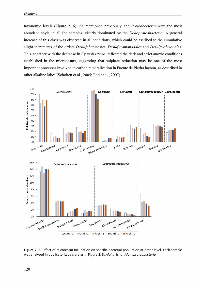
Chapter 2
120
taxonomic levels (Figure 2. 6). As mentioned previously, the Proteobacteria were the most
abundant phyla in all the samples, clearly dominated by the Deltaproteobacteria. A general
increase of this class was observed in all conditions, which could be ascribed to the cumulative
slight increments of the orders Desulfobacterales, Desulfuromonadales and Desulfovibrionales.
This, together with the decrease in Cyanobacteria, reflected the dark and strict anoxic conditions
established in the microcosms, suggesting that sulphate reduction may be one of the most
important processes involved in carbon mineralization in Fuente de Piedra lagoon, as described in
other alkaline lakes (Scholten et al., 2005; Foti et al., 2007).
Figure 2. 6. Effect of microcosm incubation on specific bacterial population at order level. Each sample was analysed in duplicate. Labels are as in Figure 2. 3. Alpha. is for Alphaproteobacteria.

Chapter 2
121
Myxococcales abundance also showed a positive trend during incubation, especially in the
non-polluted controls. Members of the Myxobacteria are generally found in soil ecosystem, and
few recent studies revealed their presence in hypersaline environment (Kambura et al., 2016; Mohr
et al., 2016). These organisms have been well studied because of their enormous potential to
produce bioactive secondary metabolites, their unique cooperative social behaviour and their
ability to survive in nutrient deficient environments (Mohr et al., 2016). Myxobacteria have been
considered obligate aerobic bacteria (Dawid, 2000), although anaerobic strains have been isolated
(Sanford et al., 2002), and their finding in anoxic sediments have been described (Acosta-Gonzalez
et al., 2013). Some authors hypothesised the possibility of an ancestral anaerobic lifestyle for this
bacterial group (Shimkets and Woese, 1992) given their phylogenetic proximity with the
Deltaproteobacteria, thus their occurrence in anaerobic environment it is not surprising.
The abundance of gammaproteobacterial sequences related to the Thiotrichales and
Chromatiales is consistent with previous studies (Mesbah et al., 2007). These two groups are
common inhabitants of soda lakes and are involved in the oxidation of the sulphide produced by
sulphate reducers in the sediment. Most members of the Chromatiales are capable of anoxic
photosynthesis, whilst Thiotrichales are not. This may explain the slight increase in the
Thiotrichales/Chromatiales ratio, since incubations were carried out in the dark. Rhodobacterales
sequences dominated the Alpharoteobacteria in all libraries as would be expected in a hypersaline
environment (Sorokin et al., 2014), although we observed a net decrease in the abundance of this
order in all the sample, especially after long-term incubation. The phylum Bacteroidetes was
mainly composed by Bacteroidia, Flavobacteriia, Rhodothermi and Saprospirae classes. All these
phylotypes showed a tendency to decrease in both microcosm conditions (control and
naphthalene), although the decrement of the Bacteroidia was less important in the PAH amended
cores, which could reflect a greater resistance of these organisms to hydrocarbon contamination,
or even an active role in hydrocarbon degradation. The most important family within the
Chloroflexi was the Anaerolineae, which showed a marked increment as compared to the original
sediment, especially after four months of incubation; however, no significant correlation with the
presence of PAHs could be found. Within the Firmicutes, Bacilli and Clostridia were majority,
and both classes increased in all the samples with incubation time. Members of both phylotypes
include aerobic and anaerobic oil-degrading strains (Kunapuli et al., 2007; da Cruz et al., 2011).

Chapter 2
122
2.3.4. Bacterial community evolution in naphthalene enrichments
We initiated a series of enrichment cultures starting from sediments collected during the first
sampling campaign (enrichments in SMM medium with HMN-dissolved naphthalene and 2MN
as carbon source) and from the pre-enriched naphthalene microcosms described above (cores 10
and 12, plus core 5 as unamended control) (enrichments in SMM medium and in filtered Fuente
de Piedra lagoon water) with naphthalene provided as crystals; see enrichment set-up scheme in
figure 2. 2). The microbial communities in the enrichment cultures at different time points were
analysed through massive sequencing of the bacterial V3-V4 16S rRNA gene region using the
Illumina-MiSeq platform. After processing and chimera removal, a total of 1,718,618 sequences
were obtained from the 16 libraries. An average of 107,414 reads were generated for each sample
(Table 2. 4 ). The sequences were grouped into OTUs at 97% identity. The rarefaction curves
reached the asymptote (supplementary Figure S1), suggesting that only a small fraction of the
ecosystem biodiversity remained unexplored. Moreover, the Good’s coverage estimator reached
an average value of 97.92%, indicating the sequencing depth was enough toeevaluate bacterial
valuate
Table 2. 4. Comparison of OTU number, diversity, evenness indices and coverage for the enrichment culture of the first and second sampling event.
Samplea Sampling campaign
NSb OTUsc OTUs (60000)d
Chao1 (60000)e Shannon
Coveragef
(%) Fdp-I 1 104864 5695 4549 7029.57 9.15 97.96 FdP-N 1 115640 4746 3582 5820.26 8.19 98.37 FdP-2MN 1 116683 4838 3667 5958.83 8.33 98.39 FdP-HMN 1 102655 4391 3496 5720.78 8.04 98.25 Core5-I 2 127020 6763 4953 7975.76 9.20 97.98 Core5-FPW-C1 2 101583 6123 4970 7584.59 9.61 97.76 Core5-FPW-C2 2 84313 5785 5051 7801.57 9.76 97.34 Core5-SMM-C2 2 94050 5363 4424 7307.70 8.80 97.59 Core10-I 2 114852 6470 4923 7931.54 9.05 97.78 Core10-FPW-N1 2 85036 5616 4853 7807.92 9.18 97.29 Core10-FPW-N2 2 102156 5664 4503 7536.37 8.41 97.66 Core10-SMM-N2 2 110607 5828 4500 7440.79 9.12 97.86 Core12-I 2 117092 6062 4603 7501.59 9.00 97.99 Core12-FPW-N1 2 98989 6112 4942 7933.36 9.36 97.49 Core12-FPW-N2 2 129043 4215 3003 5036.21 6.21 98.64 Core12-SMM-N2 2 114035 4101 3091 5200.54 7.40 98.48
a I refer to the initial sample of each enrichment; 1 and 2 refer to the subcultures at different time (0,1 and 2); FPW refer to Fuente de Piedra water used as culture media; SMM refer to SRB artificial see water media. bNumber of sequences for each library filtered for chimera and singletons. cOTU numbers calculated with all sequences at the 3% distance level. dOTU numbers calculated for a randomized subset of 60000 reads per sample at the 3% distance level. eChao index calculated with 60,000 subsampled sequences. f Good’s sample coverage estimator.

Chapter 2
123
diversity in all the samples. The results showed that the initial samples had similar richness index
values. In the enrichments from the first sampling campaign we observed a similar decrease of the
diversity index in the three cultures (naphthalene, 2MN and HMN), suggesting that culture media
had a stronger impact on the microbial diversity than exposure to PAHs. In contrast, the
enrichments originating from the sediment microcosms did not show changes in OTU richness
and Chao index when only Fuente de Piedra’s water was used as culture medium, except for the
Core12-FPW-N2 sample. Interestingly, richness indices decreased when and artificial salt medium
(SRB-SMM) was used (Table 2. 4). These findings highlight the importance of the choice of
culture media and conditions for microbial enrichments, where different factors as pH, salinity and
temperature could influence bacterial community diversity.
Naphthalene addition to the cultures resulted in only a slight decrease of the alpha diversity
indices of the enrichments from Core-10. In contrast, we found significant variation in the
enrichments from Core-12, especially after long time incubation: the Core12-FPW-N2 and
Core12-SMM-N2 samples showed the lowest OTU number and Chao index values of all samples.
The only difference between the enrichments from both cores was that Core-12 was exposed to
naphthalene for a longer time (12 months) as compared to Core-10 (4 months) before starting the
enrichments cultures, which was compensated by a shorter incubation time of the enrichment
before the first transfer (Figure 2. 2). In fact, Core-12 microcosm was the most affected by
naphthalene incubation, showing the lowest richness values (Figure 2. 3). Successive subcultures
of this sediment led to a further decrease in OTU number and Chao index.
The analysis of the of PCoA, based on weighted UniFrac distance metrics, of all the
enrichments and initial sediment samples provided an overview of the differences between
bacterial community structures resulting from the enrichment process (β-diversity). Figure 2. 7A
shows that the PCoA plot captured 53.21% of the observed variation between samples. Samples
could be grouped in four clusters: all the sediment samples used as starting material for the
enrichments clustered together (INI), which suggested similar patterns of community distribution
in the sediments collected in the two sampling campaigns and also after core pre-enrichment, as
shown previously (Figure 2. 5). The enrichments from the first sampling campaign constituted a
separated cluster from all the other samples, suggesting a significantly different community
structure of these cultures. Since the initial sediment used in these enrichments was close to the
remaining initial samples, this divergence was probably not due to the sampling time, but may
rather reflect the culture medium used, and especially the way in which the PAHs were added to

Chapter 2
124
the cultures (provided dissolved in HMN instead of as crystals). Moreover, these samples had been
incubated under strict anoxic culture conditions for the longest periods (27 months overall).
However, the community response to the presence of PAHs was similar to that observed in the
HMN control.
The third group comprised all the microcosm control cultures (i.e., unamended cultures)
together with the naphthalene-amended cultures after the first incubation time. Finally, the longest
incubation times sampling of naphthalene microcosm enrichments clustered together. These
samples did not form a very compact group, and in fact the Core10-SMM-N2 sample clustered
with the control and incubation time 1 samples. Still, a gradual shift from the original samples and
control cultures towards the longer incubation times could be observed. The furthermost sample
was Core12-FPW-N2, suggesting that naphthalene significantly affected the bacterial community
composition and abundance of this culture.
We further analysed β-diversity using the UPGMA clustering method considering unweighted
UniFrac distances (thus considering only the OTU presence/absence). Figure 2. 7 shows a slightly
different clustering of the samples in the UPGMA tree to the one obtained in the PCoA plot: in
this case, the first sampling campaign initial sediment clustered together with the enrichments
derived from it. Enrichments were grouped according to the core they originated from, except for
Figure 2. 7. Microbial community analysis of the sediment samples and enrichment cultures using weighted and unweighted UniFrac distance. UniFrac sequences were rarefied at the same sequencing depth (60,000). A) Plot of the weighted Principal Coordinate Analysis (PCoA), explaining 53.21% of variation. Colours indicate the different type of samples: orange, initial sediment samples used for the enrichments; red, control cultures; blue, naphthalene enrichments; green, 2MN enrichments. B) UPGMA clustering of original samples and the different enrichments. The bootstrapping values are indicated in each node.

Chapter 2
125
cultures from core 12, which grouped apart from all the other samples, indicating a significantly
different bacterial composition both in Fuente de Piedra water and in SMM artificial culture
medium.
Altogether these findings evidenced that three major factors structured the microbial
communities: composition of the culture media, incubation time and exposure to naphthalene.
2.3.5. Patterns of microbial diversity in the anaerobic enrichments
2.3.5.1. First sampling campaign: enrichments with PAHs dissolved in HMN.
To identify the bacterial taxa responsible for the shifts in bacterial community structure
attributable to the PAHs during the enrichment, we investigated the relative abundance of the
phylotypes present at the phylum and lower taxonomic levels (Figure 2. 8). Proteobacteria was
the most abundant phylum in the three cultures from the first campaign. The group was mainly
composed by Deltaproteobacteria, followed by Gamma- Alpha- and Epsilonproteobacteria.
Interestingly, the Betaproteobacteria were nearly absent from these sediments. The
Deltaproteobacteria were dominated by the order Desulfobacterales, which is exclusively
composed of sulphate-reducing bacteria. A marked decrease of Gamma- and
Epsilonproteobacteria was observed in the three culture conditions as compared to the original
sediment, emphasizing the possible media effect on these phylotypes (Figure 2. 8, left).
Proteobacterial classes did not show major differences with respect to the control enrichment,
except for the Deltaproteobacteria, which in the naphthalene culture had a lower relative
abundance (19.80%) than in the control HMN culture (27.77%) suggesting a possible toxic effect
of the PAH on this group. This result contrasts with the previous finding that strains of the order
Desulfobacterales were the only pure culture of SRB obtained that anaerobically degraded
naphthalene (Galushko et al., 1999). Although Bacteroidetes was the second phylum in abundance
in the initial sample, as expected in an athalassohaline lake, we observed a remarkable reduction
of this phylum in the enrichment cultures. The decrease was even more evident in the PAH
enrichments, suggesting a toxic effect on this population. Chloroflexi presented the major variation
as compared to the original sample, increasing from 5.5% to 25% in the naphthalene enrichment,
but no significant differences were observed between the PAH enrichment and the control. As for
the microcosms, Chloroflexi population was dominated by Anaerolineae, fermentative organisms
mainly retrieved from anoxic environments and often present in anaerobic culture (Martirani-Von
Abercron et al., 2016). The group has been frequently found in activated sludge where they are

Chapter 2
126
associated to sludge granulation. Although Anaerolineaceae have been recovered from
hydrocarbon-impacted environment (Sherry et al., 2013), in our enrichments they are not likely to
be directly linked to hydrocarbons metabolism, but they probably grow on carbohydrates, peptides
and/or cells debris (Yamada and Sekiguchi, 2009).
Intriguingly, some phylotypes including Firmicutes, Actinobacteria, Thermi and
Gemmatimonadetes were enriched, as revealed by the significant increase in relative abundance
in the naphthalene and 2MN cultures. Firmicutes, in which both Clostria and Bacilli dominated,
and Actinobacteria were often associated to hydrocarbon tolerance and/or degradation (Sun et al.,
2014; Yang et al., 2014). Thermus (Deinococci) group increased in all the culture as compared to
the original sample. The increase was more important in the naphthalene enrichment, where the
relative abundance shifted from 1.4% (FdP-I) to 6.24% (FdP-N). The Deinococcus-Thermus
Figure 2. 8. Relative abundance of bacterial phyla or class (only for Proteobacteria) in the Fuente de Piedra enrichments. Data from the first and second sampling campaigns are separated by a doted line.

Chapter 2
127
phylum comprises organisms with a high resistance to radiation, oxidizing agents and desiccating
conditions which has made them suitable to be used in the bioremediation of radiation-
contaminated sites (Misra et al., 2006; Griffiths and Gupta, 2007; Ho et al., 2016). The greater
increase in the PAH cultures, primarily in the naphthalene one, suggests a further interesting ability
of this phylotypes such as hydrocarbon resistance. To date, only one study has reported the
capacity of this group to transform crude oils (Hao et al., 2004).
2.3.5.2. Second sampling campaign: enrichment with naphthalene provided as crystals.
In the enrichment cultures with the Fuente de Piedra sediments from the microcosms
described in 2.3.3 (Figure 2. 2), pure naphthalene crystals were used as sole carbon source (Core10
and Core12) after the two incubation times. A culture initiated from the control microcosm (Core5)
was included as control, in which no carbon source was added. Naphthalene and control
enrichments were prepared in filtered FdP water. The use of FdP water as culture medium was
designed to promote the growth of natural and unexplored microbial populations. In fact,
athalassoaline lakes are ecosystems characterized by specific ranges of salinity, pH and nutrient
which are difficult to reproduce in the laboratory, so that artificial media could select bacterial
communities different from those naturally dominant in the habitat (Connon and Giovannoni,
2002). After the first incubation period, parallel subcultures in SRB SMM medium were included
in the experiment (Figure 2. 2).
Although the relative abundance of the main taxa varied significantly under the different
treatments, five phyla including Proteobacteria (27.8%-56.7%), Chloroflexi (3.4%-16.2%),
Bacteroidetes (3.3%-9.0%), Firmicutes (4.6%-42.1%) and Actinobacteria (0.6%-6.9%) were the
most abundant, accounting for a total of 61.7%-87.2% of the whole community (Figure 2. 8, right).
These phylotypes are considered the main bacterial inhabitants of hypersaline environments
(Mesbah et al., 2007; Sorokin et al., 2014).
In our enrichments, the Proteobacteria phyla showed a tendency to decrease in the cultures
with FdP lagoon water (FdPW-enrichments) and to increase in the cultures with SMM, reaching
56.7% of the community in the Core12-SMM-N2 sample. Alpha and especially
Gammaproteobacteria decreased in all enrichments. The Deltaproteobacteria class did not show
significant changes during the enrichment in FdP water, but we observed that the SMM medium
clearly enriched this group in both control and naphthalene amended cultures, although the
increase was especially remarkable in Core12-SMM-N2 sample, where Deltaproteobacteria
reached up to 45% of the community. The SMM medium is specifically designed for the

Chapter 2
128
cultivation of SRB, and has been previously used to isolate pure cultures of Deltaproteobacteria
able to degrade naphthalene (Galushko et al., 1999). The increment of members of this class was
especially notable for the order of Desulfobacterales, a sulfate-reducing group commonly found
in soda lake (Scholten et al., 2005). Within this order, the abundance of an OTU affiliated to the
Desulfobulbaceae significantly increased in all the cultures prepared in SMM medium, although
especially in Core12-SMM-N2 sample, where the values were 10 times higher (21% of the
community) than in the initial sample (as compared to the increase in the control Core5-SMM-C2
sample, where it reached 11% of the community) (Table S1). Members of the Desulfobulbaceae
have been previously described as toluene and naphthalene degraders under sulfate-reducing
condition (Muller et al., 2009; Pilloni et al., 2011; Kümmel et al., 2015) suggesting that this taxon
could be directly involved in naphthalene degradation. In the Core 12 samples, the
Desulfovibrionales was a second Deltaproteobacteria class positively influenced by the presence
of naphthalene, increasing from values below 0.01% in the initial core sample to 3 and 8% of the
community after 12 months incubation in FdP water and SMM medium, respectively. Two OTUs
belonging to the Desulfomicrobiaceae were mainly responsible for the increment. Surprisingly the
Epsilonproteobacteria were also favoured in naphthalene enrichments, especially in the Core12-
FPW-N2 sample, where it increased from 0.17% in the initial Core sample to 11.2%. This was
essentially due to an unclassified OTU belonging to the Campylobacteriaceae that dominated the
Campylobacteriales class in the naphthalene-amended cultures. Sequence reads belonging to the
Campylobacteriales have been retrieved from municipal wastewater (Ruiz et al., 2014), in reactors
with multiple electron acceptors under sulfur-cycling conditions (Zhao et al., 2013) and recently
they have been associated to higher concentrations of PAHs in soil (Ma et al., 2016).
Unfortunately, little is known about the possible function of the members of this class detected in
environmental samples.
Firmicutes was the phylum that showed the most dramatic changes directly related to the
presence of naphthalene in the enrichments. The phylotype was enriched in the naphthalene
cultures with FdP water as culture medium in both Core10 (FPW-N1: 13.8%; FPW-N2: 25.5%)
and Core12 (FPW-N1: 9.6%; FPW-N2: 42.16%) cultures, whilst the relative abundance in the
control culture in the same medium remained around 8% in both subcultures. Members of Bacilli
and Clostridia classes were the principal component of the Firmicutes in all the enrichments,
although the Clostridia showed the major increases. Particularly, the genus Dethiosulfatibacter
was strongly selected for in FdP water cultures with naphthalene, reaching a relative abundance
of 18% in Core10-FPW-N2 and Core12-FPW-N2 (its abundance was below 0.1% in the initial
core samples, and remaining at this level in the unamended cultures from the control Core5).

Chapter 2
129
Dethiosulfatibacter aminovorans is the only cultivated strain of this genus and was first isolated
from coastal marine sediments of the eutrophic Tokyo Bay, Japan (Takii et al., 2007). The strain
is a halophilic, thiosulfate-reducing, strict anaerobe. Previous studies suggested that
Dethiosulfatibacter was among the main players in PAH degradation in mangrove sediments
(Muangchinda et al., 2013). This strain has also been detected in crude oil microbial communities,
although at very low abundance (Cai et al., 2015). The strong enrichment of Dethiosulfatibacter
in our naphthalene enrichments is remarkable and could likely be associated to the anaerobic
degradation of naphthalene. Interestingly two other members of Clostridiales, Halanaerobiaceae
and Acidaminobacteraceae, were strongly enriched with naphthalene, although in this case only
in the Core12-FPW-N2 culture. Unclassified members of the Halanaerobiaceae family accounted
for the 11.2% of the whole community. Representatives of this family are obligate halophilic
anaerobes with a fermentative metabolism, and have been found and isolated from a wide variety
of anaerobic hypersaline environments (Kivisto and Karp, 2011). They have also been reported to
reduce nitro-substituted aromatic compounds to the corresponding amine, although they were not
incorporated by the cells (Oren et al., 1991). We have repeatedly observed the selection of
fermentative organisms in the presence of PAHs, which we generally attribute to PAH toxicity
resulting in cell lysis, the debris of which could be used as substrate by fermenters that are resistant
to the PAHs. The relative abundance of Acidaminobacteraceae (mainly represented by the
uncultured genus NP25) in the enrichments from Core-12 increased progressively from 0.2% in
the original sample up to 10.8% in Core12-FPW-N2 culture. Unfortunately, very little is known
about this group. They have been detected in oil-amended salt-marsh sediment microcosms, where
their belonged to the so-called “late-responders” to crude oil. The relative abundance of
unclassified Acidaminobacteraceae in these conditions only increased three weeks after oil
treatment, and further increased with the incubation time (Koo et al., 2015). They have also been
detected in biogas digesters (Müller et al., 2016). It is thus difficult to predict their ecological role,
although the strong increase of this group in the naphthalene amended Core-12 enrichments in FdP
water medium suggests a possible role in anaerobic naphthalene degradation. A similar increase
was not observed in the parallel enrichments from Core-10, probably because of the almost
undetectable levels of this genus in the initial sample (below 0.1%). Interestingly, this increase
was not observed in the enrichment in SMM medium, which started from the 4 months enrichment
in FdP water, where the Acidaminobacteraceae NP25 community had already reached 2.2% of the
community. Additional 8 months incubation in SMM medium reduced the abundance of this genus
back to levels similar to Core-12 initial values. This is a further example of the dramatic influence
of the medium composition on the community structure evolution, and of the importance to use

Chapter 2
130
media as close to natural conditions as possible, as it is the case of the FdP water used here. In
fact, often the change from natural environment to artificial media could de-structure the
community in such a way as to render its degradative capacities less efficient, or support bacteria
that do not have these abilities.
2.4. Concluding remarks
Most ecological studies on hypersaline ecosystems to date have focused on the water bodies.
Therefore, the data about bacterial communities present in hypersaline sediments provides relevant
information as to the community structures in these ecosystems. Overall, we observed remarkable
levels of diversity, which were amongst the highest described for microbial communities and in
the range of marine and freshwater sediments (Wang et al., 2012). The structural complexity of
the soil and sediment matrix favours spatial resource partitioning, thus creating a variety of
microhabitats. This provides a distinct mechanism for shaping diversity that differs essentially
from the situation in aqueous environments (Torsvik et al., 2002), which probably contributes to
the high diversity observed in this ecosystem. In fact, the differences in sample clustering observed
when comparing weighted and unweighted community analysis suggest an important contribution
of the so-called “rare” community to the overall diversity. Rare taxa are considered as “seed bank”
organisms which could become activated upon changes in the environmental conditions (Pedrós-
Alió, 2006).
In our microcosm experiments, naphthalene treatment did not produce a clear shift of the
bacterial community. It must be noted that the communities were dominated by
Proteobacteria, Bacteroidetes, Firmicutes and Chloroflexi, taxa of which members are capable of
metabolizing or tolerant to complex hydrocarbons (An et al., 2013; Sherry et al., 2013; Yang et
al., 2014). The resulting bacterial structure might be more dependent on the strict anoxic
conditions imposed in the microcosms. Still, the changes after 12 months of anoxic incubation
were small, and probably reflect the anoxic nature of these sediments below the upper oxic layer,
which allowed the establishment of a community already adapted to strict anoxic conditions. It is
worth noting that FdP sediment already had a basal level of hydrocarbons contamination (aliphatic
and aromatics) (Martirani-Von Abercron et al., 2016), which could explain why further addition
of PAHs may not have had the expected toxic effect, especially if the community structure is
resilient.
In contrast, enrichment cultures showed that the presence of PAHs induced a clear shift in the
bacterial community structure. Members of Firmicutes, Actinobacteria and Thermi increased in
Chapter 2
131
the naphthalene and 2MN cultures initiated after the first sampling campaign and set up with the
PAHs dissolved in HMN, supporting previous studies that identified these organisms as related to
PAH degradation. More interesting were the changes produced by naphthalene in the enrichments
initiated from microcosms that had been pre-treated with naphthalene. We found that the
cultivation-dependent approach enriched a few specific populations that might be involved in the
aromatic biodegradation process. Specifically, members of Desulfobulbaceae, detected in the
SMM medium, and members of Dethiosulfatibacter, Halanaerobiaceae and
Acidaminobacteraceae, in the FdP water, were clearly selected for in the presence of naphthalene,
reaching important relative abundances. The actual capacity of these strain to degrade PAHs
deserve further analysis. Finally, we verified that natural media, such as FdP water, could be the
best option to enrich bacteria potentially useful in bioremediation.
2.5. References
Acosta-González, A., Rosselló-Mora, R., and Marqués, S. (2013). Characterization of the anaerobic microbial community in oil-polluted subtidal sediments: aromatic biodegradation potential after the Prestige oil spill. Environ Microbiol 15: 77-92.
Adrian, R., O’reilly, C.M., Zagarese, H., Baines, S.B., Hessen, D.O., Keller, W., Livingstone, D.M., Sommaruga, R., Straile, D., Van Donk, E., Weyhenmeyer, G.A., and Winder, M. (2009). Lakes as sentinels of climate change. Limnol Oceanogr 54: 2283-2297.
Aguirre-Garrido, J.F., Ramirez-Saad, H.C., Toro, N., and Martinez-Abarca, F. (2016). Bacterial Diversity in the Soda Saline Crater Lake from Isabel Island, Mexico. Microb Ecol 71: 68-77.
An, D., Brown, D., Chatterjee, I., Dong, X., Ramos-Padron, E., Wilson, S., Bordenave, S., Caffrey, S.M., Gieg, L.M., Sensen, C.W., and Voordouw, G. (2013). Microbial community and potential functional gene diversity involved in anaerobic hydrocarbon degradation and methanogenesis in an oil sands tailings pond. Genome 56: 612-618.
Anderson, N.J. (2007). The effect of evapoconcentration on dissolved organic carbon concentration and quality in lakes of SW Greenland. Freshwat Biol v. 52: pp. 280-289-2007 v.2052 no.2002.
Brune, A., Frenzel, P., and Cypionka, H. (2000). Life at the oxic-anoxic interface: microbial activities and adaptations. FEMS Microbiol Rev 24: 691-710.
Cai, M., Nie, Y., Chi, C.-Q., Tang, Y.-Q., Li, Y., Wang, X.-B., Liu, Z.-S., Yang, Y., Zhou, J., and Wu, X.-L. (2015). Crude oil as a microbial seed bank with unexpected functional potentials. Sci Rep 5: 16057.
Caporaso, J.G., Bittinger, K., Bushman, F.D., Desantis, T.Z., Andersen, G.L., and Knight, R. (2010). PyNAST: a flexible tool for aligning sequences to a template alignment. Bioinformatics 26: 266-267.
Caporaso, J.G., Kuczynski, J., Stombaugh, J., Bittinger, K., Bushman, F.D., Costello, E.K., Fierer, N., Pena, A.G., Goodrich, J.K., Gordon, J.I., Huttley, G.A., Kelley, S.T., Knights, D., Koenig, J.E., Ley, R.E., Lozupone, C.A., Mcdonald, D., Muegge, B.D., Pirrung, M., Reeder, J., Sevinsky, J.R., Turnbaugh, P.J., Walters, W.A., Widmann, J., Yatsunenko, T., Zaneveld, J., and Knight, R. (2010). QIIME allows analysis of high-throughput community sequencing data. Nat Methods 7: 335-336.
Cerniglia, C.E. (1992). Biodegradation of polycyclic aromatic hydrocarbons. Biodegradation 3: 351-368. Connon, S.A., and Giovannoni, S.J. (2002). High-throughput methods for culturing microorganisms in
very-low-nutrient media yield diverse new marine isolates. Appl Environ Microbiol 68: 3878-3885. Da Cruz, G.F., De Vasconcellos, S.P., Angolini, C.F.F., Dellagnezze, B.M., Garcia, I.N.S., De Oliveira,
V.M., Dos Santos Neto, E.V., and Marsaioli, A.J. (2011). Could petroleum biodegradation be a

Chapter 2
132
joint achievement of aerobic and anaerobic microrganisms in deep sea reservoirs? AMB Express 1: 47-47.
Dawid, W. (2000). Biology and global distribution of myxobacteria in soils. FEMS Microbiol Rev 24: 403-427.
Debruyn, J.M., Nixon, L.T., Fawaz, M.N., Johnson, A.M., and Radosevich, M. (2011). Global Biogeography and Quantitative Seasonal Dynamics of Gemmatimonadetes in Soil. Appl Environ Microbiol 77: 6295-6300.
Demergasso, C., Casamayor, E.O., Chong, G., Galleguillos, P., Escudero, L., and Pedros-Alio, C. (2004). Distribution of prokaryotic genetic diversity in athalassohaline lakes of the Atacama Desert, Northern Chile. FEMS Microbiol Ecol 48: 57-69.
Dong, H., Zhang, G., Jiang, H., Yu, B., Chapman, L.R., Lucas, C.R., and Fields, M.W. (2006). Microbial Diversity in Sediments of Saline Qinghai Lake, China: Linking Geochemical Controls to Microbial Ecology. Microb Ecol 51: 65-82.
Edgar, R.C., Haas, B.J., Clemente, J.C., Quince, C., and Knight, R. (2011). UCHIME improves sensitivity and speed of chimera detection. Bioinformatics 27: 2194-2200.
Eiler, A., Farnleitner, A.H., Zechmeister, T.C., Herzig, A., Hurban, C., Wesner, W., Krachler, R., Velimirov, B., and Kirschner, A.K. (2003). Factors controlling extremely productive heterotrophic bacterial communities in shallow soda pools. Microb Ecol 46: 43-54.
Foght, J. (2008). Anaerobic biodegradation of aromatic hydrocarbons: Pathways and prospects. J. Mol. Microbiol. Biotech. 15: 93-120.
Foti, M., Sorokin, D.Y., Lomans, B., Mussman, M., Zacharova, E.E., Pimenov, N.V., Kuenen, J.G., and Muyzer, G. (2007). Diversity, activity, and abundance of sulfate-reducing bacteria in saline and hypersaline soda lakes. Appl Environ Microbiol 73: 2093-2100.
Galushko, A., Minz, D., Schink, B., and Widdel, F. (1999). Anaerobic degradation of naphthalene by a pure culture of a novel type of marine sulphate-reducing bacterium. Environ Microbiol 1: 415-420.
García, C.M., García-Ruiz, R., Rendón, M., Niell, F.X., and Lucena, J. (1997). Hydrological cycle and interannual variability of the aquatic community in a temporary saline lake (Fuente de Piedra, Southern Spain). Hydrobiologia 345: 131-141.
Ghosal, D., Ghosh, S., Dutta, T.K., and Ahn, Y. (2016). Current State of Knowledge in Microbial Degradation of Polycyclic Aromatic Hydrocarbons (PAHs): A Review. Front Microbiol 7: 1369.
Griffiths, E., and Gupta, R.S. (2007). Identification of signature proteins that are distinctive of the Deinococcus-Thermus phylum. Int Microbiol 10: 201-208.
Hao, R., Lu, A., and Wang, G. (2004). Crude-oil-degrading thermophilic bacterium isolated from an oil field. Can J Microbiol 50: 175-182.
Heredia, J., García De Domingo, A., Ruiz, J.M., and Araguás, L. (2010). Fuente de piedra lagoon (Málaga, Spain): A deep karstic flow discharge point of a regional hydrogeological system. Environ Earth Sci: 231-236.
Ho, J., Adeolu, M., Khadka, B., and Gupta, R.S. (2016). Identification of distinctive molecular traits that are characteristic of the phylum “Deinococcus-Thermus” and distinguish its main constituent groups. Syst Appl Microbiol 39: 453-463.
Jiang, H., Dong, H., Zhang, G., Yu, B., Chapman, L.R., and Fields, M.W. (2006). Microbial diversity in water and sediment of Lake Chaka, an athalassohaline lake in northwestern China. Appl Environ Microbiol 72: 3832-3845.
Kambura, A.K., Mwirichia, R.K., Kasili, R.W., Karanja, E.N., Makonde, H.M., and Boga, H.I. (2016). Bacteria and Archaea diversity within the hot springs of Lake Magadi and Little Magadi in Kenya. BMC Microbiol 16: 136.
Kim, J.-S., Makama, M., Petito, J., Park, N.-H., Cohan, F.M., and Dungan, R.S. (2012). Diversity of Bacteria and Archaea in hypersaline sediment from Death Valley National Park, California. MicrobiologyOpen 1: 135-148.
Kirchman, D.L. (2002). The ecology of Cytophaga-Flavobacteria in aquatic environments. FEMS Microbiol Ecol 39: 91-100.
Kivisto, A.T., and Karp, M.T. (2011). Halophilic anaerobic fermentative bacteria. J Biotechnol 152: 114-124.
Koo, H., Mojib, N., Huang, J.P., Donahoe, R.J., and Bej, A.K. (2015). Bacterial community shift in the coastal Gulf of Mexico salt-marsh sediment microcosm in vitro following exposure to the Mississippi Canyon Block 252 oil (MC252). 3 Biotech 5: 379-392.

Chapter 2
133
Kummel, S., Herbst, F.A., Bahr, A., Duarte, M., Pieper, D.H., Jehmlich, N., Seifert, J., Von Bergen, M., Bombach, P., Richnow, H.H., and Vogt, C. (2015). Anaerobic naphthalene degradation by sulfate-reducing Desulfobacteraceae from various anoxic aquifers. FEMS Microbiol Ecol 91.
Kunapuli, U., Lueders, T., and Meckenstock, R.U. (2007). The use of stable isotope probing to identify key iron-reducing microorganisms involved in anaerobic benzene degradation. ISME J 1: 643-653.
Lanzen, A., Simachew, A., Gessesse, A., Chmolowska, D., Jonassen, I., and Ovreas, L. (2013). Surprising prokaryotic and eukaryotic diversity, community structure and biogeography of Ethiopian soda lakes. PLoS One 8: e72577.
Le Borgne, S., Paniagua, D., and Vazquez-Duhalt, R. (2008). Biodegradation of organic pollutants by halophilic bacteria and archaea. J Mol Microbiol Biotechnol 15: 74-92.
Lefebvre, O., and Moletta, R. (2006). Treatment of organic pollution in industrial saline wastewater: A literature review. Water Res 40: 3671-3682.
Lizama, C., Monteoliva-Sanchez, M., Prado, B., Ramos-Cormenzana, A., Weckesser, J., and Campos, V. (2001). Taxonomic study of extreme halophilic archaea isolated from the "Salar de Atacama", Chile. Syst Appl Microbiol 24: 464-474.
Lozupone, C., and Knight, R. (2005). UniFrac: a New Phylogenetic Method for Comparing Microbial Communities. Appl Environ Microbiol 71: 8228-8235.
Lozupone, C.A., Hamady, M., Kelley, S.T., and Knight, R. (2007). Quantitative and Qualitative β Diversity Measures Lead to Different Insights into Factors That Structure Microbial Communities. Appl Environ Microbiol 73: 1576-1585.
Ma, J., Zhang, W., Chen, Y., Zhang, S., Feng, Q., Hou, H., and Chen, F. (2016). Spatial Variability of PAHs and Microbial Community Structure in Surrounding Surficial Soil of Coal-Fired Power Plants in Xuzhou, China. Int J Env Res Public Health 13: 878.
Marhuenda-Egea, F.C., and Bonete, M.J. (2002). Extreme halophilic enzymes in organic solvents. Curr Opin Biotechnol 13: 385-389.
Martirani-Von Abercron, S.-M., Pacheco, D., Benito-Santano, P., Marín, P., and Marqués, S. (2016). Polycyclic Aromatic Hydrocarbon-Induced Changes in Bacterial Community Structure under Anoxic Nitrate Reducing Conditions. Front Microbiol 7: 1775.
Meckenstock, R., Safinowski, M., and Griebler, C. (2004). Anaerobic degradation of polycyclic aromatic hydrocarbons. FEMS Microbiol Ecol 29: 27-36.
Meckenstock, R.U., Annweiler, E., Michaelis, W., Richnow, H.H., and Schink, B. (2000). Anaerobic naphthalene degradation by a sulfate-reducing enrichment culture. Appl Environ Microbiol 66: 2743-2747.
Mesbah, N.M., Abou-El-Ela, S.H., and Wiegel, J. (2007). Novel and unexpected prokaryotic diversity in water and sediments of the alkaline, hypersaline lakes of the Wadi An Natrun, Egypt. Microb Ecol 54: 598-617.
Misra, H.S., Khairnar, N.P., Kota, S., Shrivastava, S., Joshi, V.P., and Apte, S.K. (2006). An exonuclease I-sensitive DNA repair pathway in Deinococcus radiodurans: a major determinant of radiation resistance. Mol Microbiol 59: 1308-1316.
Mohr, K.I., Stechling, M., Wink, J., Wilharm, E., and Stadler, M. (2016). Comparison of myxobacterial diversity and evaluation of isolation success in two niches: Kiritimati Island and German compost. MicrobiologyOpen 5: 268-278.
Montoya, L., Vizioli, C., Rodriguez, N., Rastoll, M.J., Amils, R., and Marin, I. (2013). Microbial community composition of Tirez lagoon (Spain), a highly sulfated athalassohaline environment. Aquat Biosyst 9: 19.
Muangchinda, C., Pansri, R., Wongwongsee, W., and Pinyakong, O. (2013). Assessment of polycyclic aromatic hydrocarbon biodegradation potential in mangrove sediment from Don Hoi Lot, Samut Songkram Province, Thailand. J Appl Microbiol 114: 1311-1324.
Müller, B., Sun, L., Westerholm, M., and Schnürer, A. (2016). Bacterial community composition and fhs profiles of low- and high-ammonia biogas digesters reveal novel syntrophic acetate-oxidising bacteria. Biotechnol Biofuels 9: 48.
Muller, S., Vogt, C., Laube, M., Harms, H., and Kleinsteuber, S. (2009). Community dynamics within a bacterial consortium during growth on toluene under sulfate-reducing conditions. FEMS Microbiol Ecol 70: 586-596.

Chapter 2
134
Musat, F., Galushko, A., Jacob, J., Widdel, F., Kube, M., Reinhardt, R., Wilkes, H., Schink, B., and Rabus, R. (2009). Anaerobic degradation of naphthalene and 2-methylnaphthalene by strains of marine sulfate-reducing bacteria. Environ Microbiol 11: 209-219.
Newton, R.J., Jones, S.E., Eiler, A., Mcmahon, K.D., and Bertilsson, S. (2011). A guide to the natural history of freshwater lake bacteria. Microbiol Mol Biol Rev 75: 14-49.
Oren, A. (2010). Industrial and environmental applications of halophilic microorganisms. Environ Technol 31: 825-834.
Oren, A., Gurevich, P., and Henis, Y. (1991). Reduction of nitrosubstituted aromatic compounds by the halophilic anaerobic eubacteria Haloanaerobium praevalens and Sporohalobacter marismortui. Appl Environ Microbiol 57: 3367-3370.
Ortega-Retuerta, E., Pulido-Villena, E., and Reche, I. (2007). Effects of Dissolved Organic Matter Photoproducts and Mineral Nutrient Supply on Bacterial Growth in Mediterranean Inland Waters. Microb Ecol 54: 161-169.
Pedrós-Alió, C. (2006). Marine microbial diversity: can it be determined? Trends Microbiol 14: 257-263. Pilloni, G., Von Netzer, F., Engel, M., and Lueders, T. (2011). Electron acceptor-dependent identification
of key anaerobic toluene degraders at a tar-oil-contaminated aquifer by Pyro-SIP. FEMS Microbiol Ecol 78: 165-175.
Ruiz, V., Ilhan, Z.E., Kang, D.-W., Krajmalnik-Brown, R., and Buitrón, G. (2014). The source of inoculum plays a defining role in the development of MEC microbial consortia fed with acetic and propionic acid mixtures. J Biotechnol 182–183: 11-18.
Sanford, R.A., Cole, J.R., and Tiedje, J.M. (2002). Characterization and description of Anaeromyxobacter dehalogenans gen. nov., sp. nov., an aryl-halorespiring facultative anaerobic myxobacterium. Appl Environ Microbiol 68: 893-900.
Scholten, J.C., Joye, S.B., Hollibaugh, J.T., and Murrell, J.C. (2005). Molecular analysis of the sulfate reducing and archaeal community in a meromictic soda lake (Mono Lake, California) by targeting 16S rRNA, mcrA, apsA, and dsrAB genes. Microb Ecol 50: 29-39.
Sherry, A., Gray, N.D., Ditchfield, A.K., Aitken, C.M., Jones, D.M., Röling, W.F.M., Hallmann, C., Larter, S.R., Bowler, B.F.J., and Head, I.M. (2013). Anaerobic biodegradation of crude oil under sulphate-reducing conditions leads to only modest enrichment of recognized sulphate-reducing taxa. Int Biodeterior Biodegrad 81: 105-113.
Shimkets, L., and Woese, C.R. (1992). A phylogenetic analysis of the myxobacteria: basis for their classification. Proc Natl Acad Sci USA 89: 9459-9463.
Sorokin, D.Y., Berben, T., Melton, E.D., Overmars, L., Vavourakis, C.D., and Muyzer, G. (2014). Microbial diversity and biogeochemical cycling in soda lakes. Extremophiles 18: 791-809.
Sorokin, D.Y., Gorlenko, V.M., Namsaraev, B.B., Namsaraev, Z.B., Lysenko, A.M., Eshinimaev, B.T., Khmelenina, V.N., Trotsenko, Y.A., and Kuenen, J.G. (2004). Prokaryotic communities of the north-eastern Mongolian soda lakes. Hydrobiologia 522: 235-248.
Sorokin, D.Y., Kuenen, J.G., and Muyzer, G. (2011). The microbial sulfur cycle at extremely haloalkaline conditions of soda lakes. Front Microbiol 2: 44.
Sun, G.-D., Xu, Y., Liu, Y., and Liu, Z.-P. (2014). Microbial community dynamics of soil mesocosms using Orychophragmus violaceus combined with Rhodococcus ruber Em1 for bioremediation of highly PAH-contaminated soil. Appl Microbiol Biotechnol 98: 10243-10253.
Takii, S., Hanada, S., Tamaki, H., Ueno, Y., Sekiguchi, Y., Ibe, A., and Matsuura, K. (2007). Dethiosulfatibacter aminovorans gen. nov., sp. nov., a novel thiosulfate-reducing bacterium isolated from coastal marine sediment via sulfate-reducing enrichment with Casamino acids. Int J Syst Evol Microbiol 57: 2320-2326.
Torsvik, V., Ovreas, L., and Thingstad, T.F. (2002). Prokaryotic diversity-magnitude, dynamics, and controlling factors. Science 296: 1064-1066.
Wang, Y., Sheng, H.F., He, Y., Wu, J.Y., Jiang, Y.X., Tam, N.F., and Zhou, H.W. (2012). Comparison of the levels of bacterial diversity in freshwater, intertidal wetland, and marine sediments by using millions of illumina tags. Appl Environ Microbiol 78: 8264-8271.
Wetzel, R.G. (2001). "Rivers and lakes — Their distribution, origins, and forms," in Limnology (Third Edition). (San Diego: Academic Press), 15-42.
Widdel, F., and Bak, F. (1992). "Gram-Negative Mesophilic Sulfate-Reducing Bacteria," in The Prokaryotes: A Handbook on the Biology of Bacteria: Ecophysiology, Isolation, Identification,

Chapter 2
135
Applications, eds. A. Balows, H.G. Trüper, M. Dworkin, W. Harder & K.-H. Schleifer. (New York, NY: Springer New York), 3352-3378.
Widdel, F., Kohring, G.-W., and Mayer, F. (1983). Studies on dissimilatory sulfate-reducing bacteria that decompose fatty acids. Arch Microbiol 134: 286-294.
Yamada, T., and Sekiguchi, Y. (2009). Cultivation of uncultured Chloroflexi subphyla: significance and ecophysiology of formerly uncultured chloroflexi 'subphylum i' with natural and biotechnological relevance. Microbes Environ 24: 205-216.
Yang, J., Ma, L.A., Jiang, H., Wu, G., and Dong, H. (2016). Salinity shapes microbial diversity and community structure in surface sediments of the Qinghai-Tibetan Lakes. Sci Rep 6: 25078.
Yang, S., Wen, X., Zhao, L., Shi, Y., and Jin, H. (2014). Crude Oil Treatment Leads to Shift of Bacterial Communities in Soils from the Deep Active Layer and Upper Permafrost along the China-Russia Crude Oil Pipeline Route. PLoS ONE 9: e96552.
Zhao, H.-P., Ilhan, Z.E., Ontiveros-Valencia, A., Tang, Y., Rittmann, B.E., and Krajmalnik-Brown, R. (2013). Effects of Multiple Electron Acceptors on Microbial Interactions in a Hydrogen-Based Biofilm. Environ Sci Technol 47: 7396-7403.
Zhou, J., Bruns, M.A., and Tiedje, J.M. (1996). DNA recovery from soils of diverse composition. Appl Environ Microbiol 62: 316-322.

Chapter 2
136
2.6. Supplementary material
Figure S1. Rarefaction curves at a phylogenetic distance of 0.03 for partial (V3-V4) 16S rRNA genes of microbial populations of the different samples analysed in this study.

137
CHAPTER 3:
Naphthalene biodegradation under oxygen limiting conditions: community dynamics and the relevance of biofilm formation
capacity
This chapter was published as:
Martirani-Von Abercron SM, Marín P, Solsona-Ferraz M, Castañeda-Cataña MA & Marqués S. Naphthalene biodegradation under oxygen-limiting conditions: community dynamics and the relevance of biofilm formation capacity. Microb Biotechnol, 2017 DOI: 10.1111/1751-7915.12842

138

139
Anyone who has never made a mistake has never tried anything new.
Albert Einstein

140

Chapter 3
141
3.1. Introduction
Polycyclic aromatic hydrocarbons (PAHs) are toxic compounds of particular environmental
concern because of their marked stability and resistance to degradation. They are released into the
environment either from natural sources or as a result of industrial activities. The simplest PAH
naphthalene is included among the 16 PAHs considered to be priority pollutants by the US EPA.
International efforts to improve remediation strategies for hydrocarbon polluted sites focus on
biological processes mediated by bacteria. Bacteria have developed a striking adaptive capacity for
degrading natural and synthetic aromatics to CO2 and water, which is the basis for the use of
bioremediation approaches to clean aromatics polluted areas.
Bacteria able to degrade PAHs under strict aerobic conditions are widespread and have been
widely described. The biodegradation pathway involves the deeply characterized aromatic mono- and
dioxygenases for ring activation (hydroxylation) and ring cleavage (Vaillancourt et al., 2006; Ullrich
and Hofrichter, 2007). The genes and gene organization of PAH degradation operons are reasonably
conserved among PAH degrading isolates, especially among the Pseudomonads, which are
predominant in the databases of PAH degraders. However, the availability of oxygen is often limited
in natural environments because oxygen can be rapidly consumed in the aerobic biodegradation
processes, resulting in a decreasing oxygen concentration gradient whereupon reduction of other
electron acceptors such as nitrate, iron or sulphate becomes energetically favourable (Rivett et al.,
2008b). A similar pattern is observed in habitats that are under permanent anoxic conditions, such as
flooded sediments in marine and fresh-water environments (Brune et al., 2000). Because nitrate
respiration is not a strictly anoxic process, aerobic and nitrate reducing metabolisms generally coexist
at the boundaries of polluted sites, where microaerophilic conditions are generally found (Wilson and
Bouwer, 1997).
Under strict anoxic conditions PAH biodegradation has only been reliably proved in sulphate-
reducing bacteria (SRB), although only one Deltaproteobacteria strain and a two-strain consortium
able to degrade naphthalene have been isolated and characterized to date (Meckenstock et al., 2016).
Anaerobes use a reductive strategy to overcome the stability of the aromatic ring. Naphthalene must
be converted to 2-naphtoic acid (NA) (Zhang and Young, 1997) previous to its activation to
naphthoyl-CoA, which is then dearomatized, starting from the non-activated ring, by the activity of
two specific reductases: a naphthoyl-CoA reductase (ncr gene) followed by a 5,6-dihydro-2-naphthyl-
CoA reductase (DHNCR) (Eberlein et al., 2013a, Estelmann et al., 2015). The subsequent
dearomatization of the activated ring requires the activity of a 5,6,7,8-tetrahydro-2-naphthoic acid
reductase (THNCR) (Eberlein et al., 2013b). Degradation of 2-methylnaphthalene (2MN) requires

Chapter 3
142
the addition of its methyl group to fumarate through a naphthyl-2-methyl succinate synthase (nmsABC
genes), followed by β-oxidation-like steps to finally render naphtoyl-CoA (Meckenstock et al., 2016).
The degradation of PAHs under nitrate reducing conditions has been detected in the environment and
the presence of nitrate reducing bacteria (NRB) communities able to degrade PAHS in environmental
samples has been quantified in a number of sites (Eriksson et al., 2003a; Uribe-Jongbloed and Bishop,
2007; Acosta-González et al., 2013a; Martirani-Von Abercron et al., 2016). However, to date no
NRB isolate able to reproducibly degrade PAHs under strict anoxic conditions has been reported.
Recently, the description of intracellular oxygen production from nitrate respiration through a
putative nitric oxide dismutase (Zedelius et al., 2011; Ettwig et al., 2012) and the discovery of a high
abundance of the corresponding nod gene in the environment (Zhu et al., 2017a), have opened up the
possibility of the utilization of aerobic degradation pathways under anoxic, denitrifying conditions.
However, the actual functioning of this supposed oxygen-producing pathway under real conditions
for the degradation of hydrocarbons and other pollutants has not been proven. Furthermore, most
NRB are facultative anaerobes that may alternate nitrate and oxygen respiration according to the
availability of terminal electron acceptors, which is likely to fluctuate in polluted environments.
Under microaerophilic prevailing conditions, minute amounts of oxygen suffice to promote the
aerobic degradation of hydrocarbons (Yerushalmi et al., 2002; Táncsics et al., 2013).
Experimental approaches in the lab have mainly focussed on PAH degradation by organisms in
the planktonic state. However, in the environment, most microbial processes occur through the
development of multispecies biofilm where several groups of bacteria coexist and interact. Natural
biofilms are complex supracellular structures composed of different species that are self-organized
and coordinated to generate an appropriated habitat for the community (Flemming et al., 2016).
Growth in biofilm structures provides a number of advantages for the members of the consortium as
compared to growth in the planktonic state (Roder et al., 2016). Fundamental to these biofilm
properties is the ability to produce extracellular polymeric substances (EPS), which support the
architecture of the biofilm and provides mechanical stability, physical proximity, protection against
stressors and predators, water retention, and passive sorption of solutes, amongst other benefits.
Furthermore, interaction between species, which are favoured in complex biofilms, may lead to the
development of new capabilities resulting either from the joint activity of members of the consortium
(i.e. synergy (Ren et al., 2015)), or from the transfer and combination of functions in a single strain
(i.e. horizontal gene transfer (Madsen et al., 2012; Roberts and Kreth, 2014)).
Here, we investigate the fate of microbial populations exposed to PAHs under conditions
resembling natural situations. To this end, we started from a natural bacterial biofilm sample collected

Chapter 3
143
at the oil-water interface of a hydrocarbon polluted aquifer, and we used parallel approaches under
different oxygen limiting conditions that bacteria may face in the environment: complete anoxia with
nitrate as electron acceptor, which are found when the available oxygen is completely consumed and
there is no replenishment, and microaerophilic conditions, which are those generally found at the
boundaries of contaminated plumes. We followed changes in the microbial communities during
enrichment with PAHs in static setups. In these conditions, we expected active enrichment of those
strains adapted to thrive more efficiently under a respiratory regime that resembles the conditions
found in natural polluted sites using the provided PAH as carbon source.
3.2. Experimental Procedures
3.2.1. Sampling and site description.
The ground water sample used in this study was collected in sterile bottles at 25 m depth from a
hydrocarbon contaminated aquifer located under an oil refinery (REPSOL) in Valle de Escombreras
(Murcia, Spain) (37°34'20.6"N 0°55'24.7"W). The bottles were sealed under nitrogen gas, and stored
at 4 °C until used. The aerobic and anaerobic enrichment culture of this work started from the biofilm
present at the interface between the aquifer water and the hydrocarbon layer (Figure 3. 1).
3.2.2. Most Probable Number enumeration of bacteria.
Aerobic and nitrate reducing PAH-degrading bacterial populations were enumerated using the
most probable number (MPN) technique. The aerobic and the anaerobic media composition were
similar and were prepared as described previously (Martirani-Von Abercron et al., 2016). The
medium (9 ml) was distributed in 12 ml vials (LABCO Limited, UK) where previously naphthalene,
Figure 3. 1. Aquifer sample showing a visible biofilm growing at the interface between the oil and the water layers.

Chapter 3
144
2-methylnaphthalene, naphtoic acid and anthracene had been added to the tubes from stock diethyl
ether solutions to reach a final concentration of 0.025‰, 0.028‰, 0.035‰ and 0.035‰, respectively,
and the solvent had been evaporated before dispensing the medium. Acetate was used as a positive
control to a final concentration of 5 mM, and controls with no added carbon source were included.
For each sample, triplicate tubes were inoculated with 1 ml of the corresponding dilution in 10-fold
serial dilutions. For the NRB enumeration the minimal medium containing 5 mM sodium nitrate as
the electron acceptor was purged with sterile nitrogen gas and 9 ml were poured into oxygen-free
tubes. Cultures were incubated at 28°C in the dark and anaerobic growth of NRB was checked during
6 months as the nitrite produced from nitrate respiration as described (Martirani-Von Abercron et al.,
2016). Aerobic cultures were incubated with slow shaking (100 rpm) during 1 month and growth was
determined as turbidity. The data are the result of three replicate per sample within the 95%
confidence interval.
3.2.3. Culture conditions, enrichment, and isolation procedures.
For the cultivation of nitrate-reducing bacteria (NRB) under strict anoxic conditions 25 ml of
sample were transferred to 120 ml serum bottles containing 75 ml of modified non-reduced Widdel
mineral medium. Medium preparation and composition was as described previously (Martirani-Von
Abercron et al., 2016). Naphthalene (NAP) and 2-methylnaphthalene (2MN) prepared in 2 ml sterile
anoxic 2,2,4,4,6,8,8-heptamethylnonane (HMN) (20 g l-1) as carrier were added in each serum bottle.
A control with only HMN was included. The cultures were prepared under nitrogen atmosphere and
sealed with Teflon-lined 1 cm thick stoppers. All culture manipulations were carried out under
nitrogen atmosphere. To determine growth, the cultures were monitored periodically for nitrite
produced from nitrate respiration or for nitrate consumption by ion chromatography as described
(Martirani-Von Abercron et al., 2016). Transfers of NRB cultures were made every three months by
inoculating 10% (v/v) of the cultures into fresh medium. This operation was repeated three times, and
after an additional two-month incubation samples were taken for pyrosequencing. For the
microaerophilic enrichments, the medium composition and the preparation were the same as for the
anaerobic one, except that naphthalene provided as crystals was used as the sole carbon source and
the rubber-stoppers were completely wrapped in a Teflon tape. Teflon tape is slightly permeable to
air and thus allows a slow rate oxygen flow-through into the culture vessels (see below). In the
absence of inoculum, oxygen only reached saturation after 5-7 days (Figure 3. 2). A similar oxygen
evolution rate could be obtained by inserting a 30 gauge needle through the rubber stopper instead of
wrapping it with teflon tape (not shown). Microaerophilic enrichment cultures were incubated at 28°C
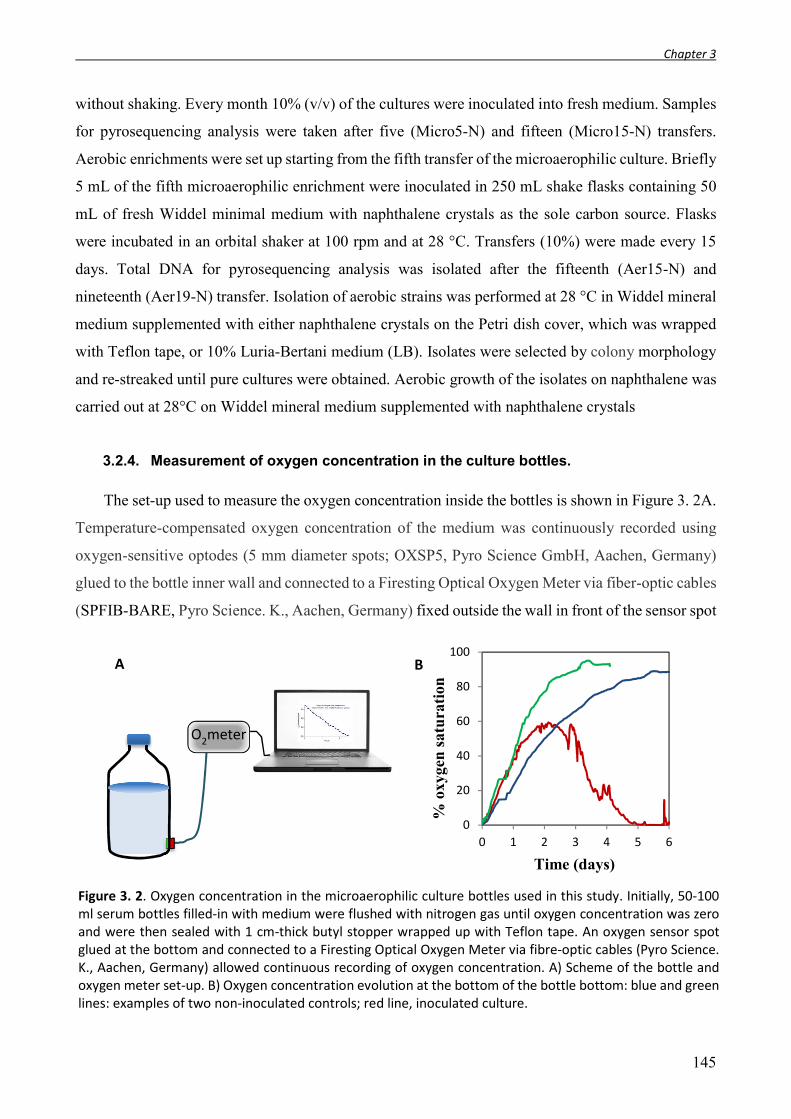
Chapter 3
145
without shaking. Every month 10% (v/v) of the cultures were inoculated into fresh medium. Samples
for pyrosequencing analysis were taken after five (Micro5-N) and fifteen (Micro15-N) transfers.
Aerobic enrichments were set up starting from the fifth transfer of the microaerophilic culture. Briefly
5 mL of the fifth microaerophilic enrichment were inoculated in 250 mL shake flasks containing 50
mL of fresh Widdel minimal medium with naphthalene crystals as the sole carbon source. Flasks
were incubated in an orbital shaker at 100 rpm and at 28 °C. Transfers (10%) were made every 15
days. Total DNA for pyrosequencing analysis was isolated after the fifteenth (Aer15-N) and
nineteenth (Aer19-N) transfer. Isolation of aerobic strains was performed at 28 °C in Widdel mineral
medium supplemented with either naphthalene crystals on the Petri dish cover, which was wrapped
with Teflon tape, or 10% Luria-Bertani medium (LB). Isolates were selected by colony morphology
and re-streaked until pure cultures were obtained. Aerobic growth of the isolates on naphthalene was
carried out at 28°C on Widdel mineral medium supplemented with naphthalene crystals
3.2.4. Measurement of oxygen concentration in the culture bottles.
The set-up used to measure the oxygen concentration inside the bottles is shown in Figure 3. 2A.
Temperature-compensated oxygen concentration of the medium was continuously recorded using
oxygen-sensitive optodes (5 mm diameter spots; OXSP5, Pyro Science GmbH, Aachen, Germany)
glued to the bottle inner wall and connected to a Firesting Optical Oxygen Meter via fiber-optic cables
(SPFIB-BARE, Pyro Science. K., Aachen, Germany) fixed outside the wall in front of the sensor spot
Figure 3. 2. Oxygen concentration in the microaerophilic culture bottles used in this study. Initially, 50-100 ml serum bottles filled-in with medium were flushed with nitrogen gas until oxygen concentration was zero and were then sealed with 1 cm-thick butyl stopper wrapped up with Teflon tape. An oxygen sensor spot glued at the bottom and connected to a Firesting Optical Oxygen Meter via fibre-optic cables (Pyro Science. K., Aachen, Germany) allowed continuous recording of oxygen concentration. A) Scheme of the bottle and oxygen meter set-up. B) Oxygen concentration evolution at the bottom of the bottle bottom: blue and green lines: examples of two non-inoculated controls; red line, inoculated culture.
O2meter
A
B
0
20
40
60
80
100
0 1 2 3 4 5 6
Time (days)
% o
xyge
n s
atu
rati
on

Chapter 3
146
with the aid of a Basic Spot Adapter (SPADBAS). The fiber was connected to a fiber-optic oxygen
meter (Fire Sting O2, Pyro Science GmbH, Aachen, Germany) linked to a computer with Pyro
Oxygen Logger software. Incubations were carried out at 28ºC. A bottle filled with water and fitted
with a temperature sensor was placed in the same chamber for temperature record. The temperature-
compensated dissolved oxygen tension was recorded continuously and expressed as percentage of air
saturation. Figure 3. 2B shows the oxygen evolution in sterile and inoculated culture bottles equipped
with an oxygen-sensitive optode placed at the bottom of the bottle wall.
3.2.5. Synthetic microbial community.
Strains were first picked from colony into Widdel mineral medium supplemented with 1/10 LB.
Late–exponential-phase cells were harvested and washed twice with fresh medium without any
carbon source. Cell densities were determined based on OD600 readings. Co-culture growth was
initiated by inoculating 0.02 OD660 units of each strain in N2 flushed Widdel mineral medium
supplemented with a naphthalene crystal as the sole carbon source. Serum bottles were sealed with
Teflon tape-wrapped rubber-stoppers as for the microaerophilic enrichments. Culture was incubated
at 28ºC without shaking.
3.2.6. Chemical analysis.
For the analysis of nitrate and sulfate we used duplicate 5 ml aliquots of the original aquifer
sample, and quantification was done by ion chromatography (IC) using a Metrohm-761 Compact Ion
Chromatograph equipped with a Metrosep A Supp 4-250 column with chemical suppression (50 mM
H2SO4) as described previously (Martirani-Von Abercron et al., 2016). Duplicate samples of the
initial contaminated aquifer (5 ml) were used to extract aliphatic and aromatic hydrocarbon
compounds and the deuterated Mix 37 (manufactured by Dr. Ehrenstorfer) and 5-alpha-cholestane
(Aldrich) were added as internal standards before the extraction. Hydrocarbon analysis was carried
out at the Instrumental Technical Services of the Estación Experimental del Zaidín (CSIC), Granada,
Spain by GC-MS as in (Martirani-Von Abercron et al., 2016).
3.2.7. Total DNA extraction, 16S rRNA gene 454-Pyrosequencing and data analysis.
DNA extraction for the initial environmental sample was performed as described (Martirani-Von
Abercron et al., 2016). For the enrichment cultures and isolated strains, genomic DNA was purified
using the Wizard Genomic DNA Purification Kit as recommended by the manufacturer (Promega,

Chapter 3
147
Madison, WI, USA). Nucleic acid quantity and purity were determined with a NanoDrop ND-1000
Spectrophotometer (NanoDrop Technologies). A multiplex pyrosequencing amplicon approach was
used for the characterization of the bacterial communities. The PCR amplifications of the 16S rRNA
gene V1–V3 hypervariable region were carried out using the bacterial universal primers 6F and 532R
containing 5′ tags with multiplex identifier and sequencing adapters (Tables S1). PCR amplifications
were performed in 50 μl reactions containing 1 x PCR Buffer (Biorad), 200 μM dNTPs (Roche), 0.5
μM of each primer, 1U of iProof™ High-Fidelity DNA Polymerase (Biorad) and 20 ng of target
DNA. The PCR program consisted of an initial denaturation step at 98°C for 30 s, followed by 25
cycles at 98°C for 10 s, 50°C for 20 s, and 72°C for 30 s, with a final extension at 72°C for 5 min.
PCR reactions were cleaned up using the Qiaquick MiniElute columns (Qiagen) and checked in 1.5%
agarose gel. Amplicons were quantified using QubitTM fluorometer (Invitrogen), pooled at an
equimolar ratio and sequenced using a 454 Titanium amplicon sequencing kit and a Genome
Sequencer FLX 454 at either Citius (University of Seville) or Macrogen (Korea). Analysis of the 16S
454 pyrosequencing data was performed following the Quantitative Insights Into Microbial Ecology
(QIIME v. 1.9.0) pipeline (Caporaso et al., 2010b). Demultiplexing, primer removal, quality-filtering,
chimeras and singletons removal, and alpha and beta diversity studies were done as defined in
(Martirani-Von Abercron et al., 2016).
3.2.8. Full-length 16S rRNA gene amplification and clone library construction.
Near-full-length 16S rRNA genes were amplified using bacterial universal primers GM3F and
GM4R (Muyzer et al., 1995). PCR were performed in 50 μl reactions as previously described (Acosta-
González et al., 2013a) and the products (~1500-bp) were cloned in pGEMT (Invitrogen). Positive
clones were checked by PCR with vector primers prior to Sanger sequencing. Phylogenetic analysis
was done with the ARB package (Ludwig et al., 2004) using the online SINA alignment service and
Silva database version SSU Ref 119. OTU’s assignation was done at ≥ 97% sequence similarity.
Phylogenetic tree was estimated by the neighbour-joining method.
3.2.9. Functional gene amplification.
Gene fragments of bssA, ncr, nmsA, nahA, dbd and nod were amplified by PCR using available
primer sets and the cycling conditions originally described (Table S2). Genomic DNA from NaphS2
and Thauera aromatica K172 strains was used as a positive control for nmsA, ncrA, and bssA
amplification, respectively. All PCR reactions were performed in 50 μl containing 1 x PCR Buffer
(Biorad), 200 μM dNTPs (Roche), 0.5 μM of each primer (Sigma), 1 U of iProof™ High-Fidelity

Chapter 3
148
DNA Polymerase (Biorad) and 10 to 20 ng of target DNA. The appropriately sized amplicons were
purified and cloned in pCR2.1 (TA Cloning Kit, Invitrogen), and 16 positive clones for each gene
were Sanger sequenced (IPBLN, CSIC, Granada, Spain). In addition to the available primers, six
naphthalene dioxygenase (ndo) degenerated primers (Table S2) were designed using the CODEHOP
(COnsensus-DEgenerate Hybrid Oligonucleotide Primers) software
(http://blocks.fhcrc.org/codehop.html) (Rose et al., 2003) based on the alignment of 29 sequences of
dioxygenase proteins of different species (Table S3). A gradient PCR was performed with annealing
temperatures ranging from 50°C to 63°C for all primer combinations to determine optimal annealing
temperatures. Touchdown PCR was used to avoid amplifying nonspecific sequences: after 1 min of
denaturation at 98°C, the first 10 thermal cycles were 10 s at 98ºC, 15 s at 63ºC to 53ºC (the annealing
temperature was reduced 1°C per cycle from 63ºC to 53ºC) and 20 s at 72ºC. The remaining 20 cycles
were 98ºC for 10 s, 55ºC for 15 s and 72ºC for 20 s, with the last cycle followed by a 5 min extension
at 72°C. Genomic DNA from P. putida KT2440 (NAH7) (Fernández et al., 2012) was used as positive
control.
3.2.10. RT-PCR assays.
Reverse transcriptase PCR (RT-PCR) of a 200 bp fragment of dbdCa was done with 75 ng of
total RNA in a final volume of 50 μl using the Titan OneTube RT-PCR system according to the
manufacturer’s instructions (Roche Laboratories). Positive (S. novella strain N1B DNA) and negative
(absence of reverse transcriptase in the assay) controls were included. The primers used were Xdbd
and XdbdI (Table S2).
3.2.11. Biofilm quantification in multi-well plates (crystal violet assay).
Biofilm formation at the air/liquid interphase was analysed in LB medium or LB medium
saturated with naphthalene under static conditions in polystyrene 96-well microtiter plates using a
modification of the method of Barahona et al., 2010. Overnight cultures of each isolate grown in LB
medium were diluted in the corresponding medium to an OD600 of 0.05. Then, 200 μl of each culture
were dispensed in triplicate into the multi-well plates and incubated at 30°C without shaking. Because
the biofilm formation kinetics varied for each strain and condition, biofilms were quantified at
different time points for 50 hours as follows: the cell suspension was removed, the wells were washed
with 200 μl of sterile water, the same volume of crystal violet solution (0.4%, w/v) was added to each
well and incubated for 20 min to allow staining of adhered cells. Excess stain was eliminated by
rinsing with water. Plates were air dried and 200 μl of 95% ethanol (v/v) were added to each well to

Chapter 3
149
extract the crystal violet from cells. Distaining was performed overnight with shaking (40 rpm) after
which the DO540 was measured on a microplate reader. The values when biofilm formation was
highest were selected for each strain.
3.2.12. Scanning electron microscopy (SEM).
Specimen preparation and SEM were performed at the Scientific Instrumentation Centre (CIC)
of the University of Granada. Biofilm samples from microaerophilic cultures growing on naphthalene
were fixed for 2 h at 4ºC in 2.5% glutaraldehyde prepared in cacodylate buffer, pH 7.4. The fixed
samples were rinsed three times (15 min each) with the same buffer at 4ºC and incubated for 1 h with
1% osmium tetroxide at room temperature, rinsed three times (5min) with water and dehydrated in
an increasing ethanol concentration gradient from 50% to 100%. Samples were further desiccated
with carbon dioxide in a Leica EM CPD300 critical point dryer according to Anderson (1951).
Samples were carbon coated in an EMTECH K975X evaporator and examined in a Zeiss
SUPRA40VP scanning electron microscope equipped with a Schottky type emission gun.
3.2.13. Nucleotide sequence accession numbers.
The 454 pyrosequencing raw reads have been deposited in the NCBI short-reads archive database
(accession number SRR5417958 to SRR5417967). The partial dbdB gene sequences obtained from
the clone libraries have been deposited in GenBank under accession numbers MF537652 to
MF537654. The 16S rRNA gene clone library sequences have been deposited in GenBank under
accession numbers MF156538 to MF156549.

Chapter 3
150
3.3. Results and discussion
3.3.1. Betaproteobacteria are dominant in a hydrocarbon polluted aquifer
In the initial sample collected from a 25 m-deep well of a hydrocarbon polluted aquifer, a biofilm
spreading between the water phase and the upper 10 cm oil layer was clearly visible (Figure 3. 1).
The most abundant PAHs in the water phase were two-ring aromatics (0,14 ± 0.082 ppm of
naphthalene, 0,20 ± 0.119 ppm of 2-methylnapthalene) followed by one order of magnitude lower
levels of aromatics with three or more rings (Table 3. 1). The alkane distribution was coherent with
contamination with a mixture of diesel and gasolines (Figure 3. 3). The concentration of nitrate and
sulphate in the aqueous phase was in the range of natural background levels in oligotrophic
groundwaters (Einsiedl and Mayer, 2005; Arauzo and Martínez-Bastida, 2015) (Table 3. 2).
Table 3. 1. Aromatic hydrocarbon composition and abundance in the contaminated aquifer initial samples.
Compound ppm (µg/g)
Naphthalene 0.1377±0.082 1-methylnaphthalene 0.1868±0.081 2-methylnaphthalene 0.2002±0.119
1,4,5-trimethylnaphthalene nd 1,4,6,7-tetramethylnaphthalene 0.0145±0.009
Dibenzothiophene 0.0299±0.026 Phenanthrene 0.0244±0.015 Anthracene 0.0245±0.005
Benzo[a]anthracene nd Pyrene 0.0066±0.0007
C2-naphthalene 0.0294±0.018 Fluoranthene nd
Chrysene nd
Total 0.654
nd: not detected.
Table 3. 2. Characterization of the initial polluted aquifer sample. The detailed hydrocarbon composition of the sample can be found in Table 3. 1 and Figure 3. 3.
Sample Name
Description Location Coordinates Nitrate (μM)
Sulphate (μM)
Total hydrocarbons
(μg/kg)
Aliphatic Aromatics
INI Aquifer below an oil refinery, 25 m depth
Valle de Escombreras, Murcia (Spain)
37°34'20.6"N 0°55'24.7"W
84.64±0.24 19.27±0.72 2130.80 654.00

Chapter 3
151
The potential of the initial biofilm community to degrade PAHs was determined using the MPN
enumeration of aerobic and nitrate reducing bacteria. Naphthalene (NAP), 2-methylnaphthalene
(2MN), 2-naphtoic acid (2NA, a known intermediate in anaerobic naphthalene degradation) and
anthracene (ANT) were used as carbon sources. A positive control with acetate as carbon source, and
a negative control with no addition, were included in the analysis to estimate the size of the nitrate
reducing community and the basal growth on the sample’s intrinsic organic matter, respectively.
Under anoxic conditions, the high counts (105 cells/ml) of NRB able to grow with acetate
revealed the presence of a significant community of NRB in the biofilm (Figure 3. 4, Table S4).
However, no growth above the basal level was observed with any of the aromatics used, except for a
minor increase with 2MN, indicative of the absence of NRB able to use naphthalenes or anthracene.
This contrasted with the results obtained under aerobic conditions; substantial growth on the three
aromatics was observed, which ranged between 4.3 x 10² cells/ml of naphthalene degraders and 2.4
x 104 cells/ml of 2MN degraders, consistent with the higher levels of 2MN in the polluted aquifer
(Table 3. 1). These values represented only a minor fraction of the culturable aerobic community
present, which reached 1.4 x 10⁷ cells/ml when acetate was used as the carbon source.
Figure 3. 3. Aliphatic hydrocarbon distribution (%) in the initial contaminated aquifer sample. The total amount of aliphatic hydrocarbons in the sample was 0.654 µg/ml.

Chapter 3
152
We isolated the total DNA from duplicate biofilm samples taken at the oil-water interphase to
determine the structure of the community present using 16S rRNA gene V1-V3 region
pyrosequencing. Total read numbers generated from all the samples analysed in this study and the
corresponding diversity indices are summarized in Table 3. 3. The rarefaction curves (Figure S1) and
the Good’s sample coverage estimator suggested that sampling depth was sufficient in all cases to
estimate the microbial diversity (Table 3. 3). The observed OTU number, diversity indices and OTU
distribution of the initial sample were reproducible between replica (weighted Unifrac significant test
P value 0.33).
Figure 3. 5 shows that the biofilm community was clearly dominated by the Proteobacteria,
where the Betaproteobacteria accounted for more than 60% of the bacterial population.
Bacteroidetes, Chloroflexi and the uncultured lineage SAR406 (also referred to as Marine Group A
(Wright et al., 2014)) were also present at substantial levels. The major presence of
Betaproteobacteria, Bacteroidetes and Chloroflexi in this hydrocarbon polluted site is in agreement
with the strains frequently found and isolated from aquifers and groundwater environments (Griebler
1E+00
1E+01
1E+02
1E+03
1E+04
1E+05
1E+06
1E+07
1E+08
na
NAP
2M
N
2NA
ANT
Ace na
NAP
2M
N
2NA
ANT
Ace
MPN
(cel
ls/m
l)
Carbon source
Anoxic Aerobic
Figure 3. 4. MPN enumeration in the aquifer initial sample of nitrate reducing and aerobic bacteria able to grow on naphthalene (NAP), 2-methyl-naphthalene (2MN), 2-naphtoic acid (2NA), anthracene (ANT), acetate (Ace) and with no added carbon source (na). Counts were made in triplicate. Numerical values (95% C.I.) can be found in supplementary Table S4. Dotted lines indicate the basal growth with no added carbon source.

Chapter 3
153
Table 3. 3. Comparison of OTU number, diversity, evenness indices and coverage for the different samples.
Samplea NSb OTUsc OTUs (1500)d
Chao1 (1500)e Shannon Coveragef
INIa 1807 167 158 259.08 4.08 95.87%
INIb 1762 174 168 248.50 4.29 96.19%
Anox-NAP 3611 138 101 207.25 4.24 98.22%
Anox-2MN 6778 161 84 189.86 4.48 99.13%
Anox-HMN 3992 148 104 198.23 4.57 98.57%
Micro5-N 34611 115 42 61.13 2.66 99.94%
Micro15-N 22599 69 26 65.00 1.35 99.91%
Micro-12s-N 23347 101 43 58.00 2.33 99.91%
Aer15-N 29889 111 37 64.20 1.58 99.89%
Aer19-N 20685 116 45 129.33 2.46 99.79% a INIa and INIb refer to the two replica of the initial samples. bNumber of sequences for each library filtered for chimera and singletons. cOTU numbers calculated with all sequences at the 3% distance level. dOTU numbers calculated for a randomized subset of 1500 reads per sample at the 3% distance level. eChao index calculated with 1500 subsampled sequences. f Good’s sample coverage estimator.
and Lueders, 2009), especially when carbon source availability is high (Fierer et al., 2007). The
Betaproteobacteria community in the sample was relatively simple, dominated by two operational
taxonomic units (OTUs): one belonging to the Comamonadaceae (29.5%) and a Rhodocyclaceae
closely related to Azovibrio (30.6%) (Figure 3. 5B). Betaproteobacteria belonging to the
Comamonadaceae occur in diverse pristine and polluted habitats such as soil, freshwater,
groundwater, and industrial processing water, and can use a variety of substrates, including
hydrocarbons, as carbon source (Willems, 2014). Interestingly, members of this group were also
highly prevalent in toluene-polluted model wetlands and in BTEX-contaminated groundwater
(Táncsics et al., 2013; Martinez-Lavanchy et al., 2015). The Gammaproteobacteria were almost
exclusively composed of a single OTU closely related to Pseudoxanthomonas (3.8%) . Using samples
from this biofilm layer, we set-up a series of enrichment cultures under two different oxygen
limitation regimes to select for PAH degrading communities.

Chapter 3
154
3.3.2. Changes in the community structure under strictly anoxic denitrifying conditions.
Enrichment cultures with nitrate as the electron acceptor and naphthalene and 2MN as the only
carbon source were prepared under strict anoxic conditions. To reduce the PAH concentration in the
water phase, which might limit growth of anaerobes (Galushko et al., 1999a), the PAHs were provided
dissolved in HMN. After 12 months and 3 subsequent subcultures, DNA from the anoxic cultures
was isolated and analysed as above by 16S rRNA 454 pyrosequencing to determine changes in the
community structure (samples Anox-NAP, Anox-2MN, Anox-HMN) (Table 3. 3). A moderate loss
of diversity was observed in all the nitrate reducing cultures, together with a clear shift in the
community structure that may be attributed to the imposed change in the respiratory regime. Overall,
the changes were comparable in the presence of the PAHs and when only the carrier HMN was added
to the cultures (Figure 3. 5A). Whilst the dominant Commamonadaceae detected in the starting
material was still present at high levels in the NAP, HMN, and to a lesser extent in the 2MN cultures,
the dominant Azovibrio OTU disappeared almost completely in the three enrichments, partially
balanced by an increase in Betaproteobacteria representatives belonging to the Burkholderiaceae,
Rhodoferax and especially to the Rhodocyclaceae genus Dok59 in the case of the 2MN cultures
(Figure 3. 5B). Members of the Bacteroidetes, which constituted more than 3% of the initial
community, decreased to levels below 0.2% in all conditions. The strong decrease of some groups in
all the enrichments suggests their high sensitivity to the new culture conditions, i.e., strict anoxic
nitrate respiration and presence of PAHs. In contrast, an increase of the Alphaproteobacteria was
observed in all conditions, especially represented by the Sphingomonadaceae, Acetobacteraceae and
Rhizobiaceae. This might be explained by the denitrifying capacity of some members of the
Sphinomonadaceae and Rhizobiaceae, in addition to their ability to tolerate the presence of toxic
hydrocarbons (Carareto Alves et al., 2014; Cua and Stein, 2014). The Anaerolineae within the
Chloroflexi also increased significantly in the 2MN and HMN enrichments. Since members of this
class seem incapable of nitrate respiration, enrichment of this group is probably a consequence of
their capacity for fermentative metabolism on cell remains (Yamada and Sekiguchi, 2009). Some
specific changes were observed that could be attributed to the presence of PAHs: the Actinobacteria,
which were almost undetectable in the initial biofilm sample, increased up to 18% in the HMN and
2MN cultures, and to almost 30% of the community in the NAP enrichment. Although an increase in
the abundance of Actinobacteria has been related to the degradation of PAHs in aerobic conditions
(Muangchinda et al., 2017; Zhu et al., 2017b), there is no evidence of the contribution of this phylum
to anaerobic degradation. Interestingly, an increase in the Acidobacteria candidate class iii1-8 (order
DS-18) from undetectable levels in the initial biofilm sample to more than 9% of the community in

Chapter 3
155
the 2MN enrichment was also observed (Figure 3. 5A), in agreement with the previous observation
of a dramatic increase of this poorly characterized group under nitrate reducing conditions, and
especially in the presence of PAHs (Martirani-Von Abercron et al., 2016). This supports the
suggestion that the group is composed of slow-growing oligotrophs (Fierer et al., 2007), probably
highly resistant to PAHs (Martirani-Von Abercron et al., 2016). During the enrichment process under
anoxic conditions, the minor changes in PAH concentration were not statistically significant (not
shown), which reflects the absence of anaerobic PAH degraders determined with the MPN
Figure 3. 5. A) Cumulative plot of bacterial phyla detected in the initial environmental samples and in the anaerobic enrichments amended with naphthalene (NAP), 2-methylnaphthalene (2MN) and heptamethylnonane (HMN). The Proteobacteria are described at the class level. The initial samples were analyzed in duplicate (labelled 1 and 2). B) Detailed cumulative plot of the Betaproteobacteria detected in the same samples. The average values of the duplicate initial samples shown in A are included for comparison, labelled as INI. [R], Rhodocyclaceae; [O] Oxalobacteraceae; [H] Hydrogenophilaceae; [C] Comamonadaceae.

Chapter 3
156
enumeration. Furthermore, our attempts to detect genes involved in anaerobic PAH degradation in
the initial sample and in the enrichment cultures using primers against ncr and nmsA gave negative
results. Neither was any amplification product obtained with primers against nitric oxide dismutase
(nod). To date, no growth of NRB isolates with PAHs as carbon source has been observed under strict
anoxic conditions, and in most cases laboratory conditions can only reproduce aerobic degradation
(Yagi et al., 2010). The observed shift in the bacterial community describes the adaptation to the new
respiratory regime. However, NRBs are facultative anaerobes prepared to thrive in the oxic-anoxic
transition zone, and in these environments anaerobic respiration and PAH degradation (aerobic) may
be spatially separated concomitant processes.
3.3.3. Microaerophilic conditions strongly select for two dominant strains.
To mimic the natural habitat of most NRB, which are facultative anaerobes that can thrive at low
oxygen concentrations, we set-up microaerophilic enrichment cultures. To this end, culture bottles
were initially flushed with nitrogen gas to reach anoxic conditions; the caps of the bottles were
prepared to allow a slow influx of air (see Experimental Procedures section) so that in the absence of
inoculum oxygen saturation in the medium could be reached after approximately one week (Fig. 3.
2B). In this setup, inoculation of the medium resulted in oxygen consumption, which increased
gradually to reach rates at equilibrium with the oxygen influx rate. Further growth of the culture
caused oxygen consumption at higher rates than diffusion, leading to a progressive decay in the
oxygen saturation level, which finally reached undetectable values in spite of the constant oxygen
influx. We initiated an enrichment culture using naphthalene as the representative PAH, provided as
the sole carbon source in the form of crystals. A fraction of 10% of the culture was regularly
transferred to fresh medium. After three months, a fully developed biofilm was clearly visible at 0.5-
1 cm around the naphthalene crystal in all the cultures of the enrichment process (Figure 3. 6A). The
subsequent subcultures were characterized by the formation of a similar biofilm structure at a short
distance from the naphthalene crystal, which was progressively consumed until it completely
disappeared. Oxygen concentration at the biofilm level remained close to zero once the biofilm had
developed, as previously shown for single species bacterial biofilms (Staal et al., 2011). Scanning
electron microscopy (SEM) of the developed biofilm revealed the presence of a mixed community of
bacteria with different sizes and shapes, adsorbed or attached onto filamentous, sometimes
amorphous, EPS material (Figure 3. 6B). From subcultures 5 (8 months, 5 transfers) and 15 (26
months, 15 transfers), total DNA was isolated and the structure of the community in the enrichments

Chapter 3
157
was analysed as above (samples Micro5-N and Micro15-N, Table 3. 3). Chao indices and rarefaction
curves indicated that during microaerophilic enrichment, the diversity of the bacterial community was
gradually reduced, reaching the lowest values in Subculture 15 (Figure S1). Accordingly, a
progressive selection for specific groups was observed in the community: the enrichment process
initially selected for four dominant Proteobacteria in Subculture 5, which in some cases could be
affiliated to the genus level after comparing the pyrosequencing reads with the 16S rRNA sequence
of strains isolated from the enrichments (see below) (Figure 3. 7). The four selected taxa belong to
the Xanthobacteraceae (Xanthobacter and Starkeya), the Commamonadaceae (Variovorax), and the
Nocardiaceae. Further enrichment under these conditions (Subculture 15) finally selected for two co-
dominant strains which constituted 97% of the community: Variovorax (54%) and Starkeya (43%),
and consequently the slowest Shannon evenness index characterized this community (Table 3. 3). It
is worth noting that the two enriched strains were almost undetectable in the initial sample. From
Subculture 5 we were able to isolate a number of strains on rich medium and on minimal medium
with naphthalene as carbon source that were further characterized (Table 3.4; Figure 3.8). Taxonomic
Figure 3. 6. A) Enrichment culture bottle under microaerophilic conditions showing the characteristic bacterial biofilm developed around the naphthalene crystal. B) Scanning electron microscopy (SEM) micrograph showing details of the multispecies biofilm formed around the naphthalene crystal during growth under microaerophilic conditions. Scale bar = 1 μm.
A
1 μm
B

Chapter 3
158
affiliation of the strains matched those taxa which were among the most abundant in the enrichment,
although we were not able to isolate some groups, such as Actinotalea and the Nocardiaceae. Because
pyrosequencing only targeted the V1-V3 fragment of 16S rRNA, several OTUs in the pyrosequencing
data were assigned to the taxon “Xanthobacteraceae”, resulting in an ambiguous linking of the OTUs
to the isolates. This ambiguity was solved by constructing a phylogenetic tree based on the V1-V3
region of all Xanthobacteraceae present in the pyrosequencing data and of the isolates (Fig. S2).
Results assigned the most abundant Xanthobacteraceae in the final enrichment (Subculture 15) to
Starkeya novella, which corresponded to strain 964 isolated on solid medium with naphthalene as the
sole carbon source and was designated as S. novella strain N1B. The most abundant strain in the
Figure 3. 7. Cumulative plot of bacterial taxons present at more than 1% of the community detected in the microaerophilic, aerobic and synthetic microaerophilic enrichments amended with crystals of naphthalene as carbon source. The deepest taxonomic affiliation reached (phyla indicated in brackets) is shown. The average values of the duplicate initial samples (Figure 3. 4A) are included for comparison, labelled as INI.

Chapter 3
159
enriched community was affiliated to Variovorax paradoxus (Figure 3. 8), which corresponded to
strain 968 isolated on rich medium and was designated as V. paradoxus strain N2.
Interestingly, the isolate 966, which constituted almost 12% of the Subculture 5 community,
disappeared almost completely in the final enrichment. This strain was finally assigned to Xanthobacter
polyaromaticivorans (99% 16S rRNA gene identity) based on the Xanthobacteracea phylogenetic tree,
although it was also closely related to Aquabacter spiritensis (98%) (Figure 3. 8). The remaining
isolates represented strains that were present at levels below 1% in the final Micro15-N enrichment
(Table 3. 4).
958 968 denovo31221_B2.LE.A10_27361_f__Comamonadaceaeg__Variovorax denovo30278_B4.LE.Q10_1376_f__Comamonadaceaeg__Variovorax__paradoxus HQ005420.1_Variovorax_paradoxus_strain_HB44 denovo9575_B5.LE.12STRAIN_59053_f__Xanthomonadaceaeg__Pseudoxanthomonass__
denovo6311_AQ1_29514_f__Xanthomonadaceaeg__Pseudoxanthomonass__ denovo25802_AQ1_42044_f__Xanthomonadaceaeg__Pseudoxanthomonass__ 962 CP003093.2:3414512-3416060_Pseudoxanthomonas_spadix_BD-a59_complete_genome denovo14227_B1.LE.0_106423
972 NR_115989.1_Epilithonimonas_lactis_strain_H1 KT766029.1_Chryseobacterium_sp._ERMR1:04 denovo14536_B2.LE.A10_21993_f__Pseudomonadaceaeg__Pseudomonas
KF863235.1_Pseudomonas_stutzeri_strain_N2 943 denovo9091_AQ2MN_19000_f__Pseudomonadaceaeg__Pseudomonas
963 LC007966.1_Pseudomonas_denitrificans_isolate:_05CF15-5C NR_028706.1_Pseudomonas_veronii_strain_CIP_104663 KF975414.1_Brevundimonas_vesicularis_strain_KK6
AJ244706.1_Brevundimonas_mediterranea_strain_V4.BO.18 denovo716_B5.LE.12STRAIN_1942_f__Caulobacteraceaeg__s__ 951 denovo31963_AQ2MN_3643f__Rhizobiaceaeg__s__
960 NR_044216.1_Rhizobium_selenitireducens_strain_B1 NR_074219.1_Starkeya_novella_strain_DSM_506 964 denovo24185_B1.LE.0_26733_f__Xanthobacteraceaeg__s__ denovo11305_B1.LE.0_6427_f__Xanthobacteraceae FR733686.1_Aquabacter_spiritensis_type_strain_DSM9035T
NR_112205.1_Xanthobacter_polyaromaticivorans_strain_127W denovo27209_B5.LE.12STRAIN_23404_f__Xanthobacteraceaeg__Xanthobacters__autotrophicus denovo670_B1.LE.0_327_f__Xanthobacteraceae denovo10294_B1.LE.0_19862_f__Xanthobacteraceae 966 952
983 denovo18658_B5.LE.12STRAIN_148598_f__Microbacteriaceaeg__Microbacteriums__maritypicum LN890176.1_Microbacterium_oxydans_strain_M90 KX369591.1_Microbacterium_oxydans_strain_AE038-20 JN881579.1_Candidatus_Caldiarchaeum_subterraneum_clone_PNG_TBR_A55
0,05
Variovarax
Pseudoxanthomonas
Epilithonimonas
Pseudomonas
Brevundimonas
Rhizobium
Starkeya
Xanthobacter
Microbacterium
Figure 3. 8. Neighbour-joining tree of the 16S rRNA gene of the bacterial strains isolated in this study and their closest relatives in the databases. Representative closest QIIME OTUs for each strain (labelled as “denovo”) are included. The bar represents 0.05 substitutions per site.

Chapter 3
160
3.3.4. A pathway with high affinity for oxygen is favoured under microaerophilic
conditions.
Eight strains amongst the isolates, including the dominant Variovorax and Starkeya strains, were
able to grow aerobically with naphthalene as the carbon source (Table 3. 4). To identify the genes
involved in naphthalene degradation, we used previously described primers and newly designed
degenerated primers based on the alignment of 29 nahA sequences for naphthalene dioxygenases
belonging to different taxonomic groups (Table S3, see the Experimental Procedure section). PCR
analysis with several sets of primers to amplify naphthalene dioxygenase gene were carried out with
DNA from the enrichments and from all the isolates. Only Pseudomonas veronii and the total DNA
from Subculture 5 gave positive results with nahA primers. The P. veronii nahA gene product
clustered with genes for class A ring-hydroxylating dioxygenases an was predicted to utilize alkyl
substituted monoaromatics as well as PAHs as substrate, whilst Subculture 5 product was predicted
to prefer hetero-polycyclic hydrocarbons and arylbenzenes, according to the ring-hydroxylating
oxygenase database (Chakraborty et al., 2014) (Figure 3. 9). As X. polyaromaticivorans was present
among the isolates, and was the predominant strains in the Subculture 5 community, we designed and
assayed primers for the dbdB gene coding for a dioxygenase from X. polyaromaticivorans strain
127W naphthalene/dibenzothiophene degradation pathway (Hirano et al., 2004). Only the isolates
belonging to the Xanthobacteraceae, S. novella and X. polyaromaticivorans, produced the
corresponding PCR amplification products, which showed 99% identity with the X.
polyaromaticivorans dbdB sequence (Figure 3. 9). We further investigated the presence in S. novella
strain N1B of the complete PAH degradation cluster identified in X. polyaromaticivorans. We were
able to amplify the genes for dbdABCaCbCcDE from the S. novella strain N1B DNA (Table 3. 5) and
showed a remarkable gene identity (99%) with the described X. polyaromaticivorans PAH
degradation gene cluster, including a traA conjugation gene located upstream dbdA.
The high percentage of identity of the sequences at the DNA level and the presence of flanking
transfer genes suggest that S. novella strain N1B could have acquired the naphthalene degradation
pathway through a recent horizontal gene transfer event, favoured by the stable physical environment
for cell-to-cell contact provided by the biofilm matrix. In fact, we were still able to detect X.
polyaromaticivorans in a gene library of whole-length 16S rRNA gene constructed with DNA from
subculture 9 (not shown), and this could even be isolated on naphthalene plates from subculture 14.
It is worth noting that no plasmid was detected in S. novella strain N1B genome. The DbdCa protein,
which was shown to code for the alpha subunit of a naphthalene hydroxylating dioxygenase, is closer

Chapter 3
161
Table 3. 4. Strains isolated from the microaerophilic enrichment cultures.
Clon # Isolated from Subculture
Closest relatives (Class)1 % ident.
Pyroseq QIIME assigned taxon
% in the Subcultures
Growth on NAP2
Biofilm3 dbdB nahA Sub 5 Sub 15
964 5, 15 Starkeya novella (B) 99% Xanthobacteraceae, Other 26.71 43.19 ++ ++ + -
966 5 Xanthobacter polyaromaticivorans (B) 99% Xanthobacteraceae, g_ 11.77 0.38 +++ - + - 960 5 Rhizobium naphthalenivorans (A) 99% Rhizobiaceae, g_ 1.13 <0.01 + - - - 951 5 Brevundimonas mediterranea (A) 100% Caulobacteraceae 0.34 <0.01 ++ - - - 952 5 Microbacterium paraoxydans (Act) 99% Microbacterium 0.04 <0.01 ± - - - 983 5 Microbacterium sp. (Act) 99% Microbacterium <0.01 <0.01 ++ - - - 958 5, 15 Variovorax ginsengisoli (B) 99% Variovorax
41.46 54.22 - - - -
968 5, 15 Variovorax paradoxus (B) 99% Variovorax ++ - - - 962 5 Pseudoxanthomonas spadix (G) 99% Pseudoxanthomonas 1.13 <0.01 + - - - 943 5 Pseudomonas nitroreducens (G) 99% Pseudomonas
0.37? <0.01 - - - -
963 5 Pseudomonas veronii (G) 99% Pseudomonas ± - - +
972 5 Chryseobacterium sp. (Bact) 98% Flavobacteriaceae, Other 0.07 <0.01 - - - -
(B), Betaproteobacteria; (A), Alphaproteobacteria; (Act), Actinobacteria; (G); Gammaproteobacteria; (Bact), Bacteroidetes. 2 +++ OD>0.9 ; ++ OD>0.4; + OD>0.3; ± OD<0.1. 3 Biofilm developed around the naphthalene crystal.

Chapter 3
162
Table 3. 5. Naphthalene degradation gene sequences identified in S. novella strain N1B.
Genes Amplicon Size
% Id. Function
dbdAB’ 994 98% ferredoxin (dbdA); gentisate 1,2-dioxygenase (dbdB) (preceded by a partial traA gene coding for a conjugative transfer relaxase)
dbdCa 1398 99% naphthalene 1,2-dioxygenase alpha subunit
dbdCbCc 1556 99% naphthalene 1,2-dioxygenase beta subunit (dbdCb) Ferredoxin (dbdCc)
dbdD 1078 99% dihydrodiol dehydrogenase
to group IV dioxygenases utilizing benzene/toluene/biphenyl as substrate, although it showed a
preference for larger substrates and was functional at extremely low oxygen concentrations (Hirano
et al., 2006).
The dbdB gene codes for a gentisate1,2-dioxygenase with high affinity for oxygen (Hirano et al.,
2007). This suggests that the microaerophilic conditions were selecting for a degradation pathway
that included enzymes with high affinity for oxygen. The selection for organisms bearing specific
aromatic degradation enzymes observed under microaerophilic conditions both in competition
experiments and in natural environments has been attributed the high affinity for oxygen of the
selected mono- and dioxygenases (Balcke et al., 2008; Kiesel et al., 2008; Táncsics et al., 2013;
Martinez-Lavanchy et al., 2015). This supports the idea that high oxygen tensions (i.e., “absolute”
aerobic conditions) are not essential for aromatic degradation as long as the appropriate organisms
are present. RT-PCR amplification of a dbdCa gene fragment from Subculture 15 showed that the
gene was expressed in these conditions (Figure 3. 10). The naphthalene degradation genes of
Variovorax and of the remaining strains except Pseudomonas could not be identified with the
different sets of primers tested. Variovorax strains are frequently found in polluted environments and
some of them, which bore genes for aromatic ring-hydroxylating dioxygenases with diverse suggested
functions, were capable of aromatic degradation (Satola et al., 2013; Posman et al., 2017). However,
a specific gene for naphthalene degradation has not been described in this genus. Previous studies
have shown that the diversity of the aromatic dioxygenases alpha subunit is underrepresented in the
databases, which are dominated by Pseudomonas-related sequences (Gomes et al., 2007; Iwai et al.,
2011), and the use of primers designed to select for substrate specificity do not guaranty the detection
of the targeted specific activities (Witzig et al., 2006).

Chapter 3
163
320
242 190
M + c D
Figure 3. 9. Phylogeny of the alpha subunit oxygenase component of hydroxylating naphthalene dioxygenase (upper panel) and gentisate 1,2‐dioxygenase (lower panel) partial amino acid sequence retrieved from Subculture 5 and different isolates obtained in this study. The trees were rooted with unrelated bssA gene product as out‐group.
Figure 3. 10. RT‐PCR analysis of total RNA extracted from subculture 15 (+). A negative control (c) with RNA where the reverse transcription step had been omitted and a positive control with DNA (D) from S. novella strain N1B were included. M, molecular weight marker.

Chapter 3
164
3.3.5. Evolution of a synthetic community under microaerophilic conditions
To gain further insight into the community structure dynamics, we reproduced an artificial
microbial community by mixing equal proportions of the twelve isolates (Table 3. 4) in a single
microaerophilic culture, which was then subjected to a similar selective pressure using naphthalene
as the sole carbon source for 12 weeks. The structure of the resulting community was analysed by
16S rRNA pyrosequencing as above (Table 3. 3, sample Micro-12s-N). Because the only possible
strains present in this culture were the twelve isolates, we used the pyrosequencing data from this
community to build the phylogenetic tree of the different taxons to establish the link between QIIME
affiliation and strain isolates (Figure 3. 8 and not shown). Figure 3. 7 (sample Micro-12s) shows that
the community had evolved from an initially even situation with equally abundant strains, to a
community where the naphthalene degrading strains X. polyaromaticivorans, and to a lesser extent
Brevundimonas and Variovorax dominated. Some Brevundimonas species are characterised for their
dependence on microaerophilic conditions for growth (Buczolits et al., 2001). In contrast the
abundance of Microbacterium, Starkeya, Pseudoxanthomonas and Rhizobium, which were also
capable of naphthalene degradation, was reduced to between 1.5 and 2% of the community. Finally,
the two Pseudomonas strains and Chryseobacterium sp., unable to used naphthalene as a carbon
source, almost disappeared, in accordance with their low abundance in the enriched subcultures 5 and
15. Overall, the distribution resembled the community structure in Subculture 5, and probably
represented an intermediate step in the evolution of the community.
3.3.6. Aerobic growth allows higher bacterial diversity
To analyze the impact of oxygen tension on the evolution of the community, Subculture 5 of the
microaerophilic enrichment was used as starting material to set-up enrichment cultures under higher
oxygen concentrations provided through continuous shaking at 100 rpm. The oxygen saturation level
in the cultures in these conditions was 92%. Samples were taken for community analysis as above
after 5 months (10 transfers, sample Aer15-N) and 9 months (14 transfers, Aer19-N). Enrichment
under aerobic conditions initially favoured Variovorax to the detriment of X. polyaromaticivorans,
and further evolved to a structure closer to Subculture 5 as regards the dominant OTUs, though with
a higher Starkeya to Variovorax ratio (Figure 3. 7). In addition, the final sample Aer19-N also showed
a clear increase in overall bacterial diversity (Table 3. 3), where the previously least favoured groups
such as Pseudomonas, Rhizobiaceae, Caulobacteraceae and Microbacterium recovered levels well
above 1% of the community. Despite the constant shaking of the flaks, the cells showed a strong
tendency to form clumps in the medium, which probably creates microhabitats of different oxygen

Chapter 3
165
concentration, which may explain the fluctuations observed in the structure of the community with
time.
3.3.7. Starkeya novella strain N1B is responsible for biofilm formation and naphthalene
degradation.
We analyzed the capacity of the different isolates to reproduce the biofilm structure around the
naphthalene crystal. Almost all the isolates were capable to certain extent of forming biofilm at the
air/liquid interphase in rich medium, as determined with the standard crystal violet assay (Figure 3.
11), both in the presence and absence of naphthalene crystals. However, only Starkeya novella strain
N1B was able to develop a well-defined biofilm structure in minimal medium around the naphthalene
crystal similar to the one developed by the bacterial community in the microaerophilic enrichments.
No growth was observed in these conditions when the naphthalene crystal was omitted from the
medium. Interestingly, X. polyaromaticivorans was among those strains that were less efficient in
standard biofilm formation, together with Pseudoxanthomonas and Rhizobium. In contrast, the two
Variovorax isolates were characterized by the formation of clumps during growth, both in the
presence and absence of naphthalene, indicative of the production of an EPS matrix, which
characterizes some Variovorax strains (Jamieson et al., 2009). However, none of these strains were
Figure 3. 11. Biofilm formation at the air/liquid interphase by different isolates in LB (light blue) or LB supplemented with a naphthalene crystal (dark blue). Assays were carried out in triplicate in microtiter plates as described in the Experimental procedures section.

Chapter 3
166
capable of developing a structured biofilm around the naphthalene crystal. The closest relative of S.
novella strain N1B is S. novella type strain (99% identity of 16S rRNA gene) (Kappler et al., 2001),
which is a strict aerobe incapable of naphthalene degradation and lacking any naphthalene
degradation gene within its genome (Kappler et al., 2012). In contrast, S. novella N1B harbors a
naphthalene degradation pathway which is almost identical to the X. polyaromaticivorans pathway,
characterized by the presence of key dioxygenases with high affinity for oxygen. X.
polyaromaticivorans was originally described as capable of growth and PAH degradation under
extremely low oxygen tensions (below 0.2 ppm) (Hirano et al., 2004). However, in our
microaerophilic enrichments, the S. novella strain N1B was able to totally outcompete X.
polyaromaticivorans, probably after having horizontally acquired its dbd pathway. Thus, it seems
that the capacity of the S. novella strain N1B to develop a structured biofilm in close proximity to the
naphthalene crystal is a key factor that provides a strong advantage to the strain for growth with
naphthalene under these oxygen limiting conditions. In fact, it has been hypothesized from simulated
biofilm competition experiments that polymer producers are evolutionary favoured at the expenses
of non-producers (Xavier and Foster, 2007). The ecological benefit driving this selection in our
enrichment may be to provide a better competition for the substrate by locating the biofilm producers
at the optimum distance from the carbon source to allow efficient mass transfer, simultaneously
maintaining a sufficient distance to avoid a possible toxic effect of naphthalene. The stable presence
of V. paradoxus in the biofilm suggests that its EPS production capacity favors its presence in the
community, probably contributing to the structure of the biofilm.
3.4. Concluding remarks
Mixed bacterial biofilms are increasingly being considered as attractive tools in bioremediation
protocols for their increased stability and resistance (Demeter et al., 2015). In fact, bacteria in the
sessile state which form part of biofilms are more resistant to pollutants (Khoei et al., 2016) and more
effective in pollutant degradation (Edwards and Kjellerup, 2013) than as free-living cells. In this
study, the initial sample collected at the oil-water interphase had a medium to low phylogenetic
diversity, numerically dominated by Betaproteobacteria, which represented more than 60% of the
community. Enrichment under microaerophilic conditions resembling those naturally found in real
environmental polluted sites produced a drastic change in the structure of the community, pointing to
oxygen limitation as the main factor driving the structure of the community, selecting for strains that
were undetectable in the starting material. Community members bearing degradation pathway
enzymes with high affinity for oxygen were selected for, whilst additionally the capacity to produce

Chapter 3
167
EPS and to develop biofilms was crucial for the final selection. Interestingly, this enrichment
evidenced the function of biofilm structure to favour gene transfer, thus generating a naphthalene
degrading strain able to grow at very low oxygen tensions and with a striking capacity to develop a
physical structure to locate the cells at the appropriate distance from the toxic carbon source. Non-
polar substances such as hydrocarbons can accumulate in the EPS matrix (Späth et al., 1998), which
would help increase the local concentration of the substrate in proximity to the biofilm producers and
biofilm associated strains. Finally, no growth was observed when oxygen was completely omitted
from the cultures and nitrate was the only terminal electron acceptor (anoxic enrichments), which
supports the general view that NRB are actually adapted to microaerophilic conditions, where the
different reactions involved in hydrocarbon degradation could be spatial and/or temporally separated
in the bacterial communities (Yagi et al., 2010).
3.5. References
Acosta-González, A., Rossello-Mora, R., and Marqués, S. (2013). Characterization of the anaerobic microbial community in oil-polluted subtidal sediments: aromatic biodegradation potential after the Prestige oil spill. Environ Microbiol 15: 77-92.
Anderson, T. (1951). Techniques for the preservation of three dimensional structure in preparing specimens for the electron microscope. Trans NY Acad Sci 13: 130-133.
Arauzo, M., and Martínez-Bastida, J.J. (2015). Environmental factors affecting diffuse nitrate pollution in the major aquifers of central Spain: groundwater vulnerability vs. groundwater pollution. Environ Earth Sci 73: 8271-8286.
Balcke, G.U., Wegener, S., Kiesel, B., Benndorf, D., Schlomann, M., and Vogt, C. (2008). Kinetics of chlorobenzene biodegradation under reduced oxygen levels. Biodegradation 19: 507-518.
Barahona, E., Navazo, A., Yousef-Coronado, F., Aguirre De Carcer, D., Martinez-Granero, F., Espinosa-Urgel, M., Martin, M., and Rivilla, R. (2010). Efficient rhizosphere colonization by Pseudomonas fluorescens f113 mutants unable to form biofilms on abiotic surfaces. Environ Microbiol 12: 3185-3195.
Brune, A., Frenzel, P., and Cypionka, H. (2000). Life at the oxic-anoxic interface: microbial activities and adaptations. FEMS Microbiol Rev 24: 691-710.
Buczolits, S., Rosengarten, R., Hirt, R., and Busse, H.J. (2001). Classification of a Brevundimonas strain detectable after PCR with a Helicobacter-specific primer pair. Syst Appl Microbiol 24: 368-376.
Caporaso, J.G., Kuczynski, J., Stombaugh, J., Bittinger, K., Bushman, F.D., Costello, E.K., Fierer, N., Pena, A.G., Goodrich, J.K., Gordon, J.I., Huttley, G.A., Kelley, S.T., Knights, D., Koenig, J.E., Ley, R.E., Lozupone, C.A., Mcdonald, D., Muegge, B.D., Pirrung, M., Reeder, J., Sevinsky, J.R., Turnbaugh, P.J., Walters, W.A., Widmann, J., Yatsunenko, T., Zaneveld, J., and Knight, R. (2010). QIIME allows analysis of high-throughput community sequencing data. Nat Methods 7: 335-336.
Carareto Alves, L.M., De Souza, J.a.M., Varani, A.D.M., and Lemos, E.G.D.M. (2014). "The Family Rhizobiaceae," in The Prokaryotes: Alphaproteobacteria and Betaproteobacteria, eds. E. Rosenberg, E.F. Delong, S. Lory, E. Stackebrandt & F. Thompson. (Berlin, Heidelberg: Springer Berlin Heidelberg), 419-437.
Chakraborty, J., Jana, T., Saha, S., and Dutta, T.K. (2014). Ring-Hydroxylating Oxygenase database: a database of bacterial aromatic ring-hydroxylating oxygenases in the management of bioremediation and biocatalysis of aromatic compounds. Environ Microbiol Rep 6: 519-523.

Chapter 3
168
Cua, L.S., and Stein, L.Y. (2014). Characterization of denitrifying activity by the alphaproteobacterium, Sphingomonas wittichii RW1. Front Microbiol 5: 404.
Demeter, M.A., Lemire, J.A., Yue, G., Ceri, H., and Turner, R.J. (2015). Culturing oil sands microbes as mixed species communities enhances ex situ model naphthenic acid degradation. Front Microbiol 6: 936.
Eberlein, C., Estelmann, S., Seifert, J., Von Bergen, M., Muller, M., Meckenstock, R.U., and Boll, M. (2013). Identification and characterization of 2-naphthoyl-coenzyme A reductase, the prototype of a novel class of dearomatizing reductases. Mol Microbiol 88: 1032-1039.
Eberlein, C., Johannes, J., Mouttaki, H., Sadeghi, M., Golding, B.T., Boll, M., and Meckenstock, R.U. (2013). ATP-dependent/-independent enzymatic ring reductions involved in the anaerobic catabolism of naphthalene. Environ Microbiol 15: 1832-1841.
Edwards, S.J., and Kjellerup, B.V. (2013). Applications of biofilms in bioremediation and biotransformation of persistent organic pollutants, pharmaceuticals/personal care products, and heavy metals. Appl Microbiol Biotechnol 97: 9909-9921.
Einsiedl, F., and Mayer, B. (2005). Sources and Processes Affecting Sulfate in a Karstic Groundwater System of the Franconian Alb, Southern Germany. Environ Sci Technol 39: 7118-7125.
Eriksson, M., Sodersten, E., Yu, Z., Dalhammar, G., and Mohn, W.W. (2003). Degradation of polycyclic aromatic hydrocarbons at low temperature under aerobic and nitrate-reducing conditions in enrichment cultures from northern soils. Appl Environ Microbiol 69: 275-284.
Estelmann, S., Blank, I., Feldmann, A., and Boll, M. (2015). Two distinct old yellow enzymes are involved in naphthyl ring reduction during anaerobic naphthalene degradation. Mol Microbiol 95: 162-172.
Ettwig, K.F., Speth, D.R., Reimann, J., Wu, M.L., Jetten, M.S., and Keltjens, J.T. (2012). Bacterial oxygen production in the dark. Front Microbiol 3: 273.
Fernández, M., Niqui-Arroyo, J.L., Conde, S., Ramos, J.L., and Duque, E. (2012). Enhanced tolerance to naphthalene and enhanced rhizoremediation performance for Pseudomonas putida KT2440 via the NAH7 catabolic plasmid. Appl Environ Microbiol 78: 5104-5110.
Fierer, N., Bradford, M.A., and Jackson, R.B. (2007). Toward an ecological classification of soil bacteria. Ecology 88: 1354-1364.
Flemming, H.C., Wingender, J., Szewzyk, U., Steinberg, P., Rice, S.A., and Kjelleberg, S. (2016). Biofilms: an emergent form of bacterial life. Nat Rev Microbiol 14: 563-575.
Galushko, A., Minz, D., Schink, B., and Widdel, F. (1999). Anaerobic degradation of naphthalene by a pure culture of a novel type of marine sulphate-reducing bacterium. Environ Microbiol 1: 415-420.
Gomes, N.C.M., Borges, L.R., Paranhos, R., Pinto, F.N., Krögerrecklenfort, E., Mendonça-Hagler, L.C.S., and Smalla, K. (2007). Diversity of ndo Genes in Mangrove Sediments Exposed to Different Sources of Polycyclic Aromatic Hydrocarbon Pollution. Appl Environ Microbiol 73: 7392-7399.
Griebler, C., and Lueders, T. (2009). Microbial biodiversity in groundwater ecosystems. Freshwat Biol 54: 649-677.
Hirano, S., Haruki, M., Takano, K., Imanaka, T., Morikawa, M., and Kanaya, S. (2006). Gene cloning and in vivo characterization of a dibenzothiophene dioxygenase from Xanthobacter polyaromaticivorans. Appl Microbiol Biotechnol 69: 672-681.
Hirano, S., Kitauchi, F., Haruki, M., Imanaka, T., Morikawa, M., and Kanaya, S. (2004). Isolation and characterization of Xanthobacter polyaromaticivorans sp. nov. 127W that degrades polycyclic and heterocyclic aromatic compounds under extremely low oxygen conditions. Biosci Biotechnol Biochem 68: 557-564.
Hirano, S., Morikawa, M., Takano, K., Imanaka, T., and Kanaya, S. (2007). Gentisate 1,2-dioxygenase from Xanthobacter polyaromaticivorans 127W. Biosci Biotechnol Biochem 71: 192-199.
Iwai, S., Johnson, T.A., Chai, B., Hashsham, S.A., and Tiedje, J.M. (2011). Comparison of the specificities and efficacies of primers for aromatic dioxygenase gene analysis of environmental samples. Appl Environ Microbiol 77: 3551-3557.
Jamieson, W.D., Pehl, M.J., Gregory, G.A., and Orwin, P.M. (2009). Coordinated surface activities in Variovorax paradoxus EPS. BMC Microbiol 9: 124.
Kappler, U., Davenport, K., Beatson, S., Lucas, S., Lapidus, A., Copeland, A., Berry, K.W., Glavina Del Rio, T., Hammon, N., Dalin, E., Tice, H., Pitluck, S., Richardson, P., Bruce, D., Goodwin, L.A., Han, C., Tapia, R., Detter, J.C., Chang, Y.J., Jeffries, C.D., Land, M., Hauser, L., Kyrpides, N.C., Goker, M., Ivanova, N., Klenk, H.P., and Woyke, T. (2012). Complete genome sequence of the facultatively chemolithoautotrophic and methylotrophic alpha Proteobacterium Starkeya novella type strain (ATCC 8093(T)). Stand Genomic Sci 7: 44-58.

Chapter 3
169
Kappler, U., Friedrich, C.G., Truper, H.G., and Dahl, C. (2001). Evidence for two pathways of thiosulfate oxidation in Starkeya novella (formerly Thiobacillus novellus). Arch Microbiol 175: 102-111.
Khoei, N.S., Andreolli, M., Lampis, S., Vallini, G., and Turner, R.J. (2016). A comparison of the response of two Burkholderia fungorum strains grown as planktonic cells versus biofilm to dibenzothiophene and select polycyclic aromatic hydrocarbons. Can J Microbiol 62: 851-860.
Kiesel, B., Balcke, G.U., Dietrich, J., Vogt, C., and Geyer, R. (2008). Microbial community shifts as a response to efficient degradation of chlorobenzene under hypoxic conditions. Biodegradation 19: 435-446.
Ludwig, W., Strunk, O., Westram, R., Richter, L., Meier, H., Yadhukumar, Buchner, A., Lai, T., Steppi, S., Jobb, G., Forster, W., Brettske, I., Gerber, S., Ginhart, A.W., Gross, O., Grumann, S., Hermann, S., Jost, R., Konig, A., Liss, T., Lussmann, R., May, M., Nonhoff, B., Reichel, B., Strehlow, R., Stamatakis, A., Stuckmann, N., Vilbig, A., Lenke, M., Ludwig, T., Bode, A., and Schleifer, K.H. (2004). ARB: a software environment for sequence data. Nucleic Acids Res 32: 1363-1371.
Madsen, J.S., Burmolle, M., Hansen, L.H., and Sorensen, S.J. (2012). The interconnection between biofilm formation and horizontal gene transfer. FEMS Immunol Med Microbiol 65: 183-195.
Martinez-Lavanchy, P.M., Chen, Z., Lunsmann, V., Marin-Cevada, V., Vilchez-Vargas, R., Pieper, D.H., Reiche, N., Kappelmeyer, U., Imparato, V., Junca, H., Nijenhuis, I., Muller, J.A., Kuschk, P., and Heipieper, H.J. (2015). Microbial Toluene Removal in Hypoxic Model Constructed Wetlands Occurs Predominantly via the Ring Monooxygenation Pathway. Appl Environ Microbiol 81: 6241-6252.
Martirani-Von Abercron, S.-M., Pacheco, D., Benito-Santano, P., Marín, P., and Marqués, S. (2016). Polycyclic Aromatic Hydrocarbon-Induced Changes in Bacterial Community Structure under Anoxic Nitrate Reducing Conditions. Front Microbiol 7: 1775.
Meckenstock, R.U., Boll, M., Mouttaki, H., Koelschbach, J.S., Cunha Tarouco, P., Weyrauch, P., Dong, X., and Himmelberg, A.M. (2016). Anaerobic Degradation of Benzene and Polycyclic Aromatic Hydrocarbons. J Mol Microbiol Biotechnol 26: 92-118.
Muangchinda, C., Yamazoe, A., Polrit, D., Thoetkiattikul, H., Mhuantong, W., Champreda, V., and Pinyakong, O. (2017). Biodegradation of high concentrations of mixed polycyclic aromatic hydrocarbons by indigenous bacteria from a river sediment: a microcosm study and bacterial community analysis. Environ Sci Pollut R 24: 4591-4602.
Muyzer, G., Teske, A., Wirsen, C.O., and Jannasch, H.W. (1995). Phylogenetic relationships of Thiomicrospira species and their identification in deep-sea hydrothermal vent samples by denaturing gradient gel electrophoresis of 16S rDNA fragments. Arch Microbiol 164: 165-172.
Posman, K.M., Derito, C.M., and Madsen, E.L. (2017). Benzene Degradation by a Variovorax Species within a Coal Tar-Contaminated Groundwater Microbial Community. Appl Environ Microbiol 83.
Ren, D., Madsen, J.S., Sorensen, S.J., and Burmolle, M. (2015). High prevalence of biofilm synergy among bacterial soil isolates in cocultures indicates bacterial interspecific cooperation. ISME J 9: 81-89.
Rivett, M.O., Buss, S.R., Morgan, P., Smith, J.W., and Bemment, C.D. (2008). Nitrate attenuation in groundwater: a review of biogeochemical controlling processes. Water Res 42: 4215-4232.
Roberts, A.P., and Kreth, J. (2014). The impact of horizontal gene transfer on the adaptive ability of the human oral microbiome. Front Cell Infect Microbiol 4: 124.
Roder, H.L., Sorensen, S.J., and Burmolle, M. (2016). Studying Bacterial Multispecies Biofilms: Where to Start? Trends Microbiol 24: 503-513.
Rose, T.M., Henikoff, J.G., and Henikoff, S. (2003). CODEHOP (COnsensus-DEgenerate Hybrid Oligonucleotide Primer) PCR primer design. Nucleic Acids Res 31: 3763-3766.
Satola, B., Wübbeler, J.H., and Steinbüchel, A. (2013). Metabolic characteristics of the species Variovorax paradoxus. Appl Microbiol Biotechnol 97: 541-560.
Späth, R., Flemming, H.-C., and Wuertz, S. (1998). Sorption properties of biofilms. Water Science &Technology 37: 207-210.
Staal, M., Borisov, S.M., Rickelt, L.F., Klimant, I., and Kuhl, M. (2011). Ultrabright planar optodes for luminescence life-time based microscopic imaging of O2 dynamics in biofilms. J Microbiol Methods 85: 67-74.
Táncsics, A., Farkas, M., Szoboszlay, S., Szabó, I., Kukolya, J., Vajna, B., Kovács, B., Benedek, T., and Kriszt, B. (2013). One-year monitoring of meta-cleavage dioxygenase gene expression and microbial community dynamics reveals the relevance of subfamily I.2.C extradiol dioxygenases in hypoxic, BTEX-contaminated groundwater. Syst Appl Microbiol 36: 339-350.

Chapter 3
170
Ullrich, R., and Hofrichter, M. (2007). Enzymatic hydroxylation of aromatic compounds. Cell Mol Life Sci 64: 271-293.
Uribe-Jongbloed, A., and Bishop, P. (2007). Comparative study of PAH removal efficiency under absence of molecular oxygen: effect of electron acceptor and hydrodynamic conditions. J Environ Eng Sci 6: 367-376.
Vaillancourt, F.H., Bolin, J.T., and Eltis, L.D. (2006). The ins and outs of ring-cleaving dioxygenases. Crit Rev Biochem Mol Biol 41: 241-267.
Willems, A. (2014). "The Family Comamonadaceae," in The Prokaryotes – Alphaproteobacteria and Betaproteobacteria, ed. E.R.E.A. (Eds.). (Berlin Heidelberg: Springer-Verlag), 777-851.
Wilson, L.P., and Bouwer, E.J. (1997). Biodegradation of aromatic compounds under mixed oxygen/denitrifying conditions: a review. J Ind Microbiol Biotechnol 18: 116-130.
Witzig, R., Junca, H., Hecht, H.J., and Pieper, D.H. (2006). Assessment of toluene/biphenyl dioxygenase gene diversity in benzene-polluted soils: links between benzene biodegradation and genes similar to those encoding isopropylbenzene dioxygenases. Appl Environ Microbiol 72: 3504-3514.
Wright, J.J., Mewis, K., Hanson, N.W., Konwar, K.M., Maas, K.R., and Hallam, S.J. (2014). Genomic properties of Marine Group A bacteria indicate a role in the marine sulfur cycle. ISME J 8: 455-468.
Xavier, J.B., and Foster, K.R. (2007). Cooperation and conflict in microbial biofilms. Proc Natl Acad Sci USA 104: 876-881.
Yagi, J.M., Suflita, J.M., Gieg, L.M., Derito, C.M., Jeon, C.O., and Madsen, E.L. (2010). Subsurface cycling of nitrogen and anaerobic aromatic hydrocarbon biodegradation revealed by nucleic Acid and metabolic biomarkers. Appl Environ Microbiol 76: 3124-3134.
Yamada, T., and Sekiguchi, Y. (2009). Cultivation of uncultured chloroflexi subphyla: significance and ecophysiology of formerly uncultured chloroflexi 'subphylum i' with natural and biotechnological relevance. Microbes Environ 24: 205-216.
Yerushalmi, L., Lascourreges, J.F., and Guiot, S.R. (2002). Kinetics of benzene biotransformation under microaerophilic and oxygen-limited conditions. Biotechnol Bioeng 79: 347-355.
Zedelius, J., Rabus, R., Grundmann, O., Werner, I., Brodkorb, D., Schreiber, F., Ehrenreich, P., Behrends, A., Wilkes, H., Kube, M., Reinhardt, R., and Widdel, F. (2011). Alkane degradation under anoxic conditions by a nitrate-reducing bacterium with possible involvement of the electron acceptor in substrate activation. Environ Microbiol Rep 3: 125-135.
Zhang, X., and Young, L.Y. (1997). Carboxylation as an initial reaction in the anaerobic metabolism of naphthalene and phenanthrene by sulfidogenic consortia. Appl Environ Microbiol 63: 4759-4764.
Zhu, B., Bradford, L., Huang, S., Szalay, A., Leix, C., Weissbach, M., Tancsics, A., Drewes, J.E., and Lueders, T. (2017a). Unexpected Diversity and High Abundance of Putative Nitric Oxide Dismutase (nod) Genes in Contaminated Aquifers and Wastewater Treatment Systems. Appl Environ Microbiol 83.
Zhu, F.X., Storey, S., Ashaari, M.M., Clipson, N., and Doyle, E. (2017b). Benzo(a)pyrene degradation and microbial community responses in composted soil. Environ Sci Pollut R 24: 5404-5414.

Chapter 3
171
3.6. Supplementary material
3.6.1. Supplementary tables
Table S1. 16S rRNA primers 6F and 532R targeting V1-V3 region containing 5′ tags with multiplex identifier (MID) and sequencing adapters used for pyrosequencing analysis.
Sample name Primer name Adaptor Key MID Primer Primer position Ia M1A CGTATCGCCTCCCTCGCGCCA TCAG ACGAGTGCGT TCAGAGTTTGATCCTGGCTCAG 6F
M1B CTATGCGCC TTGCCAGCCCGC TCAG ACGAGTGCGT CACCGCGGCKGCTGGCAC 532R Ib M2A CGTATCGCCTCCCTCGCGCCA TCAG ACGCTCGACA TCAGAGTTTGATCCTGGCTCAG 6F
M2B CTATGCGCCTTGCCAGCCCGC TCAG ACGCTCGACA CACCGCGGCKGCTGGCAC 532R Anox-N 11A CGTATCGCCTCCCTCGCGCCA TCAG TGATACGTCT TCAGAGTTTGATCCTGGCTCAG 6F
11B CTATGCGCCTTGCCAGCCCGC TCAG TGATACGTCT CACCGCGGCKGCTGGCAC 532R Anox-2MN 84A CGTATCGCCTCCCTCGCGCCA TCAG CGAGACACTAT TCAGAGTTTGATCCTGGCTCAG 6F
84B CTATGCGCCTTGCCAGCCCGC TCAG CGAGACACTAT CACCGCGGCKGCTGGCAC 532R Anox-HMN M2A CGTATCGCCTCCCTCGCGCCA TCAG ACGCTCGACA TCAGAGTTTGATCCTGGCTCAG 6F
M2B CTATGCGCCTTGCCAGCCCGC TCAG ACGCTCGACA CACCGCGGCKGCTGGCAC 532R Micro5-N 86A CGTATCGCCTCCCTCGCGCCA TCAG CGAGTCATCGT TCAGAGTTTGATCCTGGCTCAG 6F
86B CTATGCGCCTTGCCAGCCCGC TCAG CGAGTCATCGT CACCGCGGCKGCTGGCAC 532R Micro15-N 92A CGTATCGCCTCCCTCGCGCCA TCAG CGTAGCTCTCT TCAGAGTTTGATCCTGGCTCAG 6F
92B CTATGCGCCTTGCCAGCCCGC TCAG CGTAGCTCTCT CACCGCGGCKGCTGGCAC 532R Micro12s-N M4A CGTATCGCCTCCCTCGCGCCA TCAG AGCACTGTAG TCAGAGTTTGATCCTGGCTCAG 6F
M4B CTATGCGCCTTGCCAGCCCGC TCAG AGCACTGTAG CACCGCGGCKGCTGGCAC 532R Aer15-N 91A CGTATCGCCTCCCTCGCGCCA TCAG CGTACAGATAT TCAGAGTTTGATCCTGGCTCAG 6F
91B CTATGCGCCTTGCCAGCCCGC TCAG CGTACAGATAT CACCGCGGCKGCTGGCAC 532R Aer19-N 87A CGTATCGCCTCCCTCGCGCCA TCAG CGATCGTATAT TCAGAGTTTGATCCTGGCTCAG 6F
87B CTATGCGCCTTGCCAGCCCGC TCAG CGATCGTATAT CACCGCGGCKGCTGGCAC 532R

Chapter 3
172
Table S2. Functional gene primers used in this study.
Target gene Primer set 5′-3′ sequence Reference
bssA 7772f 8546r
GACATGACCGACGCSATYCT TCGTCGTCRTTGCCCCAYTT
(Winderl et al., 2007)
bssA, nmsA 7768f 8543r
CAAYGATTTAACCRACGCCAT TCGTCRTTGCCCCAYTTNGG
(von Netzer et al., 2013)
nmsA 7363f 7374f 8543r
TCGCCGAGAATTTCGAYTTG TTCGAYTTGAGCGACAGCGT TCGTCRTTGCCCCAYTTNGG
(von Netzer et al., 2013)
ncr
Ncr1f Ncr1r
CGTTATWCKCCYTGCCGTG CGATAAGCCATRCADATRGG
(Morris et al., 2014)
Ncr2f Ncr2r
TGGACAAAYAAAMGYACVGAT GATTCCGGCTTTTTTCCAAVT
(Morris et al., 2014)
nahA
pPAH-F pPAH-NR700
GGYAAYGCNAAAGAATTCGTNTGYWSHTAYCAYGGITGGG CCAGAATTCNGTNGTRTTHGCATCRATSGGRTKCCA
(Hedlund et al., 1999)
DP1Rieske_f ARHD2R
TGYMGNCAYMGNGG AANTKYTCNGCNGSNRMYTTCCA
(Iwai et al., 2011)
ndo NAPH-1F NAPH-1R
TGGCTTTTCYTSACBCATG DGRCATSTCTTTTTCBAC
(Gomes et al., 2007)
ndo
NAH-306F AAGGGCTTCGTGTGCAACTAYCAYGGNTG
This study
NAH-621F CGTGGGCGACGCCTAYCAYRTNGG
NAH-627F GACGCCTACCACGTGGGNTGGRMNCA
NAH-612R TGGTAGGCGTCGCCCRYRAARTTYTC NAH-633R GGCGTGGGTCCAGCCNAYRTGRTA NAH-1110R TGTTCATCTGGGTCACGGTCNHCATRTTNTC
dbdB dbdB-F GAGGGCCACATATGTCATTCGCG (Hirano et
al., 2007) dbdB-R CCGAAGCGGGTCGACTTTCAC
dbdAB XdbA_up GTAGAGAAGGAGAAGGCCAT
This study
Dbd_R_280 GTCGTGATCCGCGATTGTCC
dbdCa XdbdIII GGACCGCGGCAATCAGGAGTGA XdbdI GCCGGACTCTTCGGCGTAGG
dbdCaCbCc Xdbd ATGGTCCGCACCTTCTCGGC XdbdI-Rev CCGGCGATCGAGGCGTTGTA
dbdDE XdbdII AGGCGATCGTCGAGCGGTTC XdbdII-Rev CAGCAGCATGCGCCGATGAC
dbdCa XdbdIII GGACCGCGGCAATCAGGAGTGA XdbdI GCCGGACTCTTCGGCGTAGG
nod nod684Fv2 GGCTTSGCRATCCAGTAGAAG (Zhu et al.,
2017) nod1706Rv2 TGYMGNCAYMGNGG

Chapter 3
173
Table S3. Naphthalene dioxygenase sequences used to design the CODEHOP primers used in this study.
Strain Accession number
Pseudomonas fluorescens AAL07262 Pseudomonas putida WP_011117400 Burkholderia sp. ACT53249 Pseudomonas stutzeri AAD02136 Paraburkholderia sartisoli AAD09872 Marinomonas profundimaris WP_024024133 Nevskia ramosa WP_022978279 Polaromonas naphthalenivorans CJ2 AAZ93388 Alteromonas naphthalenivorans WP_013785992 Diaphorobacter sp. DS2 AGH09226 Comamonas testosterone AAF72976 Delftia sp. Cs1-4 WP_013801305 Acidovorax sp. JS42 AAB40383 Acidovorax sp. NA3 ACG70971 Sphingomonas sp. VKM B-2434 AHF58581 bacterium enrichment pahAc3 AFH77961 Paraburkholderia fungorum AGN90996 Novosphingobium aromaticivorans WP_011906634 Cycloclasticus zancles WP_016389425 Cycloclasticus sp. NY93E ADD10619 Sphingomonas polyaromaticivorans PhnA 2CKF_A Novosphingobium sp. PP1Y WP_013834067 Sphingomonas sp. A4 BAD34447 Comamonas sp. MQ AEV91670 Pseudomonas chlororaphis ADM26645 Burkholderia cepacia AAL50021 Variovorax paradoxus WP_041942906.

Chapter 3
174
Table S4. Most probable number counts of nitrate reducing and aerobic bacteria in the initial contaminated aquifer sample. Carbon source are naphthalene (NAP), 2-methylnaphthalene (2MN), 2-naphtoic acid (2NA), anthracene (ANT), acetate (Ace) and no added carbon source (na).
Sample name C source MPN counts 95% C.I.
Anoxic
na 3,79E+00 9,00E-01 1,27E+01
NAP 2,86E+00 6,30E-01 1,05E+01
2MN 7,28E+00 1,71E+00 2,12E+01
2NA 4,27E+00 1,03E+00 1,39E+01
ANT 3,79E+00 9,00E-01 1,27E+01
Ace 9,33E+04 2,07E+04 2,71E+05
Aerobic
na 9,30E+00 2,07E+00 2,71E+01
NAP 4,27E+02 1,03E+02 1,38E+03
2MN 2,40E+04 4,76E+03 9,65E+04
2NA 1,47E+03 2,78E+02 6,32E+03
ANT 1,14E+03 2,37E+02 3,56E+03
Ace 1,38E+07 2,68E+06 6,32E+07

Chapter 3
175
3.6.2. Supplementary figures
Figure S1. Rarefaction curves for 16S rRNA genes of microbial communities from the initial sample and enrichment cultures. Upper panel, anoxic enrichments; Middle panel, microaerophilic enrichments; Lower panel, aerobic enrichments. Labels are as in Table 2. Clones were grouped into phylotypes at a level of ≥ 97% sequence similarity.
0
20
40
60
80
100
120
140
160
180
0 500 1000 1500
Obs
erve
d Sp
ecie
s
Sequences per sample
Aq-IaAq-IbAq-NAq-2MNAq-HMN
INIa INIb Anox-NAP Anox-2MN Anox-HMN
0
20
40
60
80
100
120
140
160
180
0 500 1000 1500O
bser
ved
Spec
ies
Sequences per sample
Aq-IaAq-IbLE-0LE-Q10LE-12Strain
INIa INIb Micro5-N Micro15-N Micro-12s-N
0
5
10
15
20
25
30
35
40
45
50
0 500 1000 1500
Obs
erve
d Sp
ecie
s
Sequences per sample
LE-0
LE-A10
LE-A14
Micro5-NAer15-N Aer19-N

Chapter 3
176
Figure S2. Xanthobacteraceae phylogenetic tree using the 16S rRNA V1-V3 region of the isolates and pyrosequencing reads in the microaerophilic, aerobic and synthetic enrichments. Sequences labelled with “denovo” are the pyrosequencing read OTUs retrieved from each dataset, as they are classified by QIIME. The closest type strains are highlighted in bold.
HQ882805.1_Xanthobacter_sp._W30
NR_112205.1_Xanthobacter_polyaromaticivorans_strain_127W
denovo31548_Aer19-N_196203_f__Xanthobacteraceaeg__Xanthobacters__autotrophicus
denovo17083_12Strains_20917_f__Xanthobacteraceaeg__Xanthobacters__autotrophicus
denovo4695_Micro5-N_48060_f__Xanthobacteraceaeg__Xanthobacters__autotrophicus
denovo670_Micro5-N_327_f__Xanthobacteraceae
denovo10294_Micro5-N_19862_f__Xanthobacteraceae
denovo11279_Aer15-N_131361_f__Xanthobacteraceae
denovo24662_Micro5-N_32754_f__Xanthobacteraceae
denovo30474_12Strains_308895_f__Xanthobacteraceaeg__Xanthobacters__autotrophicus
denovo36483_Micro5-N_176601_f__Xanthobacteraceaeg__Xanthobacters__autotrophicus
denovo29250_Aer15-N_97553_f__Xanthobacteraceae
denovo22704_Micro5-N_300608___Xanthobacteraceaeg__Xanthobacters__autotrophicus
denovo16626_Micro5-N_165653_f__Xanthobacteraceae
denovo19162_12Strains_234137_f__Xanthobacteraceaeg__Xanthobacters__autotrophicus
denovo12408_12Strains_3605_f__Xanthobacteraceae
denovo18445_Aer19-N_183899_f__Xanthobacteraceaeg__Xanthobacters__autotrophicus
denovo23078_Aer19-N_52381_f__Xanthobacteraceaeg__Xanthobacters__autotrophicus
denovo24967_Micro5-N_207913_f__Xanthobacteraceae
NR_074255.1_Xanthobacter_autotrophicus_strain_Py2
FR733686.1_Aquabacter_spiritensis_type_strain_DSM9035T
NR_104747.1_Aquabacter_spiritensis_strain_SPL-1
AB245351.1_Xanthobacteraceae_bacterium_Gsoil_062
denovo5725_Aer19-N_187763_f__Xanthobacteraceae
JQ659763.1_Starkeya_novella_strain_R5-394-1
JQ659901.1_Starkeya_sp._R7-573
denovo11305_Micro5-N_6427_f__Xanthobacteraceae
denovo24185_Micro5-N_26733_f__Xanthobacteraceaeg__s__
denovo9070_Aer19-N_28427_f__Xanthobacteraceae
NR_074219.1_Starkeya_novella_strain_DSM_506
JQ659760.1_Starkeya_novella_strain_R5-393
denovo17311_12Strains_63640_f__Xanthobacteraceaeg__s__
denovo32578_Micro5-N_94666_f__Xanthobacteraceae
denovo34290_Micro5-N_152404_f__Xanthobacteraceae
NR_026153.1_Thauera_aromatica__train_K172
0.05
Clon 966
Clon 964

Chapter 3
177
References
Gomes, N.C.M., Borges, L.R., Paranhos, R., Pinto, F.N., Krögerrecklenfort, E., Mendonça-Hagler, L.C.S., and Smalla, K. (2007) Diversity of ndo Genes in Mangrove Sediments Exposed to Different Sources of Polycyclic Aromatic Hydrocarbon Pollution, Appl Environ Microbiol 73: 7392-7399.
Hedlund, B.P., Geiselbrecht, A.D., Bair, T.J., and Staley, J.T. (1999) Polycyclic Aromatic Hydrocarbon Degradation by a New Marine Bacterium, Neptunomonas naphthovorans gen. nov., sp. nov, Appl Environ Microbiol 65: 251-259.
Hirano, S., Morikawa, M., Takano, K., Imanaka, T., and Kanaya, S. (2007) Gentisate 1,2-dioxygenase from Xanthobacter polyaromaticivorans 127W, Biosci Biotechnol Biochem 71: 192-199.
Iwai, S., Johnson, T.A., Chai, B., Hashsham, S.A., and Tiedje, J.M. (2011) Comparison of the specificities and efficacies of primers for aromatic dioxygenase gene analysis of environmental samples, Appl Environ Microbiol 77: 3551-3557.
Morris, B.E., Gissibl, A., Kummel, S., Richnow, H.H., and Boll, M. (2014) A PCR-based assay for the detection of anaerobic naphthalene degradation, FEMS microbiol lett 354: 55-59.
von Netzer, F., Pilloni, G., Kleindienst, S., Kruger, M., Knittel, K., Grundger, F., and Lueders, T. (2013) Enhanced gene detection assays for fumarate-adding enzymes allow uncovering of anaerobic hydrocarbon degraders in terrestrial and marine systems, Appl Environ Microbiol 79: 543-552.
Winderl, C., Schaefer, S., and Lueders, T. (2007) Detection of anaerobic toluene and hydrocarbon degraders in contaminated aquifers using benzylsuccinate synthase (bssA) genes as a functional marker, Environ microbiol 9: 1035-1046.
Zhu, B., Bradford, L., Huang, S., Szalay, A., Leix, C., Weissbach, M., et al. (2017) Unexpected Diversity and High Abundance of Putative Nitric Oxide Dismutase (Nod) Genes in Contaminated Aquifers and Wastewater Treatment Systems, Appl Environ Microbiol 83.

General discussion
178

General discussion
179
V. GENERAL DISCUSSION

General discussion
180
General discussion
181
Overall this thesis investigated the microbial diversity related to PAH degradation in many
different environments under oxygen limiting condition. NRB and SRB communities were
analysed, including those from the unexplored hypersaline anoxic sediment of the athalassoaline
lake of Fuente de Piedra. Furthemore, a study of a multispecies biofilm in microaerophilic
condition was carried out. Anaerobic bacteria, extreme habitats and multispecies biofilms are very
common in nature although they have been poorly studied, probably due to the difficulties to
investigate these organisms and ecosystems. Anyhow, understanding the bacterial diversity and
the PAHs biodegradation mechanisms in these conditions is crutial for the development of
bioremediation tools.
Biodegradation mediated by bacteria is one of the most popular technologies for the recovery
of aromatic polluted sites. It is currently based on the diversity of aerobic bacteria and their
capacity to degrade almost any natural compound and many man-made aromatic structures to CO2
and water. The use of natural communities for the remediation processes requires a deep
knowledge of the diversity of the organisms available, their performance under different
conditions, and the mechanisms that drive their adaptation to changing environments such as those
imposed by the pollutant. The use of aerobic processes requires the polluted areas be under
permanent aerobic conditions. It is known that in many polluted sites, the initial
consumption/degradation of the contaminant by aerobic microorganisms results in the exhaustion
of the available oxygen, which then limits aerobic bacterial performance for the removal of the
pollutants. When oxygen is consumed, only those strains able to use alternative compounds as
terminal electron acceptors can be effective in the degradation of the contaminant. The anaerobic
processes related to aromatic degradation are less understood than their aerobic counterpart, and
only in recent years has their actual performance in the field started to be addressed (Kümmel et
al., 2016; Rabus et al., 2016). Extensive research on the biochemistry and genetics of the
pathways, the availability of an increasing number of isolates, sequenced genomes and
metagenomes, and more recently transcriptomics and proteomics approaches have contributed to
improve our picture of the anaerobic aromatic degradation in bacteria and its diversity (Kube et
al., 2005; Butler et al., 2007; DiDonato et al., 2010; Selesi et al., 2010; Bergmann et al., 2011;
Holmes et al., 2012; Rabus et al., 2014; Sierra-Garcia et al., 2014). Whilst this is essentially true
for most monoaromatic compounds in bacteria with different anaerobic respiratory metabolisms,
the anoxic degradation of PAHs is still an important bottleneck in our knowledge of anaerobic
aromatic degradation by microorganisms.

General discussion
182
1. Anaerobic naphthalene degradation: the problem.
As mentioned in the different chapters of this thesis, it is remarkable that to date only two
examples of naphthalene degrading SRBs have been described and isolated, one of which is a two-
strain consortium (N47) that cannot be obtained as a pure culture (Galushko et al., 1999;
Meckenstock et al., 2000; Musat et al., 2009). The pathways involved in naphthalene and 2MN
degradation in the two isolates have been almost completely identified, showing that the general
strategy resembles that used for monoaromatic compounds (Zhang and Young, 1997; Musat et al.,
2009; DiDonato et al., 2010; Selesi et al., 2010), although some singularities are also present
(Eberlein et al., 2013; Estelmann et al., 2015; Meckenstock et al., 2016; Weyrauch et al., 2017).
The actual functioning of the pathway in the environment was only proven recently by analysing
microcosms amended with naphthalene, which linked the enrichment of Desulfobacterium strains
related to N47 with naphthalene degradation under sulphate reducing conditions. However, the
strains responsible for naphthalene metabolism were not isolated (Kümmel et al., 2015). Sulphate
reduction-dependent anaerobic degradation of PAHs is relevant in ecosystems where sulphate
reduction conditions prevail, such as marine and brackish sediments (Foti et al., 2007; Muyzer and
Stams, 2008; Leloup et al., 2009; Enning et al., 2012), and it has also been observed in
groundwater, freshwater sediments (Kümmel et al., 2015) in addition to marine sediments (Coates
et al., 1996; Rothermich et al., 2002). However, this process is slow as compared to the aerobic
degradation. Furthermore, in many polluted sites other types of respiratory metabolism are more
relevant. Among them, denitrification is probably the most efficient energy metabolism after
aerobic respiration, and the possibility of nitrate-reducing PAH degradation processes would be
of special interest for the restoration of these ecosystems.
Although naphthalene degradation under nitrate-reducing conditions was reported as early as
in 1988 (Mihelcic and Luthy, 1988), to date there is no culture available capable of naphthalene
degradation using nitrate as terminal electron acceptor, despite attempts made in different
laboratories. The main aim of this thesis was to characterize the changes of the natural bacterial
communities when exposed to PAHs under anoxic conditions, focusing on the simplest model
compound naphthalene and its methylated derivative in 2 position. An underlying goal in this
approach was the enrichment and possible isolation of new PAH-degrading anaerobes, especially
under nitrate-reducing conditions, which has been elusive for years. Rockne et al. (2000) reported
the isolation of three naphthalene degrading NRB, although the actual anaerobic naphthalene
degradation capacity of these strains could never be reproduced (Rockne et al., 2000; Meckenstock
and Mouttaki, 2011; Meckenstock et al., 2016). The difficulty to isolate PAH degrading NRB is

General discussion
183
unexpected, considering that naphthalene is a natural compound with which bacteria have
coexisted for a long time. Furthermore SRB, which have a lower energy-yielding metabolism than
NRB, are able to use naphthalene and 2MN as carbon source under strict anoxic conditions. This
contrasts the situation for benzene degradation, a non-substituted mono-aromatic with a structure
similar to that of naphthalene. The special interest of both compounds relies on the difficulty to
activate the non-substituted aromatic ring in the absence of oxygen (Meckenstock and Mouttaki,
2011). In both cases activation proceeds through direct carboxylation of the aromatic ring to render
benzoic or naphthoic acid, respectively (Zhang and Young, 1997; Abu Laban et al., 2010;
DiDonato et al., 2010). The first isolated strains capable of anaerobic benzene degradation were
in fact nitrate-reducing bacteria (Coates et al., 2001; Chakraborty et al., 2005; Kasai et al., 2006;
Vogt et al., 2011), although recently, the actual anaerobicity of benzene degradation by these
strains was put into question (Meckenstock et al., 2016), and still needs to be unequivocally
proven. The recently discovered oxygenic dismutation into N2 and O2 of the NO produced during
nitrate respiration was suggested to account for methane and alkane oxidation in an anaerobic
methanotrophic bacterium and in an alkane-oxidizing gammaproteobacterium, respectively
(Ettwig et al., 2010; Zhu et al., 2016). The presence of high abundance and diversity of the putative
nod gene coding for the nitric oxide dismutase in contaminated aquifer and waster waters (Zhu et
al., 2016), opened up the possibility of the functioning of this pathway with hydrocarbons other
than alkane, which would allow their aerobic degradation (i.e., using aerobic pathways) under
anoxic conditions. Still, there is no biochemical evidence of the oxygenic activity of the proposed
dismutase, and this must be clarified before further speculation on the relevance of this process in
the environment.
As shown in the first chapter of this study, the efforts in our laboratory to isolate NRB able to
grow on naphthalene were unsuccessful. A set of samples initially selected based on the presence
of a nitrate-reducing community able to grow on naphthalene, as determined by the MPN (Acosta-
González et al., 2013; Chapter 1) were used to set up enrichment cultures using different
approaches and culture media, to isolate candidate strains able to degrade PAHs under nitrate-
reducing conditions. The analysis of a number of candidates selected on solid media following
different approaches revealed that the PAH degradation capacity could not be reproduced in liquid
media. Furthermore, tests performed with 13C-labelled naphthalene with the most promising
isolates confirmed the absence of degradation (Martirani-Von Abercron, unpublished). The reason
for the contradictory results in the two culture conditions is unknown.
Thus, under our enrichment conditions, we could not cultivate neither select the initially
detected community able to use naphthalene and 2MN as carbon source with nitrate as terminal

General discussion
184
electron acceptor. PAH degradation is a complex process that implies a high number of genes, and
produces a high number of intermediates. In the environment, such complex processes are likely
to be shared by different members of the community, which would hamper the possibility to obtain
a single isolate as anaerobic naphthalene degrader. We cannot discard that there might be an
intrinsic hindrance for the complete anaerobic degradation of unsubstituted aromatics under
nitrate-reducing conditions. It has been suggested that the failure to isolate naphthalene-degrading
NRB could be due to the production of highly reactive nitrogen species such as NO during nitrate
respiration, which could react with intermediates in the degradation pathway and block further
metabolism, or even be toxic to the cell. This possibility needs to be seriously evaluated. On the
other hand, NRB are generally facultative anaerobes adapted to live in conditions of fluctuating
oxic/anoxic conditions, and in these conditions PAH degradation would take advantage of the
coordinated activity of both types of metabolisms as described for other aromatics (Valderrama et
al., 2012), somehow “preventing” the need for a natural selection for naphthalene degradation
pathways under strict nitrate-reducing conditions. In fact, we have observed by following 13CO2
evolution from 13C-labellet naphthalene that under microaerophilic conditions, PAH
biodegradation occurs concomitantly with a significant nitrate respiration activity (Martirani-von
Abercron, unpublished).
A second approach of this thesis focused the isolation of SRB able to grow on naphthalene
starting from sulphate-rich sediments where basal levels of PAHs had been detected. We used a
pre-enrichments strategy by incubating the undisturbed sediments with naphthalene crystals prior
to the initiation of the enrichment cultures (Chapter 2). The shifts observed in the community
structure attributable to the presence of naphthalene suggested the enrichment of a naphthalene
degrading community. A main problem when studying anaerobic PAH degradation is the
extremely slow growth rates obtained, especially with SRB, which show doubling times in the
range of several weeks to months and low biomass yield (Meckenstock and Mouttaki, 2011).
Unfortunately, in this study we were not able to come to the isolation phase, but these enrichments
are a promising possibility for the future isolation of new naphthalene degrading SRB.
2. Changes in the microbial communities: effect of the culture conditions.
In our hands, a recurring observation during enrichment incubations using different media
(under both nitrate and sulphate reducing conditions) was the strong effect that the incubation
conditions exerted by themselves on the community structure. In environments that are not under
permanent anoxic conditions, anoxia is a transient state and the communities are probably adapted
to changing oxygen concentrations. We observed that after the establishment in the laboratory of

General discussion
185
strict anoxic conditions in samples collected from different environments, a notable shift of the
communities was observed, which was independent of the presence of PAHs, suggesting that
either the imposed anoxic conditions or the composition of the culture media were exerting a
strong selective pressure on the community. The effect was less evident when the samples
originated from environments that were closer to strict and permanent anoxic conditions, such as
marine and athalassohaline lagoon sediments. However, a dramatic shift in the community
composition was observed when the Fuente de Piedra sediments were transferred from the original
sulphate-reducing conditions to a nitrate-reducing medium, suggesting that the drastic shift in
respiratory metabolism was increasing the niche possibilities of the community, allowing
previously dormant populations to emerge, confirming the resilient nature of these highly diverse
assemblages.
The effect of the incubation conditions was further evidenced when we compared the
community structure of Fuente de Piedra sediments in the control enrichments using an artificial
mineral medium, with that obtained when only filtered lagoon water was used as basal medium.
Whilst incubation in an artificial medium produced a marked change in the community structure
and a decrease in diversity, parallel incubation in FdP water resulted in less pronounced changes
in the community structure and conservation of diversity values. This highlights the importance of
selecting appropriated media when searching for specific bacterial traits, and supports the idea that
culturing in the laboratory with artificial media implies an unavoidable bias towards already
known, culturable microbes for which the media were originally designed, with the consequent
loss in culturability. In an attempt to improve culturability, we assayed media as close as possible
to the natural conditions, an approach that has already given interesting results (Pascual et al.,
2016). The use of FdP water as basal medium in the enrichments with PAHs as substrate resulted
in the significant selection of some bacterial groups that could be good candidates for future
isolation of PAH-degrading SRB. This confirms that the use of culture media reproducing natural
conditions, especially emphasizing the presence of low nutrient concentrations, is an efficient
approach for the isolation of previously uncultured organisms (Zengler et al., 2002).
The methods used to provide the poorly soluble PAHs to the media were an additional factor
influencing the final community composition. We observed that HMN, used as solvent to provide
PAHs at lower than saturating concentrations, produced a strong shift in the community even in
the absence of PAHs. These changes were essentially different to those observed when
naphthalene was provided by directly adding crystals of the compound to the culture media. Whilst
there is no doubt that HMN produces and effect on the communities, the saturating naphthalene
concentrations reached when naphthalene is provided directly may results in a higher toxicity for

General discussion
186
certain communities, which may also decrease the overall culturability. Furthermore, the use of
saturating concentrations of PAHs hampers the experimental determination of low consumption
rates, which would be masked by the constant dissolution of the compound when consumed. It
must be noted than after prolonged incubations, the size of the naphthalene crystals in the sulphate-
reducing FdP cultures decreased or even disappeared.
3. Changes in the microbial communities: effect of PAHs.
As mentioned above, the different incubations of samples from different origins under
denitrifying conditions in the presence of PAHs produced some changes in the bacterial
community structure. In most cases, the differences between the control and the presence of PAHs
were subtle. Moreover, the groups selected for and against varied according to the particular
community composition in the initial samples, so that the response was different in each case, and
different groups were enriched from the different environments. An exception to this observation
was the candidate class iii1-8 of the Acidobacteria, with no cultured representative, which was
significantly and repetitively enriched in denitrifying cultures in the presence of PAHs from at
least three types of environment (compost pile, different rice-paddy soils and water, and water
from a polluted aquifer). The available information on this bacterial group is scarce, their
respiratory metabolism is unknown, thus little conclusions can be extracted from this observation.
The approach used is this study and the slow growth rates obtained did not allow us to link the
presence of this group to PAH degradation, and other hypothesis, such as a high resistance of the
group to PAH toxicity, could be considered to explain the enrichment of organisms of this class
(Mukherjee et al., 2014; Ren et al., 2015). Not so striking but also relevant was the increase in the
abundance of Bacilli in NRB enrichments from different origin in the presence of PAHs. Members
of the Bacilli constitute a very diverse and versatile community, which highly diverse metabolic
capabilities, including the capacity to degrade oil components both under aerobic and anaerobic
conditions. As above, a direct linked between the increases in this population and anaerobic PAH
degradation needs to be determined, and alternative explanations cannot be discarded (da Cruz et
al., 2011).
In contrast, some other groups were drastically reduced in the presence of PAHs under
denitrifying conditions, such as Bacteroidetes and Actinobacteria, which we attributed to
sensitivity of members of these groups to the PAH toxicity. The disappearance of certain groups
from the community, sometimes to undetectable levels, implies cell mortality with the consequent
destruction of cell structures and release of cell debris and metabolic compounds into the medium.
This probably explains the selection under our conditions of certain bacterial groups such as

General discussion
187
Chloroflexi, known to be incapable of nitrate respiration and able to carry out a fermentative
metabolism, which are probably using the cell remains as carbon source (Yamada and Sekiguchi,
2009). Furthermore, some denitrifying groups could also be efficiently using the cell remains as
carbon source, thus increasing their relative abundance in the community.
The enrichments under sulphate-reducing conditions produced a quite different picture. These
experiments were carried out starting from samples with an expected high activity of SRB. We
used sediments from the Fuente de Piedra athalassohaline lagoon as starting material, and
implemented two different approaches. Whilst in the experiment performed in microcosm set-ups,
keeping the sediment structures as undisturbed as possible, little changes were observed after PAH
amendment, the cultures initiated thereafter from this material using different mineral media
evidenced progressive changes in the community that were attributable to the presence of
naphthalene. The changes were essentially observed when naphthalene was provided as crystals.
An important increase in the relative abundance of certain groups known to carry out a sulphate-
reducing type of respiration was observed. Members of the Firmicutes, and especially the genus
Dethiosulfatibacter, were progressively and significantly enriched in the presence of naphthalene.
Two other groups were also favoured by the imposed culture conditions: the relative abundance
of members of the Acidaminobacteraceae, ascribed to the uncultured genus NP25, increased
notably in the presence of naphthalene. This group had previously been associated to the response
of salt-marsh communities to the presence of crude-oil (Koo et al., 2015). The clear increase of
these two groups could have a direct link to naphthalene degradation in these conditions, which
need to be further analysed. Members of the Halanaerobiaceae also showed a significant increase
in their relative abundance. However, members of this family have been described as obligate
fermentative anaerobes (Kivisto and Karp, 2011), and are probably linked to the metabolism of
cell remains.
The above-described changes were observed when filtered FdP water was used as culture
medium. However, when an artificial mineral medium designed for the cultivation of sulphate-
reducing bacteria belonging to the Deltaproteobacteria was used instead, the community shifted
towards different groups. Certain groups of the Deltaproteobacteria accumulated the major
changes, which was not unexpected. Notably, members of the family Desulfobulbaceae showed a
significant increase in their relative abundance when naphthalene was present in the cultures.
Desulfobulbaceae were identified in the analysis of the community dynamics of SRB related to
the presence and degradation of PAHs and monoaromatics as toluene in environmental samples
(Pilloni et al., 2011; Kümmel et al., 2015), and could probably be playing a similar role in the
enrichments on artificial medium described here. Unfortunately, growth of SRB is slow, and in

General discussion
188
the frame of this thesis we could not reach a point to initiate the isolation of naphthalene degraders.
Furthermore, actual naphthalene degradation needs to be assessed in these enrichments. To that
end, new culture conditions should be set up in order to be able to precisely measure small changes
in naphthalene concentration. As an alternative, following 13CO2 evolution in cultures spiked with
stable isotope-labelled naphthalene would provide evidence of anaerobic naphthalene degradation.
4. Naphthalene-degrading communities under microaerophilic conditions.
As it has been repeatedly mentioned throughout this thesis, the natural niche for NRB is
probably an environment with fluctuating oxygen concentrations in the low range, probably under
almost permanent microaerophilic conditions. We therefore set up a series of enrichments to
analyse the community response to naphthalene under very low oxygen tensions, which were
established when oxygen consumption by the growing populations overcame oxygen inflow
through the permeable stopper or through a narrow-gauge needle. With this experimental set-up,
and starting from samples from a gasoline-polluted aquifer, we observed that growth was much
faster and with a higher biomass yield than under anoxic conditions, as expected. Interestingly the
community was able to develop a similar biofilm structure around the naphthalene crystal, which
had a similar appearance in each enrichment step. The initial sample was water from the aquifer
collected at the boundary between the water phase and the overlaying oil phase, where a natural
biofilm had developed. Biofilms are among the most distributed and effective ways of microbial
life in nature, and have colonized all kinds of habitats. They are defined as complex sessile
aggregations, encapsulated in an exopolymeric matrix (Flemming et al., 2016). The bacterial
assemblage constituting the initial biofilm community had probably been selected for their
cooperative efficiency in the development of such structures. Analysis of the community in the
biofilm during the enrichment on naphthalene evidenced a dramatic shift in the predominant
populations during cultivation, which finally reached a structure with two co-dominant strains:
Starkeya novella strain N1B and Variovorax sp. strain N2. Our results suggest that the two strains
were selected for their capacity to use naphthalene as growth substrate (not all the strains present
in the biofilm were capable of naphthalene degradation), and S. novella because it was competitive
at low oxygen concentrations. Furthermore, this strain had acquired, through a probably recent
horizontal gene transfer event, a naphthalene degradation pathway bearing enzymes with high
affinity for oxygen. Interestingly, Xanthobacter aromaticivorans, a strain with similar features
with respect to oxygen affinity for both respiration and naphthalene degradation (Hirano et al.,
2004), was also present in the initial culture and was the probable donor of the degradation
pathway. However, this strain was outcompeted during enrichment. A relevant characteristic that

General discussion
189
distinguished the two strains was that S. novella was able on its own to develop a biofilm structure
that placed the cells around and at a distance from the naphthalene crystal, whilst X.
polyaromaticivorans was inefficient in the formation of biofilm structures.
Since S. novella strain N1B was capable on its own to efficiently grow on naphthalene and
develop a consistent biofilm around the crystal, the reason for the co-selection of Variovorax sp.
strain N2 at the same level during enrichment is unclear, but it is assumable that it should provide
some benefit to the community. We have shown that among the strains isolated from the
enrichment cultures, Variovorax sp. was amongst the most efficient in the formation of biofilm in
the air-water interphase, which implies that it is capable of producing some kind of extracellular
polymeric substances at least under certain conditions. We could thus assume that in the
multispecies biofilm, Variovorax sp. is somehow contributing to the formation of the biofilm
structure. It is worth noting that this structure in the multispecies biofilm (Subculture-15) was more
consistent and harder than the biofilm formed by pure cultures of S. novella strain N1B, suggesting
that additional components not synthesized by this strain were present in matrix that contributed
to biofilm texture. Variovorax would be one of the candidates to produce such substances.
5. References
Abu Laban, N., Selesi, D., Rattei, T., Tischler, P., and Meckenstock, R.U. (2010). Identification of enzymes involved in anaerobic benzene degradation by a strictly anaerobic iron-reducing enrichment culture. Environ Microbiol 12: 2783-2796.
Acosta-González, A., Rosselló-Mora, R., and Marqués, S. (2013). Characterization of the anaerobic microbial community in oil-polluted subtidal sediments: aromatic biodegradation potential after the Prestige oil spill. Environ Microbiol 15: 77-92.
Bergmann, F., Selesi, D., Weinmaier, T., Tischler, P., Rattei, T., and Meckenstock, R.U. (2011). Genomic insights into the metabolic potential of the polycyclic aromatic hydrocarbon degrading sulfate-reducing Deltaproteobacterium N47. Environ Microbiol 13: 1125-1137.
Butler, J.E., He, Q., Nevin, K.P., He, Z., Zhou, J., and Lovley, D.R. (2007). Genomic and microarray analysis of aromatics degradation in Geobacter metallireducens and comparison to a Geobacter isolate from a contaminated field site. BMC Genomics 8: 180.
Chakraborty, R., O'connor, S.M., Chan, E., and Coates, J.D. (2005). Anaerobic degradation of benzene, toluene, ethylbenzene, and xylene compounds by Dechloromonas strain RCB. Appl Environ Microbiol 71: 8649-8655.
Coates, J.D., Anderson, R.T., and Lovley, D.R. (1996). Oxidation of Polycyclic Aromatic Hydrocarbons under Sulfate-Reducing Conditions. Appl Environ Microbiol 62: 1099-1101.
Coates, J.D., Chakraborty, R., Lack, J.G., O'connor, S.M., Cole, K.A., Bender, K.S., and Achenbach, L.A. (2001). Anaerobic benzene oxidation coupled to nitrate reduction in pure culture by two strains of Dechloromonas. Nature 411: 1039-1043.
Da Cruz, G.F., De Vasconcellos, S.P., Angolini, C.F., Dellagnezze, B.M., Garcia, I.N., De Oliveira, V.M., Dos Santos Neto, E.V., and Marsaioli, A.J. (2011). Could petroleum biodegradation be a joint achievement of aerobic and anaerobic microrganisms in deep sea reservoirs? AMB Express 1: 47.
Didonato, R.J., Jr., Young, N.D., Butler, J.E., Chin, K.J., Hixson, K.K., Mouser, P., Lipton, M.S., Deboy, R., and Methe, B.A. (2010). Genome sequence of the deltaproteobacterial strain NaphS2 and analysis of differential gene expression during anaerobic growth on naphthalene. PLoS One 5: e14072.

General discussion
190
Eberlein, C., Estelmann, S., Seifert, J., Von Bergen, M., Muller, M., Meckenstock, R.U., and Boll, M. (2013). Identification and characterization of 2-naphthoyl-coenzyme A reductase, the prototype of a novel class of dearomatizing reductases. Mol Microbiol 88: 1032-1039.
Enning, D., Venzlaff, H., Garrelfs, J., Dinh, H.T., Meyer, V., Mayrhofer, K., Hassel, A.W., Stratmann, M., and Widdel, F. (2012). Marine sulfate-reducing bacteria cause serious corrosion of iron under electroconductive biogenic mineral crust. Environ Microbiol 14: 1772-1787.
Estelmann, S., Blank, I., Feldmann, A., and Boll, M. (2015). Two distinct old yellow enzymes are involved in naphthyl ring reduction during anaerobic naphthalene degradation. Mol Microbiol 95: 162-172.
Ettwig, K.F., Butler, M.K., Le Paslier, D., Pelletier, E., Mangenot, S., Kuypers, M.M., Schreiber, F., Dutilh, B.E., Zedelius, J., De Beer, D., Gloerich, J., Wessels, H.J., Van Alen, T., Luesken, F., Wu, M.L., Van De Pas-Schoonen, K.T., Op Den Camp, H.J., Janssen-Megens, E.M., Francoijs, K.J., Stunnenberg, H., Weissenbach, J., Jetten, M.S., and Strous, M. (2010). Nitrite-driven anaerobic methane oxidation by oxygenic bacteria. Nature 464: 543-548.
Flemming, H.C., Wingender, J., Szewzyk, U., Steinberg, P., Rice, S.A., and Kjelleberg, S. (2016). Biofilms: an emergent form of bacterial life. Nat Rev Microbiol 14: 563-575.
Foti, M., Sorokin, D.Y., Lomans, B., Mussman, M., Zacharova, E.E., Pimenov, N.V., Kuenen, J.G., and Muyzer, G. (2007). Diversity, activity, and abundance of sulfate-reducing bacteria in saline and hypersaline soda lakes. Appl Environ Microbiol 73: 2093-2100.
Galushko, A., Minz, D., Schink, B., and Widdel, F. (1999). Anaerobic degradation of naphthalene by a pure culture of a novel type of marine sulphate-reducing bacterium. Environ Microbiol 1: 415-420.
Hirano, S., Kitauchi, F., Haruki, M., Imanaka, T., Morikawa, M., and Kanaya, S. (2004). Isolation and characterization of Xanthobacter polyaromaticivorans sp. nov. 127W that degrades polycyclic and heterocyclic aromatic compounds under extremely low oxygen conditions. Biosci Biotechnol Biochem 68: 557-564.
Holmes, D.E., Risso, C., Smith, J.A., and Lovley, D.R. (2012). Genome-scale analysis of anaerobic benzoate and phenol metabolism in the hyperthermophilic archaeon Ferroglobus placidus. ISME J 6: 146-157.
Kasai, Y., Takahata, Y., Manefield, M., and Watanabe, K. (2006). RNA-based stable isotope probing and isolation of anaerobic benzene-degrading bacteria from gasoline-contaminated groundwater. Appl Environ Microbiol 72: 3586-3592.
Kivisto, A.T., and Karp, M.T. (2011). Halophilic anaerobic fermentative bacteria. J Biotechnol 152: 114-124.
Koo, H., Mojib, N., Huang, J.P., Donahoe, R.J., and Bej, A.K. (2015). Bacterial community shift in the coastal Gulf of Mexico salt-marsh sediment microcosm in vitro following exposure to the Mississippi Canyon Block 252 oil (MC252). 3 Biotech 5: 379-392.
Kube, M., Beck, A., Meyerdierks, A., Amann, R., Reinhardt, R., and Rabus, R. (2005). A catabolic gene cluster for anaerobic benzoate degradation in methanotrophic microbial Black Sea mats. Syst Appl Microbiol 28: 287-294.
Kümmel, S., Herbst, F.A., Bahr, A., Duarte, M., Pieper, D.H., Jehmlich, N., Seifert, J., Von Bergen, M., Bombach, P., Richnow, H.H., and Vogt, C. (2015). Anaerobic naphthalene degradation by sulfate-reducing Desulfobacteraceae from various anoxic aquifers. FEMS Microbiol Ecol 91.
Kümmel, S., Starke, R., Chen, G., Musat, F., Richnow, H.H., and Vogt, C. (2016). Hydrogen Isotope Fractionation As a Tool to Identify Aerobic and Anaerobic PAH Biodegradation. Environ Sci Technol 50: 3091-3100.
Leloup, J., Fossing, H., Kohls, K., Holmkvist, L., Borowski, C., and Jørgensen, B.B. (2009). Sulfate-reducing bacteria in marine sediment (Aarhus Bay, Denmark): abundance and diversity related to geochemical zonation. Environ Microbiol 11: 1278-1291.
Meckenstock, R.U., Annweiler, E., Michaelis, W., Richnow, H.H., and Schink, B. (2000). Anaerobic naphthalene degradation by a sulfate-reducing enrichment culture. Appl Environ Microbiol 66: 2743-2747.
Meckenstock, R.U., Boll, M., Mouttaki, H., Koelschbach, J.S., Cunha Tarouco, P., Weyrauch, P., Dong, X., and Himmelberg, A.M. (2016). Anaerobic Degradation of Benzene and Polycyclic Aromatic Hydrocarbons. J Mol Microbiol Biotechnol 26: 92-118.
Meckenstock, R.U., and Mouttaki, H. (2011). Anaerobic degradation of non-substituted aromatic hydrocarbons. Curr Opin Biotechnol 22: 406-414.

General discussion
191
Mihelcic, J.R., and Luthy, R.G. (1988). Degradation of polycyclic aromatic hydrocarbon compounds under various redox conditions in soil-water systems. Appl Environ Microbiol 54: 1182-1187.
Mukherjee, S., Juottonen, H., Siivonen, P., Lloret Quesada, C., Tuomi, P., Pulkkinen, P., and Yrjala, K. (2014). Spatial patterns of microbial diversity and activity in an aged creosote-contaminated site. ISME J 8: 2131-2142.
Musat, F., Galushko, A., Jacob, J., Widdel, F., Kube, M., Reinhardt, R., Wilkes, H., Schink, B., and Rabus, R. (2009). Anaerobic degradation of naphthalene and 2-methylnaphthalene by strains of marine sulfate-reducing bacteria. Environ Microbiol 11: 209-219.
Muyzer, G., and Stams, A.J.M. (2008). The ecology and biotechnology of sulphate-reducing bacteria. Nat Rev Micro 6: 441-454.
Pascual, J., García-López, M., Bills, G.F., and Genilloud, O. (2016). Longimicrobium terrae gen. nov., sp. nov., an oligotrophic bacterium of the under-represented phylum Gemmatimonadetes isolated through a system of miniaturized diffusion chambers. Int J Syst Evol Microbiol 66: 1976-1985.
Pilloni, G., Von Netzer, F., Engel, M., and Lueders, T. (2011). Electron acceptor-dependent identification of key anaerobic toluene degraders at a tar-oil-contaminated aquifer by Pyro-SIP. FEMS Microbiol Ecol 78: 165-175.
Rabus, R., Boll, M., Heider, J., Meckenstock, R.U., Buckel, W., Einsle, O., Ermler, U., Golding, B.T., Gunsalus, R.P., Kroneck, P.M., Kruger, M., Lueders, T., Martins, B.M., Musat, F., Richnow, H.H., Schink, B., Seifert, J., Szaleniec, M., Treude, T., Ullmann, G.M., Vogt, C., Von Bergen, M., and Wilkes, H. (2016). Anaerobic Microbial Degradation of Hydrocarbons: From Enzymatic Reactions to the Environment. J Mol Microbiol Biotechnol 26: 5-28.
Rabus, R., Trautwein, K., and Wohlbrand, L. (2014). Towards habitat-oriented systems biology of "Aromatoleum aromaticum" EbN1: chemical sensing, catabolic network modulation and growth control in anaerobic aromatic compound degradation. Appl microbiol biotechnol 98: 3371-3388.
Ren, G., Ren, W., Teng, Y., and Li, Z. (2015). Evident bacterial community changes but only slight degradation when polluted with pyrene in a red soil. Front Microbiol 6: 22.
Rockne, K.J., Chee-Sanford, J.C., Sanford, R.A., Hedlund, B.P., Staley, J.T., and Strand, S.E. (2000). Anaerobic Naphthalene Degradation by Microbial Pure Cultures under Nitrate-Reducing Conditions. Appl Environ Microbiol 66: 1595-1601.
Rothermich, M.M., Hayes, L.A., and Lovley, D.R. (2002). Anaerobic, Sulfate-Dependent Degradation of Polycyclic Aromatic Hydrocarbons in Petroleum-Contaminated Harbor Sediment. Environ Sci Technol 36: 4811-4817.
Selesi, D., Jehmlich, N., Von Bergen, M., Schmidt, F., Rattei, T., Tischler, P., Lueders, T., and Meckenstock, R.U. (2010). Combined genomic and proteomic approaches identify gene clusters involved in anaerobic 2-methylnaphthalene degradation in the sulfate-reducing enrichment culture N47. J Bacteriol 192: 295-306.
Sierra-Garcia, I.N., Correa Alvarez, J., De Vasconcellos, S.P., Pereira De Souza, A., Dos Santos Neto, E.V., and De Oliveira, V.M. (2014). New hydrocarbon degradation pathways in the microbial metagenome from Brazilian petroleum reservoirs. PloS one 9: e90087.
Valderrama, J.A., Durante-Rodriguez, G., Blazquez, B., Garcia, J.L., Carmona, M., and Diaz, E. (2012). Bacterial degradation of benzoate: cross-regulation between aerobic and anaerobic pathways. J Biol Chem 287: 10494-10508.
Vogt, C., Kleinsteuber, S., and Richnow, H.H. (2011). Anaerobic benzene degradation by bacteria. Microb biotechnol 4:710-724.
Weyrauch, P., Zaytsev, A.V., Stephan, S., Kocks, L., Schmitz, O.J., Golding, B.T., and Meckenstock, R.U. (2017). Conversion of cis-2-carboxycyclohexylacetyl-CoA in the downstream pathway of anaerobic naphthalene degradation. Environ Microbiol, in press.
Yamada, T., and Sekiguchi, Y. (2009). Cultivation of uncultured chloroflexi subphyla: significance and ecophysiology of formerly uncultured Chloroflexi 'subphylum i' with natural and biotechnological relevance. Microbes Environ 24: 205-216.
Zengler, K., Toledo, G., Rappe, M., Elkins, J., Mathur, E.J., Short, J.M., and Keller, M. (2002). Cultivating the uncultured. Proc Natl Acad Sci U S A 99: 15681-15686.
Zhang, X., and Young, L.Y. (1997). Carboxylation as an initial reaction in the anaerobic metabolism of naphthalene and phenanthrene by sulfidogenic consortia. Appl Environ Microbiol 63: 4759-4764.

General discussion
192
Zhu, B., Bradford, L., Huang, S., Szalay, A., Leix, C., Weissbach, M., Táncsics, A.S., Drewes, J.R.E., and Lueders, T. (2017). Unexpected diversity and high abundance of putative nitric oxide dismutase (Nod) genes in contaminated aquifers and wastewater treatment systems. Appl Environ Microbiol 83.

Conclusiones
193
VI. CONCLUSIONES

194

Conclusiones
195
1. El medio utilizado para los enriquecimientos a partir de muestras ambientales es un factor clave para la selección de cepas con características de interés, que hay tener en cuenta antes de establecer los protocolos experimentales. El uso de aguas procedentes del sitio de muestreo como medio de cultivo ha permitido el enriquecimiento de grupos que se pierden utilizando medios artificiales. 2. El análisis de las comunidades bacterianas durante los procesos de enriquecimiento de bacterias anaerobias degradadoras de PAHs muestra el enriquecimiento recurrente de diversos grupos bacterianos, algunos sin representante cultivado, a pesar de el origen muy diverso de las muestras. Esto nos indica que la mayoría de los taxa se pueden encontrar en muchos tipos diferentes del ambiente, y destaca la dificultad para el aislamiento de bacterias con las características deseadas. 3. La abundancia relativa de miembros de la clase candidata iii1-8 dentro de las Acidobacteria aumentó de forma recurrente en presencia de naftaleno o 2MN en cultivos de enriquecimiento preparados a partir de muestras de origen muy diverso, lo que podría indicar una alta resistencia a la presencia de estos compuestos, o un papel relevante en la respuesta de la comunidad bacteriana a la presencia de PAHs. 4. El cultivo de muestras de sedimentos de la laguna Fuente de Piedra en condiciones de sulfato-reducción en presencia de naftaleno ha permitido enriquecer de forma progresiva y significativa grupos específicos pertenecientes a Firmicutes y Deltaproteobacteria, que probablemente participen en la degradación de los PAHs. 5. En la selección en condiciones microaerófilas estáticas de la comunidad bacteriana co-dominada por los géneros Starkeya y Variovorax a partir de muestras de acuífero contaminado con gasolinas, los factores determinantes fueron la capacidad de crecer a bajas tasas de oxígeno, de portar una ruta de degradación de naftaleno con alta afinidad por el oxígeno, y ser eficientes en la producción de sustancias poliméricas extracitoplásmicas. 6. Starkeya novella N1B, seleccionada a partir de muestras de acuífero contaminado, es la cepa de la comunidad bacteriana responsable de la formación de la biopelícula de sustancias poliméricas que se sitúa de forma regular a 0,5-1 cm de distancia alrededor del cristal de naftaleno.


Imagen de portada: Micrografía electrónica de barrido (SEM) de la biopelícula formada por un consorcio microbiano alrededor de un cristal de naftaleno (se estudia en el capítulo 3).


