temporal bone cancer: frequently originating from ... · temporal bone cancer: frequently...

1
Temporal Bone Cancer: Temporal Bone Cancer: Frequently Frequently Originating from Auricular Originating from Auricular Cutaneous Malignancies Cutaneous Malignancies Ayesha N. Khalid MD, Fred G. Fedok MD, Sunny S. Park MD MPH, Jon Isaacson MD & David Goldenberg MD Division of Otolaryngology, Head and Neck Surgery, Department of Surgery, Penn State University College of Medicine, Milton S. Hershey Medical Center, Hershey, Pennsylvania Clinical Presentation 0 2 4 6 8 10 S kin lesion Otorrhea Hearing los s Otalgia Facial Weakness Abstract Background: De novo ear and temporal bone malignancies are uncommon. In contrast, malignancies involving the auricle and periauricular skin are more common and notoriously difficult to control. The morbidity and mortality associated with the spread of periauricular skin malignancies to the temporal bone underscores the importance of aggressive surgical therapy of the initial neoplasm. METHODS: A retrospective review was conducted on patients treated for ear and temporal bone cancer at the Penn State Hershey Medical Center between the years 2001 and 2006. These were analyzed according to disease site and subsite, association with prior skin cancer, prior Mohs surgery, definitive surgery, radiation therapy, margin status and outcome. RESULTS: Eighteen cases were identified for analysis. Sixteen (89%) patients had squamous cell carcinoma (SCC) while 2 (11%) had basal cell carcinoma (BCC) of the ear and temporal bone. The most common presenting symptom was an auricular skin lesion seen in 9 patients (50%) followed by otorrhea (6 patients, 33%). 14 patients had undergone prior Mohs surgery for their peri-auricular cutaneous malignancy. Definitive surgical treatment included 15 lateral temporal bone resections (LTBR) and 3 radical mastoidectomies. Parotidectomy was performed on 11patients and 10 patients had a neck dissection. CONCLUSION: Our experience with carcinoma of the temporal bone and related structures suggests a large proportion of patients have a history of previous treated cutaneous peri-auricular malignancies. . Conclusion Invasion of the temporal bone by cutaneous carcinoma of the auricle and peri- auricular skin is an ominous sign and an attempt to obtain a clean deep margin via a temporal bone resection remains the appropriate treatment. Post-operative radiation therapy may be beneficial particularly in the presence of close or positive margins. Results Eighteen patients who underwent surgical treatment for malignancies involving the temporal bone were identified, of these patients 14 had a history of invasive cutaneous malignancy and 4 had de novo temporal bone malignancies. There were 15 men and 3 women in our series. The age range was 60 to 85 years (Mean of 77). Sixteen of the patients (89%) had squamous cell carcinoma (SCC) while 2 of the patients (11%) had basal cell carcinoma (BCC). The most common presention was an auricular skin lesion seen in 9 patients (50%) followed by otorrhea (6 patients, 33%). (Table 4) Fifteen (83%) patients had carcinoma involving the EAC, the mastoid was invaded in 8 (44%) patients; the petrous apex was invaded in 3 (17%) patients. 14 patients had undergone prior Mohs surgery for their periauricular cutaneous malignancy. Two patients had palpable cervical adenopathy and 2 patients had palpable parotid invasion at presentation. Patients with SCC were staged using the modified University of Pittsburg and patients with BCC were staged using the AJCC staging systems. Four patients (22%) were stage T1, 6 (86%) patients were stage T3 and 5 (71%) patients were stage T4. Surgical intervention included 15 lateral temporal bone resections (LTBR) and 3 radical mastoidectomies. Parotidectomy was performed on 11patients and a neck dissection was performed in 10 patients. 12 patients received postoperative radiation therapy and 1 patient had received prior radiation therapy. Reconstruction of the postsurgical site was carried out in a variety of methods. Six patients had a pectoralis flap , 7 patients had temporalis flaps, 3 had rectus abdominus free flaps, one had a rotational scalp flap and 1 had a full thickness skin graft as their reconstructive technique. Examination of the pathologic specimens revealed positive margins in 6 cases (33%). In 2 of these cases the deep bony margins were positive and in 1 case the dural margin was positive. At the time of this presentation 5 patients have died of their disease, 7 patients are alive without disease 2 patients are alive with recurrent or metastasic disease, 4 patients were alive and free of their disease at last visit but have been lost to followup. Methods and Material A retrospective chart review was conducted at our Institution for the period from July 2001 through July 2006. Patients who underwent surgical treatment for malignancies involving the external auditory canal (EAC), middle ear (ME), mastoid and/or the petrous apex were identified by using ICD-9 and CPT codes. Eighteen patients who underwent surgical treatment for malignancies involving the ear and temporal bone were identified. Clinical presentation, site and subsite of current cancer, history of previous skin cancers, prior Mohs surgical treatment of their skin malignancy, and definitive surgery for their temporal bone malignancy and outcome were recorded References 1.Arena, S. and M. Keen, Carcinoma of the middle ear and temporal bone. Am J Otol, 1988. 9(5): p. 351-6. 2.Horn, J., Surveillance epidemiology and end results. 1987, Rockville: National Cancer Institute. 3.Kuhel, W.I., C.R. Hume, and S.H. Selesnick, Cancer of the external auditory canal and temporal bone. Otolaryngol Clin North Am, 1996. 29(5): p. 827-52. 4.Robins, P. and M. Nix, Analysis of persistent disease on the ear following Mohs surgery. Head Neck Surg, 1984. 6(6): p. 998-1006. 5.Lee, D., M. Nash, and G. Har-El, Regional spread of auricular and periauricular cutaneous malignancies. Laryngoscope, 1996. 106(8): p. 998-1001. 6.Batsakis, J., Epidermal carcinomas of the integument of the nose and ear. 2nd ed. Tumors of the Head and Neck: Clinical and Pathologic Considerations ed. J. Batsakis. 1979, Baltimore: Williams & Wilkins. 7.Moody, S.A., B.E. Hirsch, and E.N. Myers, Squamous cell carcinoma of the external auditory canal: an evaluation of a staging system. Am J Otol, 2000. 21(4): p. 582-8. 8.Breau, R.L., E.K. Gardner, and J.L. Dornhoffer, Cancer of the external auditory canal and temporal bone. Curr Oncol Rep, 2002. 4(1): p. 76-80. 9.Yeung, P., et al., Malignancies of the external auditory canal and temporal bone: a review. ANZ J Surg, 2002. 72(2): p. 114-20. 10.Lim, L.H., et al., Malignancy of the temporal bone and external auditory canal. Otolaryngol Head Neck Surg, 2000. 122(6): p. 882-6. 11.Pensak, M.L., et al., Temporal bone carcinoma: contemporary perspectives in the skull base surgical era. Laryngoscope, 1996. 106(10): p. 1234-7. 12.Gal, T.J., et al., Auricular carcinoma with temporal bone invasion: outcome analysis. Otolaryngol Head Neck Surg, 1999. 121(1): p. 62-5. Discussion Cancers arising in the external auditory canal and temporal bone are rare, comprising only 6 cases per million. In contrast, peri-auricular skin malignancies both SCC and BCC are relatively common. Skin cancer of the auricle represents 5 to 10% of new cases of malignant cutaneous neoplasm diagnosed annually. Peri-auricular and auricular skin cancers are challenging to manage and control. The thin skin overlying cartilage offers little resistance to spread of tumor and may even contribute to subclinical extension and horizontal growth along the dermis and perichondrium. Auricular and periauricular lesions may also spread along the embryological fusion planes of the ear. Reports in the literature state that invasive cutaneous SCC occurring on the auricle is more likely to metastasize than invasive SCC arising in other areas. Several studies have shown a higher rate of recurrence and metastasis and a poorer prognosis for invasive SCC of the external ear compared with SCC in other cutaneous sites.[5] Batsakis et al have suggested specifically that SCC of the pinna carried a worse prognosis than other non aural site of the head and neck. [6] Periauricular cutaneous malignancies, when inadequately excised, may infiltrate nerves, the parotid, auditory tissue and re-invade the skin [9] Invasion of the temporal bone by cutaneous carcinoma of the auricle and peri-auricular skin is a poor prognostic indicator. Once an auricular carcinoma has extended beyond the auricle into the ear canal, middle ear, mastoid, temporal bone, parotid, or mandible, it poses a serious threat for increased morbidity and mortality. In our study, 55% of the patients with a temporal bone cancer had auricular skin involvement at the time of presentation. 72% of patients had a previous history of auricular skin malignancy. In all, 78% of patients in this study had a temporal bone cancer which originated as a peri-auricular skin malignancy while only 22% of patients had de novo temporal bone cancer. A history of prior limited surgical excision or Mohs procedure was elicited in 78% of patients in our series. This pattern of disease is similar to that found in other series. [9] The surgical aim in this series was en bloc excision with clear excision margins if possible. In our series, as expected, higher stage and positive surgical margins were associated with a poorer outcome. Others agree in this regard and recommended surgical extirpation with clear margins for best survival.[9] In our series 7 patients have either died of their disease or are living with recurrent or metastasic disease. Admittedly, the follow-up period for the remainder is brief. Operative complications were few and reconstruction with regional or free tissue flaps was overwhelmingly successful. Facial nerve rehabilitation when necessary was undertaken using muscle slings, tarsorrhaphy or gold weight implantation. Although our study is not large enough to significantly identify an association between the peri-auricular skin malignancy and temporal bone cancer, our findings are both suggestive of this and in line with other similar studies. As in the case of numerous studies of temporal bone cancer, small sample size can be cited as a deficiency in this series. We believe that aggressive primary surgery and close surveillance should be considered for auricular and peri-auricular skin cancers and that the potential for silent spread to the external auditory canal and temporal bone should be recognized. Fig 2. Axial CT scan showing erosion of periauricular skin cancer into temporal bone Figure 5 Intra-operative photo following neck dissection and mastoidectomy portion of LTBR. Figure 1: Periauricular cutaneous malignancy invading into temporal bone: Pre-operative and at 2 year post-operative follow-up Figure 4 Fig 3. Excised EAC and tympanic membrane

Upload: vutram

Post on 01-May-2018

217 views

Category:

Documents


2 download

TRANSCRIPT

Temporal Bone Cancer: Temporal Bone Cancer: Frequently Frequently

Originating from Auricular Originating from Auricular Cutaneous MalignanciesCutaneous Malignancies

Ayesha N. Khalid MD, Fred G. Fedok MD, Sunny S. Park MD MPH, Jon Isaacson MD & David Goldenberg MDDivision of Otolaryngology, Head and Neck Surgery, Department of Surgery,

Penn State University College of Medicine, Milton S. Hershey Medical Center, Hershey, Pennsylvania

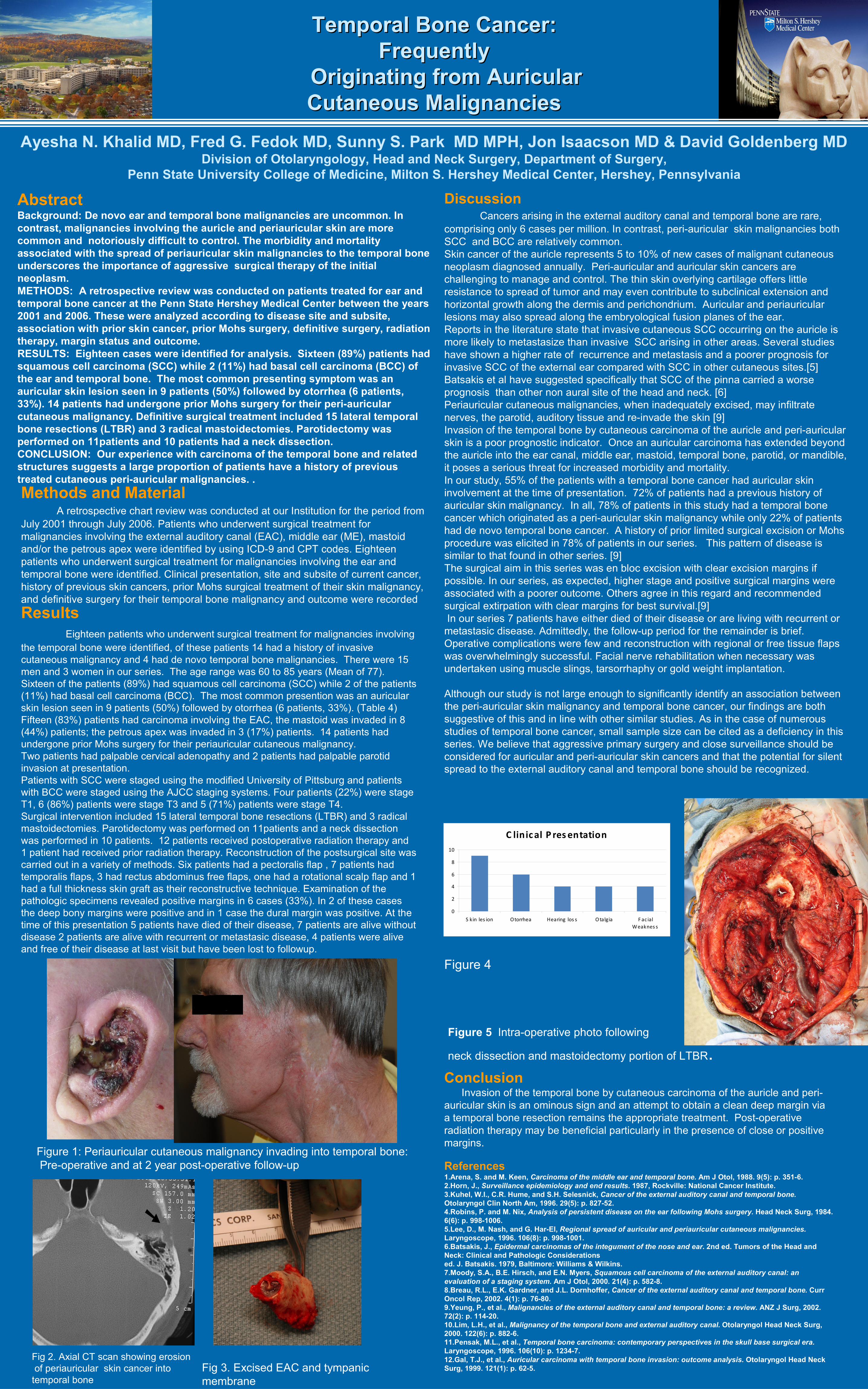
C linical P res entation

0

2

4

6

8

10

S kin  les ion Otorrhea Hearing   los s Otalgia F ac ialWeaknes s

AbstractBackground: De novo ear and temporal bone malignancies are uncommon. In contrast, malignancies involving the auricle and periauricular skin are more common and notoriously difficult to control. The morbidity and mortality associated with the spread of periauricular skin malignancies to the temporal bone underscores the importance of aggressive surgical therapy of the initial neoplasm. METHODS: A retrospective review was conducted on patients treated for ear and temporal bone cancer at the Penn State Hershey Medical Center between the years 2001 and 2006. These were analyzed according to disease site and subsite, association with prior skin cancer, prior Mohs surgery, definitive surgery, radiation therapy, margin status and outcome.RESULTS: Eighteen cases were identified for analysis. Sixteen (89%) patients had squamous cell carcinoma (SCC) while 2 (11%) had basal cell carcinoma (BCC) of the ear and temporal bone. The most common presenting symptom was an auricular skin lesion seen in 9 patients (50%) followed by otorrhea (6 patients, 33%). 14 patients had undergone prior Mohs surgery for their peri-auricular cutaneous malignancy. Definitive surgical treatment included 15 lateral temporal bone resections (LTBR) and 3 radical mastoidectomies. Parotidectomy was performed on 11patients and 10 patients had a neck dissection.CONCLUSION: Our experience with carcinoma of the temporal bone and related structures suggests a large proportion of patients have a history of previous treated cutaneous peri-auricular malignancies. .

ConclusionInvasion of the temporal bone by cutaneous carcinoma of the auricle and peri-

auricular skin is an ominous sign and an attempt to obtain a clean deep margin via a temporal bone resection remains the appropriate treatment. Post-operative radiation therapy may be beneficial particularly in the presence of close or positive margins.

ResultsEighteen patients who underwent surgical treatment for malignancies involving

the temporal bone were identified, of these patients 14 had a history of invasive cutaneous malignancy and 4 had de novo temporal bone malignancies. There were 15 men and 3 women in our series. The age range was 60 to 85 years (Mean of 77). Sixteen of the patients (89%) had squamous cell carcinoma (SCC) while 2 of the patients (11%) had basal cell carcinoma (BCC). The most common presention was an auricular skin lesion seen in 9 patients (50%) followed by otorrhea (6 patients, 33%). (Table 4) Fifteen (83%) patients had carcinoma involving the EAC, the mastoid was invaded in 8 (44%) patients; the petrous apex was invaded in 3 (17%) patients. 14 patients had undergone prior Mohs surgery for their periauricular cutaneous malignancy.Two patients had palpable cervical adenopathy and 2 patients had palpable parotid invasion at presentation. Patients with SCC were staged using the modified University of Pittsburg and patients with BCC were staged using the AJCC staging systems. Four patients (22%) were stage T1, 6 (86%) patients were stage T3 and 5 (71%) patients were stage T4. Surgical intervention included 15 lateral temporal bone resections (LTBR) and 3 radical mastoidectomies. Parotidectomy was performed on 11patients and a neck dissection was performed in 10 patients. 12 patients received postoperative radiation therapy and 1 patient had received prior radiation therapy. Reconstruction of the postsurgical site was carried out in a variety of methods. Six patients had a pectoralis flap , 7 patients had temporalis flaps, 3 had rectus abdominus free flaps, one had a rotational scalp flap and 1 had a full thickness skin graft as their reconstructive technique. Examination of the pathologic specimens revealed positive margins in 6 cases (33%). In 2 of these cases the deep bony margins were positive and in 1 case the dural margin was positive. At the time of this presentation 5 patients have died of their disease, 7 patients are alive without disease 2 patients are alive with recurrent or metastasic disease, 4 patients were alive and free of their disease at last visit but have been lost to followup.

Methods and MaterialA retrospective chart review was conducted at our Institution for the period from

July 2001 through July 2006. Patients who underwent surgical treatment for malignancies involving the external auditory canal (EAC), middle ear (ME), mastoid and/or the petrous apex were identified by using ICD-9 and CPT codes. Eighteen patients who underwent surgical treatment for malignancies involving the ear and temporal bone were identified. Clinical presentation, site and subsite of current cancer, history of previous skin cancers, prior Mohs surgical treatment of their skin malignancy, and definitive surgery for their temporal bone malignancy and outcome were recorded

References1.Arena, S. and M. Keen, Carcinoma of the middle ear and temporal bone. Am J Otol, 1988. 9(5): p. 351-6.2.Horn, J., Surveillance epidemiology and end results. 1987, Rockville: National Cancer Institute.3.Kuhel, W.I., C.R. Hume, and S.H. Selesnick, Cancer of the external auditory canal and temporal bone.Otolaryngol Clin North Am, 1996. 29(5): p. 827-52.4.Robins, P. and M. Nix, Analysis of persistent disease on the ear following Mohs surgery. Head Neck Surg, 1984. 6(6): p. 998-1006.5.Lee, D., M. Nash, and G. Har-El, Regional spread of auricular and periauricular cutaneous malignancies.Laryngoscope, 1996. 106(8): p. 998-1001.6.Batsakis, J., Epidermal carcinomas of the integument of the nose and ear. 2nd ed. Tumors of the Head and Neck: Clinical and Pathologic Considerationsed. J. Batsakis. 1979, Baltimore: Williams & Wilkins.7.Moody, S.A., B.E. Hirsch, and E.N. Myers, Squamous cell carcinoma of the external auditory canal: an evaluation of a staging system. Am J Otol, 2000. 21(4): p. 582-8.8.Breau, R.L., E.K. Gardner, and J.L. Dornhoffer, Cancer of the external auditory canal and temporal bone. Curr Oncol Rep, 2002. 4(1): p. 76-80.9.Yeung, P., et al., Malignancies of the external auditory canal and temporal bone: a review. ANZ J Surg, 2002. 72(2): p. 114-20.10.Lim, L.H., et al., Malignancy of the temporal bone and external auditory canal. Otolaryngol Head Neck Surg, 2000. 122(6): p. 882-6.11.Pensak, M.L., et al., Temporal bone carcinoma: contemporary perspectives in the skull base surgical era.Laryngoscope, 1996. 106(10): p. 1234-7.12.Gal, T.J., et al., Auricular carcinoma with temporal bone invasion: outcome analysis. Otolaryngol Head Neck Surg, 1999. 121(1): p. 62-5.

DiscussionCancers arising in the external auditory canal and temporal bone are rare,

comprising only 6 cases per million. In contrast, peri-auricular skin malignancies both SCC and BCC are relatively common. Skin cancer of the auricle represents 5 to 10% of new cases of malignant cutaneous neoplasm diagnosed annually. Peri-auricular and auricular skin cancers are challenging to manage and control. The thin skin overlying cartilage offers little resistance to spread of tumor and may even contribute to subclinical extension and horizontal growth along the dermis and perichondrium. Auricular and periauricular lesions may also spread along the embryological fusion planes of the ear. Reports in the literature state that invasive cutaneous SCC occurring on the auricle is more likely to metastasize than invasive SCC arising in other areas. Several studies have shown a higher rate of recurrence and metastasis and a poorer prognosis for invasive SCC of the external ear compared with SCC in other cutaneous sites.[5] Batsakis et al have suggested specifically that SCC of the pinna carried a worse prognosis than other non aural site of the head and neck. [6]Periauricular cutaneous malignancies, when inadequately excised, may infiltrate nerves, the parotid, auditory tissue and re-invade the skin [9]Invasion of the temporal bone by cutaneous carcinoma of the auricle and peri-auricular skin is a poor prognostic indicator. Once an auricular carcinoma has extended beyond the auricle into the ear canal, middle ear, mastoid, temporal bone, parotid, or mandible, it poses a serious threat for increased morbidity and mortality.In our study, 55% of the patients with a temporal bone cancer had auricular skin involvement at the time of presentation. 72% of patients had a previous history of auricular skin malignancy. In all, 78% of patients in this study had a temporal bone cancer which originated as a peri-auricular skin malignancy while only 22% of patients had de novo temporal bone cancer. A history of prior limited surgical excision or Mohs procedure was elicited in 78% of patients in our series. This pattern of disease is similar to that found in other series. [9] The surgical aim in this series was en bloc excision with clear excision margins if possible. In our series, as expected, higher stage and positive surgical margins were associated with a poorer outcome. Others agree in this regard and recommended surgical extirpation with clear margins for best survival.[9] In our series 7 patients have either died of their disease or are living with recurrent or metastasic disease. Admittedly, the follow-up period for the remainder is brief. Operative complications were few and reconstruction with regional or free tissue flaps was overwhelmingly successful. Facial nerve rehabilitation when necessary was undertaken using muscle slings, tarsorrhaphy or gold weight implantation.

Although our study is not large enough to significantly identify an association between the peri-auricular skin malignancy and temporal bone cancer, our findings are both suggestive of this and in line with other similar studies. As in the case of numerous studies of temporal bone cancer, small sample size can be cited as a deficiency in this series. We believe that aggressive primary surgery and close surveillance should be considered for auricular and peri-auricular skin cancers and that the potential for silent spread to the external auditory canal and temporal bone should be recognized.

Fig 2. Axial CT scan showing erosionof periauricular skin cancer into

temporal bone

Figure 5 Intra-operative photo following

neck dissection and mastoidectomy portion of LTBR.

Figure 1: Periauricular cutaneous malignancy invading into temporal bone: Pre-operative and at 2 year post-operative follow-up

Figure 4

Fig 3. Excised EAC and tympanic membrane