telomeres, telomerase, and immortality
TRANSCRIPT
SPECIAL ARTICLETelomeres, Telomerase, and Immortality
Michelle S. Rhyu*
A current hypothesis gaining prominence proposes that ac-tivation of the enzyme telomerase is necessary for cells to be-come immortal, or capable of proliferating indefinitely. Thetheory suggests that almost all cancer cells must attain im-mortality for progression to malignant states and, hence, re-quire activation of telomerase. This article reviews thefunction and formation of telomeres as background toevaluating the "telomere hypothesis." Experiments in sup-port of and experiments that challenge the hypothesis areexamined. Possible approaches to telomerase inhibition arediscussed. [J Natl Cancer Inst 87:884-894,1995]
Cancer is believed to be caused by multiple mutations thatcumulatively subvert the normal growth controls of a cell. Al-though different cancers may involve mutations in a particulargene, it appears that individual types of cancer may be distin-guished by mutations in characteristic sets of genes. In spite ofthese distinct etiologies, recent findings support the model thatactivation of the enzyme telomerase may be important in manymalignant tumor cells.
After much anticipation, a collaborative team led by CalvinHarley at Geron Corporation (Menlo Park, CA) and Jerry Shayat The University of Texas, Southwestern Medical Center (Dal-las), has shown a provocative correlation between tumor cellsand increased activity of the enzyme telomerase (1). Using anew, highly sensitive assay for activity of the enzyme, these re-searchers were able to show that tissue extracts from 90 of 101distinct tumors representing 12 cancer types and 98 of 100 inde-pendent immortalized cell lines (cells capable of growing in-definitely in culture) contain telomerase activity, whereas noenzyme activity could be detected in benign tumors, somatic(non-germline) tissues, and mortal cell lines examined. Such astrong correlation initially suggested that telomerase may play acritical role in the progression or maintenance of the malignantstate. The observation that telomerase activity is absent or pre-sent at low levels in most somatic tissues, combined with specu-lation about an obligate role for telomerase in tumorigenesis,has fueled interest in targeting telomerase for anticancertherapy.
What is the relevance of this finding given the landscape ofexisting telomerase research, and what therapeutic hope doesthe telomerase-inhibiting strategy offer? To better understandthe biological basis of this potential anticancer therapy, this ar-
ticle describes key experiments that have led to our currentknowledge of telomeres and telomerase, with special attentionto the evolution of the theory that telomerase activation is re-quired for the continued proliferation of tumor cells. Followingthis survey of the basic research, this article evaluates the exist-ing evidence for the telomerase activation theory, discussingfurther experiments necessary to validate the theory. Finally, afew approaches to isolating a telomerase inhibitor are described.
Telomeres
Miiller (2) and McClintock (3) in the 1930s and 1940s werethe first to recognize that chromosome ends, or telomeres, areessential to maintain chromosomal integrity. Chromosomes withtruncated ends are unstable, fusing with other chromosomes orbecoming lost upon cell division. More recently, investigatorssought to elaborate the molecular basis of the telomere's impor-tant properties. Capitalizing on the large number of nuclearchromosomes (and, accordingly, the large number of telomeres)in the ciliate Tetrahymena, Blackburn and Gall (4) determinedthat Tetrahymena telomeres consist of the short sequence 5'GGGGTT 3' repeated in tandem arrays. The number of theserepeats is variable at each chromosome end, causing restrictionendonuclease fragments that bear telomeric repeats to migrate asdiffuse bands in electrophoretic gels (Fig. 1, B).
After identification of the telomeric repeats in Tetrahymena,similar repeats were identified in a variety of organisms (5). Thesequence of the repeating unit was found to vary among dif-ferent species, but most known repeats are 5-8 base pairs (bp) inlength and are rich in G bases on the DNA strand that extends 5'to 3' toward the chromosome end. Human telomeres contain therepeat TTAGGG, which may be reiterated in tandem for up to15 kilobases (kb) (6-8). Initial work with ciliates and yeast sug-gested that "telomeric DNA" associates with specific proteins toform a telomeric nucleoprotein complex (9). Binding of thenecessary proteins may rely on the sequence of the repeat, as al-teration of the telomeric sequence in both human and Tetrahy-mena cells causes formation of incompetent telomeres (10-12).Human and other telomeres have been demonstrated to associate
*M. S. Rhyu, Ph.D., is a Technology Transfer Fellow at the Journal of the Na-tional Cancer Institute, National Cancer Institute, Bethesda, Md.
See "Note" section following "References."
884 SPECIAL ARTICLE Journal of the National Cancer Institute, Vol. 87, No. 12, June 21, 1995
at Stanford Medical C
enter on October 6, 2012
http://jnci.oxfordjournals.org/D
ownloaded from

Fig. 1. Structure of a telo-mere. A) Telomere (whitesection depicted for one endof blue chromosome) is madeup of a DNA component andmultiple protein components.The DNA consists of a shortsequence (for Tetrahymena,5' GGGGTT 3') repeated intandem for many kilobases.The telomeric region isdouble stranded, except forthe extreme 3' end, where theshort single-stranded stretchmay assume a characteristicstructure (structure notshown). Telomere-bindingproteins (in purple andmagenta) associate with thedouble- and single-strandedregions and may be criticalfor preventing degradationand recombination. B)Length of telomeres fromsperm (lane 1) and adult skinfibroblasts (lane 2) are visual-ized by digesting DNA with a restriction enzyme that cuts just proximal to the telomere. After probing the gel with a 32P end-labeled telomere-specific oligonucleo-tide (CCCTAA)3, the released terminal fragments (telomere restriction fragments [TRF]) appear as a smear in the lane, reflecting TRF lengths that vary in size. Thesize and position of the molecular weight markers (lane 3) are indicated on the right. The average TRF of sperm is longer than the average TRF from the fibroblasts(gel provided by R. C. Allsopp and C. Harley).
with the nuclear matrix protein fraction, which may includenuclear envelope and other nuclear proteins (13). Although theprecise molecular structure of any telomere has yet to be eluci-dated, it appears to carry out at least two essential functions.
The observations by Miiller (2) and McClintock (3) suggestthat telomeres stabilize chromosome ends, protecting them fromrecombination and end-degrading enzymes. In yeast, it has beenshown that removal of the telomere of a nonessential chromo-some causes a dramatic loss of that chromosome (14). It is un-known how the telomeric structure affords this protection, but ithas been suggested that interactions with proteins in the nuclearmembrane may effectively shield chromosomal ends fromdegrading enzymes (13).
Another function of chromosomal ends was first recognizedafter the double-stranded structure of DNA and its semiconser-
vative mode of replication were discovered (75). This functionaddresses a dilemma called the "end replication problem." Ac-cording to our understanding of DNA replication, the poly-merase that copies the strands of DNA prior to each cell divisionabsolutely requires a short "RNA primer" sequence to beginDNA polymerization in the 5' to 3' direction. After DNApolymerization, the RNA primers are degraded and replaced byDNA synthesis extending from an upstream primer. As shownin Fig. 2, the primers that are annealed to the extreme 3' end ofeach strand cannot be replaced. This replication strategy there-fore predicts the progressive shortening of chromosomal DNAat the 3' ends over multiple cycles of replication—a potentiallycatastrophic trend. Telomeric repeats could temporarily nullifythis trend by providing a cushion of expendable noncoding se-quence at the chromosome ends.
3' <
<v
3' <
51
c - .-<
f
^m b'layyiny sliaiid
w 3' leading strand
5' lagging strand
^ .T leading strand
Fig. 2. End replication problem. Original chromosomestrands are depicted in blue. As the replication forkproceeds from left to right, the leading strand (blackarrow directed rightward) proceeds continuously to repli-cate one strand of original DNA. The direction of the lag-ging strand is opposite to the direction of the replicationfork and relies on the ligation of Okazaki fragments(leftward arrows), which are primed with short stretchesof RNA (red boxes). Most RNA primers are replaced withDNA from an upstream Okazaki fragment, but the ter-minal RNA primer (solid red box) is never replaced withDNA. Consequently, each round of replication producesdaughter chromosomes, which lack the sequences cor-responding to the original 3' ends (red bracket). (The ter-minal primer actually may not anneal to the extreme 3'end, contributing to further loss of end sequences.)
Journal of the National Cancer Institute, Vol. 87, No. 12, June 21, 1995 SPECIAL ARTICLE 885
at Stanford Medical C
enter on October 6, 2012
http://jnci.oxfordjournals.org/D
ownloaded from

Telomerase Adds Telomeric Sequences
Evolutionary conservation of related telomeric repeats amongdifferent species affirmed the importance of these ends, but thequestion of how the repeats are propagated remained unresolveduntil 1985. Shampay et al. (16) had demonstrated that Tetrahy-mena telomeric DNA introduced into yeast became elongatedwith yeast telomeric sequences. From this result, they proposedthat each organism possesses the ability to transfer host-specificterminal sequences onto exogenous DNA. This model wastested directly and verified by Greider and Blackburn (17), who,using Tetrahymena extracts, detected an activity that addedtelomeric sequences to single-stranded telomeric DNA oligonu-cleotide primers. Their experiments elegantly proved that theterminal transferase, or telomerase, activity was independent ofDNA polymerase-a and of a DNA template but that the activityrequired an oligonucleotide primer containing repeats of a telo-meric sequence. This sequence could be from Tetrahymena oryeast. These groundbreaking experiments confirmed the exist-ence of telomerase activity but also unearthed a new crop ofmysteries: How could telomerase add sequences de novo,without a template, and why did it require a primer containingtelomeric sequences? Further purification of the telomerase ac-tivity provided answers to both queries.
Greider and Blackburn (18) found that, if extracts containingtelomerase activity from Tetrahymena are treated with theRNA-degrading enzyme ribonuclease (RNase) A, the extract isno longer competent to elongate primers. Concluding that thisresult reflected an essential role for RNA, in addition to pro-teins, in the telomerase complex, they isolated a 159 nucleotideRNA that copurified with telomerase activity. In the original ar-ticle describing this work, Greider and Blackburn (18) theorizeda specialized role for the RNA:
It is tempting to speculate that the RNA component of the telomerasemight be involved in determining the sequence of the telomeric repeatsthat are synthesized and/or the specific primer recognition. If the RNA oftelomerase contains the sequence CCCCAA, this sequence could act as aninternal guide sequence....
The sequence of the 159 nucleotide RNA remarkably fulfilledthese predictions. In the third of the trio of articles by Greiderand Blackburn that provided the basis of our understanding oftelomerase, the authors (19) cloned the single gene encodingthis RNA and identified the region from positions 43 to 51within the RNA as having the sequence 5' CAACCCCAA 3'(Fig. 3). They showed that preincubation of telomerase with anantisense deoxyoligonucleotide complementary to this region(i.e., containing the sequence 5' TTGGGGTTG 3') interferedwith primer elongation activity, while DNA oligomers directedto other regions of the RNA had no effect. When the RNA/DNAduplex-degrading enzyme RNase H was added together with theantisense oligonucleotide, the telomerase RNA was cleaved inthe region to which the oligonucleotide had hybridized. In sum,these results strongly implicated the CAACCCCAA sequence asthe template for repeat synthesis. This conclusion was later con-firmed by site-specific mutations in this region, which yieldedtelomerases that now synthesized repeats containing the cor-responding change in the cell (70). These results also showed
1. Bind telomenc pnmer
3. Translocate
4 Polymerize opposite strand
5- Repeat
Fig. 3. Mechanism of telomere addition. Chromosomal DNA end is shown inorange, telomerase RNA is indicated in purple, and telomerase proteins areshown in gray. Step 1: Chromosomal DNA binds part of the template region intelomerase RNA. Step 2: Telomerase RNA provides the template for elongatingthe 3' end of the chromosome (newly synthesized sequence in yellow). Step 3:The chromosome is translocated and repositioned to repeat the polymerizationstep. Step 4: The complementary strand is synthesized (orange outlined circles),presumably by a standard DNA polymerase. Step 5: The process is reiterated toadd multiple copies of the telomeric repeat sequence.
that telomerase was responsible for telomere synthesis in thecell as well as in the test tube.
Greider and Blackburn (19) recognized that the templateRNA contains approximately one and one-half times the com-plement of the GGGGTT repeat and speculated that the surplussequence enables telomerase to hybridize to an existingtelomeric repeat via several bases and to extend the repeat usingthe remaining bases as a template (Fig. 3). This model wouldexplain the need for a primer containing telomeric sequences.These authors further suggested that the observation thatheterologous telomeric repeats from other species successfullyprime the Tetrahymena telomerase reaction reflects a conservedsecondary structure formed by the repeats that can be recog-nized by the enzyme. Although some strong evidence now ex-ists in support of both contentions, these issues are still beingpursued.
It was thus found that telomerase is a ribonucleoprotein(RNP) complex that utilizes a region in the enzyme's RNA
886 SPECIAL ARTICLE Journal of the National Cancer Institute, Vol. 87, No. 12, June 21, 1995
at Stanford Medical C
enter on October 6, 2012
http://jnci.oxfordjournals.org/D
ownloaded from
component as a template for repeat synthesis. In other words,telomeric repeats are added de novo to the 3' end of chromo-somes via an RNA-dependent DNA polymerase (i.e., a reversetranscriptase) mechanism. It is speculated that a standard DNApolymerase completes the duplex telomere by synthesizing theDNA strand complementary to the telomerase-generated region.Telomerase RNAs from many single-cell organisms have nowbeen characterized, and each contains a potential template se-quence complementary to its respective telomeric repeat.
Isolation of the protein components of the telomerase enzymehas proved more elusive. At last, the prolonged drought in thefield of telomerase proteins may be over, as Greider et al. (20)recently described the purification and cloning of two polypep-tide constituents of Tetrahymena telomerase.
Human Telomerase
The discovery of telomerase and its RNA dependence was awatershed in the understanding of telomere biology. This workfostered a renewed interest in the function of chromosome ends,leading to inquiries into the role of telomeres and telomerase inhuman cells. While initial attempts to find telomerase activity inhuman tissues encountered considerable difficulties, Morin (8)ultimately identified the activity in immortalized HeLa cells.This telomerase activity is RNase A sensitive, suggesting that,like the Tetrahymena telomerase, it is an RNP. At the time ofMorin's discovery, the implications of finding telomerase in animmortalized cell line were not widely appreciated.
Now, with the latest reports of telomerase activity in malig-nant and immortalized cells and the reported cloning of thehuman telomerase RNA sequence (27), this field may be poisedfor another breakthrough. The assertion, first put forth by Har-ley, Futcher, and Greider (22) in 1990, is that telomerase activa-tion is a necessary step toward immortalization. Immortalizationhas since been proposed to be a necessary step in the patho-genesis of tumor malignancy (23-25). With the steady ac-cumulation of data consistent with this proposal, the theory isgaining acceptance. Does this signal a major paradigm shift, orare these conclusions premature? The next section explains theexperiments and observations that directed the development ofthis hypothesis of telomerase's role in cancer.
Telomere as a Mitotic Clock?
The Impetus: Telomerase Is Inactive in Normal SomaticCells
If telomeres are fundamental to chromosomal stability, whywould activation of telomerase be associated with aberranttumor cells? This question broaches the puzzle of telomerase ac-tivity in human cells, a riddle that has, until recently, beenobscured by the difficulty in measuring telomerase activity inhuman tissues. Unlike the ciliate Tetrahymena, which containsup to 40 000 telomeres per macronucleus and a correspondinglyrobust telomerase activity, human cells with the normal 46chromosomes (and 92 telomeres) appear to contain dramaticallylower levels of telomerase activity. Furthermore, primary tissuesamples are not available in the large quantities possible whenculturing ciliates.
The telomerase detection problem was initially addressed in-directly by measuring changes in the average telomere length ofchromosomes in various tissue types and inferring telomeraseactivity by monitoring changes in this length. A battery of suchstudies suggested that telomerase activity is regulated in a celltype-specific manner: Sperm cells were found to contain longtelomeres (10-14 kb) whose length does not appear to shortenwith increased age (26). Telomeres from these germline cellsare consistently several kilobases longer than telomeres ofsomatic cells (peripheral blood cells or fibroblasts) from thesame individual (7J,27,2S). In vivo, these somatic cell telomereswere calculated to shrink by about 15-40 bp per year (26,29).These results suggest that telomerase sustains and replenishestelomeric sequences specifically in germ cells (sperm andoocytes) but is inactive in most somatic tissues, allowing thegradual depletion of the chromosomes' terminal sequences.These predictions have since been validated by direct testing oftelomerase activity in various tissue types.
Shrinking Telomeres Reflect Mitotic Age: the Model andEvidence
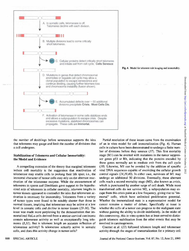
Publishing in the early 1970s, the Soviet scientist A. M. Olov-nikov (75) was the first to suggest that gradual loss ofchromosome ends could lead to an exit from the cell cycle.Spurred by mounting experimental support for this theory, Har-ley et al. (22) introduced a revised theory in 1990; theyproposed that the shrinking telomere may be the cell's measureof the mitotic age of the organism. Specifically, this hypothesisposits that, as chromosomes are incompletely replicated witheach division, terminal sequences are inevitably lost to the pointwhere they no longer protect the chromosome end from recom-bination and degradation (Fig. 4, A). Such a phenomenon couldrepresent a molecular equivalent of aging, with the end pointbeing an exit from the cell cycle and senescence. It should benoted that mortality by senescence, or the loss of the cell'sability to propagate, is to be distinguished from apoptosis, theactivation of a cell death program. Although senescence couldprecede the activation of programmed cell death, such arelationship has yet to be established.
The strongest evidence for telomere involvement in senes-cence is the finding by Allsopp et al. (26) that telomere lengthsare predictive of the replicative capacity of fibroblasts in cul-ture. These authors collected cells from donors aged zero (fetaltissues) to 93 years and established primary cultures from thesesamples. By measuring telomere length early in the estab-lishment of the culture and determining the life span of each cul-ture, they found that the age of the donor did not correspondwell to fibroblast telomere length and did not correlate stronglywith the proliferative ability of the cells. Instead, the number ofdivisions that the cells could undergo in culture was directlyproportional to the initial length of their telomeres when placedin culture. Allsopp et al. further determined that cell samplesfrom individuals with the premature aging disease Hutchinson-Gilford progeria contained telomeres with a significantly shorteraverage length than telomeres from normal donors of the sameage. In culture, the cells from progeria patients exhibitedreduced proliferative ability. Although these experiments do notdemonstrate cause, the correlation between telomere length and
Journal of the National Cancer Institute, Vol. 87, No. 12, June 21,1995 SPECIAL ARTICLE 887
at Stanford Medical C
enter on October 6, 2012
http://jnci.oxfordjournals.org/D
ownloaded from
Ay
A. In somatic cells, telomerase is off.Telomeres decline with each division.
B. Multiple divisions lead to some criticallyshort telomeres.
C. Cellular proteins detect critically short telomeresand initiate exit from cell cycle; Cells Senesce
D. Mutations in genes that detect chromosomalanomolies or regulate cell cycle may allow aclonal population to escape senescence andcontinue dividing, causing further telomere lossand chromosome instability (fusion shown).
T
E. Accumulated defects over ~ 50 additionaldivisions precipitate Crisis. Most Cells Die.
F. Activation of telomerase in some cells stabilizes endsand allows a subpopulation to escape crisis. Despiteexcessive mutations, stabilized chromosomes canpropagate. These cells are Immortal.
Fig. 4. Model for telomere role in aging and immortality.
the number of doublings before senescence supports the ideathat telomeres may gauge and limit the number of divisions thata cell undergoes.
Stabilization of Telomeres and Cellular Immortality:the Model and Evidence
A compelling extension of the theory that marginal telomeresinduce cell mortality is the suggestion that reactivation oftelomerase may enable cells to prolong their life span; i.e., theimmortal character of tumor cells may rely on the aberrant reac-tivation of the telomerase enzyme. While the measurement oftelomeres in sperm and fibroblasts gave support to the hypothe-sized role of telomeres in cellular mortality, telomere lengths intumor tissues appeared to contradict the idea that telomerase ac-tivation is necessary for immortality. Telomeres from a varietyof tumor types were found to be notably shorter than those innormal tissues, implying that telomerase may be active at a lowlevel in somatic cells and decline in tumor tissues (29-36). Theissue was made more perplexing by the demonstration that im-mortalized HeLa cells derived from a uterine cervical carcinomacontain telomerase activity as well as exceptionally long telo-meres (8^1). But is telomere length an accurate reflection oftelomerase activity? Is telomerase actually active in somaticcells, and does this activity change in tumor cells?
Partial resolution of these issues came from the examinationof an in vitro model for cell immortalization (Fig. 4). Humancells in culture have been demonstrated to undergo a finite num-ber of divisions before they senesce (37). This first mortalitystage (Ml) can be averted with mutations in the tumor suppres-sor genes p53 or Rb, indicating that the proteins encoded bythese genes normally act to mediate exit from the cell cycle(38). Likewise, Ml can be avoided by the addition of specificviral DNA sequences capable of overriding the cellular growthcontrol signals (24,39,40). In either case, survivors of Ml mayundergo an additional 50 divisions. Eventually, these aberrantcells reach a second mortality stage (M2), also known as crisis,which is punctuated by another surge of cell death. While mosttransformed cells do not survive M2, a subpopulation may es-cape from this crisis point at a low frequency, giving rise to "im-mortal" cells, which have unlimited proliferative potential.Whether the immortalized state is a representative model forcancer remains a matter of debate. Specifically at issue iswhether the cells of a tumor can progress to the malignant statewithout gaining the ability to divide indefinitely. In the midst ofthis controversy, this in vitro system has at least served to distin-guish telomere stabilization from the other events that may beinvolved in carcinogenesis.
Counter et al. (23) followed telomere length and telomeraseactivity through the stages of immortalization for a primary cell
SPECIAL ARTICLE Journal of the National Cancer Institute, Vol. 87, No. 12,June21,1995
at Stanford Medical C
enter on October 6, 2012
http://jnci.oxfordjournals.org/D
ownloaded from

line transformed with simian virus 40 or adenovirus DNA. Theyobserved that telomere attrition continued through M2, oftencoinciding with the appearance of abnormal or fused chromo-somes, but that the exceedingly short telomeres became stableupon immortalization. These authors were further able to assaytelomerase activity in extracts from these cells and determinedthat telomere stabilization during immortalization correspondsto the onset of telomerase activity. These experiments resolvedthe conundrum of short telomeres existing in the presence of ac-tive telomerase: Telomerase could be activated after dramatictelomere loss, acting to stabilize the degraded ends. Though per-suasive, this in vitro evidence fell short of demonstratingtelomerase activity in malignant cells in vivo.
Measuring Telomerase in Primary Tissues
The standard method of measuring the terminal transferaseactivity of telomerase was initially to assay the ability of the cellextract to add telomeric repeats to the 3' end of a syntheticdeoxyoligonucleotide primer (8,18). The incorporation of radio-actively labeled nucleotides could be observed by separating thereaction products on a polyacrylamide gel and exposing the gelto autoradiographic film. Because telomerase appears to pauseafter synthesis of each set of six nucleotides, products of a reac-tion yield a typical telomerase pattern of bands spaced at sixnucleotide intervals. Because of the low levels of telomerase inmammalian cells, interfering activities, and limited quantities oftumor samples, detecting telomerase activity by this methodproved difficult in primary tumor samples. Two groups nonethe-less managed to use this technique to demonstrate activetelomerase in extracts of ovarian epithelial carcinoma (41) andmalignant hematopoietic carcinoma cells (42), lending supportto the theory that reactivation of telomerase accompanies im-mortalization.
Further evidence for this association was obtained by Kim etal. (1) through two technical achievements that substantially im-proved the sensitivity of the telomerase assay. The first advanceoptimized extract preparation, allowing efficient telomerase ex-traction from a smaller starting quantity of cells via detergent-mediated cell lysis. (The old method employed hypotonic lysis.)The second, most significant, breakthrough allowed exponentialamplification of the products generated in a telomerase reaction(Fig. 5). This amplification was accomplished by using theproducts of a telomerase-catalyzed primer extension reaction astemplates for a polymerase chain reaction (PCR). Through suc-cessive rounds of oligonucleotide-primed DNA synthesis andstrand separation, the telomerase reaction products were ampli-fied many times over, enhancing the sensitivity of the conven-tional assay KJ'-fold.
This sensitized assay, termed the TRAP (telomere repeatamplification protocol) assay, enabled large-scale testing fortelomerase activity in a collection of 101 tumor biopsy speci-mens and 100 immortal cell populations as well as in their nor-mal counterparts. Consistent with previous reports, 98 of the100 immortal lines exhibited telomerase activity. But most com-pelling was the telomerase activity found in 90 of 101 actualtumor tissues, representing 12 distinct tumor types. No telo-merase activity was seen in 22 mortal cultured cell populationsand 50 normal somatic tissues tested. Benign proliferating fi-
1. Detergent lysis extraction
2. Telomerase reaction
dNTPs
TAQPolymerase I Denature, anneal primers,
elongate
Repeat 27 cycles
I4. Run Products on gel
1 2 3 4
3. PCR reaction
Fig. 5. TRAP assay for telomerase activity. Step 1: Extract is prepared fromprimary tissues or established cell lines by detergent lysis or the previously es-tablished hypotonic lysis method. Step 2: Telomerase in the extract elongates aprimer (Primer A, in red) using added deoxynucleotide triphosphates (dNTPs).Since telomerase is a processive enzyme, the products of this reaction will beheterogeneous in length (only one length shown). Step 3: Each product of thetelomerase reaction is amplified by PCR: Primer B (in blue), which contains se-quences complementary to the telomerase repeat, anneals with the telomerasereaction products; TAQ polymerase extends Primer B to synthesize duplex ver-sions of each telomerase product; these double-stranded fragments are dena-tured, annealed to primers again, and duplicated. This step is repeated for 27cycles. Step 4: The amplified products are run in a polyacrylamide gel, wherethe fragments of heterogeneous length create a "ladder^' pattern of bands (gelprovided by J. Shay). Gel shows results of TRAP assay using the following tis-sue samples: lane 1, squamous cell carcinoma; lane 2, normal tissue adjacent tosquamous cell carcinoma; lane 3, endometrial carcinoma; and lane 4, normal en-dometrium adjacent to endometrial carcinoma.
Journal of the National Cancer Institute, Vol. 87, No. 12, June 21, 1995 SPECIAL ARTICLE 889
at Stanford Medical C
enter on October 6, 2012
http://jnci.oxfordjournals.org/D
ownloaded from

broid tissues also tested negative, while telomerase activity wasat last demonstrated for normal ovaries and testes (presumablyas a result of activity in the germ cells). Corroborating the pre-vious reports (41,42) of telomerase activity in ovarian andhematopoietic carcinomas, this analysis established a strong cor-relation between cancerous tissues, immortal cell lines, and ac-tivation of telomerase. Telomere lengths in the immortal linesvaried from less than 4 kb in some lines to greater than 10 kb inothers, confirming that, in immortalized cells, telomere lengthdoes not accurately reflect telomerase activity. Since theoriginal report of the TRAP assay (1), more than 400 inde-pendent tumor samples have been tested for telomerase activityusing this assay (43,44). The recent data confirm that a high per-centage (84.8%) of malignant tumors exhibit telomerase ac-tivity. These findings are summarized in Table 1.
Authors' Interpretations
Kim et al. (1) proposed that the finding that telomerase is ac-tive in a wide array of immortal-human cell lines strengthens theassertion that this enzyme may be necessary for achieving theimmortal state. The additional connection between telomerasereactivation and malignant tumor tissues led these authors tospeculate that malignant tumor cells are immortal and, conse-quently, rely on telomerase. They proposed that early stagetumors are akin to transformed cells in culture and may similar-
ly lose telomeric DNA until a crisis point is reached. (In vitro,the crisis is M2.) Only by stabilizing the telomeres (through theinduction of telomerase) can these cells propagate indefinitely,developing and perpetuating their malignant characteristics.From this theory follows the prediction that disruption oftelomerase activity would impede the ability of a tumor to growindefinitely. Since reproductive cells are the only cell popula-tion shown to normally have high levels of telomerase, therapyaimed at this enzyme would be expected to have few adverse ef-fects in other cells.
Other Considerations
The predictions of Kim et al. (1) have fueled enthusiasm for in-hibition of telomerase as a means to combat cancer. As more atten-tion is turned to this strategy, this is a timely juncture to take acloser look at the body of existing work on telomerase and its rela-tion to cancer. Critical examination of the data points to three im-portant issues that warrant consideration when pursuing thetelomerase inhibition anticancer strategy: -1) The link betweentelomerase and cancer is still only correlative; 2) even if causalrelationships can be established and telomere stabilization enablessurvival through the crisis stage (M2), telomerase-independentmechanisms for extending telomeric DNA may thwart the effec-tiveness of antitelomerase drugs; and 3) in some tumors, this ap-proach may be limited by timing and specificity constraints.
Table 1. Published telomerase activity in human tissues—May 1995
Site or type of tumor: investigators (ref. No.), y Normal, adjacent to tumor, or benign tissues* Tumor tissues* (%)
Lung: Hiyama et al. (44), 1995
Breast: Kim et al. (1), 1994
Prostate:Kimetal. CO, 1994Sommerfield et al. (55), 1995
Colon:Kimetal. (/), 1994Chadeneau et al. (56), 1995
Liver: Kim etal. (/), 1994
Ovarian: Counter et al. (41), 1994
Renal: Mehle et al. (57), 1995
Neuroblastoma:Kimetal. (/), 1994Hiyama et al. (43), 1995
Hematological:|Lymphoma: Kimetal. (/), 1994CLL: Kimetal. (/), 1994ALL: Kim et al. (1), 1994
Brain: Kim et al. (/), 1994
Miscellaneous: Wilms', head and neck, rhabdomyosarcoma,leiomyosarcoma: Kim et al. (/), 1994
Total
3/68 adjacent
0/8 normal2/20 adjacent
0/8 normal1/10 benign prostatic hyperplasia
0/24 adjacent0/20 benign polyps0/1 adenoma
0/8 normal ascites
0/55 adjacent
0/13 adjacent0/4 ganglioneuroma
6/16 adjacent head and neck2/6 adjacent Wilms' tumor0/10 normal myometrium0/11 leiomyoma (fibroids)0/50 postmortem tissues14/332 = 4.2%
109/136(80.1)
19/24(79.0)
23/27(85.1)
22/23 (95.6)
1/1 (100)
7/7(100)
40/55 (72.7)
94/100 (94.2)
5/52/2(91.3)14/16
6/8 (75)
24/26 (92.3)
365/430 (84.8)
•Values = number of telomerase-positive samples/total number of samples.jCLL = chronic lymphocytic leukemia; ALL = acute lymphocytic leukemia.
890 SPECIAL ARTICLE Journal of the National Cancer Institute, Vol. 87, No. 12, June 21, 1995
at Stanford Medical C
enter on October 6, 2012
http://jnci.oxfordjournals.org/D
ownloaded from

Correlation Versus Cause
The distinction between causality and correlation is vitalwhen choosing a focus for therapeutic attack. Although experi-ments have not disproved the model specifying an obligatoryrole of telomerase in malignant tumor formation, the model hasnot been rigorously tested. Two tenets of the model rely almostentirely on correlative data. First, the model poses that the crisisin M2 is caused solely by erosion of the telomere. While it hasbeen shown that some cultured human cells at this stage have,on average, short telomeres and a high frequency of abnormalchromosomes, it has yet to be established that loss of telomericsequences triggers the chromosomal instability. Some immortaltissues even have exceedingly long telomeres, arguing againstthe supposition that crisis occurs as a consequence of thediminution of terminal sequences (1,31). If the chromosomalaberrations observed during crisis are not the result of unstableends, then emergence from crisis may have no dependence ontelomerase activation. To date, the best evidence that shortenedtelomeres induce chromosome instability exists for yeast (14).In yeast, however, the isolation of a mutant that uncouplestelomere length and senescence suggests that telomere lengthdoes not dictate the life span of the cell (45).
The second instance where the data show correlation, notnecessarily cause, is the demonstration of telomerase activity inan array of human tumor and immortalized cells. Hiyama et al.(43) have expanded on this work to show that telomerase ac-tivity is at higher levels in the more severe grades of neuroblas-toma examined. Furthermore, they showed that, in a subclass ofadvanced neuroblastoma called 4s, telomerase activity is notdetected. This complementing correlation is particularly intrigu-ing, since 4s metastases are sometimes observed to undergospontaneous remission. Despite these provocative correlations, acausative relationship remains to be demonstrated. These obser-vations could, for example, be explained if the process of im-mortalization activates telomerase secondarily. In such a case,activation may commonly be seen when a cell is immortalized,but telomerase activity would not necessarily be required for im-mortalization.
The need for establishing cause is emphasized by the exist-ence of some data that appear to contradict an obligate role fortelomerase in immortalization. For example, in some immortal-ized and tumor cell lines, telomerase activity has not been detec-table, even with the extremely sensitive TRAP assay(1^25,43,44,46). While it remains possible that the assay mayfail to detect a low or transient telomerase activity for technicalreasons, these cells may be evidence of a telomerase-inde-pendent pathway to immortalization. Alternatively, some meta-static tumors simply may not require immortalization (43,44).
The length of the average mouse telomere poses another ap-parent challenge to the telomerase activation theory. Mousetelomeres are typically between 40 kb and 50 kb in length anddo not appear to decay at a faster rate than human telomeres(47,48), yet the advantage of up to 10-fold more telomeric DNAdoes not give murine cells a corresponding increase in life span.If senescence in mice is actually caused by telomere erosion, itmay be that dangerously short telomeres exist within the popula-tion whose average telomere length is 40-50 kb. These critically
short telomeres could still induce chromosomal instability. Al-ternatively, it has been suggested that the 40-50 kb measured formouse telomeres may include sequences that are similar to butdistinct from actual telomeric repeats. However, if no "criticallyshort" telomeres exist within the population and telomericrepeats do constitute 40-50 kb of sequence, the mouse examplewould be evidence that telomere length does not endow cellswith increased life span.
Telomerase-Independent Telomere Addition
Even if telomerase activation is required to achieve the im-mortalized state, experiments in the yeast Kluyveromyces lactisand one immortal human cell line suggest that inhibition oftelomerase may not prevent the stabilization of telomeres viaanother mechanism. Indeed, while most K. lactis cells graduallylose terminal sequences and senesce when mutated for the geneencoding its telomerase RNA, such selective pressure leads tothe emergence of a subpopulation that appears capable of rapid-ly adding long stretches of telomeric repeats to the chromosomaltermini (49). In contrast to the rather gradual telomeric repeataddition seen with telomerase-mediated elongation, these ter-minal sequences appear to undergo abrupt, dramatic changes inlength.
A recent report by Murnane et al. (46) described a com-parable telomerase-independent mechanism in an immortalizedhuman cell line that does not appear to contain detectabletelomerase activity. Rather than looking at average telomerelengths, as has been done in all previous studies, Murnane et al.followed telomeres of single chromosomes and found that theyundergo rapid changes in length. Though little else is knownabout these telomerase-independent mechanisms, their mere ex-istence indicates that cells may evolve ways to circumventtelomerase inhibition in order to stabilize chromosomes. Iftumor cells can usurp this mechanism to stabilize the chromo-somes of cells in crisis, telomerase-blocking anticancer agentsmight have minimal effect.
Timing and Specificity
Telomere lengths vary broadly in the tumor and immortalizedcells that have been examined. In the absence of telomerase ac-tivity, these telomeres would no longer be stabilized, but theyalso would not be rapidly degraded. Before encountering crisis,these cells would experience the normal telomere attrition thataccompanies continued rounds of cell division. Thus, in tumorsinitiated by cells with extremely long telomeres, telomerase in-hibitors may potentially require an exceedingly long time to beeffective.
The initial observation (1,13,27^28) that telomerase is normal-ly expressed only in sperm and oocytes has led to speculationthat side effects of blocking its activity would be limited to thesegerm cells. But recent reports (44£0£l) suggest that hema-topoietic stem cells may also have low levels of telomerase ac-
- tivity. Although the long telomeres found in these cells may beprotective if their telomerase activity is disrupted, the evidenceof activity in these vital cells presents the possibility that drugstargeting telomerase activity may have greater adverse side ef-fects than originally anticipated.
Journal of the National Cancer Institute, Vol. 87, No. 12, June 21, 1995 SPECIAL ARTICLE 891
at Stanford Medical C
enter on October 6, 2012
http://jnci.oxfordjournals.org/D
ownloaded from

It Still Might Work
Having elaborated the pitfalls, it is important to reiterate thattelomerase remains a strong candidate for cancer therapeutics.Expanding this area of research could rapidly overcome the ex-perimental difficulties that have thus far prohibited thedemonstration of cause. That is, if telomerase inhibitors preventtumor progression, this would in itself be a demonstration of thecausal role of telomerase. Furthermore, even though telomeraseexpression may not be the exclusive mode to stabilize telomeresin the cell, it may be consequential enough to slow the prolifera-tion of tumor cells if inhibited. Thus, this approach may be ef-fective if used in combination with existing therapeutic agentsor treatments.
If telomerase activation is proved to be required for con-tinuous cell proliferation, detection of telomerase may be a use-ful indicator of the capacity of a primary tumor to metastasize.More extensive studies are required to investigate whethertelomerase is activated early enough to be a useful diagnostictool and to determine if detection of active telomerase is a suffi-cient prognosticator of later malignancy. Should these relation-ships be established, the sensitized telomerase assay andimproved methods of identifying and extracting early stagetumors will serve to potentiate this application.
A Target for Cancer Therapy: Strategies forTelomerase Inhibition
How can inhibitors of telomerase be found? Several generalapproaches might be taken. A classical approach to drug dis-covery is to test a battery of candidate small molecules for theireffects on telomerase activity. This brute force approach maynot immediately identify an inhibitor that is specific for thetelomerase enzyme alone, but it may discover classes of in-hibitors that, through further chemical modification, may proveto be optimal therapeutic drugs.
A more focused approach to inhibiting telomerase employsgenetic therapy. Although effective use of this strategy as a can-cer treatment awaits the development of a method of expressingthe desired DNA in all tumor cells, the extensive knowledge oftelomerase gained from studies in ciliates and yeast presents anexciting opportunity to pursue targeted genetic approaches. Forexample, the essential function of the RNA in the telomerasecomplex has been demonstrated in Tetrahymena. In addition toshowing that a segment of the RNA serves as the template fortelomeric sequences, manipulation of the RNA sequence hasgenerated telomerase mutants that behave enzymatically likesome mutants in protein components of DNA polymerases(Blackburn EH: personal communication). This observationraises the intriguing possibility that the RNA may itself possessenzymatic activity in the telomerase complex and reiterates theRNA as a reasonable target for inhibition. '
The strategy taken by Greider and Blackburn (19) forobstructing telomerase RNA activity through an antisenseoligonucleotide targeted to the template region is one approach"that could be applied to the human telomerase RNA (Fig. 6, A).Another inhibition strategy with a precedent in basic research isthe generation of mutant telomerase RNAs (Fig. 6, B). WhenRNA containing mutations in the template region is expressed at
A. Excess Antisense Oligonucleotide Displaces Chromosome Endfrom Template
Oligonucleotide
3
B. Excess Mutated Telomerase RNA Associates with TelomeraseProteins and Adds Wrong Telomeric Repeat to ChromosomeEnds, Making Incompetent Telomere
Fig. 6. Genetic strategies for telomerase inhibition. A) Antisense strategy. Ex-pression of high levels of a DNA oligonucleotide (turquoise) whose sequence iscomplementary to the sequence in the telomerase template may displace thechromosome end from the template, thereby preventing addition of telomeric se-quences to the chromosome. B) Mutated telomerase RNA. Expression of highlevels of mutated telomerase RNA (green circles among purple) may cause thepreferential assembly of telomerase enzymes that contain the mutated RNA.These telomerases may synthesize telomeric repeats that contain mutations(violet circles among orange), producing telomeres that are incompetent to bindtelomere-associated proteins.
high levels in normal Tetrahymena cells, the mutated RNAcompetes with endogenous wild-type RNA for the telomeraseproteins. Because the mutated RNA is expressed at many timesthe quantity of the wild-type RNA, most of the telomerase com-plexes formed contain the manipulated RNA. These impairedtelomerases attach the wrong sequence to chromosome termini,resulting in telomeres that fail to stabilize chromosomes and thatconsequently induce senescence in the ciliates (10). In spite ofthe current limitations in gene therapy, these two targeted ap-proaches may be the most direct methods to test in vitro whethertelomerase is required for immortality and malignancy. For ex-ample, if telomerase is inhibited in already immortal cells, willthese cells revert to crisis and die?
The protein components of telomerase would present anotherviable target for inhibition if the human telomerase proteinscould be identified. Progress toward this end may be acceleratedif sequence elements from telomerase proteins in other or-ganisms are conserved in the human counterpart. Proteins fromTetrahymena telomerase have now been partially purified andcharacterized (20). With the identification last fall of telomeraseRNA from the yeast Saccharomyces cerevisiae (52), genetic
892 SPECIAL ARTICLE Journal of the National Cancer Institute, Vol. 87, No. 12, June 21, 1995
at Stanford Medical C
enter on October 6, 2012
http://jnci.oxfordjournals.org/D
ownloaded from
isolation of proteins that interact with this RNA is widely an-ticipated.
It is noted that telomerase RNAs from different species con-spicuously lack primary sequence conservation (53). Instead, itis the conserved secondary structure, resulting from base pairingbetween compatible residues within the RNA, that appears to beimportant for its conserved activity across species. It is not yetknown whether or how much the proteins that associate withtelomerase RNA are conserved. Nonetheless, while it was notpossible to isolate mammalian telomerase RNA via homology toother telomerase RNAs, nucleotide sequence homology may beeffective in identifying genes encoding telomerase proteins.
It may also be possible to identify the biological repressor oftelomerase. Cell hybridization studies have shown that fusion ofa telomerase-expressing cell with a cell lacking telomerase ac-tivity yields a hybrid cell that has a limited lifespan (54). Such adominant effect suggests that somatic cells may express an in-hibitor or a repressor of telomerase activity. The mechanism andtarget of this putative repressor could eventually be utilized inapproaches to telomerase inhibition.
Unanswered Questions
Intriguing possibilities are raised by the prospect that mostcancers rely on activating the enzyme telomerase. Even if thiswere true of only some cancers, drugs aimed at telomerase in-hibition could provide a therapy with relatively limited side ef-fects. But these hopes rest on two still unresolved issues: Doshrinking telomeres cause senescence, and does activation oftelomerase stabilize the telomeres of cells that have escaped cellcycle controls, allowing them to continue dividing indefinitely?The causal relationships in question could be tested bymanipulating telomere lengths or telomerase activity. Key infor-mation could also come from determining how and whentelomerase is activated in malignant cells. For instance, does theregulation of the RNA or the proteins determine activity? Arethe RNA and proteins regulated at the level of transcription orpost-transcriptionally? At what tumor stage is telomerase ac-tivated?
The exciting new developments in this field, including thecloning of mammalian telomerase RNA, isolation and cloningof Tetrahymena telomerase components, and the TRAP assay,have equipped researchers with invaluable tools to address thesequestions. With regard to these long-awaited answers, the endmay finally be in sight.
References
(1) Kim NW, Piatyszek MA, Prowse KR, et al: Specific association of humantelomerase activity with immortal cells and cancer. Science 266:2011-2015, 1994
(2) Miiller HJ: The remaking of chromosomes. The collecting net. WoodsHole 13:181-198,1938
(3) McClintock B: The stability of broken ends of chromosomes in Zea mays.Genetics 41:234-282,1941
(4) Blackburn EH, Gall JG: A tandemly repeated sequence at the termini ofthe extrachromosomal RNA genes in Tetrahymena. J Mol Biol 120:33-53,1978
(5) Blackburn EH: Structure and function of telomeres. Nature 350:569-573,1991
(6) Allshire RC, Gosden JR, Cross SH, et al: Telomeric repeat from T. ther-mophila cross hybridizes with human telomeres. Nature 332:656-659,1988
(7) Moyzis RK, Buckingham JM, Cram LS, et al: A highly conserved repeti-tive DNA sequence, (TTAGGG)n, present at the telomeres of humanchromosomes. Proc Natl Acad Sci U S A 85:6622-6626,1988
(8) Morin GB: The human telomere terminal transferase enzyme is aribonucleoprotein that synthesizes TTAGGG repeats. Cell 59:521-529,1989
(9) Gilson E, Laroche T, Gasser SM: Telomeres and the functional architec-ture of the nucleus. Trends Cell Biol 3:128-134,1993
(70) Yu GL, Bradley JD, Attardi LD, et al: In vivo alteration of telomere se-quences and senescence caused by mutated Tetrahymena telomerase RNAs[see comment citation in Medline]. Nature 344:126-132, 1990
(11) Hanish JP, Yanowitz JL, de Lange T: Stringent sequence requirements forthe formation of human telomeres. Proc Natl Acad Sci U S A 91:8861-8865, 1994
(12) Sheng H, Hou Z, Schierer T, et al: Identification and characterization of aputative telomere end-binding protein from Tetrahymena thermophila. MolCell Biol 15:1144-1153, 1995
(13) de Lange T: Human telomeres are attached to the nuclear matrix. EMBO J11:717-724,1992
(14) Sandell LL, Zakian VA: Loss of a yeast telomere: arrest, recovery, andchromosome loss. Cell 75:729-739, 1993
(15) Olovnikov AM: A theory of marginotomy. The incomplete copying oftemplate margin in enzymic synthesis of polynucleotides and biologicalsignificance of the phenomenon, J Theor Biol 41:181 -190, 1973
(16) Shampay J, Szostak JW, Blackburn EH: DNA sequences of telomeresmaintained in yeast. Nature 310:154-157, 1984
(17) Greider CW, Blackburn EH: Identification of a specific telomere terminaltransferase activity in Tetrahymena extracts. Cell 43:405-413, 1985
(18) Greider CW, Blackburn EH: The telomere terminal transferase ofTetrahymena is a ribonucleoprotein enzyme with two kinds of primerspecificity. Cell 51:887-898,1987
(19) Greider CW, Blackburn EH: A telomeric sequence in the RNA ofTetrahymena telomerase required for telomere repeat synthesis. Nature337:331-337,1989
(20) Greider CW, Autexier C, Buchkovich K, et al: Telomerase biochemistryand regulation in cellular immortalization. Proc Am Assoc Cancer Res36:672,1995
(21) Harley CB, Andrews W, Chiu CP, et al: Human telomerase inhibition andcancer. Proc Am Assoc Cancer Res 36:671, 1995
(22) Harley CB, Futcher AB, Greider CW: Telomeres shorten during ageing ofhuman fibroblasts. Nature 345:458-460, 1990
(23) Counter CM, Avilion AA, LeFeuvre CE, et al: Telomere shortening as-sociated with chromosome instability is arrested in immortal cells whichexpress telomerase activity. EMBO J 11:1921-1929, 1992
(24) Shay JW, Wright WE, Werbin H: Loss of telomeric DNA during agingmay predispose cells to cancer. Int J Oncol 3:559-563,1993
(25) Harley CB, Kim NW, Prowse KR, et al: Telomerase, cell immortality, andcancer. Cold Spring Harb Symp Quant Biol. In press
(26) Allsopp RC, Vaziri H, Patterson C, et al: Telomere length predicts replica-tive capacity of human fibroblasts. Proc Natl Acad Sci U S A 89:10114-10118,1992
(27) Cooke HJ, Smith BA: Variability at the telomeres of the human X/Y pseu-doautosomal region. Cold Spring Harb Symp Quant Biol 51:213-219, 1986
(28) Allshire RC, Dempster M, Hastie ND: Human telomeres contain at leastthree types of G-rich repeats distributed non-randomly. Nucleic Acids Res17:4611-4627,1989
(29) Hastie ND, Dempster M, Dunlop MG, et al: Telomere reduction in humancolorectal carcinoma and with ageing [see comment citation in Medline].Nature 346:866-868,1990
(30) Cooke HJ, Brown WR, Rappold GA: Hypervariable telomeric sequencesfrom the human sex chromosomes are pseudoautosomal. Nature 317:687-692,1985
(31) de Lange T, Shiue L, Myers RM, et al: Structure and variability of humanchromosome ends. Mol Cell Biol 10:518-527,1990
(32) Adamson DJ, King DJ, Haites NE: Significant telomere shortening inchildhood leukemia. Cancer Genet Cytogenet 61:204-206,1992
(33) Yamada O, Oshimi K, Motoji T, et al: Telomeric DNA in normal andleukemic blood cells. J Clin Invest 95:1117-1123,1995
(34) Rogalla P, Kazmierczak B, Rohen C, et al: Two human breast cancer celllines showing decreasing telomeric repeat length during early in vitro pas-saging. Cancer Genet Cytogenet 77:19-25, 1994
(35) Shirotani Y, Hiyama K, Ishioka S, et al: Alteration in length of telomericrepeats in lung cancer. Lung Cancer 11:29-41, 1994
(36) Odagiri E, Kanada N, Jibiki K, et al: Reduction of telomeric length and c-erbB-2 gene amplification in human breast cancer, fibroadenoma, and
Journal of the National Cancer Institute, Vol. 87, No. 12, June 21, 1995 SPECIAL ARTICLE 893
at Stanford Medical C
enter on October 6, 2012
http://jnci.oxfordjournals.org/D
ownloaded from

gynecomastia. Relationship to histologic grade and clinical parameters.Cancer 73:2978-2984,1994
(37) Hayflick L, Moorhead PS: The serial cultivation of human diploid strains.Exp Cell Res 25:585-621,1961
(38) Shay JW, Pereira-Smith OM, Wright WE: A role for both RB and p53 inthe regulation of human cellular senescence. Exp Cell Res 196:33-39,1991
(39) Sack GH: Human cell transformation by simian virus 40—a review. InVitro 17:1-19, 1981
(40) DiPaolo JA: Relative difficulties in transforming human and animal cellsin vitro. J Natl Cancer Inst 70:3-8, 1983
(41) Counter CM, Hirte HW, Bacchetti S, et al: Telomerase activity in humanovarian carcinoma [see comment citation in Medline]. Proc Natl Acad SciUSA91:2900-2904, 1994
(42) Nilsson P, Mehle C, Remes K, et al: Telomerase activity in vivo in humanmalignant hematopoietic cells. Oncogene 9:3043-3048, 1994
(43) Hiyama E, Hiyama K, Yokoyama T, et al: Correlating telomerase activitylevels with human neuroblastoma outcomes. Nature Med 1:249-255,1995
(44) Hiyama K, Hiyama E, Ishioka S, et al: Telomerase activity in small-celland non-small-cell lung cancers. J Natl Cancer Inst 87:000-000, 1995
(45) Kennedy BK, Austriaco NR Jr, Zhang J, et al: Mutation in the silencinggene SIR4 can delay aging in S. cerevisiae. Cell 80:485-496, 1995
(46) Mumane JP, Sabatier L, Marder BA, et al: Telomere dynamics in an im-mortal human cell line. EMBO J 13:4953-4962, 1994
(47) Starling JA, Maule J, Hastie ND, et al: Extensive telomere repeat arrays inmouse are hypervariable. Nucleic Acids Res 18:6881-6888, 1990
(48) Kipling D, Cooke HJ: Hypervariable ultra-long telomeres in mice. Nature347:400-402,1990
(49) McEachern MJ, Cohn M, Blackbum EH: The yeast Kluyveromyces lactisas a model system to study telomere length regulation and the ability of
cells to grow in the absence of telomerase. Proc Am Assoc Cancer Res36:670,1995
(50) Chiu CP, Dragowski V, Kim NW, et al: Telomerase activity inhematopoietic progenitor cell extracts from adult bone marrow. Proc AmAssoc Cancer Res 36:abstr 3306,1995
(5/) Counter CM, Gupts J, Harley CB, et al: Telomerase activity in normalleukocytes and in hematologic malignancies. Blood. In press
(52) Singer MS, Gottschling DE: TLC1: Template RNA component of Sac-charomyces cerevisiae telomerase [see comment citation in Medline].Science 266:404-409,1994
(55) Romero DP, Blackbum EH: A conserved secondary structure fortelomerase RNA. Cell 67:343-353, 1991
(54) Pereira-Smith OM, Smith JR: Evidence for the recessive nature of cellularimmortality. Science 221:964-966,1983
(55) Sommerfield HJ, Meeker AK, Piatyszek MA, et al: Telomere shorteningand telomerase activity in human prostate tissue. Proc Am Assoc CancerRes 36:abstr 3311,1995
(56) Chadeneau C, Gallinger S, Hirte HW, et al: Temporal pattern oftelomerase activation in human and mouse tumorigenesis. Proc Am AssocCancer Res 36:abstr 3307, 1995
(57) Mehle C, Piatyszek MA, Ljungberg B, et al: Telomerase activity in renalcell carcinoma. Proc Am Assoc Cancer Res 36:abstr 3309,1995
Note
I am grateful to Drs. Jerry Shay, Gregg Morin, and Elizabeth Blackbum fortheir critical readings of the manuscript. I also thank Dr. Shay for providingTable 1 and the gel in Fig. 5. Many thanks to Drs. Richard Allsopp and CalvinHarley of Geron Corporation for providing the gel in Fig. 1, B, and for furnish-ing unpublished papers.
PREDOCTORAL STIPENDS IN NUTRITIONSCIENCES/CANCER PREVENTION
The University of Alabama at Birmingham
NIH-fiinded predoctoral positions are available forpursuit of the Ph.D. in Nutrition Sciences. The trainingincludes courses in basic and clinical nutrition and inpublic health and chronic disease prevention; exposure topatients with nutritional disorders and to diseaseprevention projects involving nutritional intervention; andresearch, which will be related to prevention of chronicdisease, especially cancer. Support includes stipend,tuition and fees, supplies, and travel to scientific meet-ings. Applicants must be U.S. citizens or permanentresidents. Minority applicants are strongly encouraged.Interested persons should send a letter describing theirinterest, plus curriculum vitae to Dr. Douglas C.Heimburger, Department of Nutrition Sciences, UABStation, Birmingham, AL 35294-3360; or call 205-934-7058.
The University of Alabama at Birmingham is anAffirmative Action/Equal Opportunity Employer.
894 SPECIAL ARTICLE Journal of the National Cancer Institute, Vol. 87, No. 12, June21, 1995
at Stanford Medical C
enter on October 6, 2012
http://jnci.oxfordjournals.org/D
ownloaded from