tectoreticular pathwayis in the turtle, pseudemys scripta...
TRANSCRIPT

THE JOURNAL OI' COMPARATIVE NEUROLOGY 233~48-90 (1985)
Tectoreticular Pathwayis in the Turtle, Pseudemys scripta. I. Morphology of
Tectoreticular Axons
MARTIN 1. SERENO Committees on Neurobiology and on The Conceptual Foundations of Science,
The University of Chicago, Chicago, IL 60637
ABSTRACT Tectoreticular projections in turtles were examined by reconstructing
from serial sections axons that were anterogradely filled with horseradish peroxidase after tectal injections. Three tectoreticular pathways each con- tain extensively collateralized axons. The crossed dorsal pathway (TBd) contains large and small caliber axons. After leaving the tectum, TBd axons emit collaterals into the ipsilateral profundus mesencephali rostralis and then give off a main rostral branch that bears secondary collaterals in the ipsilateral interstitial nucleus of the medial longitudinal fasciculus and the suprapeduncular nucleus. The main trunks cross the midline and descend in the predorsal bundle, generating collatera.ls at regular intervals. These terminate mostly in the medial half of the reticular core from the midbrain to the caudal medulla. Axons in the uncrossed intermediate pathway also emit collaterals into a midbrain reticular nucleus (profundus mesencephali caudalis) and often have a thick rostral branch. The main caudal trunks, however, remain ipsilateral and travel in a diffuse, laterally placed tract, where each emits a long series of collaterals into the lateral half of the reticular core. The uncrossed ventral pathway (TBv) contains medium and small caliber axons. TBv axons often have collaterals within the tectum and apparently lack main rostral branches. Their caudal trunks run in the tegmental neuropile below the TBi where they collateralize less exuberantly than do TBd and TBi axons.
The morphology of axons in all three pathways suggests that projections from disjunct tectal loci converge at many rostrocaudal levels within the reticular formation. This point was examined explicitly in experiments in which two disjunct injections were placed in one tectal lobe. Intermediate pathway axons traced from the two loci initially formed two distinct bundles but then intermingled in the reticular formation.
Key words: tectum, sensorimotor, reticular formation, non-topographic, eye movements
The optic tectum contains topologically organized affer- ent maps of the retinal surface (e.g., McIlwain, '75; Graham et al., '81), of auditory space (e.g., Harris et al., '80; Jay and Sparks, '82), and of cutaneous receptors (e.g., Nagata and Kruger, '79; Stein and Gaither, '81). Weakly electric fish, in addition, have a tectal electrosensory map (Bastian, '82) while heat-sensitive snakes have a map of pit organ infared receptors (Hartline et al., '78). There are also inputs of unknown topography from muscle receptors (Abrahams and Rose, '75a,b) and from several vestibular nuclei (Maeda et al., '79).
Electrical stimulation and lesion experiments implicate the tectum in the regulation of orienting movements (Ada- miik, 1870; Fernier, 1886; Akert, '49; recent reviews: Wurtz
0 1985 ALAN R. LISS, INC.
and Albano, '80; Sparks and Mays, '81; Ingle '82; Hartline, '84). The tectum in cats, for instance, appears to initiate coordinated movements of the eyes, head, pinnae, and vi- brissate toward spatially localized visual, auditory, and cu- taneous target stimuli (Grantyn and Grantyn, '76; Grantyn and Berthoz, '77; Guitton et al., '80; Roucoux et al., '80; Stein and Clamann, '81). The tectum in frogs is involved in the accurate sagittal and horizontal rotation of the body
Accepted October 11, 1984. Martin Sereno's present address is Division of Biology 216-76,
California Institute of Technology, Pasadena, CA 91125. Address reprint requests there.

TECTORETICULAR AXONS 49
toward a visual prey stimulus just prior to a tongue flip (Ewert, '70; Grobstein et al., '83). In turtles, the tectum mediates orienting movements of the eyes, neck, and body toward visual stimuli (Bass et al., '73; Bass, '77; Mrosovsky et al., '79).
Thus, the tectum may play a general role in the transfor- mation of topographically organized sensory inputs into the graded motor outputs that underlie orienting movements. A detailed understanding of the organization of tectoreti- cular pathways linking the tectum to premotor centers in the brainstem reticular formation is essential to an analy-
sis of such orienting behaviors. Previous anatomical studies of tectoreticular neurons have concentrated principally on the laminar distribution of their somata or on the location of tracts and terminal fields. A pattern of tectal efferent projections consistent across vertebrate classes has emerged, including several apparently non-topographic descending pathways that terminate in the reticular formation (e.g., Altman and Carpenter, '61; Rubinson, '68; Kawamura et al., '74; Foster and Hall, '75; Hunt and Kiinzle, '76; Ulinski, '77; Harting, '77; Harting et al., '80; Smeets, '81; Luiten '81; Burne et al. '81; Huerta and Harting, '82). Information
A bhreoiations
BON BOT cEnt CG CM D DC DLA dLFB dLGN dNPC DMA EW HP ICO i-IT Imc Imlf
Imr IP IPd IPV LHA LM Mes V MFB MLF nLL Nmlf N. 111 N. IV N.V N. VIII N. IX OT P Pa PC PD Pe PMc PMr PMrl PMr-Tect PMv Po PR Pt Re rEnt Ri RID RM RML RN Rot
Basal optic nucleus Basal optic tract Caudal entopeduncular nucleus Central gray Caudomedial segment of nucleus rotundus Dorsal hypothalamic nucleus Dorsal cluster of reticularis inferioris dorsalis Dorsolateral anterior nucleus Dorsal peduncle of the lateral forebrain bundle Dorsal lateral geniculate nucleus Dorsal nucleus of the posterior commissure Dorsomedial anterior nucleus Edinger-Westphal nucleus Habenulopeduncular tract Intercollicular nucleus Ipsilateral isthmotectal tract (from Imc) Caudal magnocellular nucleus isthmi Iriterstitial nucleus of the medial longitudinal fasciculus Rostral magnocellular nucleus isthmi Parvocellular nucleus isthmi Dorsal interpeduncular nucleus Ventral interpeduncular nucleus Lateral hypothalamic area Nucleus lentiformis mesencephali Mesencephalic trigeminal nucleus Medial forebrain bundle Medial longitudinal fasciculus Nucleus of the lateral lemniscus Nucleus of the medial longitudinal fasciculus Oculomotor nerve Trochlear nerve Trigeminal nerve Auditory-vestibular nerve Glossopharyngeal nerve Optic tract Periventricular hypothalamic nucleus Paraventricular hypothalamic nucleus Posterior commissure Nucleus posterodorsalis of the pretectum External pretectal nucleus Profundus mesencephali caudalis Profundus mesencephali rostralis Profundus mesencephali rostrolateralis PMr-tectal tract Profundus mesencephali ventralis Posterior hypothalamic nucleus Prerubral area Nucleus pretectalis Nucleus reuniens Rostral entopeduncular nucleus Inferior raphe nucleus Reticularis inferioris dorsalis Reticularis medius Reticularis medius lateralis Red nucleus Nucleus rotundus
RSL(dm)
RSL(v1)
Rsl RSM Rsm SAC sc SFGS SGC SGP SN SN pr so SP spo TBd(lg)
TBd(sm)
TBi TBv(med)
TBv(sm)
Tect-Imc Torc Tor1 1 T h V VeDL vEnt VEO VeVL VeVM vLFB VL VM VMH VTA x-IT x-SC-Tect x-TBd x-?TH x-Ve-Tect 111 N V ds V mot V mr v Pr V t r VI VII XI1
Reticularis superioris lateralis, dorsomedial segment Reticularis superioris lateralis, ventrolateral segment Lateral superior raphe nucleus Reticularis superioris medius Medial superior raphe nucleus Stratum album centrale Small-celled nucleus Stratum fibrosum e t griseum superficiale Stratum griseum centrale Stratum griseum periventriculare Substantia nigra Substantia nig-a pars reticulata Stratum opticum Suprapeduncular nucleus Superior olive Dorsal tectobulbar pathway, large caliber component Dorsal tectobulaar pathway, small caliber component Intermediate tectobulbar pathway Ventral tectobulbar pathway, medium caliber component Ventral tectobulbar pathway, small caliber component Tectoisthmi tract (to Imc) Torus semicircularis, central nucleus Torus semicircularis, laminar nucleus Tectothalamic tract Ventral thalamic nucleus Dorsolateral vestibular nucleus Ventral entopeduncular nucleus Ventricular ependymal organ Ventrolateral vestibular nucleus Ventromedial vestibular nucleus Ventral peduncle of the lateral forebrain bundle Ventrolateral thalamic nucleus Ventromedial thalamic nucleus Ventromedial hypothalamic nucleus Ventral tegmental area Crossed isthmotectal tract Crossed SC-tectal tract Contralateral part of the dorsal pathway Crossed tectothalnmic tract Crossed vestibulotectal tract (from VeVL) Oculomotor nucleus Trochlear nucleus Descending trigeminal nucleus Trigeminal motor nucleus Mesencephalic root of the trigeminal nerve Principal sensory trigeminal nucleus Descending tract of the trigeminal complex Abducens motor nucleus Facial motor nuccleus Hypoglossal motor nucleus
50 M.I. SERENO
A B C D E F G H I J K L M
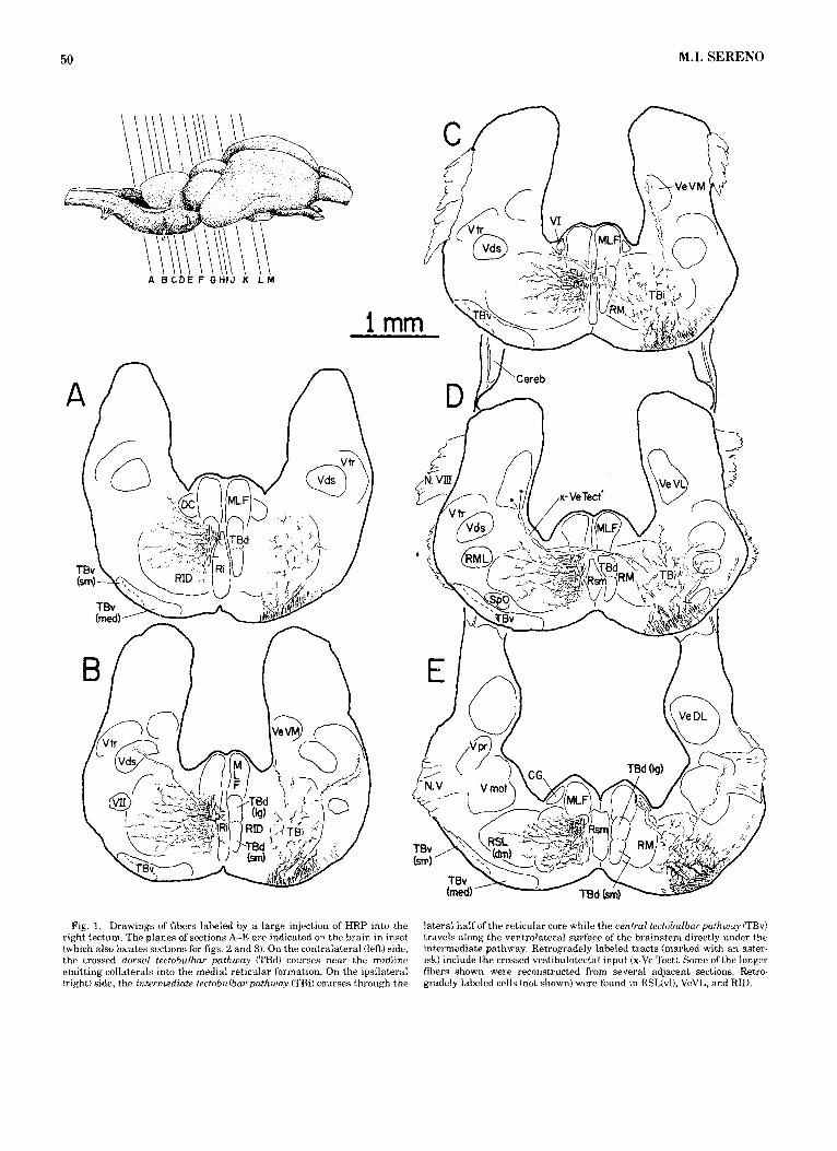
Fig. 1. Drawings of fibers labeled by a large injection of HRP into the right tectum. The planes of sections A-E are indicated on the brain in inset (which also locates sections for figs. '2 and 3). On the contralateral (left) side, the crossed dorsal tectobulbar pathway (TBd) courses near the midline emitting collatera.ls into the medial reticular formation. On the ipsilateral (right) side, the intermediate tectobulbar pathway (TBil courses through the
lateral half of the reticular core while the aentral tectobulbarpathway (TBv) travels along the ventrolateral surface of the brainstem directly under the intermediate pathway. Retrogradely labeled tracts (marked with an aster- isk) include the crossed vestibulotectal input (x-Ve Tect). Some of the longer fibers shown were reconstructed from several adjacent sections. Retro- gradely labeled cells (not shown) were found in RSL(vI), VeVL, and RID.

TECTORETICULAR AXONS 51
'CI;
1 mm Fig. 2 . Drawings of fibers labeled by a large unilateral HRP injection.
See Figure 1 for location of section planes. The dorsal, intermediate, and ventral tectobulbar pathways can all be seen leaving the tectum in section I. The dorsal pathway (TBd) emits rostrally directed ipsilateral branches (see also Fig. 3) before decussating (section I) and turnin;: caudally to run just lateral to the midline of the tegmentum in sections H, G, and F. The intermediate (TBi) and ventral (TBv) pathways emerge caudal to the dorsal
pathway (sections H and G) and remain ipsilateral to the injection for their entire course. The densely labeled tectoisthmi pathway (Tect-Imc, section F) is also illustrated. Retrogradely labeled tracts (marked with asterisks) in- clude the ipsilateral isthmotectal tract &IT, from Imc), the crossed isthmo- tectal tract (x-IT), and the crossed tract from SC (x-SC Tect). Retrogradely labeled cells (not shown) were found in isthmic (SC, Imc, Ip, Imr) and mesencephalic (KO, Torl, SN, PMc, PMr) nuclei.

52 M.J. SERENO
BOT
1 mm Fig. 3. Drawings of fibers labeled by a large unilateral HRP injection.
For location of section planes see Figure 1. A dense mat of collaterals from dorsal pathway axons occupies the midbrain nucleus profundus mesence- phali rostralis (PMr, section J; see also Fg. 4C). Further ventrally, dorsal pathway axons also give off “main rostra1 collaterals” that pass just medial to the red nucleus (section d) and just lateral to the large cells in the interstitial nucleus of the medial longitudinal fasciculus (Imlf, section K) to eventually terminate in the suprapeduncular nucleus (SP) of the ventral thalamus (sections L and M). The densely labeled tectopretcxtal pathway (emitted from injection site in section K), which projects to lentiformis mesencephali (LM) and a region dorsal to dLGN, and the tectorotundal
pathway (Trh) , which projects to nucleus pretcctalis (Pt), the nucleus of the 1Th (n‘lTh), and nucleus rotundus (Rot), arc also illustrated. Retropadely filled axons in the optic tract are indicated by hatching. Other retrogradely lahelcd tracts (marked with asterisks) include PMr-tectal axons (PMr-Tect), the ipsilateral bundle from the dorsal nucleus of the postcrior commissure (dNPC-Tecti, and the crossed isthmotectal tract WT). Retrogradely labeled cells (not shown) were found in mesencephalic (PMr, PMrll, pretectal (Pt, LM, Pe, dNl’C), and diencephalic (cEnt, Vl nuclei. Finally, retinal terminals in dLGN and Pe filled through their tectal branches are omitted in sections K-M.

Fig. 4. Photomicrographs of injection sites and HRP-filled neurons. Two small tectal injection sites (I, 11) in the horizontal section in B each emit thin bundles of tectobulbar axons apparent several sections ventrally in A. The retrogradely labeled tracts from profundus mesencephali rostralis (PMr- Tect) and the caudal magnocellular nucleus isthmi (i-IT) are also visible in A. C is a transverse view of the dorsal pathway terminal field in profundus
mesencephali rostralis (PMr) (see Fig. 35) labeled after an extensive tectal HRP injection. The injection also back-filled large PMr cells which have long, sparsely branched dendrites arrayed in the transverse plane. D is a high-magnification view of a putative contact between solid-filled boutons from a dorsal pathway axon and the soma of a lightly back-filled PMr cell.

Rost rat
- 2 mm
Figure 5

TECTORETICULAR A X C " 55
on the detailed organization of the tectoreticular pathways, by contrast, is just beginning to appear (Grantyn and Gran- tyn, '82; Grantyn et al., '82; Dacey '82).
The present study examines the morphology of tectoreti- cular axons at the single-cell level by using small extracel- lular horseradish peroxidase (HRP) injections and serial section reconstruction techniques. The following paper (Ser- eno and Ulinski, '85) illustrates the morphology of the dendrites of tectoreticular neurons and discusses the results in the light of current ideas about the nature of the senso- rimotor transformation underlying tectally mediated ori- enting movements.
MATERIALS AND METHODS Twenty-five pond turtles (Pseudemys scripta elegans and
Chrysemys picta) weighing 0.5-1.5 kg were used. Animals were anesthetized with a small (0.3 mlkg) dose of Brevital (Wang et al., '77) and packed in ice for surgery. This tech- nique provides stable anesthesia and a helpful reduction in blood pressure. A craniotomy was performed, and either a 1 p1 Hamilton syringe with a sharpened tip or a micropi- pette was introduced into the tectum. In nine animals, multiple pressure injections (total of 0.2-0.8 pl) or ionto- phoretic injections (2-4 pA for 20 minutes at each site) of concentrated Sigma Type VI HRP in pH 8.6 Tris buffer were made at two to four sites in one tectal hemisphere. Thirteen other animals received small iontophoretic injec- tions (1 pA pulsed for 20-100 seconds with a 5-10 pm I.D. tip) at a single site or at two widely separated sites. Three animals received unilateral subtotal tectal lesions.
Animals with HRP injections survived for 3 days at 20°C before intracardial perfusion with pH 7.4 phosphate-bmff- ered saline followed by a buffered solution of 1% parafor- maldehyde and 3% glutaraldehyde. Brains were removed, soaked in 30% sucrose, embedded in gelatin, and sectioned the next day on a freezing microtome at 110 or 120 pm. Transverse and horizontal serial sections were processed according to the cobalt-enhanced diaminobenzidine method of Adams ('77). The gelatin was removed and sections were carefully aligned on slides to facilitate serial reconstruc- tions. Sections were usually counterstained with cresyl vi- olet before coverslipping. In other cases, reconstructions were done from unstained sections and the coverslips were removed for counterstaining afterward.
Sections shrunk significantly during drying. When cut at 120 pm they often ended up only 45 pm thick (60-65% shrinkage) as measured by a stage micrometer under oil. Shrinkage in directions parallel to the plane of section was much less (0-5%), apparently because the section becomes bonded to the slide before it dries. This anisotropy in shrinkage results in an artifactual "crumpling" of axons perpendicular to the slide not seen in Golgi preparations where the tissue is infiltrated with a supporting medium before sectioning. Since tectobulbar axons tend to travel
and emit collaterals in a horizontal plane, most single axon reconstructions were done from horizontal sections to min- imize this artifact.
Low-power chartings of the distribution of efferents la- beled by large injections were made with a drawing tube. Axons were drawn semischematically at about four times actual diameter to illustrate their morphology better. Sin- gle HRP-filled axons were reconstructed with a drawing tube from a number of adjacent sections under a 100 x oil objective to illustrate their detailed morphology. This mag- nification resulted in very large drawings but was neces- sary to distinguish closely apposed processes within a section. Slight color differences were also helpful in this regard. The tendency for cut ends of filled axons to be darkly stained at the surfaces of a section (perhaps due to better access to the staining solutions) greatly facilitates locating a process in adjacent sections and even fine branches could be confidently traced. To locate a process in the adjacent section, a drawing tube and a 16x objective were first used to mark down the exit diameters and direc- tions of labeled axons in the current section (including the target axon and several surrounding axons) on a scratch sheet. Then, while viewing the proper surface of the next section, the scratch sheet was moved under the drawing tube until the appropriate pattern of axon diameters and directions was detected. In most cases, the target axon could be located in a few seconds; with crowded fields, it was sometimes necessary to repeat the procedure at a higher magnification. The reconstruction process was most practical when less than 20 main axon trunks were labeled in a given pathway; consequently, injections for a single axon reconstructions were usually less than 250 pm in diameter (under 1% of the tectal surface). Low-power trac- ings of sections containing reconstructed axons were done with a drawing tube. Since a single section usually con- tained only disconnected segments of a given axon, a recon- structed portion made from up to five sections was photographically reduced to the appropriate dimensions and superimposed on a low-power tracing of a single section whenever the surrounding structures had borders approxi- mately perpendicular to the plane of section.
A stereodiagram was made by hand (Glenn and Burke, '81) from sixteen 110 pm serial, horizontal sections for one of the double injection cases. Main tectoreticular axon trunks in each section were traced at 190 x total magnifi- cation ( 1 6 ~ objectve). A disparity of 1 mm per section (at 190 X ) was then introduced, resulting in some compression of the depth axis but allowing the entire reconstruction to be displayed in one panel. The stereodiagram can be viewed by ocular deviation or by using a standard stereoviewer.
The nomenclature of cell groups and fiber tracts generally follows Cruce and Nieuwenhuys ('74) and ten Donkelaar and Nieuwenhuys ('79). Nomenclature for the brainstem reticular fields follows Newman et al. ('83), while that for- rostral midbrain and thalamic structures follows Papez ('35) as modified by Rainey ('78), Balaban and Ulinski ('811, and Bass and Northcutt ('81). The identification of the substan-
Fig. 5. Schematic summary of the ipsilateral collaterals of a dorsal path- way axon. The first branches shown in A arborize in the midbrain nucleus profundus mesencephali rostralis (PMr). In B, the PMr branches have been removed to better illustrate the "main rostra1 collateral." It first courses under PMr sending branches UP into PMr, and then continues through the
tia nigra and surrounding structures was based on the histochemical studies Of Parent ('79) and Brauth et ('83). The tectal lamination scheme is from Huber and Crosby ('33) exceDt that laver 8 of Ramon (1896) has been included
interstitial nucleus of the medial longitudinal fasciculus (Imlf) to end in the suprapeduncular nucleus (SP) of the ventral thalamus. Small clumps of boutons arise throughout its course. The main trunk crosses the midline to
in the ce&ral gray. several new midbrain tegmental cell groups and fiber tracts were identified and named in the
run in the contralateral predorsal bundle. present study.

FiLwre 6

TECTORETICULAR AXONS 57
RESULTS The results are divided into three parts. First, a trans-
versely sectioned case with a large injection is illustrated and described to demonstrate the overall organization of the three tectoreticular pathways. Second, examples of sin- gle axons from each pathway are described on the basis of serial reconstructions, principally from horizontal sections. Finally, the results of a double injection experiment de- signed to directly examine the convergent nature of the tectoreticular projections are presented.
Organization of tectoreticular pathways There are three tectoreticular pathways in pond turtles-
a dorsal pathway (TBd) with large and small calibei com- ponents, a medium caliber intermediate pathway (TE i; a key to nomenclature abbreviations precedes Fig. l), and a ventral pathway (TBv), with medium and small caliber com- ponents. These pathways are illustrated in Figures 1-3. A large HRP injection in this case had an effective uptake zone covering 30-40% of the tectal surface as judged by the extent of label in the optic tract (marked by hatching in Fig. 3L,M) and by the restricted band of label in the topo- graphically organized caudal magnocellular nucleus isthmi (Fig. 2F). Pathways and nuclei are sometimes identified on the unlabeled side to avoid covering up labeled axons.
Dorsal tectobulbar pathway (TBd). Axons forming this pathway travel deep in the stratum album centrale (SAC), exit the tectum as a sheet about 100 pm thick and 800 pm long, and pass lateral to the central nucleus of the torus semicircularis (Torc) before dividing into a medial, large caliber (4-6 pm) and a lateral, small caliber (1-2 pm) com- ponent (Fig. 21). The rostral edges of these components encroach on the caudal pole of the large-celled profundus mesencephali rostralis (PMr). Both components emit collat- erals into this nucleus as they pass behind it (Figs. 35,4C). A few collaterals reach the small-celled profundus mesen- cephali rostrolateralis (PMrl, Fig. 3 4 while others encroach on the central gray as well as the cell-free zone medial to it. The collaterals in profundus mesencephali rostralis con- tinue forward and end ventral to the dorsal nucleus of the posterior commissure (dNPC) a t the caudal face of the dien- cephalon (Fig. 3K).
The main trunks of large and small TBd axons continue ventrally between the substantia nigra and the medial longitudinal fasciculus where robust collaterals arise (Fig. 21) and travel rostrally through the interstitial nucleus of the medial longitudinal fasciculus (Imlf, = interstitial nu- cleus of Cajal) emitting secondary collaterals and terminals
Fig. 6. Partial reconstruction from transverse sections of the ipsilateral collaterals of a dorsal pathway neuron. A large tectal neuron (see inset) was filled through a distal dendrite. It gives off a robust axon that courses just behind profundus mesencephali rostralis (PMr) in the large caliber compo- nent of the dorsal pathway, eventually crossing to run in the predorsal bundle. The first two collaterals at A and B course rostrally (into the picture) to enter PMr. They branch almost immediately, spanning the dor- soventral extent of PMr. Stars indicate where each branch leaves the ros- tral-most section used for the reconstruction; the outline of PMr is taken
(Fig. 3J,K). The main collaterals continue rostrally into the suprapeduncular nucleus (SP, Fig. 3L) emitting terminals and abruptly turning laterally to end at mid-rotunda1 levels (Fig. 3M).
The main trunks next cross the midline, passing through both oculomotor nerves, and then turn caudally to form the paramedian predorsal bundle just ventral to the medial longitudinal fasciculus. The small caliber axons remain ventral to the large caliber axons throughout the brain- stem. A few robust collaterals are emitted rostrally into the contralateral interstitial nucleus of the medial longitudinal fasciculus soon after the main trunks turn caudally. These eventually reach the SP to form a small contralateral com- plement of a predominantly ipsilateral rostral pathway. Profundus mesencephali rostralis also receives a small con- tralateral projection from TBd at this level.
Larger numbers of collaterals begin arising from both components at the level of the trochlear nucleus. The initial branches follow the upward sweep of the rostral parts of reticularis superioris medialis (RSM) and the dorsomedial segment of reticularis superioris lateralis (RSL(dm)) (Fig. 2F,G). The majority terminate in reticularis superioris me- dialis, but some collaterals extend into the dorsomedial segment of reticularis superioris lateralis and a small-celled nucleus (SC) located ventral to the caudal magnocellular nucleus isthmi (Imc) (Fig. 2F). As the main trunks of TBd axons continue caudally, their collaterals have less of a tendency to curve dorsally. These branches extend 500-800 pm into the reticular core, forming a rather sparse terminal zone within the medial half of reticularis medius (RM) and reticularis inferioris dorsalis (RID) (Fig. 1A-E). Some TBd collaterals reach the spinal trigeminal nucleus while others are emitted medially into the raphe (Fig. lA,B) just caudal to the level where very large cells are seen in the inferior raphe nucleus mi). Staining faded in the caudal medulla while the main trunks were still thick (4-5 pm for the large component), suggesting that these axons were not com- pletely filled. Consequently, it was not possible to deter- mine if any TBd axons project to the spinal cord.
In summary, TBd axons have a widespread distribution to medial “reticular” structures from the diencephalon to the caudal medulla. The only apparent difference between small and large caliber components is size; their overall distributions are remarkably similar.
Intermediate tectobulbar pathway (TBi). The TBi path- way resembles the TBd pathway, but is primarily ipsilat- era1 and is more laterally placed. Medium caliber (1.5-2.5 pm) axons in this pathway travel in the middle layers of the stratum albnm centrale, leave the tectum in a diffuse bundle lateral to the TBd sheet, and bend laterally along with the TBd as it divides into large and small components (Fig. 21). The TBi bundle turns caudally after leaving the tectum, emitting a mass of terminals in profundus mesen- cephali caudalis (PMc) that extends to the central gray in sections caudal to profundus mesencephali rostralis. Collat- erals arise as TBi axons course near or through the inter- collicular nucleus (KO, a target of the spinal cord-Kunzle and Woodson, ’82). There appears to be little overlap be- tween the TBi projection to profundus mesencephali cau- dalis (and ICo) and the TBd proiection to profundus
from that level. Many preterminal branches bearing small clumps of bou- mesecephali rostralis (Figs. 2H,I, 3J):A few TBi hbers also project to the large-celled profundus mesencephali ventralis tons are subsequently given off (not shown). The “main rostral collateral”
arises at C as the main trunk passes the oculomotor nucleus. Collateral C continues rostrally for several millimeters, eventually reaching the supra- (PMV, “nucleus profundus mesencephali“ of ’35; peduncular nucleus. Nodes without collaterals are indicated by small pairs Brauth et al., ’83) (Fig. 2H). Single-axon reconstructions of filled triangles.

58 M.I. SERENO

Rostra1
Medial Lateral
Caudal

60 M.I. SERENO
Medial
Rostra1
Lateral + Caudal
PMr cell dendrite (cont.)
145 collateral
(cont.)
25 11
Fig. 8. High-power views of boxed regions in F i p r e 7. In a, two preterminal branches given off by a niyelinated dorsal pathway (TBd) collateral make 14 apparent contacts with the proximal dendrites of 21 back-filled profundus mesencephali rostralis (PMr) cell while in b, another preterminal branch froin the same collateral makes nine more apparent contacts with a distal dendrite of the same cell.
reveal thick collaterals of TBi axons that arise at the level of profundus mesencephali caudalis and pass rostrally through the TBd, afterward penetrating the central gray to run rostroventrally in its medial neuropile to the caudal diencephalon (Fig. 3K).
Moving caudoventrally, the main caudal trunks of TBi axons pass into the rostra1 pole of the dorsomedial segment of reticularis superioris lateralis emitting regular branches. Some very fine collaterals enter the contiguous SC (= “nu- cleus of the lateral lemniscus” of Cruce and Nieuwenhuys, ’74) at this level. The tract then runs through or just lateral to the magnocellular elements that appear caudally in the dorsomedial segment of reticularis superioris lateralis. Many collaterals at this and more caudal levels turn medi- ally, although they usually avoid entering deeply into the medial TBd terminal field. Continuing caudally, TBi axons give off collaterals that occupy the lateral halves of reticu- laris medius and then reticularis inferioris dorsalis (Fig. 1A-E). A few ipsilateral TBi collaterals reach the caudal central gray (Fig. lE), a vestibular nucleus (VeVM), and the trigeminal complex, while others arborize in the Ri or cross the midline dorsal to the TBd and arborize near the contra- lateral abducens nucleus (Fig. 1C). The main trunks of TBi axons were visible further caudally than TBd axons since they do not decussate. In contrast to TBd axons, most had thinned to less than half their original diameters by the time they entered reticularis inferioris dorsalis. Although the main trunks of TBi axons do not form a tight bundle, they remain dorsal to the ventral pathway (TBv) through- out their trajectory. A fkw large axons (4-5 pm) were pres- ent among the medium caliber axons of the TBi. When
followed through serial sections, a number of these turned out to be retrogradely labeled axons of large neurons in the contralatleral ventrolateral vestibular nucleus (Fig. 1D).
Ventral tectohulbar pathway (TBu). The ventral tecto- bulbar pathway contains small and medium caliber axons that travel in the more superficial layers of the SAC. Small caliber TlBv axons also travel in a thinner fiber layer near the top of the stratum griseum centrale (SGC). TBv axons leave the tectum lateral to the TBi band and divide into a medially placed medium caliber component (2.5-3.5 pm) and a more lateral, small caliber component (1 pm or less). Immediately after leaving the tectum, the small caliber component of the TBv enters the retrogradely labeled ros- tral magnocellular nucleus isthmi (Imr, = “profundus mes- encephali” of Foster and Hall, ’75) where it swells into an almond-shaped mass of fine fibers and small and large boutons (Fig. 2G). A tract that travels with the TBv(sm) as it leaves the tectum is the ipsilateral tectoisthmi tract (Tect- Imc, Fig. :!F,G). Caudal to rostral magnocellular isthmi, the medium caliber axons of the tectoisthmi tract become dis- tinguishable as they separate medially from TBv(sm1 and begin entlering the rostrolateral surface of caudal magno- cellular isthmi (Fig. 2F). The medium caliber component of the TBv runs just medial to rostral magnocellular isthmi (Figs. 2F,G, 20, 21). Finally, there is a small unnamed efferent tract (seen also in lesion cases) that issues from the rostrolateiral face of the rostral magnocellular isthmi com- plex (ventrolateral-most fibers in Fig. 21). At the level of the basal optic nucleus, medially directed collaterals arise from it (Fig. 3J,K) and enter the cell-poor zone of the teg- mentum that overlies the basal optic nucleus.

TECTORETICULAR AXONS 61
Soon after TBv(sm) emerges from rostral magnocellular isthmi, and TBv(med) passes the medial edge of that nu- cleus, the tracts condense into distinct lateral and medial bundles. At this level, both give off fine, medially directed collaterals that give rise to a terminal field in the SC in the dorsolateral mesencephalic tegmentum. That nucleus be- gins rostrally just behind the large-celled profundus mes- encephali ventralis (Fig. 2G) and ends under the middle of the caudal magnocellular isthmi. The two TBv components give off collaterals only to the dorsal half of the SC; the ventral half characteristically contains cell clusters that are not seen among the evenly spaced cells of the dorsal half. TBv(sm) in addition gives off a sheet-like terminal zone embedded in the pathway itself that is especially dense in the neuropile lateral to the caudal part of that nucleus (Fig. 2F).
A number of retrogradely labeled tracts enter the tectum near the exit of the anterogradely labeled TBv(med) and TBv(sm), and it was important to clearly distinguish these two types of labeling. Before continuing, then, the retro- gradely labeled tracts will be described, starting with the most lateral. The thin band of fibers lateral to TBv(sm) in the stratum opticum (SO) in Figure 2H is the retrogradely labeled crossed isthmotectal tract (x-IT). Traced out of the tectum, it joins the anterogradely labeled crossed tectoro- tundal tract in the thalamus (x-TTh in Fig. 3M). Both tracts eventually cross in the supraoptic decussation. Second, the large neurons in rostral magnocellular isthmi project to the tectum via the TBv(sm). Third, the retrogradely labeled ipsilateral isthmotectal tract (I-IT) has a complex relation- ship with the more medially located component of the TBv. The i-IT is a distinct axon bundle that arises from the rostromedial face of caudal magnocellular isthmi, makes a sharp, dorsal turn, and courses toward the tectum (Figs. 2G, 19), which it enters to run in the upper part of the stratum griseum centrale superficial to TBv(med) axons in the stratum album centrale. As the TBv(med) runs past the rostral magnocellular isthmi, it must therefore pass through the I-IT to reach the lateral surface of the brainstem. Fi- nally, there is a small bundle of large caliber axons that crosses between the dorsal and ventral interpeduncular nuclei (x-SC Tect in Fig. 2G,H) and enters the tectum within the medium caliber component of the TBv. These turned out to be retrogradely labeled axons of crossed tectal-proj- ecting cells located in the caudal parts of the SC ventral to the caudal magnocellular isthmi. Recently, Schnyder and Kiinzle (’83) back-labeled cells in the SC after contralateral intraocular injections in turtles. It seems possible, in view of their results, that some of the labeled cells in that nu- cleus in the present material are in fact retinal-projecting neurons whose axons were interrupted by the large injec- tion as they passed through the tectum en route to the optic tract and retina. The lateral to medial sequence at the level of rostral magnocellular isthmi, then, is (1) x-IT, (2) TBv(sm) plus Tect-Imc, (3) TBv(med) plus i-IT plus x-SC Tect, and (4) TBi.
Fig. 9. Reconstruction from seven horizontal sections of the third and fourth collaterals emitted by the main TBd axon trunk shown in Figure 7. The collateral arising at C is the myelinated “main rostral collateral” seen on all TBd axons. It arises as the parent trunk passes between the oculomotor nucleus and the substantia nigra. As collateral C passes beneath profundus mesencephali rostralis (PMr), it emits two secondary collaterals (at C1 and C1) that travel vertically for about 700 pm to reach PMr, where they arborize among the preterminal branches shown in Figure 7. The small collateral at D arborized near the oculomotor nu^
cleus. The inset shows the location of the high-power view as well as the location of PMr in overlying sections.
100 CI
Rostra1
Caudal

100 u
Rostra1
\ Caudal
\
Continuing now with the anterograde results, just ven- tral and caudal to the decussating SC axons, the TBv(med) begins giving off medially directed collaterals again, this time into the small-celled ventrolateral segment of reticu- lark superioris lateralis (Fig. 2F,G). At first, these collater- als encroach only slightly on the dorsomedially contiguous TBi terminal field in the dorsomedial segment of reticularis superioiris lateralis, but by the time the rostral pole of the trigeminal complex is reached, there is an extensive over- lap in the terminal fields of these two tracts. The TBv(sm), by contrast, continues to generate only a sheet-like termi- nal zone embedded within the pathway. As the tracts pass under reticularis medius, the TBv(med) collaterals take a more dorsal course and some extend dorsolaterally by the level of the abducens nucleus. Caudal to the facial nucleus, TBv(med) collaterals travel laterally. The shift results from the more medial placement of the parent axons at caudal levels in the brainstem. Substantial overlap with the TBi terminal field continues. The sheet-like terminal zone pro- duced by TBv(sm) appears to end in the medulla, but a few by now thin TBv(med) axons still emit collaterals at the level of the hypoglossal nucleus. Staining in these axons fades at, the obex. To summarize, TBv(med) generates a continuous terminal field throughout the ventrolateral as- pect of t,he reticular core while TBv(sm) generates a more restricted sheet on the ventral surface of the brainstem.
It seems unlikely that more than a few axons in the body of TBv were retrogradely filled for several reasons. First, although there is a sparse spinotectal projection (Kiinzle and Woodson, '82), it is contralateral and it decussates rostral to the obex. Caudal reticulotectal or trigeminotectal projections were also very sparse in the present material. However, one possibility cannot be ruled out. In the garter snake (Dacey, '821, a tectal-projecting nucleus just caudal to the obex and dorsal to the spinal canal gives off very large axons that enter the TBv in that animal. In the pond turtle, several very large axons (8 pm) were seen in the TBv(med) with large injections, but these faded without thinning at medullary levels.
Morphology of tectoreticular axons The large injection just described filled many axons, mak-
ing serial reconstruction of tectoreticular axons in each pathway impractical. By contrast, small injections filled only a fiew efferent axons in a given pathway, permitting single axons to be followed through serial sections. The following description is based on the substantially complete serial reconstruction of 19 TBd axons, 23 TBi axons, and 16
Fig. 10. Continuation of the main rostral branch shown in Figure 9. Another secondary collateral is given off at C3 but unlike the collaterals at C , and Cz, C 3 travels rostrally along with the parent trunk. Both C and C, give off small clumps of boutons at regular intervals. These branches pass just lateral to the magnocellular spinal-projecting neurons in the interstitial nucleus of the medial longitudinal fasciculus (ImlD. The rectangle in the inset indicates the position of this reconstruction.
Fig. 11. Continuation of the rostral branches shown in Figures 9 and 10. The fourth and fifth secondary collaterals arise a t ( 2 , and Cs. At Cg, the main branch makes a sharp turn to the right. Similar abrupt turns occurred in the main rostral branches of most TBd neurons once they were in the suprapeduncular nucleus (SP) although sometimes the turns were directed medially. The distal ends of these collaterals were rather lightly stained and it is possible that some may have penetrated the optic tract. The rectangle in the inset indicates the position of this reconstruction.

Medial Lateral
Caudal
100 jl
Rostra1
I
Figure 11

64 M.I. SERENO
TBv axons. Many m o ~ e of each type were examined locally, or were partially reconstructed to determine typical pat- terns of branching.
Dorsal tectobulbar axom (TBd axom). Figure 5 is a summary of the branching pattern of TBd axons. These axons typically had three major parts: (1) rostrally directed branches in nucleus profundus mesencephali rostralis, (2) a robust, rostrally directed branch to the interstitial nucleus of the medial longituldinal fasciculus and the suprapedun- cular nucleus, and (3) a caudally directed trunk that crosses the midline to run in .the predorsal bundle.
Collaterals i n profundus mesencephali rostralis (PMr). Rostrally directed collaterals to profundus mesen- cephali are schematically indicated in Figure 5A. The dor- soventral extent of these arbors is best seen in transverse reconstruction. Figurle 6 is a partial reconstruction from six transverse sections of the ipsilateral collaterals of a TBd(lg) axon. Only the proximal portions of these branches are shown for clarity. Collaterals A and B were traced rostrally into profundus mesecephali rostralis as they branched. The stars indicate where these branches exit the front face of the most rostral section. Clearly, the branches span most of the dorsoventral extent of the nucleus. Bou- tons are not illustrated (most were rostral to this recon- struction) but arose from the main branches in small clusters throughout the volume of profundus mesencephali rostralis. One unusual branch of collateral B travels ven- trally out of the nucleus to terminate in the interstitial nucleus of the medial longitudinal fasciculus. Continuing ventrally along the main trunk, collateral C is the thick rostrally directed branch that eventually terminates in the suprapeduncular nucleus. In ti. is example, a proximal branch of collateral C almost peiLatrates the oculomotor nucleus. However, there was no collateral near the ventral aspect of the oculomotor nucleus from this axon like the ones seen on other TBd axons.
A different perspective is afforded by Figures 7-15, which comprise a complete reconstruction of a large caliber TBd axon and all its labeleid collaterals, assembled from twenty- one 110 pm serial horizontal sections. The reconstructed TBd axon was traced out of the anterior of two small injection sites shown in Figure 4B. Each site emits a thin bundle of ventromedially directed TBd and TBi axons vis- ible several sections ventrally in Figure 4A. The two bun- dles converge rostroca.udally before they leave the tectum. No collaterals were seen on TBd axons within the tectum.
After leaving the tectum, the main TBd trunk emits two rostrally directed collaterals (A and B) into profundus mes- encephali rostralis (Fig. 7). Collateral A apparently be- comes myelinated and then branches into three myelinated trunks that emit thin strings of boutons a t intervals. The myelinated trunks themselves eventually thin out and be- gin to show boutons en passage after traveling about 500 pm (Fig. 4C). Collateral B is not myelinated. The main branches of collaterals A and B span almost the entire dorsoventral extent of profundus mesencephali rostralis (500 pm), penetrating variable distances into it in a rostro- medial direction (i.e., along the rostrocaudal axis) before making 90" bends to run rostrolaterally along the den- drites of profundus mesencephali rostralis cells, which are confined mostly to planes approximately perpendicular to the rostrocaudal axis of the brainstem.
Boutons from anterolyadely labeled TBd collaterals could often be observed contacting retrogradely labeled den- drites and somata in profundus mesencephali rostralis, in
a manner suggestive of synapses. For example, the TBd collateral in Figure 8 (high-magnification view of boxed regions in Fig. 7) makes 14 apparent contacts on the prox- imal dendrites of a retrogradely filled profundus mesence- phali cell and nine more on one of its distal dendrites. Collateral A also made a lesser number of apparent con- tacts with the dendrites (stippled shafts in Fig. 7) of five other back-labeled cells in the nucleus and the labeled soma of another. Figure 4D is a photomicrograph of appar- ent contacts from a TBd axon onto a profundus mesence- phali rostralis neuron from a different case. Although the majority of the boutons supported by collaterals A and B are confined to profundus mesencephli rostralis, a few col- laterals stray laterally into the tectothalamic tract (TTh), another meanders rostrally into the pretectum, and a few enter the central gray.
Main rostral collateral. In Figure 5B, the first branches have been omitted and the thick rostral branch that arises between the medial longitudinal fasciculus and the sub- stantia nigra is schematically indicated in its long course under profundus mesencephali rostralis, through the inter- stitial nucleus of the medial longitudinal fasciculus, and finally into the suprapeduncular nucleus. The main rostral collateral in the horizontal reconstruction in Figure 7 is collateral C; it arises as the main trunk passes the oculo- motor nucleus. This collateral is illustrated in detail in Figures 9-11. All TBd axons examined had a robust collat- eral at this level. These collaterals are as thick as 3 pm on TBd(1g) axons and 1 pm on TBd(sm) axons. As collateral C travels rostral and ventral to profundus mesencephali ros- tralis (Fig. 91, it gives off secondary collaterals (C1 and Cz) which course dorsally for 500 to 700 pm, whereupon they arborize into fine branches that overlap those from collat- erals A and B. Moving rostrally (Fig. lo), collateral C passes lateral to the large cells in the interstitial nucleus of the medial longitudinal fasciculus (Imlf). Another sec- ondary collateral (C3) arises, but unlike C 1 and Cz, travels forward, parallel to C. C and C3 give off short strings of five to 15 boutons into the nearby tegmentum at regular intervals. They continue rostrally into the diencephalon where tlhey enter the suprapeduncular nucleus (SP). At this point (Fig. ll), the secondary collaterals (Cq and C5) proceed laterally and probably end near the optic tract. The main collateral C, by contrast, makes an almost 90" lateral bend at this level (at (35). Similar bends appeared in most collaterals of this type, as if they had suddenly run into invisible barriers. Occasionally, the collaterals turned medially into nucleus reuniens (Re). As in profundus mes- encephali rostralis and the interstitial nucleus of the me- dial longitudinal fasciculus, moderate-sized clusters of boutons are distributed throughout the volume of the su- prapeduncular nucleus.
Crosse,d collaterals. The morphology of the contralat- era1 branches will be illustrated by two examples. The first
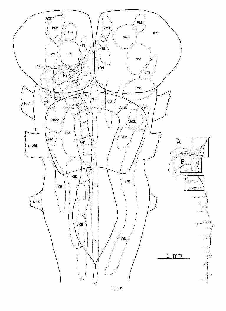
Fig. 12. Reconsfmction froin twelve horizonlal sections of'the contralat- era1 course (through the predorsal bundle) of a dorsal pathway (TBd) axon. The ipsilatcral collaterals of this axon were illustrated in Figures 7 and 9- 11. The axcn is plotted onto a horizontal reconstruction of a turtle brainstem made from alternate serial transverse sections. This axon emitted collater- als a t regular intervals. Most of the thousand or so boutons supported by this axon are located in the medial half of the I-eticular formation. A few branches enter the raphc caudally. The axon was not completely filled past the level of the trigeminal motor nucleus. The rectangular regions in the inset 1abelt.d A-C are illustrated at a greater magnification in Figures 13- 15.
I I I
4
Ri
Ri
V I
Figure 12

66 M.I. SERENO
Fig. 13. High-magnification view of region A of the dorsal pathway axon illustrated in Figure 12. The first contralateral collaterals (E, F) travel near the main trunk for a while before turning laterally, and thus avoid entering the substantia nigra. Subsequent branches course laterally from the start. Each contralateral collateral branches from a node in the thick (5-6 pm) myelinated main trunk with a.n initial diameter well under 1 pm. Portions of many collaterals thicken (up to 2 pm) and apparently rnyelinate (e.g., the initial 150 pm of collateral GI. Small clumps of five to 20 boutons arise from unmyelinated preterminal strands. These clumps are distributed throughout the medial reticular fbrmation. The inset shows one of the horizontal sections used to reconstruct this region of the axon and the rectangle indicates the location of the high-magnification view. Far laterally (outside the rectangle), collaterals thin to less than 1 pm and begin to wander caudally, giving rise to a few additional boutons in the small-celled nucleus (SC) ventral to Imc. In this and the succeeding high- power views, houtons have been drawn about twice their actual size to make their location apparent.
100 u


68 M.I. SERENO
is the same axon shown in Figures 7-11. Its main trunk continues toward the midline after giving rise to the ros- trally directed branclnes and emits a collateral (D in Fig. 9) ventral to the oculomotor complex that gives off a handful of varicosities just outside the medial longitudinal fascicu- lus and the oculomotor complex. A few other TBd axons did not have a collateral at this point, but such collaterals
longitudinal fasciculus, and eventually the suprapeduncu- lar nucleus. Thus, some TBd axons project to the suprape- duncular nucleus bilaterally. The more common pattern by far, lhowever, is an ipsilateral-only connection.
An example of a TBi =On is illustrated in Figures 17-25. It was recon. StructetJ from twenty-one 110 pm serial horizontal sections,
Inter,me&ate tectobulbar OW (Tsi).
were identified in examples of both TBd(1g) and TBd(sm) Figure 17 is a low-power reconstruction of most of this axon axons. Figure 12 is a reconstruction of the contralateral plotted to scale onto the same topographical reconstruction course of the axon plotted to scale on a horizontal recon- used in Figure 12. This axon was traced into injection site struction from serial transverse sections. Figures 13-15 A in Figure 27. ~i~~~~ 18, 19, and 22-25 are high-power are higher-power views of the regions labeled A-C in Fig- views of the regions labeled B,A, C-F in Figure 17. Figures ure 12. It is clear that the axon Was not Completely filled 20 and 21 are high-power views of the end of the thick caudally because boutons were not visible Past the abdu- rostral collateral; these regions are located beyond the ros- cens nucleus, and large caliber collaterals faded without tral edge of the low-power reconstruction in Figure 17. The thinning. However, filling was still good Over 6 mm from main collaterals are lettered sequentially in the high-power the injection site, in region C (Fig. 15), while in region A, views. collaterals could be traced until they became extremely Figur,e 18 illustrates the first collaterals visible after the thin and began "meandering," bearing only occasional myelinated, 2.5 pm diameter main trunk emerges from the (but well-filled) boutons. injection site. The main trunk travels (almost perpendicu-
The main trunk of the axon runs caudally in the Predor- lar to thie plane of section) through profundus mesencephali sal bundle, sweeping laterally around the bulge in the caudalis (PMc). The branches labeled A-D distribute strings midline nuclei at this level before coursing along the mid- of 25-7;; boutons to the medial third of the nucleus. Some line in the caudal pons and medulla. Contralateral collat- collater,als also reach the laminated and central nuclei of erals arise regularly at intervals of 100-150 I*m in the the torus semicircularis (Tor1 and Torc) and the central gray pons. The spacing between COlhterak Seems wider mu- (CG). M:any partially reconstructed T B ~ axOnS showed a dally, but it is Possible that SOme were not They similar overall pattern, distributing collaterals to only a have initial diameters under 1 pm and are nearly invisible part of profundus mesencephali caudalis. A point-to-point for the first 10 or 20 pm. Many thicken and Probably topography, however, was not evident. In contrast to the become myelinated after these initial constrictions. The common observation of TBd boutons in apparent contact first contralateral co~laterals (E, F in Fig. 13) travel mu- with back-labeled profundus mesencephali rostralis cells, dally for a considerable distance before turning into the TBi boutons were not observed to contact back-labeled pro- reticular formation, thus avoiding the substantia ni@a fundus lmesencephali caudalis cells; in a number of cases, (SN) and the adjacellt PMv (Fig. 12). Subsequent collater- labeled (cells in the caudal nucleus were seen lateral to the a h extend from the main trunk at almost right TBi terminal field. Profundus mesencephali caudalis cells (Figs. 14, 151, gene.rating a continuous terminal field also had less extensive dendritic fields and smaller somata throughout the reticular core. Clusters or short strings of than prcfundus mesencephali rostralis cells. five to 20 boutons are commonly seen. Some collaterals The rostrally directed collateral a t B is unique in that it extend laterally up to 1 mm, entering the terminal field of is by far the thickest. It branches from the main trunk with the intermediate pat.hway, and a few travel UP into the a diameter of over 1 pm and then expands to almost 2 pm, small-celled nucleus and terminate Sparsely there. How- apparently becoming myelinated for most of its course. By ever, the density ofboutons is much higher in the contrast, the other collaterals are constricted to well under half of the reticular core. Thus, the terminal zone of the 1 pn1 in diameter at their point of origin and are not mye- Predorsal Part of a sil%le TBd muron occuPies~ as was the linated. The majority of TBi axons had one robust, rostrally case with its ipsilateral branches, virtually the Same re- directed branch among the first several collaterals. These gion as does the total projection. branchem; avoided profundus mesencephali rostralis, either A portion of a second TBd axon in the predorsal bundle passing medial or ventral to it. In this example, collateral
is illustrated in Figure 16. This axon ran in the small B runs between the medial edge of that nucleus and the caliber component of the TBd. It was traced out of a more central gray (Figs. 18, 19), pierces the central gray at the caudal tectal site than the TBd(lg) fiber above, and gave posterior commissure, and courses rostrally through the off two ipsilateral, rostrally directed branches into profun- neuropile between the central gray and the ependyma for dus mesencephali rostralis, a long rostral branch to the almost a millimeter (Fig. 201, until it passes by the dorsal suprapeduncular nucleus, and a fine twig just under the oculomotor nucleus before crossing to run in the small ___ caliber part of the predorsal bundle. It is also similar to the previous TBd example in the caudal course of its initial ~ i ~ . 14. Hlgh-magnification view of' region B of the dorsal pathmay axon contralateral collaterals, in the tendency for branches to illustrated in Figure I'L. A s in Figure 13, collaterals arise a t regular inter- occasiona~~y penetrate the TBi field, and in the clumpy vals and distribute small clumps of' houtons to many parts of the medial distribution of boutons. one difference is that there are reticular formation. A slightly increased density of termination is evident
near the origins of' collaterals I and J. The axon i s p about three times as mlany boutons situated within I*m caudal polo of the trochlear nucleus at this level (see Fig. 12). The main ofthe trochlear nucleLLs. A second is that the axon also has trunk begins to curve back toward the midline after traveling around the a contru~u~eru~ branch to the suprapeduncu~ar nucleus hulge in inidline structures that i s created by the superior raphe nuclei and
the interpeduncular nuclei (Rsl, Rsm, Ipd, and Ipv). The location of the high- arising from collateral D. It initially f o ~ ~ o w s the surround- ing branches caudally before making a 180" turn and trav- magnification view is shown in the Inset. Coilaterais t ha t reached the
lateral reticular formation (RSL. lateral to the rectangle in the inset) be- eling upward to enter the interstitial nucleus of the medial came very thin and gave off only four more boutons
.

I 0 0 4

70 M.I. SERENO
edge of the nucleus of the medial longitudinal fasciculus (Nmlf, Fig. 21). Just rostral to that nucleus, the collateral penetrates back through the pretectal central gray, contin- ues rostrally, and then abruptly turns into the lateral hy- pothalamic area (Fig. 21) where thin unmyelinated stretches begin to appear in the main collateral trunk. It continues laterally until it enters the ventral peduncle of the lateral forebrain bundle where it turns and runs cau- dally for several millimeters, after which it was lost. The main myelinated trunk of collateral B gives off short branching strings of ten to 75 boutons at intervals of ap- proximately 100 pm throughout most of its course. No strings are emitted fior a 400 pm stretch as the collateral passes through the bundle of ventrally directed TBd axons (Fig. 191, and only a few boutons arise in the lateral hypo- thalamic area and even fewer in the long meandering path back through the lateral forebrain bundle.
Figures 22-25 (regions C-F in Fig. 17) illustrate selected portions of the main caudal trunk and its collaterals. The axon was densely filled to the midpoint of the facial motor nucleus, which is about 7 mm from the injection site. The main trunk turns 1at.erally (Figs. 22, 23) as it passes the bulge in the midline nuclei near the pons-midbrain junc- tion. It then recurves medially (Figs. 24, 251, but remains in the lateral half of the reticular core. Collaterals are emitted at approximately 100 pm intervals until the main trunk passes well into reticularis inferioris dorsalis, where branches arise less frequently. As with TBd collaterals, TBi collaterals are given off from the main myelinated trunk with a diameter well under 1 pm, and often travel a short distance closely apposed to the main trunk before turning laterally. In this TBi axon, most collaterals course medially but, collaterals sometimes travelled laterally in other axons where the main trunk was located a little closer to the midline (e.g., photomicrograph in Fig. 26A). Each collateral bears 25-75 synaptic boutons, often arranged into small clumps of five to 20 boutons. The majority of the terminals are in the lateral reticular formation-the dorsomedial seg- ment of reticularis superioris lateralis and the lateral half of reticularis medius and reticularis inferioris. A number of collaterals penetra.ted the medial TBd terminal field (Figs. 23-25) but deposited relatively few terminals there. Collateral G entered the small-celled nucleus and gave off a small branch bearing very fine boutons (Fig. 22-low- power drawing). A similar overall pattern of arborization in the lateral half of the reticular core could be distin- guished in other TBi axons. In some axons, medially di- rected collaterals reached the caudal central gray at the level of the trigeminal motor nucleus. Thus, as with TBd axons, the terminal zone of a single TBi axon occupies almost the same region as the total projection. The axon in Figures 17-25 emitted a little over 2,700 boutons in its visible course. Perhaps 200-500 additional boutons might have been missed by light filling far caudally and by con- servative reconstruction practices.
Ventral tectobulbar axom (TBu axons). Single ventral pathway axons, like intermediate and dorsal pathway ax- ons, have extensive brainstem distributions. However, TBv axons apparently do n,ot have thick rostral collaterals, and their caudal trunks branch less extensively. Furthermore, TBv axons often have intratectal collaterals and, probably, commissural branches to the contralateral tectum (Sereno and Ulinski, '85) both of which were never seen in TBd and TBi axons. It was difficult to get a complete picture of TBv axonal morphology at. the single-cell level because even
small injections resulted in a formidable tangle of afferents and othler efferents near the point where TBv axons leave the tectum. For the small caliber component of the TBv, this was remedied by reconstructing axons that were retro- gradely labeled from the tegmentum (see Materials and Methods of the following paper).
Small caliber TBu axom. Figure 27 is a serial recon- struction of the intratectal course of a retrogradely labeled TBv(sm) axon and a schematic diagram of its course through the brainstem. The 1 pm diameter axon arises from the descending radial dendrite of a small bitufted cell in the stratum fibrosum et griseum superficiale (SFGS) and runs just below the deepest retinal terminal zone, between layers 7 and 8 of Ramon (1896). Collaterals bear- ing many boutons en passage arise every 100-200 pm and descend through the central gray (SGC) and white (SAC) layers, sometimes entering the deep periventricular gray (SGP). Collaterals strictly avoided the retinal recipient stratum fibrosum et griseum supefiiciale in this and other examples. This collateral field ends abruptly as the main axon trunk leaves the tectum and enters the rostral mag- nocellular isthmi (Imr). It passes all the way through the almond-shaped mass of fibers and terminals that engulfs the nucleus (only lightly labeled by this tegmental injec- tion) without generating any collaterals. Soon after pass- ing .out o f rostral magnocellular isthmi, the axon makes a downward bend into the fine caliber component of the ventral pathway just ventrolateral the caudal magnocel- lular nucleus isthmi (Imc). Here, several long collaterals are given off into the dorsal half of the small-celled nucleus just under caudal magnocellular isthmi. The axon then runs along the ventrolateral surface of the brainstem emit- ting short collaterals into the neuropile there, until it was lost entering the injection site. Several other small bitufted cells in ithe stratum fibrosum et griseum superficiale that were rcconstructed also had axons with intratectal collat- erals, :L lack of collaterals as the main trunk passed through rostral r-Jagnocellular isthmi, long collaterals to the small-
us, and short collaterals in the neuropile at the surface of the brainstem. The source of the
almond.-:;haped mass of fine terminals in rostral magnocel- isthmi (Fig. 2G) and of the dense field of fine
terminalls in the neuropile lateral to the small-celled nu- cleus (Fig. 2F) was not determined. A possible source is the population of small radial cells in the SGP that have the smallest caliber axons of any tectobulbar cells.
Medium caliber TBu axons. The following paper sug- gests, on the basis of retrogradely labeled material, that TBv(mec1) axons also tend to have intratectal collaterals. In several cases, putative TBv(med) cells had an axon branch that crossed in the tectal commissure (e.g., Fig. 10 from Sereno and Ulinski, '85). After TBv(med1 axons pass through the ipsilateral LIT, it becomes possible to follow single axons through serial sections in anterogradely la- beled material. These axons are the main source of collat- erals to the small-celled nucleus. As the parent axon passes the lateral edge of the nucleus, two or three branches arise,
Fig. 15. High-magnification view of region C of the dorsal pathway axon illustrated in Figure 12. At this level (6-7 mm from the injection site) the HRP filling begins to lighten (e.g., see the lateral end of collateral 0). The main trunk remains thick and could he followed for 3 more rnrn. It continued to give off ~:ollatcrals a t regular intervals but these were too lightly stained to distinguish boutons. The main trunk faded in the medullary reticular formation.

Ros
tra1
Late
ral
Med
ial
Cau
dal
100 u
72
subdivide, and give rise to about 30-100 boutons distrib- uted across a significant portion of its dorsal segment. Almost all TBv(med) axons had branches to the small- celled nucleus. Caudal to it, the main trunks of the axons travel ventrally for about a millimeter until they reach the ventrolateral segment of the reticularis superioris la- teralis (RSL(vl)), where they emit several collaterals (Fig. 28). These branches usually avoid the more medially lo- cated TBi terminal field in the dorsomedial segment of reticularis superioris lateralis. Further caudally, in reti- cularis medius and reticularis inferioris dorsalis, however, there is significant orerlap between the TBi and TBv(med) terminal fields. The morphology of those TBv(med) collat- erals is similar to the ones shown in Figure 28. Compared to TBi axons, TBv(nied) axons branch less frequently in the tegmentum and (could often be followed for a millime- ter at a time without a branch. No topographic pattern was discernible but the sample of reconstructed axons was small. A few TBd(med) axons were still visible at the level of the hypoglossal nucleus.
Topography of the TBi projection There was no obvious topographic pattern in the tectore-
ticular projections as the location of the injection site in the tectum was varied. However, cases with two injections at different loci sometimes resulted in separated clumps of main axon trunks in the pontine reticular formation. Fur- thermore, the TBi “tract” spans up to 750 pm dorsoven- trally in contrast ito the more localized (and hence recognizable) predorsal bundle (TBd axons). Given that the majority of the boutons arising from a TBi axon are found within 200 pm of the main trunk, and often in dorsoven- trally flattened arrays, a crude dorsoventral andor medi- olateral mapping of tectal loci seemed possible. Also, given the low density of the projection, such a topography might have been overlooked with conventional plotting tech- niques. Therefore it was decided to examine the topography at the level of single axons. Figure 29 is a stereoscopic view of the main trunks of 17 TBi axons emerging from two punctate injection sites and coursing into the lateral pon- tine reticular formation. Two small HRP injections at A and B labeled eight (Nos. 1-8) and nine (Nos. 9-17) TBi axons, respectively, each of which was traced through 16 horizontal serial sections. The axons all entered the dorso- medial segment of rleticularis superioris lateralis where they sorted into two distinct clumps. The caudal ends of axons 1,4, 5 , 7, 10-13, 16, and 17 are located in a flattened clump about 500 pm dorsal to axons 2, 3,6, 8,9, 14, and 15. Most axons remained with one clump throughout the dor- somedial segment of r(eticu1aris superioris lateralis; axon 8, by contrast, started out in the dorsal group but then made an abrupt jump to the ventral group that is visible at the middle of the figure. Each clump obviously contains a mix- ture of axons from the two injection sites, suggesting that there is no point-to-point topography at the level of small groups of axons. The axon previously illustrated in detail in Figures 17-25 is axon number 2 (circled); it emerged from site A and ended up in the ventral clump. All 17 axons were between 1.5 and 2.5 pm in diameter. For clarity, how- ever, the axons from injection site A have been drawn thicker to distinguish them from site B axons. Thirteen of the 17 axons shown had one thick, rostrally directed collat- eral that arose as the axons turned caudally; these are not drawn in Figure 29.
M.I. SERENO
DISCUSSION These results indicate that tectoreticular projections in
turtles comprise three main pathways containing axons of several sizes. The pathways are schematically illustrated in Figure 30. Single tectoreticular axons in each pathway terminate sparsely and non-topographically in a variety of reticu1a.r structures throughout the brainstem. One axon typically supports several thousand boutons. Comparable numbers of boutons are supported by single axons in topo- graphically organized pathways like the ipsilateral isth- motectal pathway (from caudal magnocellular isthmi) where each axon gives rise to a dense, spatially restricted termi- nal thicket whose location varies systematically with the map location of the neuron’s dendritic field in the nucleus (Sereno., ’83). The boutons of a tectoreticular axon, by con- trast, are distributed throughout an immensely greater volume and are intermingled with boutons originating from tectobulbar neurons a t many different, nonadjacent tectal loci. This discussion will consider some general features of the three pathways. Functional implications of the organi- zation of the pathways will be discussed in the accompany- ing paper after the morphology of the parent cells has been establis,hed.
The dorsal tectobulbar pathway is constant across verte- brate classes. It has been characterized at the single-cell level in turtles, snakes (Dacey, ’82), and cats (Grantyn and Grantyn, ’82; Grantyn et al., ’82). In all three animals, dorsal pathway axons lack intratectal collaterals and have ipsilatei-a1 branches into the rostral mesencephalic tegmen- tum and central gray, as well as a main rostral branch that reaches the ventral thalamus (via “medial tectothalamic tract” in Dacey, ’82). In cats, this last branch arises at the level of the oculomotor nucleus and travels just lateral to the interstitial nucleus of Cajal ( = interstitial nucleus of the medlial longitudinal fasciculus in reptiles) to reach the rostral interstitial nucleus of the medial longitudinal fasci- culus (see Buttner-Ennever and Buttner, ’78) and the zona incerta (probably together equivalent to the reptilian SP). In turtles, visual and somatosensory responses have been recorded in the suprapeduncular nucleus, and visually dri- ven activity there is almost completely abolished by ipsilat- era1 tectal lesions (Belekhova, ’79).
A similar pattern of ipsilateral branching can be inferred from anterograde tracer and degeneration experiments in a number of other animals. In monkeys, for example, Hart- ing et al. (‘80) showed a rostrally directed “fine caliber component” of the dorsal pathway arising just before the thick predorsal bundle axons decussate. These thinner ax- ons travel lateral to the interstitial nucleus of Cajal to reach thle rostral interstitial nucleus of the medial longitu- dinal fasciculus where some turn laterally into the zona incerta. In birds, Hunt and Kunzle (’76, their Fig. 4B) demonstrated a rostrally directed fascicle arising from the dorsal tectobulbar pathway that eventually terminates in the ipsilateral nucleus subrotundus. In Iguana, Foster and Hall (’75, their Figs. 5,6) showed a similar projection to the “ventromnedial thalamic nucleus” while Ebbesson and Vanegac; (’76) illustrate a “recurrent rostral bundle” leav- ing the dorsal pathway to enter the ventral thalamus in two teleost species.
The detailed pattern of contralateral branching is also uniform across vertebrates. For example, a second thick rostral collateral occasionally arises after the main trunk

TECTORETICULAR AXONS 73
200 u
Fig. 16. Reconstruction of the first four contralateral branches of an axon in the small caliber component of the dorsal pathway, TBdkm). As with large caliber dorsal pathway axons, this axon had ipsilateral collaterals (not shown) including branches to profundus mesencephali rostralis (PMr), a “main rostra1 collateral” eventually reaching the suprapeduncular nucleus (SP), and a small twig near the oculomotor nucleus. The first contralateral collaterals trend caudally. The main trunk (2 pm diameter) is myelinated as are some of the thicker collaterals (parts of collateral D). This axon has a contralateral branch to the suprapeduncular nucleus in addition to a more usual ipsilateral branch; it arises from collateral D, initially runs caudally, and then makes a 180” turn to run rostrally through the interstitial nucleus of the medial longitudinal fasciculus (Imlf), eventually reaching SP. This axon also has about three times as many boutons within 200 pm of the overlying trochlear nucleus as did the previous example in Figures 12-15 (for a similar-sized brain). Since the original drawing was reduced more in this illustration, boutons were drawn at about four times actual size to make them visible.

74 M.I. SERENO
has decussated; such a collateral can reach the contralat- era1 ventral thalamus in turtles (Fig. 16, collateral D) as well as in cats (collateral 3 in Fig. 3 of Grantyn et al., '82). Caudally, the extensive collaterals of dorsal pathway axons in cat, snake, and turtle apparently differ only in scale. Thus, a 5 pm diameter main trunk gives off branches every 150 pm in the turtle wlhile a 9 pm predorsal axon in the cat emits approximately t:he same number of collaterals (over a course three times as long) at about 700 pm intervals. A few medially directed collaterals travel into or near the raphe just caudal to the abducens nucleus in cats and tur- tles. The paramedian terminal field of the predorsal bundle is present in all other vertebrates examined, the primary difference in bony fish and amphibians being that the par- ent axons are more ventrally placed (Rubinson, '68; Luiten, '81). A small caliber component of the dorsal pathway ven- tral to the large caliber component has not yet been recog- nized in animals besides turtles and snakes. However, it may have been mistaken for a terminal field in anterograde tracer studies. Small axons were not recovered after intra- cellular injections in t.he predorsal bundle (Grantyn and Grantyn, '82), but this could be due to a sampling problem.
The intermediate tectobulbar pathway is known at the single-cell level only in turtles and snakes where it can be aptly described as a more laterally terminating, ipsilateral version of the dorsal pathway. As with dorsal pathway axons, intermediate pathway axons have one main rostral collateral. However, these branches were not present in every case and their rostral course through the pretectal central gray near the nucleus of the medial longitudinal fasciculus was somewhat variable. Thus, the intermediatt pathway axon illustrated by Dacey ('82) apparently lacked an identifiable main rostral branch, though these were present on other axons (personal observation). Harting et al. ('80) has demonstraked a. deep tectal projection to the rostral central gray in monkeys. Nevertheless, given the variable course of these branches, it will be necessary to characterize axons in this pathway at the single cell level before more firm comparisons can be made.
The turtle and snake material is similar at caudal levels. In both animals, single intermediate pathway axons termi- nate first in the caudal mesencephalic tegmentum and then throughout the lateral iceticular core, occasionally also giv- ing off branches to the caudal central gray and the trige- minal complex. The intermediate pathway is especially well developed in cartilaginous fishes (Smeets, '81) but can be clearly distinguished in other reptiles (Foster and Hall, '75) and birds (Hunt and Kunzle, '76; labeled "ventral tectobul- bar pathway") as well. Most who have worked on mammals have not explicitly distinguished intermediate and ventral pathways (e.g., Burne et al., '81; Harting and Huerta, '82). Nevertheless, the recent experiments of Holcombe and Hall ('81a,b) using restricted injections demonstrate that differ- ent sets of tectal neuroins project to the ventrolateral pon- tine tegmentum on one hand, and the underlying dorsolateral pontine nuclei on the other. In squirrels, cats, and monkeys, the intermediate pathway emits a dense terminal field in the caudal part of the cuneiform nucleus before terminating more lightly in the lateral half of the pontine reticular core (Harting, '77; Holcombe and Hall, '81a; Harting and Huerta, '82).
The ventral tectobulb.ur pathway is the most variable, probably in part the result of its involvement with the pontine nuclei, which appear in birds and mammals (Clarke, '77; Brown-Gould, '80; Bangma and ten Donkelaar,
'82). Single-cell information about this pathway, however, is only available in turtles and snakes, which themselves show significant differences. Turtle ventral pathway axons have intratectal and commissural collaterals that were not seen in snakes. Turtle axons then pass without branching through -the rostral (non-topographic) nucleus isthmi (Imr) and colla.teralize in a small-celled nucleus ventral to the caudal (topographic) nucleus isthmi (Imc), rather than in the topographic nucleus isthmi itself as do snake axons. Finally, the turtle ventral pathway has a medium caliber component that emits collaterals partially overlapping the intermediate pathway terminal field in the caudal pons and medulla, in addition to the small caliber pathway terminat- ing exclusively in the ventrolateral neuropile that is seen by itself in snakes. In birds, Hunt and Kunzle ('76) have described a tectopontine pathway that appears similar to the ventral pathway. It passes through the non-topographic nucleus isthmi magnocellularis (probably equivalent to tur- tle Imr) and then passes just lateral to the topographic nucleus isthmi parvocellularis (probably equivalent to tur- tle Imc) before terminating non-topographically in the lat- eral pontine nucleus. Recently it has been suggested that the rat tectopontine projection is topographically organized (Burne et al., '81). Cats and primates, too, exhibit a complex tectopontiine projection (Harting, '77; Harting and Huerta, '82). Sincte retrograde tracer experiments have uncovered no evidence of cerebellar projecting neurons in the reptilian ventral pons (Bangma and ten Donkelaar, '821, it appears that the ventral tectobulbar pathway has been secondarily recruited into a tecto-ponto-cerebellar circuit.
ACKNOWLEDGMENTS I thank Dr. P.S. Ulinski and Dr. D.M. Dacey for advice
and encouragement. This work was supported by PHS grant NS 12518 and an NSF predoctoral fellowship.
LITERATURE CITED Abrahams, V.C., and P.K. Rose (1975a) The spinal course and distribution
of fore and hind limb muscle afferent projections to the superior collicu- lus of the cat. J. Physiol. (Lond.) 247:117-130.
Abrahams, V.C., and P.K. Rose (1975b) Projections of extraocular, neck muscle, and retinal afferents to superior colliculus in the cat: Their connections to cells of origin of the tectospinal tract. J. Neurophysiol. .78:10-18.
Adams, J.C. (1977) Technical considerations on the use of horseradish per- oxidase EIS a neuronal marker. Neuroscience 2141-145.
Adamiik, E. (1870) h e r die Innervation der AugenheweL-ngen. Zentrabl. Med. Wi:is. 8.65.
Akert. K. (1945) Der visuclle Greifreflex. Helv. Physiol. Acta. 7.112-134.
Altman, J., .and M.C. Carpenter (1961) Fiber projections of the superior colliculu: in the cat. J. Comp. Neurol. 116.157-178.
Balaban, C.I)., and P.S. Ulinski (1981) Organization of thalamic afferents to anterior dorsal ventricular ridge in turtles. I. Projections of thalamic nuclei. J. Comp. Neurol. 200:95-129.
Fig. 17. Reconstruction from twenty-one 110 pm serial horizontal sections of an intermediate pathway (TBi) axon. As with dorsal pathway axons, this axon has a rnyelinated main rostral branch, and it gives off collaterals bearing small clumps of boutons at many levels throughout the brainstem. The main differences are: (1) the great majority of the boutons are in the lateral half of the reticular core, and (2) the caudal trunk does not cross. This axon was densely filled to the midpoint of the facial motor nucleus but quite faint by the time it passed the hypoglossal nucleus. It had thinned considerably before fading completely. The rectangular regions in the inset labeled A-F are illustrated at greater magnification in the next figures in the order B, PL, and the C-F.

Figure 17

Rostra I
Lateral --:- Medial
Caudal
Fig. 18. High-magnification view of region B of the intermediate pathway axon illustrated in Figure 17. The medium caliber myelinated main trunk (2.5 Wm diameter) begins giving off collaterals (lettered sequentially) immediately after leaving the tectum. Collaterals A-C support terminals in parts of profundus mesencephali caudalis (PMc), the intercollicular nucleus (ICo), and the central Dray (CG). Collateral B is the “main rostral branch.” It is by far the thickest collateral, approaching the diameter of the caudal trunk, and the only one that is consistently myelinated. The secondary collaterals emitted by collateral B are identified with subscripts. Most of the unmyelinated preterminal strands emitted from the rostral and caudal trunks remain close to the parent axon or travel medially, supporting small clusters of boutons scattered throughout a large volume. The inset shows one of the five sections used in this portion of the reconstruction. The rostral trunk is continued in Figures 19-21, while the caudal trunk is continued in Figures 22-25.

100 jl
Rostra1
Lateral 4- Medial
Caudal
Fig. 19. High-magnification view of region A of the intermediate pathway axon illustrated in Figure 17. The myelinated main rostral branch begins emitting collaterals again (at B,) immediately after passing through the bundle of large caliber dorsal pathway axons (TBd). These preterminal branches distribute small clumps of boutons to the neuropile between the central gray (CG) and profundus mesencephali rostralis (PMr). The main rostral collaterals of this and other TBi axons always avoided entering PMr. The inset shows one of the four sections used in this portion of the reconstruction.

Rostra1
Mediol Lateral
+ Caudal
Fig. 20. High-magnification view of a portion of the intermediate pathway axon illustrated in Figure 17 that is just rostral to region A. The myelinated main rostral branch penetrates the central gray to run rostroventrally in the neuropile adjacent to the ependyma, where it emits a series of short collaterals. Ventrally, it reaches the rostral tip of the nucleus of the medial longitudinal fasciculus (Nmlf). The inset shows four of the eight sections used in this portion of the reconstruction.

TECTORETICULAR AXONS 79
Rostra1
Medial Lateral
Fig. 21. High-magnification view of a portion of the intermediate path^ way axon illustrated in Figure 17 that I S just rostral and ventral to the region shown in Figure 20. ':'he main rmstrlil branch continues medililly past the nucleus of the MLF before penetrating laterally back through the central gray to enter the lateral hypothalamic area (LHA), where it begins to show unmyelinated stretches. It continues laterally until it reaches the ventral peduncle of the lateral f~~reb ra in bundle (vI,FB) where it runs caudally for several millimeters as a thin unniyelinated strand bearing k w boutons. Only a few other TBi rostral trunks enter the LHA.

Fig. 22. High-magnification view of region C of the intermediate pathway axon illustrated in Figure 17. The main caudal trunk enters the dorsomedial segment of reticularis superioris lateralis (RSUdm)), where it emits mostly medially or caudally directed preterminal strands at regular intervals bearing small clumps of medium-simd boutons. The main trunk turns ventrally (into the page) past collateral F and thus appears foreshortened until collateral I. RSUdm) contains no magnocellular elements a t this level. The inset shows one of the s,ections used in this portion of the reconstruction. Collateral G enters the small-celled nucleus (SC) ventral to the caudal magnocellular nucelus isthmi (Imc, not in section) and gives off a fine preterminal branch (see inset) bearing very small boutons.

81 TECTORETICULAR AXONS
100 JJ
Rostra1
Medial Lateral
Caudal
Fig. 23. High-magnification view of region D of the intermediate pathway axon illustrated in Figure 17. The main caudal trunk continues through the dorsomedial segment of reticuIaris superioris lateralis (RSL(dm)) emitting medially directed collaterals that sometimes penetrate the dorsal pathway terminal field in reticularis superioris medialis (RSM) (e.g., collaterals J and K). Conspicuous large cells appear in RSL(dm) at this level. The main trunk begins to recurve medially with other lateral brainstem structures after passing the bulge in the midline nuclei. The inset shows one of the sections used in this portion of the reconstruction.

Rostra1
Lateral + Medial
Caudal
Fig. 24. High-magnification view of region E of the intermediate pathway axon illustrated in Figure 17. The main caudal trunk continues through the dorsomedial segment of reticularis lateralis (RSL(dm)), which still contains prominent. magnocellular elements a t this level. Several of the medially directed collaterals penetrate reticularis superioris medialis (RSMI but as before, most of the boutons are located in the lateral half of the reticular formation, The inset shows t.he location of this portion of the axon.

Rostra1
Lateral 7- Medial
Caudal
Fig. 25. High-magnification view of region F of the intermediate pathway axon illustrated in Figure 17. The main caudal trunk enters the medial half of reticularis medius (RM) where it emits a particularly thick medially directed collateral that almost reaches the midline, but supports few boutons in the medial parts of RM. The main trunk begins to thin as it continues caudally, emitting collaterals a t regular intervals. It eventually enters the lateral parts of reticularis inferioris dorsalis (RID, see inset). HRP filling was less dense caudal to the portion of the reconstruction shown in the inset. As before, most of the boutons are located lateral to the TBd terminal field.
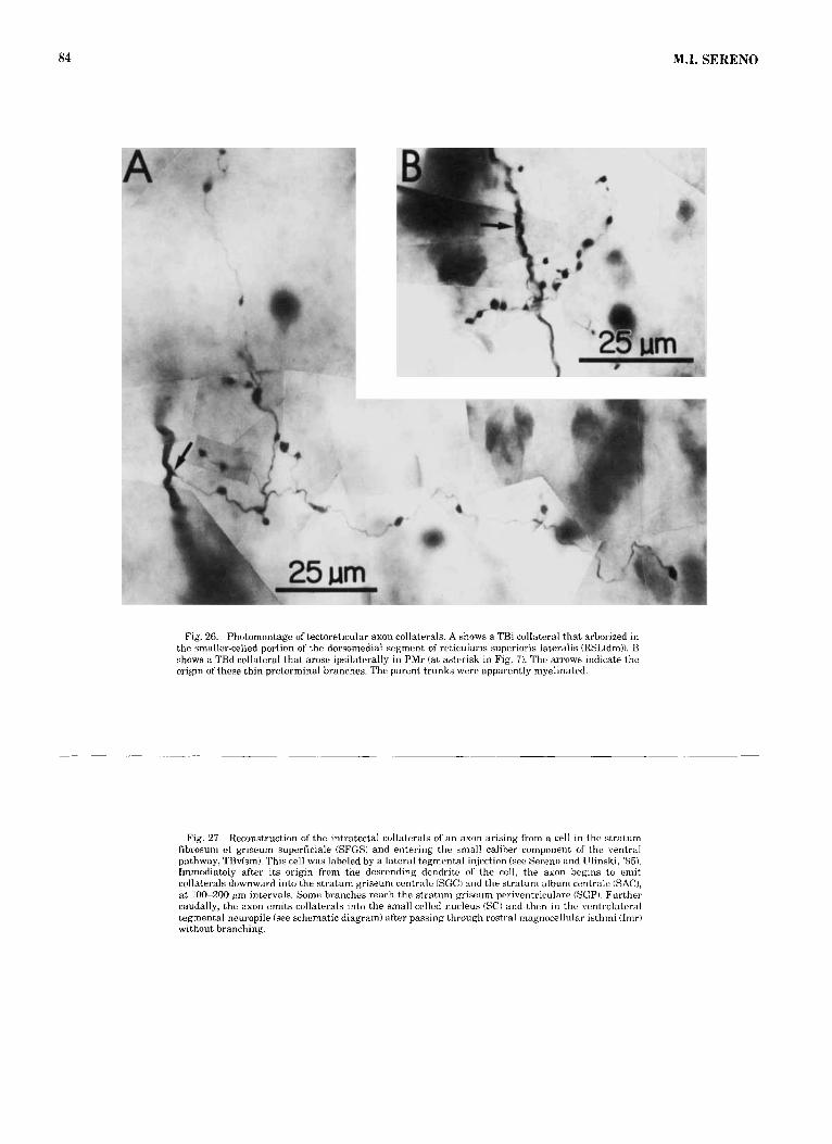
84 M.I. SERENO
Fig. 26. Photomontage of tectoreticular axon collaterals. A shows a TBi collateral that arborized in the smaller-celled portion of the dorsomediaf segment of reticularis superioris lateralis (RSL(dm)). B shows a TBd collateral that arose ipsilaterally in PMr (at asterisk in Fig. 7 ) . The arrows indicate the origin of these thin preterminal branches. The parent trunks were apparently myelinated.
Fig. 27. Reconstruction of the intratectal collaterals of an axnn arising from a cell in the stratum fibrosum et griseum superficiale (SFGSi and entering the small caliber component of the ventral pathway, TBvkmi. This cell was labeled by a lateral tegmental injection (see Sereno and Ulinski, '85). Immediately after its origin from the descending dendrite of' the cell, the axon begins to emit collaterals downward into the stratum y i seum centrale (SGCI and the stratum album centrale (SAC), a t :100-200 pm intervals. Some branches reach the stratum kvitieum periventriculare (SCP). Further caudally, the axon emits collaterals into the small-celled nucleus (SC) and then in the ventrolateral tegmental neuropile (see schematic diagram) after passing through rostra1 magnocellular isthmi (Imr) without branching.

t- N

Ros
tra1 t
Late
ral
-t-
Med
ial
t C
auda
l
Fig.
28.
R
econ
stru
ctio
n fr
om h
oriz
onta
l se
ctio
ns o
f tw
o co
llate
rals
ari
sing
fro
m a
n ax
on i
n th
e m
ediu
m c
alib
er c
ompo
nent
of
the
vent
ral
path
way
, T
Bv(
med
). T
hese
bra
nche
s w
ere
conf
ined
to
the
vent
rola
tera
l se
gmen
t of
ret
icul
aris
sup
erio
ris
late
rali
s (R
SL(v
1)). T
he m
ain
trun
k of
thi
s ax
on w
as
trac
ed t
owar
d th
e te
ctal
inj
ectio
n si
te t
hrou
gh t
en 1
10 p
m s
eria
l se
ctio
ns b
ut s
how
ed n
o ad
ditio
nal
bran
ches
bef
ore
it w
as l
ost
near
ros
tra1
mag
noce
llula
r is
thm
i (I
mr)
. Cau
dally
, se
vera
l m
ore
wid
ely
spac
ed b
ranc
hes
aros
e. T
ypic
ally
, T
Bv(
med
) axo
ns d
o no
t br
anch
as
regu
larl
y as
do
TB
i an
d T
Bd
axon
s.
Fig.
29.
St
ereo
scop
ic v
iew
of
the
mai
n tr
unks
of
17 i
nter
med
iate
pat
hway
(T
Bi)
axon
s em
ergi
ng
from
two
punc
tate
inje
ctio
n si
tes
and
cour
sing
into
the
late
ral
pont
lne
reti
cula
r fo
rmat
ion.
Tw
o sm
all
HR
P in
ject
ions
at A
and
B (i
nset
s) la
bele
d ei
ght (
Nos
. 1-8
) and
nin
e (N
os. 9
-17)
TB
i axo
ns, r
espe
ctiv
ely.
T
hese
axo
ns w
ere
trac
ed t
hrou
gh s
ixte
en 1
10 p
m s
eria
l ho
rizo
ntal
se
ctio
ns i
nto
the
dors
omed
ial
segm
ent o
f ret
icul
aris
sup
erio
ris
late
rali
s (R
SL(d
m))
, whe
re th
ey f
orm
ed tw
o sp
atia
lly d
iscr
ete
clum
ps.
each
, how
ever
, con
tain
ing
a m
ixtu
re o
f ax
ons
from
the
two
inje
ctio
n si
tes.
The
mor
e ve
ntra
l cl
ump,
fo
r in
stan
ce, c
onsi
sts
of a
xons
num
bers
2, 3
, 6, a
nd 8
(sit
e A
) and
num
bers
9, 1
4, a
nd 1
5 ki
te B
). T
he
axon
s em
ergi
ng fr
om s
ite A
are
dra
wn
thic
ker
only
to
mak
e it
easi
er t
o di
stin
guis
h th
em f
rom
sit
e B
ax
ons;
all
axon
s in
fac
t w
ere
of s
imila
r ca
liber
(1.
5-2.
5 W
m).
The
ste
reod
iagr
am c
an b
e vi
ewed
by
ocul
ar d
evia
tion
or b
y us
ing
a st
anda
rd s
tere
ovie
wer
. Axo
n nu
mbe
r 2
(cir
cled
) is t
he a
xon
reco
nstr
ucte
d in
det
ail i
n Fi
gure
s 17
-25.

Rostra1
Lateral 250 u t Medial
Caudal Figure 29
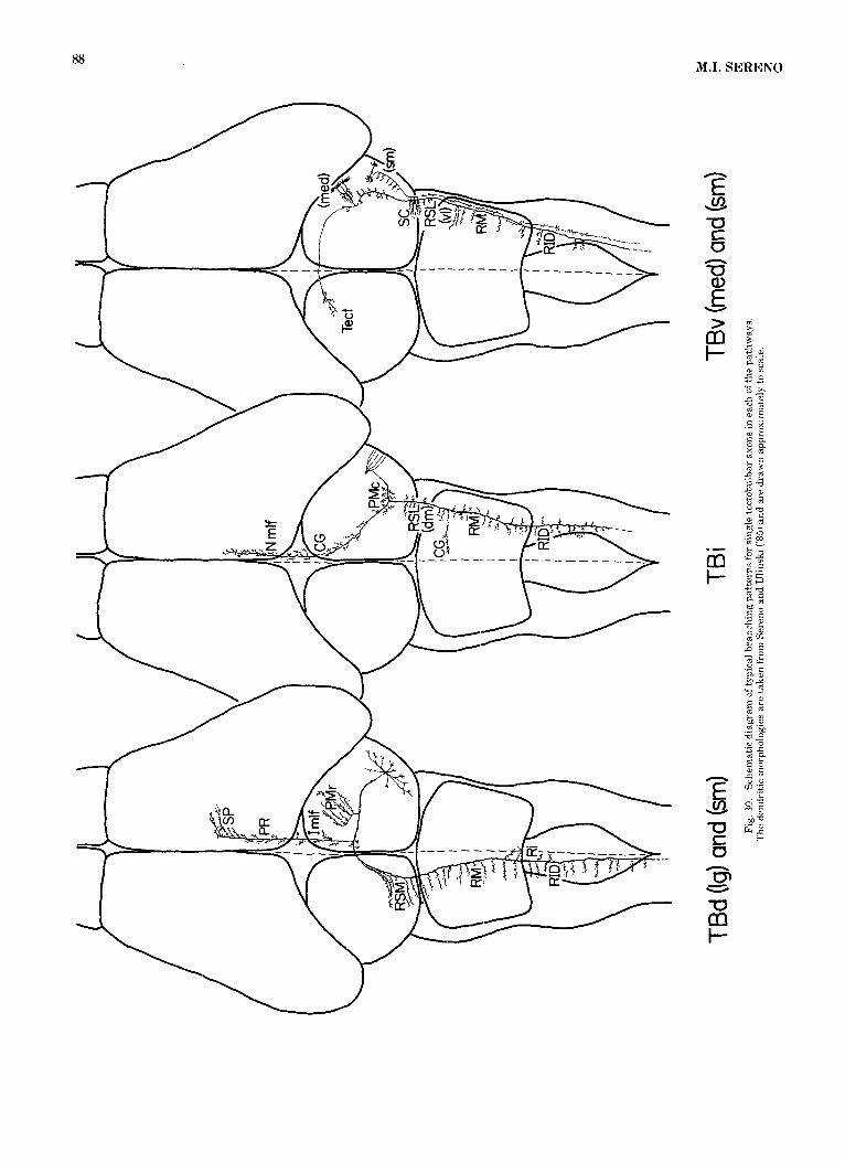
88 M.I. SERENO

TECTORETICULAR AXONS 89
Bangma, G.C., and H.J. ten Donkelaar (1982) Afferent connections of the cerebellum in various types of reptiles, J. Comp. Neurol. 207:255-273.
Bass, A.H. (1977) Effects of lesions of the optic tectum on the abilities of turtles to locate food stimuli. Brain Behav. Evol. 14:251-260.
Bass, A.H., and R.G. Northcutt (1981) Retinal recipient nuclei in the painted turtle, Chrysem?js picta: An autoradiographic and HRP study. J. Comp. Neurol. 199:97-112.
Bass, A.H., M.B. Pritz, and R.G. Northcutt (1973) Effects of telencephalic and tectal ablations on visual behavior in the side-necked turtle, Podoc- nemius unifilis. Brain Res. 55:455-460.
Bastian, J. (1982) Vision and electroreception: Integration of sensory infor- mation in the optic tectum of the weakly electric fish Apteronotus alhifrons. J. Comp. Phsyiol. 147.987-297.
Belekhova, M.G. (1979) Neurophysiology of the forebrain. In C. Gans, R.G. Northcutt, and P. Ulinski (eds): BiolokSy of the Reptilia, Vol. 10, Neurol- ogy B. London: Academic, pp. 247-359.
Brauth, S.E., A. Reiner, C.A. Kitt, and H.J. Karten (1983) The substance P~ containing striatotegmental path in reptiles: An immunohistochemical study. J. Comp. Neurol. 219:305-327.
Brown-Gould, B. (1980) Organization of Afferents from the Brain Stem Nuclei to the Cerebellar Cortex in the Cat. Adv. Anat. Embryol. Cell Biol., Vol. 62. New York: Springer-Verlag.
Burne, R.A., S.A. Azizi, G.A. Milhailoff, and D.J. Woodward (1981) The tectopontine projection in the rat with comments on the visual pathways to basilar pons. J. Comp. Neurol. 202:287-307.
Buttner-Ennever, J.A., and U. Buttner (1978) A cell group associated with vertical eye movements in the rostra1 mesencephalic reticular formation of the monkey. Brain Res. 151:31-47.
Clarke, P.G.H. (1977) Some visual and other connections to the cerebellum of the pigeon. J. Comp. Neurol. 174535552.
Cruce, W.L.R., and R. Nieuwenhuys (1974) The cell masses in the brain stem of the turtle Testido hermanni: A topographical and topological analysis. J. Comp. Neurol. 156277-306.
Dacey, D.M. (1982) Neural Organization of the Optic Tectum in the Eastern Garter Snake, Thamnophis sirtalis: Structural Analysis of a Sensori- motor Transformation. Ph.D. dissertation, University of Chicago.
Ebbesson, S.O.E., and H. Vanegas (1976) Projections of the optic tectum in two teleost species. J. Comp. Neurol. 165:161-180.
Ewert, J.-P. (1970) Neural mechanisms of prey-catching and avoidance be havior in the toad (Bufo hufo L.). Brain Behav. Evol. 3:36-56.
Ferrier, D. (1886) The Functions of the Brain. New York: Smith, Elder, and CO.
Foster, R.E., and W.C. Hall (1975) The connections and laminar organiza- tion of the optic tectum in a reptile (Iguana iguana). J. Comp. Neurol. 163:397-426.
Glenn, L.L., and R.E. Burke (1981) A simple and inexpensive method for 3-dimensional visualization of neurons reconstructed from serial sec- tions. J. Neurosci. Methods 4:127-134.
Graham, J., H.E. Pearson, N. Berman, and E.H. Murphy (1981) Laminar organization of superior colliculus in the rabbit: A study of receptive field properties of single units. J. Neurophysiol. 45:915-932.
Grantyn, A.A., and A. Berthoz (1977) Synaptic actions of the superior colliculus on medial rectus motoneurons in the cat. Neuroscience 2:945- 951.
Grantyn, A., and R. Grantyn (1976) Synaptic actions of tectofugal pathways on abducens motoneurons in the cat. Brain Res. 105:269-285.
Grantyn, A., and R. Grantyn (1982) Axonal patterns and sites of termina- tion of cat superior colliculus neurons projecting in the tecto-bulbo- spinal tract. Exp. Brain Res. 46;243-256.
Grantyn, A., R. Grantyn, and A. Berthoz (1982) Tectal control of vertical eye movements: A search for the underlying circuits in the mesenceph- alon. In A. Roucoux and M. Crommelinck (eds): Physiological and Path- ological Aspects of Eye Movements. The Hague: Dr. W. Junk Publishers,
Grobstein, P., C. Comer, and S.K. Kostyk (1983) Frog prey capture behavior: between sensory maps and directed motor output. In J.-P. Ewert, R.R. Capranica, and D.J. Ingle (eds): Advances in Vertebrate Neuroethology. New York: Plenum, pp. 331-347.
Guitton, D., M. Crommelinck, and A. Roucoux (1980) Stimulation of the superior colliculus in the alert cat. I. Eye movements and neck EMG activity when head is restrained. Exp. Brain Res. 39353-73.
Harting, J.K. (1977) Descending pathways from the superior colliculus: An autoradio?g+aphic analysis in the rhesus monkey (Macaca mulatta). J. Comp. Neurol. 173:583-612.
pp. 337-344.
Harting, J.K., M.F. Huerta, A.J. Frankfurter, N.L. Strominger, and G.J. Royce (1980) Ascending pathways from the monkey superior colliculus: An autoradiographic analysis. J. Comp. Neurol. 192:852-882.
Hartline, P.H., L. Kass, and M.S. Loop (1978) Merging of modalities in the optic tectum: Infrared and visual integration in rattlesnakes. Science 199: 1225-1 229.
Hartline, P.H. (1984) The optic tectum of reptiles: Neurophysiological stud- ies. In H. Vanegas (ed): Comparative NeuroIobT of the Optic Tectum. New York: Plenum, pp. 601-618.
Holcombe, V., and W.C. Hall (1981a) Lammar origin of ipsilateral tectopon- tine pathways. Neuroscience 6t255-260.
Holcombe, V., and W.C. Hall i1981b) The laminar origin and distribution of the crossed tectoreticular pathways. J. Neurosci. 1:1103-1112.
Huber, G.C., and E.C. Crosby (1933) The reptilian optic tectum. J. Comp. Neurol. 57:57-164.
Huerta, M.F., and J.K. Harting (1982) Tectal control of spinal cord activity: Neuroanatomical demonstration of pathways connecting the superior colliculus with the cervical spinal cord grey. In H.G.J.M. Kuypers and G.F. Martin (eds): Descending Pathways to the Spinal Cord. Prog. Brain Res. 57:293-328.
Hunt, S.P., and H. Kunzle (1976) Observations on the projections and intrin~ sic organization of the pigeon optic tectum: An autoradiographic study based on anterograde and retrograde. axonal and dendritic flow. J. Comp. Neurol. 170:153-172.
Ingle, D.J. (1982) Organization of visuomotor behaviors in vertebrates. In D.J. Ingle, M.A. Goodale, and R.J.W. Mansfield (eds): Analysis ofvisual Behavior. Cambridge: M.I.T. Press, pp. 67-109.
Jay, M.F., and D.L. Sparks (1982) Auditory and saccade-related activity in the superior colliculus of the monkey. Soc. Neurosci. Ahstr. 8:951.
Kawamura, K., A. Brodal, and G. Hoddevik (1974) The projection of the superior colliculus onto the reticular formation of the brainstem: An experimental anatomical study in the cat. Exp. Brain Res. 29t1-19.
Kunzle, H., and W. Woodson (1982) Mesodiencephalic and other target regions of ascending spinal projections in the turtle, Pseudcvnys srripta elegans. J. Comp. Neurol. 212:349-364.
Luiten, P.G.M. (1981) Afferent and efferent connections of the optic tectum in the carp (Cryprinus carpio L.). Brain Res. 2205-65.
McIlwain, J.T. (1975) Visual receptive fields and their images in superior colliculus of the cat. J. Neurophysiol. 38:219-230.
Maeda, M., T. Shibazaki, and K. Yoshida (1979) Labyrinthine and visual inputs to superior colliculus neurons. In R. Granit and 0. Pompeiano (eds). h o g . Brain Res. 50335-743.
Mrosovsky, N., A.M. Granda, and T. Hay (1979) Seaward orientation of hatchling turtles: Turning systems in the optic tectum. Brain Behav. Evol. 16203-221.
Nagata, T. and L. Kruger (1979) Tactile neurons of the superior colliculus of the cat: Input and physiological properties. Brain Res. 174:19-37.
Newman, D.B., W.L.R. Cruce, and L.L. Bruce (1983) The sources of supra- spinal afferents to the spinal cord in a variety of limbed reptiles. I. Reticulospinal systems. J. Comp. Neurol. 215:17-32.
Palmer, A.R., and A.J. King (1982) The representation of auditory space in the mammalian superior colliculus. Nature 299:248-249.
Papez, J.W. (1935) Thalamus of turtles and thalamic evolution. J. Comp. Neurol. 6Zt433-475.
Parent, A. (1979) Monoaminergic systems of the brain. In C. Gans, R.G. Northcutt, and P. Ulinski (eds) : Biology of the Reptilia, Vol. 10, Neurol-
ogy B. London: Academic, pp. 247-285.
Rainey, W.T. (1978) Organization of nucleus rotundus, a tectofugal thalamic nucleus in turtles. I. Nissl and Golgi analysis. J. Comp. Neurol. I60:121- 142.
Rambn, P. (1896) Estructura de encefalo dcl camaleon. Rev. Trim. Micro- graf. 1:146-182.
Roucoux, A., D. Guitton, and M. Crommelinck (1980) Stimulation of the superior colliculus in the alert cat. 11. Eye and head movements evoked when the head is unrestrained. Exp. Brain Res. 3935-85.
Rubinson, K. (1968) Projections of the optic tectum of the frog. Brain Behav. Evol. 1.529-561.
Schnyder, H., and H. Kunzle (1983) The retinopetal system in the turtle Pseudemys scripta elegans. Cell Tissue Res. 234:219-224.
Sereno, M.I. (1983) Dendritic and axonal morphology of tectal-projecting neurons in the isthmus region of a turtle, P.seudem.ys scripta. Proc. Soc. Neurosci. 9r818 (Abstract).
Sereno, M.I., and P.S. Ulinski (1985) Tectoreticular pathways in the turtle

90 M.I. SERENO
Pseudemys scrzppta. 11. Morphology of tectoreticular cells. J. Comp. Neu- rol. 233:91-114.
Smeets, W.J.A.J. (1981) Efferent tectal pathways in two chondrichthyans, the shark Scyliorhinus canicula and the ray Raja C ~ U U U ~ Q . J. Comp. Neural. 195: 13-23.
Sparks, D.L., and L.E. Mays (1981) The role of the monkey superior collicu- lus in the control of saccadic eye movements: A current perspective. In A.F. Fuchs and W. Becker (eds): Progress in Oculomotor Research. New York Elsevier, pp. 137-144.
Stein, B.E., and H.P. Clamann (1981) Control of pinna movements and sensorimotor register in cat superior colliculus. Brain Behav. Evol. 19: 180-192.
optic tectum: Some comparisons with mammals. J Comp. Neural. Stein, B.E., and N.S. Caither (1981) Sensory representation in reptllian
20250-87. Stein, B.E., and N.S. Gaither (1983) Receptive-iield properties in reptil-
ian optic tectum: Some comparisons with mammals. J. Neurophysiol. 50: 102-124.
ten Donkelaar, H.J., and R. Nieuwenhuys (1979) The brainstem. In C. Gans, R.G. Northcutt, and P. Ulinski (eds): Biology of the Reptilia, Vol. 10, NeurolobT B. London: Academic, pp. 133-200.
Ulinski, P.S. (1977) Tectal efferents in the handed water snake. J. Comp. Neurol. 17.?:251-274.
Wang, R., J.L. Kuhie, and M. Halpern (1977) Brevital Sodium: An effective anesthetic agent far performing surgery on small reptiles. Copeia 1977338-743.
Wurtz, R.H., and J.E. Albano (1980) Visual-motor functions of the primate superior colliculus. Annu. Rev. Neurosci. 3,189-226.