technological perspectives for endovascular therapy...
TRANSCRIPT
Technological Perspectives forEndovascular Therapy of the Lower Limb
Prof. Thomas ZellerDepartment AngiologyClinic for Cardiology and Angiology IIUniversity Heart-Center Freiburg - Bad Krozingen Bad Krozingen , Germany
Disclosure
2
Speaker name: Thomas Zeller
I have the following potential conflicts of interest to report:
Consulting
Employment in industry
Stockholder of a healthcare company
Owner of a healthcare company
Other(s)
I do not have any potential conflict of interest
X
X
Recanalization Techniques
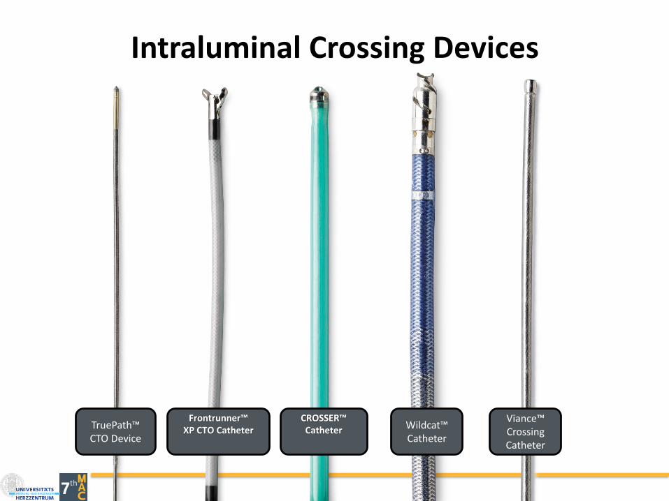
TruePath™ CTO Device
CROSSER™Catheter
Frontrunner™XP CTO Catheter Wildcat™
Catheter
Viance™ Crossing Catheter
Intraluminal Crossing Devices
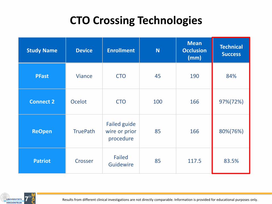
CTO Crossing Technologies
Study Name Device Enrollment NMean
Occlusion (mm)
Technical Success
PFast Viance CTO 45 190 84%
Connect 2 Ocelot CTO 100 166 97%(72%)
ReOpen TruePathFailed guide wire or prior
procedure85 166 80%(76%)
Patriot CrosserFailed
Guidewire85 117.5 83.5%
Results from different clinical investigations are not directly comparable. Information is provided for educational purposes only.
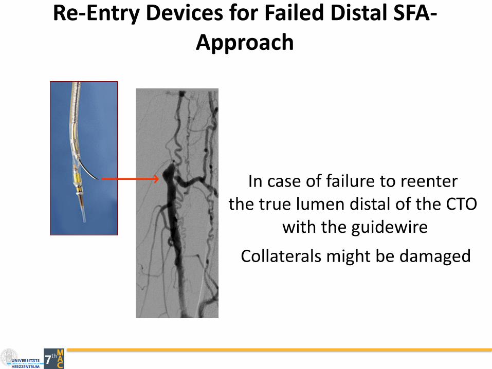
Re-Entry Devices for Failed Distal SFA-Approach
In case of failure to reenterthe true lumen distal of the CTO
with the guidewire
Collaterals might be damaged
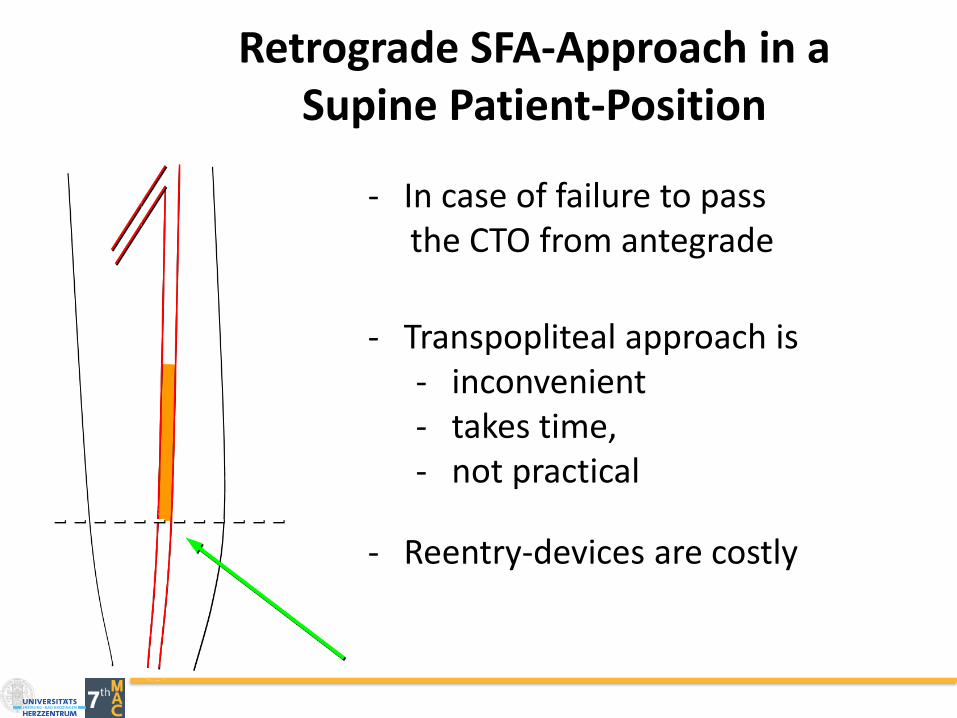
Retrograde SFA-Approach in a Supine Patient-Position
- In case of failure to pass the CTO from antegrade
- Transpopliteal approach is- inconvenient- takes time, - not practical
- Reentry-devices are costly
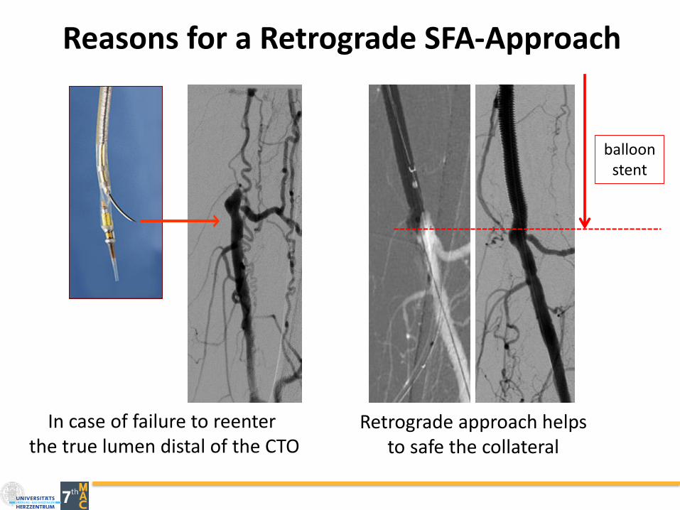
Reasons for a Retrograde SFA-Approach
In case of failure to reenterthe true lumen distal of the CTO
Retrograde approach helpsto safe the collateral
balloonstent
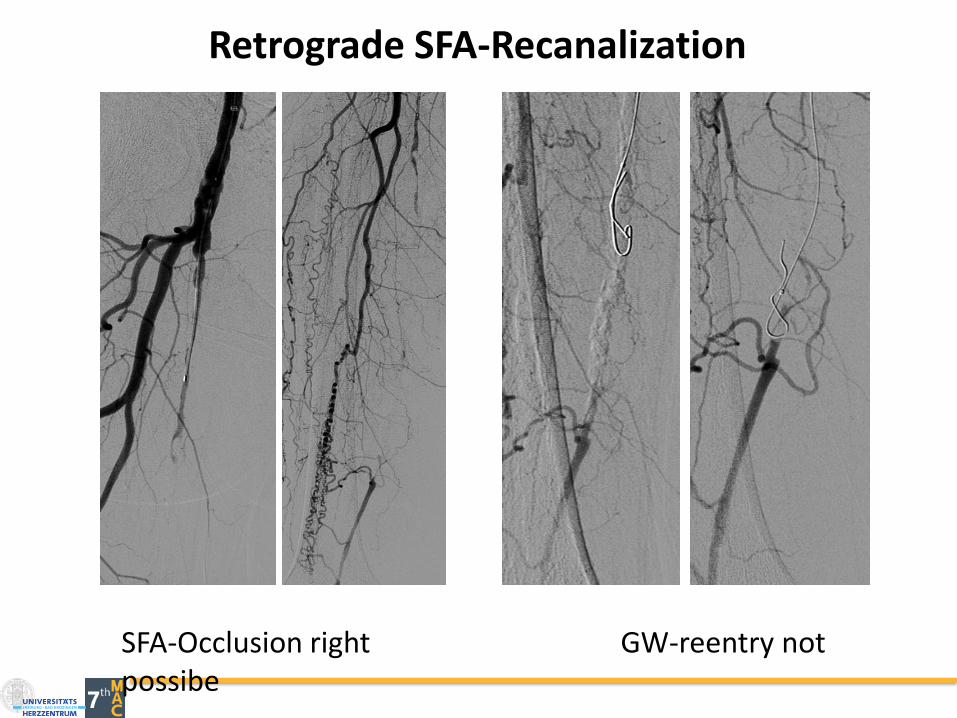
Retrograde SFA-Recanalization
SFA-Occlusion right GW-reentry not possibe
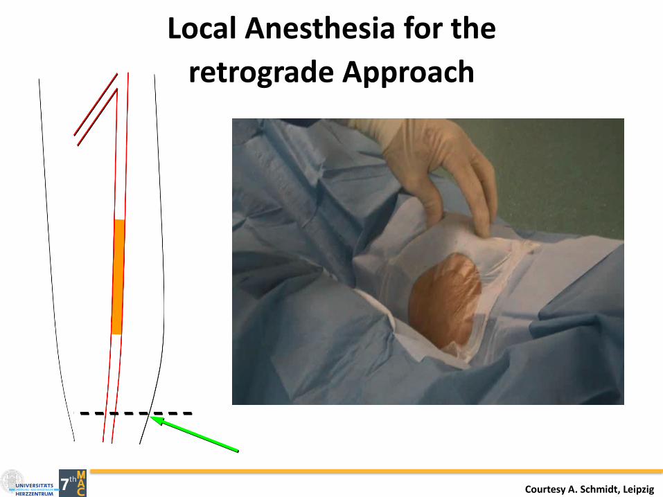
Local Anesthesia for the
retrograde Approach
Courtesy A. Schmidt, Leipzig
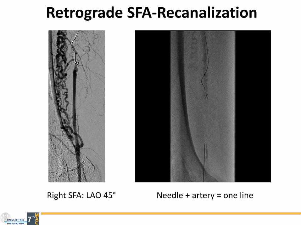
Retrograde SFA-Recanalization
Right SFA: LAO 45° Needle + artery = one line
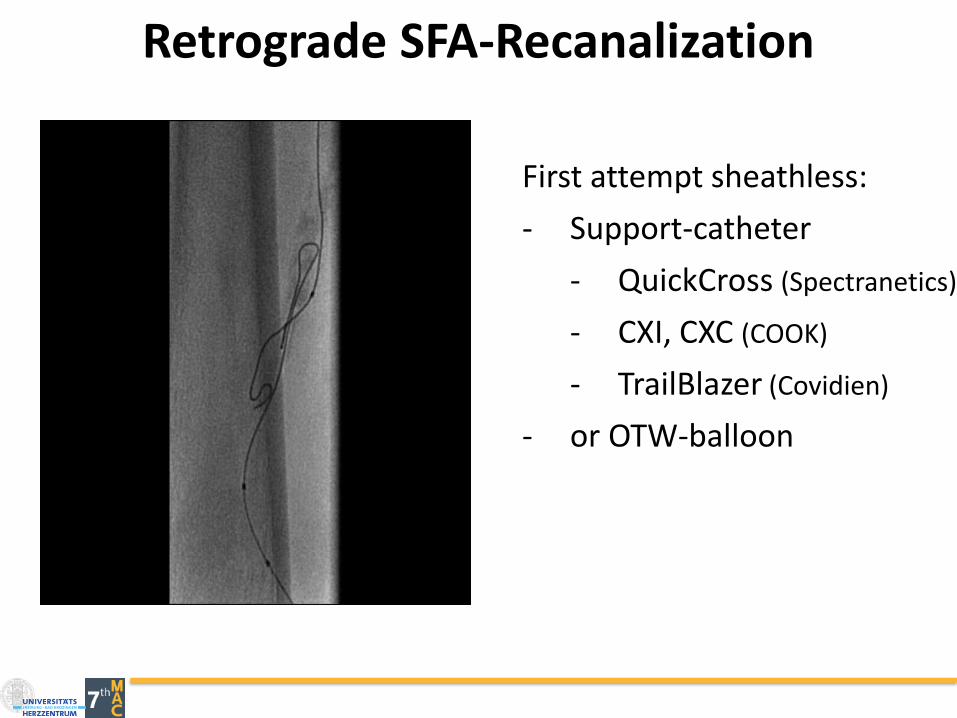
Retrograde SFA-Recanalization
First attempt sheathless:
- Support-catheter
- QuickCross (Spectranetics)
- CXI, CXC (COOK)
- TrailBlazer (Covidien)
- or OTW-balloon
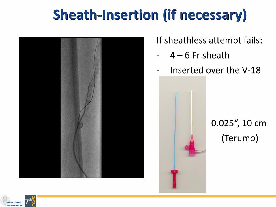
Sheath-Insertion (if necessary)
If sheathless attempt fails:
- 4 – 6 Fr sheath
- Inserted over the V-18
0.025“, 10 cm
(Terumo)

In case GW-Passage fails from retrograde
3. Re-back techniquereentry-device intoballoon
1. Antegrade balloon(CART-technique)
2. Double-balloon
Outback
Courtesy A. Schmidt, Leipzig
Calcium Solutions
12 Month Primary Patency100% 100% 100%
90% 90% 88%
50% 50%
0%
20%
40%
60%
80%
100%
1a 1b 2a 2b 3a 3b 4a 4b
Minimal Calcificationa High Calcification
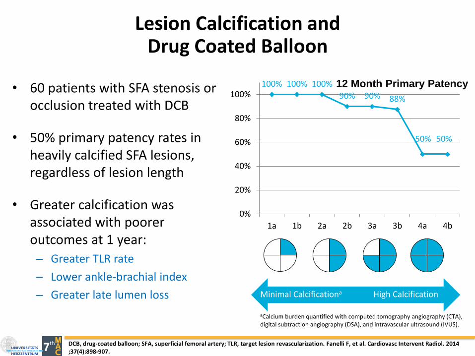
Lesion Calcification andDrug Coated Balloon
• 60 patients with SFA stenosis or occlusion treated with DCB
• 50% primary patency rates in heavily calcified SFA lesions, regardless of lesion length
• Greater calcification was associated with poorer outcomes at 1 year:
– Greater TLR rate
– Lower ankle-brachial index
– Greater late lumen loss
DCB, drug-coated balloon; SFA, superficial femoral artery; TLR, target lesion revascularization. Fanelli F, et al. Cardiovasc Intervent Radiol. 2014 ;37(4):898-907.
aCalcium burden quantified with computed tomography angiography (CTA), digital subtraction angiography (DSA), and intravascular ultrasound (IVUS).
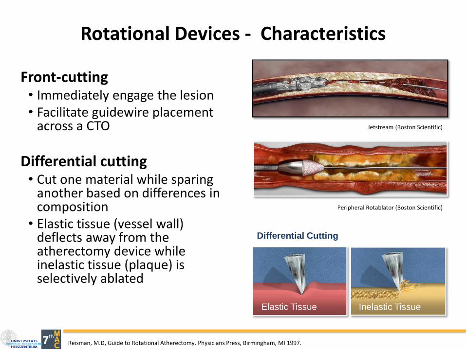
Reisman, M.D, Guide to Rotational Atherectomy. Physicians Press, Birmingham, MI 1997.
Differential Cutting
Elastic Tissue Inelastic Tissue
Peripheral Rotablator (Boston Scientific)
Front-cutting• Immediately engage the lesion• Facilitate guidewire placement
across a CTO
Differential cutting• Cut one material while sparing
another based on differences in composition
• Elastic tissue (vessel wall) deflects away from the atherectomy device while inelastic tissue (plaque) is selectively ablated
Jetstream (Boston Scientific)
Rotational Devices - Characteristics
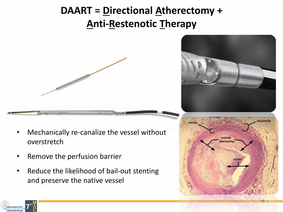
DAART = Directional Atherectomy + Anti-Restenotic Therapy
• Mechanically re-canalize the vessel without overstretch
• Remove the perfusion barrier
• Reduce the likelihood of bail-out stenting and preserve the native vessel
18 |

• Severe calcified lesions that underwent intravascular ultrasound guided DA and Drug Coated Balloon (In-Pact admiral)
• All procedures performed using a distal protection device.
• Patients followed up to 12 mo.
RESULTS:
• Procedural and clinical success, was achieved in all cases.
• Bail-out stenting was necessary in only two (6.5%).
• At 1-year primary patency rate and freedom from TLR rate: 90%
Directional Atherectomy and DCBHeavily Calcified Fempop lesions
Cioppa A, Stabile E, Popusoi G et al. Cardiovasc Revasc Med. 2012 Jul-Aug;13(4):219-23

• DEFINITIVE AR: directional atherectomy + DCB vs DCB alone
Atherectomy and DCB: DEFINITIVE AR
Zeller T et al. Circulation CI 2017.
DCB Ath + DCB
Technical Success* 64.2% 89.6%
Bail-out Stent 3.7% 0%
Flow-limiting Dissection 19% 2%
Procedural Results
86%
63%
90%97%
70%
93%
0%
20%
40%
60%
80%
100%
Lesions >10 cm Severely Calcified All patients
DCB DCB + Ather
n=23 n=8 n=27 n=54 n=48n=31
Duplex Ultrasound Patency at 12-months
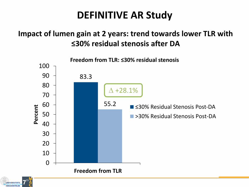
DEFINITIVE AR Study
Impact of lumen gain at 2 years: trend towards lower TLR with ≤30% residual stenosis after DA
83.3
55.2
0
10
20
30
40
50
60
70
80
90
100
Freedom from TLR
Pe
rce
nt ≤30% Residual Stenosis Post-DA
>30% Residual Stenosis Post-DA
Freedom from TLR: ≤30% residual stenosis
+28.1%

Lithoplasty® TechnologyLocalized Lithotripsy to Treat Vascular Calcium
• Designed to normalize vessel wall compliance prior to controlled, low pressure dilatation
• Effective lesion expansion with minimized impact to healthy tissue• Familiar Balloon-based endovascular technique• “Front-line” balloon strategy (.014”compatible)
Sonic Pressure Waves are Tissue-selective:
• Hard on hard tissue, Soft on soft tissue
Waves, unfocused and spherical in shape, travel outside balloon:
• Designed to disrupt both superficial, deep calcium
Lesion modification using lithotripsy in a balloon
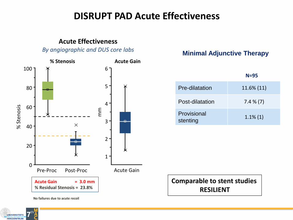
DISRUPT PAD Acute Effectiveness
Pre-dilatation 11.6% (11)
Post-dilatation 7.4 % (7)
Provisional
stenting1.1% (1)
Minimal Adjunctive Therapy
N=95
% Stenosis Acute Gain
Pre-Proc Post-Proc Acute Gain
mm
% S
ten
osi
s
0
20
40
60
80
100
1
2
3
5
6
4
Acute EffectivenessBy angiographic and DUS core labs
Acute Gain = 3.0 mm% Residual Stenosis = 23.8%
No failures due to acute recoil
Comparable to stent studiesRESILIENT
Clinical data on file at Abbott Vascular.
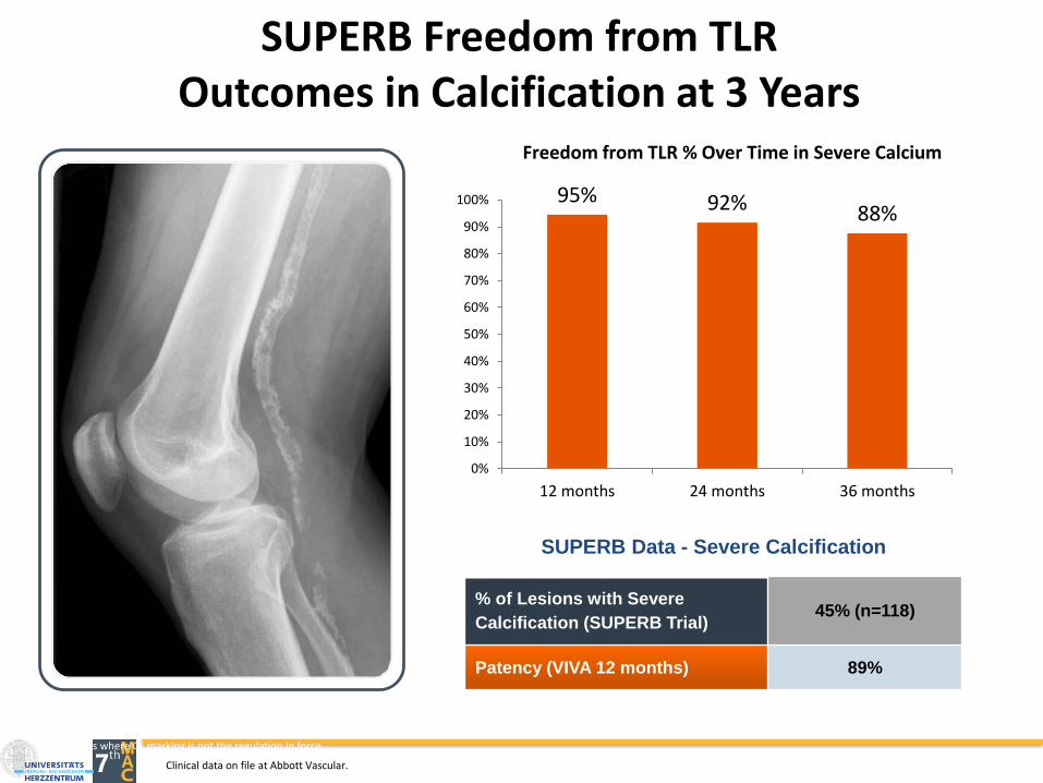
SUPERB Freedom from TLROutcomes in Calcification at 3 Years
SUPERB Data - Severe Calcification
% of Lesions with Severe
Calcification (SUPERB Trial)45% (n=118)
Patency (VIVA 12 months) 89%
95% 92% 88%
0%
10%
20%
30%
40%
50%
60%
70%
80%
90%
100%
12 months 24 months 36 months
Freedom from TLR % Over Time in Severe Calcium
Information contained herein for distribution outside the U.S. only. Check the regulatory status of the device in areas where CE marking is not the regulation in force.
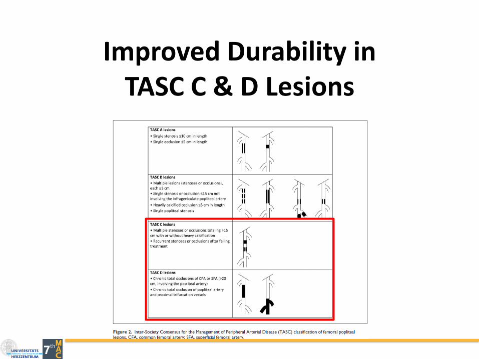
Improved Durability in TASC C & D Lesions
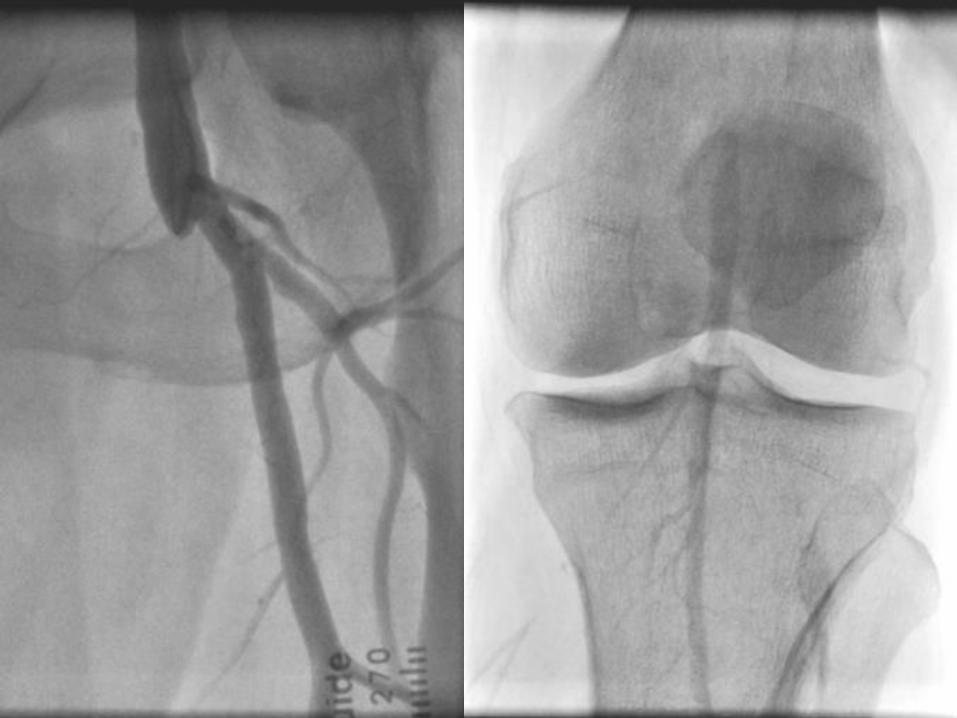
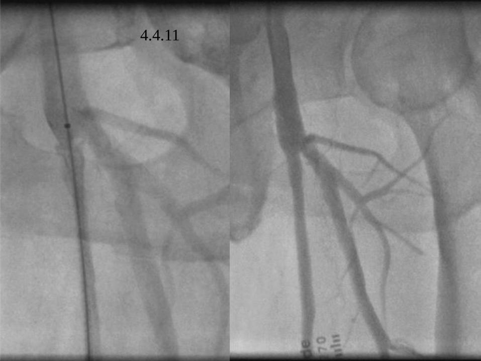
4.4.11
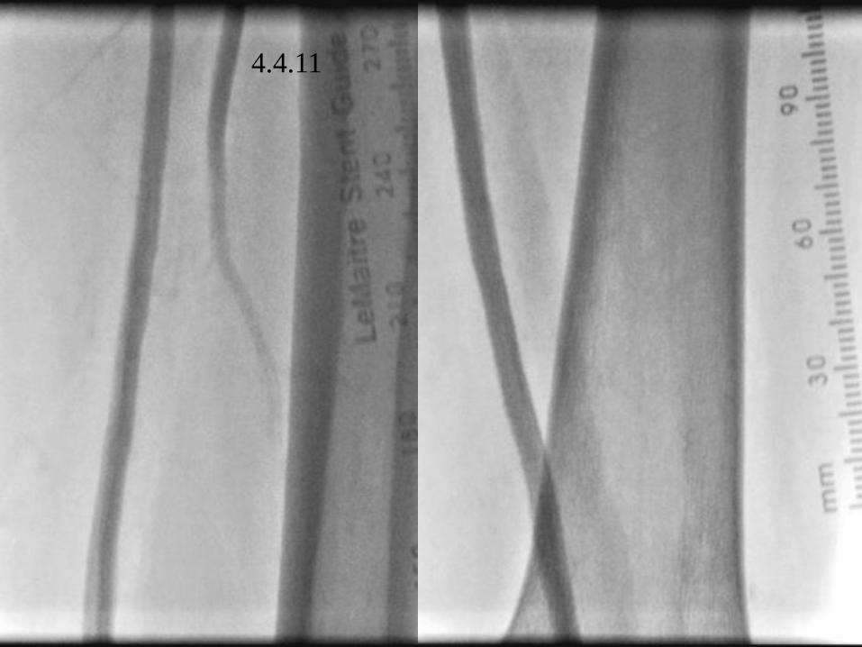
4.4.11
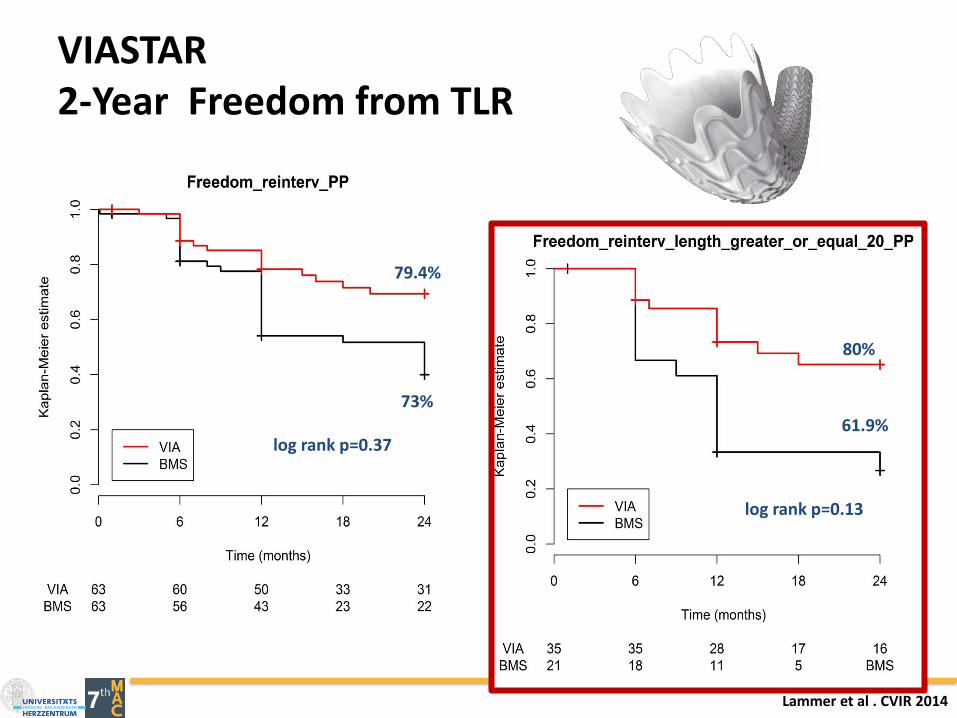
VIASTAR2-Year Freedom from TLR
79.4%
73%
log rank p=0.37
80%
61.9%
log rank p=0.13
Lammer et al . CVIR 2014
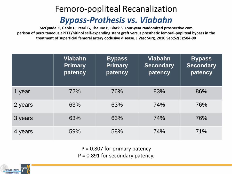
Femoro-popliteal RecanalizationBypass-Prothesis vs. Viabahn
McQuade K, Gable D, Pearl G, Theune B, Black S. Four-year randomized prospective comparison of percutaneous ePTFE/nitinol self-expanding stent graft versus prosthetic femoral-popliteal bypass in the
treatment of superficial femoral artery occlusive disease. J Vasc Surg. 2010 Sep;52(3):584-90
Viabahn
Primary
patency
Bypass
Primary
patency
Viabahn
Secondary
patency
Bypass
Secondary
patency
1 year 72% 76% 83% 86%
2 years 63% 63% 74% 76%
3 years 63% 63% 74% 76%
4 years 59% 58% 74% 71%
P = 0.807 for primary patencyP = 0.891 for secondary patency.
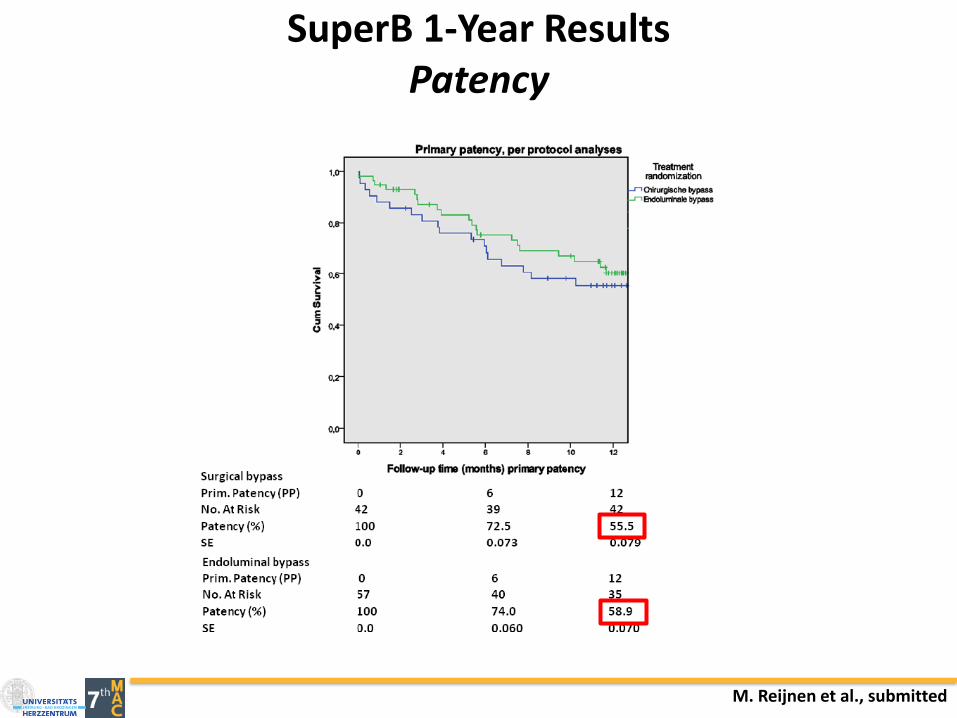
M. Reijnen et al., submitted
SuperB 1-Year ResultsPatency
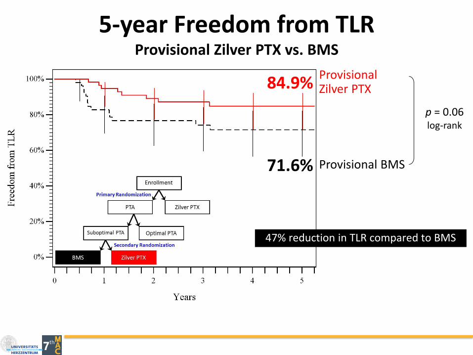
5-year Freedom from TLRProvisional Zilver PTX vs. BMS
Provisional BMS
Provisional Zilver PTX84.9%
71.6%
p = 0.06log-rank
47% reduction in TLR compared to BMS
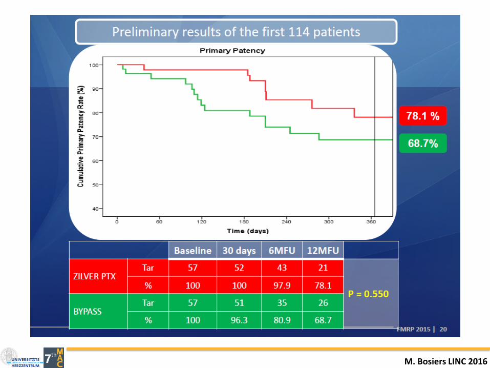
M. Bosiers LINC 2016

M. Bosiers LINC 2016
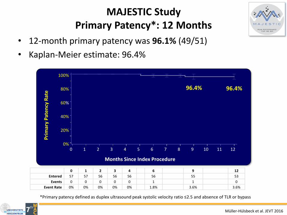
MAJESTIC StudyPrimary Patency*: 12 Months
• 12-month primary patency was 96.1% (49/51)
• Kaplan-Meier estimate: 96.4%P
rim
ary
Pat
en
cy R
ate
0%
20%
40%
60%
80%
100%
Months Since Index Procedure
0 1 2 3 4 5 6 7 8 9 10 11 12
0 1 2 3 4 6 9 12
Entered 57 57 56 56 56 56 55 53
Events 0 0 0 0 0 1 1 0
Event Rate 0% 0% 0% 0% 0% 1.8% 3.6% 3.6%
*Primary patency defined as duplex ultrasound peak systolic velocity ratio ≤2.5 and absence of TLR or bypass
96.4% 96.4%
Müller-Hülsbeck et al. JEVT 2016
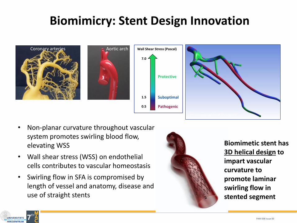
Biomimicry: Stent Design Innovation
• Non-planar curvature throughout vascular system promotes swirling blood flow, elevating WSS
• Wall shear stress (WSS) on endothelial cells contributes to vascular homeostasis
• Swirling flow in SFA is compromised by length of vessel and anatomy, disease and use of straight stents
Protective
Suboptimal
Pathogenic
1.5
0.5
7.0
Wall Shear Stress (Pascal)Coronary arteries Aortic arch
Biomimetic stent has 3D helical design to impart vascular curvature to promote laminar swirling flow in stented segment
PAM 098 Issue 00
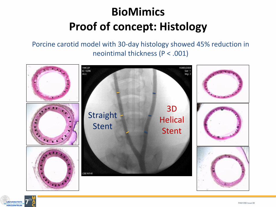
BioMimicsProof of concept: Histology
Porcine carotid model with 30-day histology showed 45% reduction in neointimal thickness (P < .001)
Straight Stent
3D Helical Stent
PAM 098 Issue 00
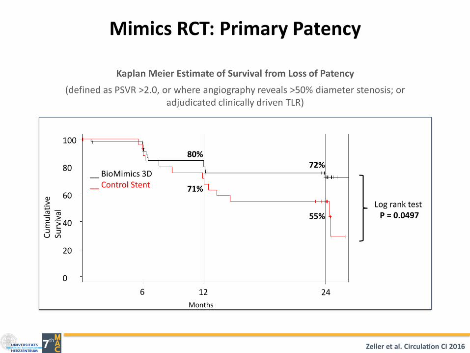
Kaplan Meier Estimate of Survival from Loss of Patency
(defined as PSVR >2.0, or where angiography reveals >50% diameter stenosis; or adjudicated clinically driven TLR)
Mimics RCT: Primary Patency
Log rank test P = 0.0497
80%72%
71%
55%
Months
246 12
0
20
40
60
80
100
Cu
mu
lati
ve
Surv
ival
__ BioMimics 3D__ Control Stent
Zeller et al. Circulation CI 2016
Months
2412 18
0
20
40
60
80
100
Cu
mu
lati
ve S
urv
ival
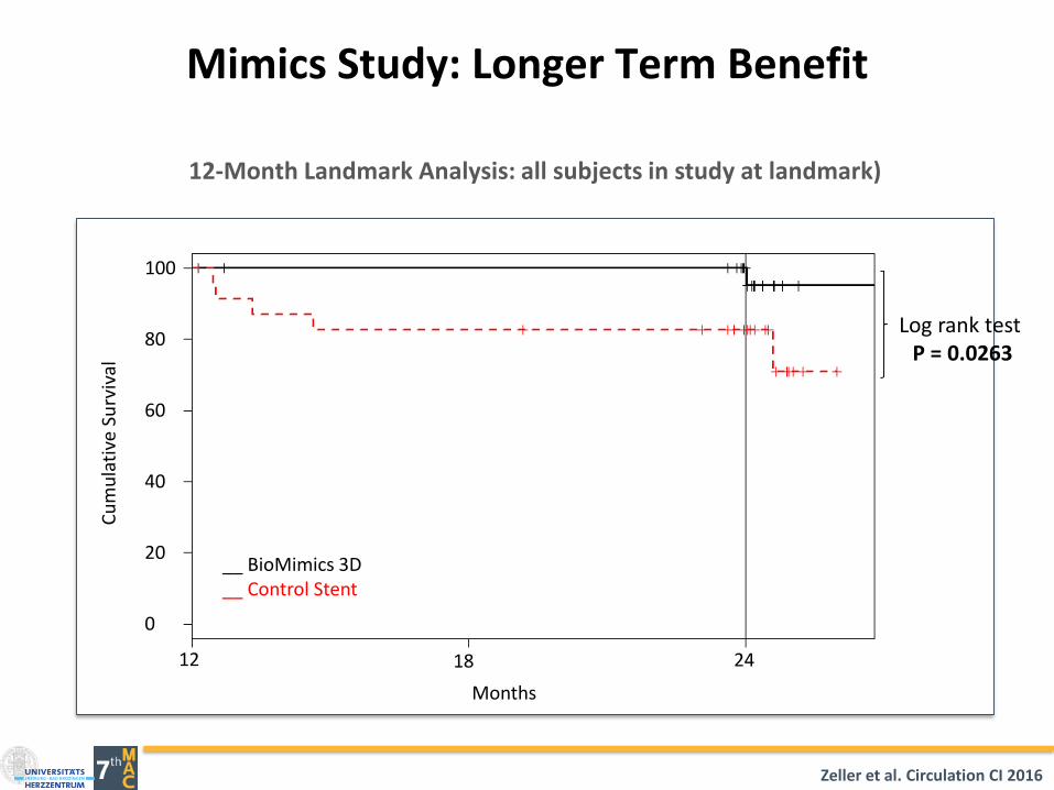
Log rank test P = 0.0263
Kaplan Meier Estimate of Survival from CDTLR
(Clinically-driven TLR determined through Event Adjudication)
Mimics Study: Longer Term Benefit
12-Month Landmark Analysis: all subjects in study at landmark)
__ BioMimics 3D__ Control Stent
Zeller et al. Circulation CI 2016
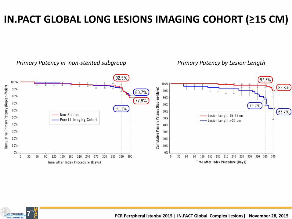
IN.PACT GLOBAL LONG LESIONS IMAGING COHORT (≥15 CM)
40
Primary Patency in non-stented subgroup Primary Patency by Lesion Length
PCR Perrpheral Istanbul2015 | IN.PACT Global Complex Lesions| November 28, 2015

A. Micari. The drug-eluting balloon superficial femoral artery - long study: the DEB SFA-LONG Study. euro PCR 2015
• 105 patients enrolled in the prospective, multicenter DEB-SFA-LONG study, led by Dr.Antonio Micari.
• 105 femoropopliteal lesions (94.6% de novo), lesion length of 251.71 ±78.89 mm.
– CTOs: 49.5%
– Provisional stenting: 10.5%
• Freedom from CD-TLR = 96%
• MAE composite at 12m = 6.9%
• Thrombosis = 1% (1 event)
Italian DEB-SFA-Long studyFirst independent study confirming IN.PACT™ DCB effectiveness as a stand-alone
therapy in long, complex SFA lesions
Primary Patency at 360 days
89.3%
27.6%
61.9%
8.6%1.9%
RCC 2 RCC 3 RCC 4 RCC 5
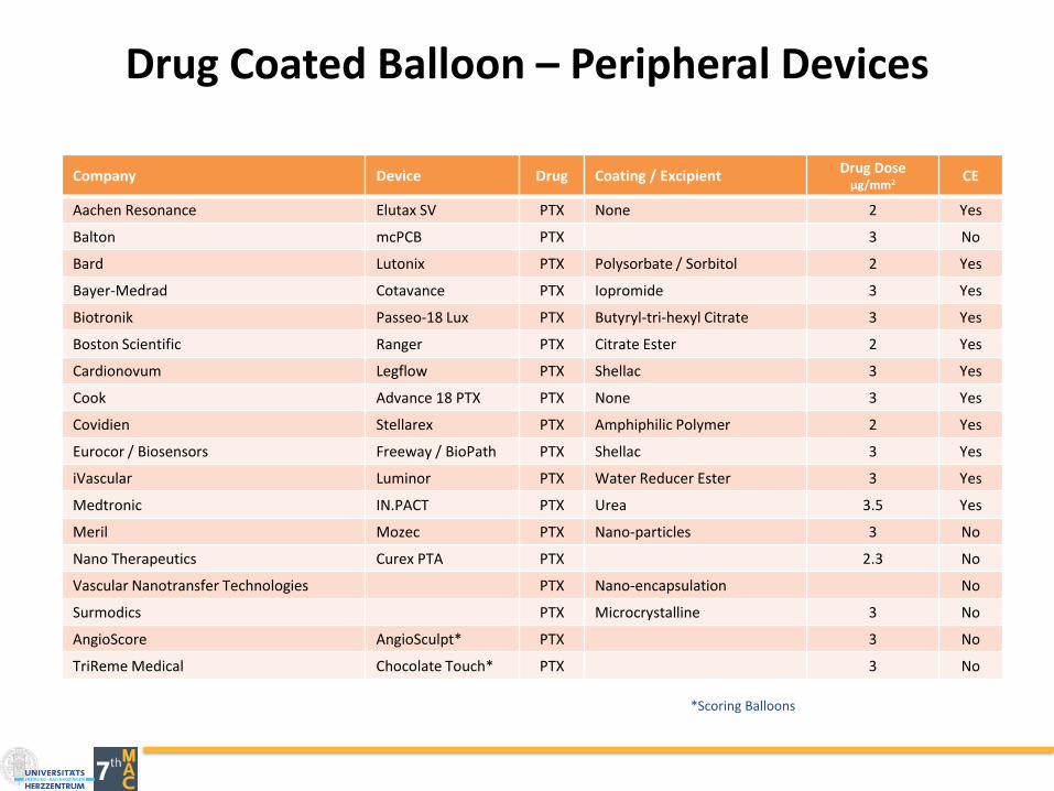
Drug Coated Balloon – Peripheral Devices
Company Device Drug Coating / ExcipientDrug Dose
μg/mm2 CE
Aachen Resonance Elutax SV PTX None 2 Yes
Balton mcPCB PTX 3 No
Bard Lutonix PTX Polysorbate / Sorbitol 2 Yes
Bayer-Medrad Cotavance PTX Iopromide 3 Yes
Biotronik Passeo-18 Lux PTX Butyryl-tri-hexyl Citrate 3 Yes
Boston Scientific Ranger PTX Citrate Ester 2 Yes
Cardionovum Legflow PTX Shellac 3 Yes
Cook Advance 18 PTX PTX None 3 Yes
Covidien Stellarex PTX Amphiphilic Polymer 2 Yes
Eurocor / Biosensors Freeway / BioPath PTX Shellac 3 Yes
iVascular Luminor PTX Water Reducer Ester 3 Yes
Medtronic IN.PACT PTX Urea 3.5 Yes
Meril Mozec PTX Nano-particles 3 No
Nano Therapeutics Curex PTA PTX 2.3 No
Vascular Nanotransfer Technologies PTX Nano-encapsulation No
Surmodics PTX Microcrystalline 3 No
AngioScore AngioSculpt* PTX 3 No
TriReme Medical Chocolate Touch* PTX 3 No
*Scoring Balloons
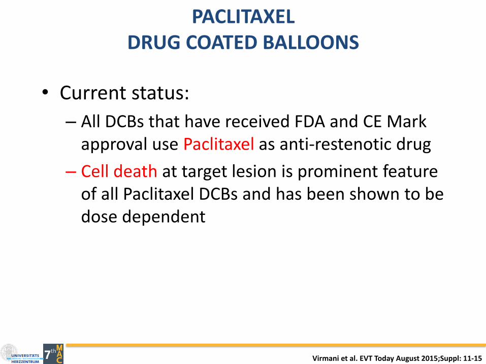
PACLITAXELDRUG COATED BALLOONS
• Current status:
– All DCBs that have received FDA and CE Mark approval use Paclitaxel as anti-restenotic drug
– Cell death at target lesion is prominent feature of all Paclitaxel DCBs and has been shown to be dose dependent
Virmani et al. EVT Today August 2015;Suppl: 11-15

Secondary Safety Outcomes
12- month Safety DEB PTA p
Major Amputation 8.8% (20/227) 3.6% (4/111) 0.080
All-Cause Mortality 10.1% (23/227) 8.1% (9/111) 0.551
Death and Amputations1 35.2% (80/227) 25.2% (28/111) 0.064
Death, Major Amp, CD-TLR2
26.9% (61/227) 23.4% (26/111) 0.496
Amputation Free Survival 81.1% (184/227) 89.2% (99/111) 0.057
Wound Healing (sitereported)
73.8% (121/164) 76.9% (70/91) 0.579
IN.PACT DEEP Trial: Secondary Endpoints
Trend towards higher Major Amputation Rate in DCB Arm
Zeller, T. JACC 2014;64(15):1568-76.
Sirolimus Coated Balloon
Benefits, Challenges & Solution

Paclitaxel vs. SirolimusMargin of Safety
46
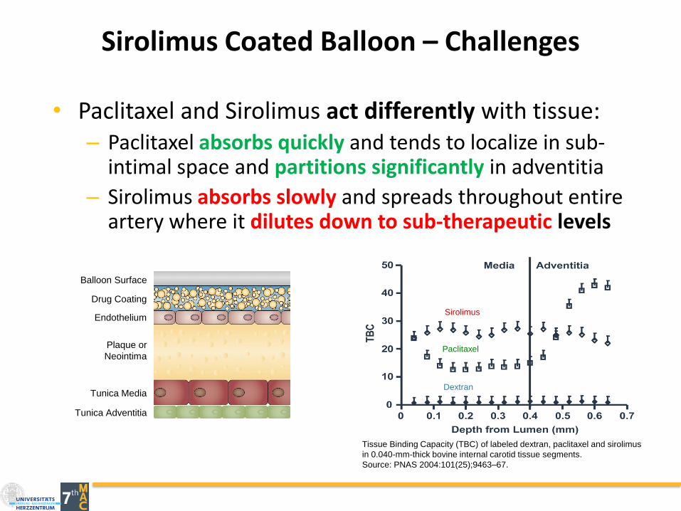
Sirolimus Coated Balloon – Challenges
• Paclitaxel and Sirolimus act differently with tissue: – Paclitaxel absorbs quickly and tends to localize in sub-
intimal space and partitions significantly in adventitia
– Sirolimus absorbs slowly and spreads throughout entire artery where it dilutes down to sub-therapeutic levels
Sirolimus
Paclitaxel
Dextran
Tissue Binding Capacity (TBC) of labeled dextran, paclitaxel and sirolimus
in 0.040-mm-thick bovine internal carotid tissue segments.
Source: PNAS 2004:101(25);9463–67.
Balloon Surface
Drug Coating
Endothelium
Plaque or
Neointima
Tunica Media
Tunica Adventitia

MED ALLIANCESELUTION™ SIROLIMUS DCB
• Micro-reservoirs made out of biodegradable polymer intermixed with Sirolimus:
Controlled and sustained drug release mechanism
Maintains therapeutic effect in tissue over longperiod of time
• Novel Cell Adherent Technology – CAT™:
CAT™ transfer membrane houses and protects micro-reservoirs during balloon insertion, lesion crossing and expansion
CAT™ transfer membrane with embedded micro-reservoirs releases from balloon delivery system and adheres to vessel lumen during short balloon inflation
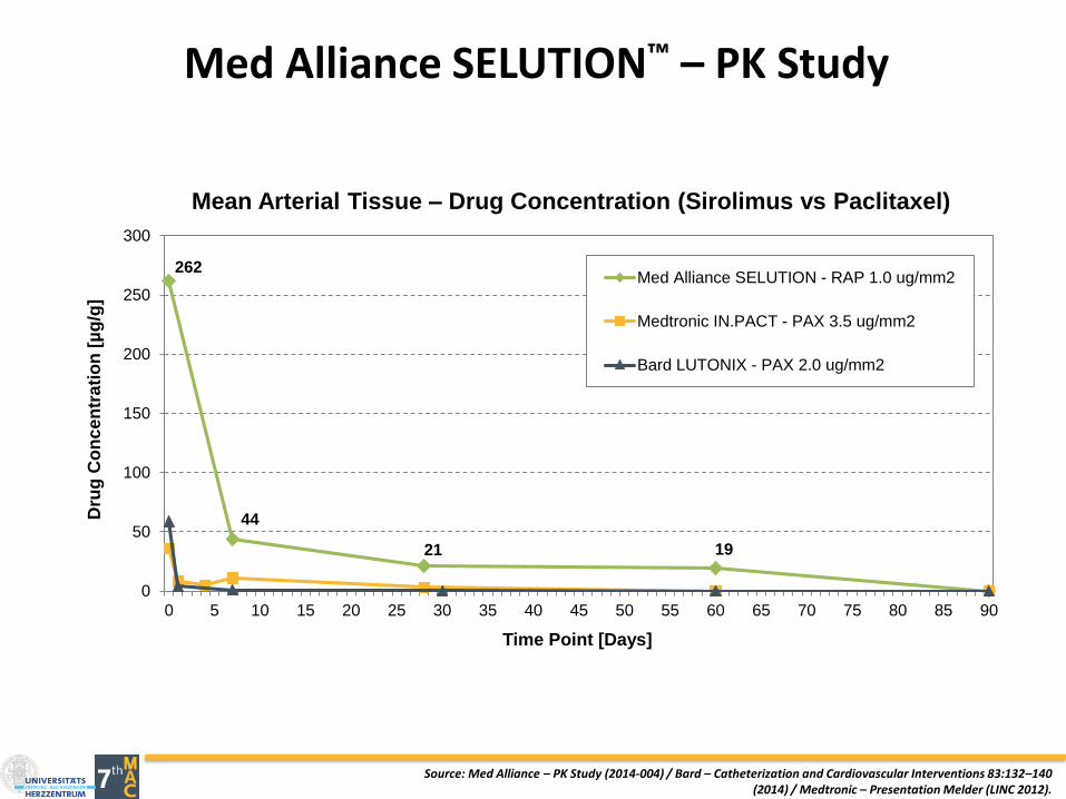
Med Alliance SELUTION™ – PK Study
262
44
21 19
0
50
100
150
200
250
300
0 5 10 15 20 25 30 35 40 45 50 55 60 65 70 75 80 85 90
Dru
g C
on
ce
ntr
ati
on
[µ
g/g
]
Time Point [Days]
Mean Arterial Tissue – Drug Concentration (Sirolimus vs Paclitaxel)
Med Alliance SELUTION - RAP 1.0 ug/mm2
Medtronic IN.PACT - PAX 3.5 ug/mm2
Bard LUTONIX - PAX 2.0 ug/mm2
Source: Med Alliance – PK Study (2014-004) / Bard – Catheterization and Cardiovascular Interventions 83:132–140 (2014) / Medtronic – Presentation Melder (LINC 2012).

SummaryFemoropopliteal Artery-Revascularization
▪ Improved recanalization techniques including retrogradeaccess techniques have increased acute treatment successof femoro-popliteal occlusions to almost 100%.
▪ Even complex lesions such as calcified and long lesions canbe treated with endovascular techniques with comparabledurability as compared to bypass surgery.
▪ Thus, bypass surgery is no longer first line strategy in thetreatment of TASC C & D femoro-popliteal lesions▪ Limited availability of the conduit being considered optimal – the
vein▪ Harvested for coronary bypass grafts▪ Vein stripping or endo-treatment for varicose disease