techniques, clinical applications and limitations of 3d ... · this pictorial essay describes the...
TRANSCRIPT

Korean J Radiol 5(1), March 2004 55
Techniques, Clinical Applications andLimitations of 3D Reconstruction inCT of the Abdomen
Enhanced z-axis coverage with thin overlapping slices in breath-hold acquisi-tions with multidetector CT (MDCT) has considerably enhanced the quality ofmultiplanar 3D reconstruction. This pictorial essay describes the improvements in3D reconstruction and technical aspects of 3D reconstruction and rendering tech-niques available for abdominal imaging. Clinical applications of 3D imaging inabdomen including liver, pancreaticobiliary system, urinary and gastrointestinaltracts and imaging before and after transplantation are discussed. In addition, thisarticle briefly discusses the disadvantages of thin-slice acquisitions includingincreasing numbers of transverse images, which must be reviewed by the radiol-ogist.
tate-of-the-art cross-sectional imaging techniques allow radiologists tovisualize disease with greater certainty by subtracting the impact ofoverlying tissues, thus allowing separate evaluation of individual organs,
which aids in the detection and characterization of pathology.Radiologists discovered in the late 1970s that although diagnosis based on axial CT
images alone was more sophisticated than with plain radiography, the lack of a thirddimension (e.g. sagittal and coronal dimensions) was frequently frustrating. Manyreferring physicians with no basic training in cross-sectional imaging still encounterdifficulties in appreciating normal anatomy and pathology on transverse CT images,being more familiar with anatomy depicted in the coronal plane.
Following the somewhat crude three-dimensional computer rendering algorithmsinitially developed in late 1970s, that allowed formation of images in the thirddimension from data acquired in the axial plane, the development of single-slicehelical CT and more recently multidetector CT (MDCT) scanners has opened newchapters in 3D imaging. These advances were made possible by the rapid acquisitionof volumetric data in the lower case z-axis using thin slices and improved renderingalgorithms, which facilitate exquisite 3D reformats, devoid of degradation by respira-tion and other physiological movements. The pace of progress is being hastened withthe rapid developments in MDCT technology (1). We describe 3D renderingtechniques available for abdominal imaging including multiplanar reformations,surface rendering, virtual endoscopy, volume rendering and maximum intensityprojections. We also discuss clinical applications of 3D imaging in the abdomen includ-ing the liver, pancreaticobiliary system, urinary and gastrointestinal tracts.
This paper also discusses the steps required for introducing 3D reconstruction into aradiology department. The present article also discusses the disadvantage of reducedcollimation MDCT scanning, which results in increasing numbers of images for review.
Michael M. Maher, MDMannudeep K. Kalra, MDDushyant V. Sahani, MDJames J. Perumpillichira, MDStephania Rizzo, MDSanjay Saini, MDPeter R. Mueller, MD
Index terms:Computed TomographyAbdomenMultidetector, 3D
Korean J Radiol 2004;5:55-67Received June 2, 2003; accepted after revision January 26, 2004.
Division of Abdominal Imaging andIntervention, Department of Radiology,Massachusetts General Hospital andHarvard Medical School
Address reprint requests to:Dr. Michael M. Maher, MD, Division ofAbdominal Imaging and Intervention,Department of Radiology, White 270,Massachusetts General Hospital, 55 FruitSt. Boston, MA 02184.Tel. (617) 726 8396Fax. (617) 726 4891e-mail: [email protected]
S

TECHNICAL IMPLEMENTATION OF 3DRECONSTRUCTION
Introduction of 3D ImagingHaving arrived at a point of recognition that 3D imaging
techniques are valuable, the challenge facing an institutionis to develop a practical basis for performing 3D imaging.At our institution, we have set up a 3D imaging laboratoryfor the purpose of developing special expertise that iseasily accessible.
The process of 3D imaging began with image acquisitionprotocols, which were optimized for subsequent post-processing. We have taken the step of identifying certainCT protocols that would always require 3D analysis andmodified these protocols so that 3D imaging is an integralpart of these studies. Referring physicians ordering theseexaminations, now understand that they are simultane-ously requesting additional 3D analysis. However, thereare certain studies in which 3D imaging does not appear
important prior to imaging the patient, but on reviewingaxial images the radiologist may feel that 3D reconstruc-tion may offer a problem solving option. In thesesituations, it is preferable to reconstruct the volumetrichelical data in thinner sections and use this data forappropriate 3D reconstructions.
Based on the standardized protocols, the technologists inthe 3D laboratory apply the appropriate 3D renderingsoftware to create a data set of 3D images that is returnedto the Picture Archiving Communications System (PACS)associated with the source images. Development of closecommunication between physicians and 3D technologistsand close proximity of the 3D laboratory to the CTinterpretation area allows radiologists to participate in 3Dreconstruction with the technologists. In addition to 3Drenderings there are a number of applications, whichrequire quantitative measurements. These include planningprior to aortic stent graft placement, or volumetric analysisprior to liver resection (2) and living related organdonation (3, 4).
Maher et al.
56 Korean J Radiol 5(1), March 2004
Fig. 1. Liver donor evaluation in a 50-year-old man with CT angiog-raphy. Maximum intensity projection image (A) demonstrateshepatic arterial anatomy with left hepatic (LH) artery originatingfrom celiac trunk (arrow) and right hepatic (RH) artery arising fromsuperior mesenteric (SM) artery. 3D surface rendering in the samepatient depicts entire hepatic lobe volume (B) and left lobe volume(C) to determine liver volume in living-related liver donor.
C
A B

Protocol DesignPreparation for 3D CT should begin prior to the arrival
of the patient in the CT scanner. It is important in mostcases, to withhold positive oral and rectal contrast agentsto obtain satisfactory 3D CT images. This is particularlyimportant for CT angiogiography, and CT urography when3D reconstruction is planned. For pancreatic protocolMDCT scanning, water can be used as a negative oralcontrast agent to aid in imaging of the duodenum andampullary region. Optimum bowel cleansing or labeling offecal matter with agents, which facilitate digital post-processing image subtraction is critical for virtualendoscopy reconstruction.
Optimum contrast volume, rate of injection, and timingof the scan at the peak concentration are pre-requisites formost 3D angiography applications. Only good source axial
image data can provide reasonable quality 3D reconstruc-tion. It is therefore critical to obtain thin, overlappingsections of the region of interest with minimum motionartifacts.
3D RENDERING TECHNIQUES AVAILABLE FORABDOMINAL IMAGING
Multiplanar reconstruction (MPR) provides efficientcomputation of images that lie along the non-acquiredorthogonal orientations of the scanned volume by re-addressing the order of voxels in the scanned volume. It isa fast and interactive algorithm that can represent severalarbitrary planes at once and create multiplanar display inreal-time. Generally, it is helpful whenever pathologycannot be accurately assessed on axial plane images alone.
Techniques, Clinical Applications and Limitations of 3D Reconstruction in Abdominal CT
Korean J Radiol 5(1), March 2004 57
Fig. 2. Unexpected finding in a 35-year-old woman who underwent CT evaluation prior to liver donor surgery. Axial (A), multiplanarvolume rendering (B) and maximum intensity projection (C) images show dissection of hepatic artery (arrow) with tortuous, dilated andfeatureless false lumen (arrowheads) without branching, which was not appreciated on conventional angiogram (D). The false lumen ofcommon hepatic artery dissection is seen only on CT angiography. Distal communication of true and false lumens maintains the patencyand perfusion of right and left hepatic arteries.
C D
A B
Surface rendering (shaded surface display, SSD) is asurface-fitting algorithm that creates triangle-based isosur-faces within 3D space. For surface rendering, the userspecifies a threshold attenuation value close to the centerof the signal difference between the brightest pixel foundwithin the object of interest and the signal in the surround-ing structures. Then, the surface-rendering algorithm loopson successive slices and determines whether its cornervalues straddle the threshold value. Non-straddling cellsare discarded, while straddling pixel values are chosen togenerate surface rendering datasets. With the surfacecreated from the data, the remainder of the data isdiscarded.
Although this technique is associated with loss of densityinformation, it can aid in preoperative planning forinterventional endovascular procedures and visualizationof complex anatomic situations such as pathology of thethoracic and abdominal aorta.
Virtual endoscopy (VE) processes scan datasets toprovide simulated visualizations of specific organs similarto those produced by standard endoscopic procedures (5).With increasing availability of MDCT scanners and acquisi-tion of isotropic data, virtual endoscopy can now beperformed using SSD or volume rendering techniques togenerate high-resolution VE images in three-dimensionsand perform “fly-through displays” of the trachea, esopha-gus, and colon, which are comparable to standardendoscopic views (5). In addition, VE can define preciselocation, size, and shape of lesions, both within and outsideof the region of interest, information which is not alwayseasily available with conventional endoscopy. However,despite being comparable to conventional endoscopy, VEdoes not provide information on the “flat lesions” or color
of the mucosa and suffers the obvious disadvantage of notproviding immediate biopsy specimens of lesions seen.
Volume rendering (multiplanar volume rendering,MPVR) is the visualization and manipulation of objectsrepresented as sampled data in three or more dimensions.The technique interpolates the entire data set rather thanediting a single scan to generate 3D images directly fromscanned volume data. Unlike other projection techniquessuch as SSD and MIP, MPVR does not distort objects in thereconstructed planes. It allows “quick view” of largeMDCT scan data sets with comprehensive details of theanatomic orientation of lesion or structures of interest.
The maximum intensity projection (MIP) techniquedisplays the pixels of greatest intensity along a predefined
Maher et al.
58 Korean J Radiol 5(1), March 2004
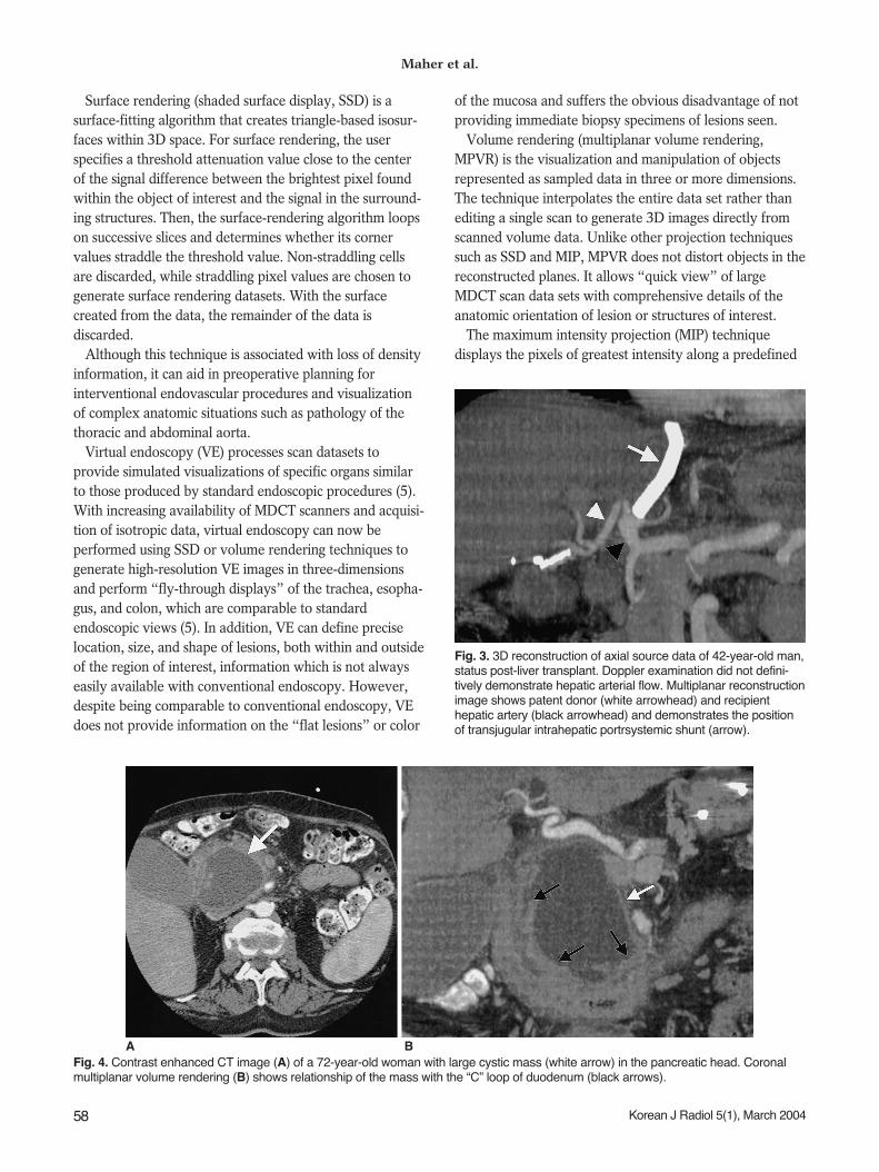
Fig. 4. Contrast enhanced CT image (A) of a 72-year-old woman with large cystic mass (white arrow) in the pancreatic head. Coronalmultiplanar volume rendering (B) shows relationship of the mass with the “C” loop of duodenum (black arrows).
A B
Fig. 3. 3D reconstruction of axial source data of 42-year-old man,status post-liver transplant. Doppler examination did not defini-tively demonstrate hepatic arterial flow. Multiplanar reconstructionimage shows patent donor (white arrowhead) and recipienthepatic artery (black arrowhead) and demonstrates the positionof transjugular intrahepatic portrsystemic shunt (arrow).

axis of the image. It is useful for the depiction of vascularanatomy when there is a large difference between attenua-tion values (Hounsfield value, HU) of vessels opacified bycontrast agent, and the surrounding tissues. MIP is usefulfor all types of CT angiography and has also been used forCT Urography (6, 7).
Minimum intensity projection (MinIP) is the opposite ofMIP technique (7). Whereas, the latter technique is usedfor displaying high attenuation structures, the formerdisplays 3D images of structures with lower attenuationvalues such as the bile ducts, pancreatic duct or theunopacified renal collecting system or ureters. On MinIP,structures such as the hepatic biliary tree and dilatedunopacified ureter are displayed in three-dimensionalplane and the margins of such structures are also muchmore apparent (7).
CLINICAL APPLICATIONS
There are many situations in which 3D imaging simplyprovides a “pretty picture” and does not provideadditional diagnostic information and therefore additionalcost cannot be justified. As experience is gained with 3Dreconstruction, the goal is no longer the acquirement of a“pretty picture” but the production of additional images,which will have additional diagnostic value. There aremany conditions in which the classical radiologic descrip-tion was made on plain radiographs or contrast examina-tions and which are not as easily depicted on transverse CTimages (7). 3D can be helpful in the characterization ofthese conditions. These conditions will be discussed andillustrated with examples.
LiverIncreasingly the use of MDCT with CT angiography is
Techniques, Clinical Applications and Limitations of 3D Reconstruction in Abdominal CT
Korean J Radiol 5(1), March 2004 59
Fig. 6. CT cholangiogram of dilated biliary ductal system in a 47-year-old woman with cholangiocarcinoma at porta hepatis (arrow). AxialCT image (A) shows dilated intrahepatic biliary system with hypodense mass at the porta hepatis. Minimum intensity projection (B)rendering provides a “cholangiographic” view of the dilated intrahepatic ductal system with mass in the porta hepatis region.
A B
Fig. 5. Multiplanar volume renderingimage (A) of axial source data of a 60-year-old man with pancreatic adenocarci-noma (white arrows) encasing the portalvein and distal superior mesenteric vein(black arrows). Intraluminal filling defectsuggestive of a thrombus is seen in thesuperior mesenteric vein. Note theexquisite display of 3D vascular anatomyof superior mesenteric artery (blackarrowheads) in relation with the tumor(B).
A B

replacing conventional catheter angiography for transplantwork-up (3, 4). The routine utilization of 3D reconstruc-tion as part of CT protocols in the evaluation of the recipi-ent and donor prior to living-related transplantation allowscomplete work-up in a single imaging study. Thiseliminates the need for other invasive radiologicalprocedures.
Liver transplantation: All organ recipients with end-stageliver failure are evaluated prior to liver transplantationwith dual phase MDCT of the liver. The acquired imagedata in the arterial and portal venous phases allows state-of-the-art CT angiography, portal and hepatic venographyto be performed (3, 4). Protocols for 3D CT angiographyshould not include oral contrast administration. 3Dreformats can depict pre-operative and post-transplantvascular anatomy and anomalies particularly of the hepaticartery, portal and hepatic veins (Figs. 1 3) (3, 4).
To evaluate living related transplant donors, firstly, theliver parenchyma is assessed for focal liver lesions, and
fatty infiltration, which can contraindicate transplantationand can negatively impact on the quality of the graft andresult in the need for re-transplantation. Next, the arterialand hepatic venous anatomy needs to be thoroughlymapped and important vascular variant anatomy can beidentified which is fundamental to surgical planning (3, 4).3D reconstructions provide accurate information aboutnormal vascular supply and some critical vascularanomalies that may contradict organ donation due toexcessive risk either to the donor or recipient. 3D CTimages can detect and identify all major variations of thehepatic vein confluences, portal vein trifurcations and allhepatic arterial variants. 3D reconstruction delineatesportal and hepatic venous anatomy equally or better thanconventional angiography and can identify the hepaticartery and its branches well enough to consider replacingconventional angiography, thus reducing cost and potentialrisk to the organ donor.
With paint brush technique, 3D reconstruction aids in
Maher et al.
60 Korean J Radiol 5(1), March 2004
Fig. 8. Virtual endoscopy of pancreaticduct in a 62-year-old woman.Curved multiplanar reconstruction imageshows a small hypodense mass in thepancreatic head (arrow) with dilatedpancreatic duct (A). Virtual endoscopy(B) of pancreatic duct shows the attenu-ated lumen of the pancreatic duct due toextrinsic compression from periampullarycarcinoma.
A B
Fig. 7. Double duct sign on 3D CT in a 65-year-old man with biopsy proven ampullary carcinoma (white arrowheads). Axial CT image (A)shows dilated common bile duct (black arrow) and pancreatic duct (white arrow). Curved multiplanar reconstruction (B) images showsmooth dilatation of pancreatic duct (white arrow) and common bile duct (black arrow).
A B
estimation of liver volumes at either side of the proposedresection line that is crucial for determining whetherremaining liver volume is sufficient to satisfy the metabolicneeds of the donor (1). The volume of liver resected isimportant, as the proposed graft needs to be sufficient tomaintain the needs of the recipient (3). A potential limita-tion of MDCT and 3D reconstruction, is their inability toprovide a surgical roadmap of biliary anatomy andanomalous anatomy in both the donor and recipient.Failure to appreciate anomalous biliary anatomy can leadto graft failure and post-procedure bile leakage.Intraoperative cholangiography is currently advised todelineate biliary anatomy. Recently, MRCP and in particu-lar, mangafodipir-enhanced T1 MRCP has been reportedas a non-invasive means of evaluating biliary anatomyprior to transplantation (3).
Hepatic Resection: Prior to liver resection, all patientsare evaluated with dual phase MDCT of the liver andimage data are acquired in the arterial and portal venous
phases which allows state-of-the-art CT angiography,portal and hepatic venography to be performed (3). Thisassesses the exact location of the liver tumor and itsanatomic relationship to hepatic vasculature.
Virtual hepatectomy with 3D images and liver volumeestimation before surgery is useful in planning the extentand nature of hepatic resection prior to liver resection.Estimation of liver volume using 3D techniques, whencombined with clinical and laboratory evaluation of liverfunction, can facilitate the prediction of postoperative liverfailure in patients undergoing resection, assist in volume-enhancing embolization procedures and the planning ofstaged hepatic resection for bilobar disease. Thus, 3D-reconstruction and pre-operative virtual hepatic resectionhas an impact on surgical planning and has obviated theneed for conventional angiography in many centers.
Pancreaticobiliary ImagingThe significant improvements in 3D CT angiography as
Techniques, Clinical Applications and Limitations of 3D Reconstruction in Abdominal CT
Korean J Radiol 5(1), March 2004 61
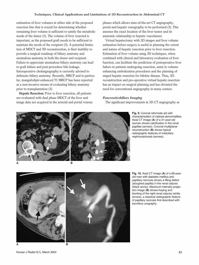
Fig. 10. Axial CT image (A) of a 68-year-old man with diabetes mellitus andpapillary necrosis shows a filling defect(sloughed papilla) in the renal calyces(black arrow). Maximum intensity projec-tion image (B) shows fraying andblunting of the right renal calyces (whitearrows), a classical radiographic featureof papillary necrosis first described withexcretory urography.
A B
Fig. 9. Coronal reformats aid withcharacterization of caliceal abnormalities.Axial CT image (A) of a 31-year-oldwoman shows calcification in the renalpapillae (arrows). Coronal multiplanarreconstruction (B) shows typicalradiographic features of medullarynephrocalcinosis (arrows).
A B
an adjunct to axial images have completely eliminated theneed for catheter angiography in the diagnosis, staging androad mapping of peripancreatic vascular anatomy prior tosurgery in many centers. Important factors in preoperativeevaluation of patients with pancreatic cancer which can beaddressed by 3D CT angiography include definition ofperipancreatic vascular anatomy and its relationship to thepancreatic tumor as well as local invasion of structuressuch as duodenum, liver or tranverse colon (Fig. 4). Theseimportant factors can now be evaluated in a single pancre-atic protocol CT scan. 3D reconstruction is now a routinestandard component of most pancreatic protocol CT scans(8).
Encasement of the superior mesenteric artery and/orvein renders the pancreatic tumor surgically unresectable.3D reconstructions now provide exquisite anatomic detailof vascular structures in vicinity of focal pancreatic lesions(Fig. 5). 3D CT angiography has advantages over catheterangiography in its ability to illustrate anatomic structuresoutside the vascular lumen and therefore the relationshipof the vessels to the tumor can be more clearly illustrated.
3D Reconstructions can aid in answering severalpertinent questions regarding resectability of a malignantpancreatic mass. Axial source images depict the involve-ment of superior mesenteric vein, but sagittal reformats arebest for demonstrating superior mesenteric artery involve-ment. Coronal reformats help in demonstrating localextension to stomach and duodenum, which can aid insurgical planning and has major implications in planningthe appropriate palliative treatment for patients withunresectable pancreatic tumors (8).
In addition, for detection of small pancreatic tumors, 3Dreconstruction can aid in determining the level of dilated
pancreatic and biliary ducts which is fundamental tolocalization of small or subtle pancreatic tumors, if viewedin conjunction with axial source images (9). Curved MPRcan clearly demonstrate the dilated main pancreatic duct inpatients with chronic pancreatitis, pancreatic cancer andmucin-producing pancreatic tumors (9, 10). Multiplanar CTpancreatography and distal cholangiography improvesdepiction of the pancreatic duct and bile ducts with imagequality, which approaches that of ERCP. Park et al. (10)have reported the value of 3D CT cholangiography usingMinIP projection to determine the level and cause ofbiliary obstruction (Figs. 6 8).
Maher et al.
62 Korean J Radiol 5(1), March 2004
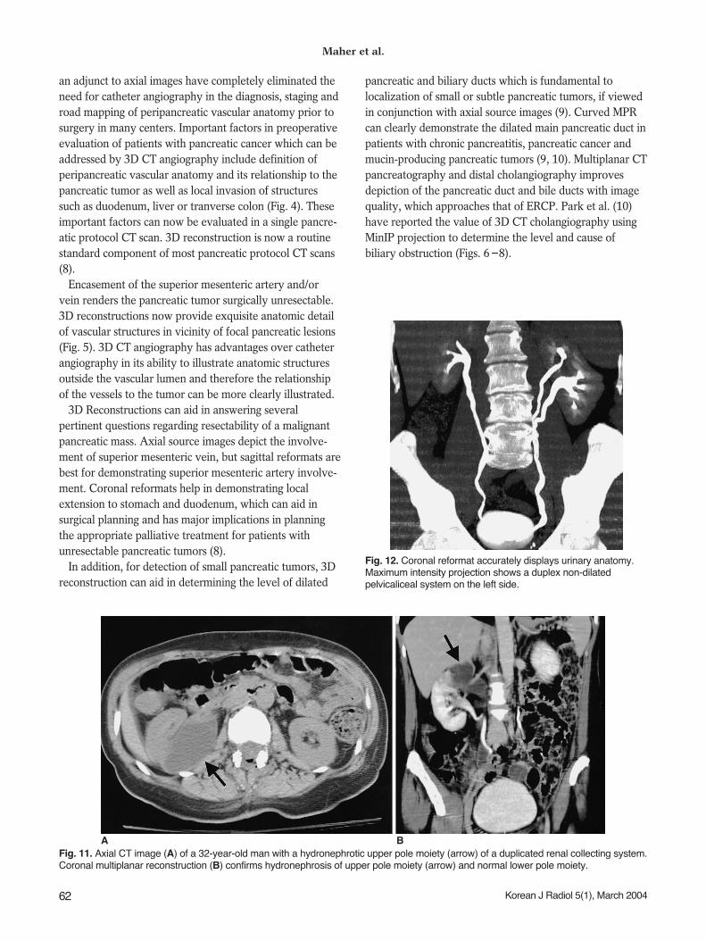
Fig. 12. Coronal reformat accurately displays urinary anatomy.Maximum intensity projection shows a duplex non-dilatedpelvicaliceal system on the left side.
Fig. 11. Axial CT image (A) of a 32-year-old man with a hydronephrotic upper pole moiety (arrow) of a duplicated renal collecting system.Coronal multiplanar reconstruction (B) confirms hydronephrosis of upper pole moiety (arrow) and normal lower pole moiety.
A B

Urinary Tract3D reconstruction has aided in convincing urologists of
the advantages of CT urography over excretory urographyin the evaluation of urinary tract pathology, as it allowedurologists to view images in the coronal plane, which weresimilar to excretory urography images (6). In addition, forradiologists, experience in the characterization of certainpathologies particularly those of the renal calices andpapillae, such as renal tubular ectasia and papillarynecrosis had been gained by evaluation of excretoryurography images (Figs. 9, 10) (6). 3D reconstructionsperformed in the coronal plane closely resemble excretoryurography images and review of these reconstructedimages is very useful for characterizing these conditions.Indeed, Caoili et al. (6) have reported that 3D CT imagesare useful to radiologists and urologists as a “bridge”
between excretory urography data and transverse CT data.3D reconstruction also aids in evaluation of patients withanatomic variation of the urinary tract, contour abnormal-ity of the renal outline and in confirmation of the site ofureteric obstruction (Figs. 11 13) (11). However, it shouldbe emphasized that many 3D reformations suffer the samedisadvantages as excretory urography if the transverseimages are not reviewed in association. Many ureteral andbladder wall abnormalities are frequently detected ontransverse images and can be missed on 3D reformats (6).
In management of renal masses, laparoscopic nephrec-tomy and partial nephrectomy are frequently technicallymore demanding procedures than conventional nephrec-tomy especially when performed using a retroperitonealapproach (12). These procedures are more difficult becausevisualization of renal hilar vascular anatomy is much more
Techniques, Clinical Applications and Limitations of 3D Reconstruction in Abdominal CT
Korean J Radiol 5(1), March 2004 63
Fig. 13. 3D CT can combine advantages of intravenous urography and voiding cystourethrogram and retrograde urethrogrogram. AxialCT image (A) in 47-year-old man with recurrent urinary tract infection shows a dilated, ectopic left ureter (arrow) within the prostate.Sagittal multiplanar reconstruction (B) shows a dilated, ectopic ureter (arrow) opening into the posterior urethra. 3D surface rendering (C)demonstrates anatomic relationship of the ectopic ureter to the regional anatomy.
A B C
Fig. 14. Virtual cystoscopy is useful in evaluation of bladder tumors.Axial (A) and virtual cystoscopy (B) images in a 67-year-old man demonstrate bladder wall thickening and irregularity (arrow). Virtualcystoscopy depicts the surface of the bladder mucosa and shows the size and site of the bladder neoplasm (arrow).
A B

difficult intraoperatively than with conventional opennephrectomy. In addition, with retroperitoneal laparo-scopic surgery, the plane in which the vessels are viewed isdifferent (dorsocaudal) than with conventional surgery(12). Urologists have reported the important role of 3D CTangiography, which aids in planning laparoscopic nephrec-tomy, particularly in the detection of accessory renalarteries and veins. 3D CT angiography allows analysis ofthe extent of the renal mass and its relationship to renalvasculature and hilar structures in a variety of planes,which is fundamental to preoperative surgical planning.
Virtual CT cystoscopy evolved with CT colonography asa means of evaluating the bladder mucosa although it ismuch less widely utilized in clinical practice (Fig. 14). Theadditional information acquired at virtual CT cystoscopy
can potentially aid in the planning of cytoscopy andcystoscopic resection of bladder tumors (13, 14). Forpolypoidal tumors, reports in the literature have suggestedthat virtual endoscopy of urinary bladder has equivalentaccuracy to conventional cystoscopy, and may be veryuseful in the follow-up of patients following cytoscopicresection or other local treatments of bladder tumors thusreducing the costs and morbidity associated with conven-tional cytoscopy (13, 14). Virtual cystoscopy is particularlyadvantageous to patients in whom conventionalcystoscopy is impossible due to urethral stricture.
Gastrointestinal TractThe utilization of 3D reconstruction is reserved for
problem solving in gastrointestinal pathology. Coronal,
Maher et al.
64 Korean J Radiol 5(1), March 2004
Fig. 16. Screening CT colonoscopy was performed in a 55-year-old man. Coronal reformat (A) and virtual colonoscopy (B) imagesdemonstrate a polyp (arrow) in the ascending colon. 3D reconstruction in CT colonoscopy also helps in differentiating normal mucosalfolds from intraluminal masses.
A B
Fig. 15. 3D CT of malignant colonic stricture in a 73-year-old man. Axial CT (A) and coronal multiplanar reconstruction (B) images showcircumferential short segment thickening of the sigmoid colon (arrow) suggestive of colon cancer. 3D CT colonography (C) as a “double-contrast barium enema” simulating image, reveals short segment “apple-core” lesion (arrow) in the sigmoid colon.
A B C
sagittal and oblique reconstructions can aid in identifyingprecise anatomic location of stomach, small bowel andcolonic lesions (Fig. 15).
CT colonoscopy: CT colonoscopy utilizes an array ofmultiplanar and endoluminal reconstructions to permitdetailed evaluation of the entire large bowel (Fig. 16) (5).Data from our institution and others have demonstratedsatisfactory sensitivity and specificity for detection ofpolyps greater than 1 cm, coincidentally, the lesions thatare most likely to harbor malignancy (Fig. 16). Oneindisputable advantage of CT colonography has been inthe completion of conventional colonography examina-tions when the endoscopist cannot reach the cecum.
Colonic Stricture: The characterization of a colonicstricture can frequently be more thoroughly performed by3D reconstruction of the area of narrowing. The stricturecan then be examined in various planes for the features ofmalignant stricture initially described on single and doublecontrast barium enema. These imaging findings includemucosal destruction, ‘shouldering’ at the site of stricture,
and length of the stricture can be more easilydemonstrated with the aid of 3D reconstructions. We havefound that 3D reconstruction can be useful in the diagnosisand confirmation of the presence of intussuseption (Fig.17).
Bowel Ischemia: In the clinical setting of ischemic bowel,a CT angiogram can aid in thorough diagnostic evaluation.The use of dual phase acquisition CT scan coupled with 3Drendering allows the creation of SMA and SMVangiograms and can detect thrombosis or stenosis of thesevessels (15). This data can be combined with enhancementcharacteristics of bowel to further question the possibilityof acute or chronic bowel ischemia.
Inflammatory Bowel Disease: CT angiography maps ofsmall and large bowel have been reported as having apotential role in the assessment of activity in inflammatorybowel disease. Enlarged distal branch vessels andhyperemia have been reported to be associated withincreased activity (15) (Fig. 18).
Techniques, Clinical Applications and Limitations of 3D Reconstruction in Abdominal CT
Korean J Radiol 5(1), March 2004 65
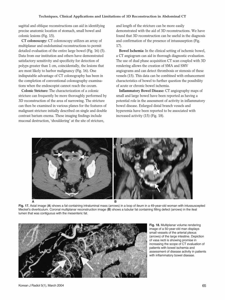
Fig. 17. Axial image (A) shows a fat containing intraluminal mass (arrows) in a loop of ileum in a 49-year-old woman with intussusceptedMeckel’s diverticulum. Coronal multiplanar reconstruction image (B) shows a tubular fat containing filling defect (arrows) in the ileallumen that was contiguous with the mesenteric fat.
A B
Fig. 18. Multiplanar volume renderingimage of a 50-year-old man displayssmall vessels of the arterial plexus(arrows) of the large intestine. Depictionof vasa recti is showing promise inincreasing the scope of CT evaluation ofpatients with bowel ischemia andassessment of disease activity in patientswith inflammatory bowel disease.

16 SLICE MDCT AND BEYOND
The latest development of MDCT is the 16 slice CTscanner. MDCT can acquire sub-millimeter slices with thepromise of equal image quality in the Z-axis (Fig. 19). Themajor impact of this rapidly evolving technology will bethe refinement and optimization of 3D reformation withelimination of common artifacts such as “stair step”artifacts.
PROBLEMS ASSOCIATED WITH MDCT
From a practical standpoint, developments in MDCTscanning allow multiple thin slices to be acquired withincreased z-axis coverage in a single breath-hold, whichgenerates an extraordinary increase in the quantity ofacquired data. Review of an enormous number of imagesposes significant constraints on radiologists’ efficiency andmay be simply impractical. Furthermore, the sheer numberof images raises additional problems and expense for thosedepartments, which have not yet converted to PACS andstill use film for reading.
Rapid 3D reconstructions directly from the scannerconsole are currently being promoted by industry as apotential solution to these difficulties. This revolutionarythinking may alter the current practice of always reviewingaxial ‘source’ images to using 3D reconstructions forprimary interpretation. This would result in fewer imagesfor review in most cases. However this approach will needthorough investigation and comparison to conventional CTpractices before it can be widely embraced.
CONCLUSION
In certain circumstances, 3D reformatted images candemonstrate multiple abnormalities in a simplereconstructed image, which is ideal for consultation withreferring physicians. A potential disadvantage of sophisti-cated 3D rendering techniques includes necessity ofspecialized personnel to generate quality images forevaluating specific clinical or imaging issues. However,when source images are equivocal, 3D reconstructionsoffer a problem-solving option by providing exquisitedepiction of anatomy and pathology in multiple planes ofchoice.
References1. Hu H, He HD, Foley WD, Fox SH. Four multidetector -row
helical CT: image quality and volume coverage speed.Radiology 2000;215:55-62
2. Wigmore SJ, Redhead DN, Yan XJ, et al. Virtual hepaticresection using three-dimensional reconstruction of helicalcomputed tomography angioportograms. Ann Surg2001;233:221-226
3. Kamel IR, Kruskal JB, Keogan MT, Goldberg SN, WarmbrandG, Raptopoulos V. Multidetector CT of potential right-lobe liverdonors. AJR Am J Roentgenol 2001;177:645-651
4. Bogetti JD, Herts BR, Sands MJ, Carroll JF, Vogt DP,Henderson JM. Accuracy and utility of 3-dimensional computedtomography in evaluating donors for adult living related livertransplants. Liver Transpl 2001;7:687-692
5. Rubin GD, Beaulieu CF, Argiro V, et al. Perspective volumerendering of CT and MR images: applications for endoscopicimaging. Radiology 1996;199:321-323
6. Caoili EM, Cohan RH, Korobkin M, et al. Urinary tractabnormalities: initial experience with multi-detector row CT
Maher et al.
66 Korean J Radiol 5(1), March 2004
Fig. 19. 3D reconstruction with 16-slice CT scanner. Coronal multiplanar volume rendering (A) and minimum intensity projection (B)images reconstructed from sub-millimeter images (0.625 mm) of a 60-year-old woman show right renal mass. Note the exquisitedemonstration of relation of the mass with right renal artery and an accessory renal artery (arrow) (General Medical Systems, WaukeshaWis, U.S.A.).
A B

urography. Radiology 2002;222:353-3607. Cody DD. AAPM/RSNA physics tutorial for residents: topics in
CT. Image processing in CT. RadioGraphics 2002;22:1255-12688. O’Malley ME, Boland GW, Wood BJ, Fernandez-del Castillo C,
Warshaw AL, Mueller PR. Adenocarcinoma of the head of thepancreas: determination of surgical unresectability with thin-section pancreatic-phase helical CT. AJR Am J Roentgenol 1999;173:1513-1518
9. Takeshita K, Furui S, Yamauchi T, et al. Minimum intensityprojection image and curved reformation image of the mainpancreatic duct obtained by helical CT in patients with mainpancreatic duct dilation. Nippon Igaku Hoshasen Gakkai Zasshi1999;59: 146-148
10. Park SJ, Han JK, Kim TK, Choi BI. Three-dimensional spiral CTcholangiography with minimum intensity projection in patientswith suspected obstructive biliary disease: comparison with
percutaneous transhepatic cholangiography. Abdom Imaging2001; 26:281-286
11. Schreyer HH, Uggowitzer MM, Ruppert-Kohlmayr A. HelicalCT of the urinary organs. Eur Radiol 2002;12:575-591
12. Marukawa K, Horiguchi J, Shigeta M, Nakamoto T, Usui T, ItoK. Three dimensional navigator for retroperitoneal laparoscopicnephrectomy using multidetector row computerized tomogra-phy. J Urol 2002;168:1933-1936
13. Song JH, Francis IR, Platt JF, et al. Bladder tumor detection atvirtual cystoscopy. Radiology 2001;218:95-100
14. Kim JK, Ahn JH, Park T, Ahn HJ, Kim CS, Cho KS. Virtualcystoscopy of the contrast material-filled bladder in patientswith gross hematuria. AJR Am J Roentgenol 2002;179:763-768
15. Horton KM, Fishman EK. Volume-rendered 3D CT of themesenteric vasculature: normal anatomy, anatomic variants, andpathologic conditions. RadioGraphics 2002;22:161-172
Techniques, Clinical Applications and Limitations of 3D Reconstruction in Abdominal CT
Korean J Radiol 5(1), March 2004 67