technical and personnel requirements - bilder.buecher.de · involved in performing cardiac ct,...
TRANSCRIPT

Technical and Personnel RequirementsM. Dewey
2.1 Technical Requirements ........................... 3
2.2 Purchasing a Scanner ............................... 3
2.3 Personnel Requirements .......................... 5
2.3.1 Guidelines of the ACR ............................... 7
2.3.2 Guidelines of the ACC ............................... 8
Recommended Reading ........................... 9
Abstract
In this chapter, w e summarize the requirements for setting up a coronary CT angiography practice.
2.1 Technical Requirements
Noninvasive coronary angiography is a n as cending c lini-cal application that requires very high spatial and temporal resolution. Thus, CT scanners with multiple detector rows (multislice CT [MSCT]), sho rt gantry rotation times, a nd thin-slice collimation are essential for establishing a success-ful cardiac CT imaging center. Because 64-slice CT is supe-rior to 16-slice CT in terms of image quality and diagnostic accuracy, we believe that (at least) 64-slice technology is nec-essary f or p erforming noninvasive c oronary a ngiography (List 2.1). CT wi th 64-slice t echnology not only increases the quality of the images (Figs. 2.1–2.3) but also improves the workflow because scanning and breath-hold times are shorter (Table 2.1). The shorter breath-hold time is also very relevant for patients after coronary bypass grafting, for w hom 64-slice CT r educes the breath-hold time t o about 15 s ( Fig. 2.4). The fast er ga ntry r otation sp eed (List 2.1) improves temporal resolution and dramatically reduces the likelihood of relevant motion artifacts.
Temporal r esolution ca n b e signif icantly im proved by u sing t wo si multaneous X -ray s ources ( dual-source
CT, Siemens) and adaptive multisegment reconstruction (Toshiba and Philips). We b elieve t hat one of t hese two approaches should be implemented on cardiac CT s can-ners to reduce the influence of heart rate on image quality (List 2.1 ). In addition to t hese technical improvements, beta b locker administra tion sho uld b e us ed w henever possible to lower the heart rate to below approximately 65 beats per min, because slowing the heart rate to this level further improves both the image quality and the diagnos-tic accuracy (Chaps. 6 and 7). Finally, an ECG, a dual-head contrast agent injector, and an automatic 3D cardiac anal-ysis workstation are required for cardiac CT (List 2.1).
2.2 Purchasing a Scanner
The purchase costs of 16 and 64-slice CT scanners still differ eno rmously. F or a pplications o ther t han ca rdiac imaging, 16-slice CT s canners a re c learly suf ficient to a nswer t he vast ma jority o f c linical q uestions.
List 2.1. Technical requirements for coronary CT angiography
1. CT scanner with at least 64 simultaneous slices2. CT scanner with a gantr y rotation time of belo w
400 ms3. Adaptive multiseg ment r econstruction or dual-
source CT4. ECG for gating and triggeringa of acquisitions5. Dual-head contrast agent injector for saline flush6. Workstation with aut omatic cur ved multiplanar
reformation and 3D data segmentation and ana-lysis capabilities
a This r efers t o the ac quisition method: r etrospective (EC G gating) or prospective (ECG triggering)
2

4 Chapter 2 ● Technical and Personnel Requirements
2
For cardiac applications, however, at least 64-slice techno-logy is clearly needed. The decision to purchase a scan-ner from any particular manufacturer not only depends on i ts meetin g t he r elevant t echnical cr iteria, suc h as those mentioned earlier, but will definitely also be influ-enced by local pricing policies and, more important, by the q uality o f t he ma intenance a nd s ervice su pport ( List 2.2). How to perform cardiac CT exams using scanners from different vendors is explained in Chap. 8.
List 2.2. Factors to consider in deciding to purchase a particular CT scanner
1. Local situation and mixture of different examina-tion types
2. Quality of technical and maintenance support3. Availability of high temporal and spatial resolution4. Quality and durability of the application support5. Integration int o existing pic ture ar chiving and
communication systems6. Local pricing policies
Multislice CT has a va riety o f o ther a pplications in addition t o c ardiac i maging, a nd C T s canners u sed solely for cardiac applications are very unlikely to reach the break-even point. Thus, we believe that a mixt ure of different CT applications is a prerequisite for clinical and economic success. In the US, the Center for Medi-care and Medicaid Services (CMS) recently decided after a n ext ensive r eview t hat no M edicare na tional coverage o f co ronary CT a ngiography is a ppropri-ate at this time. In the decision memo, it is co ncluded
Fig. 2.1 Comparison of 16-slice (Panel A) and 64-slice CT coro-nary angiography (Panel B ) of the right c oronary ar tery (cur ved multiplanar r eformation) in a 61- year-old male patient. 64-slic e CT sho ws longer v essel seg ments, especially in the peripher y (arrow). This enhanc ed per formance can be explained b y f ewer motion ar tifacts (due t o breathing, ex trasystoles, or variations in the length of the car diac cycle) and the bett er contrast between arteries and veins resulting from the faster scan and consequently better depiction of the ar terial phase. The improved depiction of the arterial phase using 64-slice CT is also demonstrated in Fig. 2.2. Panel B also illustrates the slightly higher image noise with 64-slice CT, which can be compensated for by the better depiction of the arterial phase and the higher intravascular density. Ao aorta
Fig. 2.2 The impr oved depic tion of the ar terial phase using 64-slice ( Panel B ), when c ompared with 16-slic e, C T c oronary angiography (Panel A) is illustr ated by a double -oblique coronal slice along the lef t ventricular outflow tract, with the aor tic valve nicely depicted (Ao). In the craniocaudal dir ection, the densit y in the aorta and left ventricle shows less variation and decline when 64 simultaneous detector rows are used (Panel B). Use of 64-slice CT improves image quality and facilitates the application of auto-matic coronary vessel and cardiac function analysis tools
5 2
that no adeq uately powered study has est ablished that improved health outcomes can be causally attributed to coronary CT angiography for any well-defined clinical indication. Thus, coverage will be determined by local contractors t hrough t he lo cal coverage det ermination process o r cas e-by-case ad judication. H owever, t he debate is o ngoing and f urther decisions are exp ected. Chapter 5 presents clinically most relevant indications for coronary CT angiography.
2.3 Personnel Requirements
Having well-trained technicians who are knowledgeable in cardiac CT a pplications is a p rerequisite for success (List 2.3). It is better to have a limited number of special-ized technicians who perform coronary CT angiography than t o ha ve all t echnicians p erform t his t est. On t he one hand, having specialized staff members can ensure a consistently high level of image quality, and these expe-rienced technicians can assist in further educating other coworkers about the entire scanning and reconstruction procedure. On t he other hand, if mo re technicians are involved in performing cardiac CT, coronary CT angi-ography can easily be offered at night; doing so would,
however, also require a physician trained in reading the images. What we consider most helpful in terms of train-ing is to give constant feedback to the technicians about good as well as bad examinations. This approach ensures that a high level of quality is maintained, and small mis-takes are prevented from creeping in. Moreover, provid-ing positive feedback about high-quality examinations is very motivating.
List 2.3. Personnel requirements for coronary CT angiography
1. Well-trained and experienced CT technicians2. Physician k nowledgeable in C T and r adiation
protection3. Physician knowledgeable in cardiac anatomy and
pathophysiology4. Team focused on quality assurance
There a re tw o ma jor p rerequisites f or p hysicians, in addition t o g ood a natomical, t echnical (inc l. radia tion issues), and clinical knowledge: (1) a clear understanding of the entire examination procedure, and (2) the ability
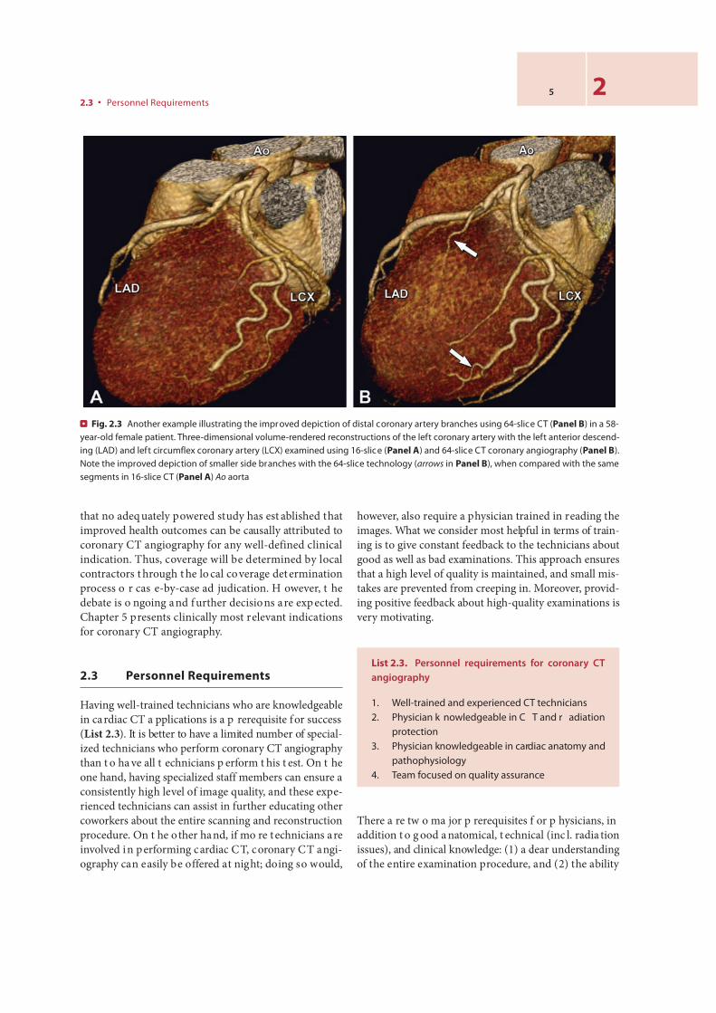
Fig. 2.3 Another example illustrating the improved depiction of distal coronary artery branches using 64-slice CT (Panel B) in a 58-year-old female patient. Three-dimensional volume-rendered reconstructions of the left coronary artery with the left anterior descend-ing (LAD) and left circumflex coronary artery (LCX) examined using 16-slice (Panel A) and 64-slice CT coronary angiography (Panel B). Note the improved depiction of smaller side branches with the 64-slice technology (arrows in Panel B), when compared with the same segments in 16-slice CT (Panel A) Ao aorta
2.3 ● Personnel Requirements
6 Chapter 2 ● Technical and Personnel Requirements
2
to independently interpret three-dimensional coronary CT angiography datasets on workstations.
Chapters 6 a nd 7 will dis cuss ho w t o p repare t he patient for coronary CT angiography and how to perform the procedure. Being present during examinations is the key to understanding the work of the technicians and the special requirements of cardiac CT. It is als o enlighten-ing for physicians to perform examinations themselves, because doing s o can yield im portant insights into t he procedural steps and problems that can be encountered
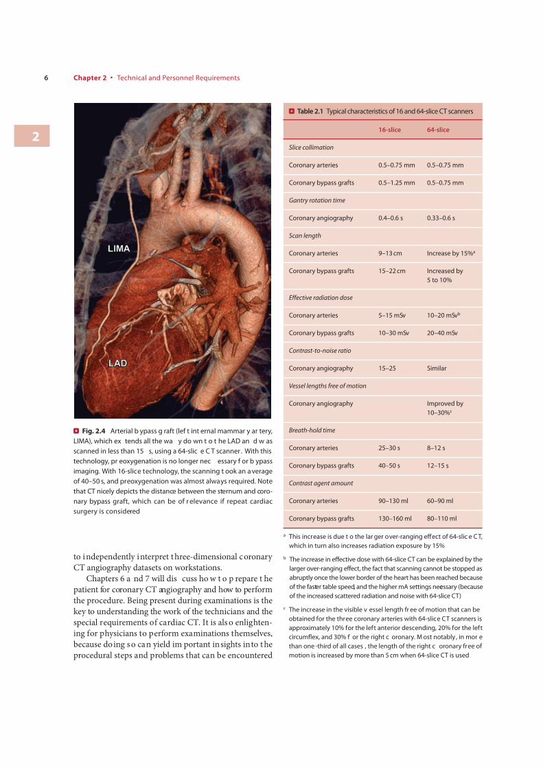
Fig. 2.4 Arterial b ypass g raft (lef t int ernal mammar y ar tery, LIMA), which ex tends all the wa y do wn t o t he LAD an d w as scanned in less than 15 s, using a 64-slic e C T scanner. With this technology, pr eoxygenation is no longer nec essary f or b ypass imaging. With 16-slice technology, the scanning t ook an a verage of 40–50 s, and preoxygenation was almost always required. Note that CT nicely depicts the distance between the sternum and coro-nary bypass graft, which can be of r elevance if repeat cardiac surgery is considered
Table 2.1 Typical characteristics of 16 and 64-slice CT scanners
16-slice 64-slice
Slice collimation
Coronary arteries 0.5–0.75 mm 0.5–0.75 mm
Coronary bypass grafts 0.5–1.25 mm 0.5–0.75 mm
Gantry rotation time
Coronary angiography 0.4–0.6 s 0.33–0.6 s
Scan length
Coronary arteries 9–13 cm Increase by 15%a
Coronary bypass grafts 15–22 cm Increased by 5 to 10%
Effective radiation dose
Coronary arteries 5–15 mSv 10–20 mSvb
Coronary bypass grafts 10–30 mSv 20–40 mSv
Contrast-to-noise ratio
Coronary angiography 15–25 Similar
Vessel lengths free of motion
Coronary angiography Improved by 10–30%c
Breath-hold time
Coronary arteries 25–30 s 8–12 s
Coronary bypass grafts 40–50 s 12–15 s
Contrast agent amount
Coronary arteries 90–130 ml 60–90 ml
Coronary bypass grafts 130–160 ml 80–110 ml
a This increase is due t o the lar ger over-ranging effect of 64-slic e C T, which in turn also increases radiation exposure by 15%
b The increase in effective dose with 64-slice CT can be explained by the larger over-ranging effect, the fact that scanning cannot be stopped as abruptly once the lower border of the heart has been reached because of the faster table speed, and the higher mA settings necessary (because of the increased scattered radiation and noise with 64-slice CT)
c The increase in the visible v essel length fr ee of motion that can be obtained for the three coronary arteries with 64-slice CT scanners is approximately 10% for the left anterior descending, 20% for the left circumflex, and 30% f or the right c oronary. M ost notably , in mor e than one -third of all cases , the length of the right c oronary free of motion is increased by more than 5 cm when 64-slice CT is used

7 2
during scanning. This hands-on training also strengthens the p osition o f t he p hysician as a n ed ucator o f o ther physicians or technicians. In larger centers, it is good to identify two to three doctors who will be considered the primary contacts for cardiac CT imaging for the techni-cians as well as the referring physicians.
Competence in imag e interpretation is b est achieved by co rrelating co nventional co ronary a ngiograms wi th CT angiography results. How to read and interpret car-diac CT scans is explained in Chap. 9. To understand and gain skill in usin g t he w orkstations, p hysicians sho uld practice operating them without time pressure. The time necessary to feel comfortable with the workstations will depend on an individual’s general computer skills, but 2–4 co ntinuous w eeks sho uld b e s ufficient, a nd a ttend-ing one of the true hands-on workshops is a good way to begin the learning process. Such workshops should ide-ally offer direct comparison of CT findings (on interactive workstations) with conventional angiography findings or the results of cardiac stress tests. This is t he only way of acquiring a thorough understanding of coronary and car-diac pathology. Good cardiac CT courses also offer active participation in patient preparation and scanning. Never-theless, the learning curve for centers with early experi-ence has been shown to last at least 6 months.
However, learning does not stop after a few weeks of intensive familiarization with the workstations or a short course: Even in a t eam of exp erienced readers, cer tain coronary lesions will sometimes be misinterpreted (over-called o r e ven o verlooked). Th us, co ntinuous le arning
efforts (with comparison of CT to the invasive coronary angiography findings, e.g., in joint interdisciplinary con-ferences) are necessary to maintain high quality.
There is also a formal accreditation of the physicians’ skills a nd kno wledge. The American C ollege o f R adi-ology (ACR) a nd t he Amer ican C ollege o f C ardiology (ACC) have established guidelines for assessing clinical competence in performing and interpreting cardiac CTs. These guidelines p lay a n incr easing r ole in ob taining certification for coronary CT angiography and claiming reimbursement in the US. Those outside the US may find it useful to study these guidelines as a basis f or starting discussions about certification of coronary CT angiography readers and centers in their own countries.
In G ermany, f or in stance, t he la w r equires t hat every p hysician p erforming CT (o f a ny o rgan) ho ld the Fachkunde (“technical qualification”) for CT, which requires ha ving co nducted 1,000 exa minations o ver a period of at least 12 months and participating in a course on radiation protection. Such regulations offer promise for increasing radiation safety, and they emphasize the relevance of the ongoing discussion on requirements for coronary CT angiography.
2.3.1 Guidelines of the ACR
Several A CR guidelines are r elevant t o co ronary CT angiography. M ost im portant is t he “ ACR P ractice Guideline for the Performance and Interpretation of
2.3 ● Personnel Requirements
Table 2.2 ACR physician requirements for coronary CT angiography
Not trained in general or thoracic CT Board-certified radiologistsa
CME (category I) Completion of an ACGME approved training program in the specialty practiced 200 hours in cardiac CTb
Training in cardiac CT in an ACGME approved training program 30 h in cardiac anatomy, physiology, pathology, and cardiac CT
Interpretation, reporting, and/or supervised reviewc
500 CT examinationsd 50 cardiac CT examinations
Maintaining competence 75 contrast-enhanced cardiac CT examinations every 3 years 150 h of CME every 3 years
ACGME Accreditation Council for Graduate Medical Education
a In addition, at least 100 CT examinations are required during each of the past 3 years, as also at least 100 CT examinations per year to maintain competence according to the ACR practice guideline for performing and interpreting diagnostic CT
b Including at least 30 h in cardiac anatomy, physiology, pathology, and cardiac CT
c Examinations (noncontrast examinations do not count) in a supervised environment during the past 3 y ears; supervising physician needs to meet the ACR requirements
d At least 100 must be a c ombination of thor acic C T or thoracic C T angiography (exclusive of calcium sc oring exams). A t least 50 contrast-enhanced cardiac CT examinations must also be included

8 Chapter 2 ● Technical and Personnel Requirements
2
Table 2.3 ACC physician requirements for coronary CT angiography
Level 2a Level 3b
CME (category I) 20 h in cardiac CT 40 h in cardiac CT
Trainingc 8 weeks 6 months
Interpretation, reporting, and/or supervised review
50 noncontrast cardiac CT examinations 150 contrast-enhanced cardiac CT examinationsd
100 noncontrast cardiac CT examinations 300 contrast-enhanced cardiac CT examinationsd
Maintaining competence 50 contrast-enhanced cardiac CT examinations every year 20 h of CME in cardiac CT every 3 years
100 contrast-enhanced cardiac CT examinations every year 40 h of CME in cardiac CT every 3 years
a Allows independent performance and interpretation of cardiac CT
b Allows serving as a director of an independent cardiac CT center
c Training must be conducted under the super vision of a level 3 physician. Each week consists of at least 35 h. The time commitment does not go into effect until July 2010
d Physically present and involved in the acquisition, performance, and interpretation of 50 (level 2) or 100 (level 3) contrast-enhanced cardiac CT examinations. The noncontrast examinations can be performed in the same patients who undergo contrast-enhanced CT
Cardiac Computed Tomography” (http://www.acr.org/SecondaryMainMenuCategories/quality_safety/guidelines/dx/cardio/ct_cardiac.aspx). Ot her im portant guidelines are the “ACR Clinical S tatement on Nonin-vasive C ardiac Imaging,” “ACR Practice Guideline for the Performance a nd I nterpretation o f CT An giogra-phy,” and the “ACR Practice Guideline for Performing and I nterpreting D iagnostic C omputed Tomography.” Later we briefly outline and discuss the recommenda-tions arising from the guidelines that directly relate to coronary CT angiography.
The ACR defines cardiac CT as a chest CT performed primarily for the evaluation of the heart (including the cardiac c hambers, val ves, m yocardium, ao rta, central pulmonary v essels, p ericardium, co ronary a rteries, and veins). However, noncardiac structures are included and must b e e valuated by a tra ined physician. Trained physicians a re def ined in t he “ACR P ractice G uideline for Performing and Interpreting Diagnostic C omputed Tomography” as b oard-certified radio logists w ho ha ve interpreted and reported at le ast 100 CT exa minations over e ach o f t he past 3 y ears a nd in terpret a nd r eport at least 100 CT exa minations p er y ear t o ma intain competence. These physicians can achieve competence in t he p erformance and i nterpretation of c oronary C T angiography b y a t le ast 30 h o f CME in ca rdiac a nat-omy, physiology, p athology, a nd c ardiac CT, p lus t he interpretation, r eporting, a nd/or su pervised r eview of at least 50 cardiac CT exa minations during the past
3 y ears ( Table 2.2 ). P hysicians w ho a re n ot d efined i n this guideline as trained physicians in diagnostic CT ca n achieve competence in t he p erformance and interpreta-tion of coronary CT angiography by at least 200 h of CME in the performance and interpretation of cardiac CT, plus the interpretation, reporting, and/or supervised review of at least 500 chest CT examinations (including 50 cardiac CT examinations) during the past 3 years (Table 2.2). The ACR stresses t hat all p hysicians p erforming ca rdiac CT need to be knowledgeable about the administration, risks, and contraindications of beta blockers and nitroglycerin.
2.3.2 Guidelines of the ACC
The “ACC Clinical C ompetence S tatement o n C ardiac Imaging with Computed Tomography and Magnetic Res-onance” (h ttp://www.acc.org/qualityandscience/clinical/competence/imaging/index.pdf) states that it is intended to be complementary to the recommendations of the ACR on noninvasive cardiac imaging. Cardiac CT is defined in this guideline as t he imaging of anatomy, function, coro-nary calcium, noncalcified p laque, and congenital he art disease. The guideline def ines three levels of competence in co ronary CT a ngiography, o f w hich tw o a re relevant here. Level 2 allows independent performance and inter-pretation of cardiac CT a nd requires 8 w eeks (each con-sisting of at least 35 h) of cumulative training in a clinical cardiac CT laboratory plus 150 contrast-enhanced and 50

noncontrast cardiac CT exa minations. A p hysician will-ing to achieve level 2 co mpetence needs t o be physically present and involved in the acquisition and performance of 50 of the 150 contrast-enhanced cardiac CT examina-tions (Table 2.3). Level 3 allo ws serving as a dir ector of an independent cardiac CT center and requires 6 months of cumulative training in a clinical cardiac CT laboratory plus 300 contrast-enhanced and 100 noncontrast cardiac CT examinations. A p hysician willing to achieve level 3 competence needs to be physically present and involved in the acquisition and performance of 100 of the 300 con-trast-enhanced cardiac CT examinations (Table 2.3). An additional r ecommendation f or “ Training i n A dvanced Cardiovascular I maging (C omputed Tomography)” has been r eleased b y t he ACC. The A CC str esses t hat all physicians performing cardiac CT need to be knowledge-able a bout radia tion r isks a nd no ncardiac f indings o n coronary CT angiography.
Recommended Reading
1 Budoff MJ, Achenbach S, Berman DS, et al. Task force 13: training in advanced cardiovascular imaging (computed tomography) endorsed by the American Society of Nuclear Cardiology, Society of Athero-sclerosis Imaging and Prevention, Society for Cardiovascular Angi-ography and Interventions, and Society of Cardiovascular Computed Tomography. J Am Coll Cardiol 2008; 51:409–14
2 Budoff MJ, Cohen MC, Garcia MJ, et al. ACCF/AHA clinical compe-tence statement on cardiac imaging with computed tomography and magnetic resonance. J Am Coll Cardiol 2005; 46:383–402
3 Chin S, On g T, Chan W, et al . 64 r ow multi-detector computed tomography coronary image from a centre with early experience:
first il lustration of learning curve. J G eriatric Cardiology 2006; 3:29–34
4 Dewey M, H amm B . C ost ef fectiveness o f co ronary a ngiography and calcium scoring using CT and stress MRI for diagnosis of coro-nary artery disease. Eur Radiol 2007; 17:1301–9
5 Dewey M, Hoffmann H, Hamm B. CT coronary angiography using 16 and 64 simultaneous detector rows: intraindividual comparison. Fortschr Röntgenstr 2007; 179:581–86
6 Hamon M, Morello R, Riddell JW. Coronary arteries: diagnostic performance o f 16 v ersus 64-s ection sp iral CT co mpared wi th invasive co ronary a ngiography–meta-analysis. R adiology 2007; 245:720–31
7 Hausleiter J, Meyer T, Hadamitzky M, et al. Non-invasive coronary computed t omographic a ngiography f or pa tients wi th s uspected coronary artery disease: the Coronary Angiography by Computed Tomography with the Use of a Submillimeter resolution (CACTUS) trial. Eur Heart J 2007; 28:3034–41
8 Jacobs JE, B oxt LM, D esjardins B , Fishma n EK, L arson PA, Schoepf J . A CR p ractice guideline f or t he p erformance a nd interpretation of cardiac computed tomography (CT). J Am C oll Radiol 2006; 3:677–85
9 Pannu HK, Alvarez W, Jr., Fishman EK. Beta-blockers for cardiac CT: a primer for the radiologist. AJR Am J Roentgenol 2006; 186:S341–5
10 Weinreb JC, Larson PA, Woodard PK, et al. ACR clinical statement on noninvasive cardiac imaging. J Am Coll Radiol 2005; 2:471–7
The ACR p ractice guideline f or t he p erformance a nd in terpretation o f cardiac CT (Jacobs et al.) can be accessed at:
http://www.acr.org/SecondaryMainMenuCategories/quality_safety/guidelines/dx/cardio/ct_cardiac.aspx
The guideline of the ACC (Budoff et al.) can be accessed at:http://www.acc.org/qualityandscience/clinical/competence/imaging/
index.pdfhttp://www.escr.orghttp://www.nasci.orghttp://www.scct.org
2.3 ● Personnel Requirements9 2