teat condition - prevention and cure through teat dips · teat condition - prevention and cure...
TRANSCRIPT
Proceedings of the British Mastitis Conference (2002) Brockworth, p 1-14 Institute for Animal Health/Milk Development Council
1
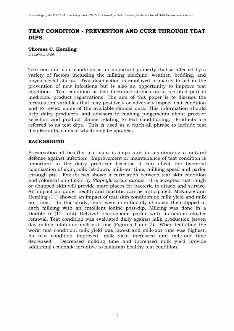
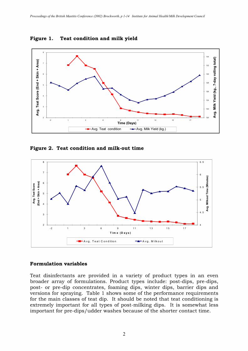
TEAT CONDITION - PREVENTION AND CURE THROUGH TEAT DIPS Thomas C. Hemling DeLaval, USA Teat end and skin condition is an important property that is affected by a variety of factors including the milking machine, weather, bedding, and physiological status. Teat disinfection is employed primarily to aid in the prevention of new infections but is also an opportunity to improve teat condition. Teat condition or teat tolerance studies are a required part of medicinal product registrations. The aim of this paper is to discuss the formulation variables that may positively or adversely impact teat condition and to review some of the available clinical data. This information should help dairy producers and advisors in making judgements about product selection and product claims relating to teat conditioning. Products are referred to as teat dips. This is used as a catch-all phrase to include teat disinfectants, some of which may be sprayed. BACKGROUND Preservation of healthy teat skin is important in maintaining a natural defense against infection. Improvement or maintenance of teat condition is important to the dairy producer because it can affect the bacterial colonization of skin, milk let-down, milk-out time, milking speed and parlor through put. Fox (6) has shown a correlation between teat skin condition and colonization of skin by Staphylococcus aureus. It is accepted that rough or chapped skin will provide more places for bacteria to attach and survive. An impact on udder health and mastitis can be anticipated. McKinzie and Hemling (11) showed an impact of teat skin condition on milk yield and milk out time. In this study, teats were intentionally chapped then dipped at each milking with an emollient iodine post-dip. Milking was done in a Double 6 (12 unit) DeLaval herringbone parlor with automatic cluster removal. Teat condition was evaluated daily against milk production (seven day rolling total) and milk-out time (Figures 1 and 2). When teats had the worst teat condition, milk yield was lowest and milk-out time was highest. As teat condition improved, milk yield increased and milk-out time decreased. Decreased milking time and increased milk yield provide additional economic incentive to maintain healthy teat condition.
Proceedings of the British Mastitis Conference (2002) Brockworth, p 1-14 Institute for Animal Health/Milk Development Council
2
Figure 1. Teat condition and milk yield
Figure 2. Teat condition and milk-out time Formulation variables Teat disinfectants are provided in a variety of product types in an even broader array of formulations. Product types include: post-dips, pre-dips, post- or pre-dip concentrates, foaming dips, winter dips, barrier dips and versions for spraying. Table 1 shows some of the performance requirements for the main classes of teat dip. It should be noted that teat conditioning is extremely important for all types of post-milking dips. It is somewhat less important for pre-dips/udder washes because of the shorter contact time.
2
3
4
5
6
7
8
-2 1 3 6 9 1 1 1 3 1 5 1 7
T im e (D a y s )
Avg. Teat S
core
(End +
Skin
+ A
rea)
4
4 .5
5
5 .5
6
6 .5
Avg. M
ilkout Tim
e (M
inute
s)
A v g . T e a t C o n d it io n A v g . M ilk o u t
2
3
4
5
6
7
8
-2 1 3 6 9 11 13 15 17
Time (Days)
Avg. Teat S
core
(E
nd +
Skin
+ A
rea)
120
122
124
126
128
130
132
134
Avg. M
ilk Y
ield
(kg., 7
-day r
ollin
g tota
l)
Avg. Teat condition Avg. Milk Yield (kg.)
`
Proceedings of the British Mastitis Conference (2002) Brockworth, p 1-14 Institute for Animal Health/Milk Development Council
3
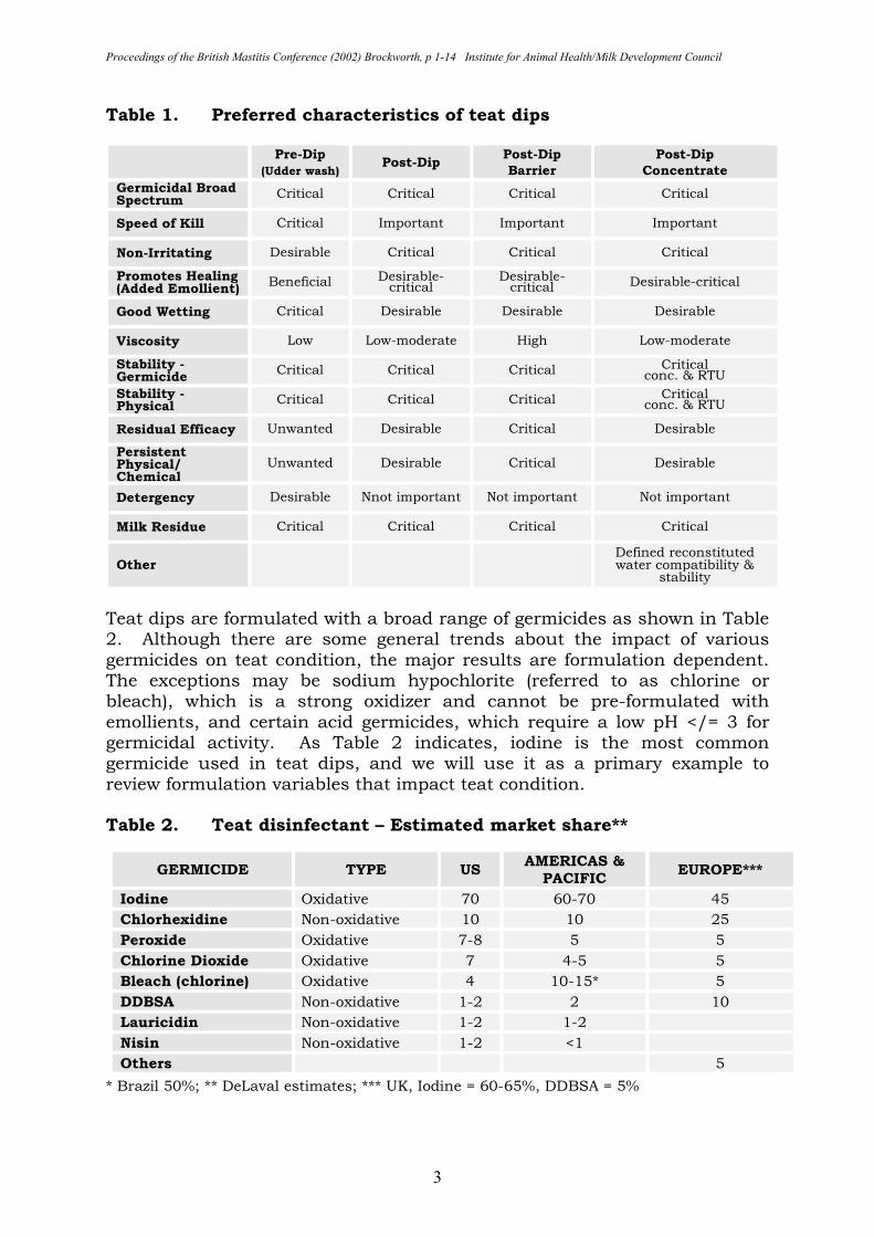
Table 1. Preferred characteristics of teat dips
Pre-Dip
(Udder wash) Post-Dip
Post-Dip
Barrier
Post-Dip
Concentrate
Germicidal Broad Spectrum
Critical Critical Critical Critical
Speed of Kill Critical Important Important Important
Non-Irritating Desirable Critical Critical Critical
Promotes Healing (Added Emollient)
Beneficial Desirable-critical
Desirable-critical Desirable-critical
Good Wetting Critical Desirable Desirable Desirable
Viscosity Low Low-moderate High Low-moderate
Stability - Germicide
Critical Critical Critical Critical conc. & RTU
Stability - Physical
Critical Critical Critical Critical conc. & RTU
Residual Efficacy Unwanted Desirable Critical Desirable
Persistent Physical/ Chemical
Unwanted Desirable Critical Desirable
Detergency Desirable Nnot important Not important Not important
Milk Residue Critical Critical Critical Critical
Other Defined reconstituted water compatibility &
stability
Teat dips are formulated with a broad range of germicides as shown in Table 2. Although there are some general trends about the impact of various germicides on teat condition, the major results are formulation dependent. The exceptions may be sodium hypochlorite (referred to as chlorine or bleach), which is a strong oxidizer and cannot be pre-formulated with emollients, and certain acid germicides, which require a low pH </= 3 for germicidal activity. As Table 2 indicates, iodine is the most common germicide used in teat dips, and we will use it as a primary example to review formulation variables that impact teat condition.
Table 2. Teat disinfectant – Estimated market share**
GERMICIDE TYPE US AMERICAS & PACIFIC
EUROPE***
Iodine Oxidative 70 60-70 45 Chlorhexidine Non-oxidative 10 10 25
Peroxide Oxidative 7-8 5 5
Chlorine Dioxide Oxidative 7 4-5 5 Bleach (chlorine) Oxidative 4 10-15* 5
DDBSA Non-oxidative 1-2 2 10 Lauricidin Non-oxidative 1-2 1-2
Nisin Non-oxidative 1-2 <1 Others 5
* Brazil 50%; ** DeLaval estimates; *** UK, Iodine = 60-65%, DDBSA = 5%
Proceedings of the British Mastitis Conference (2002) Brockworth, p 1-14 Institute for Animal Health/Milk Development Council
4
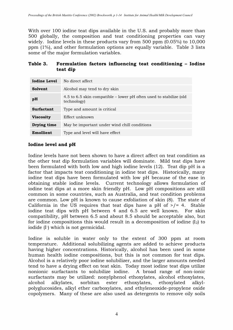
With over 100 iodine teat dips available in the U.S. and probably more than 500 globally, the composition and teat conditioning properties can vary widely. Iodine levels in these products vary from 500 ppm (0.05%) to 10,000 ppm (1%), and other formulation options are equally variable. Table 3 lists some of the major formulation variables. Table 3. Formulation factors influencing teat conditioning – Iodine
teat dip Iodine Level No direct affect
Solvent Alcohol may tend to dry skin
pH 4.5 to 6.5 skin compatible – lower pH often used to stabilize (old technology)
Surfactant Type and amount is critical
Viscosity Effect unknown
Drying time May be important under wind chill conditions
Emollient Type and level will have effect
Iodine level and pH Iodine levels have not been shown to have a direct affect on teat condition as the other teat dip formulation variables will dominate. Mild teat dips have been formulated with both low and high iodine levels (12). Teat dip pH is a factor that impacts teat conditioning in iodine teat dips. Historically, many iodine teat dips have been formulated with low pH because of the ease in obtaining stable iodine levels. Current technology allows formulation of iodine teat dips at a more skin friendly pH. Low pH compositions are still common in some countries, such as Australia, and teat condition problems are common. Low pH is known to cause exfoliation of skin (8). The state of California in the US requires that teat dips have a pH of >/= 4. Stable iodine teat dips with pH between 4 and 6.5 are well known. For skin compatibility, pH between 6.5 and about 8.5 should be acceptable also, but for iodine compositions this would result in a decomposition of iodine (I2) to iodide (I-) which is not germicidal. Iodine is soluble in water only to the extent of 300 ppm at room temperature. Additional solubilizing agents are added to achieve products having higher concentrations. Historically, alcohol has been used in some human health iodine compositions, but this is not common for teat dips. Alcohol is a relatively poor iodine solubilizer, and the larger amounts needed tend to have a drying effect on teat skin. Today most iodine teat dips utilize nonionic surfactants to solubilize iodine. A broad range of non-ionic surfactants may be utilized: nonylphenol ethoxylates, alcohol ethoxylates, alcohol alkylates, sorbitan ester ethoxylates, ethoxylated alkyl-polyglucosides, alkyl ether carboxylates, and ethyleneoxide-propylene oxide copolymers. Many of these are also used as detergents to remove oily soils
Proceedings of the British Mastitis Conference (2002) Brockworth, p 1-14 Institute for Animal Health/Milk Development Council
5
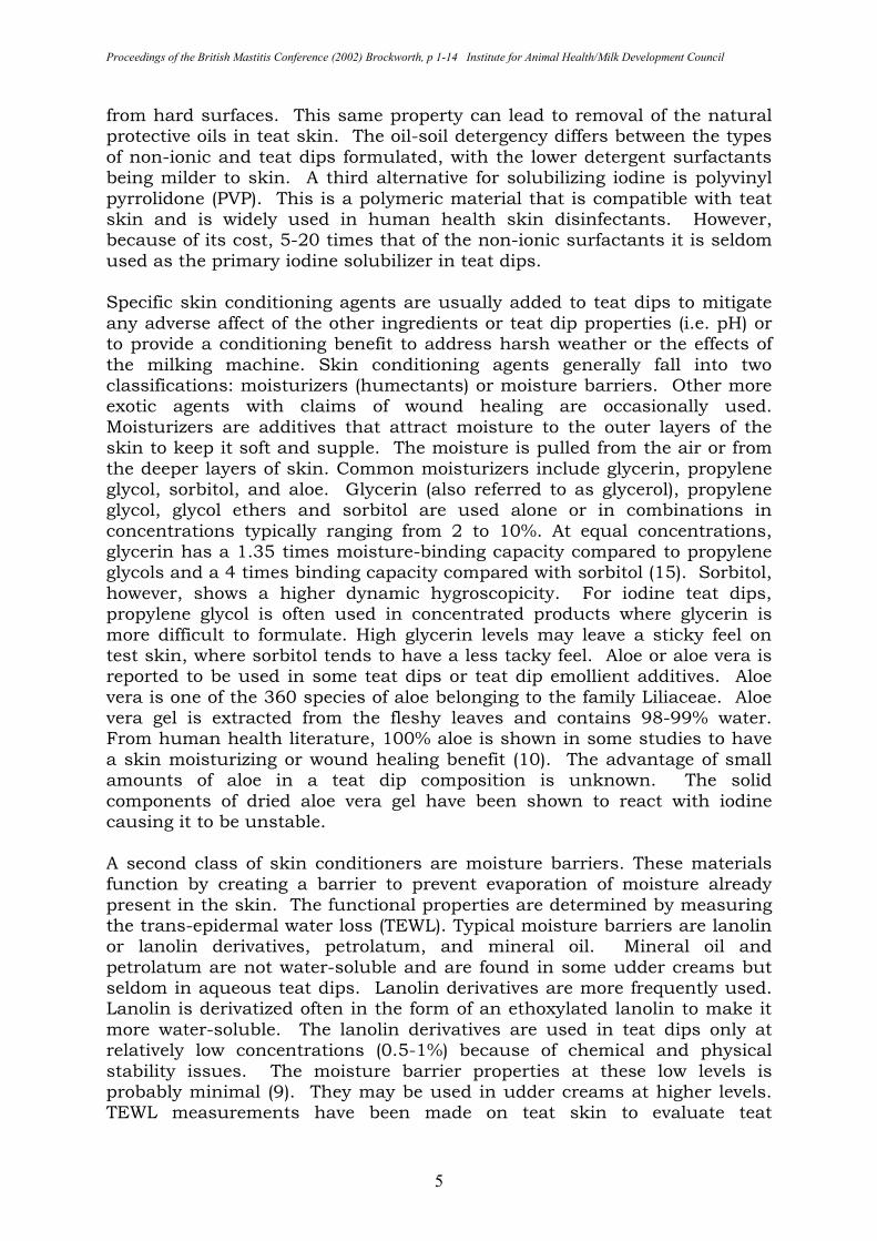
from hard surfaces. This same property can lead to removal of the natural protective oils in teat skin. The oil-soil detergency differs between the types of non-ionic and teat dips formulated, with the lower detergent surfactants being milder to skin. A third alternative for solubilizing iodine is polyvinyl pyrrolidone (PVP). This is a polymeric material that is compatible with teat skin and is widely used in human health skin disinfectants. However, because of its cost, 5-20 times that of the non-ionic surfactants it is seldom used as the primary iodine solubilizer in teat dips. Specific skin conditioning agents are usually added to teat dips to mitigate any adverse affect of the other ingredients or teat dip properties (i.e. pH) or to provide a conditioning benefit to address harsh weather or the effects of the milking machine. Skin conditioning agents generally fall into two classifications: moisturizers (humectants) or moisture barriers. Other more exotic agents with claims of wound healing are occasionally used. Moisturizers are additives that attract moisture to the outer layers of the skin to keep it soft and supple. The moisture is pulled from the air or from the deeper layers of skin. Common moisturizers include glycerin, propylene glycol, sorbitol, and aloe. Glycerin (also referred to as glycerol), propylene glycol, glycol ethers and sorbitol are used alone or in combinations in concentrations typically ranging from 2 to 10%. At equal concentrations, glycerin has a 1.35 times moisture-binding capacity compared to propylene glycols and a 4 times binding capacity compared with sorbitol (15). Sorbitol, however, shows a higher dynamic hygroscopicity. For iodine teat dips, propylene glycol is often used in concentrated products where glycerin is more difficult to formulate. High glycerin levels may leave a sticky feel on test skin, where sorbitol tends to have a less tacky feel. Aloe or aloe vera is reported to be used in some teat dips or teat dip emollient additives. Aloe vera is one of the 360 species of aloe belonging to the family Liliaceae. Aloe vera gel is extracted from the fleshy leaves and contains 98-99% water. From human health literature, 100% aloe is shown in some studies to have a skin moisturizing or wound healing benefit (10). The advantage of small amounts of aloe in a teat dip composition is unknown. The solid components of dried aloe vera gel have been shown to react with iodine causing it to be unstable. A second class of skin conditioners are moisture barriers. These materials function by creating a barrier to prevent evaporation of moisture already present in the skin. The functional properties are determined by measuring the trans-epidermal water loss (TEWL). Typical moisture barriers are lanolin or lanolin derivatives, petrolatum, and mineral oil. Mineral oil and petrolatum are not water-soluble and are found in some udder creams but seldom in aqueous teat dips. Lanolin derivatives are more frequently used. Lanolin is derivatized often in the form of an ethoxylated lanolin to make it more water-soluble. The lanolin derivatives are used in teat dips only at relatively low concentrations (0.5-1%) because of chemical and physical stability issues. The moisture barrier properties at these low levels is probably minimal (9). They may be used in udder creams at higher levels. TEWL measurements have been made on teat skin to evaluate teat
Proceedings of the British Mastitis Conference (2002) Brockworth, p 1-14 Institute for Animal Health/Milk Development Council
6
conditioning properties of treatments, but with limited success (3). The lack of success is likely the result of the inability to control all of the environmental factors to which the teat is exposed. A number of other human health or cosmetic ingredients have been incorporated into teat dips. These include alpha hydroxy acids, allantoin, collagen, vitamins and other ingredients for skin conditioning or wound healing properties. Although data exist to show some effect on human skin, or in pig or rat skin models, little information is available on the benefit in teat dips. In some countries, teat skin emollient products are sold separately to be added to teat dip solutions on farm. Unless the teat dip and skin conditioning agents are both labeled with specific directions on combining the two products, this practice is discouraged. The mixing of the two products could cause a chemical or physical incompatibility that negates either the germicidal effect of the teat dip, the skin conditioning effect of the emollient, or both. Viscosity The viscosity of commercial teat dips varies from essentially water-like (1 centipoise) to the more viscous barrier teat dips (150-500 centipoise). Common post-dips that are suited for dipping or spraying have a viscosity for about 5 to 30 centipoise. Increased viscosity will generally result in a thicker layer of product on the teat, especially the teat end. Viscosity alone is not expected to impact teat condition, except under low temperature “wind chill” conditions where increased viscosity may prolong evaporation and cause increased chapping, frost bite or teat end freezing. Under other conditions, the increased thickness of teat dip on the teat skin could be expected to act as a multiplier of the conditioning properties. Harsh products will have more of an adverse effect. Conditioning properties will deliver more benefit. Pre-milking dips/udderwash and post-dip interactions Pre-dips have relatively short contact time on teats and the impact on teat condition is expected to be minimal. Dedicated pre-dips are normally formulated with low levels of emollients and usually have germicidal properties that provide rapid kill. Good pre-milking teat cleaning achieved by pre-dipping may reduce abrasion caused by the rubbing effect of the teat liner on soil on the teat that would otherwise be “dry milked”. Questions of possible pre-dip:post-dip interactions have been raised (5), especially for pre-dip:post-dip combinations with different germicides, but there is limited published clinical trial data. The minimal contact time for the pre-dip and the small amount of post-dip likely to remain on the teat at the next milking would suggest little chance for adverse reaction. Barrier dips may be an exception, as the amount of product remaining on the teat at the next milking would be increased. One retrospective survey was conducted that showed some influence of the pre-dip:post-dip combination in teat chapping

Proceedings of the British Mastitis Conference (2002) Brockworth, p 1-14 Institute for Animal Health/Milk Development Council
7
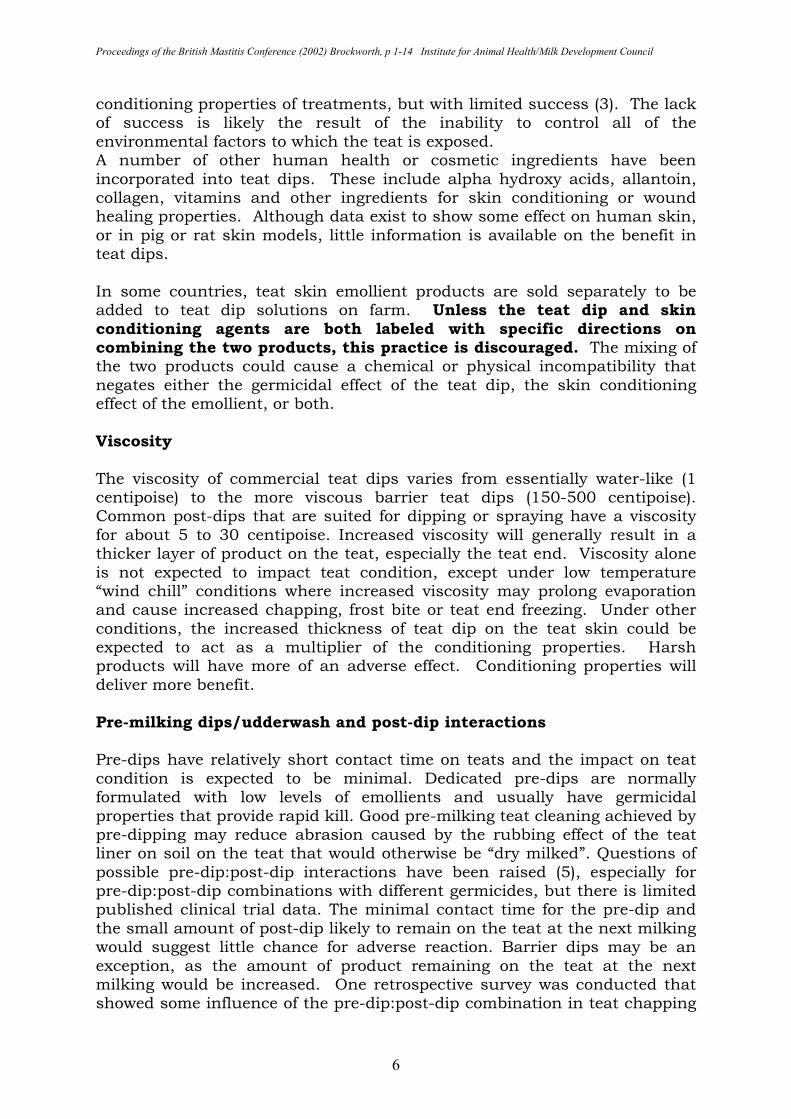
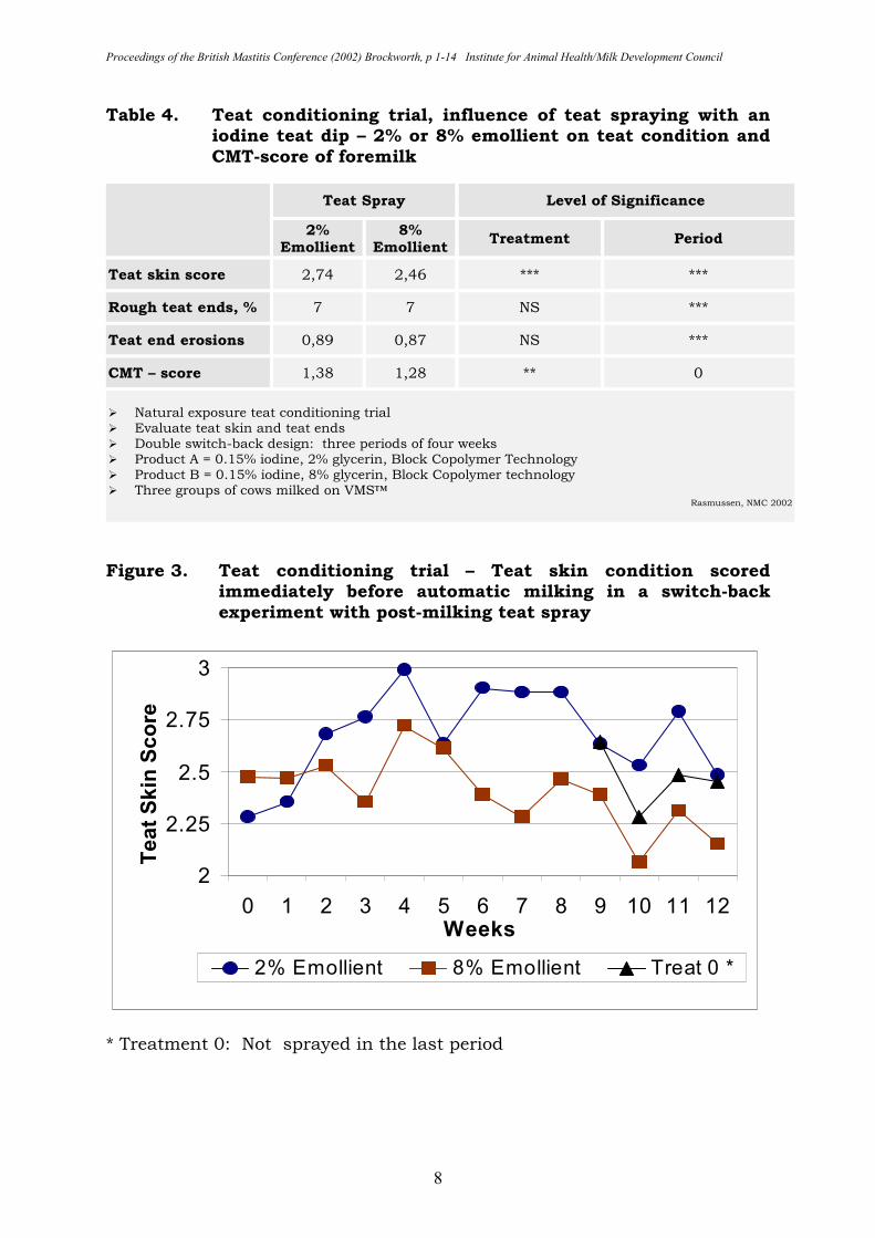
(2), but these conclusions were not supported by data from a controlled clinical study (4). TEAT CONDITIONING DATA Although teat dips have been sold and promoted as having teat conditioning benefits for years, scientific studies on the teat conditioning effects of teat dip compositions have only been common during the past 10-15 years. With the efforts to standardize scoring systems and methods, research in this area is expected to expand. I summarize here some of the studies that support some of the discussions presented above. These studies use a 1-5 scoring system for teat skin evaluation that is recommended by Teat Club International. Scoring systems for teat ends either evaluates smoothness-roughness, or incorporates some measure of ring-formation-hyperkeratosis. For both teat skin and teat end, the lower score indicates better condition. In the reported studies, the timing of teat skin evaluation varies depending on the object of the study and what is possible at the trial site and is not consistent between the studies. Effect of emollient level Rasmussen and Hemling (14) reported a study of two iodine products with identical, mild surfactant compositions differing in the level of glycerin: 2% versus 8%. The cows in this study were milked with identical VMSTM robotic milkers. The products were evaluated in a double switchback design, including three periods of 4 weeks, with teat skin and teat end evaluations being done prior to milking. The trial showed a significantly better teat skin condition for the product with 8% glycerin (Table 4, and Figure 3). This trial did not show any adverse effect of increasing milking frequency on teat end or teat skin condition. This could be a result of the high emollient, mild surfactant teat dips, or the use of quarter level automatic take-offs on the VMS robot. A second trial (13), evaluated the effects of 10% glycerin (glycerol), a chlorine dioxide teat dip, and a chlorine dioxide teat dip with 10% glycerin. The three products were compared in a four-week natural exposure trial with teat skin and teat end condition measured 3 to 4 hours after milking. In this trial, 10% glycerin alone provided the best teat skin and teat end condition. Chlorine dioxide with 10% glycerin provided better teat skin and end condition compared to chlorine dioxide (Table 5). This study shows the benefit of emollients like glycerin and also the emollient germicide combination. This data support the conclusion that teat conditioning properties are a result of the teat dip composition and not the specific germicide or the emollient.
Proceedings of the British Mastitis Conference (2002) Brockworth, p 1-14 Institute for Animal Health/Milk Development Council
8
Table 4. Teat conditioning trial, influence of teat spraying with an iodine teat dip – 2% or 8% emollient on teat condition and CMT-score of foremilk
Teat Spray Level of Significance
2% Emollient
8% Emollient
Treatment Period
Teat skin score 2,74 2,46 *** ***
Rough teat ends, % 7 7 NS ***
Teat end erosions 0,89 0,87 NS ***
CMT – score 1,38 1,28 ** 0
� Natural exposure teat conditioning trial � Evaluate teat skin and teat ends � Double switch-back design: three periods of four weeks � Product A = 0.15% iodine, 2% glycerin, Block Copolymer Technology � Product B = 0.15% iodine, 8% glycerin, Block Copolymer technology � Three groups of cows milked on VMS™
Rasmussen, NMC 2002
Figure 3. Teat conditioning trial – Teat skin condition scored
immediately before automatic milking in a switch-back experiment with post-milking teat spray
* Treatment 0: Not sprayed in the last period
2
2.25
2.5
2.75
3
0 1 2 3 4 5 6 7 8 9 10 11 12Weeks
Teat S
kin
Score
2% Emollient 8% Emollient Treat 0 *
Proceedings of the British Mastitis Conference (2002) Brockworth, p 1-14 Institute for Animal Health/Milk Development Council
9
Table 5. Teat conditioning trial – Score of teat skin condition after four weeks of post-milking teat spray
Teat Spray Lactating Cows
Teat Middle Teat End
Glycerol 2.13a 2.08a
Glycerol & Chlorine Dioxide
2.38a 2.21ab
Chlorine Dioxide 3.00b 2.83c
No Teat Spray 3.00b 2.71bc
STD 0.400 0.38
� Natural exposure teat conditioning trial � Evaluate teat skin and teat ends, 1(smooth)-6 (rough or damaged) scale � Evaluate after four weeks � a,b,c: numbers with different superscripts are different (p < 0.05)
Rasmussen, Acta vet. scand. 1998, 39, 443-452
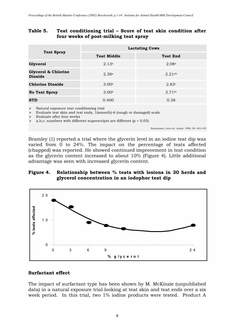
Bramley (1) reported a trial where the glycerin level in an iodine teat dip was varied from 0 to 24%. The impact on the percentage of teats affected (chapped) was reported. He showed continued improvement in teat condition as the glycerin content increased to about 10% (Figure 4). Little additional advantage was seen with increased glycerin content. Figure 4. Relationship between % teats with lesions in 30 herds and
glycerol concentration in an iodophor teat dip
Surfactant effect The impact of surfactant type has been shown by M. McKinzie (unpublished data) in a natural exposure trial looking at teat skin and teat ends over a six week period. In this trial, two 1% iodine products were tested. Product A
0
1 0
2 0
0 3 6 9 1 2 1 5 1 8 2 1 2 4
% g l y c e r o l
% teats
aff
ecte
d

Proceedings of the British Mastitis Conference (2002) Brockworth, p 1-14 Institute for Animal Health/Milk Development Council
10
contained 10% glycerin and utilized nonylphenol ethoxylates as the iodine complexor. Product B contained 4% glycerin and utilized ethylene oxide-propylene oxide compolymers as the iodine complexor. Both products improved the teat condition score for the first two weeks of the trial, but teat condition for the Product A group deteriorated during weeks 4 to 6. The trial shows the significant effect of the surfactant type in iodine teat dips, which is more important than the difference in glycerin level (Figure 5). This trial is also an interesting example of the change of teat condition over time. From Figure 5 one can speculate that some adverse event (perhaps a weather or milking system change) occurred around week three that led to a change in teat condition. Product B was better able to maintain good teat condition during this period. Figure 5. Natural exposure teat conditioning – effect of surfactant
type
� Natural exposure teat conditioning trial � Evaluate teat skin and teat ends � Report combined score as total teat score � Product A = 1% iodine, 10% glycerin Conventional NPE Complexor, (Teat Kote 10/III) � Product B = 1% iodine, 4% glycerin, Patented Block Copolymer Technology, (West Dip)
McKinzie, Hemling, NMC 1995
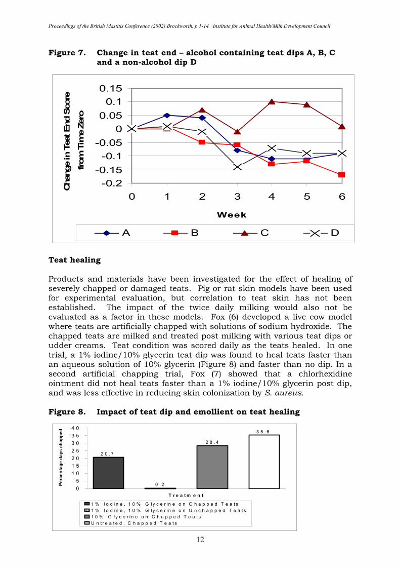
Solvent effects A six week, split udder, natural exposure trial (Table 6, Figures 6, 7) was run to compare three alcohol containing teat dips with an iodine product (un-published data). The three alcohol products were smooth, or slippery, to the touch and are marketed as being good skin conditioning products. During the six-week trial, teat ends improved for three of the products, but deteriorated for the high viscosity alcohol product (C). The other three products showed a similar positive effect on teat ends, with the emollient iodine composition (D) showing a more rapid effect. The low viscosity iodine
Natural Exposure Teat Conditioning
Effect of Surfactant Type
3 .2
3 .4
3 .6
3 .8
4
0 1 2 3 4 5 6
W e e k s
Tota
l Teat S
core
B lo c k C o p o ly m e r 4 % G ly c e r in N P E 1 0 % G ly c e r in
Proceedings of the British Mastitis Conference (2002) Brockworth, p 1-14 Institute for Animal Health/Milk Development Council
11
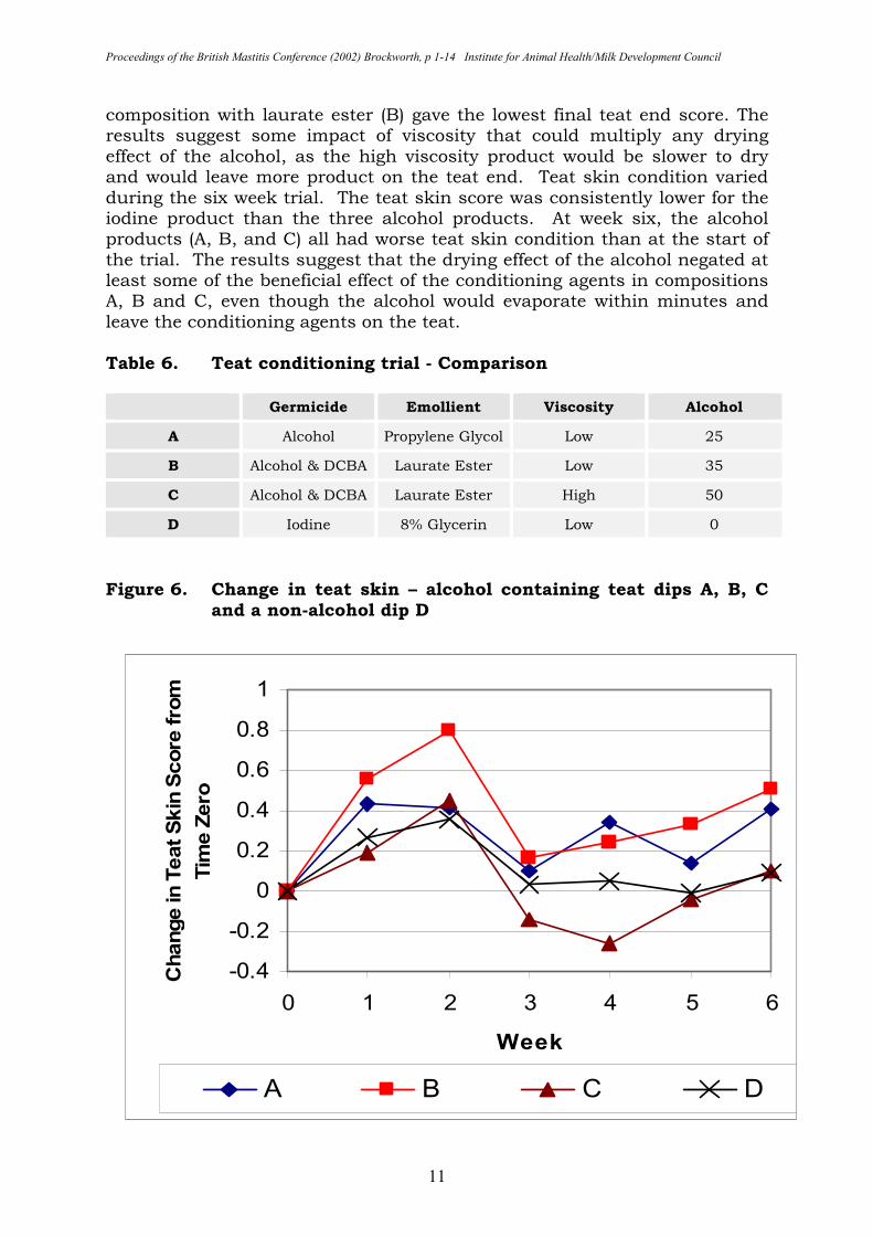
composition with laurate ester (B) gave the lowest final teat end score. The results suggest some impact of viscosity that could multiply any drying effect of the alcohol, as the high viscosity product would be slower to dry and would leave more product on the teat end. Teat skin condition varied during the six week trial. The teat skin score was consistently lower for the iodine product than the three alcohol products. At week six, the alcohol products (A, B, and C) all had worse teat skin condition than at the start of the trial. The results suggest that the drying effect of the alcohol negated at least some of the beneficial effect of the conditioning agents in compositions A, B and C, even though the alcohol would evaporate within minutes and leave the conditioning agents on the teat. Table 6. Teat conditioning trial - Comparison
Germicide Emollient Viscosity Alcohol
A Alcohol Propylene Glycol Low 25
B Alcohol & DCBA Laurate Ester Low 35
C Alcohol & DCBA Laurate Ester High 50
D Iodine 8% Glycerin Low 0
Figure 6. Change in teat skin – alcohol containing teat dips A, B, C
and a non-alcohol dip D
-0.4
-0.2
0
0.2
0.4
0.6
0.8
1
0 1 2 3 4 5 6
Week
Change in T
eat Skin
Score
fro
m
Tim
e Z
ero
A B C D
Proceedings of the British Mastitis Conference (2002) Brockworth, p 1-14 Institute for Animal Health/Milk Development Council
12
Figure 7. Change in teat end – alcohol containing teat dips A, B, C and a non-alcohol dip D
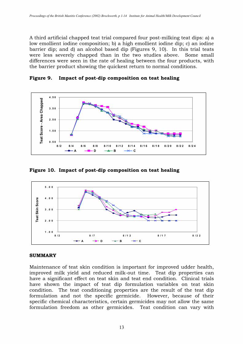
Teat healing Products and materials have been investigated for the effect of healing of severely chapped or damaged teats. Pig or rat skin models have been used for experimental evaluation, but correlation to teat skin has not been established. The impact of the twice daily milking would also not be evaluated as a factor in these models. Fox (6) developed a live cow model where teats are artificially chapped with solutions of sodium hydroxide. The chapped teats are milked and treated post milking with various teat dips or udder creams. Teat condition was scored daily as the teats healed. In one trial, a 1% iodine/10% glycerin teat dip was found to heal teats faster than an aqueous solution of 10% glycerin (Figure 8) and faster than no dip. In a second artificial chapping trial, Fox (7) showed that a chlorhexidine ointment did not heal teats faster than a 1% iodine/10% glycerin post dip, and was less effective in reducing skin colonization by S. aureus. Figure 8. Impact of teat dip and emollient on teat healing
2 0 . 7
0 . 2
2 8 . 4
3 5 . 6
0
5
1 0
1 5
2 0
2 5
3 0
3 5
4 0
T r e a t m e n t
Perc
enta
ge d
ays c
happed
1 % I o d in e , 1 0 % G ly c e r in e o n C h a p p e d T e a t s
1 % I o d in e , 1 0 % G ly c e r in e o n U n c h a p p e d T e a t s
1 0 % G l y c e r i n e o n C h a p p e d T e a t s
U n t r e a t e d , C h a p p e d T e a t s
-0.2
-0.15
-0.1
-0.05
0
0.05
0.1
0.15
0 1 2 3 4 5 6
Week
Change in T
eat End S
core
from
Tim
e Z
ero
A B C D
Proceedings of the British Mastitis Conference (2002) Brockworth, p 1-14 Institute for Animal Health/Milk Development Council
13
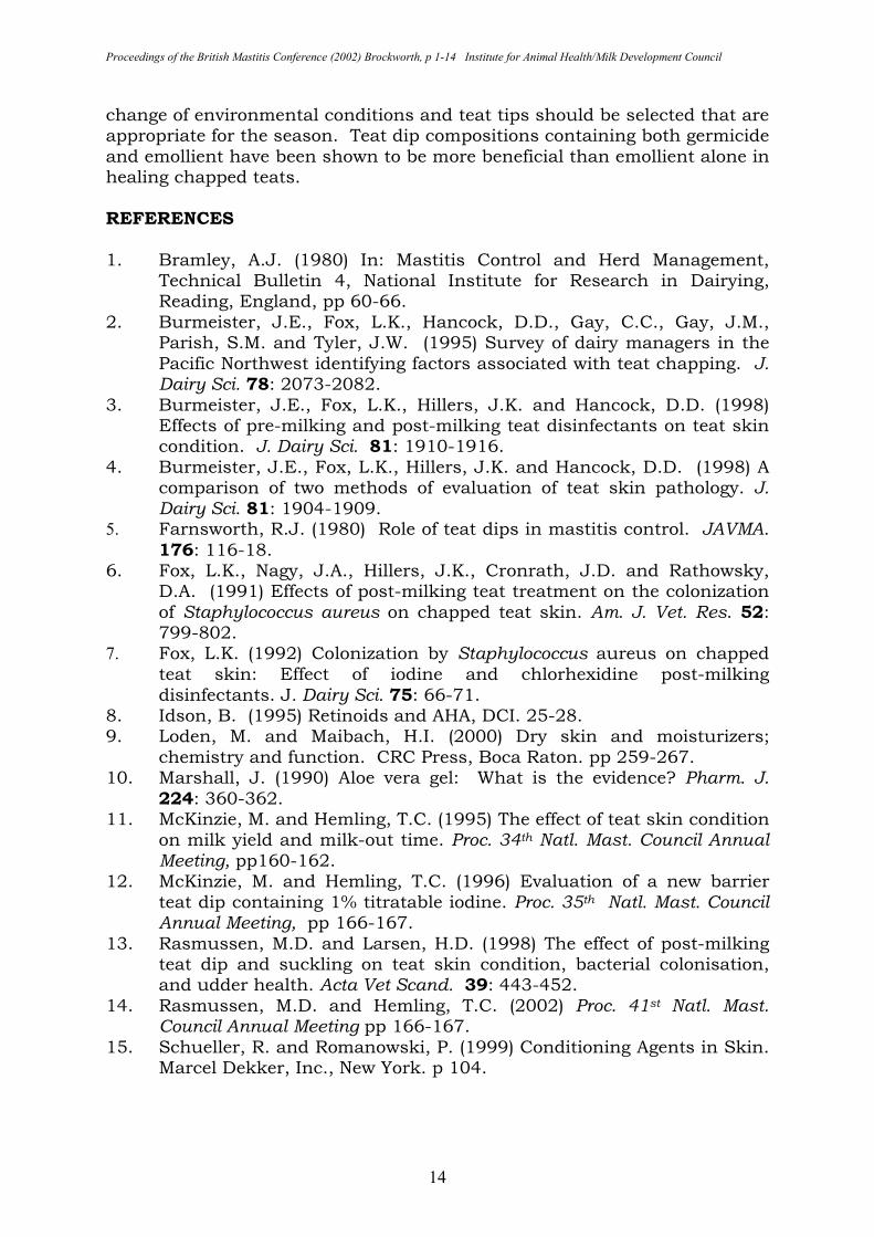
A third artificial chapped teat trial compared four post-milking teat dips: a) a low emollient iodine composition; b) a high emollient iodine dip; c) an iodine barrier dip; and d) an alcohol based dip (Figures 9, 10). In this trial teats were less severely chapped than in the two studies above. Some small differences were seen in the rate of healing between the four products, with the barrier product showing the quickest return to normal conditions. Figure 9. Impact of post-dip composition on teat healing Figure 10. Impact of post-dip composition on teat healing
SUMMARY Maintenance of teat skin condition is important for improved udder health, improved milk yield and reduced milk-out time. Teat dip properties can have a significant effect on teat skin and teat end condition. Clinical trials have shown the impact of teat dip formulation variables on teat skin condition. The teat conditioning properties are the result of the teat dip formulation and not the specific germicide. However, because of their specific chemical characteristics, certain germicides may not allow the same formulation freedom as other germicides. Teat condition can vary with
0 .5 0
1 .5 0
2 .5 0
3 .5 0
4 .5 0
8 /2 8 /4 8 /6 8 /8 8 /1 0 8 /1 2 8 /1 4 8 /1 6 8 /1 8 8 /2 0 8 /2 2 8 /2 4
Teat S
core
- A
rea C
happed
A D B C
1 . 0 0
2 . 0 0
3 . 0 0
4 . 0 0
5 . 0 0
8 / 2 8 / 7 8 / 1 2 8 / 1 7 8 / 2 2
Teat S
kin
Score
A D B C
Proceedings of the British Mastitis Conference (2002) Brockworth, p 1-14 Institute for Animal Health/Milk Development Council
14
change of environmental conditions and teat tips should be selected that are appropriate for the season. Teat dip compositions containing both germicide and emollient have been shown to be more beneficial than emollient alone in healing chapped teats. REFERENCES 1. Bramley, A.J. (1980) In: Mastitis Control and Herd Management,
Technical Bulletin 4, National Institute for Research in Dairying, Reading, England, pp 60-66.
2. Burmeister, J.E., Fox, L.K., Hancock, D.D., Gay, C.C., Gay, J.M., Parish, S.M. and Tyler, J.W. (1995) Survey of dairy managers in the Pacific Northwest identifying factors associated with teat chapping. J. Dairy Sci. 78: 2073-2082.
3. Burmeister, J.E., Fox, L.K., Hillers, J.K. and Hancock, D.D. (1998) Effects of pre-milking and post-milking teat disinfectants on teat skin condition. J. Dairy Sci. 81: 1910-1916.
4. Burmeister, J.E., Fox, L.K., Hillers, J.K. and Hancock, D.D. (1998) A comparison of two methods of evaluation of teat skin pathology. J. Dairy Sci. 81: 1904-1909.
5. Farnsworth, R.J. (1980) Role of teat dips in mastitis control. JAVMA. 176: 116-18.
6. Fox, L.K., Nagy, J.A., Hillers, J.K., Cronrath, J.D. and Rathowsky, D.A. (1991) Effects of post-milking teat treatment on the colonization of Staphylococcus aureus on chapped teat skin. Am. J. Vet. Res. 52: 799-802.
7. Fox, L.K. (1992) Colonization by Staphylococcus aureus on chapped teat skin: Effect of iodine and chlorhexidine post-milking disinfectants. J. Dairy Sci. 75: 66-71.
8. Idson, B. (1995) Retinoids and AHA, DCI. 25-28. 9. Loden, M. and Maibach, H.I. (2000) Dry skin and moisturizers;
chemistry and function. CRC Press, Boca Raton. pp 259-267. 10. Marshall, J. (1990) Aloe vera gel: What is the evidence? Pharm. J.
224: 360-362. 11. McKinzie, M. and Hemling, T.C. (1995) The effect of teat skin condition
on milk yield and milk-out time. Proc. 34th Natl. Mast. Council Annual Meeting, pp160-162.
12. McKinzie, M. and Hemling, T.C. (1996) Evaluation of a new barrier teat dip containing 1% titratable iodine. Proc. 35th Natl. Mast. Council Annual Meeting, pp 166-167.
13. Rasmussen, M.D. and Larsen, H.D. (1998) The effect of post-milking teat dip and suckling on teat skin condition, bacterial colonisation, and udder health. Acta Vet Scand. 39: 443-452.
14. Rasmussen, M.D. and Hemling, T.C. (2002) Proc. 41st Natl. Mast. Council Annual Meeting pp 166-167.
15. Schueller, R. and Romanowski, P. (1999) Conditioning Agents in Skin. Marcel Dekker, Inc., New York. p 104.
Proceedings of the British Mastitis Conference (2002) Brockworth, p 15-19 Institute for Animal Health/Milk Development Council
15
TEAT DIPPING TROUBLE Peter W. Edmondson Shepton Veterinary Group, Allyn Saxon Drive, Shepton Mallet, Somerset BA4 5QH Post-milking teat disinfection is one of the most important components of mastitis control. It is essential that the entire surface of the teat is coated with the solution as soon as possible after milking. The entire surface needs to be coated, as it will have been in contact with the milk and the machine, either of which may contaminate it with pathogenic bacteria. METHOD OF APPLICATION
Dip
Dipping is the preferred way to apply teat disinfectant. It uses less solution than spraying and provided that it is carried out thoroughly, it will provide excellent cover of the teat. In order to teat disinfect, it is essential that the teat disinfectant cup is large enough to contain the entire length of the teat. It should be designed in such a way that the spillage of the teat disinfectant solution is minimal. There are a variety of designs of teat disinfectant cups on the market which can achieve this goal. It is important that the cup is kept clean throughout milking. At the end of milking, any remaining solution should be discarded and the pot thoroughly cleaned and refilled prior to the next milking. During milking, it is possible that contamination can enter the teat disinfectant cup. This will be easily seen in lighter coloured solutions such as chlorhexidine or hypochlorite. It may be more difficult to see in iodine or dark coloured solutions. If contaminated, the solution should be discarded, and the cup cleaned and refilled. On average, the amount of teat disinfectant used per cow per milking will be 10 ml per cow per dipping. Usage may increase if the teat disinfectant cups are kicked or tipped over, or do not have an anti-spill design. Anti-spill cups are preferable as they are more economical in use and are less likely to become contaminated. Spray Teat spraying can also be very effective, but needs to be carried out thoroughly. Many people prefer spraying as they consider it to be quicker than dipping. In general, you need to rotate the lance of a sprayer twice around the teats in order to give sufficient cover. The teat spray lances must be long enough to be able to reach underneath the udder, and also have spray nozzles that are effective in action.
Proceedings of the British Mastitis Conference (2002) Brockworth, p 15-19 Institute for Animal Health/Milk Development Council
16
The teat spraying will use 15 ml of solution per cow per milking. Teat sprayers are more expensive than teat disinfectant cups and need to be maintained. If the nozzles become blocked, or if the spray pattern is reduced, then the coverage of the teat may also become poorer. In some parlours, the milkers begin to teat spray as they open the gate to release the cows from the parlour. Cows receive a quick spray as they walk past, but this provides a very poor coverage of the teat. If these cows were to be examined outside the parlour, the observer would be able to identify which cows were milked through the left and right sides of the parlour, as only one half of the teat is likely to be thoroughly coated. Spray nozzles need to be checked regularly to ensure that they are providing a cone of spray and that they are not leaking throughout milking which will result in a costly waste of post-dip solution. Automatic teat sprayers (ATS) Automatic teat sprayers have been installed in some milking parlours. The aim is to reduce the number of tasks the milker has to perform and thereby speed up the throughput of cows. The ATS is situated at or towards the exit from the parlour and is triggered by an electronic eye, which is activated as the cow walks past. The spray nozzle then releases a burst of disinfectant spray from the nozzle or a raised bar on the floor and directs it towards the udder. ATS systems have been in existence for some 20 to 30 years. The concept of reducing the number of tasks for the milker is perfectly sound. The big problem is that ATS systems are ineffective at providing a thorough coating on the entire surface of each teat of every cow after milking. In addition, they also use significant amounts of teat disinfectant, somewhere in the region of 20 to 30 ml per cow per milking. This is between two and three times the amount used when manually teat disinfecting. The main disadvantages of ATS systems include:
� The nozzle may become blocked or the machine runs out of solution. The milker is unable to see this from the pit
� The magic eye is defective or dirty
� The spray is unable to coat the entire surface of every teat as it has one nozzle (earlier systems had 2 or 3 nozzles but used even more disinfectant
� There may be a significant delay from the time the cow finishes milking until it passes through the ATS and the teat canal has started to close
Proceedings of the British Mastitis Conference (2002) Brockworth, p 15-19 Institute for Animal Health/Milk Development Council
17
� Some cows rush or walk slowly through the race and the teats are missed entirely
� Some cows push through the race, causing the ATS to see only one long cow and so triggering only one burst of spray after the last cow pushed through
� If situated outside the parlour, the spray may be deflected by the wind
� Faeces deposited on the spray head by one cow may be sprayed on to other cows
� Cows with high udders may not get coated
� Some spray systems have a jetter bar which could make contact with the teats and udders of cows with pendulous udders, thereby contaminating them rather than disinfecting them.
For all the above reasons, the use of ATS systems is not recommended. STORAGE OF TEAT DISINFECTANTS Teat disinfectants need to be stored securely and in areas where they will not freeze. In some dairies, the teat disinfectant may be stored at the front of the parlour with an open lid on a drum, or even in open buckets. As the parlour is hosed out and washed, and as the cows exit the parlour, there is plenty of opportunity for dirty water to contaminate the teat disinfectant. It is important that teat disinfectants are stored carefully and with minimal risk of contamination occurring. RTU (Ready to Use) SOLUTIONS AND SOLUTIONS THAT NEED TO BE DILUTED Some teat disinfectants come only in an RTU format while others have to be diluted according to the manufacturer’s recommendations. RTU solutions are easy since all the farmer has to do is use them. Solutions, which have to be diluted, require more attention. It is important that they are diluted with potable water (water free from faecal contamination) and at the correct rate of dilution. Some people make a guestimate of the dilution required which can result in solutions being too weak, or too strong. If too weak, then the killing power of the disinfectant is likely to be compromised. If it is too strong, this is going to be costly and secondly may cause some irritation to the teat. There are some brands of teat disinfectant on the market, which do not contain adequate levels of teat conditioners. Some farmers try and compensate by adding glycerine when diluting these teat disinfectants. This may provide a solution which is less effective in killing bacteria at the end of milking, although may help in conditioning the teat.
Proceedings of the British Mastitis Conference (2002) Brockworth, p 15-19 Institute for Animal Health/Milk Development Council
18
If a teat disinfectant does not condition teats correctly, rather than add glycerine and various other conditioners to the solution on a ‘let’s hope this will do’ basis, one should change to a better brand which will improve teat condition. COMMON PROBLEMS WITH POST-MILKING TEAT DISINFECTION A variety of problems may be encountered.
� Poor coverage of teats through a poor application technique through spraying, ATS systems, or using a teat disinfectant cup of the wrong shape or design.
� Incorrect dilution of teat disinfectant
� Diluting teat disinfectant excessively so that it can be used as a pre- and post-milking teat disinfectant
� Adding high levels of glycerine to poorer quality teat disinfectants to try to achieve high levels of teat conditioning
� Use of ATS systems; is there an ATS system that provides adequate cover?
� Contamination of teat disinfectant cups during milking
� Dilution of teat disinfectant using contaminated water. This is especially true when using hypochlorite or other solutions which are easily inactivated by organic matter
� Blocked spray nozzles, or spray lances which provide a poor spray pattern
� Seasonal spraying of teat disinfectant. Every teat must be dipped after every milking throughout the lactation
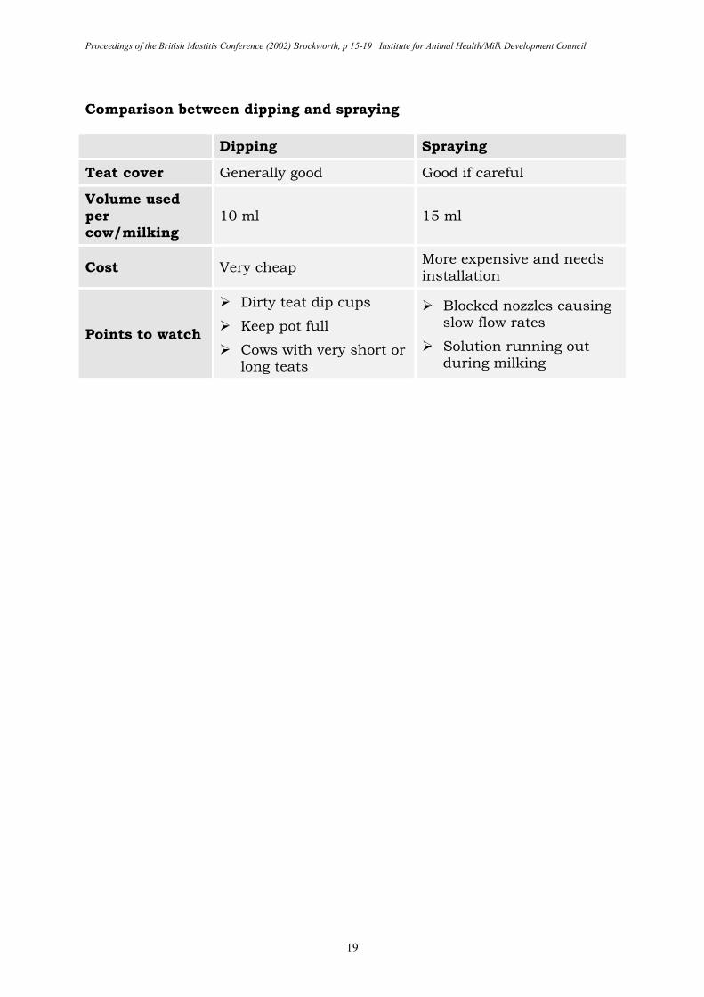
SUMMARY Post-milking teat disinfecting is essential to control the spread of mastitis-causing organisms. The entire surface of each teat needs to be thoroughly coated after each milking throughout the lactation. Teat disinfectant solutions need to be used according to the manufacturer’s recommendations. The ideal form of application is by teat dipping, which will generally achieve a better coating of the teats than spraying and will use considerably less solution. Spraying can be just as effective, provided it is applied diligently, but farmers must accept that they will use up to 50% more solution. Many farmers are reluctant to change from a cheaper teat disinfectant to a branded quality product. Simply a change from spraying to dipping, means that not only can the branded product be used, but also better teat disinfection is likely to result.
Proceedings of the British Mastitis Conference (2002) Brockworth, p 15-19 Institute for Animal Health/Milk Development Council
19
Comparison between dipping and spraying
Dipping Spraying
Teat cover Generally good Good if careful
Volume used per cow/milking
10 ml 15 ml
Cost Very cheap More expensive and needs installation
Points to watch
� Dirty teat dip cups
� Keep pot full
� Cows with very short or long teats
� Blocked nozzles causing slow flow rates
� Solution running out during milking
Proceedings of the British Mastitis Conference (2002) Brockworth, p 20-29 Institute for Animal Health/Milk Development Council
20
ANTIMICROBIAL TREATMENT OF MASTITIS – CHOICE OF THE ROUTE OF ADMINISTRATION AND EFFICACY Satu Pyörälä University of Helsinki, Faculty of Veterinary Medicine, Department of Clinical Veterinary Science, Saari Unit, FIN-04920 Saarentaus, Finland INTRODUCTION Bovine mastitis has been treated with antimicrobials for more than fifty years, and we still lack consensus about the most efficient and economical treatment practices. Mastitis is the most frequent reason for the use of antimicrobials in dairy herds (20), but results from these treatments are less than optimal. The aim of this paper is to review current knowledge of the antimicrobial treatment of mastitis during lactation. GENERAL ASPECTS OF ANTIMICROBIAL TREATMENT The extent to which a drug has access into milk when given systemically or is absorbed and distributes throughout the udder when given intramammarily, depends on its main pharmacokinetic (PK) properties: lipid solubility, degree of ionization, and extent of binding to serum and udder proteins (63). As regards intramammary preparations, the type of vehicle is also important (21). Weak organic bases tend to accumulate in milk in the ionized form after parenteral administration, and attain concentrations higher than those in blood. On the contrary, concentrations of weak acids in milk are much less than those in blood (41). Despite the long history of the use of antimicrobials to treat infections in dairy cows, knowledge of pharmacokinetics of many substances is still limited. Many antibiotic preparations are old and the requirements for authorization at the time they were launched to the markets did not meet the current criteria for PK studies in the target animals. In addition to PK considerations, attention should be paid to pharmacodynamics (PD), which studies the interaction between the bacteria and the drug, and should support PK studies in determining the optimum dosages of the antimicrobials. Very little is known about PD aspects of antimicrobials used in mastitis therapy, because these studies have appeared quite late in veterinary science. Antimicrobials can be divided into concentration-dependent and time-dependent drugs. In the first group (e.g. aminoglycosides and fluoroquinolones) concentration of several times the minimum inhibitory concentration (MIC) for the target organisms at the infection site increases the efficacy. In the latter group (e.g. penicillins and macrolides) the efficacy depends on the time during which the concentration of the drug exceeds the MIC, but high concentrations do not increase efficacy (7). In fact, this characteristic of penicillin G was found very early in streptococcal infections (11).
Proceedings of the British Mastitis Conference (2002) Brockworth, p 20-29 Institute for Animal Health/Milk Development Council
21
An ideal drug for mastitis therapy should have a low MIC for mastitis pathogens. As treatment should be efficient and targeted towards specific infections, Gram-negative and Gram-positive infections in fact would require different antimicrobials (21,43). Anti-mastitis drugs should preferably have bactericidal action, as phagocytosis is impaired in the mammary gland (49). The activity of antimicrobial substances should not be reduced by the presence of milk, but this has been shown for many including macrolides, tetracyclines and trimethoprim-sulphonamides (16,31). INTRAMAMMARY TREATMENT The most common route of administration of antimicrobials in mastitis is the intramammary (IMM) route (21). The advantages of this route are high concentrations of antibiotics achieved in the milk compartment of the mammary gland (21,36), and low consumption of the antimicrobial substances as the drug is administered straight to the infection site. Disadvantages could be the uneven distribution of many substances throughout the udder, risk for contamination when infusing the drug via the teat canal, and possible irritation of the mammary tissue caused by the drug (21). In addition, some in vitro studies have shown that antibiotics may disturb phagocytosis when given IMM (37,64). Clinical relevance of this finding has not been shown. A new technique using an isolated, perfused, bovine udder to study drug distribution in the udder was recently introduced by German authors (12,13). Numerous intramammary products seem to have appeared on the market without supportive scientific data on their efficacy. Although all mastitis tubes carry a label claim for staphylococcal mastitis, the cure rates can be negligible, especially in chronic infections (60). There is little data demonstrating their efficacy for mastitis caused by environmental pathogens (22). In published studies, clinical cure rates have been lower than 60% and bacteriological cure rates as low as 10-40% (1,2,9,55). The requirements for authorization of veterinary drugs at least in the centralized procedure in the EU have become stricter, and efficacy claims must be supported with scientific data (4). Intramammary preparations with combinations of two or even three antibiotics were introduced to mastitis therapy due to suggested synergistic action and to cover all pathogens, Gram-negative bacteria included. The evidence of their efficacy against coliform mastitis is still lacking, and synergistic action was never proven in vivo (59). The idea of fixed combination tubes is outdated; they could be removed from the market, as they have shown no superiority over single components in controlled clinical trials (38,45).
Proceedings of the British Mastitis Conference (2002) Brockworth, p 20-29 Institute for Animal Health/Milk Development Council
22
PARENTERAL TREATMENT The parenteral (systemic) route of administration was introduced into mastitis therapy in the 1970s, mainly after Israeli work (63). Twenty years earlier Swedish researchers had shown by radiographic studies that penicillin G was distributed unevenly when administered by the IMM route (56). It was suggested that systemic treatment would penetrate throughout the udder better and be more efficient in therapy of mastitis. Systemic treatment of mastitis was widely adopted in the Nordic countries and this practice still continues (3,20). However, the superiority of systemic treatment of mastitis over IMM treatment has never been proven in comparative clinical trials. Pharmacokinetics of antimicrobials after systemic administration into adult ruminants is problematic (41). Ruminants eliminate xenobiotics very fast and half-lives of many antibiotics are short. It is difficult to achieve and maintain therapeutic concentrations in milk or udder tissue via systemic administration (63). Intravenous administration would in general produce higher concentrations in milk, but it is often unpractical in field conditions, and not possible for preparations in oily vehicles. The slowly absorbed antibiotic preparations for intramuscular use are the worst choice in mastitis, because they do not generally produce therapeutic concentrations in milk or tissues (5,63). One additional problem for the practitioner is that dosage recommendations of many antibiotic preparations for adult cattle are too low with regard to the MIC of the target bacteria, but residue studies have been carried out using the recommended dosages (26). Repeated intramuscular injections of large volumes of antibiotics are not ideal from the animal welfare point of view. There are very few substances, which from both the PK and PD point of view, would be ideal for systemic mastitis treatment. Even if the drug has ideal characteristics in theory, the treatment results from clinical trials may still be disappointing, as in the case of fluoroquinolones or florfenicol (16,27,43,52). Many broad-spectrum antibiotics, such as oxytetracycline and ceftiofur, have been tested for systemic mastitis treatment or prevention with no effect (10,14,15,39). At least in the latter case, the PK is not suitable for mastitis treatment (15). Macrolides, which are narrow spectrum drugs with activity against Gram-positive bacteria only, would have ideal PK (18,48), but they have problems in PD. They are bacteriostatic and milk strongly interferes with their activity (31). Good penetration into cells does guarantee intracellular killing of bacteria (32). These may be the reasons for the reported poor efficacy of macrolides in mastitis treatment (39,43). With high dosing of spiramycin some authors have shown better results (50), but residues may then cause problems. One of the most commonly used drugs for systemic treatment is penicillin G, but as a weak acid it penetrates poorly into the mammary gland (18). However, as the MIC values of susceptible organisms are low, efficient concentrations can be achieved and maintained in milk using reasonable
Proceedings of the British Mastitis Conference (2002) Brockworth, p 20-29 Institute for Animal Health/Milk Development Council
23
dosing regimens (17,62). Milk does not interfere with the activity of penicillin G (31). Penethamate is a more liphophilic penicillin G formulation and diffuses better than penicillin G procaine into milk (62). INTRAMAMMARY OR PARENTERAL TREATMENT? The ultimate question is, if the antibiotic will accumulate in the milk or in the udder tissue? This may depend on the infection: mastitis streptococci are known to stay in the milk compartment, but Staphylococcus aureus can penetrate into udder tissue and cause a deep infection (49). Coliforms generally are eliminated spontaneously from the udder, and antibiotics are not required at all (8,28,46). In serious cases, however, there can be a risk for bacteriaemia, which supports the use of systemic administration of antibiotics (58). Randomized, comparative field trials using IMM versus parenteral treatment of mastitis with the same antibiotic do not exist. Different systemic or combined regimens using penicillin G procaine to treat mastitis caused by penicillin-susceptible bacteria have been tested in several uncontrolled trials (19,24,43,57). In the study mostly cited, combined treatment was compared with IMM treatment only in experimental S. aureus mastitis with promising results, but different beta-lactam drugs were used and no information about the penicillin susceptibility of the bacterial strain was available (40). In one recent study, treatment with parenteral penethamate hydroiodide was compared with IMM treatment with IMM penicillin-dihydrostreptomycin treatment, and no difference was seen (34). From comparisons between separate studies, it seems that the only type of mastitis where systemic treatment would be clearly advantageous is mastitis caused by S. aureus. Widely distributed penicillin resistance among S. aureus isolates has made use of penicillin G difficult in many countries (41). Cure rates for mastitis caused by penicillin-resistant isolates seems to be inferior to those of penicillin-susceptible isolates (43,44,53,62). It is not known if this is due to pharmacologic problems of the drugs used, or the virulence factors other than β-lactamase production of the resistant isolates. In mastitis caused by penicillin-susceptible S. aureus strains best results were achieved using a combination of systemic and IMM treatment with penicillin G (45). In infections of the milk compartment such as streptococcal mastitis, there is probably no advantage of systemic administration indeed the concentration of penicillin G in milk remains 100-1000 fold lower than when given intramammarily (13,18,36). Based on the results from different studies, cure rates in streptococcal mastitis using IMM treatment are equal or even better than using systemic administration (34,57,61). In coliform mastitis, parenteral administration of antimicrobials has been suggested in severe cases, due to the risk of bacteriaemia (58). Generally, the efficacy of the antimicrobial treatment in coliform mastitis has been
Proceedings of the British Mastitis Conference (2002) Brockworth, p 20-29 Institute for Animal Health/Milk Development Council
24
questioned, as cure rates have been as high with or without antimicrobials or with drugs inefficient in vitro (25,42). Frequent milking with oxytocin has often been recommended for treatment of coliform mastitis (46). This treatment has been reported to give equal or better results than treatment with antimicrobials (22,54). In serious Escherichia coli mastitis with heavy growth of bacteria in the udder, use of systemic antimicrobial treatment may be beneficial (28,47). In an experimental E. coli mastitis model, cefquinome, an advanced-spectrum cephalosporin drug, showed beneficial effects compared to the combination ampicillin-cloxacillin (51). THE EFFECT OF DURATION OF TREATMENT One reason for poor cure rates is probably the short duration of standard treatments (29). Mastitis due to S. aureus, and probably also due to Streptococcus uberis, benefits from a long duration of treatment (19,35). The better efficacy of long treatment in staphylococcal mastitis was already suggested by some authors decades ago (19,62) but more recent studies have confirmed this (43,53). Treatment should be carried out without breaks; the use of so-called extended (pulse) treatment has no scientific justification; it was introduced from the USA, where treatment must be discontinued for the legal withdrawal period between the treatment episodes (55). Regarding some pathogens other than S. aureus, e.g. coagulase-negative staphylococci and mastitis streptococci causing contagious mastitis, a shorter antibiotic treatment is enough both from efficacy and economical points of view. Cost-benefit analysis is essential for treatment decisions (8,30), but we need more knowledge about the efficacy of different treatment regimens. CONCLUSIONS Countries differ in their practices and policies to treat mastitis. In many countries, antimicrobials are available to the farm personnel, and treatment decision and drug selection is made by them (23). In those conditions it is hard to imagine how new information about the PK and PD of mastitis drugs and advances in mastitis therapy could be taken into the field. Diagnosis of mastitis and assessment of prognosis needs also improvement; the concept of one broad-spectrum antibiotic treatment of standard duration for all mastitis types is outdated. Broad-spectrum intramammaries such as 3rd or 4th generation cephalosporins are in some countries marketed for all mastitis treatment. This does not agree with prudent use guidelines (3), and may enhance emergence of wide-spectrum beta-lactamase production among bacteria (6,33). These substances are less efficient than narrow-spectrum preparations against Gram-positive mastitis pathogens, as they are more targeted towards Gram-negative bacteria (41). In streptococcal mastitis (enterococci excluded) and mastitis due to penicillin-susceptible
Proceedings of the British Mastitis Conference (2002) Brockworth, p 20-29 Institute for Animal Health/Milk Development Council
25
staphylococci, penicillin G should be the drug of first choice. In general, a short withdrawal time alone cannot be the sole basis for treatment if the efficacy and safety are questionable. In acute clinical mastitis, a rapid diagnosis is necessary. For this purpose, selective diagnostic media (e.g. Selma selective agar, SVA, Uppsala, Sweden; ColiMast, ICP, Auckland, New Zealand) are available to allow rapid (overnight) diagnosis; treatment can then be re-evaluated and targeted towards the specific pathogen (30).
REFERENCES
1. Aungier, S.P.M. and Austin, F.H.A. (1987) Study of the efficacy of
intramammary antibiotics in the treatment of clinical mastitis Br. Vet. J. 143: 88-90.
2. Anonymous (1994) Poor cure rates limit lactation therapy effectiveness. Udder Topics 17: 5.
3. Anonymous (1996) Use of antimicrobial agents in animals. Report of the working group on antimicrobial agents. Ministry of Agriculture and Forestry in Finland. MAFF Publications 3.
4. Anonymous (2002) http://www.eudra.org/emea.html. 5. Blanchflower, S.E. (1983) Antibiotic concentration in milk from
normal, endotoxin challenged and mastitic quarters of cows after parenteral dosign with amoxycillin. Vet. Res. Commun. 7: 259-260.
6. Bradford, P.A., Petersen, P.J., Fingerman, I.M. and White, D.G. (1999) Characterization of expanded-spectrum cephalosporin resistance in E coli isolates associated with bovine calf diarrhoeal disease. J. Antimicr. Chemother. 44: 607-610.
7. Craig, W. (1993) Pharmacodynamics of antimicrobial agents as basis for determining dosage regimens. J. Clin. Microbiol. Infect. Dis. Suppl. 1: 6-8.
8. Craven, N. (1987) Efficacy and financial value of antibiotic treatment of bovine clinical mastitis during lactation - a review. Br. Vet. J. 143: 410-422.
9. Deluyker, H.A., Chester, S.T. and Van Oye, S.N. (1999) A multilocation clinical trial in lactating dairy cows affected with clinical mastitis to compare the efficacy of treatment with intramammary infusions of a lincomycin/neomycin combination with an ampicillin/cloxacillin combination. J. vet. Pharmacol. Ther. 22: 274-282.
10. Duenas, M.I., Paape, M.J., Wettemann, R.P. and Douglass, L.W. (2001) Incidence of mastitis in beef cows after intramuscular administration of oxytetracycline. J. Anim. Sci. 79: 1996-2005.
11. Eagle, H. and Musselman, A.D. (1948) The rate of bactericidal action of penicillin in vitro as a function of its concentration, and its paradoxically reduced activity at high concentrations against certain organisms. J. Exp. Med. 58: 99-131.
Proceedings of the British Mastitis Conference (2002) Brockworth, p 20-29 Institute for Animal Health/Milk Development Council
26
12. Ehinger, A.M. and Kietzmann, M. (2000a) Tissue distribution of oxacillin and ampicillin in the isolated perfused bovine udder. J. Vet. Med. A. 47: 157-168.
13. Ehinger, A.M. and Kietzmann, M. (2000b) Tissue distribution of benzylpenicillin after intramammary administration in the isolated perfused bovine udder. J. vet. Pharm. Ther. 23: 303-310.
14. Erskine, R.J., Barlett, P.C., Crawshaw, P.C. and Gombas, P.M. (1994) Efficacy of intramuscular oxytetracycline as a dry cow treatment for Staphylococcus aureus mastitis. J. Dairy Sci. 77: 3347-3353.
15. Erskine, R.J., Barlett, P.C., Johnson, G.L. and Halbert, L.W. (1996) Intramuscular administration of ceftiofur sodium versus intramammary infusion of penicillin/novobiocin for treatment of Streptococcus agalactiae mastitis in dairy cows. J. Am. Vet. Med. Assoc. 208: 258-260.
16. Fang, W. and Pyörälä, S. (1996) Mastitis causing Escherichia coli: serum sensitivity and susceptibility to selected antibacterials in milk. J. Dairy Sci. 79: 76-82.
17. Franklin, A., Holmberg, O., Horn af Ranzien, M. and Åström, G. (1984) Effect of procaine benzylpenicillin alone or in combination with dihydrostreptomycin on udder pathogens in vitro and in experimentally infected bovine udders. J. Am. Vet. Res. 45: 1398-1402.
18. Franklin, A., Horn af Ranzien, M., Obel, N., Östensson, K. and Åström, G. (1986) Concentrations of penicillin, streptomycin, and spiramycin in bovine udder tissue liquids. J. Am. Vet. Res. 47: 804-807.
19. Funke, H. (1982) Practical experiences in the treatment of clinical mastitis, Proc. Symp. Mast. Contr. Ther. Novo Nordisk, Copenhagen, Denmark. pp 1-2.
20. Grave, T., Greko, C., Nilsson, L., Odensvik, K., Mörk, T. and Rönning, M. (1999) The usage of veterinary antibacterial drugs for mastitis in cattle in Norway and Sweden during 1990-1997. Prev. Vet. Med. 42: 45-55.
21. Gruet, P., Maincent, P., Berthelot, X. and Kaltsatos, V. (2001) Bovine mastitis and intramammary drug delivery: review and perspectives. Adv. Drug Deliv. Rev. 50: 245-259.
22. Guterbock, W.M., van Eenennaam, A.L., Anderson, R.J., Gardner, I.A., Cullor, J.S. and Holmberg, C.A. (1993) Efficacy of intramammary antibiotic therapy for treatment of clinical mastitis caused by environmental pathogens. J. Dairy Sci. 76: 3437-3444.
23. Guard, C. (1999) Disease treatment programs for farm personnel: an example for a program for clinical mastitis. Proc. NMC Reg. Meeting, Waterloo, Ontario, pp 30-34.
24. Jarp, J., Bugge, H.P. and Larsen, S. (1989) Clinical trial of three therapeutic regimens for bovine mastitis. Vet. Rec. 124: 630-634.
25. Jones, G.F. and Ward, G.E. (1990) Evaluation of systemic administration of gentamicin for treatment of coliform mastitis in cows. J. Am. Vet. Med. Assoc. 197: 731-735.
Proceedings of the British Mastitis Conference (2002) Brockworth, p 20-29 Institute for Animal Health/Milk Development Council
27
26. Kaartinen, L., Löhönen, K., Wiese, B., Franklin, A. and Pyörälä, S. (1999) Pharmacokinetics of sulphadiazine-trimethoprim in lactating dairy cows. Acta vet. Scand. 40: 271-278.
27. Kaartinen, L., Salonen, M., Älli, L. and Pyörälä, S. (1995) Pharmacokinetics of enrofloxacin after single intravenous, intramuscular and subcutaneous injections in lactating cows. J. vet. Pharm. Ther. 18: 357-362.
28. Katholm, J. (2001) Treatment of coliform mastitis in bovine practice: can antibiotics be avoided? Proc. 11th Int. Conf. Prod. Dis. Copenhagen, Denmark. p 33.
29. Knight, C.H., Fitzpatrick, J.L., Logue, D.N. and Platt, D.J. (2000) Efficacy of two non-antibiotic therapies and topical liniment, against bovine staphylococcal mastitis. Vet. Rec. 146: 311-316.
30. Leslie, K. and Keefe, G. (1998) Decision-making in clinical mastitis therapy programmes. IDF Bulletin. 330: 21-22.
31. Louhi, M., Inkinen, K., Myllys, V. and Sandholm, M. (1992) Relevance of sensitivity testings (MIC) of S. aureus to predict the antibacterial action in milk. J. Vet. Med. B 39: 253-262.
32. Madgwick, L., Mayer, S. and Keen, P. (1989) Penetration of antibiotics into bovine neutrophils and their activity against Staphylococcus aureus. J. Antimicr. Chemother. 24: 709-718.
33. Mayer, K.H., Opal, S.M. and Medeiros, E.A. (1994) Mechanisms of antibiotic resistance. In: Mandell et al (eds.), Principles and practice of infectious diseases. 4th ed. Churchill Livingstone, New York, 212-225.
34. McDougall, S.Mc. (1998) Efficacy of two antibiotic treatments in curing clinical and sub-clinical mastitis in lactating dairy cows. New. Z. Vet. J. 46: 226-232.
35. Milne, M.H., Barrett, D.C., Fitzpatrick, J.L. and Biggs, A.M. (2000) Survey of bacterial causes of clinical mastitis and a pilot investigation of the response to treatment of cases caused by Streptococcus uberis. Proc. IDF Symp. Immunol. Rumin. Mamm. Gland. Stresa, Italy. pp 379-381.
36. Moretain, J.P. and Boisseau, J. (1989) Excretion of penicillins and cephalexin in bovine milk following intramammary administration. Food Add. Contamin. 6: 79-90.
37. Nickerson, S.C., Paape, M.J., Harmon, R.J. and Ziv, G. (1986) Mammary leukocyte response to drug therapy. J. Dairy Sci. 69: 1733-1742.
38. Oedegaard, S. and Sviland, S. (2001) Comparison of intramammary antibiotic preparations for the treatment of clinical bovine mastitis caused by bacteria sensitive to penicillin. Proc. 2nd Int. Symp. Mast. Milk Quality, Vancouver, Canada. pp 502-503.
39. Owens, W.E., Nickerson, S.C. and Ray, C.H. (1999) Efficacy of parenterally or intramammarily administered tilmicosin or ceftiofur against Staphylococcus aureus mastitis during lactation. J. Dairy Sci. 82: 645-647.
Proceedings of the British Mastitis Conference (2002) Brockworth, p 20-29 Institute for Animal Health/Milk Development Council
28
40. Owens, W.E., Watts, J.L., Boddie, R.L. and Nickerson, S.C. (1988) Antibiotic treatment of mastitis: Comparison of intramammary and intramammary plus intramuscular therapies. J. Dairy Sci. 71: 3143-3147.
41. Prescott, J.F., Baggot, J.D. and Walker, R.D. (eds) (2000) Antimicrobial therapy in veterinary medicine. 3. ed. Iowa State University Press, Ames, Iowa.
42. Pyörälä, S., Kaartinen, L., Käck, H. and Rainio, V. (1944) Efficacy of Two Therapy Regimes for Treatment of Experimentally Induced Escherichia coli Mastitis in the Bovine. J. Dairy Sci. 77: 453-461.
43. Pyörälä, S. and Pyörälä, E. (1998) Efficacy of parenteral administration of three antimicrobial agents in treatment of clinical mastitis in lactating cows: 487 cases (1989-1995). J. Am. Vet. Med. Assoc. 212: 407-412.
44. Pyörälä, S., Taponen, S., Jantunen, A. and Pyörälä, E. (2000) Efficacy of targeted 5-day parenteral and intramammary treatment of clinical Staphylococcus aureus mastitis caused by penicillin-susceptible or penicillin-resistant bacterial strain. Proc. IDF Symp. Immunol. Rumin. Mamm. Gland. Stresa, Italy. pp 382-384.
45. Pyörälä, S., Taponen, S., Dredge, K., Henriksson, B., Pyyhtiä, A., Suojala, L., Junni, R. and Heinonen, K. (2001) Efficacy of intramammary treatment with procaine penicillin G vs procaine penicillin G plus neomycin in bovine clinical mastitis – a double blind field study. Proc. 2nd Int. Symp. Mast. Milk Quality. Vancouver, Canada. pp 219-223.
46. Radostits, O.M., Gay, C.C., Blood, D.C. and Hinchcliff, K.W. (2000) Coliform mastitis caused by Escherichia coli, Klebsiella spp., and Enterobacter aerogenes. In: Veterinary Medicine: A Textbook of the Diseases of Cattle, Sheep, Pigs, Goats and Horses. 9. ed. W.B. Saunders Company Ltd, New York, USA, 639-650.
47. Rantala, M., Kaartinen, L., Välimäki, E., Styrman, M., Hiekkaranta, M., Niemi, A., Saari, L. and Pyörälä, S. ( ) Efficacy and pharmacokinetics of enrofloxacin and flunixin meglumine for treatment of cows with experimentally induced Escherichia coli mastitis. J. vet. Pharm. Ther. In press.
48. Sanders, P., Moulin, G., Guillot, P., Dagorn, M., Perjant, P., Delepine, B., Gaudiche, C. and Mourot, D. (1992) Pharmacokinetics of spiramycin after intravenous, intramuscular and subcutaneous administration in lactating cows. J. vet. Pharm. Ther. 15: 53-61.
49. Sandholm, M., Kaartinen, L. and Pyörälä. S. 1990. Bovine Mastitis - Why does antibiotic therapy not work? An overview. J. vet. Pharm. Ther. 13: 248-260.
50. Schällibaum, M., Nicolet, J. and Bosson, J. (1981) Die Behandlung von chronisch-subklinischen Staphylokokkenmastitiden beim laktierenden Rind durch die Verabreichung von hohen Spiramycindosen. Schweiz. Arch. Tierheilk. 123: 277-292.
Proceedings of the British Mastitis Conference (2002) Brockworth, p 20-29 Institute for Animal Health/Milk Development Council
29
51. Shpigel, N.Y., Levin, D., Winkler, M., Saran, A., Ziv, G. and Böttner, A. (1997) Efficacy of cefquinome for treatment of cows with mastitis experimentally induced using Escherichia coli. J. Dairy Sci. 80: 318-323.
52. Soback, S., Paape, M.J., Filep, R., Varma, K.J. (1995) Florfenicol pharmacokinetics in lactating cows after intravenous, intramammary and intramuscular administration. Proc. 3rd Int. IDF Mast. Sem. Tel-Aviv, Israel. pp 39-43.
53. Sol, J., Sampimon, O.C., Barkema, H.W. and Schukken, Y.H. (2000) Factors associated with cure after therapy of clinical mastitis caused by Staphylococcus aureus. J Dairy Sci. 83: 278-284.
54. Stämpfli, H.R., Nemeth, J., Leslie, K., Gyles, C.L. and Muckle. C.A. (1994) Clinical response and antimicrobial residue test results in non-antibiotic treated cows with induced acute coliform mastitis. Proc. XVIII World Buiatrics Congr. Bologna, Italy. pp 869-871.
55. Timms, L. (1998) Evaluation of recommended and extended pirlimycin mastitis therapy for recent and chronic high SCC cows in two herds. Proc. 37. Annual NMC Meeting, Missouri. pp 271-272.
56. Ullberg, S., Hansson, E. and Funke, H. (1958) Distribution of penicillin in mastitic udders following intramammary injection – an autoradiographic study. Am. J. Vet. Res. 19: 84-92.
57. Waage, S. (1997) Comparison of two regimens for the treatment of clinical bovine mastitis caused by bacteria sensitive to penicillin. Vet. Rec. 141: 616-620.
58. Wenz, J.R., Barrington, G.M., Garry, F.B., McSweeney, K.D., Dinsmore, R.P., Goodell, G. and Callan, R.J. (2001) Bacteremia associated with naturally occurring acute coliform mastitis in dairy cows. J. Am. Vet. Med. Assoc. 219: 976-981.
59. Whittem, T. and Hanlon, D. (1997). Dihydrostreptomycin or streptomycin in combination with penicillin in dairy cattle therapeutics: A review and re-analysis of published data, Part 1: Clinical pharmacology. New Zealand Vet. J. 45: 178-184.
60. Wilson, D.J., Gonzales, R.N., Case, K.l., Garrison, L.L. and Gröhn, Y.T. (1999) Comparison of seven antibiotic treatments with no treatment against bovine mastitis pathogens. J. Dairy Sci. 82: 1664-1670.
61. Wilson, C.D. (1980) Antibiotic therapy in mastitis control. In: Bramley et al (eds), Mastitis Control and Herd Management. NIRD Technical Bulletin. 4: 113-127.
62. Ziv, G. and Storper, M. (1985) Intramuscular treatment of sub-clinical staphylococcal mastitis in lactating cows with penicillin G, methicillin and their esters. J. Vet. Pharm. Ther. 8: 276-283.
63. Ziv, G. (1980) Drug selection and use in mastitis: systemic vs. local therapy. J. Am. Vet. Med. Assoc. 176: 1109-1115.
64. Ziv, G., Paape, M. J. and Dulin, M.T. (1983) Influence of antibiotics and intramammary antibiotic products on phagocytosis of Staphylococcus aureus by bovine leukocytes. Am. J. Vet. Res. 44: 385-388.

Proceedings of the British Mastitis Conference (2002) Brockworth, p 30-36 Institute for Animal Health/Milk Development Council
30
HOW TO GET ANTIBIOTIC TO THE SITE OF AN INTRAMAMMARY INFECTION Keith Lawrence Elanco Animal Health, Kingsclere Road, Basingstoke, Hants RG21 6XA The title of this paper is a description of a journey without a clear destination, so I will first clarify where the antibiotic needs to get. Within the udder there are a number of sites where bacteria involved in mastitis may be found. Indeed some types of bacteria may pass through all these sites as the manifestation of mastitis moves from sub-clinical to acute clinical to chronic. The most obvious site is the milk followed by healthy tissue, scarred tissue and finally inside both white blood cells and the cells lining the ducts and secretary tissues in the udder. Infections in these sites are attacked via a range of antibiotics administered either by intramammary infusions or injection. It is the choice of delivery route coupled to duration of treatment that can lead to a successful outcome in both clinical and bacteriological terms. This point needs further reinforcement, as clinical cure and bacteriological cure are very difficult to achieve with infections caused by Staphylococcus aureus and with some Streptococcus uberis infections. The 80 to 90% clinical cures recorded for certain strains of bacteria hide an underlying bacteriological cure rate of only 25 to 30%. This has become important as somatic cell count (SCC) drive quality payments – clinical cure leading to the milk looking normal and being returned to the bulk tank is worth little if the bacteria causing the disease remain to cause elevated SCC and subsequent cases of clinical mastitis. Our target should be a bacteriological cure that may require long treatment periods and significant milk discard and not ‘get the milk back in to the tank as quickly as possible’. We must also accept that there are some sites in the udder, especially in cows chronically infected with S. aureus, that are out of reach of antibiotics and the cow should be culled or as is the case in the US the affected quarter culled. (This technique tends to be used in high merit cattle with unresponsive mastitis in a single quarter). Let’s first discuss the enemy before we assemble our armoury:- Staphylococcus aureus – this passes through the milk in to normal udder tissue and over time produces significant scarring. It is found inside macrophages in the milk and within udder tissue as well as potentially being engulfed by cells lining the teat and lactiferous sinuses during the early dry period. These cells engulf milk constituents after drying off and may also engulf adherent S. aureus. The bacteria can grow a shaggy, slimy coat when in milk and avoid the attentions of patrolling white blood cells (macrophages).
Proceedings of the British Mastitis Conference (2002) Brockworth, p 30-36 Institute for Animal Health/Milk Development Council
31
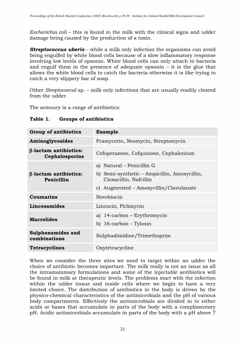
Escherichia coli – this is found in the milk with the clinical signs and udder damage being caused by the production of a toxin. Streptococcus uberis – while a milk only infection the organisms can avoid being engulfed by white blood cells because of a slow inflammatory response involving low levels of opsonin. White blood cells can only attach to bacteria and engulf them in the presence of adequate opsonin – it is the glue that allows the white blood cells to catch the bacteria otherwise it is like trying to catch a very slippery bar of soap. Other Streptococcal sp. – milk only infections that are usually readily cleared from the udder. The armoury is a range of antibiotics: Table 1. Groups of antibiotics
Group of antibiotics Example
Aminoglycosides Framycetin, Neomycin, Streptomycin
ββββ-lactam antibiotics: Cephalosporins
Cefoperazone, Cefquinone, Cephalonium
ββββ-lactam antibiotics: Penicillin
a) Natural – Penicillin G
b) Semi-synthetic – Ampicillin, Amoxycillin, Cloxacillin, Nafcillin
c) Augmented – Amoxycillin/Clavulanate
Coumarins Novobiocin
Lincosamides Lincocin, Pirlimycin
Macrolides a) 14-carbon – Erythromycin
b) 16-carbon – Tylosin
Sulphonamides and combinations
Sulphadimidine/Trimethoprim
Tetracyclines Oxytetracycline
When we consider the three sites we need to target within an udder the choice of antibiotic becomes important. The milk really is not an issue as all the intramammary formulations and some of the injectable antibiotics will be found in milk at therapeutic levels. The problems start with the infection within the udder tissue and inside cells where we begin to have a very limited choice. The distribution of antibiotics in the body is driven by the physico-chemical characteristics of the antimicrobials and the pH of various body compartments. Effectively the antimicrobials are divided in to either acids or bases that accumulate in parts of the body with a complimentary pH. Acidic antimicrobials accumulate in parts of the body with a pH above 7
Proceedings of the British Mastitis Conference (2002) Brockworth, p 30-36 Institute for Animal Health/Milk Development Council
32
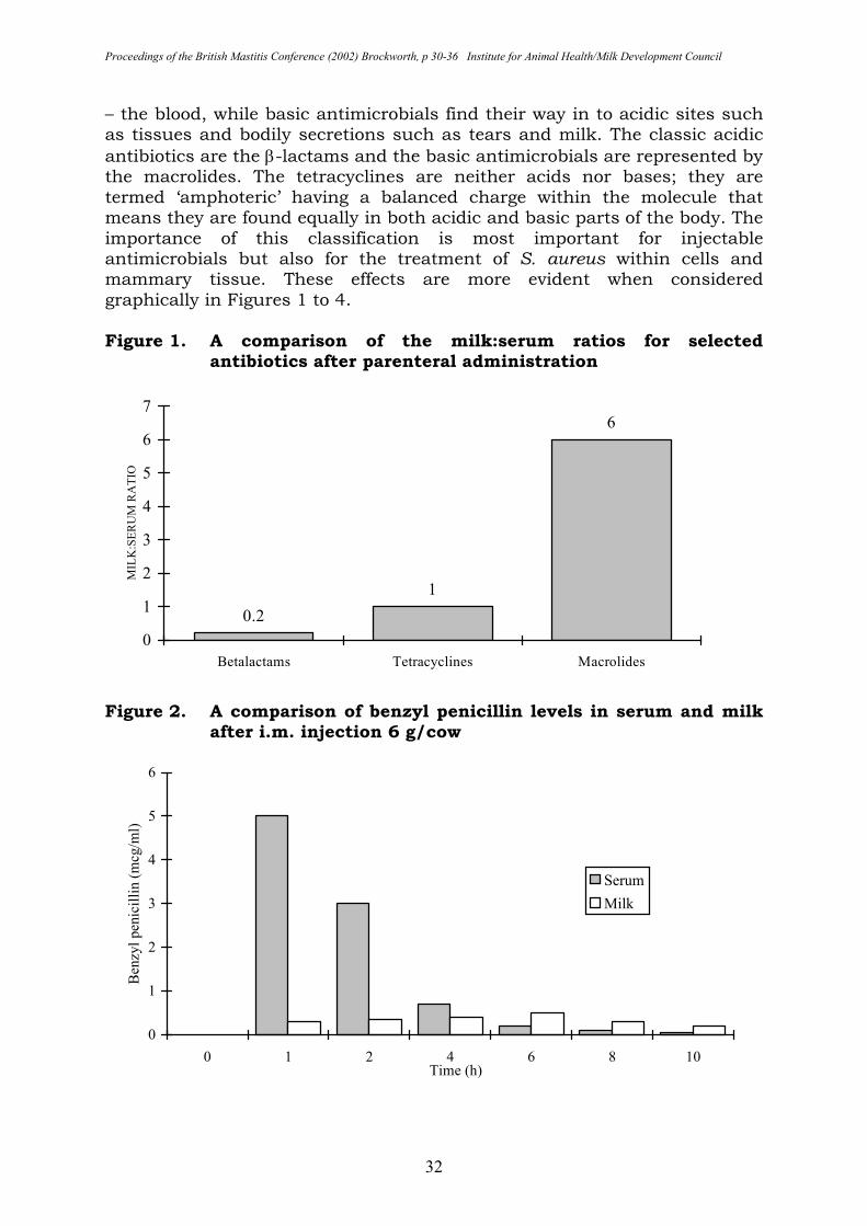
– the blood, while basic antimicrobials find their way in to acidic sites such as tissues and bodily secretions such as tears and milk. The classic acidic antibiotics are the β-lactams and the basic antimicrobials are represented by the macrolides. The tetracyclines are neither acids nor bases; they are termed ‘amphoteric’ having a balanced charge within the molecule that means they are found equally in both acidic and basic parts of the body. The importance of this classification is most important for injectable antimicrobials but also for the treatment of S. aureus within cells and mammary tissue. These effects are more evident when considered graphically in Figures 1 to 4. Figure 1. A comparison of the milk:serum ratios for selected
antibiotics after parenteral administration
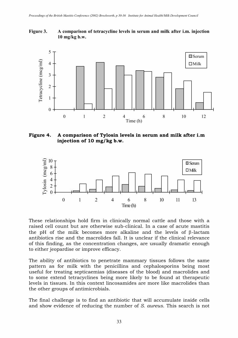
Figure 2. A comparison of benzyl penicillin levels in serum and milk
after i.m. injection 6 g/cow
0.2
1
6
0
1
2
3
4
5
6
7
Betalactams Tetracyclines Macrolides
MIL
K:S
ER
UM
RA
TIO
0
1
2
3
4
5
6
0 1 2 4 6 8 10Time (h)
Ben
zyl pen
icil
lin (
mcg
/ml)
Serum
Milk
Proceedings of the British Mastitis Conference (2002) Brockworth, p 30-36 Institute for Animal Health/Milk Development Council
33
Figure 3. A comparison of tetracycline levels in serum and milk after i.m. injection
10 mg/kg b.w.
Figure 4. A comparison of Tylosin levels in serum and milk after i.m injection of 10 mg/kg b.w.
These relationships hold firm in clinically normal cattle and those with a raised cell count but are otherwise sub-clinical. In a case of acute mastitis the pH of the milk becomes more alkaline and the levels of β-lactam antibiotics rise and the macrolides fall. It is unclear if the clinical relevance of this finding, as the concentration changes, are usually dramatic enough to either jeopardise or improve efficacy. The ability of antibiotics to penetrate mammary tissues follows the same pattern as for milk with the penicillins and cephalosporins being most useful for treating septicaemias (diseases of the blood) and macrolides and to some extend tetracyclines being more likely to be found at therapeutic levels in tissues. In this context lincosamides are more like macrolides than the other groups of antimicrobials. The final challenge is to find an antibiotic that will accumulate inside cells and show evidence of reducing the number of S. aureus. This search is not
0
2
4
6
8
10
0 1 2 4 6 8 10 11 13Time (h)
Tylo
sin (m
cg/m
l)
Serum
Milk
0
1
2
3
4
5
0 1 2 4 6 8 10 12Time (h)
Tet
racy
clin
e (m
cg/m
l)
Serum
Milk
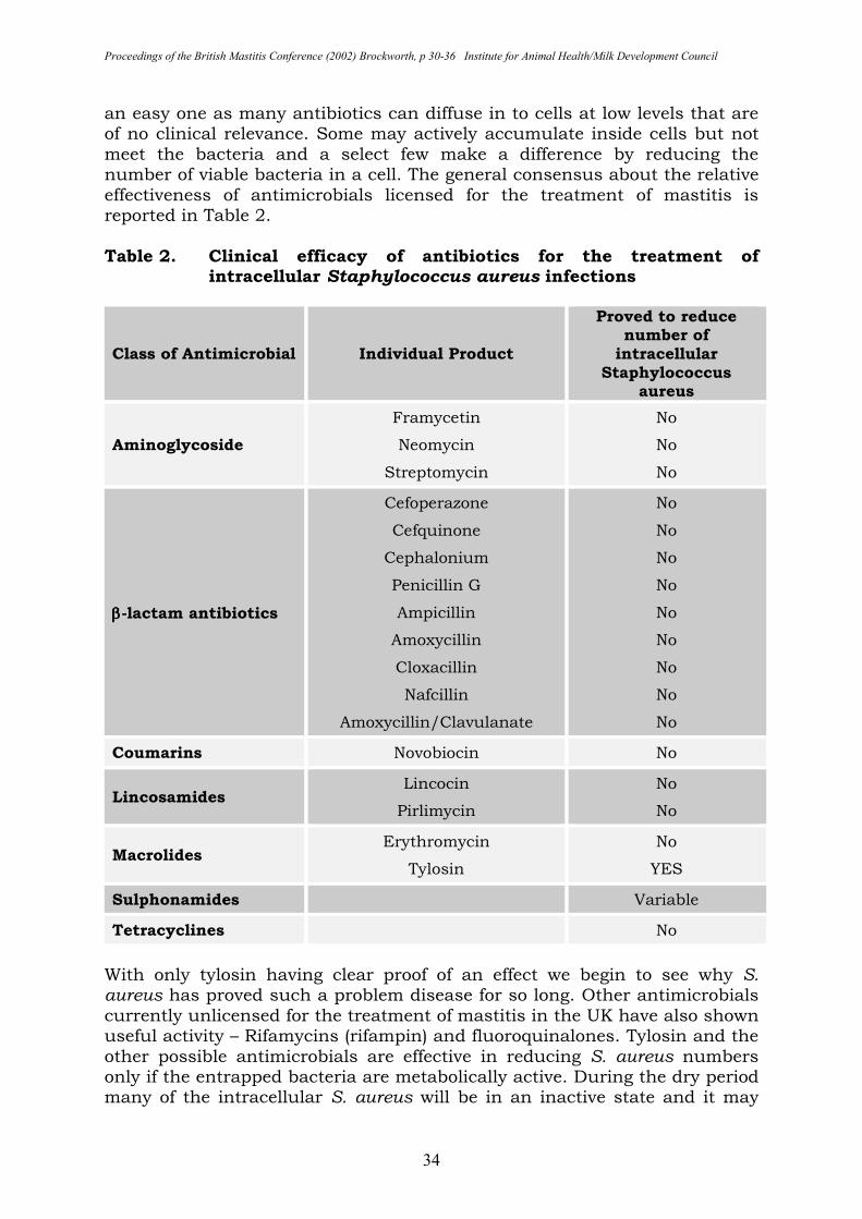
Proceedings of the British Mastitis Conference (2002) Brockworth, p 30-36 Institute for Animal Health/Milk Development Council
34
an easy one as many antibiotics can diffuse in to cells at low levels that are of no clinical relevance. Some may actively accumulate inside cells but not meet the bacteria and a select few make a difference by reducing the number of viable bacteria in a cell. The general consensus about the relative effectiveness of antimicrobials licensed for the treatment of mastitis is reported in Table 2. Table 2. Clinical efficacy of antibiotics for the treatment of
intracellular Staphylococcus aureus infections
Class of Antimicrobial Individual Product
Proved to reduce number of intracellular
Staphylococcus aureus
Aminoglycoside
Framycetin
Neomycin
Streptomycin
No
No
No
ββββ-lactam antibiotics
Cefoperazone
Cefquinone
Cephalonium
Penicillin G
Ampicillin
Amoxycillin
Cloxacillin
Nafcillin
Amoxycillin/Clavulanate
No
No
No
No
No
No
No
No
No
Coumarins Novobiocin No
Lincosamides Lincocin
Pirlimycin
No
No
Macrolides Erythromycin
Tylosin
No
YES
Sulphonamides Variable
Tetracyclines No
With only tylosin having clear proof of an effect we begin to see why S. aureus has proved such a problem disease for so long. Other antimicrobials currently unlicensed for the treatment of mastitis in the UK have also shown useful activity – Rifamycins (rifampin) and fluoroquinalones. Tylosin and the other possible antimicrobials are effective in reducing S. aureus numbers only if the entrapped bacteria are metabolically active. During the dry period many of the intracellular S. aureus will be in an inactive state and it may
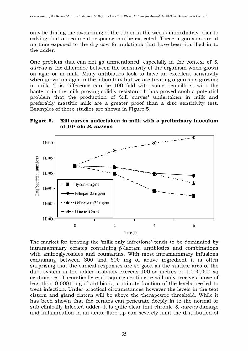
Proceedings of the British Mastitis Conference (2002) Brockworth, p 30-36 Institute for Animal Health/Milk Development Council
35
only be during the awakening of the udder in the weeks immediately prior to calving that a treatment response can be expected. These organisms are at no time exposed to the dry cow formulations that have been instilled in to the udder. One problem that can not go unmentioned, especially in the context of S. aureus is the difference between the sensitivity of the organism when grown on agar or in milk. Many antibiotics look to have an excellent sensitivity when grown on agar in the laboratory but we are treating organisms growing in milk. This difference can be 100 fold with some penicillins, with the bacteria in the milk proving solidly resistant. It has proved such a potential problem that the production of ‘kill curves’ undertaken in milk and preferably mastitic milk are a greater proof than a disc sensitivity test. Examples of these studies are shown in Figure 5. Figure 5. Kill curves undertaken in milk with a preliminary inoculum
of 107 cfu S. aureus
The market for treating the ‘milk only infections’ tends to be dominated by intramammary cerates containing β-lactam antibiotics and combinations with aminoglycosides and coumarins. With most intramammary infusions containing between 300 and 600 mg of active ingredient it is often surprising that the clinical responses are so good as the surface area of the duct system in the udder probably exceeds 100 sq metres or 1,000,000 sq centimetres. Theoretically each square centimetre will only receive a dose of less than 0.0001 mg of antibiotic, a minute fraction of the levels needed to treat infection. Under practical circumstances however the levels in the teat cistern and gland cistern will be above the therapeutic threshold. While it has been shown that the cerates can penetrate deeply in to the normal or sub-clinically infected udder, it is quite clear that chronic S. aureus damage and inflammation in an acute flare up can severely limit the distribution of
1.E+00
1.E+02
1.E+04
1.E+06
1.E+08
1.E+10
0 2 4 6
Time (h)
Log b
acte
rial
num
ber
s
Tylosin 4 mcg/ml
Pirlimycin 2.5 mgc/ml
Cefaperazone 2.5 mcg/ml
Untreated Control
Proceedings of the British Mastitis Conference (2002) Brockworth, p 30-36 Institute for Animal Health/Milk Development Council
36
the antibiotic. It is also probably not widely known that a significant proportion of a dose of antibiotic instilled in to the udder can be absorbed in to the blood and, depending on the individual antibiotic under consideration, is then unavailable to treat the infection. As long as the organisms involved in the infection are sensitive to the chosen antibiotic there seems little to choose between the intramammary tubes for the treatment of Streptococcus agalactiae and Streptococcus dysgalactiae. E. coli is also a milk infection but being a Gram-negative organism there is a more limited range of options in the antibiotic armoury. The use of antibiotics in a disease driven by toxin production is a secondary consideration to fluid therapy and the possible role of anti-inflammatory drugs. S. aureus is the outstanding problem if it allowed to establish in the udder before action is taken. With a recent infection, even in a lactating cow, there is a chance of complete resolution as long as the infection is taken seriously. Do not just treat with a single course of intramammary tubes and expect to clear the organism – hit it with the ‘kitchen sink’ – at least double the recommended course of tubes plus an injected antibiotic preferably a macrolide. Indeed early treatment of the first infection of S. aureus can prove highly effective with bacteriological cures being nearer 90% than the often-quoted 25%. As soon as such a prolonged course of treatment is considered there should be real attention to the milk withdrawal period. Unless the proposed usage is specifically licensed and depending on the milk contract this prolonged combination treatment is either a 7-day milk withdrawal (standard withdrawal) or until a negative Delvo SP test. There is absolutely no excuse to try and get the milk from these cows back in to the tank as soon as it is possible – the only objective should be to clear the infection. The younger the cow and the fewer the cases of clinical mastitis she has suffered the more likely a successful outcome. In cattle with no evidence of clinical mastitis and no palpable changes in the udder, but with a raised SCC a prolonged course of pirlimycin may well prove an effective alternative. SUMMARY Long-standing prescribing and usage habits can no longer drive our response to a clinical case of mastitis. A basic understanding of the different organisms associated with mastitis coupled with serious thought about where antibiotics go in the udder can drive a change in expectations. We must judge our treatment regimen by the bacteriological cure rate not by how quickly we can get milk back in to the bulk tank. Apparently normal milk is not the end point of a course of treatment for mastitis – concentrate on the SCC and the disease causing organisms.
Proceedings of the British Mastitis Conference (2002) Brockworth, p 37-50 Institute for Animal Health/Milk Development Council
37
PRACTICAL USE OF ANTIBIOTICS IN CLINICAL AND SUB-CLINICAL MASTITIS Andrew Biggs, The Vale Veterinary Centre, The Laurels, Station Road, Tiverton, Devon EX16 4LF Email [email protected] SUMMARY The use of antibiotics to treat intramammary infections is only part of a farm mastitis control plan. The effects of antibiotics on prevalence and incidence of intramammary infections in a herd are exerted by their influence on the outcome of treatment of clinical and sub-clinical mastitis either in lactation or during the dry period. The bacteriological cure rates are determined in part by the dose and duration of treatment of susceptible infections. Economic constraints and concerns about antibiotic residues have encouraged short treatment courses with short withhold periods. This is probably contrary to the requirement of ideal treatment protocols. The benefits and pitfalls of extended and or aggressive treatment are discussed with the aid of treatment protocol examples. INTRODUCTION The aims of all mastitis control programs include reducing both the rate of new infections and the duration of existing infections. The successful treatment of existing infections has an impact mainly on the duration of infection but exerts some effect on new infection rate by reducing the prevalence of infected quarters within the herd. Part of any mastitis control programme will include early identification and prompt treatment of clinical mastitis. The requirement for high quality milk, in particular low somatic cell count (SCC), has sharpened the focus on sub-clinical mastitis. Over time this had led to the development of various management tools to help identify sub-clinical infections which in turn has created a demand for ways to eliminate these sub-clinical infections. The importance of farm actions to prevent new infections remain. The requirement to identify sub-clinical mastitis, assess and where appropriate eliminate infection either by treatment or by culling the infected cow should now be part of all mastitis control programs. THE OBJECTIVES WHEN TREATING MASTITIS Some fundamental questions should be asked.
Proceedings of the British Mastitis Conference (2002) Brockworth, p 37-50 Institute for Animal Health/Milk Development Council
38
Do I need to treat?
Perhaps the question should be - Why should I treat? A common belief is that treatment will result in a benefit to the cow or herd (health, welfare gain) and/or benefit the dairy farmer (limit financial loss). The objectives are to resolve clinical signs, rapidly re-permit milk sales, limit udder damage and prevent spread of infection. The likelihood of spontaneous recovery must be weighed against the prospects of successful therapy and the additional costs incurred or benefits arising. High spontaneous clinical recovery rates in the absence of therapy and the often limited success of antibiotic treatment in effecting bacteriological cures should not be interpreted as a reason for abandoning treatment of mild clinical cases. Reduction in bacterial numbers shed from infected quarters as a result of antibiotic treatment helps reduce spread of infection and to improve bacteriological quality of bulk milk. Both are instrumental in maintaining premium milk quality (11). The use of non-antibiotic preparations to treat mastitis may allow large numbers of bacteria to enter the bulk tank with a resulting quality payment penalty (8). What are the chances of success? The infection status of a quarter can be evaluated in a number of ways. The change of status with or without treatment can lead to a disappearance of clinical signs (clinical cure), an absence of causal pathogen (bacteriological cure) and a return to cow somatic cell counts of below 200,000 cell per ml (cell count cure). When assessing cure rates it is as well not to attribute all outcomes to antibiotic therapy. It is worth remembering that on occasions infections improve despite treatment, as a consequence of self-cure. If a change of treatment results in a clinical improvement that this might have been going to happen anyway. Sometimes with an apparent clinical failure of treatment there may in fact be a bacteriological cure. The udder damage may be so severe that a clinical cure may never occur (lost quarter) or will only be evident once the udder has had sufficient time to repair. Somatic cell count may remain elevated despite a bacteriological cure. Antibiotics only kill bacteria they do not heal udders. Clinical and bacteriological cure rates vary according to a number of factors including pathogen involved, previous infection history and duration of infection. A study of a non-treatment approach to mild mastitis (mostly Staphylococcus aureus infections) in one herd resulted in an 87% spontaneous clinical cure but only a 20% bacterial cure rate based on the assessment criteria used (10). It is likely that both the rate of spontaneous clinical cure, and in particular bacteriological cure, will be much lower in more severe mastitis cases. It should be noted that the incidence of clinical mastitis caused by S. aureus increased in this herd after cessation of antibiotic treatment during lactation. In general, bacterial cure rates are lower than clinical cure rates. Bacteriological cure rates are highest for mastitis caused by Gram-negative bacteria, will be relatively unaffected by antibiotic treatment and may also approach 100% in mild cases.
Proceedings of the British Mastitis Conference (2002) Brockworth, p 37-50 Institute for Animal Health/Milk Development Council
39
Bacteriological cure rates for mastitis caused by Gram-positive bacteria other than Streptococcus agalactiae such as S. aureus or Streptococcus uberis are significantly lower and achieved by antibiotic treatment. Bacteriological cure rates for S. aureus infections treated with antibiotic at drying off may well be 65 to 75% whereas treatment during lactation may result in bacteriological cure rates as low as 25 to 30% (20). Case selection and treatment can improve success rates dramatically and may result in bacteriological cure rates in lactation of greater than 70%, even with S. aureus infections. A realistic approach must be taken, especially with pathogens such as S. aureus, where chronic (long duration) infections are common place, success rates are low, and removal of the cow from the herd (culling) is often the most appropriate treatment. Culling of cows achieves a 100% cure rate (1), the infection is removed from the herd, in that cow at least, but often the infection has spread within the herd and other cows are ready to take over the mantle of highest cell count cow in the herd. Early identification and treatment of intramammary infection will generally lead to higher cure rates. Early treatment of experimentally induced Str. uberis infections in cows previously uninfected produced bacteriological cure rates varying from zero for no antibiotic treatment to 80% for aggressive off label treatment (16,29). What drug(s) should I use?
Very often cure rates (clinical, bacteriological or cell count) are more influenced by factors other than the therapeutic agent used. The outcome of treatment is determined more by case characteristics such as causal pathogen, duration of infection and the duration of treatment than by therapeutic agent used. A study in Israel showed that bacteriological cure rates using penicillin G for chronic S. aureus infections (of at least 3 to 5 months duration) could be raised from 23% to 90% by increasing the treatment duration from the label 3 tubes to 18-20 tubes (32). A review of the literature since 1978 evaluated the available efficacy data for various antibiotics and although the calculated cure rate data was very variable it is possible to conclude that greater bacteriological cure rates result with certain pathogens such as Str. agalactiae as compared to S. aureus (20). What are the economics of treatment?
The possibility of spontaneous recovery must be weighed against the prospects of successful therapy and the additional costs incurred and benefits arising. When treating a mild case of clinical mastitis with a label treatment of 3 tubes the cost of drugs used will be 20 to 30% of direct costs whereas the cost of discarded milk during treatment and during withhold period will be 70 to 80% of direct costs. No allowance has been made for any subsequent reduced yield. When deciding whether to treat sub-clinical mastitis, other factors, such as the risk of spread of infection within the herd, the current bulk milk somatic cell count (BMSCC) and any quality payment penalties currently incurred, the long term gain of future production in the treated cow (including increased yield and the age or
Proceedings of the British Mastitis Conference (2002) Brockworth, p 37-50 Institute for Animal Health/Milk Development Council
40
potential number of subsequent lactations) may need to be balanced against the cost of culling. At a herd level the reduction in cow somatic cell counts after treatment can have a positive effect on milk value by reducing bulk milk somatic cell count. In certain instances the financial calculations can be based on predictable results e.g. whole herd blitz therapy for Str. agalactiae (3,12). However, cure rates are often more variable and cost benefits are more difficult to determine. How do I know if I have been successful?
Various techniques are available to assess cure rates and all have their limitations. The most practical method available to most producers is to monitor post treatment somatic cell counts at subsequent monthly milk recordings. An important and underused method is routine post treatment bacteriological examination of milk from treated quarters, particularly in persistent sub-clinical infections or clinical cases which have proved difficult to resolve (5). The timing of post treatment sampling for bacteriological culture is important, particularly with persistent bacteria such as S. aureus. There is a need to evaluate sufficiently long after cessation of treatment. Samples taken at 21 to 28 days post treatment may show more realistic bacteriological cure rates than the artificially high apparent bacteriological cure rates found at 7 days post treatment where suppression of infection rather than bacteriological cure may be a factor. S. aureus infections may “recover” after treatment with suppressed shedding for 2 to 4 weeks post treatment and with intermittent shedding further complicating the assessment of success rates (27). Serial quarter sampling over a period of one week with 2 samples frozen and one fresh sample may be used to increase the sensitivity of detection of S. aureus in both high somatic cell count cows and post treatment sample checks. Three samples taken over one week helps to overcome intermittent excretion whilst the frozen samples may improve the isolation of intracellular S. aureus bacteria. The expansion of ice crystals during freezing causes the neutrophils to rupture and release the S. aureus increasing the chance of positive culture. However, sampling much later after cessation of treatment does increase the chance of new infections effectively increasing the failure rate. PROS & CONS OF TREATMENT WITH ANTIBIOTIC
The Pros include
� Increased chance of treatment success particularly with Gram-positive infections (Clinical/Bacteriological/SCC).
� Reduced excretion of bacteria in milk and reduced spread within herd, which impacts on bulk milk somatic cell count (BMSCC) and payment penalties.
� More rapid resolution of clinical signs, more rapid return of milk to bulk tank and avoidance of yield depression.
� Improved quality re fat lactose and casein.
Proceedings of the British Mastitis Conference (2002) Brockworth, p 37-50 Institute for Animal Health/Milk Development Council
41
� Reduced chance of recurrence of infection (either in current or subsequent lactation).
But the Cons are
� Cost of treatment (drugs).
� Cost of milk discard especially significant in high yielding (fresh calved) cows.
� Risk of contamination of milk supply.
� Theoretical risk of increased antibiotic resistance.
� Aspirations to magic bullet rather than husbandry and management to control mastitis.
� May not significantly effect outcome especially in mild Gram-negative infections.
CASE SELECTION FOR BETTER SUCCESS RATES
Causal pathogen
Efficacy varies greatly with the pathogen e.g. Str. agalactiae is much easier to treat than S. aureus.
Duration of infection or chronicity
A new infection is much easier to treat than an established chronic infection. SCC data can help evaluate the duration of infection. If the infection has been present for several months or even the entire lactation then there is a much reduced chance of success. Age of cow
This is an “exposure over time” phenomenon. Older cows with uninfected quarters that become infected should stand a reasonable chance of treatment success. The age and increased yield may exert a minor effect on treatment success via reduced immuno-competance and dilution of antibiotic but generally success rates should only be marginally reduced compared to those seen in young cows. The chance of an older cow being infected is greater (more exposure incidents as a result of being milked more times) but without detailed knowledge it may not follow that new infections in older cows are less likely to be treated successfully. Stage of lactation
Cure rates are lower in lactation and financial losses are greater as a result of discarded milk. Treatment during the dry period is generally more
Proceedings of the British Mastitis Conference (2002) Brockworth, p 37-50 Institute for Animal Health/Milk Development Council
42
successful and early drying off with antibiotic can be useful particularly with S. aureus infections. Complicating factors reducing cure rates
These include:
� Teat end or teat damage.
� Immuno-suppression e.g. BVD infection.
� Multi-quartered infection.
� Concurrent disease.
IDEAL TREATMENT REGIMEN
Considering all of the above it is critical to have realistic expectations of what can be achieved by antibiotic therapy. The best antimicrobial treatment regimen is simple; it must deliver the drug at a dose and site that will allow accumulation in the mammary gland [pharmacokinetics]. This requires identifying the pathogen and it’s minimum inhibitory concentration (MIC) [sensitivity] so that an effective drug concentration is maintained [pharmacodynamics] (12). However, there are some doubts regarding in vitro susceptibility tests and many consider they correlate poorly with the outcome of therapy in vivo for bovine mastitis. This is in part due to the lack of pharmacokinetic data on the compatibility of drugs with milk, concentrations at the site of infection and interactions with endogenous inhibitors (24,25,26).
Clinical mastitis almost always demands treatment and as there is not time to identify the pathogen involved using a broad-spectrum intramammary antibiotic is usually the line of approach for most producers. The outcome is not always favourable but treatment is generally initiated and will reduce bacterial shedding even if a bacterial cure is not achieved. This is equally important in the treatment of sub-clinical mastitis. For example chronic S. aureus infections may be “untreatable” in terms of a true and lasting bacteriological cure. It is possible however to “buy time” for a dairy farmer by treating chronic S. aureus infections to limit spread while the real problem is addressed with management and husbandry changes. Many cows treated in this way may eventually be culled but often economics dictate that not all cows which need to be culled. Treatment successes are greater in the dry period but again financial pressure brought to bear by bulk milk somatic cell count payment penalties may bring a sense of urgency such that it is more valuable to treat persistently high cell count cows, in the absence of clinical signs, during lactation.
Proceedings of the British Mastitis Conference (2002) Brockworth, p 37-50 Institute for Animal Health/Milk Development Council
43
TREATMENT PROTOCOLS – AN EVOLUTION
The vast majority of infections of the mammary gland in the UK are treated with label treatment protocols. Clinical cases are identified and treated with 3 tubes of intramammary antibiotic at the appropriate interval and milk is discarded for the label withhold period before reconsigning to the bulk milk tank. There appears to be no evidence in the literature as to why 3 tubes per case are commonly used in mastitis treatment. Initially treatment protocols were treat once, repeat after 24 to 48 hours if no improvement was seen. Inter-treatment intervals were commonly 24 hours (once a day or every other milking) but more recently have tended to reduce to 12 hours (twice a day or every milking). The change has been partly driven by treatment efficacy, also the treatment course may be shorter (and discarded milk less) helping to make this approach more attractive to the dairyman. Thus a bacterial infection may be treated with an antibiotic at 12-hour intervals for a 36-hour period. This may be contrary to what is really needed. Effective inhibitory concentrations are not maintained for periods that would normally be applied in other areas of infectious disease treatment. What would be the likely comment from a GP if a patient took 36 hours treatment of a course of antibiotic for say a sore throat and then complained it had not got better? It has been shown that experimental Str. uberis infections treated every 12 hours had significantly better clinical cure and bacteriological cure rates than infections treated every 24 hours (15). The improvement was noted at both 3 days and 6 days duration of treatment. The 12 hourly treatment combined with parentral treatment showed yet further improvements in both clinical and bacteriological cure rates but significantly increased the total amount of antibiotic used. It is clear from intramammary sale and estimates of clinical mastitis incidence that much mastitis treatment is often in excess of label recommendations. This is likely to be as a result of practical experience of more and better cure rates with a quicker return to optimum milk quality when treatment frequency or duration is increased. This may reflect a change in emphasis in milk production from quantity to quality (15). The changing demands on dry cow therapy particularly in low somatic cell count herds, where contagious pathogens (Gram-positive infections) are well controlled, has resulted in more emphasis on prevention of new infections particularly in the late dry period and perhaps most importantly with environmental infections (9,14). Also the desire to reduce the amount of antibiotic usage by using non-antibiotic drying off treatment has been investigated. The efficacy of an internal teat seal in preventing new intramammary infections was demonstrated in the 1970s (21). More recently a UK comparative field trial in selected herds has shown a reduction in new intramammary infections during the dry period with Gram-negative bacteria in cows treated with a non-antibiotic internal teat seal as compared to cows
Proceedings of the British Mastitis Conference (2002) Brockworth, p 37-50 Institute for Animal Health/Milk Development Council
44
treated with an antibiotic dry cow intramammary tube at drying off (18). Herds were selected for the study on the basis of having a 12 month geometric somatic cell count of < 200,000 cells per ml and cows enrolled onto the study were selected on the basis of having had no clinical cases during the preceding lactation and a somatic cell count < 200,000 cells per ml. This approach may herald a rethink of the approach to the dry cow management of cows uninfected at drying off. Antibiotic dry cow therapy remains a vital part of mastitis control in cows with existing intramammary infections. Failure to treat and control intramammary infections will lead to increased clinical mastitis, sub-clinical mastitis and bulk milk somatic cell counts. The economic impact of treatment costs, discarded milk and payment penalties mean cows need to be carefully selected for eligibility for either antibiotic or non antibiotic treatment at drying off. Accurate identification of cows uninfected by a major mastitis pathogen is not easy. Routine bacteriological culturing of single samples prior to drying off is neither economic nor accurate in predicting infection status. Interpretation of the last few months, pre-drying off, somatic cell count is probably the most practical tool available under UK field conditions. However, even by using a cow level threshold of <200,000 cells per ml between 1.8 and 2.2% of quarters are infected, this figure falls to <1% if a threshold of 100,000 cells per ml is used (17, 30). These findings are not surprising when one considers the example of a cow with one quarter with a somatic cell count of 650,000 cells per ml and the 3 other quarters with a somatic cell count of 50,000 cells per ml. Providing the yields from each quarter were equal, the cow somatic cell count of the composite sample would be 200,000 cells per ml. (650+50+50+50 divided by 4) A quarter with a somatic cell count of 650,000 cells per ml is highly likely to be infected. With the demands for high quality, cleaner, low somatic cell count milk placed on the producer it is not surprising that treatment efficacy is constantly being closely scrutinised. There is a move to extended, aggressive treatment to give better cures and reduced recurrence rates. This ultimately leads to less antibiotic usage in the long term. Aggressive treatment at every milking for 3 days gave a faster clinical and bacteriological cure and in fact used less antibiotic than label treatment of 3 tubes at 24 hours or injection alone (16). The average treatment period to achieve 100% clinical cure was 3.7 days (7.3 syringes). The overall reduced antibiotic usage is based on the fact that conventional “label” treatment with Str. uberis infections is likely to result in a recurrence of clinical symptoms which would require repeat treatment. It is also not surprising that in selected cases, particularly where recurrence is a problem, “off-label” treatment protocols are being used under very close veterinary supervision to try and achieve reasonable bacteriological cure rates where that outcome often eludes the conventional “label” 3 tubes approach. The aggressive intramammary treatment regimen appeared to be the most cost effective because of the speed of response. It was also most effective in animal welfare, as there was minimal recurrence of disease (16).
Proceedings of the British Mastitis Conference (2002) Brockworth, p 37-50 Institute for Animal Health/Milk Development Council
45
OFF-LABEL TREATMENT
Types of “off-label” treatment available
Off-label treatment types can either be intramammary, parenteral or a combination of both. There are currently 2 products in the UK licensed for combination use (both using injection with a conventional 3 intramammary tube treatment). Intramammary treatment can be aggressive (more frequent treatment or a greater dose than the label recommendation) or extended (where treatment is for a longer period than label recommendations) or a combination of both. The treatment can be during lactation (generally at milking time) or during the dry period. Treatment other than in lactation is most commonly at the start or towards the end of the dry period, as these are the most at risk periods for new infections. This is often used in sub-clinical cases (generally identified as high somatic cell count cows) preferably with a bacteriological sample to identify the pathogen involved. Also, there is time to evaluate the herd situation as well as the individual cow and chose the most appropriate treatment protocol. It is also used in clinical cases, where responses to label treatment have been shown to be poor (either failure to respond to conventional treatment or recurrence of clinical signs within a short period). Ideally once a problem is identified samples prior to starting treatment of the next case or a recurrence. Precautions with “Off-label” treatment
Any treatment regimen other than what is described on the label is by definition “off-label”. All “off-label” treatment should be under direct veterinary supervision and should be a conscious decision for a specific situation so appropriate precautions can be taken. In the UK “off-label” treatment requires a minimum milk withhold of 7 days and meat withhold of 28 days after the last treatment. The practicing veterinary surgeon has an important role to help and advise producers to ensure no antibiotic violations occur as a result of off label therapy (8). There are no recognised withhold periods for off-label treatment and it is strongly advised that a milk sample from the treated cow is subjected to a recognised inhibitory substance test e.g. DelvoSP or βetastar, before the milk is consigned to the bulk milk tank.
SPECIFIC EXAMPLES OF “OFF-LABEL” TREATMENT PROTOCOLS FOR INTRAMAMMARY INFECTIONS.
The following section reports personal views and experiences of the author. No statistical analyses have been performed on the cure rate data. Off label treatment is best performed after bacteriological sampling and identification of the pathogen involved to avoid unrealistic expectations and or disappointing results. It must be remembered that:
Proceedings of the British Mastitis Conference (2002) Brockworth, p 37-50 Institute for Animal Health/Milk Development Council
46
All off-label treatment requires a minimum 7 day milk and 28 day meat withhold with milk preferably being subjected to an inhibitory substance test prior to returning milk from treated cows to the bulk tank.
TREATMENT DURING LACTATION The following examples particularly relate to S. aureus and Str. uberis infections where sub-clinical mastitis cases fail to respond to standard “label” treatment. Extended treatment In mild cases a compromise between economics and therapeutics can be used. It is 3 tubes at 12-hour intervals followed by 2 tubes at a 24-hour interval. It is possible to extend treatment further but milk discard costs tend to become prohibitive. Combination treatment Cure rate data for sub-clinical mastitis treated in lactation by combination therapy seems to be very variable. Combination of parenteral (injectable antibiotic) and intramammary antibiotic tubes for all but 2 licensed combinations namely clavulanic potentiated amoxycillin with prednisolone (Synulox, Pfizer Ltd) and Cefquinome 75 mg (Cephaguard, Intervet Ltd) are off-label in the UK. Aggressive and extended treatment For persistent Str. uberis cases a protocol using both aggressive and extended therapy can be useful (7). This entails a very high dose of intramammary penicillin combined with 5 days of daily parenteral antibiotic. The intramammary antibiotic consists of three of 5 Mega (3 g) of benzylpenicillin (Crystapen, Schering-Plough Ltd.). Each 3 g vial is made up to 20 mls with sterile water for injection. 10 mls (1.5 g) of this soluble penicillin is infused directly into the infected quarter followed by either one tube of procaine penicillin G (1 g) and Dihydrostreptomycin (500 mg) (Streptopen MC, Schering Plough Ltd.) or one tube of Cefquinome 75 mg (Cephaguard, Intervet Ltd.). This is repeated at each milking for 6 milkings, then either one Streptopen MC or Cephaguard tube is infused daily for 5 days. The milk is then subjected to an inhibitory substance test prior to reconsignment to the bulk tank. This treatment protocol was developed in response to many cases being apparently treated successfully with label therapy, when assessed clinically at 4 or 5 days post treatment, but by 7 days the clinical signs had returned. The costs (drugs and discarded milk) of such a protocol are high, however the costs of repeated recurrent cases due to repeated label treatment failure often approach or even exceed the costs of such extended aggressive therapy. The improved cure rates also help to reduce any spread of pathogens within the herd.
Proceedings of the British Mastitis Conference (2002) Brockworth, p 37-50 Institute for Animal Health/Milk Development Council
47
There are instances where extended treatment can be on-label. Pulse treatment Pulse therapy of S. aureus consists of 3 sets of label treatments with the appropriate milk withhold between each successive set of label treatment (4,23). On label pulse therapy has also been used for the treatment of Str. uberis infections which bacteriological sampling has shown have failed to respond to label treatment (22) Licensed extended non-pulse treatment More recently a product containing pirlimycin, which was previously licensed in the USA for clinical mastitis with a treatment regime of 2 tubes at 24 hours and had been used in pulse treatment of S. aureus infections, has been granted a Europe-wide license for extended treatment (8 daily treatments) of sub-clinical mastitis. (Pirsue, Pharmacia Ltd.). This is a licensed formulation following the principles of the “off-label” extended protocols used by many workers. One of the principles of extended therapy is to exceed minimum inhibitory concentrations of antimicrobials for a period beyond the expected life span of neutrophils, thereby allowing the antibiotics to be effective against intracellular bacteria (2). Neutrophils survive in the circulation for 7-14 hours and in the tissue for 2-3 days, giving a total lifespan of 3-4 days (19). References to bacterial life spans of 5–7 days (1) suggest that treatment periods of 7 to 10 days may be appropriate. An extended treatment period of ten days, using a ‘pulse’ regimen, improved the clinical cure rate of chronic Staph. aureus infection (2). TREATMENT DURING THE DRY PERIOD
The following examples again particularly relate to S. aureus and Str. uberis infections where sub-clinical mastitis cases have persisted to drying off:
� Pre-treatment with lactating tubes just prior to DO – with the possibility of using extended treatment just before DO (2).
� Parenteral antibiotic at drying off
This could be on label e.g. Tylosin (Tylan, Elanco Ltd) or off label e.g. Tilmicosin (Micotil, Elanco Ltd.) Excretion of some antibiotics when administered during the dry period are variable and unpredictable. The chance of milk residue violations must be avoided and prolonged excretion can be monitored by subjecting milk to an inhibitory substance test before consigning milk to the bulk tank after calving. A herd with a recent rise in bulk milk somatic cell count was found to have a number of S. aureus infected cows. High cell count cows were sampled twice at least one week apart and 17 quarters were identified as persistently infected with S. aureus. Chronically infected cows, as judged by somatic cell
Proceedings of the British Mastitis Conference (2002) Brockworth, p 37-50 Institute for Animal Health/Milk Development Council
48
count, were culled and 13 quarters were treated. All cows received 600 mg cloxacillin (Orbenin Extra, Pfizer Ltd.) at drying off and S. aureus infected cows received an additional 10 mg per kg of Tilmicosin (1 ml per 30 kg subcutaneously in 4 divided doses). Successful treatments were achieved in 85% of treated quarters (11 quarters) as judged by treated quarter somatic cell count remaining below 100,000 cells per ml for 4 months after calving and the absence of S. aureus from 3 samples, the last of which was 3 months after calving. The high success rate may possibly be as a result of the early identification of infection, the relatively young age of cows affected or a low virulence strain of S. aureus (6).
� Parenteral antibiotic 2 weeks before calving.
An Italian field study involving 746 quarters and 187 cows investigated the effect of pre-calving parentral antibiotic. All cows were treated with dry cow antibiotic therapy and trial cows received pre-calving Tylosin (Tylan, Elanco Ltd.) 2 weeks before calving (30 ml, 30 ml and 40 ml with a 24-hour interval). Improvements in bacteriological cure rate from 74% to 91.6% were shown overall with improvements from 63% to 88% for S. aureus and 77% to 100% for Str. uberis. The increase in cure rates mainly related to S. aureus and Str. uberis so it is advisable to selectively use the protocol in herds with high prevalence of these pathogens (31).
CONCLUSION
The selection of an appropriate treatment regimen is critical to achieving a good success rate in treating an intramammary infection. For the majority of cases, other than persistent intramammary infections, label treatment protocols seem adequate in achieving acceptable cure rates In general bacteriological cure rates are better in cows treated during the dry period. The poor bacteriological cure rates, as determined by post treatment bacteriology or recurrence of clinical cases, seen particularly with persistent S. aureus or Str. uberis intramammary infections has led to a demand for more effective treatment protocols. The current short duration, 3 tube treatment protocols, with short milk withhold gives rise to sub-optimal times of drug levels above minimum inhibitory concentration (MIC). The ultimate treatment for a chronic intramammary infection is culling but some persistent infections or resilient clinical cases respond well to aggressive and or extended treatment protocols. The key to success is case selection and not trying to treat infections that are untreatable. The improved results and the overall reduction in antibiotic usage of such protocols seem to justify their use in selected cases. The reduction in antibiotic usage despite higher dosage during treatment relies on improved cure rates resulting in fewer repeat treatments. There is a need for more research into the economics, efficacy and avoidance of potential antibiotic residues of extended treatment protocols.
Proceedings of the British Mastitis Conference (2002) Brockworth, p 37-50 Institute for Animal Health/Milk Development Council
49
REFERENCES
1. Belscher, A., Hallberg, J., Nickerson, S. and Owens, W. (1996) Staphylococcus aureus mastitis therapy revisited. Proc. 35th Natl. Mast. Council Annual Meeting pp 116-122.
2. Belschner, A.P. and Sears, P. (1998) Extended antibiotic therapy combined with Staphylococcus aureus vaccination to improve efficacy in treating chronic infections. Proc. 20th World Buiatrics Congress 1: 297-302.
3. Biggs, A. (1995) Streptococcus agalactiae to blitz or not to blitz. Proc. 19th World Buiatrics Congress 2: 296-301.
4. Biggs, A. (1997) Pulse treatment of chronic Staphylococcus aureus infection with clavulanic acid potentiated amoxycillin with prednisolone. Proc. Brit. Mast. Conf. p 59.
5. Biggs, A. (1998) Mastitis therapy on farm – keeping up with the moving goal posts. Proc. Brit. Mast. Conf. pp 15-21.
6. Biggs, A. (1998) Treatment of persistent Staphylococcus aureus infected cows at drying off with Tilmicosin (Micotil Elanco) Proc. Brit. Mast. Conf. pp 89-90.
7. Biggs, A. (1999) Mastitis Therapy. Cattle Practice 7: 15-18. 8. Biggs, A. (2000) Avoiding milk antibiotic residues – how the practitioner
can help and advise. Cattle Practice 8: 283-292. 9. Bradley, A. and Green, M. (2001) The role of dry cow therapy in the
control of clinical coliform mastitis. Proc. 40th Natl. Mast. Council Annual Meeting pp 189-190.
10. Chamings, R.J. (1984) The effect of not treating mild cases of clinical mastitis in a dairy herd. Vet. Rec. 115: 499-500.
11. Craven, N. (1991) Is treatment necessary? Proc. Brit. Mast. Conf. pp 42-49.
12. Edmondson, P. (1989) An economic justification of “blitz” therapy to eradicated Streptococcus agalactiae from a dairy herd. Vet. Rec. 125: 591-593.
13. Erskine, R. (1998) Making mastitis treatment decisions. Proc. Brit. Mast. Conf. pp 9-14.
14. Hill, A. (1995) The changing epidemiology of mastitis: Implications for dry cow therapy. Proc. 3rd Intl. Mast. Sem. II 6: 45-50.
15. Hillerton, J.E. and Kliem, K.E. (2001) Aggressive therapy of clinical Streptococcus uberis mastitis. Proc. 2nd Intl. Symp. Mastitis and Milk Quality pp 234-237.
16. Hillerton, J.E. and Kliem, K.E. (2002) Effective treatment of Streptococcus uberis clinical mastitis to minimise the use of antibiotics J. Dairy Sci. 85: 1009-1014.
17. Huxley, J., Green, M., Green, L. and Bradley, A. (2001) An assessment of the ability of historical mastitis and cell count data to predict quarter infection status at drying off. Proc. 40th Natl. Mast. Council Annual Meeting pp 191-192.
18. Huxley, J., Green, M., Green, L. and Bradley, A. (2002) Evaluation of the efficacy of an internal teat sealer during the dry period. J. Dairy Sci. 85: 551-561.
Proceedings of the British Mastitis Conference (2002) Brockworth, p 37-50 Institute for Animal Health/Milk Development Council
50
19. Jain, N.C. (1993) The Neutrophils. In: Essentials of Veterinary Haematology Lea and Febiger, Philadelphia pp 228-229.
20. Le Loudec, C. (1978) Annales de Recherche Veterinaire 9: 63-88. 21. Meaney, W. (1977) Effect of a dry period teat seal on bovine udder
infections Ir. J. Agric. Res. 16: 293-299. 22. Milne, M. and Biggs, A. (2001) Dissertation for RCVS Diploma in Cattle
health and Production. 23. Nickerson, S.C. (1993) Eliminating chronic Staphylococcus aureus
mastitis. Vet. Med. 8: 375-391. 24. Owens, W.E., Watts, J.L., Greene, B.B. and Ray, C.H. (1990) Minimum
Inhibitory concentrations and disk diffusion zone diameter for selected antibiotics against Streptococcus isolated from bovine intramammary infections. J. Dairy Sci. 73: 1225-1231.
25. Pyorala, S. (1985) Antimicrobial sensitivity of Staphylococcus aureus, Streptococcus dysgalactiae and Streptococcus uberis strains isolated from bovine mastitic milk. Proc. 5th Intl. Symp. on Mastitis Control pp 284-297.
26. Sandholm, M., Kaartinen, L. and Pyorala, S. (1990) Bovine mastitis - Why does antibiotic therapy not always work? An overview. J. Vet. Pharm. Ther. 13: 248-260.
27. Sol, J., Sampimon, O. and Snoep, J. (1995) Results of treatment of sub-clinical Staphylococcus aureus mastitis during lactation. Proc. 3rd Intl. Mastitis Seminar II 5: 68-72.
28. Sumner, J. and Harding, F. (April 1991) Milk Producer p 39. 29. Wilson, D., Case, K., Gonzalez, R. and Han, H. (1998) Proc. 37th Natl.
Mast. Council Annual meeting pp 273-277. 30. Woolford, M., Williamson, A., Day, A. and Copeman, P. (1998) The
prophylactic effect of a teat sealer on bovine mastitis during the dry period and the following lactation NZ Vet. J. 46: 12.
31. Zecconi, A., Costanzi, F., Nai, P. and Piccinini, R. (1999) Field study of intramuscular antibiotic treatment with Tylosin on IMI prevalence after calving. Proc. 38th Natl. Mast. Council Annual Meeting pp 237-238.
32. Ziv, G. (1995) Cattle Practice (BCVA) Vol 3 Part IV 325-331.
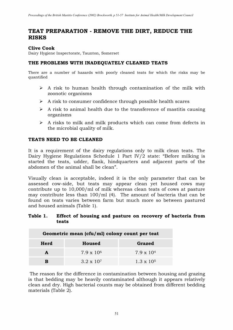
Proceedings of the British Mastitis Conference (2002) Brockworth, p 51-57 Institute for Animal Health/Milk Development Council
51
TEAT PREPARATION - REMOVE THE DIRT, REDUCE THE RISKS
Clive Cook Dairy Hygiene Inspectorate, Taunton, Somerset THE PROBLEMS WITH INADEQUATELY CLEANED TEATS There are a number of hazards with poorly cleaned teats for which the risks may be quantified
� A risk to human health through contamination of the milk with zoonotic organisms
� A risk to consumer confidence through possible health scares
� A risk to animal health due to the transference of mastitis causing organisms
� A risks to milk and milk products which can come from defects in the microbial quality of milk.
TEATS NEED TO BE CLEANED It is a requirement of the dairy regulations only to milk clean teats. The Dairy Hygiene Regulations Schedule 1 Part IV/2 state: “Before milking is started the teats, udder, flank, hindquarters and adjacent parts of the abdomen of the animal shall be clean”. Visually clean is acceptable, indeed it is the only parameter that can be assessed cow-side, but teats may appear clean yet housed cows may contribute up to 10,000/ml of milk whereas clean teats of cows at pasture may contribute less than 100/ml (4). The amount of bacteria that can be found on teats varies between farm but much more so between pastured and housed animals (Table 1). Table 1. Effect of housing and pasture on recovery of bacteria from
teats
Geometric mean (cfu/ml) colony count per teat
Herd Housed Grazed
A 7.9 x 10⁶ 7.9 x 10⁴
B 3.2 x 10⁷ 1.3 x 10⁵
The reason for the difference in contamination between housing and grazing is that bedding may be heavily contaminated although it appears relatively clean and dry. High bacterial counts may be obtained from different bedding materials (Table 2).
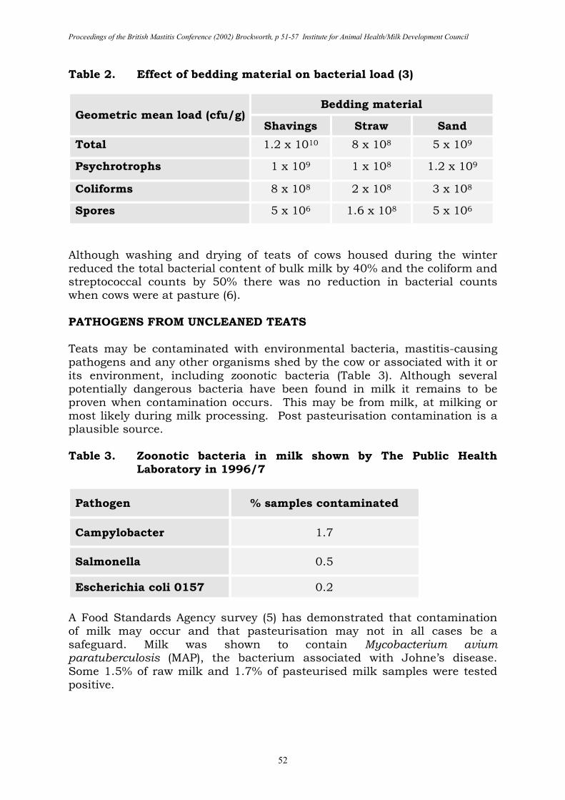
Proceedings of the British Mastitis Conference (2002) Brockworth, p 51-57 Institute for Animal Health/Milk Development Council
52
Table 2. Effect of bedding material on bacterial load (3)
Geometric mean load (cfu/g) Bedding material
Shavings Straw Sand
Total 1.2 x 10¹⁰ 8 x 10⁸ 5 x 10⁹
Psychrotrophs 1 x 10⁹ 1 x 10⁸ 1.2 x 10⁹
Coliforms 8 x 10⁸ 2 x 10⁸ 3 x 10⁸
Spores 5 x 10⁶ 1.6 x 10⁸ 5 x 10⁶
Although washing and drying of teats of cows housed during the winter reduced the total bacterial content of bulk milk by 40% and the coliform and streptococcal counts by 50% there was no reduction in bacterial counts when cows were at pasture (6). PATHOGENS FROM UNCLEANED TEATS Teats may be contaminated with environmental bacteria, mastitis-causing pathogens and any other organisms shed by the cow or associated with it or its environment, including zoonotic bacteria (Table 3). Although several potentially dangerous bacteria have been found in milk it remains to be proven when contamination occurs. This may be from milk, at milking or most likely during milk processing. Post pasteurisation contamination is a plausible source. Table 3. Zoonotic bacteria in milk shown by The Public Health
Laboratory in 1996/7
Pathogen % samples contaminated
Campylobacter 1.7
Salmonella 0.5
Escherichia coli 0157 0.2
A Food Standards Agency survey (5) has demonstrated that contamination of milk may occur and that pasteurisation may not in all cases be a safeguard. Milk was shown to contain Mycobacterium avium paratuberculosis (MAP), the bacterium associated with Johne’s disease. Some 1.5% of raw milk and 1.7% of pasteurised milk samples were tested positive.
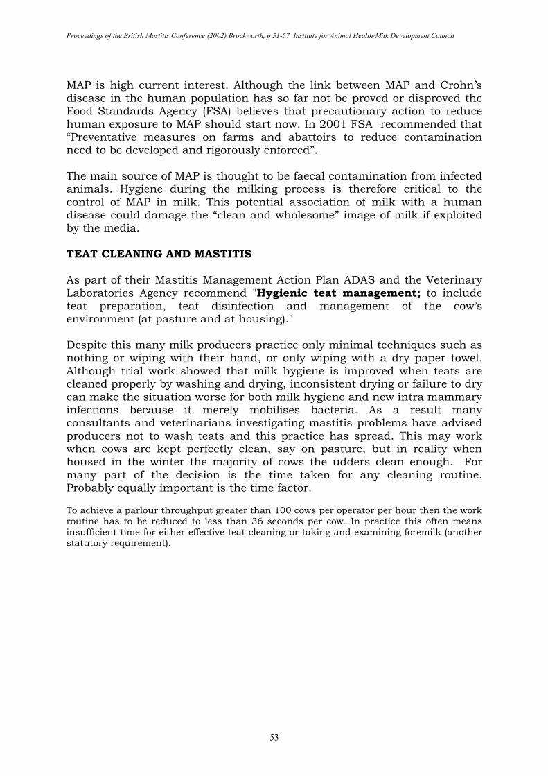
Proceedings of the British Mastitis Conference (2002) Brockworth, p 51-57 Institute for Animal Health/Milk Development Council
53
MAP is high current interest. Although the link between MAP and Crohn’s disease in the human population has so far not be proved or disproved the Food Standards Agency (FSA) believes that precautionary action to reduce human exposure to MAP should start now. In 2001 FSA recommended that “Preventative measures on farms and abattoirs to reduce contamination need to be developed and rigorously enforced”. The main source of MAP is thought to be faecal contamination from infected animals. Hygiene during the milking process is therefore critical to the control of MAP in milk. This potential association of milk with a human disease could damage the “clean and wholesome” image of milk if exploited by the media. TEAT CLEANING AND MASTITIS As part of their Mastitis Management Action Plan ADAS and the Veterinary Laboratories Agency recommend "Hygienic teat management; to include teat preparation, teat disinfection and management of the cow’s environment (at pasture and at housing)." Despite this many milk producers practice only minimal techniques such as nothing or wiping with their hand, or only wiping with a dry paper towel. Although trial work showed that milk hygiene is improved when teats are cleaned properly by washing and drying, inconsistent drying or failure to dry can make the situation worse for both milk hygiene and new intra mammary infections because it merely mobilises bacteria. As a result many consultants and veterinarians investigating mastitis problems have advised producers not to wash teats and this practice has spread. This may work when cows are kept perfectly clean, say on pasture, but in reality when housed in the winter the majority of cows the udders clean enough. For many part of the decision is the time taken for any cleaning routine. Probably equally important is the time factor. To achieve a parlour throughput greater than 100 cows per operator per hour then the work routine has to be reduced to less than 36 seconds per cow. In practice this often means insufficient time for either effective teat cleaning or taking and examining foremilk (another statutory requirement).
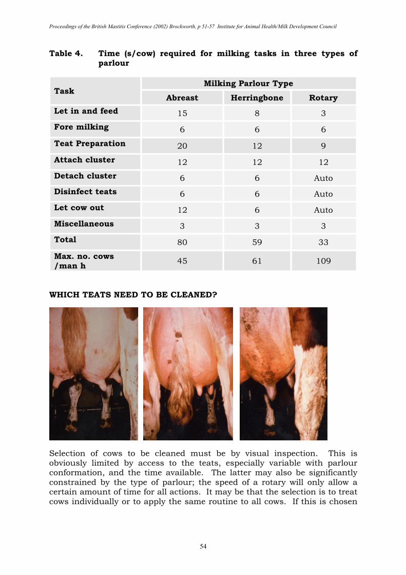
Proceedings of the British Mastitis Conference (2002) Brockworth, p 51-57 Institute for Animal Health/Milk Development Council
54
Table 4. Time (s/cow) required for milking tasks in three types of parlour
Task Milking Parlour Type
Abreast Herringbone Rotary
Let in and feed 15 8 3
Fore milking 6 6 6
Teat Preparation 20 12 9
Attach cluster 12 12 12
Detach cluster 6 6 Auto
Disinfect teats 6 6 Auto
Let cow out 12 6 Auto
Miscellaneous 3 3 3
Total 80 59 33
Max. no. cows /man h
45 61 109
WHICH TEATS NEED TO BE CLEANED?
Selection of cows to be cleaned must be by visual inspection. This is obviously limited by access to the teats, especially variable with parlour conformation, and the time available. The latter may also be significantly constrained by the type of parlour; the speed of a rotary will only allow a certain amount of time for all actions. It may be that the selection is to treat cows individually or to apply the same routine to all cows. If this is chosen
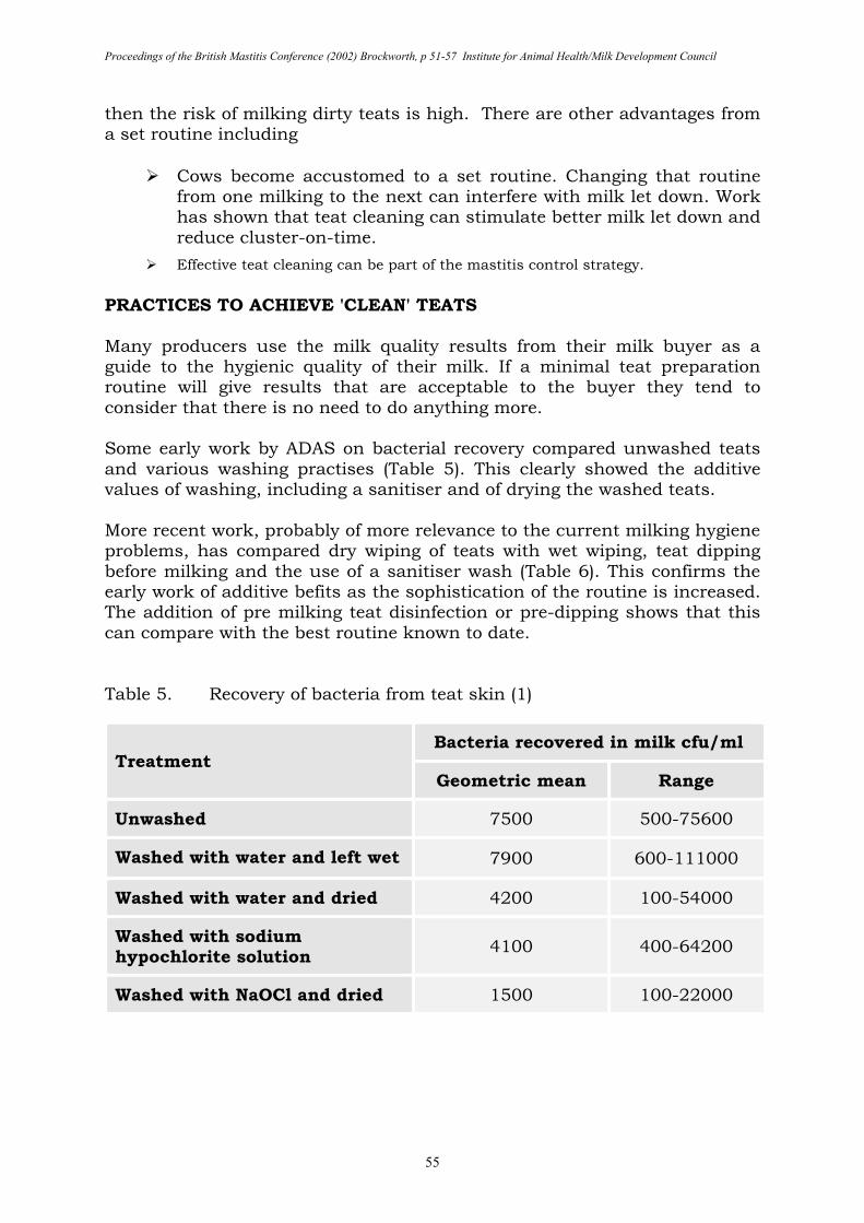
Proceedings of the British Mastitis Conference (2002) Brockworth, p 51-57 Institute for Animal Health/Milk Development Council
55
then the risk of milking dirty teats is high. There are other advantages from a set routine including
� Cows become accustomed to a set routine. Changing that routine from one milking to the next can interfere with milk let down. Work has shown that teat cleaning can stimulate better milk let down and reduce cluster-on-time.
� Effective teat cleaning can be part of the mastitis control strategy.
PRACTICES TO ACHIEVE 'CLEAN' TEATS Many producers use the milk quality results from their milk buyer as a guide to the hygienic quality of their milk. If a minimal teat preparation routine will give results that are acceptable to the buyer they tend to consider that there is no need to do anything more. Some early work by ADAS on bacterial recovery compared unwashed teats and various washing practises (Table 5). This clearly showed the additive values of washing, including a sanitiser and of drying the washed teats. More recent work, probably of more relevance to the current milking hygiene problems, has compared dry wiping of teats with wet wiping, teat dipping before milking and the use of a sanitiser wash (Table 6). This confirms the early work of additive befits as the sophistication of the routine is increased. The addition of pre milking teat disinfection or pre-dipping shows that this can compare with the best routine known to date. Table 5. Recovery of bacteria from teat skin (1)
Treatment Bacteria recovered in milk cfu/ml
Geometric mean Range
Unwashed 7500 500-75600
Washed with water and left wet 7900 600-111000
Washed with water and dried 4200 100-54000
Washed with sodium hypochlorite solution
4100 400-64200
Washed with NaOCl and dried 1500 100-22000
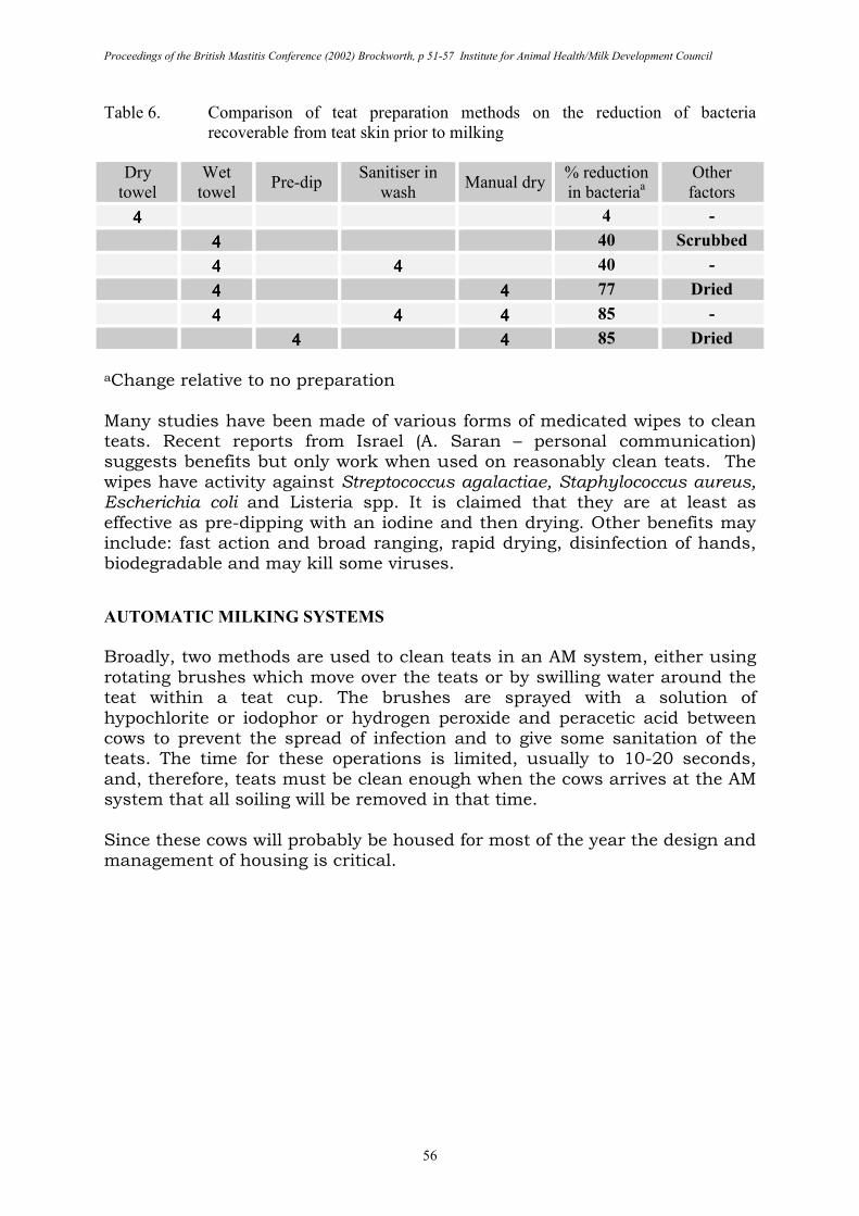
Proceedings of the British Mastitis Conference (2002) Brockworth, p 51-57 Institute for Animal Health/Milk Development Council
56
Table 6. Comparison of teat preparation methods on the reduction of bacteria
recoverable from teat skin prior to milking
Dry
towel
Wet
towel Pre-dip
Sanitiser in
wash Manual dry
% reduction
in bacteriaa
Other
factors
4444 4 -
4444 40 Scrubbed
4444 4444 40 -
4444 4444 77 Dried
4444 4444 4444 85 -
4444 4444 85 Dried
aChange relative to no preparation Many studies have been made of various forms of medicated wipes to clean teats. Recent reports from Israel (A. Saran – personal communication) suggests benefits but only work when used on reasonably clean teats. The wipes have activity against Streptococcus agalactiae, Staphylococcus aureus, Escherichia coli and Listeria spp. It is claimed that they are at least as effective as pre-dipping with an iodine and then drying. Other benefits may include: fast action and broad ranging, rapid drying, disinfection of hands, biodegradable and may kill some viruses.
AUTOMATIC MILKI.G SYSTEMS
Broadly, two methods are used to clean teats in an AM system, either using rotating brushes which move over the teats or by swilling water around the teat within a teat cup. The brushes are sprayed with a solution of hypochlorite or iodophor or hydrogen peroxide and peracetic acid between cows to prevent the spread of infection and to give some sanitation of the teats. The time for these operations is limited, usually to 10-20 seconds, and, therefore, teats must be clean enough when the cows arrives at the AM system that all soiling will be removed in that time. Since these cows will probably be housed for most of the year the design and management of housing is critical.
Proceedings of the British Mastitis Conference (2002) Brockworth, p 51-57 Institute for Animal Health/Milk Development Council
57
CONCLUSIONS Conclusions are as follows:
� All visually dirty teats need to be cleaned effectively to reduce bacterial contamination (especially from faecal material).
� Even visually clean teats during the housing period need to be cleaned effectively to reduce bacterial contamination.
� Visually clean cows at pasture can be left out of the effective cleaning routine. However, if left to operator choice there is a good chance some cows may not be cleaned when they should be.
� It is often difficult to assess teat cleanliness visually because of poor lighting in the vicinity of the teat and the difficulty of observing the “blind” side.
Finally, a comment on contamination of milk with faecal matter (C. Heggum – personal communication). The impact of milking Hygiene on a single milking is that:
� Poor hygiene results in up to 2 g faecal material in 20 kg milk
� Good hygiene results in up to 0.2 g faecal material in 20 kg milk
� Best hygiene results in up to 0.04 g faecal material in 20 kg milk
REFERENCES 1. Cousins, C.M. (1978) “Milking Techniques and the Microbial Flora of
Milk” XX1st Dairy Congress, Paris. 2. Cousins, C.M. and McKinnon, C.H. (1997) Cleaning and disinfection in milk production. In: Machine Milking Technical Bulletin 1, NIRD/HRI. 3. Food Standards Agency (2001) “A review of the evidence for a link
between exposure to Mycobacterium Paratuberculosis (MAP) and Crohns Disease in Humans”
4. McKinnon, C.H., Rowlands, J. and Bramley, A.J. (1990) The effect of udder preparation before milking and contamination from the milking plant on bacterial numbers in bulk milk of eight herds. J. Dairy Res. 57: 307-318.
Proceedings of the British Mastitis Conference (2002) Brockworth, p 58-62 Institute for Animal Health/Milk Development Council
58
DETECTING MASTITIS AUTOMATICALLY H.J. Biggadike1, I. Ohnstad1, R.A. Laven1 and J.E. Hillerton2 1ADAS Bridgets, Martyr Worthy, Hants SO21 1AP 2IAH Compton, Newbury, Berks RG20 7NN SUMMARY Individual quarter milk conductivity measurements were collected from 31 cows in a 70-cow herd in south-east England, for a period of 15 weeks. Over this period, 42% of cow-weeks and 20% of quarter-weeks had an increase in quarter conductivity of 10% or more compared to the mean quarter conductivity of the previous 14 milkings. Geometric mean SCC was higher in quarter-weeks with a 10% plus increase in conductivity compared to quarters with a conductivity change of less than 10%. The specificity of this system was estimated to be 87%, the sensitivity 50%, the negative predictive value 87% and the positive predictive value 45%. This study found that individual quarter milk conductivity was insufficiently accurate to be used as the sole criteria for the identification of mastitic quarters. INTRODUCTION Mastitis causes changes in the conductivity of milk, by damaging the mammary epithelium, and thus altering the balance of sodium, potassium and chloride ions. Measurement of changes in milk conductivity has therefore been suggested as a method for automatically detecting mastitis in cows that are milked robotically. Additionally, changes in conductivity tend to occur prior to the development of visible clinical signs of mastitis (1), thus measuring conductivity could allow early identification and treatment of mastitis potentially improving bacteriological cure rate, reducing recurrence rates and, perhaps, overall antibiotic usage (2). However, despite the clear scientific principle behind conductivity measurement, there have been few reports of its practical utility on farm. This study was designed to investigate whether, on a commercial dairy farm, changes in individual quarter milk conductivity, measured using a commercially available system, could be used as a diagnostic method for mastitis. MATERIALS AND METHODS Data were collected from 31 cows in a 70-cow herd, located in south-east England, for a period of 15 weeks. Cows were milked through a 2-box Liberty automatic milking unit, which measured and recorded individual quarter milk conductivity in-line. Conductivity was reported on an individual quarter basis in terms of the percentage increase from the mean of the previous 14 days measurements once it exceeded a 10% trigger
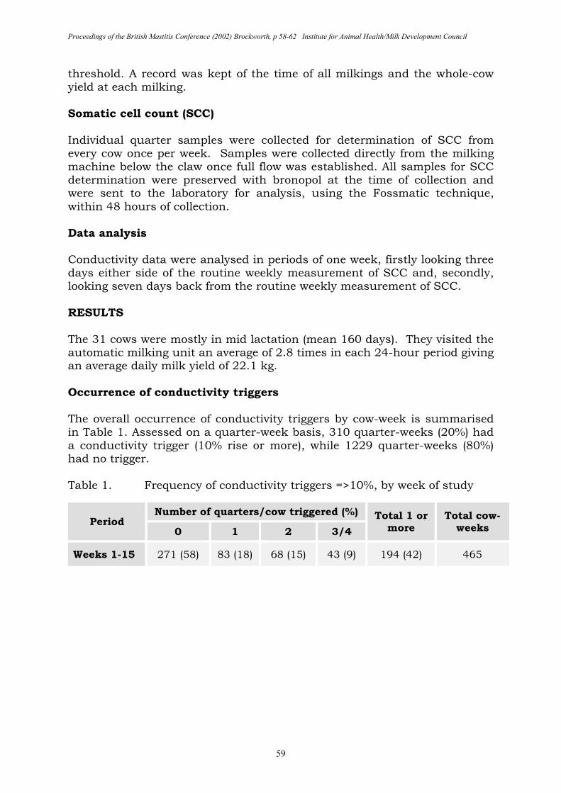
Proceedings of the British Mastitis Conference (2002) Brockworth, p 58-62 Institute for Animal Health/Milk Development Council
59
threshold. A record was kept of the time of all milkings and the whole-cow yield at each milking.
Somatic cell count (SCC)
Individual quarter samples were collected for determination of SCC from every cow once per week. Samples were collected directly from the milking machine below the claw once full flow was established. All samples for SCC determination were preserved with bronopol at the time of collection and were sent to the laboratory for analysis, using the Fossmatic technique, within 48 hours of collection.
Data analysis
Conductivity data were analysed in periods of one week, firstly looking three days either side of the routine weekly measurement of SCC and, secondly, looking seven days back from the routine weekly measurement of SCC. RESULTS
The 31 cows were mostly in mid lactation (mean 160 days). They visited the automatic milking unit an average of 2.8 times in each 24-hour period giving an average daily milk yield of 22.1 kg.
Occurrence of conductivity triggers
The overall occurrence of conductivity triggers by cow-week is summarised in Table 1. Assessed on a quarter-week basis, 310 quarter-weeks (20%) had a conductivity trigger (10% rise or more), while 1229 quarter-weeks (80%) had no trigger. Table 1. Frequency of conductivity triggers =>10%, by week of study
Period Number of quarters/cow triggered (%) Total 1 or
more Total cow-weeks 0 1 2 3/4
Weeks 1-15 271 (58) 83 (18) 68 (15) 43 (9) 194 (42) 465
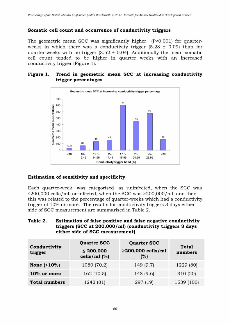
Proceedings of the British Mastitis Conference (2002) Brockworth, p 58-62 Institute for Animal Health/Milk Development Council
60
Somatic cell count and occurrence of conductivity triggers The geometric mean SCC was significantly higher (P<0.001) for quarter-weeks in which there was a conductivity trigger (5.28 ± 0.09) than for quarter-weeks with no trigger (3.52 ± 0.04). Additionally the mean somatic cell count tended to be higher in quarter weeks with an increased conductivity trigger (Figure 1). Figure 1. Trend in geometric mean SCC at increasing conductivity
trigger percentages
Geometric mean SCC at increasing conductivity trigger percentage
122982
4948
27
65
22
17
0
100
200
300
400
500
600
700
800
<10 10-
12.49
12.5-
14.99
15-
17.49
17.5-
19.99
20-
24.99
25-
29.99
>30
Conductivity trigger band (%)
Geom
etr
ic m
ean S
CC
('0
00/m
l)
Estimation of sensitivity and specificity
Each quarter-week was categorised as uninfected, when the SCC was ≤200,000 cells/ml, or infected, when the SCC was >200,000/ml, and then this was related to the percentage of quarter-weeks which had a conductivity trigger of 10% or more. The results for conductivity triggers 3 days either side of SCC measurement are summarised in Table 2. Table 2. Estimation of false positive and false negative conductivity
triggers (SCC at 200,000/ml) (conductivity triggers 3 days either side of SCC measurement)
Conductivity trigger
Quarter SCC
≤≤≤≤ 200,000 cells/ml (%)
Quarter SCC
>200,000 cells/ml (%)
Total numbers
None (<10%) 1080 (70.2) 149 (9.7) 1229 (80)
10% or more 162 (10.5) 148 (9.6) 310 (20)
Total numbers 1242 (81) 297 (19) 1539 (100)
Proceedings of the British Mastitis Conference (2002) Brockworth, p 58-62 Institute for Animal Health/Milk Development Council
61
From these data the sensitivity was calculated to be 50 per cent (148/(148+149)) and the specificity 87 per cent (1080/(1080+162)).
Examining conductivity triggers 7 days before the SCC measurement changed the distribution very little. There was a slight increase in the percentage of quarter-weeks with a raised SCC that were not identified by a conductivity trigger compared to 3 days either side of the SCC measurement. The sensitivity was reduced to 46%; the specificity remained unchanged at 87%. These data suggest that the likelihood ratio for a positive conductivity response is approximately 3.9, while that for a negative response is 0.6. These are significantly different from 1; clearly showing that conductivity is significantly related to mastitis. However, the positive predictive value was only around 45%.
DISCUSSION This study confirms the findings of Hamann and Zecconi (1), that there is a significant relationship between individual quarter milk conductivity and mastitis (as measured by SCC). However, these data suggest that under commercial conditions tested here individual quarter milk conductivity is neither sensitive nor specific enough to be used as a diagnostic test for mastitis. Firstly, too high a proportion of cows (approximately 50%) did not have a rise in conductivity at or around the time of a rise in SCC, and thus would have not have been diagnosed as having mastitis if conductivity had been the sole indicator. Furthermore, the poor specificity completely precludes using conductivity alone as <50% of cows with a conductivity trigger had an associated rise in cell count. Treatment on the basis of a conductivity rise would therefore have resulted in an unacceptable percentage of unjustified treatments. The problem of poor specificity could be overcome by combining the use of milk conductivity with other tests, such as measuring the concentration of NAGase or ATP in milk from cows with raised conductivity. However, this does not solve the problem of the lack of sufficient sensitivity. For cows milked robotically additional tests are essential to identify those cows with mastitis that have not had a conductivity rise. Currently the only available tests are the same as used in conventional parlours (e.g. measurement of milk yield and visual and tactile assessment of the udder). More research is needed on in-line tests that can be used alongside conductivity.
Proceedings of the British Mastitis Conference (2002) Brockworth, p 58-62 Institute for Animal Health/Milk Development Council
62
ACKNOWLEDGEMENTS This project was funded by the Milk Development Council. Milking facility and cows were made available and sampling undertaken by Mr C. Hamilton. Technical support was provided by Liberty Milking Systems. This paper is a shortened version of one previously published in the Veterinary Record
REFERENCES 1. Hamann, J. and Zecconi, A. (1998) Evaluation of the electrical
conductivity of milk as a mastitis indicator. Int. Dairy Fed. Bull. No. 334: 5-22.
2. Milner, P., Page, K.L. and Hillerton, J.E. (1997) The effects of early antibiotic treatment following diagnosis of mastitis detected by a change in the electrical conductivity of milk. J. Dairy Sci. 80: 859-863.
Proceedings of the British Mastitis Conference (2002) Brockworth, p 63-67 Institute for Animal Health/Milk Development Council
63
FREE ACCESS ROBOTIC MILKING: THE NEXT GENERATION?
Gavin Dick Mackies Ltd, Old Meldrum, Aberdeenshire BACKGROUND Mackies is a vertically integrated business focussed on the manufacture of premium ice-cream. Current production is based on around 5m litres/year. All milk is processed on site and sold as ice-cream to the major multiples throughout the UK. We have a new and expanding export market to South Korea. We farm 570 ha which supports a herd of 500 Jerseys. Production currently averages 5,500 litres per cow at 6.1% butterfat. Free Access Robotic Milking We started the FARM system on the 4th December 2001 with a single Astronaut automated milking unit. However, the history behind the decision to introduce automated milking probably merits a mention. Our existing twin herringbone parlours are now more than 20 years old and desperately in need of replacement. We started looking at options about 6 years ago. We examined the usual options of herringbone, tandem, rapid exit, rotary parlours and robots. Our primary objective, with 500 cows to milk, was animal welfare, or more particularly we wanted an animal friendly system. Quickly we narrowed our option to a rotary parlour. However, we were intrigued by the robotic milking opportunities. The concept is innovative technology with a reduced requirement for labour. However, there were many reports of mixed results of performance and indeed units being removed. We decided that more research was needed and we spent the next 2 to 3 years looking at various robotic systems before we were happy that the robots could do the job. We then costed fully both a rotary and a robotic system with 2 underlying assumptions.
� 15% more milk from the robot, which we knew we would get from the 3rd milking, and
� a reduction in labour of one person.
Proceedings of the British Mastitis Conference (2002) Brockworth, p 63-67 Institute for Animal Health/Milk Development Council
64
The figures came out marginally in favour of robots, but the future potential from robots was far greater. The decision to establish the FARM system was made in September 1999 and the first phase of the renovation started in May 2000. UNIT 1 – THE STORY SO FAR The Cows The FARM group started with 55 cows assigned as soon as they left the post calving group (after 21 days). Some 6 cows (11%) were subsequently removed as unsuitable for automatic milking due to too low udders or high rear teats. We added further cows to bring the total to 57. Training To commission the system the cows were put through the Astronaut once on the Monday, Wednesday and Friday, without being milked or fed, prior to starting milking on the following Tuesday. Milking started on the Tuesday afternoon with the milking order recorded. Initially the whole process took around 9 hours. The 2nd and 3rd milkings took place without a break, with the cows being milked in the same order each time. On the 4th milking the cows were allowed to come to the robot as they wished, but milked and non-milked cows were kept separate. On the 5th milking the cows were allowed to mix and come for milking as they wished. Cows going longer than 9-10 hours were fetched to the Astronaut for milking. By the weekend, normal routines and patterns had been established. Routines We have established a daily routine. 7.30 am Attention list checked
Holding pen and Astronaut washed Astronaut checked Milk filter changed Laser cleaned Cows overdue for milking fetched to the Astronaut (8.3 cows/day with an attendance interval of more than 9 hours)
Proceedings of the British Mastitis Conference (2002) Brockworth, p 63-67 Institute for Animal Health/Milk Development Council
65
12.30 Holding pen and Astronaut washed Laser cleaned Cows overdue fetched to the Astronaut
4.30 pm Holding pen and Astronaut washed Laser cleaned Milk filter changed Cows overdue fetched to the Astronaut
Total time for the fixed routine work is approximately 1½ hours/day. Astronaut statistics
We have achieved: 4.9 visits/cow/day
2.9 milkings/cow/day
7 attachment failures/day
17.9 hrs/day spent milking
We have had only one breakdown that involved calling out an engineer. We lost 2 hours in total. Fertility
We now require: 2.03 services/cow in calf
compared to: 1.82 services/cow in calf for conventional system used on the rest of the farm.
Production
Group Days in milk Total yield (l) Daily yield (l)
1 70 1612 19.1
2 130 3126 18.2
FARM 220 5430 18.3
3 245 4707 14.0
4 316 5108 10.2
Production by the FARM group is significantly ahead of other comparable animals in total yield to date and the daily yield shows a remarkable persistency of lactation. These are the outcome, predicted and expected from voluntary use of the automatic milking system.
Proceedings of the British Mastitis Conference (2002) Brockworth, p 63-67 Institute for Animal Health/Milk Development Council
66
Health In the FARM group we have recorded, from 4 December to date,
8 cases of mastitis 4 sore feet (1 ulcer; 3 digital dermatitis) 1 metabolic disorder.
Milk quality The milk is not collected separately for quality analysis but so far we have not noticed any change in the quality of our supply following the inclusion of the milk from the robot. WHAT HAVE WE LEARNED? The most important thing that we have realised is that the system is the key, not the robotic unit itself. To be successful, the system must be set up to allow the cow to make her own decisions and go about her daily routines as easily as possible. The system must be cow friendly. The management of the system must be detailed and vigorous – the information collated by the computer must be managed and acted upon, and although the overall labour requirement is less, the level of management input is significantly higher. Objectives for the robot must be clearly set and the robot set up to achieve them these include milkings/cow/day, production levels, hygiene levels etc. Initial training of the cows must be intensive and consistent. CONCLUSIONS The results from the first group of cows to go through the system have been far beyond our expectations in all aspects.
� We have found that our cows have adapted very quickly.
� The staff have adapted almost as well as the cows!
� Production is significantly improved.
� Cow health has improved.
� Udder health has improved.
� Mechanical reliability has been excellent.
Proceedings of the British Mastitis Conference (2002) Brockworth, p 63-67 Institute for Animal Health/Milk Development Council
67
There is no doubt in my mind that a FARM system allows large herds to achieve the attention to detail at a cow level which results in a greater efficiency of production than can be achieved from any other milking system currently available. FUTURE So what to the future? Of all the agricultural commodities, I believe that the milk sector has the most potential over the next 5 years. I expect that milk prices will rise by slightly more than the rate of inflation. However, to remain viable, producers must not only become ever more efficient at milk production, but must be able to spread their fixed costs over a greater output base. A FARM system is an ideal way to achieve this:
� It is flexible in that cow numbers can be increased in multiples of 60.
� The level of information produced at individual cow level allows a high degree of efficiency.
� It is both intriguing and attractive to the public, meeting many of their requirements of a food producing operation.
We are now operating 6 units with plans to increase to 10 automated milking units! We will have a fully automated milking FARM.
Proceedings of the British Mastitis Conference (2002) Brockworth, p 68-80 Institute for Animal Health/Milk Development Council
68
AUTOMATIC MILKING – CHANGES AND CHANCES Kees de Koning and Yvonne van de Vorst Research Institute for Animal Husbandry (PV), P.O. Box 2176, NL - 8203 AD LELYSTAD SUMMARY An overview is given of the development of automatic milking with special emphasis on management aspects and milk quality. Risk factors, changes, opportunities and challenges are discussed. Changing over from a milking parlour to automatic milking will lead to big changes for both herdsman and cow and can cause stress to both. Important aspects of successful implementation of an AM-system are the attitude and expectations of the dairy farmer. Results from commercial farms indicate, that in many cases milk quality is somewhat negatively effected, although the levels of bacterial counts and somatic cell counts still are relatively low and far within the penalty levels. However, after some time the BMSCC decreased in all countries to a comparable level of the conventional farms. Automatic milking systems require a higher investment than conventional milking systems. However, increased milk yields and reduced labour requirements may lead to a decrease in the fixed costs per kilogram of milk. Automatic milking today is common practice on more than 1250 farms worldwide and a further increase in the number of systems is expected, mainly in the countries with high labour costs. INTRODUCTION The first ideas about fully automating the milking process were generated in the mid-seventies. The growing costs of labour in several countries were the main reason to start the development of automatic milking. The final step in the automation of the milking process seemed to be the development of automatic cluster attachment systems. However, it took almost a decade to convert the techniques for locating teats and attaching teat cups to fully integrated and reliable automatic milking systems. In automatic milking systems (AM-systems) cows are milked by a robotic milking system without direct human supervision. The first milking robots were installed on commercial dairy farms in the Netherlands in 1992. The breakthrough of automatic milking came at the end of the nineties and at spring 2002, more than 1250 farms worldwide milked their cows automatically (1). AUTOMATIC MILKING SYSTEMS AM-systems can be divided into single stall systems and multi-stall systems. Single stall systems have an integrated robotic and milking system, while multi-stall systems have a transportable robot device. Each stall has its own milking devices, like in a milking parlour. A single stall AM-system is able to
Proceedings of the British Mastitis Conference (2002) Brockworth, p 68-80 Institute for Animal Health/Milk Development Council
69
milk 55-60 cows up to three times per day on average. Multi stall systems have 2 to 4 stalls and are able to milk a herd of 80 to 150 cows three times per day. Automatic milking relies on the cow’s motivation to visit the AM-system more or less voluntarily. The main motive for a cow to visit the AM-system is the supply of concentrates; therefore all AM-systems are equipped with concentrate dispensers. An automatic milking system has to take over the “eyes and hands” of the milker and therefore these systems should have electronic cow identification, cleaning and milking devices and computer controlled sensors to detect abnormalities in order to meet international legislation and hygiene rules from the dairy industry. The current teat cleaning systems can be divided into three main types; cleaning with brushes or rollers, cleaning inside the teat-cup and cleaning with a separate ‘teat cup like’ device. Little information is available about the efficacy of teat cleaning devices. Several trials showed that cleaning with a cleaning device is better than no cleaning, but not as good as manual cleaning by the herdsman (2). AM-systems are also equipped with a variety of sensors to observe and to control the milking process. Data are automatically stored in a database and the farmer has a management program to control the settings and conditions for cows to be milked. Attention lists and reports are presented to the farmer by screen or printer messages. However, the AM-system only notifies, the farmer has to take action. FARMS WITH AUTOMATIC MILKING SYSTEMS The first AM-systems on commercial farms were implemented in North Western Europe. The reasons for these countries to start the development of AM-systems most probably were related to the expensive labour and the farm structure with family farms. Increasing costs of labour, land and buildings and machinery, while milk prices tended to decrease, forced farmers to increase their output per man-hour. Average herd-size showed a continuous increase and this phenomenon is still ongoing. In the first years after the introduction of the first AM-systems, the adoption went slow, until 1998 (Figure 1). From that year on, in the Netherlands, automatic milking became an accepted technology by the dairy sector and in the same period also other countries adopted AM-systems, like Germany, Denmark and France. More than 90% of all dairy farms with an AM-system are located in north-western Europe. Most dairy farms with an AM-system can be found in the Netherlands. However, AM-systems are also regarded to have potential for the USA and Canada too (3).
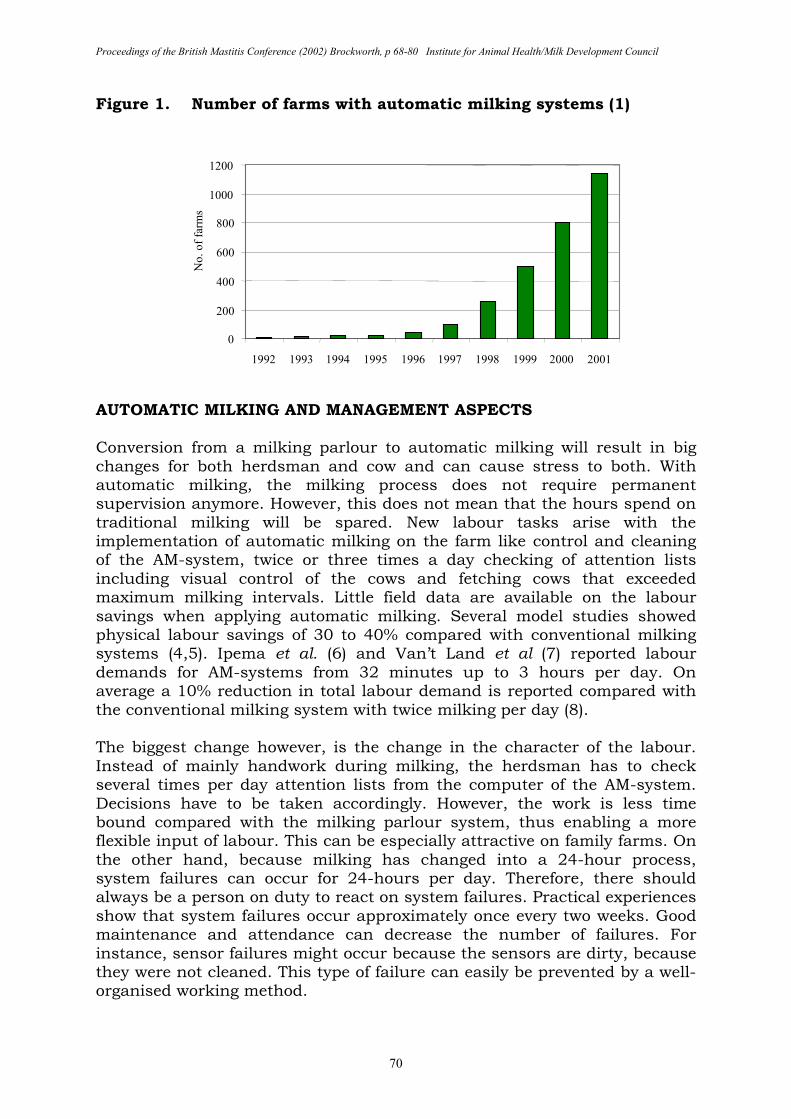
Proceedings of the British Mastitis Conference (2002) Brockworth, p 68-80 Institute for Animal Health/Milk Development Council
70
Figure 1. Number of farms with automatic milking systems (1)
AUTOMATIC MILKING AND MANAGEMENT ASPECTS Conversion from a milking parlour to automatic milking will result in big changes for both herdsman and cow and can cause stress to both. With automatic milking, the milking process does not require permanent supervision anymore. However, this does not mean that the hours spend on traditional milking will be spared. New labour tasks arise with the implementation of automatic milking on the farm like control and cleaning of the AM-system, twice or three times a day checking of attention lists including visual control of the cows and fetching cows that exceeded maximum milking intervals. Little field data are available on the labour savings when applying automatic milking. Several model studies showed physical labour savings of 30 to 40% compared with conventional milking systems (4,5). Ipema et al. (6) and Van’t Land et al (7) reported labour demands for AM-systems from 32 minutes up to 3 hours per day. On average a 10% reduction in total labour demand is reported compared with the conventional milking system with twice milking per day (8). The biggest change however, is the change in the character of the labour. Instead of mainly handwork during milking, the herdsman has to check several times per day attention lists from the computer of the AM-system. Decisions have to be taken accordingly. However, the work is less time bound compared with the milking parlour system, thus enabling a more flexible input of labour. This can be especially attractive on family farms. On the other hand, because milking has changed into a 24-hour process, system failures can occur for 24-hours per day. Therefore, there should always be a person on duty to react on system failures. Practical experiences show that system failures occur approximately once every two weeks. Good maintenance and attendance can decrease the number of failures. For instance, sensor failures might occur because the sensors are dirty, because they were not cleaned. This type of failure can easily be prevented by a well-organised working method.
0
200
400
600
800
1000
1200
1992 1993 1994 1995 1996 1997 1998 1999 2000 2001
No. of
farm
s
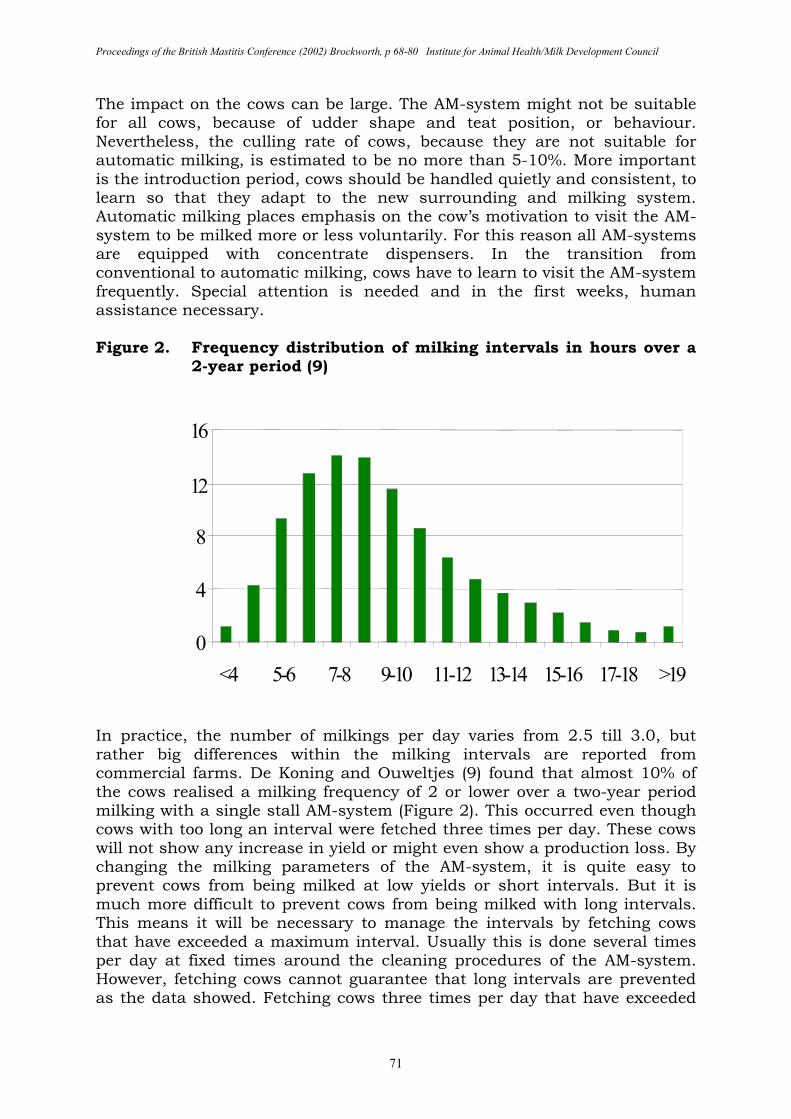
Proceedings of the British Mastitis Conference (2002) Brockworth, p 68-80 Institute for Animal Health/Milk Development Council
71
The impact on the cows can be large. The AM-system might not be suitable for all cows, because of udder shape and teat position, or behaviour. Nevertheless, the culling rate of cows, because they are not suitable for automatic milking, is estimated to be no more than 5-10%. More important is the introduction period, cows should be handled quietly and consistent, to learn so that they adapt to the new surrounding and milking system. Automatic milking places emphasis on the cow’s motivation to visit the AM-system to be milked more or less voluntarily. For this reason all AM-systems are equipped with concentrate dispensers. In the transition from conventional to automatic milking, cows have to learn to visit the AM-system frequently. Special attention is needed and in the first weeks, human assistance necessary. Figure 2. Frequency distribution of milking intervals in hours over a
2-year period (9)
In practice, the number of milkings per day varies from 2.5 till 3.0, but rather big differences within the milking intervals are reported from commercial farms. De Koning and Ouweltjes (9) found that almost 10% of the cows realised a milking frequency of 2 or lower over a two-year period milking with a single stall AM-system (Figure 2). This occurred even though cows with too long an interval were fetched three times per day. These cows will not show any increase in yield or might even show a production loss. By changing the milking parameters of the AM-system, it is quite easy to prevent cows from being milked at low yields or short intervals. But it is much more difficult to prevent cows from being milked with long intervals. This means it will be necessary to manage the intervals by fetching cows that have exceeded a maximum interval. Usually this is done several times per day at fixed times around the cleaning procedures of the AM-system. However, fetching cows cannot guarantee that long intervals are prevented as the data showed. Fetching cows three times per day that have exceeded
0
4
8
12
16
<4 5-6 7-8 9-10 11-12 13-14 15-16 17-18 >19
Proceedings of the British Mastitis Conference (2002) Brockworth, p 68-80 Institute for Animal Health/Milk Development Council
72
an interval of 12 hours, means that the maximum interval may amount to 20 hours. Fetching cows with shorter intervals is quite time-consuming. One of the main benefits of automatic milking is an increase in milk yield from more frequent milking. It is known that milk production, in terms of milk production per hour, is dependent on the milking interval. An increase in milk yield from 6 to 25% in complete lactations has been shown when increasing the milking frequency from two to three times per day (10). Dairy herd information records in The Netherlands show that daily milk production increases by 11.4%, when farms change from milking two times per day in a milking parlour to automatic milking (unpublished data). French data show an average 3% increase in milk yield up to 9% increase for farms that utilized the AM-system for more than 2 years (11,12). Important aspects of successful implementation of an AM-system are the attitude and expectations of the dairy farmer (13). When expectations are too high, disappointments will also be high. Automatic milking requires, especially initially, a high input of labour and management. Almost all manufacturers of AM-systems have had customers who afterwards, went back to a traditional milking system. The exact reasons are not always known. Key factors of a successful implementation of AM-systems are:
� Realistic expectations
� Good management support by skilled consultants before, during and after implementation
� Flexibility and discipline to control the system and the cows
� Ability to work with computers
� Much attention to the barn layout and a good functioning cow traffic
� Technical functioning of the AM-system and regular maintenance
BARN LAYOUT, CAPACITY OF THE AM-SYSTEM AND MILK COOLING Cows should have easy access to the milking system, using the cow’s motivation for eating. The main motive for a cow to visit the AM-system is the supply of concentrate. AM-systems are equipped with concentrate dispensers, to attract cows to the AM-system to be milked. Therefore, the routing in the barn should be according the Eating – Lying – Milking principle. Cows should have an easy access to the milking stall. Selection gates, long alleys and so on should be minimised. The AM-system should be a part of the free stall barn (14). A central position of the AM-system in the barn minimises the walking distances of the cows. However, for matters of hygiene, in many countries the dairy industry requires the placement of the AM-system close to the milking room. Moreover, it is required that the AM-system be reached through a clean route.
Proceedings of the British Mastitis Conference (2002) Brockworth, p 68-80 Institute for Animal Health/Milk Development Council
73
After visiting the milking system, the cow should have access to the feeding area. Using this milking-feeding-lying principle, the cows are motivated to use the AM-system. Moreover, sufficient roughage should be available 24-hours a day. This is a prerequisite to have optimal cow traffic. There does not seem to be a big difference in average milking frequency between the one-way and the free cow traffic systems in practice (6,7). The one-way cow traffic system is a very effective way to utilize the AM-system and to learn for cows. However, there is a consensus that for animal welfare, free cow traffic is better. Cows spend more time in the waiting area in a one-way cow traffic system (15). Especially for AM-systems with a high occupancy rate, this might affect the number of visits to the roughage station and thus result in a limited intake of roughage. In most European countries, grazing during summer time is routine or even compulsory. Moreover, from an ethological point of view, many consumers in North Western Europe believe grazing is essential for cows. In the Netherlands, about 53% of the farms with an AM-system graze, showing that grazing in combination with AM is possible (17). One milk buyer in The Netherlands started with paying a bonus on the milk price when grazing is applied. Capacity of an AM-system The capacity of an automatic milking system is often expressed as the number of milkings per day, but this number will largely depend on the configuration of the automatic milking system, like the number of stalls and the use of selection gates, milking frequency, machine on time, herd size and cow traffic system. Increasing the number of milkings per cow per day, does not necessarily contribute to a high output capacity in kg of milk per day. This is due to the more or less fixed handling time of the automatic milking system per milking and the decreasing amount of milk per milking when cows are milked more frequently. Milk flow rate and yield have a large impact on capacity in kg per day (9). By changing the milking criteria for individual cows, the AM-system can be optimised to realise a maximal capacity in kg per day. Besides effects on capacity, negative effects of certain milk intervals, such as increase of free fatty acids (FFA) with shorter milking intervals and possibly an increase in somatic cell counts (SCC) with long and short milking intervals must also be taken into account. Cooling of milk in an AM-system Milk should be cooled within 3 hours to a temperature below 4oC. The basic requirement is that the system can handle the specific conditions of automatic milking. In general there are four principles (19) to adjust the cooling system to automatic milking; a) indirect cooling with an ice-bank tank, b) combination of bulk and buffer tank, c) storage tank with modified cooling system and d) instant cooling. For an ice bank tank and modified cooling system, it may be useful to have an additional buffer tank, which is able to store the milk when the bulk tank is emptied and cleaned. This
Proceedings of the British Mastitis Conference (2002) Brockworth, p 68-80 Institute for Animal Health/Milk Development Council
74
enables the AM-system to continue milking, thus increasing the capacity of the system. MILK QUALITY Milk quality is without doubt one of the most important aspects of milk production on modern dairy farms. Milk payment systems are based on milk quality and consumers expect a high quality level in the milk products they buy. Although automatic milking uses more or less the same milking principles as conventional milking, there are some big differences. Results from commercial farms indicate, that milk quality is adversely effected (12,16,19,20,21) in many cases after introduction of automatic milking. Data show almost a doubling of the bacterial counts, although the levels are still relatively low and far within the penalty limits. The cleaning of the milking equipment and the cooling of the milk seem to be the most important factors regarding the increase in bacterial counts. Also cell counts are not reduced after the change to automatic milking, despite the increased milking frequency. In a Danish research (21) an increase in the number of new infections was reported during the first months after the introduction of the AM-system. It is suggested that more attention should be paid to the introductory period. Within the EU project “Implications of the introduction of automatic milking on dairy farms” (QLK5 2000-31006) as part of the EU-programme 'Quality of Life and Management of Living resources', an international study regarding milk quality aspects and automatic milking (22) was undertaken on Danish, German and Dutch farms. The farms were divided in comparable groups based on the installation date. 1) Before January 1, 1998 (AM1), no farm data were available from
Germany and Denmark. 2) January 1, 1998 - March 31, 1999 (AM2) 3) April 1, 1999 - June 30, 2000 (AM3) 4) July 1, 2000 - December, 2000 (AM4) Significant differences in milk quality were found after introduction of the AM-system. The results (predicted means and recalculated geometric means) are presented in Table 1. Averages of conventional farms are also presented as a reference, however not statistically tested in the model.
Proceedings of the British Mastitis Conference (2002) Brockworth, p 68-80 Institute for Animal Health/Milk Development Council
75
Table 1. Predicted and recalculated geometric means before versus after the introduction of the AM system with figures of conventional farms as reference (22)
Country Group No. of farms
TPC cfu/ml BMSCC Cells/ml
FP oC
FFA Meq/100 g fat
PM GM PM GM PM PM GM
DK
C2* All - 9,000 - 246,000 - - -
Before 99 2.080x 8,000 5.558x 259,000 - - -
After 99 2.633y 14,000 5.633y 279,000 - - -
D
C2* All - 21,000 - 181,000 - - -
Before 33 2.835x 17,000 5.302x 201,000 -0.521x - - After 33 3.033y 21,000 5.313x 203,000 -0.516y - -
NL
C2* 295 1.995 7,000 5.169 176,000 -0.521 -0.8142 0.44 C3* 40 2.016 8,000 5.214 184,000 -0.522 -0.5870 0.56
Before 262 2.006x 7,000 5.138x 170,000 -0.522x -0.9310x 0.39
After 262 2.559y 13,000 5.320y 204,000 -0.517y -0.5569y 0.57
* dark shaded sections not included in model x and y = averages with different superscripts within each column and country differ significantly (p<0.05) The European limit for total plate counts (TPC – 100,000 cfu/ml) and bulk milk somatic cell counts (BMSCC – 400,000 cells/ml) are laid down in legislation (Council Directive 92/46/EEC). The German penalty limit for freezing point (FP) is –0.515oC. The Dutch limit is –0.505oC. The Dutch limit for free fatty acids (FFA) is 1,0 mmol/100 g fat. From Table 1 it can be concluded that, the milk quality in all three countries deteriorated slightly after the introduction of the AM-system. This can also be concluded from Table 2, presenting the percentage of milk deliveries exceeding the European or National penalty limits after the introduction of the AM-system in comparison to before introduction. Table 2. Percentage (%) of bulk tank milk exceeding penalty limits
before versus after the introduction of the AM-system, with conventional farms as reference (22)
Country Group
TPC cfu/ml
BMSCC Cells/ml
FP oC
FFA Meq/100 g
fat
>100,000 >400,000 >-0.505 >1.0
DK Before 0.8% 9.1% - - After 2.5% 11.1% - -
D Before 2.9% 6.0% 1.1% - After 7.7% 9.3% 2.8% -
NL Before 0.8% 2.4% 0.4% 1.6% After 2.8% 5.5% 1.4% 7.3%
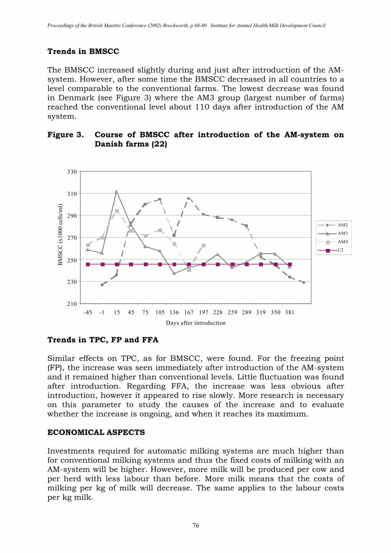
Proceedings of the British Mastitis Conference (2002) Brockworth, p 68-80 Institute for Animal Health/Milk Development Council
76
Trends in BMSCC The BMSCC increased slightly during and just after introduction of the AM-system. However, after some time the BMSCC decreased in all countries to a level comparable to the conventional farms. The lowest decrease was found in Denmark (see Figure 3) where the AM3 group (largest number of farms) reached the conventional level about 110 days after introduction of the AM system. Figure 3. Course of BMSCC after introduction of the AM-system on
Danish farms (22)
Trends in TPC, FP and FFA Similar effects on TPC, as for BMSCC, were found. For the freezing point (FP), the increase was seen immediately after introduction of the AM-system and it remained higher than conventional levels. Little fluctuation was found after introduction. Regarding FFA, the increase was less obvious after introduction, however it appeared to rise slowly. More research is necessary on this parameter to study the causes of the increase and to evaluate whether the increase is ongoing, and when it reaches its maximum. ECONOMICAL ASPECTS Investments required for automatic milking systems are much higher than for conventional milking systems and thus the fixed costs of milking with an AM-system will be higher. However, more milk will be produced per cow and per herd with less labour than before. More milk means that the costs of milking per kg of milk will decrease. The same applies to the labour costs per kg milk.
210
230
250
270
290
310
330
-45 -1 15 45 75 105 136 167 197 228 259 289 319 350 381
Days after introduction
BM
SC
C (
x1000 c
ells
/ml)
AM2
AM3
AM4
C2
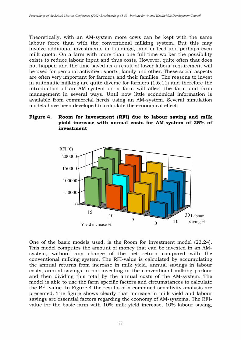
Proceedings of the British Mastitis Conference (2002) Brockworth, p 68-80 Institute for Animal Health/Milk Development Council
77
Theoretically, with an AM-system more cows can be kept with the same labour force than with the conventional milking system. But this may involve additional investments in buildings, land or feed and perhaps even milk quota. On a farm with more than one full time worker the possibility exists to reduce labour input and thus costs. However, quite often that does not happen and the time saved as a result of lower labour requirement will be used for personal activities: sports, family and other. These social aspects are often very important for farmers and their families. The reasons to invest in automatic milking are quite diverse for farmers (1,6,11) and therefore the introduction of an AM-system on a farm will affect the farm and farm management in several ways. Until now little economical information is available from commercial herds using an AM-system. Several simulation models have been developed to calculate the economical effect. Figure 4. Room for Investment (RFI) due to labour saving and milk
yield increase with annual costs for AM-system of 25% of investment
One of the basic models used, is the Room for Investment model (23,24). This model computes the amount of money that can be invested in an AM-system, without any change of the net return compared with the conventional milking system. The RFI-value is calculated by accumulating the annual returns from increase in milk yield, annual savings in labour costs, annual savings in not investing in the conventional milking parlour and then dividing this total by the annual costs of the AM-system. The model is able to use the farm specific factors and circumstances to calculate the RFI-value. In Figure 4 the results of a combined sensitivity analysis are presented. The figure shows clearly that increase in milk yield and labour savings are essential factors regarding the economy of AM-systems. The RFI-value for the basic farm with 10% milk yield increase, 10% labour saving,
30
10
1510
50
0
50000
100000
150000
200000
RFI (€)
Labour
saving %Yield increase %
Proceedings of the British Mastitis Conference (2002) Brockworth, p 68-80 Institute for Animal Health/Milk Development Council
78
medium automated milking parlour and 25% annual costs of the AM-system amounts to €125,044. The differences between the extremes are rather large, almost equal to the investment of a single stall AM-system. CONCLUSION The number of farms milking with an AM-system has increased rapidly since 1998. In regions with expensive labour or a shortage of labour, automatic milking is serious alternative for a traditional milking parlour. The introduction of automatic milking has a large impact on the farm and affects all aspects of dairy farming. Since cows visit the AM-system more or less voluntarily, a large variation in milking intervals can be observed between cows. The introduction of automatic milking has a large impact on the farm, the management and the social life of the farmer. Automatic milking systems require a higher investment than conventional milking systems. However increased milk yields and reduced labour requirements may contribute to a decrease in the fixed costs per kg milk. A successful use of automatic milking depends largely on the management skills of the farmer and the barn layout and farming conditions. REFERENCES 1. De Koning C.J.A.M., van de Vorst, Y., and Meijering, A. (2002)
Automatic milking experience and development in Europe. Proc. 1st North American Conf. on Robotic Milking, Toronto, Canada, pp I1–I11.
2. Schuiling H.J. (1992). Teat cleaning and stimulation. In: Proc. Intl. Symp. on prospects for automatic milking, EAAP publication 65, pp 164-168.
3. Rodenburg, J. and Kelton, D.F. (2001) Automatic milking systems in North America: Issues and challenges unique to Ontario. Proc. Natl. Mast. Council Annual Meeting pp 163-169, NMC, Madison, WI, USA
4. Sonck, B.R. and Donkers, J.H.W. (1995) The milking capacity of a milking robot. J. Agr. Eng. Res. 62: 25-38.
5. Schick, M., Volet, M.-R. and Kaufmann, R. (2000) Modelling of time requirements and milking capacity in automatic milking systems with one or two milking stalls. Page 32-37 In: H. Hogeveen and A. Meijering (editors) Robotic milking. Wageningen Pers, Wageningen.
6. Ipema A, Smits, A. and Jagtenberg, C. (1998) Praktijkervaringen met melkrobots. In: Praktijkonderzoek, 98-6, pp 37-39 (in Dutch).
7. Land, A. van ‘t et al, (2000) Effects of husbandry systems on the efficiency and optimisation of robotic milking performance and management. In: Robotic Milking, Proc. Intl. Symp. held in Lelystad, pp 167-176.
8. Koning, C. de, (2001) Automatic milking; chances and challenges. In: A.Rosati, S. Mihina, C. Mosconi (editors), Physiological and Technical Aspects of Machine Milking, pp 131-140, ICAR TS 7, Rome, Italy.
9. Koning C.J.A.M. de, and Ouweltjes, W. (2000) Maximising the milking capacity of an automatic milking system. In: Robotic Milking, Proc. Intl. Symp. held in Lelystad, pp 38-46.

Proceedings of the British Mastitis Conference (2002) Brockworth, p 68-80 Institute for Animal Health/Milk Development Council
79
10. Erdman, R.A. and Varner, M. (1995) Fixed yield responses to increased milking frequency. J. Dairy Sci. 78: 1199-1203.
11. Veysset P., Wallet, P. Prognard, E. (2001) Automatic milking systems: Characterising the farms equipped with AMS, impact and economic simulations. In: A. Rosati, S. Mihina, C. Mosconi (editors), Physiological and Technical Aspects of Machine Milking, pp 141-150, ICAR TS 7, Rome, Italy.
12. Billon P. (2001) Les robots de traite en France; impact sur la qualité du lait en le système de production. In: Proc. Il Robot di Mungitura in Lombardia, Cremona, Italy.
13. Hogeveen H., van der Vorst, Y., de Koning, C. and Slaghuis, B. (2001) Concepts et implications de la traite automatisée. In: Symp. sure les bovines laitiers, pp 104-120, CRAAQ, Canada.
14. Lind O., Ipema, A.H., de Koning, C., Mottram, T.T. and Herrmann, H.J. (2000) Automatic milking, Bulletin of the IDF 348/2000, pp 3-14.
15. Hogeveen, H., van Lent, A.J.H. and Jagtenberg, C.J. (1998) Free and one-way cow traffic in combination with automatic milking. In: J.P. Chastain (editor) Proc. 4th Intl. Dairy Housing Conf. ASAE 01-98, pp 80-87, Michigan, U.S.A.
16. Vorst, Y. van der, and Hogeveen, H. (2000) Automatic milking systems and milk quality in the Netherlands. In: Robotic Milking, Proc. Intl. Symp. held in Lelystad, pp 73-82.
17. Van Dooren, H.J., Spordnly, E., and Wiktorsson, H. (2002) Automatic milking and grazing, Applied grazing strategies in Europe, Deliverable D25 from EU project Implications of the introduction of automatic milking on dairy farms (QLK5 2000-31006).
18. De Koning C.J.A.M., Verstappen-Boerekamp, J.A.M. and Schuiling, H. (2002) Milk cooling systems for automatic milking. In: Proc. 1st North American Conf. on Robotic Milking, Toronto, Canada, pp V25 – V35.
19. Klungel, G.H., Slaghuis, B.A. and Hogeveen, H. (2000) The effect of the introduction of automatic milking on milk quality. J. Dairy Sci. 83:1998-2003.
20. Pomies D. and Bony, J. (2001) Comparison of hygienic quality of milk collected with a milking robot vs. with a conventional milking parlour. In: H. Hogeveen and A. Meijering (editors) Robotic Milking, pp. 122-123, Wageningen Pers, Wageningen, The Netherlands.
21. Rasmussen, M.D., Blom, J.Y., Nielsen, L.A.H. and P. Justesen, P. (2001) Udder health of cows milked automatically. Livest. Prod. Sci. 72:147-156.
22. Vorst, Y. van der, Knappstein, K. and Rasmussen, M.D. (2002) Effects of Automatic Milking on the Quality of Produced Milk, Deliverable D8 from EU project Implications of the introduction of automatic milking on dairy farms (QLK5 2000-31006).
23. Mandersloot F., van Scheppingen, A.T.J. and Nijssen, J.M.A. (1999) A dairy farm budget program as a tool for planning and evaluation. In: Book of abstracts, 50th annual meeting EAAP, p 216.
Proceedings of the British Mastitis Conference (2002) Brockworth, p 68-80 Institute for Animal Health/Milk Development Council
80
24. Arendzen, I. and van Scheppingen, A.T.J. (2000) Economical sensitivity of four main parameters defining the room for investement of AM-systems on dairy farms. In: Robotic Milking, Proc.Intl. Symp. held in Lelystad, pp 201-211.