team mimesis - raweb.inria.fr · team mimesis 3 2.2.challenges the research of the mimesis team...
TRANSCRIPT
Activity Report 2015
Team MIMESIS
Computational Anatomy and Simulation forMedicineInria teams are typically groups of researchers working on the definition of a common project,and objectives, with the goal to arrive at the creation of a project-team. Such project-teams mayinclude other partners (universities or research institutions).
RESEARCH CENTERNancy - Grand Est
THEMEComputational Neuroscience andMedecine

Table of contents
1. Members . . . . . . . . . . . . . . . . . . . . . . . . . . . . . . . . . . . . . . . . . . . . . . . . . . . . . . . . . . . . . . . . . . . . . . . . . . . . . . . . 12. Overall Objectives . . . . . . . . . . . . . . . . . . . . . . . . . . . . . . . . . . . . . . . . . . . . . . . . . . . . . . . . . . . . . . . . . . . . . . . . 2
2.1. Team Overview 22.2. Challenges 3
3. Research Program . . . . . . . . . . . . . . . . . . . . . . . . . . . . . . . . . . . . . . . . . . . . . . . . . . . . . . . . . . . . . . . . . . . . . . . . 33.1. Real-Time Patient-Specific Computational Models 33.2. Adaptive Meshing and Advanced Simulation Technologies 53.3. Image-Driven Simulation 5
4. Highlights of the Year . . . . . . . . . . . . . . . . . . . . . . . . . . . . . . . . . . . . . . . . . . . . . . . . . . . . . . . . . . . . . . . . . . . . . 64.1.1. Translational Simulation: from pre-operative to intra-operative simulation 6
4.1.1.1. Pre-operative planning 74.1.1.2. Towards intra-operative guidance 7
4.1.2. Eurographics Award 74.1.3. SOFA Consortium 84.1.4. Evaluation by IHU Strasbourg 84.1.5. Science & You 8
5. New Software and Platforms . . . . . . . . . . . . . . . . . . . . . . . . . . . . . . . . . . . . . . . . . . . . . . . . . . . . . . . . . . . . . . 85.1.1. Description 85.1.2. The SOFA Consortium 9
6. New Results . . . . . . . . . . . . . . . . . . . . . . . . . . . . . . . . . . . . . . . . . . . . . . . . . . . . . . . . . . . . . . . . . . . . . . . . . . . . . 106.1. Augmented reality for surgery 106.2. Cardiac electrophysiology 106.3. Cryoablation 116.4. Lipofilling reconstructive surgery 126.5. Neurosurgery 126.6. Physics-based registration algorithms 126.7. Radiation-less guidance during interventional radiology procedures 136.8. Regional anaesthesia 136.9. Training for retina surgery 136.10. Virtual Cutting 14
7. Bilateral Contracts and Grants with Industry . . . . . . . . . . . . . . . . . . . . . . . . . . . . . . . . . . . . . . . . . . . . . 148. Partnerships and Cooperations . . . . . . . . . . . . . . . . . . . . . . . . . . . . . . . . . . . . . . . . . . . . . . . . . . . . . . . . . . . 15
8.1. National projects 158.1.1. ADT (Aide au Développement Technologique, Inria) - DynMesh 158.1.2. ADT - Sofa 168.1.3. ADT - SofaOR 168.1.4. ANR - IDEFI 168.1.5. ANR - RESET 168.1.6. IDEX - CNRS 168.1.7. REBOAsim, Department of Defense USA 178.1.8. IHU, Strasbourg 17
8.2. National collaborations 178.3. Inria collaborations 188.4. European Initiatives 188.5. International Initiatives 198.6. International Research Visitors 20
8.6.1. Visitors 208.6.2. Internships 208.6.3. Visits to International Teams 20

2 Activity Report INRIA 2015
9. Dissemination . . . . . . . . . . . . . . . . . . . . . . . . . . . . . . . . . . . . . . . . . . . . . . . . . . . . . . . . . . . . . . . . . . . . . . . . . . . 209.1. Promoting Scientific Activities 20
9.1.1. Scientific events selection 209.1.2. Journal 219.1.3. Invited talks 21
9.2. Teaching - Supervision - Juries 229.2.1. Teaching 229.2.2. Supervision 229.2.3. Juries 23
9.3. Popularization 239.3.1. IHU Scientific Days 239.3.2. Journée Francaise de Radiologie, JFR 239.3.3. Rencontre Inria Industrie, RII 259.3.4. Journée Alsacienne d’Ophtalmologie, JAO 259.3.5. Talk at Université Paris Descartes 259.3.6. Talk at MEDinISRAEL 259.3.7. Science & You 259.3.8. Pitch at B.E.S.T. Symposium 25
10. Bibliography . . . . . . . . . . . . . . . . . . . . . . . . . . . . . . . . . . . . . . . . . . . . . . . . . . . . . . . . . . . . . . . . . . . . . . . . . . .25

Team MIMESIS
Creation of the Team: 2015 July 01
Keywords:
Computer Science and Digital Science:2.5. - Software engineering3.1.1. - Modeling, representation3.1.4. - Uncertain data3.2.2. - Knowledge extraction, cleaning5.2. - Data visualization5.3.3. - Pattern recognition5.3.4. - Registration5.4.4. - 3D and spatio-temporal reconstruction5.4.5. - Object tracking and motion analysis5.6. - Virtual reality, augmented reality6.1.1. - Continuous Modeling (PDE, ODE)6.1.5. - Multiphysics modeling6.2.8. - Computational geometry and meshes
Other Research Topics and Application Domains:2.4. - Therapies
Our offices are located in the Clovis Vincent building, near the IRCAD. Please do not hesitate to visit us!
1. MembersResearch Scientists
Stéphane Cotin [Team leader, Inria, Senior Researcher, HdR]Hadrien Courtecuisse [CNRS, University of Strasbourg, Researcher]David Cazier [CNRS, University of Strasbourg, Professor, from Apr 2015, HdR]
EngineersRemi Bessard Duparc [Inria, Engineer]Marc Legendre [Inria, Engineer, until Sep 2015]Bruno Marques [Inria, Engineer]Guillaume Paran [Inria, Engineer, from Nov 2015]Frederick Roy [Inria, Engineer]Etienne Schmitt [Inria, Engineer]Hugo Talbot [Inria, Engineer]
PhD StudentsJaime Garcia Guevara [Inria, PhD student]Christoph Paulus [Inria, PhD student]Rosalie Plantefeve [Inria, PhD student, granted by CIFRE Altran]Raffaella Trivisonne [Inria, PhD student, from Nov 2015]
Post-Doctoral FellowsLionel Untereiner [Inria, Post-doctoral fellow]Huu Phuoc Bui [CNRS, University of Strasbourg, Post-doctoral fellow]

2 Activity Report INRIA 2015
Nazim Haouchine [Inria, Post-doctoral fellow]Visiting Scientist
Igor Peterlik [Masaryk University, Czech Republic, Researcher (Collaborator)]Others
Santiago Camacho [Inria, Intern, from Jul 2015 until Dec 2015]Sabrina Izcovich [Inria, Intern, from Sep 2015]David John [Inria, Intern, from Nov 2015]
2. Overall Objectives
2.1. Team OverviewAt the end of 2011, a part of the MIMESIS team 1 moved from Lille to Strasbourg to join the newly createdIHU, which is developing novel clinical technologies at the intersection of the fields of laparoscopy, interven-tional flexible endoscopy and interventional radiology. To develop this new discipline named "image-guidedminimally invasive hybrid surgery", the IHU has established a multidisciplinary research and developmentprogram involving medical experts, scientists and industrial partners. The scientific objectives of this newteam, named MIMESIS are related, but not limited to, several scientific challenges of the IHU. Over the past4 years we have developed new approaches supporting advanced simulations in the context of simulation fortraining. The best example of our success in this area was certainly the work done in collaboration with theHelpMeSee foundation, leading to the creation of our start-up InSimo(read more in 7.1). We now propose tofocus our research on the use of real-time simulation for per-operative guidance. The underlying objectivesinclude patient-specific biophysical modeling, dedicated numerical techniques for real-time computation, dy-namic topological representations, data assimilation and image-driven simulation. This last topic is a transverseresearch theme and raises several open problems, ranging from non-rigid registration to augmented reality. Topursue these directions we have started to assemble a team with a multidisciplinary background, and haveestablished close collaborations with academic and clinical partners. One of these key partners is the ICubelaboratory. We also collaborate with members of Inria Nancy, Inria Lille, Ecole Centrale de Lille, Universityof Luxembourg, Karlsruhe Institute of Technology, and TIMC Laboratory in Grenoble and CIMIT in Boston.
Figure 1. MIMESIS team at the annual team seminar in 2015

Team MIMESIS 3
2.2. ChallengesThe research of the MIMESIS team focuses on improving the realism and fidelity of interactive simulationsof medical procedures 2. From this increase in realism, new clinical applications emerged, in particular per-operative guidance, that currently rely on imaging techniques, but could greatly benefit from our expertisein real-time numerical simulation. To reach these objectives, we addressed several challenges that lie at theintersection of several scientific domains. They include real-time biophysical models (to define new modelsdescribing soft tissue deformation or physiological phenomena such as electrophysiology), novel numericalstrategies (to enable real-time computation even with the increase in complexity of future models), dynamictopological representations (to support topological changes or adaptivity of the models in areas of interest), andimage-driven simulation (to link simulation with real world data such as the one available in an intra-operativecontext). The SOFA (Simulation Open Framework Architecture) platform, in combination with the SOFA-ORproject, is used to integrate our various contributions into a series of prototypes, facilitating validation andtechnology transfer.
The multidisciplinary nature of our research implies that our scientific objectives span across several domains.We have identified 3 main challenges which lie at the intersection of multi-physics modeling, numericalsimulation and computer vision. These challenges are summarized below:
• Real-time patient-specific computational models– Biomechanical, physiological and electrical modeling– Multi-model simulations– Validation
• Adaptive meshing and advanced simulation techniques– Multi-resolution topologies for physics-based simulation– Patient-specific modeling for numerical simulation– Numerical strategies for real-time simulation
• Image-driven Simulation– Parameter Identification / data assimilation– Linking image analysis and physics-based modeling
3. Research Program3.1. Real-Time Patient-Specific Computational Models
The main objective of this scientific challenge is the modeling of the biomechanics and physiology of certainorgans under various stimuli. This requires describing different biophysical phenomena such as soft-tissuedeformation, fluid dynamics, electrical propagation, or heat transfer. These models help simulate the impact ofcertain therapies (such as cryosurgery or radio-frequency ablation), but also represent the behavior of complexorgans such as the brain, the liver or the heart. A common requirement across these developments is the needfor (near) real-time computation and the ability to take into account for patient-specific characteristics.
An important part of our research was dedicated to the development of new accurate models that remaincompatible with real-time computation. Such advanced models do not only permit to increase the realismof future training systems, but they act as a bridge toward the development of patient-specific preoperativeplanning as well as augmented reality tools for the operating room. Yet, patient-specific planning or per-operative guidance also requires the models to be parametrized with patient-specific biomechanical data.The objective in this area is related to the study of hyper-elastic models and their validation for a range oftissues. Preliminary work in this area has been done through two collaborations, one with the biomechanicallab in Lille (LML) with which we have a joint PhD student, and the biomechanics group from the ICubelaboratory in Strasbourg on the development and validation of liver and kidney models. Another importantresearch topic was related to model reduction through various approaches, such as Proper GeneralizedDecomposition (PGD). We have already established discussions with the LEGATO team at University ofLuxembourg which has very good expertise in this area.

4 Activity Report INRIA 2015
Figure 2. Objectives of the MIMESIS team: from training to intra-operative guidance, our objective is to reachaccurate patient-specific simulations
(a) Patient-specific mechanical model ofthe liver with its vascular system
extracted from Computed TomographyAngiogram, and compatible with
real-time simulation
(b) Simulation of the electricalactivity of a human heart from
intra-operative depolarization map
(c) Thermal evolution within ahuman kidney used in the
context of cryoablation
Figure 3. Multi-physics and patient-specific models
Team MIMESIS 5
We continued our work on cardiac electro-physiology simulation, with a focus on patient-specific adaptationof the model. We also studied the simulation of heat transfer and optimization problems in the context ofheat diffusion. This work is a key element of the development of a planning system, such as for cryoablationprocedures (Figure 3).
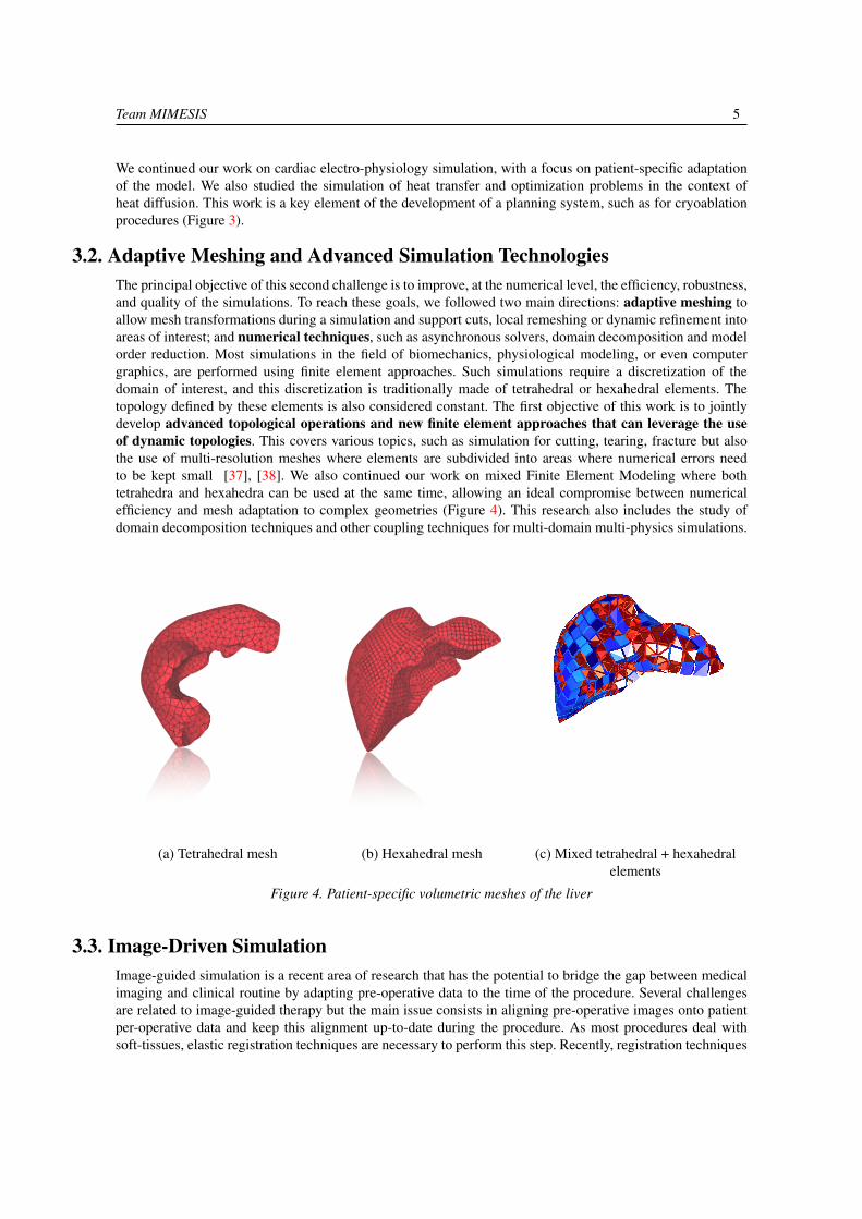
3.2. Adaptive Meshing and Advanced Simulation TechnologiesThe principal objective of this second challenge is to improve, at the numerical level, the efficiency, robustness,and quality of the simulations. To reach these goals, we followed two main directions: adaptive meshing toallow mesh transformations during a simulation and support cuts, local remeshing or dynamic refinement intoareas of interest; and numerical techniques, such as asynchronous solvers, domain decomposition and modelorder reduction. Most simulations in the field of biomechanics, physiological modeling, or even computergraphics, are performed using finite element approaches. Such simulations require a discretization of thedomain of interest, and this discretization is traditionally made of tetrahedral or hexahedral elements. Thetopology defined by these elements is also considered constant. The first objective of this work is to jointlydevelop advanced topological operations and new finite element approaches that can leverage the useof dynamic topologies. This covers various topics, such as simulation for cutting, tearing, fracture but alsothe use of multi-resolution meshes where elements are subdivided into areas where numerical errors needto be kept small [37], [38]. We also continued our work on mixed Finite Element Modeling where bothtetrahedra and hexahedra can be used at the same time, allowing an ideal compromise between numericalefficiency and mesh adaptation to complex geometries (Figure 4). This research also includes the study ofdomain decomposition techniques and other coupling techniques for multi-domain multi-physics simulations.
(a) Tetrahedral mesh (b) Hexahedral mesh (c) Mixed tetrahedral + hexahedralelements
Figure 4. Patient-specific volumetric meshes of the liver
3.3. Image-Driven SimulationImage-guided simulation is a recent area of research that has the potential to bridge the gap between medicalimaging and clinical routine by adapting pre-operative data to the time of the procedure. Several challengesare related to image-guided therapy but the main issue consists in aligning pre-operative images onto patientper-operative data and keep this alignment up-to-date during the procedure. As most procedures deal withsoft-tissues, elastic registration techniques are necessary to perform this step. Recently, registration techniques

6 Activity Report INRIA 2015
started to account for soft tissue biomechanics using physically-based methods, yet several limitations stillhinder the use of image-guided therapy in clinical routine. First, as registration methods become morecomplex, their computation times increase, thus lacking responsiveness. Second, techniques used for non-rigidregistration or deformable augmented reality only “borrow” ideas from continuum mechanics but lack somekey elements (such as identification of the rest shape, or definition of the boundary conditions). Also, theseregistration or augmented reality problems are highly dependent on the choice of image modality and requireinvestigating some aspects of computer vision or medical image processing. However, if we can properlyaddress these challenges, the combination of a real-time simulation and regular acquisitions of image dataduring the procedure opens up very interesting possibilities by using data assimilation to better adapt themodel to the intra-operative data. In the area of non-rigid registration and augmented reality, we have alreadydemonstrated the benefit of our physics-based approaches. This was applied in particular to the problem oforgan tracking during surgery (Figure 5) and led to several key publications [35], [36], [34] and awards(best paper at ISMAR 2013, second best paper at IPCAI 2014). We continued this work with an emphasison robustness to uncertainty and outliers in the information extracted in real-time from image data. We alsoimproved our computer vision techniques, in particular to guarantee a very accurate initial registration of thepre-operative model onto the per-operative surface patch extracted from monocular or stereo laparoscopiccameras.
(a) Step 1 (b) Step 2 (c) Step 3 (d) Step 4
Figure 5. An augmented elastic object undergoing large deformations and topological changes. The computation ofthe physics-based deformation, the cut detection and the topological modification of the underlying volumetric
model are all performed in real-time.
The use of simulation in the context of image-guided therapy can be extended in several other ways. Adirection we particularly want to address is the combined use of simulation and X-ray imaging duringinterventional radiology procedures. Whether it is for percutaneous procedures or catheterization, the taskof the simulation is to provide a short-term (1 to 5 seconds) prediction of the needle or catheter position.Using information extracted from the image, the parameters of the simulation can be assimilated (usingmethods such as unscented Kalman filters), so that the simulation progressively matches the real data in orderto reduce uncertainties. We have already started to create a flexible framework integrating the real-time soft-tissue simulation and state-of-the-art methods of data assimilation and filtering. The reduced-order stochasticfiltering is a computationally efficient improvement over traditional computationally expensive approacheswhich fits well the real-time and patient-specific requirements arising from our per-operative context.
4. Highlights of the Year
4.1. Highlights of the Year4.1.1. Translational Simulation: from pre-operative to intra-operative simulation
Team MIMESIS 7
In recent years, an active development of novel technologies dealing with medical training, planning and guid-ance has become an increasingly important area of interest in both research and health-care manufacturing.With a combination of advanced physical models, realistic human-computer interaction and growing compu-tational power, the MIMESIS team aims at bringing new solutions in order to help both medical students andexperts to achieve a higher degree of accuracy and reliability in surgical interventions [26].
4.1.1.1. Pre-operative planning
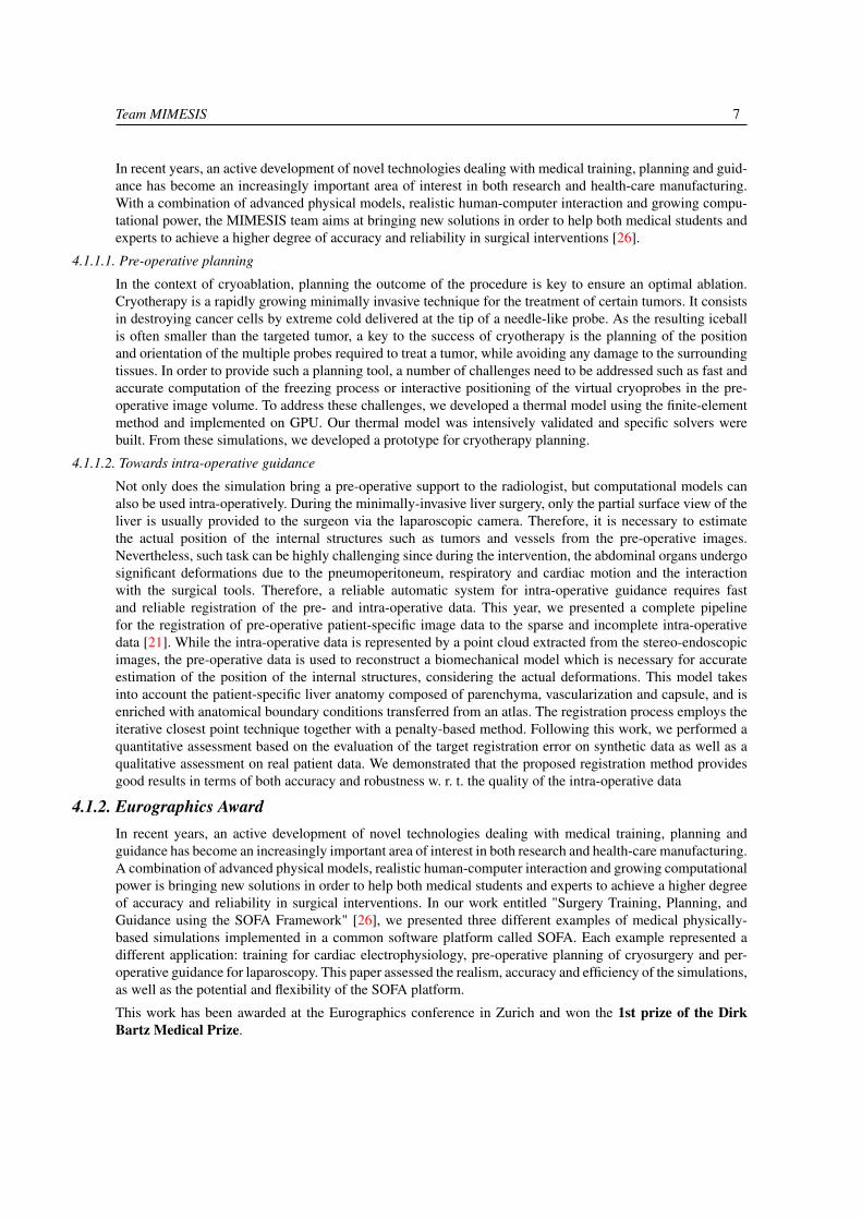
In the context of cryoablation, planning the outcome of the procedure is key to ensure an optimal ablation.Cryotherapy is a rapidly growing minimally invasive technique for the treatment of certain tumors. It consistsin destroying cancer cells by extreme cold delivered at the tip of a needle-like probe. As the resulting iceballis often smaller than the targeted tumor, a key to the success of cryotherapy is the planning of the positionand orientation of the multiple probes required to treat a tumor, while avoiding any damage to the surroundingtissues. In order to provide such a planning tool, a number of challenges need to be addressed such as fast andaccurate computation of the freezing process or interactive positioning of the virtual cryoprobes in the pre-operative image volume. To address these challenges, we developed a thermal model using the finite-elementmethod and implemented on GPU. Our thermal model was intensively validated and specific solvers werebuilt. From these simulations, we developed a prototype for cryotherapy planning.
4.1.1.2. Towards intra-operative guidance
Not only does the simulation bring a pre-operative support to the radiologist, but computational models canalso be used intra-operatively. During the minimally-invasive liver surgery, only the partial surface view of theliver is usually provided to the surgeon via the laparoscopic camera. Therefore, it is necessary to estimatethe actual position of the internal structures such as tumors and vessels from the pre-operative images.Nevertheless, such task can be highly challenging since during the intervention, the abdominal organs undergosignificant deformations due to the pneumoperitoneum, respiratory and cardiac motion and the interactionwith the surgical tools. Therefore, a reliable automatic system for intra-operative guidance requires fastand reliable registration of the pre- and intra-operative data. This year, we presented a complete pipelinefor the registration of pre-operative patient-specific image data to the sparse and incomplete intra-operativedata [21]. While the intra-operative data is represented by a point cloud extracted from the stereo-endoscopicimages, the pre-operative data is used to reconstruct a biomechanical model which is necessary for accurateestimation of the position of the internal structures, considering the actual deformations. This model takesinto account the patient-specific liver anatomy composed of parenchyma, vascularization and capsule, and isenriched with anatomical boundary conditions transferred from an atlas. The registration process employs theiterative closest point technique together with a penalty-based method. Following this work, we performed aquantitative assessment based on the evaluation of the target registration error on synthetic data as well as aqualitative assessment on real patient data. We demonstrated that the proposed registration method providesgood results in terms of both accuracy and robustness w. r. t. the quality of the intra-operative data
4.1.2. Eurographics AwardIn recent years, an active development of novel technologies dealing with medical training, planning andguidance has become an increasingly important area of interest in both research and health-care manufacturing.A combination of advanced physical models, realistic human-computer interaction and growing computationalpower is bringing new solutions in order to help both medical students and experts to achieve a higher degreeof accuracy and reliability in surgical interventions. In our work entitled "Surgery Training, Planning, andGuidance using the SOFA Framework" [26], we presented three different examples of medical physically-based simulations implemented in a common software platform called SOFA. Each example represented adifferent application: training for cardiac electrophysiology, pre-operative planning of cryosurgery and per-operative guidance for laparoscopy. This paper assessed the realism, accuracy and efficiency of the simulations,as well as the potential and flexibility of the SOFA platform.
This work has been awarded at the Eurographics conference in Zurich and won the 1st prize of the DirkBartz Medical Prize.

8 Activity Report INRIA 2015
Figure 6. First Dirk Medical Prize at Eurographics 2015
4.1.3. SOFA ConsortiumAfter ten years of development, a Consortium around the simulation platform SOFA was founded by Inriain November 2015. The MIMESIS team intensively participated in the creation of this Consortium. Theobjectives of this Consortium are to make the SOFA community grow and encourage contributions fromnew SOFA users. The Consortium should also be a way to better answer to the needs of academic or industrialpartners.
A member of the MIMESIS team is now in charge of the coordination of this Consortium. A new engineer wasalso hired to manage the support on the SOFA forum, handle the SOFA events and communicate about SOFAConsortium. The activity of the SOFA Consortium is expected to significantly grow in the coming years.
4.1.4. Evaluation by IHU StrasbourgEvery year, research done at IHU is evaluated by a group of 15 international experts, scientists and clinicians.The 2015 report highlighted our work in the field of modeling and augmented reality: "Interestingly, besides itsnumerous applications for computer assisted surgery, it paves the way to build a new science of anatomy, withthe establishment of innovative, "big data" based organ atlases. The program truly shows the most disruptiveresults. It is scientifically impressive and potentially very practical. There is no doubt that this is the domainwhere IHU is close to be the leading group. The program has a real strategy beyond distinct projects, and clearsynergies have been identified." This report attests to our involvement within the IHU Strasbourg.
4.1.5. Science & YouScience & You is an international event about scientific mediation in the field of digital technologies. In 2015,Science & You took place in Nancy from the 1st until the 6th June 2015. Inria co-organized the event withINS2I and SIF. At this occasion, the MIMESIS team presented the results and prototypes developed in theteam. This event drew a crowd and was a real success.
5. New Software and Platforms
5.1. The SOFA Framework5.1.1. Description

Team MIMESIS 9
SOFA 1 is an open-source software framework targeted at real-time multi-physics simulation, with an emphasison medical simulation. The idea of SOFA was initiated by members of the MIMESIS team, strongly supportedby Inria and still actively developed within the MIMESIS team. Based on C++, the SOFA engine providesmany algorithms, physiological models and anatomical data, made available within a plugin architecture.With its high level of modularity, SOFA appears to be an efficient tools to benchmark and develop new medicaltechnologies using existing algorithms.
Figure 7. Logo of the SOFA framework
The SOFA framework relies on a multi-model representation which allows for having several representations(e.g. mechanical, thermal and visual) of the same object. Those different representations are connected togetherthrough a mechanism called mapping. With these features, it is also possible to have models of very differentnatures interacting with each other, for instance rigid bodies, deformable objects, and fluids. CPU and GPUimplementations can be transparently combined to exploit the computational power of modern hardwarearchitectures.
SOFA is at the heart of a number of research projects, including cardiac electro-physiology modeling,interventional radiology planning and guidance, planning for cryosurgery and deep brain stimulation, robotics,percutaneous procedures, laparoscopic surgery, non-rigid registration, etc. As a proof of its success, SOFAhas been downloaded nearly 150,000 times, and is used today by many research groups around the world,as well as a number of companies. The mailing list used to exchange with the community includes severalhundreds of researchers, from about 50 different institutions. SOFA is currently used by several industrialpartners (Siemens Corporate Research, Epona Medical, Moog, SenseGraphics, etc.) and also provides the keytechnology on which our newly created start-up (InSimo) is relying. We strongly believe that today SOFA hasbecome a reference for academic research, and is increasingly gaining recognition for product prototyping anddevelopment. The best illustration of this worldwide positioning is the role of SOFA in the challenge set bythe HelpMeSee foundation to win the contract for the development of a very ambitious and high-risk projecton cataract surgery simulation.
5.1.2. The SOFA ConsortiumSOFA started ten years ago as an Inria collaborative research project. Now, SOFA includes many differentfunctionalities, several companies rely on the framework as a physics engine and a large community rose overthe years. To better meet the expectations of the community, Inria and the SOFA architects decided to createthe SOFA Consortium in which the MIMESIS is strongly involved. The official kick-off of the Consortiumtook place in Strasbourg on the 25. November 2015.
The objectives of the SOFA Consortium can be defined as:
• Represent the identity of SOFA,
• Structure and develop the community,
• Coordinate the development of SOFA to make it always more efficient and stable.
1More information about SOFA at http://www.sofa-framework.org

10 Activity Report INRIA 2015
Figure 8. The SOFA Consortium was created around the SOFA platform in 2015: here are some fields ofapplication of SOFA
The Consortium has to represent the identity of SOFA. As a consequence, the first mission of the Consortiumis to promote SOFA in conferences, forums or any other event. The Consortium must present SOFA toresearchers and industrials and inform about all activities around the simulation platform and the availableapplications. By advertising all this work, the Consortium will bring more visibility to the entire SOFAcommunity, encourage partnership and stimulate technology transfer.
Second, the Consortium now becomes a privileged contact point for any question or request. Members, users,beginners or any interested partner can contact us. We will find the answer to their needs and thus increase theinteractions outside and within the community.
Third, the Consortium is in charge of coordinating the developments made in SOFA. Through regularmeetings, and bi-annual technical committee, the Consortium makes sure the development follows the roadmap. Moreover, the Consortium sticks to the vision of SOFA as an open-source software, that has to becomemore and more stable and easy to use.
Finally, a free support is provided by the Consortium on the public version of SOFA, with the help of the entireSOFA community.
6. New Results
6.1. Augmented reality for surgeryWe have developed a method for real-time augmented reality of internal liver structures during minimally in-vasive hepatic surgery. Vessels and tumors computed from pre-operative Computed Tomography Angiograms(CTA) scans can be overlaid onto the laparoscopic view for surgery guidance. Compared to current meth-ods, our method is able to locate the in-depth positions of the tumors based on partial three-dimensional livertissue motion using a real-time biomechanical model. We are pursuing the development of this augmentedreality system by using a better biomechanical model, and by relying on parameter optimization and addi-tional per-operative information to further improve accuracy and robustness. In addition, more experiments,and also clinical studies are being performed to precisely measure the benefits and limitations of our approach.This work is strongly related to our involvement in the IHU Strasbourg and is tightly linked to the SOFA-ORproject. Many articles were published on this topic [28], [16], [17].
6.2. Cardiac electrophysiologyCardiac arrhythmia is a very frequent pathology that comes from an abnormal electrical activity in themyocardium. This pathology can be treated by catheterization and ablation of the malfunctionning cardiactissue. The skills required for such interventions are still very challenging to learn, and typically acquiredover several years. We first developed a training simulator for interventional electrocardiology and thermo-ablation of these arrhythmias. Based on physical models 9, this training system reproduces the different
Team MIMESIS 11
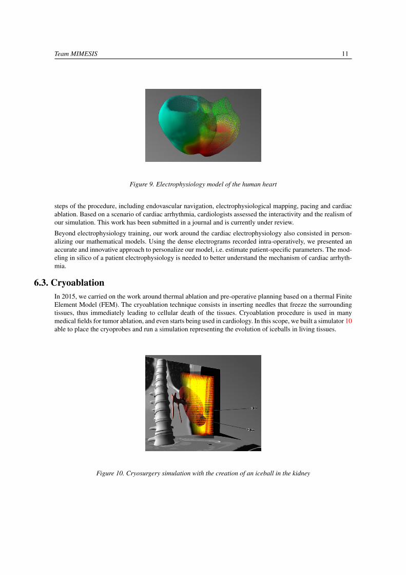
Figure 9. Electrophysiology model of the human heart
steps of the procedure, including endovascular navigation, electrophysiological mapping, pacing and cardiacablation. Based on a scenario of cardiac arrhythmia, cardiologists assessed the interactivity and the realism ofour simulation. This work has been submitted in a journal and is currently under review.
Beyond electrophysiology training, our work around the cardiac electrophysiology also consisted in person-alizing our mathematical models. Using the dense electrograms recorded intra-operatively, we presented anaccurate and innovative approach to personalize our model, i.e. estimate patient-specific parameters. The mod-eling in silico of a patient electrophysiology is needed to better understand the mechanism of cardiac arrhyth-mia.
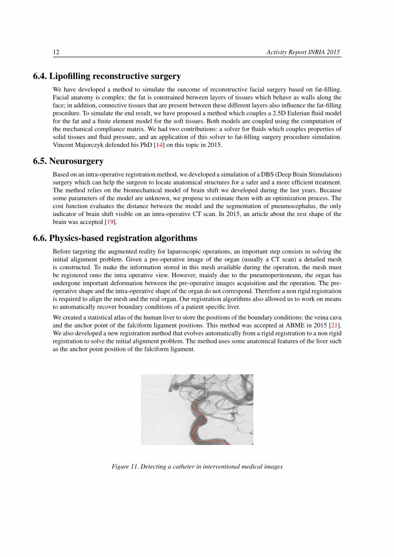
6.3. CryoablationIn 2015, we carried on the work around thermal ablation and pre-operative planning based on a thermal FiniteElement Model (FEM). The cryoablation technique consists in inserting needles that freeze the surroundingtissues, thus immediately leading to cellular death of the tissues. Cryoablation procedure is used in manymedical fields for tumor ablation, and even starts being used in cardiology. In this scope, we built a simulator 10able to place the cryoprobes and run a simulation representing the evolution of iceballs in living tissues.
Figure 10. Cryosurgery simulation with the creation of an iceball in the kidney
12 Activity Report INRIA 2015
6.4. Lipofilling reconstructive surgeryWe have developed a method to simulate the outcome of reconstructive facial surgery based on fat-filling.Facial anatomy is complex: the fat is constrained between layers of tissues which behave as walls along theface; in addition, connective tissues that are present between these different layers also influence the fat-fillingprocedure. To simulate the end result, we have proposed a method which couples a 2.5D Eulerian fluid modelfor the fat and a finite element model for the soft tissues. Both models are coupled using the computation ofthe mechanical compliance matrix. We had two contributions: a solver for fluids which couples properties ofsolid tissues and fluid pressure, and an application of this solver to fat-filling surgery procedure simulation.Vincent Majorczyk defended his PhD [14] on this topic in 2015.
6.5. NeurosurgeryBased on an intra-operative registration method, we developed a simulation of a DBS (Deep Brain Stimulation)surgery which can help the surgeon to locate anatomical structures for a safer and a more efficient treatment.The method relies on the biomechanical model of brain shift we developed during the last years. Becausesome parameters of the model are unknown, we propose to estimate them with an optimization process. Thecost function evaluates the distance between the model and the segmentation of pneumocephalus, the onlyindicator of brain shift visible on an intra-operative CT scan. In 2015, an article about the rest shape of thebrain was accepted [19].
6.6. Physics-based registration algorithmsBefore targeting the augmented reality for laparoscopic operations, an important step consists in solving theinitial alignment problem. Given a pre-operative image of the organ (usually a CT scan) a detailed meshis constructed. To make the information stored in this mesh available during the operation, the mesh mustbe registered onto the intra operative view. However, mainly due to the pneumoperitoneum, the organ hasundergone important deformation between the pre-operative images acquisition and the operation. The pre-operative shape and the intra-operative shape of the organ do not correspond. Therefore a non rigid registrationis required to align the mesh and the real organ. Our registration algorithms also allowed us to work on meansto automatically recover boundary conditions of a patient specific liver.
We created a statistical atlas of the human liver to store the positions of the boundary conditions: the veina cavaand the anchor point of the falciform ligament positions. This method was accepted at ABME in 2015 [21].We also developed a new registration method that evolves automatically from a rigid registration to a non rigidregistration to solve the initial alignment problem. The method uses some anatomical features of the liver suchas the anchor point position of the falciform ligament.
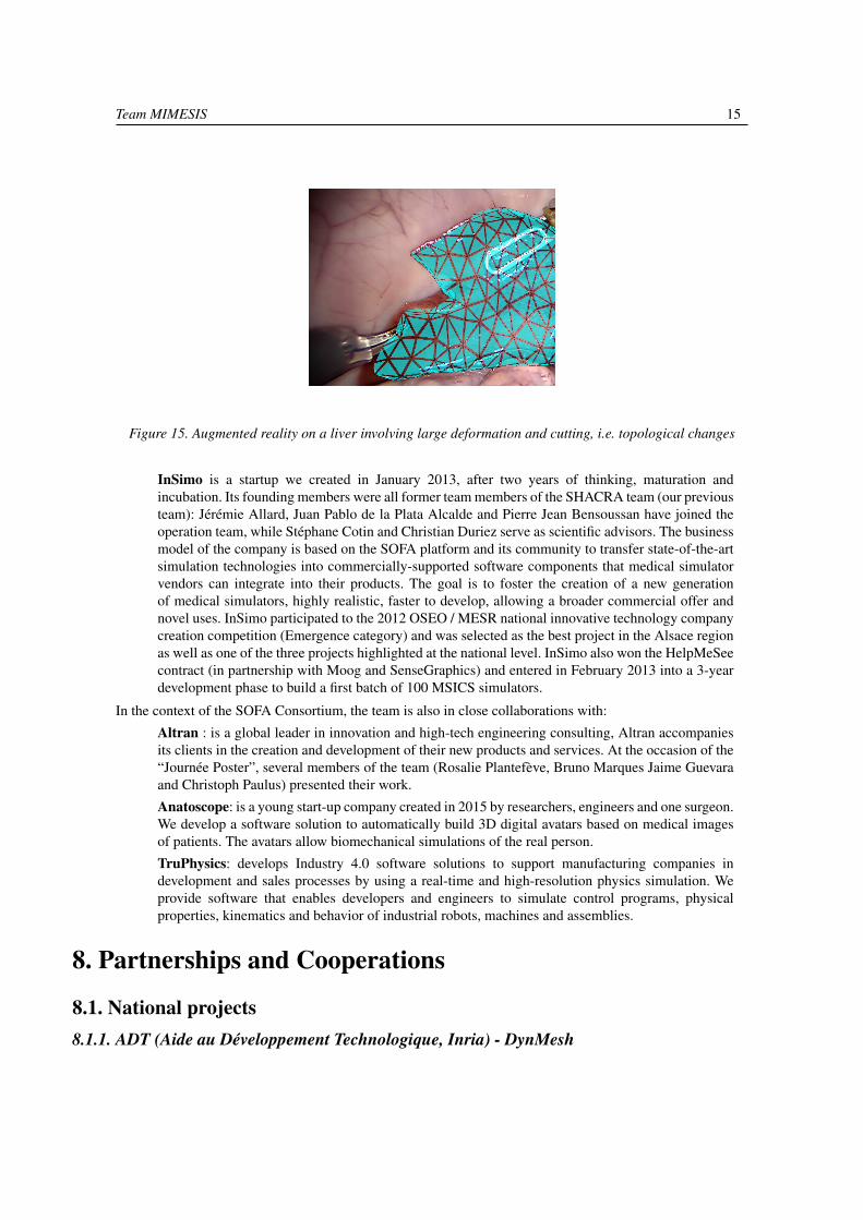
Figure 11. Detecting a catheter in interventional medical images

Team MIMESIS 13
6.7. Radiation-less guidance during interventional radiology proceduresSignificant changes have taken place over the past 20 years in medicine with the development of minimally-invasive procedures. While surgery evolved towards laparoscopy for instance, interventional radiology hasbecome another alternative for many pathologies. Yet, some limitations remain: for percutaneous procedures,soft tissue motion, either due to breathing or deformation induced by the needle, changes the location of thetarget. When using image guidance, or robotic control, this remains a major obstacle. Regarding catheter-based interventions, the lack of 3D information, and extensive use of X-ray imaging to visualize the path tobe followed, are among the main issues. We propose to address these different problems by developing anadvanced navigation system which relies on a combination of real-time simulation and information extractedfrom intra-operative images to assess the current position of the needle. Such a method would have directapplications in pre-operative planning, per-operative guidance, and control for robotics. Our approach willcombine advanced modeling of the device, soft tissue deformation, tissue-tool interactions, and planningalgorithms 11.
6.8. Regional anaesthesiaThe RASimAs project (Regional Anaesthesia Simulator and Assistant) is a European research project fundedby the European Union’s 7th Framework Program. It aims at providing a virtual reality simulator and assistantto doctors performing regional anaesthesia by developing the patient-specific Virtual Physiological Humanmodels. In this project, we are in charge of developing a simulation of a needle inserted into a leg usingthe SOFA framework 12. We especially focused on the integration of the needle simulation into SOFA. Weplanned to release the first version of the simulator by January 2016.
Figure 12. Needle insertion in a muscle in the context of local anaesthesia
In the context of RASimAs, we organized a coding sprint in Strasbourg in April 2015.
6.9. Training for retina surgeryRetina surgery is an increasingly performed procedure for the treatment of a wide spectrum of retinalpathologies. Yet, as most micro-surgical techniques, it requires long training periods before being mastered.To properly answer requests from clinicians for highly realistic training on one hand, and new requirementsfor accreditation or recertification from surgical societies on the other hand, we are developing a high-fidelitytraining system for retinal surgery. This simulator will be built upon our strong scientific expertise in thefield of real-time simulation, and a success story for technology transfer in the field of cataract surgerysimulation. Members of the consortium have a long expertise in the development of prototypes, as well ascollaborations with clinical partners. The simulation system that we propose to develop is based on the OpenSource simulation platform SOFA, and relies on expertise from our partners to ensure clinical and industrialrelevance. This work is initially funded through the ANR project RESET which started in March 2015. A

14 Activity Report INRIA 2015
Figure 13. FEM model of the eye used in our simulation of retina surgery
first version 13 of the training system has been delivered and we made a live demonstration at the JournéeAlsacienne d’Ophtalmologie.
6.10. Virtual CuttingThe simulation of cutting is a central interest in the team. We especially work on the simulation of surgicalcuts 14, tearing and other separations of materials induced by surgical tools. On the one hand, we investigatedthe theoretical aspect: using the standard finite element method (FEM) combined with a re-meshing approach,we replace locally the current structure of the mesh in order to allow for a separation. On the other hand, wedetected a separation in the motion of an object provided by a monocular video stream. With that detection,we can provide an augmented reality during the cutting and tearing of a deformable object.
Figure 14. Our cutting algorithm in SOFA
The theoretical aspect of our work has been published in an article both at the conference Computer GraphicsInternational CGI [24] and in the journal "The visual computer" [20]. The application in augmented reality 15has been published at two conferences: "Augmented Reality during Cutting and Tearing of DeformableObjects", International Symposium on Mixed and Augmented Reality (ISMAR) [30].
To read more about our projects and results, please visit our website: http://mimesis.inria.fr.
7. Bilateral Contracts and Grants with Industry7.1. Bilateral Contracts with Industry
Team MIMESIS 15
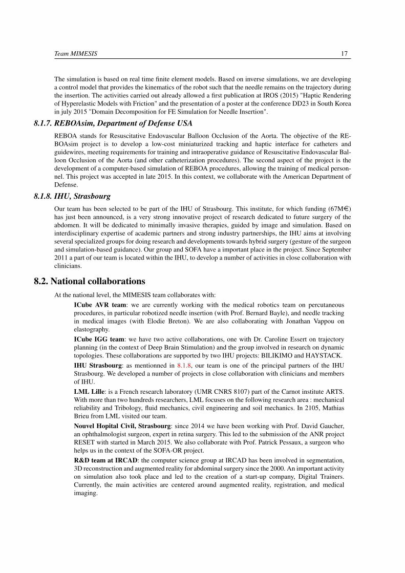
Figure 15. Augmented reality on a liver involving large deformation and cutting, i.e. topological changes
InSimo is a startup we created in January 2013, after two years of thinking, maturation andincubation. Its founding members were all former team members of the SHACRA team (our previousteam): Jérémie Allard, Juan Pablo de la Plata Alcalde and Pierre Jean Bensoussan have joined theoperation team, while Stéphane Cotin and Christian Duriez serve as scientific advisors. The businessmodel of the company is based on the SOFA platform and its community to transfer state-of-the-artsimulation technologies into commercially-supported software components that medical simulatorvendors can integrate into their products. The goal is to foster the creation of a new generationof medical simulators, highly realistic, faster to develop, allowing a broader commercial offer andnovel uses. InSimo participated to the 2012 OSEO / MESR national innovative technology companycreation competition (Emergence category) and was selected as the best project in the Alsace regionas well as one of the three projects highlighted at the national level. InSimo also won the HelpMeSeecontract (in partnership with Moog and SenseGraphics) and entered in February 2013 into a 3-yeardevelopment phase to build a first batch of 100 MSICS simulators.
In the context of the SOFA Consortium, the team is also in close collaborations with:
Altran : is a global leader in innovation and high-tech engineering consulting, Altran accompaniesits clients in the creation and development of their new products and services. At the occasion of the“Journée Poster”, several members of the team (Rosalie Plantefève, Bruno Marques Jaime Guevaraand Christoph Paulus) presented their work.
Anatoscope: is a young start-up company created in 2015 by researchers, engineers and one surgeon.We develop a software solution to automatically build 3D digital avatars based on medical imagesof patients. The avatars allow biomechanical simulations of the real person.
TruPhysics: develops Industry 4.0 software solutions to support manufacturing companies indevelopment and sales processes by using a real-time and high-resolution physics simulation. Weprovide software that enables developers and engineers to simulate control programs, physicalproperties, kinematics and behavior of industrial robots, machines and assemblies.
8. Partnerships and Cooperations
8.1. National projects8.1.1. ADT (Aide au Développement Technologique, Inria) - DynMesh

16 Activity Report INRIA 2015
The objectives of this ADT are the coupling of SOFA, the physical simulation platform supported by Inria, andCGoGN, the mesh management library developed within the ICube lab at Strasbourg. It aims at extending thephysical engine SOFA with the topological kernel of CGoGN that supports a wide variety of mesh and manylocal remeshing operations. The coupling of both software libraries will provide users of physical engines withnew tools for the development of simulations involving topological changes like cutting, fracturing, adaptationof the resolution or improving contact management or collision detection. The impacts are numerous andwill be operated directly within the MIMESIS Team, with our partners or through the establishment of newcollaborations.
8.1.2. ADT - SofaSOFA Large Scale Development Initiative (ADT) : the SOFA project is an international, multi-institution,collaborative initiative, aimed at developing a flexible and open source framework for interactive simulations.This will eventually establish new grounds for a widely usable standard system for long-term research andproduct prototyping, ultimately shared by academic and industrial sites. The SOFA project involves 4 Inriateams: ASCLEPIOS, DEFROST, IMAGINE and MIMESIS. The development program of the ADT started in2007. This ADT ended in September 2015 and the associated contract of our SOFA engineer Marc Legendreended at the same time.
8.1.3. ADT - SofaORIn December 2014, a new ADT national initiative started. The objective of this ADT is twofold: first, we aimat achieving a level of quality and robustness compatible with IEC 62304 for the core of SOFA and a reducedset of components. This does not include the certification of the code itself, but rather the implementationof a comprehensive development process that will enable the certification by companies wishing to integratethis code into their systems. The second objective is to add new features specific to the needs of using intra-operative guiding tools: interoperability with equipment from the operating room, acquisition and real-timeprocessing of full HD video streams, data assimilation and predictive filters, path planning, visualization foraugmented reality, or user interfaces dedicated to the operating room.
8.1.4. ANR - IDEFIIn the IDEFI ANR, the MIMESIS team is involved in the EVEREST project which aims to develop a newgeneration on-line training platforms, dedicated to the theory and practice of image-guided minimally invasivesurgery. A central objective is to develop a framework for the integration and the rapid spread of numericalinteractive simulation systems, associated with online assessment methodologies. The IHU Strasbourg is theANR project leader and we collaborate on the topic of virtual simulations.
8.1.5. ANR - RESETAt the end of 2014, the team has been awarded a new ANR project: RESET. This project started in March 2015.Its objective is to develop a high-fidelity training system for retinal surgery. Retina surgery is an increasinglyperformed procedure for the treatment of a wide spectrum of retinal pathologies. Yet, as most micro-surgicaltechniques, it requires long training periods before being mastered. This simulator is built upon our scientificexpertise in the field of real-time simulation, and our success story for technology transfer in the field ofcataract surgery simulation (MSICS simulation developed for the HelpMeSee foundation).
8.1.6. IDEX - CNRSThe aim of the project CONECT (Couplage de la rObotique et de la simulatioN mEdicale pour des proCéduresauTomatisées) is to develop a robotic system for needle insertion in deformable tissues which is entirelycontrolled and driven by a numerical simulation. The results of this work could be extremely beneficial formedical applications, such as brachytherapy or biopsy, given the accuracy and the precision required in thiskind of procedures. A first demonstration is currently under development where the needle will be inserted in asilicone gel samples. Given a non-straight predefined trajectory, our goal is to control a Mitsubishi MRV1 robotthat will automatically insert a needle along the predefined path, taking into account the deformation of boththe environment and the needle. The deformation of the gel is tracked with camera using the Optitrack system.
Team MIMESIS 17
The simulation is based on real time finite element models. Based on inverse simulations, we are developinga control model that provides the kinematics of the robot such that the needle remains on the trajectory duringthe insertion. The activities carried out already allowed a first publication at IROS (2015) "Haptic Renderingof Hyperelastic Models with Friction" and the presentation of a poster at the conference DD23 in South Koreain july 2015 "Domain Decomposition for FE Simulation for Needle Insertion".
8.1.7. REBOAsim, Department of Defense USAREBOA stands for Resuscitative Endovascular Balloon Occlusion of the Aorta. The objective of the RE-BOAsim project is to develop a low-cost miniaturized tracking and haptic interface for catheters andguidewires, meeting requirements for training and intraoperative guidance of Resuscitative Endovascular Bal-loon Occlusion of the Aorta (and other catheterization procedures). The second aspect of the project is thedevelopment of a computer-based simulation of REBOA procedures, allowing the training of medical person-nel. This project was accepted in late 2015. In this context, we collaborate with the American Department ofDefense.
8.1.8. IHU, StrasbourgOur team has been selected to be part of the IHU of Strasbourg. This institute, for which funding (67M¤)has just been announced, is a very strong innovative project of research dedicated to future surgery of theabdomen. It will be dedicated to minimally invasive therapies, guided by image and simulation. Based oninterdisciplinary expertise of academic partners and strong industry partnerships, the IHU aims at involvingseveral specialized groups for doing research and developments towards hybrid surgery (gesture of the surgeonand simulation-based guidance). Our group and SOFA have a important place in the project. Since September2011 a part of our team is located within the IHU, to develop a number of activities in close collaboration withclinicians.
8.2. National collaborationsAt the national level, the MIMESIS team collaborates with:
ICube AVR team: we are currently working with the medical robotics team on percutaneousprocedures, in particular robotized needle insertion (with Prof. Bernard Bayle), and needle trackingin medical images (with Elodie Breton). We are also collaborating with Jonathan Vappou onelastography.ICube IGG team: we have two active collaborations, one with Dr. Caroline Essert on trajectoryplanning (in the context of Deep Brain Stimulation) and the group involved in research on dynamictopologies. These collaborations are supported by two IHU projects: BILIKIMO and HAYSTACK.IHU Strasbourg: as mentionned in 8.1.8, our team is one of the principal partners of the IHUStrasbourg. We developed a number of projects in close collaboration with clinicians and membersof IHU.LML Lille: is a French research laboratory (UMR CNRS 8107) part of the Carnot institute ARTS.With more than two hundreds researchers, LML focuses on the following research area : mechanicalreliability and Tribology, fluid mechanics, civil engineering and soil mechanics. In 2105, MathiasBrieu from LML visited our team.Nouvel Hopital Civil, Strasbourg: since 2014 we have been working with Prof. David Gaucher,an ophthalmologist surgeon, expert in retina surgery. This led to the submission of the ANR projectRESET with started in March 2015. We also collaborate with Prof. Patrick Pessaux, a surgeon whohelps us in the context of the SOFA-OR project.R&D team at IRCAD: the computer science group at IRCAD has been involved in segmentation,3D reconstruction and augmented reality for abdominal surgery since the 2000. An important activityon simulation also took place and led to the creation of a start-up company, Digital Trainers.Currently, the main activities are centered around augmented reality, registration, and medicalimaging.

18 Activity Report INRIA 2015
TIMC, Grenoble: this large research group has a strong background in computer-aided surgery,medical imaging, registration, statistical and bio-mechanical modeling. We have regular interactionswith this various members of this group. We are collaborating with Yohan Payan (DR CNRS) on themodeling and simulation of the brain shift. A common PhD thesis started on that topic in late 2014.Other areas of interest are in the field of advanced soft tissue modeling and computer aided surgery,
8.3. Inria collaborationsWithin Inria, the MIMESIS team collaborates with:
ASCLEPIOS: although the core activities of team are in the field of medical image analysis, it alsohas a strong expertise in physics-based simulation of the heart. We collaborated on the developmentof an electro-mechanical model of the heart, and on some core components of SOFA. We collaboratewith the ASCLEPIOS team on the development of the SOFA framework and on the development ofa simulation system for radio-frequency ablation in the case of cardiac arrhythmia,
DEFROST: the team imagines future robots which don’t need to be "rigid" but made of complexdeformable structures, composed of stiff and soft regions, close to organic materials that can be foundin nature. Soft robotics opens very attractive perspectives in terms of new applications, reduction ofmanufacturing costs, robustness, efficiency and security. It could constitute a great jump in roboticsin the following years. We continue to interact with the team in Lille given our common researchbackground. A joint article of constraint-based haptic modeling has already been submitted.
IMAGINe: the team has a general focus on animation and simulation of natural objects. Weessentially collaborate with Prof. François Faure on real-time finite element techniques, collisiondetection and contact response (which led to a SIGGRAPH paper) and the development of SOFA,
MAGRIT: their research field is computer vision, with a focus on augmented reality applications.The team is also fairly involved in computer-based solutions for the planning or the simulationof interventional radiology procedures, with a strong collaboration with the CHU in Nancy. Wecollaborate with the MAGRIT team in the area of interventional radiology and augmented reality. Acommon PhD thesis, whose subject was to develop implicit representations of anatomical structuressuch as blood vessels or aneurysms, was defended in 2013. Another joint PhD thesis was defendedin January 2015 on the topic of non-rigid augmented reality and combined the computer visionexpertise of MAGRIT with our expertise on real-time simulation and biomechanical modeling.
8.4. European Initiatives8.4.1. RASimAs
2015 was the second year of the RASimAs project (STREP project funded under FP7) during which wedeveloped new models of the biomechanics of the leg and arm, as well as the simulation of the insertion of theanaesthesiology needle. Regional anaesthesia has been used increasingly during the past four decades. Thisis due to the perceived advantages of reduced postoperative pain, earlier mobility, shorter hospital stay, andsignificantly lower costs. Current training methods for teaching regional anaesthesia include cadavers, videoteaching, ultrasound guidance, and simple virtual patient modeling. These techniques have limited capabilitiesand do not consider individual anatomy. The goal of this project is to increase the application, the effectivenessand the success rates of RA and furthermore the diffusion of the method through the development VPH modelsfor anaesthesia. The goal of the MIMESIS team is to provide the computational infrastructure for the physics-based simulation and to propose new methods for patient-specific modeling and simulation of soft tissues andtheir interaction with the needle, including its effect on nerve physiology.
See http://rasimas.imib.rwth-aachen.de for more details.

Team MIMESIS 19
In the context of the RASimAS project, we collaborate with the company:
• SenseGraphics: develops next generation medical simulator software for a wide range of surgicalprocedures. It is used in simulators for training surgeons in various fields such as robotic surgery,eye surgery, dentistry, ultrasound interpretation and anesthesia. The simulators combine the latesttechnologies in real-time graphics rendering as well as advanced force feedback to allow thesurgeons to have an experience that is as close to reality as possible.
With the RASimAS project, we also collaborate with: the University Hospital Aachen, RWTH Aachen Uni-versity, Bangor University, University College Cork, Universidad Rey Juan Carlos, Foundation for Researchand Technology Hellas, Zilinska univerzita v Ziline, Katholieke Universiteit Leuven and the Stiftelsen Sintef.
8.5. International Initiatives8.5.1. Inria International Partners
At the international scale, the MIMESIS team collaborates with:
CIMIT, Boston: we are restarting our interactions on interventional radiology simulation, inparticular the design and development of a hardware interface for tracking catheters and guidewires.A joint proposal to the DoD has been submitted to this end.
Harvard Biorobotics lab, Cambridge: this group focuses on the role of sensing and mechanicaldesign in motor control, in both robots and humans. This work draws upon diverse disciplines,including biomechanics, systems analysis, and physiology. We started a collaboration on inverseproblems for identifying optimal areas of cardiac ablation using our work on electro-mechanicalmodeling of the heart. Other areas of collaboration are planned, such as cardiac valve interactionswith blood flow.
Humanoid and Intelligence Systems Lab, Karlsruhe Institute of Technology: we started acollaboration with Dr Stefanie Speidel and Dr. Stefan Suwelack on the topics of real-time soft tissuemodeling and laparoscopic augmented reality.
Institute of Computer Science, Masaryk University, Czech Republic: we have an extensivecollaboration with Igor Peterlik at the ICS, leading to 7 publications over that past 18 months. Thiscollaboration covers the fields of non-rigid registration, augmented reality and haptics.
Interactive Graphics and Simulation, Innsbruck: the IGS group in Innsbruck is a continuationof a group led at ETH by Matthias Harders. Its scientific focus is on physically-based simulation,computer haptics, and to a limited extent, augmented reality. The main application area is the medicaldomain.
Surgical Planning Lab, Boston: this research laboratory at Brigham and Women’s Hospital hasa large expertise in the analysis of diagnostic data using computational image analysis. We knowthis group very well, in particular in the field of Deep Brain Stimulation and through their work onOpen Source solutions for computer aided surgery. We are regularly interacting with them on thedevelopment of a version of SOFA dedicated to the operating room.
SINTEF, Norway: we are currently collaborating with SINTEF in the context of the europeanproject RASimAs, and also on other aspects, such as the creation of anatomically correct andaccurate datasets from patient-specific data. We are also discussing future collaborations in thecontext of hepatic surgery simulation and augmented reality (we have jointly written a H2020proposal on this topic).
Team Legato, University of Luxembourg: since last year we have active discussions with Prof.Stéphane Bordas on real-time soft tissue cutting simulation. This has already led to a journal articlein Media [33] and a co-supervision of a post-doctoral fellow.

20 Activity Report INRIA 2015
8.6. International Research Visitors8.6.1. Visitors
In 2015, MIMESIS invited several visitors:
• Jim Ueltschi (founder of the HelpMeSee non-profit organization)
• Karol Miller (Winthrop Professor, School of Mechanical and Chemical Engineering, The Universityof Western Australia)
• Stéphane Bordas (LEGATO team, Luxembourg)
• Karel van Gelder (Product manager, MOOG, Amsterdam)
• Alexandre Krupa (Inria, Rennes)
• Mathias Brieu (Laboratoire de Mécanique, Ecole Centrale Lille)
8.6.2. InternshipsIn 2015, the MIMESIS welcomed two international interns (for 6 months):
Santiago Camacho, Universidad de Buenos Aires, worked on "Improvement of Visualization Toolsfor Augmented Reality Applications"
Sabrina Izcovich, Universidad de Buenos Aires, worked on "Quadratic Tetrahedron Element forFEM simulations".
8.6.3. Visits to International Teams8.6.3.1. Explorer programme
This year, Hugo Talbot obtained an Inria Explorer grant in the context of a partnership with the HarvardBioRobotics Laboratory from Harvard, Cambridge. The Explorer programme covered the one-month visit(June 2015). This visit allowed to discuss about our respective work around simulation, especially concerningsimulation in the field of cardiology. This was also the opportunity to establish several academic and industrialcontacts in the United States. Hugo Talbot namely visited:
• Thermedical: is a company developing a new generation of radio-frequency catheters.
• Center of Medical Simulation: is a simulation center focusing on training based on mannequins.
• SimQuest: is a company developing simulation technologies for medicine, very close to the researchtopic of our team.
• Surgical Planning Laboratory (Brigham and Womens’ Hospital) is a research center very close tothe clinics and working mainly on medical imaging, but also interested in the medical simulation.
• CIMIT: is a research center developing mannequins for training.
9. Dissemination
9.1. Promoting Scientific Activities9.1.1. Scientific events selection9.1.1.1. Reviewer
Stéphane Cotin made reviews for the following conferences:
VRIPHYS
VCBM (Visual Computing in Biology and Medicine)
and MICCAI.
David Cazier made reviews for the conference:
Team MIMESIS 21
Computer Graphics International (CGI)
Nazim Haouchine made reviews for the conference:
International Symposium on Biomedical Imaging
9.1.2. Journal9.1.2.1. Reviewer - Reviewing activities
Stéphane Cotin made reviews for:
the Medical Image Analysis journal.
Hadrien Courtecuisse made reviews for:
IEEE Haptics Symposium,
Transactions on Haptics,
The Visual Computer,
Medical & Biological Engineering & Computing.
David Cazier made reviews for:
the Computer-Aided Design (CAD) journal,
the Visual Computer journal.
Huu Phuoc Bui made reviews for:
the Computational Mechanics journal,
Nazim Haouchine made reviews for the journal:
IET Computer Vision
Rosalie Plantefeve made reviews for the journal:
IJCARS
9.1.3. Invited talksStéphane Cotin has been invited speaker:
keynote lecture at "Open Your Mind" seminar series, ECAM (Paris, France),
keynote lecture at the 10th MICCAI Computational Biomechanics Workshop (Munich,Germany)
keynote lecture at Visual Computing in Biology and Medicine (Chester, UK),
invited talk at HCST Medical Robotics (Tel Aviv, Israel),
invited speaker at the International Conference on Augmented and Virtual Reality (Salento,Italy).
Hadrien Courtecuisse has been invited speaker:
in the legato team at the Computational Mechanics department of the Luxembourg Uni-versity July 2015,
at SINTEF Technology and Society Medical Technology Department. Trondheim, Nor-way,
at HCST Medical Robotics symposium. Tel-Aviv, Israel.
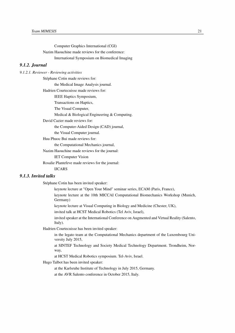
Hugo Talbot has been invited speaker:
at the Karlsruhe Institute of Technology in July 2015, Germany.
at the AVR Salento conference in October 2015, Italy.
22 Activity Report INRIA 2015
Figure 16. Presentation of SOFA at the conference AVR Salento in Lecce (Italy)
9.2. Teaching - Supervision - Juries9.2.1. Teaching
Stéphane Cotin was in charge of the following course:
Course on real-time biomechanical simulation at École Catholique d’Arts et Métiers (Paris,France)
Hadrien Courtecuisse was in charge of the following courses:
Master TIC-Santé. Télécom Physique Strasbourg : Real time simulation (30h)
Master IRMC : (10 h) Real time simulation
David Cazier was in charge of the following course:
Licence : Web technologies and programming, 96hTD, DUT2, Université de Strasbourg,France
9.2.2. SupervisionStéphane Cotin supervised
PhD defended (in co-direction with DEFROST team): Vincent Majorczyk, Modeling ofthe interactions between deformable bodies and fluids in the context of medical simulationin real-time, 1/01/2010 - 28/04/2015
PhD defended : Nazim Haouchine, Image-guided Simulation for Augmented Realityduring Hepatic Surgery, 1/10/2011 - 16/01/2015
PhD in progress : Christoph Paulus, Modélisation et simulation temps-réel pour la prise encompte des changements topologiques dans les tissus mous, 1/01/2014
PhD in progress : Rosalie Plantefeve, Augmented reality and numerical simulations forresection of hepatic tumors, 1/01/2014
Hadrien Courtecuisse supervised:
PhD in progress: Yinoussa adagolodjo, Coupling between robotics and medical simulationfor automated procedures in the scope of the CONECT project. University of Strasbourg,1/02/2015
PhD in progress (in co-direction with the TIMC laboratory): Fanny Morin, Non linear sim-ulation for intraoperative guidance for neurosurgery. Université de Grenoble, 1/10/2014
Internship : Asmaa Ait Hadouch, from Telecom Physique Strasbourg. Topic: communica-tion protocol between Sofa, Mitsubishi MRV1A robot, and optitrack system fot the projectCONECT, 1/06/2015 - 31/08/2015
Team MIMESIS 23
David Cazier supervised:
PhD defended: Thomas Pitiot, Multiresolution tools for the management of interaction inreal time simulations (Outils multirésolutions pour la gestion des interactions en simulationtemps-réel), Université de Strasbourg, 17/12/2015
PhD in progress : Christoph Paulus, Modélisation et simulation temps-réel pour la prise encompte des changements topologiques dans les tissus mous, 1/01/2014
Frédérick Roy and Rosalie Plantefeve co-supervised:
Internship : Santiago Camacho, from Universidad de Buenos Aires. Topic: software devel-opment around visualization for augmented reality applications, 1/07/2015 - 1/12/2015
Hugo Talbot supervised:
Internship : David John, from Karlsruhe Institute of Technology. Topic: research aroundsimulation of thermal effect in living tissues, development of specific solvers, 1/11/2015
Christoph Paulus supervised:
Internship : Sabrina Izcovich, from Universidad de Buenos Aires. Topic: research aroundsecond-order finite element methods and simulation of cutting, 1/09/2015
9.2.3. JuriesStéphane Cotin was part of the following juries:
PhD defense: Mariem Gargouri Osman : "Caractérisation des usagers de la route parimagerie médicale : Extraction fine des paramètres géométriques des côtes à partir devolumes d’images CT", 06/2015 (President)
PhD defense: Vincent Majorczyk "Modélisation des interactions entre solides déformableset films fluides pour la simulation médicale temps-réel", 04/2015 (Supervisor)
PhD defense: Chloe Audigier "Modélisation de l’ablation radiofreéquence pour la planifi-cation de la résection de tumeurs abdominales", 10/2015 (President)
PhD defense: Pierre Chantereau "Caractérisation biomécanique et modélisation his-tologique des mécanismes de vieillissement et d’endommagement du système pelvien",07/2015 (Reviewer)
David Cazier was part of the following juries:
PhD defense: Vincent Majorczyk, Modeling of the interactions between deformable bodiesand fluids in the context of medical simulation in real-time, 4/11/2015 (Reviewer)
PhD defense: Evans Bohl, Modélisation de fruits de leurs structures internes et de leursdéfauts, Université de Poitiers, 4/11/2015 (Reviewer)
PhD defense: Kevin Jordao, Interactive design of crowd animations in large environments,21/12/2015 (Reviewer)
9.3. Popularization9.3.1. IHU Scientific Days
At the occasion of the 4 IHU Scientific days in 2015, the MIMESIS took part and organized several meetings,presentation and discussions around simulation in medicine.
9.3.2. Journée Francaise de Radiologie, JFRThe MIMESIS team was part of the JFR 2015 event in Paris on the 16 and 17th October 2015. Hugo Talbotpresented the work and the projects achieved by the team.

24 Activity Report INRIA 2015
Figure 17. Our booth in the RII Health 2015 in Bordeaux
Figure 18. Rémi Bessard presenting our simulation at JAO 2015
Team MIMESIS 25
9.3.3. Rencontre Inria Industrie, RIIHugo Talbot presented the SOFA framework and our current work during the "Rencontre Inria Industrie" 2015which took place in Bordeaux. We had many nice discussions and feedback about SOFA.
9.3.4. Journée Alsacienne d’Ophtalmologie, JAOIn collaboration with the ophthalmologist Pr. Gauchet, several members of the team (Rémi Bessard, BrunoMarques and Stéphane Cotin) participated at the JAO (Journée Alsacienne d’Ophtalmologie) on the 28th and29th of November 2015. They presented our prototype simulator for retinal surgery.
9.3.5. Talk at Université Paris DescartesOn the 4th of December, Stéphane Cotin was invited as a speaker to the BME seminar at University ParisDescartes. His talk was entiteled "patient safety through real-time numerical simulation".
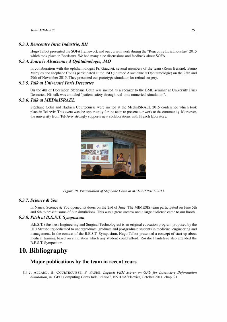
9.3.6. Talk at MEDinISRAELStéphane Cotin and Hadrien Courtecuisse were invited at the MedinISRAEL 2015 conference which tookplace in Tel Aviv. This event was the opportunity for the team to present our work to the community. Moreover,the university from Tel-Aviv strongly supports new collaborations with French laboratory.
Figure 19. Presentation of Stéphane Cotin at MEDinISRAEL 2015
9.3.7. Science & YouIn Nancy, Science & You opened its doors on the 2nd of June. The MIMESIS team participated on June 5thand 6th to present some of our simulations. This was a great success and a large audience came to our booth.
9.3.8. Pitch at B.E.S.T. SymposiumB.E.S.T. (Business Engineering and Surgical Technologies) is an original education program proposed by theIHU Strasbourg dedicated to undergraduate, graduate and postgraduate students in medicine, engineering andmanagement. In the context of the B.E.S.T. Symposium, Hugo Talbot presented a concept of start-up aboutmedical training based on simulation which any student could afford. Rosalie Plantefeve also attended theB.E.S.T. Symposium.
10. BibliographyMajor publications by the team in recent years
[1] J. ALLARD, H. COURTECUISSE, F. FAURE. Implicit FEM Solver on GPU for Interactive DeformationSimulation, in "GPU Computing Gems Jade Edition", NVIDIA/Elsevier, October 2011, chap. 21

26 Activity Report INRIA 2015
[2] J. ALLARD, F. FAURE, H. COURTECUISSE, F. FALIPOU, C. DURIEZ, P. G. KRY. Volume ContactConstraints at Arbitrary Resolution, in "ACM Transactions on Graphics (Proceedings of SIGGRAPH2010)", August 2010, vol. 29(3), https://www.sofa-framework.org/portfolio-item/volume-contact-constraints-at-arbitrary-resolution/
[3] H. COURTECUISSE, J. ALLARD, K. PIERRE, S. P.-A. BORDAS, S. COTIN, C. DURIEZ. Real-time simulationof contact and cutting of heterogeneous soft-tissues, in "Medical Image Analysis", February 2014, 20 p. ,https://hal.inria.fr/hal-01097108
[4] J. DEQUIDT, J. LENOIR, S. COTIN. Interactive Contacts Resolution Using Smooth Surface Representation, in"Proc. Medical Image Computing and Computer-Assisted Interventions (MICCAI)", 2007, pp. 850-857
[5] J. DEQUIDT, M. MARCHAL, C. DURIEZ, S. COTIN. Interactive Simulation of Embolization Coils: Modelingand Experimental Validation, in "Medical Image Computing and Computer Assisted Intervention (MICCAI)",2008, pp. 695-702
[6] C. DURIEZ, S. COTIN, J. LENOIR, P. F. NEUMANN. New Approaches to Catheter Navigation for Interven-tional Radiology Simulation, in "Computer Aided Surgery", 2006, vol. 11, pp. 300-308
[7] C. DURIEZ, F. DUBOIS, A. KHEDDAR, C. ANDRIOT. Realistic Haptic Rendering of Interacting DeformableObjects in Virtual Environments, in "IEEE Transactions on Visualization and Computer Graphics", 2006, vol.12, no 1, pp. 36–47, http://dx.doi.org/10.1109/TVCG.2006.13
[8] F. FAURE, C. DURIEZ, H. DELINGETTE, J. ALLARD, B. GILLES, S. MARCHESSEAU, H. TALBOT, H.COURTECUISSE, G. BOUSQUET, I. PETERLIK, S. COTIN. SOFA: A Multi-Model Framework for InteractivePhysical Simulation, in "Soft Tissue Biomechanical Modeling for Computer Assisted Surgery", Y. PAYAN(editor), Studies in Mechanobiology, Tissue Engineering and Biomaterials, Springer, June 2012, vol. 11, pp.283-321 [DOI : 10.1007/8415_2012_125], https://hal.inria.fr/hal-00681539
[9] N. HAOUCHINE, S. COTIN, I. PETERLIK, J. DEQUIDT, M. SANZ-LOPEZ, E. KERRIEN, M.-O. BERGER.Impact of Soft Tissue Heterogeneity on Augmented Reality for Liver Surgery, in "IEEE Transactions on Visual-ization and Computer Graphics", 2015, vol. 21, no 5, pp. 584 - 597 [DOI : 10.1109/TVCG.2014.2377772],https://hal.inria.fr/hal-01136728
[10] V. LUBOZ, X. WU, K. KRISSIAN, C. WESTIN, R. KIKINIS, S. COTIN, S. DAWSON. A segmentation andreconstruction technique for 3D vascular structures, in "Medical Image Computing and Computer AssistedIntervention (MICCAI)", 2005, pp. 43-50
[11] H. TALBOT, S. MARCHESSEAU, C. DURIEZ, M. SERMESANT, S. COTIN, H. DELINGETTE. Towardsan Interactive Electromechanical Model of the Heart, in "Interface Focus", April 2013, vol. 3, no 2, 4 p.[DOI : 10.1098/RSFS.2012.0091], https://hal.inria.fr/hal-00797354
[12] Y. WEI, S. COTIN, F. LE, J. ALLARD, P. CHUNHUONG. Toward Real-time Simulation of Blood-Coil In-teraction during Aneurysm Embolization, in "Medical Image Computing and Computer Assisted Intervention(MICCAI)", 2009
Publications of the yearDoctoral Dissertations and Habilitation Theses

Team MIMESIS 27
[13] N. HAOUCHINE. Image-guided Simulation for Augmented Reality during Hepatic Surgery, Université deLille1, January 2015, https://hal.inria.fr/tel-01254439
[14] V. MAJORCZYK. Modelling interactions between deformable solids and fluid films for real-time medicalsimulations, Université des Sciences et Technologies de Lille, April 2015, https://hal.inria.fr/tel-01165320
Articles in International Peer-Reviewed Journals
[15] N. HAMZÉ, I. PETERLÍK, S. COTIN, C. ESSERT. Preoperative trajectory planning for percutaneousprocedures in deformable environments, in "Computerized Medical Imaging and Graphics", 2015, vol. 47[DOI : 10.1016/J.COMPMEDIMAG.2015.10.002], https://hal.inria.fr/hal-01242842
[16] N. HAOUCHINE, S. COTIN, I. PETERLIK, J. DEQUIDT, M. SANZ-LOPEZ, E. KERRIEN, M.-O. BERGER.Impact of Soft Tissue Heterogeneity on Augmented Reality for Liver Surgery, in "IEEE Transactions on Visual-ization and Computer Graphics", 2015, vol. 21, no 5, pp. 584 - 597 [DOI : 10.1109/TVCG.2014.2377772],https://hal.inria.fr/hal-01136728
[17] N. HAOUCHINE, J. DEQUIDT, M.-O. BERGER, S. COTIN. Monocular 3D Reconstruction and Augmentationof Elastic Surfaces with Self-occlusion Handling, in "IEEE Transactions on Visualization and ComputerGraphics", 2015, 14 p. [DOI : 10.1109/TVCG.2015.2452905], https://hal.inria.fr/hal-01186011
[18] Z. JIANG, J.-F. WITZ, P. LECOMTE-GROSBRAS, J. DEQUIDT, C. DURIEZ, M. COSSON, S. COTIN, M.BRIEU. B-spline Based Multi-organ Detection in Magnetic Resonance Imaging, in "Strain", June 2015, vol.51, no 2, pp. 235-247 [DOI : 10.1111/STR.12136], https://hal.archives-ouvertes.fr/hal-01204589
[19] F. MORIN, H. COURTECUISSE, M. CHABANAS, Y. PAYAN. Rest shape computation for highly deformablemodel of brain, in "Computer Methods in Biomechanics and Biomedical Engineering", September 2015, vol.18 Suppl 1, pp. 2006-2007 [DOI : 10.1080/10255842.2015.1070591], https://hal.archives-ouvertes.fr/hal-01217673
[20] C. PAULUS, L. UNTEREINER, H. COURTECUISSE, S. COTIN, D. CAZIER. Virtual Cutting of DeformableObjects based on Efficient Topological Operations, in "The Visual Computer, Springer-Verlag Publ., ISSN0178-2789 (Print) 1432-2315 (Online)", 2015, vol. 31, no 6-8, pp. 831-841 [DOI : 10.1007/S00371-015-1123-X], https://hal.archives-ouvertes.fr/hal-01162099
[21] R. PLANTEFÈVE, I. PETERLIK, N. HAOUCHINE, S. COTIN. Patient-specific Biomechanical Modeling forGuidance during Minimally-invasive Hepatic Surgery, in "Annals of Biomedical Engineering", August 2015,https://hal.inria.fr/hal-01205194
[22] R. TRIVISONNE, I. PETERLIK, S. COTIN, H. COURTECUISSE. 3D Physics-Based Registration of 2DDynamic MRI Data, in "Studies in Health Technology and Informatics", April 2016, https://hal.inria.fr/hal-01254388
International Conferences with Proceedings
[23] H. COURTECUISSE, Y. ADAGOLODJO, H. DELINGETTE, C. DURIEZ. Haptic Rendering of HyperelasticModels with Friction, in "2015 IEEE/RSJ International Conference on Intelligent Robots and Systems(IROS)", Hamburg, Germany, IEEE, September 2015, pp. 591-596 [DOI : 10.1109/IROS.2015.7353432],https://hal.archives-ouvertes.fr/hal-01184113

28 Activity Report INRIA 2015
[24] C. PAULUS, L. UNTEREINER, H. COURTECUISSE, S. COTIN, D. CAZIER. Virtual Cutting of Deformable Ob-jects based on Efficient Topological Operations, in "Computer Graphics International", Unknown, Unknownor Invalid Region, 2015, https://hal.archives-ouvertes.fr/hal-01208546
[25] H. TALBOT, S. COTIN, R. RAZAVI, C. RINALDI, H. DELINGETTE. Personalization of Cardiac Electrophys-iology Model using the Unscented Kalman Filtering, in "Computer Assisted Radiology and Surgery (CARS2015)", Barcelona, Spain, June 2015, https://hal.inria.fr/hal-01195719
[26] H. TALBOT, N. HAOUCHINE, I. PETERLIK, J. DEQUIDT, C. DURIEZ, H. DELINGETTE, S. COTIN. SurgeryTraining, Planning and Guidance Using the SOFA Framework, in "Eurographics", Zurich, Switzerland, May2015, https://hal.inria.fr/hal-01160297
Conferences without Proceedings
[27] N. HAMZÉ, A. BILGER, C. DURIEZ, S. COTIN, C. ESSERT. Anticipation of Brain Shift in Deep BrainStimulation Automatic Planning, in "IEEE Engineering in Medicine and Biology Society (EMBC’15)", Milan,Italy, IEEE, August 2015, pp. 3635 - 3638 [DOI : 10.1109/EMBC.2015.7319180], https://hal.inria.fr/hal-01242851
[28] N. HAOUCHINE, A. BILGER, J. DEQUIDT, S. COTIN. Fracture in Augmented Reality, in "SIGGRAPH[Poster]", Los Angeles, United States, August 2015, https://hal.inria.fr/hal-01191090
[29] B. MARQUES, N. HAOUCHINE, R. PLANTEFEVE, S. COTIN. Improving depth perception during surgicalaugmented reality, in "SIGGRAPH [Poster]", Los Angeles, United States, August 2015, Article No. 24[DOI : 10.1145/2787626.2792654], https://hal.inria.fr/hal-01191101
[30] C. J. PAULUS, N. HAOUCHINE, D. CAZIER, S. COTIN. Augmented Reality during Cutting and Tearingof Deformable Objects, in "The 14th IEEE International Symposium on Mixed and Augmented Reality",Fukuoka, Japan, September 2015, 6 p. , https://hal.inria.fr/hal-01184495
[31] C. J. PAULUS, N. HAOUCHINE, D. CAZIER, S. COTIN. Surgical Augmented Reality with TopologicalChanges, in "Medical Image Computing and Computer Assisted Interventions", München, Germany, October2015, https://hal.inria.fr/hal-01184498
[32] H. TALBOT, F. ROY, S. COTIN. Augmented Reality for Cryoablation Procedures, in "SIGGRAPH 2015", LosAngeles, United States, August 2015, https://hal.inria.fr/hal-01180848
References in notes
[33] H. COURTECUISSE, J. ALLARD, K. PIERRE, S. P.-A. BORDAS, S. COTIN, C. DURIEZ. Real-time simulationof contact and cutting of heterogeneous soft-tissues, in "Medical Image Analysis", February 2014, 20 p. ,https://hal.inria.fr/hal-01097108
[34] N. HAOUCHINE, S. COTIN, I. PETERLIK, J. DEQUIDT, M. SANZ-LOPEZ, E. KERRIEN, M.-O. BERGER.Impact of Soft Tissue Heterogeneity on Augmented Reality for Liver Surgery, in "IEEE Transactions on Visual-ization and Computer Graphics", 2015, vol. 21, no 5, pp. 584 - 597 [DOI : 10.1109/TVCG.2014.2377772],https://hal.inria.fr/hal-01136728

Team MIMESIS 29
[35] N. HAOUCHINE, J. DEQUIDT, I. PETERLIK, E. KERRIEN, M.-O. BERGER, S. COTIN. Image-guidedSimulation of Heterogeneous Tissue Deformation For Augmented Reality during Hepatic Surgery, in "ISMAR- IEEE International Symposium on Mixed and Augmented Reality 2013", Adelaide, Australia, October 2013,https://hal.inria.fr/hal-00842855
[36] N. HAOUCHINE, J. DEQUIDT, I. PETERLIK, E. KERRIEN, M.-O. BERGER, S. COTIN. Towards an AccurateTracking of Liver Tumors for Augmented Reality in Robotic Assisted Surgery, in "International Conference onRobotics and Automation (ICRA)", Hong Kong, China, June 2014, https://hal.inria.fr/hal-01003262
[37] T. PITIOT, D. CAZIER, T. JUND, A. HABIBI, P. KRAEMER. Deformable polygonal agents in crowdsimulation, in "Computer Animation and Virtual Worlds", 2014, vol. 25, no 3-4, pp. 341–350
[38] L. UNTEREINER, D. CAZIER, D. BECHMANN. n-Dimensional multiresolution representation of subdivisionmeshes with arbitrary topology, in "Graphical Models", 2013, vol. 75, no 5, pp. 231–246