tbi report 2013 - dtic.mil. a.a. farooqui, and l.a. horrocks, ... deficient groups were subjected to...
TRANSCRIPT

AD_________________
Award Number: W81XWH-11-2-0074 TITLE: Neural Resilience to Traumatic Brain Injury: Identification of Bioactive Metabolites of Docosahexaenoic Acids Involved in Neuroprotection and Recovery PRINCIPAL INVESTIGATOR: Hee-Yong Kim CONTRACTING ORGANIZATION: National Institutes of Health/NIAAA REPORT DATE: March 2013 TYPE OF REPORT: Annual PREPARED FOR: U.S. Army Medical Research and Materiel Command Fort Detrick, Maryland 21702-5012 DISTRIBUTION STATEMENT: Approved for Public Release; Distribution Unlimited The views, opinions and/or findings contained in this report are those of the author(s) and should not be construed as an official Department of the Army position, policy or decision unless so designated by other documentation.

REPORT DOCUMENTATION PAGE Form Approved
OMB No. 0704-0188 Public reporting burden for this collection of information is estimated to average 1 hour per response, including the time for reviewing instructions, searching existing data sources, gathering and maintaining the data needed, and completing and reviewing this collection of information. Send comments regarding this burden estimate or any other aspect of this collection of information, including suggestions for reducing this burden to Department of Defense, Washington Headquarters Services, Directorate for Information Operations and Reports (0704-0188), 1215 Jefferson Davis Highway, Suite 1204, Arlington, VA 22202-4302. Respondents should be aware that notwithstanding any other provision of law, no person shall be subject to any penalty for failing to comply with a collection of information if it does not display a currently valid OMB control number. PLEASE DO NOT RETURN YOUR FORM TO THE ABOVE ADDRESS.
1. REPORT DATE March 2013
2. REPORT TYPEAnnual
3. DATES COVERED 01 March, 2012 - 28 Feb 2013
4. TITLE AND SUBTITLE Neural Resilience to Traumatic Brain Injury: Identification of Bioactive Metabolites of Docosahexaenoic Acids Involved in Neuroprotection and Recovery
5a. CONTRACT NUMBER
5b. GRANT NUMBER W81XWH-11-2-0074
5c. PROGRAM ELEMENT NUMBER
6. AUTHOR(S) Hee-Yong Kim
5d. PROJECT NUMBER
5e. TASK NUMBER
E-Mail: [email protected]
5f. WORK UNIT NUMBER
7. PERFORMING ORGANIZATION NAME(S) AND ADDRESS(ES)
8. PERFORMING ORGANIZATION REPORT NUMBER
National Institutes of Health/NIAAA Rockville, MD 20852
9. SPONSORING / MONITORING AGENCY NAME(S) AND ADDRESS(ES) 10. SPONSOR/MONITOR’S ACRONYM(S) U.S. Army Medical Research and Materiel Command
Fort Detrick, Maryland 21702-5012 11. SPONSOR/MONITOR’S REPORT NUMBER(S) 12. DISTRIBUTION / AVAILABILITY STATEMENT Approved for Public Release; Distribution Unlimited 13. SUPPLEMENTARY NOTES
14. ABSTRACT A mouse model of TBI has been established with behavioral test parameters to evaluate functional deficits. Dietary conditions to generate extreme and moderate n-3 fatty acid depletion in the mouse brain have been established for testing TBI outcome. Significant retardation of spontaneous recovery from TBI-induced motor and cognitive deficits in mice raised on an n-3 deficient diet where brain DHA was depleted by over 70%. Degradation of alpha spectrin, a marker of TBI induced injury, was elevated in the cortex of TBI-inflicted DHA-deficient mice with less NeuN-positive neurons, indicating exacerbated injury in these animals. Moderate depletion of DHA (by 30%) also showed the same trend in behavioral outcome, and biochemical characterization is in progress. Effects of DHA and synaptamide on axon growth have been established in cortical neuron cultures. Software for the isotope assisted mass spectrometric identification of metabolites has been developed which is being applied to DHA-derived metabolites formed by cortical neurons in cultures as well as TBI-inflicted brain homogenates. A nanosymposia presentation and a manuscript have been generated from this project. 15. SUBJECT TERMS
16. SECURITY CLASSIFICATION OF:
17. LIMITATION OF ABSTRACT
18. NUMBER OF PAGES
19a. NAME OF RESPONSIBLE PERSON USAMRMC
a. REPORT U
b. ABSTRACT U
c. THIS PAGEU
UU
19b. TELEPHONE NUMBER (include area code)

Table of Contents
Page
Introduction…………………………………………………………….………..………3
Body………………………………………………………………………………………5
Key Research Accomplishments…………………………………………………….11
Reportable Outcomes…………………………………………………………………..12
Conclusion………………………………………………………………………………..12
References………………………………………………………………………………...n/a
Appendices………………………………………………………………………………..n/a
Introduction
Military personnel in combat deployments are afflicted with high rates of traumatic brain injury (TBI) causing lifelong neurological and cognitive impairments, especially in learning and memory. Numerous studies have shown that docosahexaenoic acid (DHA) is essential for proper brain development and function [1,2], although the underlying mechanisms are still unfolding. Under normal conditions, DHA is present in esterified form in membrane phospholipids, especially the aminophospholipids, phosphatidylethanolamine (PE), and phosphatidylserine (PS). Despite tight regulation to maintain membrane phospholipid homeostasis, DHA enrichment can expand the PS pool in the neuronal membranes [3], as DHA-containing phospholipids serve as the most favored substrate for PS biosynthesis in mammalian tissues [4]. On the contrary, depletion of DHA has been shown to decrease PS levels significantly in brain tissues [3, 5-7]. Since PS is known to participate in key signaling events supporting cell survival and differentiation, DHA-dependent PS modulation is an important aspect of neuroprotection [8]. Following brain injury, polyunsaturated fatty acids including DHA and arachidonic acid (AA, 20:4n-6) are released from neural membranes and metabolized to many bioactive derivatives. Some of the AA-derived eicosanoids are known to be pro-inflammatory, exacerbating the initial injury [9,10]. In contrast, some DHA-derived docosanoids has been shown to ameliorate or resolve inflammatory processes [11]. Furthermore, N-docosahexaenoylethanolamide (synaptamide), a DHA metabolite of a separate class, has been recently identified as a potent neuritogenic and synaptogenic agent [12]. In this regard, the DHA content of the brain may be an important variable to consider in devising a strategy to improve neuroprotection and recovery outcome after brain injuries. 1. N. Salem, Jr., B. Litman, H.Y. Kim, and K. Gawrisch, Mechanisms of action of docosahexaenoic acid in the nervous system, Lipids 36 (2001) 945-959. 2. S.M. Innis, Dietary (n-3) fatty acids and brain development, J Nutr 137 (2007) 855-859. 3. H.Y. Kim, Novel metabolism of docosahexaenoic acid in neural cells, J Biol Chem 282 (2007) 18661-18665. 4. H.Y. Kim, J. Bigelow, and J.H. Kevala, Substrate preference in phosphatidylserine biosynthesis for docosahexaenoic acid containing species, Biochemistry 43 (2004) 1030-1036. 5. L. Hamilton, R. Greiner, N. Salem, Jr., and H.Y. Kim, n-3 fatty acid deficiency decreases phosphatidylserine accumulation selectively in neuronal tissues, Lipids 35 (2000) 863-869. 6. M. Murthy, J. Hamilton, R.S. Greiner, T. Moriguchi, N. Salem, Jr., and H.Y. Kim, Differential effects of n-3 fatty acid deficiency on phospholipid molecular species composition in the rat hippocampus, J Lipid Res 43 (2002) 611-617. 7. H.Y. Kim, M. Akbar, and A. Lau, Effects of docosapentaenoic acid on neuronal apoptosis, Lipids 38 (2003) 453-457. 8. H.Y. Kim, M. Akbar, and Y.S. Kim, Phosphatidylserine-dependent neuroprotective signaling promoted by docosahexaenoic acid, Prostaglandins Leukot Essent Fatty Acids 82 (2010) 165-172. 9. E. Candelario-Jalil, and B.L. Fiebich, Cyclooxygenase inhibition in ischemic brain injury, Curr Pharm Des 14 (2008) 1401-1418. 10. A.A. Farooqui, and L.A. Horrocks, Phospholipase A2-generated lipid mediators in the brain: the good, the bad, and the ugly, Neuroscientist 12 (2006) 245-260. 11. S. Hong, K. Gronert, P.R. Devchand, R.L. Moussignac, and C.N. Serhan, Novel docosatrienes and 17S-resolvins generated from docosahexaenoic acid in murine brain, human blood, and glial cells. Autacoids in anti-inflammation, J Biol Chem 278 (2003) 14677-14687. 12. H.Y. Kim, H.S. Moon, D. Cao, J. Lee, K. Kevala, S. Jun, D. Lovinger, M. Akbar, and B.X. Huang, N-Docosahexaenoylethanolamide promotes development of hippocampal neurons, Biochem J 435 (2011) 327-336.

Objective The major goals of this project are to develop strategies to improve neural resilience to traumatic brain
injury and facilitate recovery through mechanism-based optimization of the nutritional DHA or metabolite status in neuronal tissues.
Aim 1. To determine if diets rich in DHA afford protection to the nervous system against traumatic brain
injury in animal models Aim 2. To identify bioactive DHA metabolites formed in the brain that are involved in neuronal survival,
neurite development, learning and memory Aim 3. To determine if DHA-derived mediators improve recovery after traumatic brain injury in animal
models Aim 4. To devise therapeutic approaches for improving DHA status and/or administering specific
bioactive metabolites that facilitate recovery from traumatic brain injury. Statement of Work Year 2
Task 1: Testing effects of DHA status on TBI injury outcome using animal and dietary models established (months 9-24) Task 2: Testing bioactivity of identified DHA metabolites in cell culture systems (months 13-24) Task 3: Developing software in consultation with an instrumental company software team to establish a method to profile a broader range of DHA metabolites (months 6-18) Milestone 1: Effects of DHA status evaluated in an extreme case of DHA-deficiency in comparison to
DHA-adequate controls. Milestone 2: Bioactivity of major DHA metabolites tested for survival, neurite development and synapse
formation in neuronal culture. Milestone 3: More DHA metabolites identified Milestone 4: Publication 1 on identification of brain DHA-metabolites by isotope-assisted metabolomics
approach

Report Task 1: Testing effects of DHA status on TBI injury outcome using animal and dietary models established (months 9-24) During this report period, we established the effects of DHA status on the recovery from TBI-induced motor deficit using the CCI-model established during the last period. We also established moderately DHA-deficient model in addition to the extreme DHA-deficient model.
During the last report period, we have successfully established the CCI-model for TBI. During this period, we further refined behavioral test parameters to evaluate the functional deficit associated with TBI. We also established dietary conditions to generate moderate and extreme DHA depletion in the mouse brain by feeding mice with an omega-3 deficient special diet for one to three consecutive generations (G1-G3) (Scheme 1). The brain DHA level in the second or third generation omega-3 deficient animals was lowered by over 70%, which was mostly compensated by the increase of docosapentaenoic acid (DPA, 22:5n-6) (Fig. 1).
Scheme 1. Generation of moderate and severely DHA-deficient mice
0
5
10
15
20
25
16:0
18:0
20:0
22:0
24:0
16:1
18:1
n9
18:1
n7
20:1
n9
24:1
18:2
n6
20:3
n6
20:4
n6
22:4
n6
22:5
n6
22:6
n3
Wei
ght P
erce
nt
**
***
**
*
Adequate
Deficient
Fig. 1. Fatty acid composition of the brains from omega-3 fatty acid adequate and severely deficient mice. Data are expressed as mean ± SD (n=3 for each group). *p<0.05, **p<0.01, and ***p<0.001 compared to the adequate group
Using these animal models, we investigated the effect of DHA-depletion on the TBI outcome. Age and gender matched mice at 10-12 weeks from adequate and severely deficient groups were subjected to the CCI procedure and the TBI-induced motor and cognitive deficits were evaluated using accelerating rotarod, beam walk and novel object recognition (NOR) tests. For the accelerating rotarod test, the mice were pre-trained for the rotarod apparatus for three days and a baseline reading of the mice was recorded on the day prior to the surgery. For the beam walk test, the mice were trained to traverse a narrow beam before the surgery and the number of hind foot slips was observed after surgery and compared to sham operated animals. The rotarod and beam walk

tests were further performed from the first day after injury and each day during spontaneous recovery until day 7 after TBI. The two diet groups showed a significant difference in spontaneous recovery of motor function (Fig. 2). By day 3 after TBI the rotarod and beam walk performances of omega-3 adequate animals was recovered significantly. In contrast, the deficient group showed prolonged motor deficits until day 7 after TBI. Statistical significance between two groups was reached with 8 animals from each group.
***
*
120
140
160
180
200
220
240
260
280
300
320
Day 0 Day 1 Day 2 Day 3 Day 4 Day 5 Day 6 Day 7
Sham Adequate
Sham Def icient
TBI Adequate
TBI Def icient
***
***
Tim
e (s
)
Beam Walk Test
0
20
40
60
80
100
120
Day 0 Day 1 Day 2 Day 3 Day 4 Day 5 Day 6 Day 7
Adequate
Def icient* ****
**
Rotarod Test
Fig. 2. Effects of dietary omega-3 fatty acids on spontaneous recovery of TBI-induced motor deficits evaluated by rotarod and beam walk tests. Data are expressed as mean ± SD (n=8 for each group). *p<0.05, **p<0.01, and ***p<0.001 as compared to the TBI adequate group.
The novel object recognition (NOR) test was performed on day 7 after TBI. Mice were individually acclimatized to the test arena daily for 10 min for three days prior to the testing. On the third day, the animals were exposed to two objects for 10 min each and subsequently tested for memory after 2 hours. In sham animals, the NOR performance between omega-3 adequate and severely deficient groups was not statistically different. Nevertheless, NOR performance was significantly impaired in TBI-inflicted severely omega-3 deficient animals compared to the corresponding adequate group (Fig. 3). This data suggests that DHA-depleted animals are particularly susceptible to TBI-induced cognitive deficit and adequate omega-3 fatty acid provision may have a role in preventing such deficit.
p=0.0003
TBI Sham
% N
ove
l Ob
ject
Exp
lora
tio
n
Fig. 3. Effects of dietary omega-3 fatty acids on the TBI-induced cognitive deficit evaluated by novel object recognition test. Data are expressed as mean ± SE (n=7-17 for each group). p= 0.0003 as compared to the TBI adequate group.
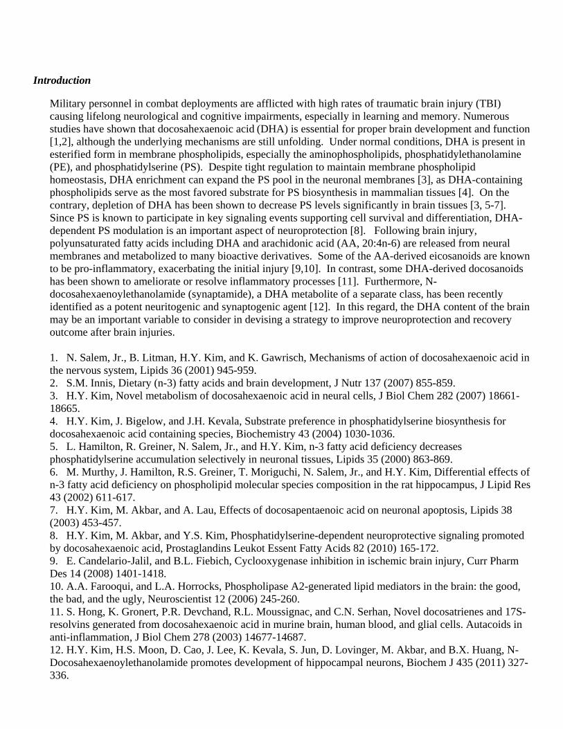
We have also evaluated the anxiety-like behavior using open field test (Fig. 4). Animals with severe omega-3 deficiency showed significantly less time in the center zone compared to the adequate animals, indicating that DHA-deficiency alone can cause increased anxiety. TBI significantly increased anxious behavior in both adequate and deficient groups; however, the significant difference between two groups remained after TBI. These data suggest that the TBI-induced anxious behavior can be exacerbated by severe omega-3 deficiency.
p=0.012
0
5
10
15
20
25
Ade Def0
10
20
30
40
50
60
70
80
Adequate Def icient
Tim
e in
Cen
ter
Zo
ne
(s)
TBI Sham
p=0.05
Fig. 4. Effects of dietary omega-3 fatty acids on the TBI-induced anxious behavior evaluated by open field test. Data are expressed as mean ± SD (n=7-8 for each group). p= 0.012 and 0.05 as compared to the corresponding TBI adequate group.
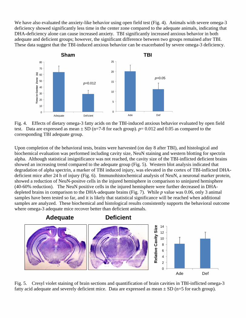
Upon completion of the behavioral tests, brains were harvested (on day 8 after TBI), and histological and biochemical evaluation was performed including cavity size, NeuN staining and western blotting for spectrin alpha. Although statistical insignificance was not reached, the cavity size of the TBI-inflicted deficient brains showed an increasing trend compared to the adequate group (Fig. 5). Western blot analysis indicated that degradation of alpha spectrin, a marker of TBI induced injury, was elevated in the cortex of TBI-inflicted DHA-deficient mice after 24 h of injury (Fig. 6). Immunohistochemical analysis of NeuN, a neuronal marker protein, showed a reduction of NeuN-positve cells in the injured hemisphere in comparison to uninjured hemisphere (40-60% reduction). The NeuN positive cells in the injured hemisphere were further decreased in DHA-depleted brains in comparison to the DHA-adequate brains (Fig. 7). While p value was 0.06, only 3 animal samples have been tested so far, and it is likely that statistical significance will be reached when additional samples are analyzed. These biochemical and histological results consistently supports the behavioral outcome where omega-3 adequate mice recover better than deficient animals.
Deficient Adequate
0
2
4
6
8
10
12
14
Ade Def
Re
lati
ve
Ca
vit
y S
ize
Fig. 5. Cresyl violet staining of brain sections and quantification of brain cavities in TBI-inflicted omega-3 fatty acid adequate and severely deficient mice. Data are expressed as mean ± SD (n=5 for each group).

0
0.05
0.1
0.15
0.2
0.25
0.3
0.35
Ade Def Ade Def0
0.05
0.1
0.15
0.2
0.25
Ade Def Ade Def
Sham TBI Sham TBI
*
***
*
***
P=0.03 P=0.01
Ade Def Ade Def150 KD145 KD
Sham TBI
Cleaved Spectrin
Actin
Cle
ave
d S
pe
ctrin
/Act
in
150 KD 145 KD
Fig. 6. Western blot analysis of cleaved spectrin in TBI-inflicted omega-3 fatty acid adequate and severely deficient mouse brains. Data are expressed as mean ± SD (n=4 for each group).
Adequate Deficient
0
10
20
30
40
50
60
70
Adequate Deficient
NeuN‐Positive Cells
(% of Control)
P=0.06
Fig. 7. NeuN immunostaining of brain sections and quantification of NeuN-positive cells in TBI-inflicted omega-3 fatty acid adequate or severely deficient mice. % of NeuN-positive cells in the injured hemisphere was calculated against NeuN-positive cells in the uninjured hemisphere. Data are expressed as mean ± SD (n=3 mice with 3 sections/mouse for each group).
Using the extreme case of DHA depletion, we established that DHA-adequate mice recover better from TBI compared to the DHA-deficient mice. We have extended this study to a model of moderate DHA deficiency which commonly occurs in humans. The moderately DHA-depleted mice (G1) were generated by feeding pregnant mice a special omega-3 deficient diet from the gestation day 12 (Scheme 1) throughout pregnancy and lactation period and offspring mice were continued on the same diet until the time of experiments (10-12 weeks old). Such deprivation of dietary omega-3 fatty acids effectively lowered brain DHA by 30% compared to the omega-3 adequate group (Fig. 8).

0
5
10
15
20
25
16:0
18:0
20:0
22:0
24:0
16:1
18:1
n9
18:1
n7
20:1
n9
24:1
18:2
n6
20:3
n6
20:4
n6
22:4
n6
22:5
n6
22:6
n3
Wei
ght P
erce
nt
Adequate
Deficient
***
******
***
***
Fig. 8. Fatty acid composition of the brains from the first generation offspring mice fed on omega-3 fatty acid adequate and deficient diets. Moderate DHA-depletion was induced. Data are expressed as mean ± SD (n=3 for each group). ***p<0.001 compared to the adequate group. Moderate DHA-depletion also had a similar impact on the TBI outcome, although the extent was not as severe as the extreme case of DHA-depletion. Both rotarod and beam walk tests indicated significant differences between adequate and deficient groups with the latter showing slower recovery (Fig. 9). In addition, cognitive function and anxious behavior were also adversely affected by the moderate omega-3 fatty acid deficiency in TBI-inflicted mice (Fig. 10).
0
50
100
150
200
250
300
350
Day 0 Day 1 Day 2 Day 3 Day 4 Day 5 Day 6 Day 7
Paired t‐test p=0.009
0
10
20
30
40
50
60
70
80
90
Day 0 Day 1 Day 2 Day 3 Day 4 Day 5 Day 6 Day 7
Ade
Def
Paired t‐test p=0.02
Beam Walk TestRotarod Test
Fig. 9. Effects of moderate omega-3 fatty acid deficiency on spontaneous recovery of TBI-induced motor deficits evaluated by rotarod and beam walk tests. Data are expressed as mean ± SE (n=8 for each group). Paired t-test indicates significant difference between two groups.

Adequate Deficient
40
50
60
70
80
90
Open Field Test
p<0.005
p<0.005
% N
ove
l Ob
ject
Exp
lora
tio
n
Novel Object Recognition Test
Fig. 10. Effects of moderate omega-3 fatty acid deficiency on the cognitive function and anxiety in TBI-inflicted mice evaluated by novel object recognition and open field tests, respectively. Data are expressed as mean ± SE. Our results indicated that the mice from both severe and moderate DHA-deficiency groups had slower recovery of vestibulomotor functions as assessed by the rotarod and beam walk tests. Unlike the n-3 adequate controls, the brain injured n-3 deficient mice also failed to discriminate between the familiar and novel objects in the object recognition test, indicating impaired memory. Histological analysis revealed an increased cavity volume in the n-3 deficient group along with less NeuN-positive neurons, indicating exacerbated injury. Western blot analysis indicated that degradation of alpha spectrin, a marker of TBI induced injury, was elevated in the cortex of TBI-inflicted DHA-deficient mice after 24 hours of injury. These results consistently indicate that DHA-adequate mice recover better from TBI. Considering that modern diets have low n-3 fatty acids and DHA deficiency is common in humans, results from this study present strong possibility of using nutritional remediation as a tool to enhance recovery from brain injury.
Task 2: Testing bioactivity of DHA metabolites in cell culture systems (months 13-24) We have previously identified N-docosahexaenoylethanolamide (synaptamide) as a potent neuritogenic and synaptogenic metabolite of DHA formed in the hippocampal and cortical neuronal cultures. In addition to dendrite extension, we tested the bioactivity of synaptamide on axon growth in cortical neurons during this period. The cortical neuron cultures were chosen, since the cortical region would be most significantly inflicted by the CCI procedure. Axon growth of cortical neurons was promoted by DHA dose-dependently in the 0.1-1 µM range. DHA-derived synaptamide was even more potent in stimulating axon growth. Significant effects were observed at a concentration as low as 5 nM while other fatty acid ethanolmines including DPAn-6 ethanolamide (DPEAn-6), anandamide (AEA) and oleoylethanolamide (OEA) exerted no effects at 10 nM (Fig. 11).
Control SYN DPEA AEA0
0.5
1
1.5
2
2.5
Axo
n l
eng
th r
elat
ive
to c
on
tro
l **
Fig. 11. Effect of polyunsaturated fatty acid ethanol amides on cortical axon growth. Cortical neuron cultures were treated with 10 nM fatty acid amides for 3 days, immunostained for axon specific marker SMI-312 and axon length was quantified by Metamorph software. **, p < 0.01 vs. control.
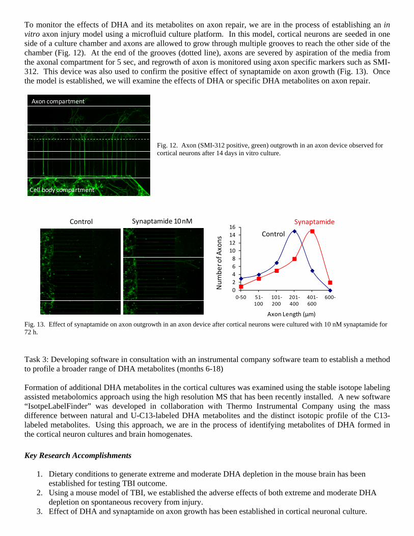
To monitor the effects of DHA and its metabolites on axon repair, we are in the process of establishing an in vitro axon injury model using a microfluid culture platform. In this model, cortical neurons are seeded in one side of a culture chamber and axons are allowed to grow through multiple grooves to reach the other side of the chamber (Fig. 12). At the end of the grooves (dotted line), axons are severed by aspiration of the media from the axonal compartment for 5 sec, and regrowth of axon is monitored using axon specific markers such as SMI-312. This device was also used to confirm the positive effect of synaptamide on axon growth (Fig. 13). Once the model is established, we will examine the effects of DHA or specific DHA metabolites on axon repair.
Fig. 12. Axon (SMI-312 positive, green) outgrowth in an axon device observed for cortical neurons after 14 days in vitro culture.
Control Synaptamide 10 nM
0
2
4
6
8
10
12
14
16
0‐50 51‐100
101‐200
201‐400
401‐600
600‐
Number of A
xons
Axon Length (µm)
Synaptamide
Control
Fig. 13. Effect of synaptamide on axon outgrowth in an axon device after cortical neurons were cultured with 10 nM synaptamide for 72 h. Task 3: Developing software in consultation with an instrumental company software team to establish a method to profile a broader range of DHA metabolites (months 6-18) Formation of additional DHA metabolites in the cortical cultures was examined using the stable isotope labeling assisted metabolomics approach using the high resolution MS that has been recently installed. A new software “IsotpeLabelFinder” was developed in collaboration with Thermo Instrumental Company using the mass difference between natural and U-C13-labeled DHA metabolites and the distinct isotopic profile of the C13-labeled metabolites. Using this approach, we are in the process of identifying metabolites of DHA formed in the cortical neuron cultures and brain homogenates.
Key Research Accomplishments
1. Dietary conditions to generate extreme and moderate DHA depletion in the mouse brain has been established for testing TBI outcome.
2. Using a mouse model of TBI, we established the adverse effects of both extreme and moderate DHA depletion on spontaneous recovery from injury.
3. Effect of DHA and synaptamide on axon growth has been established in cortical neuronal culture.
Axon compartment
Cell body compartment

4. An axon growth model using a microfluid culture platform has been established for further development into an in vitro injury model.
5. An algorism for stable isotope assisted identification of DHA metabolites has been established.
Reportable Outcomes
A presentation entitled “Omega-3 Fatty Acid Deficient Diet Worsens Traumatic Brain Injury Outcome” was selected for nanosymposia at the 2012 Society for Neuroscience meeting in New Orleans, LA, and a manuscript has been prepared for submission. Conclusion
Milestone 1: Evaluation of the effects of DHA status in an extreme case of DHA-deficiency in comparison to DHA-adequate controls has been completed.
Milestone 2: Bioactivity of synaptamide, the major DHA metabolite formed in the cortical neuron cultures, has been tested for axon growth.
Milestone 3: An algorism to identify more DHA metabolites has been developed. Milestone 4: Publication 1 on ‘Omega-3 Fatty Acid Deficient Diet Worsens Traumatic Brain Injury
Outcome”. During the second year, we have met the above specified milestones approved by the CDMRP. Since the instrument installation was significantly delayed, it was not possible to make the publication 1 on “identification of brain DHA-metabolites by isotope-assisted metabolomics approach”. Instead, we were able to complete the study on effect of the DHA status on TBI outcome and a manuscript is in final preparation for submission to PLoS One. We anticipate acceleration of further metabolomics research to identify new metabolites as we now have developed the software. We will subsequently evaluate their bioactivity on neuronal survival and neurite growth and repair using cortical neuron cultures and axon device. Animal feeding studies will continue in order to further evaluate the injury outcome in a moderately DHA-depleted status. We will continue to pursue the therapeutic dose and time windows of synaptamide treatment using FAAH KO and wild type animals.