targeting the nrf2 signaling pathway in the retina with a ... gene-delivered secretable and...
TRANSCRIPT

Biochemistry and Molecular Biology
Targeting the Nrf2 Signaling Pathway in the Retina With aGene-Delivered Secretable and Cell-Penetrating Peptide
Cristhian J. Ildefonso,1 Henrique Jaime,2 Emily E. Brown,3 Ryo L. Iwata,1 Chulbul M. Ahmed,1
Michael T. Massengill,1 Manas R. Biswal,1 Shannon E. Boye,3 William W. Hauswirth,3
John D. Ash,3 Qiuhong Li,3 and Alfred S. Lewin1
1Department of Molecular Genetics & Microbiology, University of Florida College of Medicine, Gainesville, Florida, United States2Department of Biology, University of Florida College of Liberal Arts and Sciences, Gainesville, Florida, United States3Department of Ophthalmology, University of Florida College of Medicine, Gainesville, Florida, United States
Correspondence: Alfred S. Lewin,Department of Molecular Genetics &Microbiology, University of FloridaCollege of Medicine, 1200 NewellDrive, PO Box 100266, Gainesville,FL 32610-0266, USA;[email protected].
Submitted: July 15, 2015Accepted: December 17, 2015
Citation: Ildefonso CJ, Jaime H, BrownEE, et al. Targeting the Nrf2 signalingpathway in the retina with a gene-delivered secretable and cell-penetrat-ing peptide. Invest Ophthalmol Vis
Sci. 2016;57:372–386. DOI:10.1167/iovs.15-17703
PURPOSE. Oxidative stress has been linked to several ocular diseases, initiating an inflammatoryresponse that increases tissue injury. The Nrf2 transcription factor regulates expression ofantioxidant genes and is tightly regulated by Kelch-Like ECH-Associated Protein 1 (Keap-1).We evaluate the antioxidant and anti-inflammatory properties of an adeno-associated virus(AAV) vector delivering an Nrf2-derived peptide that binds Keap-1.
METHODS. The sequence of the Nrf2 peptide was fused to a cell-penetrating peptide (Tat-peptide) sequence (TatNrf2mer). The effects of lentiviral-delivered TatNrf2mer were studiedin vitro. Transcript (quantitative [q] RT-PCR) and protein levels (ELISA and immunofluores-cence) were quantified. Cell viability was measured by MTT and Cell Titer assays. The AAVvectors were packaged with the TatNrf2mer fused to secretable green fluorescent protein(GFP) under the control of the small chicken b actin promoter. The protective effects of thisvector were evaluated in a model of RPE oxidative injury and in a mouse model of uveitis afterintravitreal injection.
RESULTS. Expression of TatNrf2mer peptide induced antioxidant gene expression, blocked IL-1b secretion, and protected cells from oxidative injury. In mice, TatNrf2mer expressionpartially protected photoreceptor function based on ERG responses and optical coherencetomography measurements in the sodium iodate (NaIO3) model. Furthermore, sGFP-TatNrf2merexpression decreased IL-1b and IL-6 in the NaIO3-treated mice, and resulted in a 54% decrease inthe number of inflammatory cells in the vitreous body of the endotoxin-induced uveitis mousemodel.
CONCLUSIONS. The intravitreally delivered AAV-TatNrf2mer has antioxidant and anti-inflamma-tory effects in widely-used models of ocular injury, suggesting it also could be useful in oculardiseases associated with oxidative stress and inflammasome activation.
Keywords: AAV, oxidative stress, inflammation, geographic atrophy, retina
Oxidative stress has been associated with neurodegenerativediseases ranging from amyotrophic lateral sclerosis (ALS)
to stroke.1,2 Within the retina, oxidative stress is an importantdriving force for the development of dry age-related maculardegeneration (dry-AMD).3–5 In age-related neurodegeneration,mitochondria are a major source of toxic oxygen radicals,though superoxide generated by nicotinamide adenine dinu-cleotide phosphate (NADPH) oxidase also is thought tocontribute to neural injury and inflammation.6 Superoxidegenerated by mitochondrial electron transport is responsible,directly or indirectly, for injurious modifications to proteins,lipids, and DNA.7,8 These oxidized molecules not only lose theirbiological function but also serve as damage-associatedmolecular pattern (DAMP) signals. Damage-associated molecu-lar pattern signals, such as the reactive aldehyde 4-hydroxyno-nenal, have been detected in the retina and RPE of patientsaffected with dry-AMD.9 An oxidation product of docosahex-anoic acid, carboxyethyl pyrrole (CEP) induces a dry-AMD–likephenotype when injected in mice.10
Human cells have overlapping defense systems to protectagainst reactive oxygen species (ROS).11 These systems includesmall molecules, such as glutathione and a-tocopherol, andenzymes, such as superoxide dismutases and catalase. Nuclearfactor (erythroid-derived 2)-like 2, or Nrf2, is a transcriptionalcoinducer of a set of genes with antioxidant and detoxifyingproperties.12 In addition, Nrf2 recently has been shown to be anegative regulator of NADPH oxidase subunit NOX2 inneuronal glial co-cultures.13 Nrf2 is tightly regulated by itsrepressor Kelch-Like ECH-Associated Protein 1 (Keap-1) which,upon binding to Nrf2, recruits ubiquitin ligases that target Nrf2for proteosomal degradation. When increases in oxidative stressoccur, sulfhydryl groups on Keap-1 are oxidized. This allowsNrf2 to be released, phosphorylated,12 and translocated intothe nucleus where it upregulates genes under the control of theantioxidant response element (ARE).14 These genes encodeproteins that are responsible for the removal of noxious ROSand reactive nitrogen species (RNS).
In recent years, modulators of the Nrf2 signaling pathwayhave been of significant interest as potential therapeutic agents.
iovs.arvojournals.org j ISSN: 1552-5783 372
This work is licensed under a Creative Commons Attribution-NonCommercial-NoDerivatives 4.0 International License.
Downloaded from iovs.arvojournals.org on 02/17/2019

Studies in animal models of acute liver failure have suggestedthat the siRNA-mediated knockdown of Keap-1 could alleviatedisease-associated pathology.15 Other groups have studied thecontrol of this signaling pathway by small molecules, such asthe phytochemical sulforaphane, which modifies sufhydryls inKeap 1 and is protective in animal models of a severaldiseases.16–18 Within the retina, the Nrf2 signaling pathway hasshown to be a promising therapeutic target. Recently, Xiong etal.19 demonstrated that delivery of NRF2 cDNA protectedphotoreceptors and retinal ganglion cells from oxidative stress.An impaired Nrf2 signaling pathway has been implicated in thedevelopment of the RPE damage seen in AMD.20 Interestingly,this same pathway seems to be of importance in thepathophysiology of ocular inflammatory diseases, such asuveitis. Nagai et al.21 demonstrated that Nrf2 protected theretina from inflammation in a mouse model of uveitis. Takentogether, these results suggest that stimulating the Nrf2signaling pathway could have broad therapeutic benefit.
One approach to modulating the NRF2 signaling pathway isthrough the use of small peptides that bind Keap-1 and lead tothe liberation of the Nrf2. Using a phage display library toscreen for peptides that stimulate the ARE response, Hancocket al.22 identified such peptides derived from p62, prothymo-sin-a and other proteins. In 2012, Steel et al.23 showed that anNrf2-derived peptide could activate downstream heme oxy-genase 1 (HO-1) gene expression and inhibit TNF-a productionin vitro. Although this peptide holds potential for therapy, itwould require repeated administration to treat chronicinflammatory disease, such as AMD. Because of its demon-strated record of safety in treating ocular disease,24–26 we askedwhether adeno-associated virus (AAV)–mediated delivery of anNrf2 peptide could alleviate oxidative stress in the retina. Wedescribed an AAV vector that delivers a secreted and cell-penetrating peptide derived from the Nrf2 protein, whichallows the nuclear translocation of endogenous Nrf2 and theinduction of ARE genes in vitro and in vivo. We alsodemonstrated that this vector can block secretion of proin-flammatory cytokine IL-1b in cell culture while decreasing therecruitment of inflammatory cells to the vitreous in an animalmodel of uveitis.
MATERIALS AND METHODS
Cell Culture
Human cell lines were grown at 378C in the presence of 5%CO2. The HEK293T cells were maintained in Dulbecco’smodified Eagle’s medium (DMEM), while ARPE-19 cells weregrown in a 1:1 mixture of DMEM and F-12 medium (DMEM/F12, 50/50). All media were supplemented with fetal bovineserum (FBS) to a final concentration of 10% and with penicillin-streptomycin in a 1% final concentration. Media were filteredsterilized using a 0.22-lm filter unit. ARPE19 cells werevalidated by ATCC (Manassas, VA, USA) and were frozenimmediately upon receipt. Only low passage cells were used inthese experiments.
Design and Cloning the TatNrf2mer Sequence Intothe pCDH-EF1-MCS-T2A-puroR Lentiviral VectorPlasmid
The Nrf2 amino acid sequence that interacts with Keap-1protein is reported to be the following: LQLDEETGEFLPIQ.This sequence was fused downstream of the HIV Tat-peptidesequence (RKKRRQRRR) to generate the Nrf2 peptide(Nrf2mer) with cell-penetration properties. The completeamino acid sequence of the TatNrf2mer gene is as follows:
RKKRRQRRRLQLDEETGEFLPIQ. Codons were selected andoptimized for expression in mammalian cells using the J-Catsoftware.27 Partially overlapping oligonucleotides were synthe-sized (Table 1). The uppercase and underlined sequence ofthese oligonucleotides represent complementary sequenceswith a melting temperature (TM) of 658C, while the uppercaseand bold sequence represents the EcoRI (1) and NotI (2)restriction sites. Oligonucleotides were mixed, and singlestranded sequences were filled-in using the Klenow fragmentof Escherichia coli DNA polymerase I. Each reaction mixturecontained 5 lg each oligo, 2 lL NEB buffer 2 (103), 1 lL dNTPsmix (10 mM each), and 6 lL deionized H2O. Hybridization ofthe oligonucleotides was achieved using the followingconditions: Three cycles of 948C for 30 seconds and 608C for30 seconds. The reaction was cooled to 158C and 1 lL Klenowfragment (5 U/lL) was added to the reaction and incubated atroom temperature for 15 minutes. The reaction was stoppedby adding 1 lL 210 mM EDTA and incubating at 728C for 20minutes. This product was purified with the GenElute PCRclean-up kit (Sigma-Aldrich Corp., St. Louis, MO, USA). Thepurified fragment and the pCDH-EF1-MCS-T2A-puroR plasmid(Systems Biosciences, Mountain View, CA, USA) were digestedwith EcoRI and NotI for 2 hours at 378C, and purified with theGelElute kit. The digested products were ligated using the T4DNA ligase (New England Biolabs, Ipswich, MA, USA) byincubating at room temperature for 2 hours. Ligation plasmidswere used to transform E. coli strain DH5a cells (Invitrogen,Grand Island, NY, USA), and transformed colonies wereselected on LB plates containing ampicillin (100 lg/ mL).
Lentiviral and AAV Vector Production
Lentiviral vectors (LV) were generated with the pCDH-EF1a-MCS-T2A-Puro plasmid from System Biosciences (MountainView, CA, USA) as we described previously.28 Transgenes weresequenced by the di-deoxynucleotide method.29
The AAV vectors were constructed by inserting theTatNrf2mer coding sequence downstream of an Igj-EGFP
gene, linked via furin cleavage site. Expression was driven bythe CMV enhancer and the chicken b actin promoter (CBApromoter) (see Fig. 4). The entire insert was placed betweeninverted terminal repeat (ITRs) of the AAV2 present in theplasmid. This plasmid was propagated amplified in Sure 2 cells(Agilent Technologies, Santa Clara, CA, USA) and DNA wasprepared by CsCl purification of DNA before packaging.Finally, AAV2(quad Y-FþT-V) viral particles were generated,purified, and titrated by the Vector Core of the Vision Center atthe University of Florida (Gainesville, FL, USA).30 This mutantvariation of AAV2 has been shown to transduce many cell typeswithin the retina after an intravitreal injection.31
Enzyme Linked Immunosorbent Assay (ELISA)
The concentration of IL-1b in the conditioned media of cellcultures was measured using a Human IL-1b ELISA kitpurchased from RayBiotech (Norcross, GA, USA). A total of100 lL media was assayed. Each biological replicate wasevaluated in triplicates according to the manufacturers’instructions. The concentration of murine IL-1b, IL-6, andMCP-1 was determined with ELISA kits from Peprotech (RockyHill, NJ, USA) according to the manufacturer’s protocol. AllELISA kits and antibodies used are listed on Table 2.
Immunofluorescence
Cells were grown in an 8-well chamber slide at 2 3 105 cellsper chamber for 24 hours. Afterwards, cells were washed oncewith PBS (pH 7.4) and incubated with 2% paraformaldehyde in
Ocular Gene Delivery of an Nrf2-Derived Peptide IOVS j February 2016 j Vol. 57 j No. 2 j 373
Downloaded from iovs.arvojournals.org on 02/17/2019

PBS for 15 minutes at room temperature. Cells were washedthree times with PBS and then incubated in PBS with 0.1%Triton X-100 (PBS-T) for 10 minutes at room temperature. Cellsthen were washed three times as done previously. Cells wereblocked by incubating with 1% BSA, 1% goat serum in PBS with0.1% Tween-20, and 0.3 M glycine for 1 hour. The anti-Nrf2antibody was diluted to 1 lg/mL in PBS-T with 1% BSA andincubated with the cells for 1 hour at room temperature in ahumidified chamber. Cells were washed as in previous stepsand then incubated with an anti-rabbit antibody conjugated toCy3 chromophore (1:500 dilution) and 40,6-diamidino-2-phe-nylendole (DAPI; 1:1000 dilution) diluted in PBS-T with 1% BSA
for 1 hour at room temperature in the dark. Cells then werewashed as in the previous step and mounted using Fluoro-mount-G (Southern Biotechnology Associates, Inc., Birming-ham, AL, USA). Pictures were taken using a fluorescencemicroscope.
Transfection
HEK293T cells were plated at 80% confluency in a 6-well plate.Plasmid DNA complexes were made by diluting 1 lg plasmidDNA and 2.5 lg linear polyethyleneimine32 (PEI) in 100 lL PBSand incubated at room temperature for 5 minutes. Complexeswere made by adding the diluted PEI to the diluted plasmidDNA and incubating for another 20 minutes at roomtemperature. The medium in each well was replaced with 2mL serum and antibiotic-free medium before adding thecomplexes. Cells were incubated in the presence of thecomplexes for 18 hours (378C, 5% CO2) before medium wasreplaced with 3 mL fresh medium containing 10% FBS and 1%Pen-Strep. Cells were collected 24 hours later by trypsindigestion.
RNA Isolation
Total RNA was isolated from cell cultures using the RNeasymini kit (QIAGEN, Valencia, CA, USA) according to themanufacturers’ protocol. RNA was quantified by 260 nmabsorbance and quality was verified by running an aliquot in a1% agarose gel.
cDNA Synthesis
Complementary DNA was synthesized with the iScript cDNAsynthesis kit (Bio-Rad, Hercules CA, USA). Briefly, 1 ng totalRNA (10 lL) was mixed with 4 lL 53 iScript reaction mix, 5 lLRNAse free water, and 1 lL iScript reverse transcriptase. Thefollowing temperatures and times were used in the synthesis ofthe cDNA: 258C for 5 minutes, 428 for 30 minutes, and 858C for5 minutes. cDNA was stored at �208C until needed.
PCR for the Detection of TatNrf2mer Expression
A PCR reaction was prepared using 1 lL cDNA isolated fromARPE-19 cells or ARPE-19 cells expressing either puromycinresistance (PuroR) gene only (control) or TatNrf2mer. Primersbinding the Tat region (Tat-F) and the PuroR region (PuroR-R)were used to detect TatNrf2mer mRNA (Table 1). Polymerasechain reaction conditions were as follows: 938C for 3 minutes,30 cycles of 938C for 30 seconds, 558C for 30 seconds, and728C for 20 seconds, followed by 728C for 10 minutes.
TABLE 1. Sequences of Oligonucleotides Used
Name Sequence (50->30)
Target
Species
TatNrf2mer-F tatGAATTCgccaccatgaggaagaagaggaggcagagGAGGAGGCTGCAGCTGGACGAG
Human
TatNrf2mer-R ataGCGGCCGCctggatgggcaggaactcgccggtctcCTCGTCCAGCTGCAGCCTCCTC
Human
Tat-F AGTTCTTGCAGCTCGGTG Human
Puro-R TCGCCACCATGAGGAAG Human
NqO1-F AAAGGACCCTTCCGGAGTAA Human
NqO1-R CCATCCTTCCAGGATTTGAA Human
GSTM1-F CTACCTTGCCCGAAAGCAC Human
GSTM1-R ATGTCTGCACGGATCCTCTC Human
GAPDH-F ACAGTCCATGCCATCACTGCC Human
GAPDH-R GCCTGCTTCACCACCTTCTTG Human
b actin-F AGCGAGCATCCCCCAAAGTT Human
b actin-R GGGCACGAAGGCTCATCATT Human
HO-1-F AGCCCCACCAAGTTCAAACA Mouse
HO-1-R GCAGTATCTTGCACCAGGCT Mouse
NqO1-F CGACAACGGTCCTTTCCAGA Mouse
NqO1-R CCAGACGGTTTCCAGACGTT Mouse
GSTM1-F GGGATACTGGAACGTCCGC Mouse
GSTM1-R GCTCTGGGTGATCTTGTGTGA Mouse
Catalase-F CGCAATCCTACACCATGTCG Mouse
Catalase-R AGTATCCAAAAGCACCTGCTCC Mouse
b actin-F CGAGCACAGCTTCTTTGCA Mouse
b actin-R TTCCCACCATCACACCCTGG Mouse
Tat, trans-activator of transcription from HIV-1; Puro-R, puromycinresistance gene; NqO1, NAD(P)H dehydrogenase quinone 1; HO-1,heme oxygenase 1; GSTM1, glutathione S-transferase mu 1; GAPDH,glyceraldehyde 3-phosphate dehydrogenase.
TABLE 2. Antibodies and ELISA Kits
Reagent Company Catalog Number
Anti-Nrf2 antibody Abcam (Cambridge, MA, USA) ab31163
Anti-Nrf2 [EP1808Y] antibody Abcam ab62352
Anti-Lamin AþC [EPR4100] antibody Abcam ab108595
Goat anti-rabbit IgG (HþL) secondary antibody, Alexa Fluor 488 conjugate Invitrogen A-11034
Anti-GFP antibody, rabbit IgG fraction Invitrogen A11122
Nitrotyrosine ELISA Kit Abcam ab113848
Anti-ZO-1 antibody Invitrogen 40-2200
Murine IL-1B Mini ELISA ABTS Development kit Peprotech 900-M47
Murine IL-6 Mini ELISA ABTS Development kit Peprotech 900-M50
Murine JE/MCP-1 Mini ELISA ABTS Development kit Peprotech 900-M126
Human IL-1 beta ELISA RayBiotech ELH-IL1b
Ocular Gene Delivery of an Nrf2-Derived Peptide IOVS j February 2016 j Vol. 57 j No. 2 j 374
Downloaded from iovs.arvojournals.org on 02/17/2019

Polymerase chain reaction products were separated in a 1.3%agarose gel.
Real-Time PCR (RT-PCR) for ARE Genes
Real-time PCR for glutathione S-transferase mu 1 (GSTM1) andNAD(P)H dehydrogenase quinone 1 (NqO1) was performedusing the SsoFast EvaGreen Supermix kit (Bio-Rad). Primersequences are listed in Table 1.
PCR reaction mixtures contained 1 lL 1:10 dilution ofcDNA library, 1 lL forward (F) primer (5 lM) and 1 lL reverse(R) primer (5 lM), 5 lL 23 SsoFast EvaGreen supermix, and 2lL dH2O. Simultaneous amplification of all genes was doneusing the following conditions: 958C for 3 minutes, followed by40 cycles of 958C for 10 seconds, and 608C for 20 seconds.Fluorescence was measured at the end of each cycle by usingthe Bio-Rad CFX96 thermocycler. Fold changes in geneexpression were determined by the DDCt method.33
MTT Assay
Cells were plated in a 96-well plate at 8 3 104 cells per well in100 lL complete growth medium and incubated overnight at378C. The next day, medium was removed, cells were washedwith PBS, then exposed to 200 lL serum- and antibiotic-freemedium containing 800 lM H2O2. Cells then were incubated at378C for 6 hours, washed with PBS, and incubated with 200 lLMTT (tetrazolium) solution diluted in RPMI-1640 (500 lg/mL)at 378C for 4 hours. Three wells without cells but containing200 lL MTT were included in the plate. After 4 hours, the MTTsolution was removed, and cells were incubated in 200 lL PBSfor 15 minutes at room temperature. The absorbance at 570nm was determined using a Varian Carry 50 plate reader (PaloAlto, CA, USA).
Cell Titer Assay
Cells were plated in a 96-well plate at 70% confluency. Thefollowing day, cells were placed in serum-free media contain-ing paraquat (Sigma-Aldrich Corp.) at the concentrationindicated and incubated for 48 hr. CellTiter Aqueous reagent(Promega, Madison, WI, USA) was added and cells wereincubated for an additional 30 minutes. Absorbance at 490 nmwas measured with a plate reader. Cell survival was calculatedby subtracting the absorbance in untreated cells as 100%.
Endotoxin-Induced Uveitis (EIU) Mouse Model
All mice in this study were treated by procedures approved bythe University of Florida Institutional Animal Care and UseCommittee and in accordance with the ARVO Statement for theUse of Animals in Ophthalmic and Visual Research. One-month-old C57BL/6J mice were injected intravitreally with 1 lLsterile saline containing 3 3 109 vector genomes of AAV vectordelivering either green fluorescent protein (GFP; left eye) orsGFP-TatNrf2mer (right eye). One month after injection, GFPexpression was observed by fluorescence funduscopy using aMicron III fundus microscope and fluorescein filters. Uveitiswas induced by intravitreal injection of LPS (25 ng/eye) and thenumber of infiltrative cells in histologic sections was quantifiedas described previously by our lab.34
Sodium Iodate (NaIO3) Mouse Model of RPEDamage
One-month-old C57BL/6J mice were injected intravitreally with3 3 109 vector genomes of AAV2 (quad Y-FþT-V) vectordelivering either GFP (left eye) or sGFP-TatNrf2mer (right eye).
One month later, mice were evaluated for expression of GFPusing fluorescent funduscopy. One week later, mice wereinjected intraperitoneally with 35 or 25 mg/kg NaIO3. After 7days, retinal function was evaluated by full field scotopicelectroretinogram (ERG) using the Espion Ganzfeld Profilesystem (Diagnosys UK Ltd., Cambridge, UK).
Electroretinogram (ERG)
Scotopic ERG analysis was used to measure the loss of rodfunction as previously published.35,36 Mice were dark adaptedovernight. The following day the mice were dilated with oculardrops of 1% atropine and 10% phenylephrine solutions. Micethen were anesthetized using a mixture of ketamine (20 mg/mL) and xylazine (0.8 mg/mL) in normal saline. Gold wireelectrodes were placed over the corneas of anesthetized mice,the reference electrode was placed in the mouth, andgrounding needle electrode was placed in the tail. Mice werestimulated with 2.7 cd.s/m2 flashes of light and light stimulatedvoltage changes were recorded as a function of time. The valueof the a-wave was measured from 0 lV reference to the peak ofthe initial negative deflection, and the b-wave was measuredfrom the absolute peak of the a-wave to the peak of thepositively deflection within 2000 ms of the flash stimulus. Themuch slower c-wave was measured from the baseline to thepeak of the positive deflection that occurs between 1 to 4seconds of the light flash.
Fundoscopy and Spectral-Domain OpticalCoherence Tomography (SD-OCT)
Fundus images were taken using a Micron III retinal imagingmicroscope (Phoenix Research Laboratories, Pleasanton, CA,USA). For SD-OCT, we used a high resolution instrument fromBioptigen (Morrisville, NC, USA). The animals were posi-tioned upright and the retina was imaged with the digitalfundus camera or the SD-OCT instrument. In both cases, theobjective lens (funduscope) or optical probe (OCT) wasmobile and was positioned near the surface of the eye. Micewere anesthetized with ketamine and xylazine and their eyesdilated28 before images were made. Segmentation analysis ofthe retina was performed using system software (Driver 2.0)from Bioptigen.
Flat Mount Immunofluorescence
Flat mounts were prepared as described previously.37 Briefly,eyes were enucleated and washed in PBS followed by a fixationin 4% PFA-PBS for 10 minutes. Afterwards, an incision wasmade through the sclera with an 18-gauge (G) needle followedby another 20 minutes incubation in 4% PFA-PBS. Eyes werewashed with PBS and the cornea, iris, lens, and neuroretinawere removed surgically leaving the RPE/choroid attached tothe eye cup. This was then sectioned into quadrants andflattened on a glass slide to stain as described in theimmunofluorescence section.
Statistical Analysis
Values are reported as the average and error bars represent thestandard error of the mean (SEM). For comparison of twogroups, Student’s t-test for paired samples was performed.When more than two groups were compared, an ANOVA testwas conducted followed by a student Newman-Keuls test toidentify the differences between each group. Statisticalsignificance was defined by a P value of �0.05. Data wereanalyzed using GraphPad Prism 5 software (La Jolla, CA, USA);*P � 0.05, **P � 0.01, ***P � 0.001.
Ocular Gene Delivery of an Nrf2-Derived Peptide IOVS j February 2016 j Vol. 57 j No. 2 j 375
Downloaded from iovs.arvojournals.org on 02/17/2019

RESULTS
Design of a Secretable and Cell-Penetrating Nrf2Peptide
Steel et al.23 reported an Nrf2 derived peptide that can inducethe expression of antioxidant genes in vitro. We developed aDNA sequence encoding an Nrf2-derived peptide that can bedelivered via AAV (Fig. 1A). Stable cell lines expressing thispeptide were created following transduction of an LVexpressing TatNrf2mer from the EF1a promoter. Cells express-ing this peptide were selected with puromycin, because thepuromycin resistance gene was linked to the Nrf2 peptideusing a 2A self-cleaving peptide, permitting separation of thetwo proteins during translation.38 Transcription of TatNrf2merin a human RPE–derived cell line (ARPE-19) was confirmed byreverse transcription PCR (Fig. 1B). Retinal pigment epithelialcells stably expressing TatNrf2mer exhibited increased expres-
sion of antioxidant genes, glutathione S-transferase mu 1
(GSTM1) and NAD(P)H dehydrogenase quinone 1 (NqO1),
which contain the ARE in their promoters39 (Fig. 1C). We
noted some induction of GSTM1 in the vector-only (LV-PuroR)
treated cells, which we believe is related to protein aggregation
caused by the ongoing puromycin selection. Nevertheless,
expression of the TatNrf2mer peptide led to higher levels of
induction of this ARE gene and of NqO1. Cells expressing the
Nrf2 peptide also showed increased resistance to oxidative
injury caused by treatment with paraquat or hydrogen
peroxide (Fig. 1D). Once more, we observed some protection
from H2O2 in the vector cells; however, the cells expressing
the TatNrf2mer peptide showed a much higher protection
from this stress. Low levels of the mitochondrial-specific toxin
paraquat killed 50% of the ARPE-19 cells, but this response was
almost completely blocked by expression of TatNrf2mer.
FIGURE 1. The TatNrf2mer peptide induces antioxidant genes and protects cells against oxidative stress. (A) Map of the plasmid containing theTatNrf2mer sequence. A DNA sequence coding for the HIV-1 Tat peptide fused to the Nrf2 peptide was designed and codon optimized forexpression in humans and mice. This sequence was cloned in a lentiviral vector plasmid fused to the puromycin resistance gene by a T2A self-cleaving peptide sequence. (B) Detection of TatNrf2mer mRNA in stably transfected ARPE-19 cells expressing TatNrf2mer. ARPE-19 cells transducedwith lentiviral vectors delivering either TatNrf2mer-PuroR or PuroR were selected in puromycin. Total RNA was isolated from both stable cells, and acDNA library was generated to detect the presence of TatNrf2mer by RT-PCR. Lentiviral plasmid containing the TatNrf2mer sequence was used as apositive control. (C) Constitutive expression of TatNrf2mer induces the expression of ARE genes. Quantitative RT-PCR detecting the GSTM1 andNqO1 mRNA was performed using the cDNA library described in (B), using b-actin as a control. ARPE-19 stably expressing TatNrf2mer had greaterexpression of ARE genes. Assays were performed in triplicate. (D) TatNrf2mer expression protects ARPE-19 cells from paraquat and H2O2 inducedoxidative stress. Stably transfected ARPE-19 cells were incubated with 55, 166, or 500 lM paraquat for 48 hours or with 800 lM H2O2 for 6 hours toinduce oxidative damage. Afterwards, cell viability was assessed with the Cell Titration or MTT assay, respectively. Assays were performed intriplicate. Values are reported as average 6 SD.
Ocular Gene Delivery of an Nrf2-Derived Peptide IOVS j February 2016 j Vol. 57 j No. 2 j 376
Downloaded from iovs.arvojournals.org on 02/17/2019

Immunofluorescence was used to determine if increases inthe expression of ARE genes was associated with activation ofNrf2. ARPE-19 cells stably expressing either the puromycinresistance gene (PuroR) only (control vector) or the TatNrf2peptide with the PuroR (TatNrf2mer) were stained with anantibody against endogenous Nrf2 (this antibody does notdetect the TatNrf2mer peptide), an isotype control antibody, orno primary antibody. Control vector treated cells showed lessintense Nrf2 staining. However, TatNrf2mer-expressing cellshad more intense Nrf2 staining that co-localized with DAPI-stained nuclei (Fig. 2). The IgG control antibody controlconfirms the specificity of the staining (Fig. 2). The level ofnuclear Nrf2 stain in TatNrf2mer treated and vector-only cellswas compared using ImageJ software to calculate the correctedtotal cell fluorescence (CTCF). To detect changes in the levelsof Nrf2 in these cells, we conducted a Western blot using celllysates and probing the membrane with an anti-Nrf2. Weobserved an increase signal for Nrf2 in cells expressing theTatNrf2mer (Supplementary Figs. S1A, S1B) indicating astabilization of the Nrf2 expression in these cells. To determineany nuclear translocation of Nrf2, cells were fractionated intocytosol (cyto) and nuclear (nuc) fractions using the REAPprotocol as described by Suzuki et al.40 When the fractionswere probed with the same anti-Nrf2 antibody, we observed anincreased Nrf2 band density in the nuclear fraction of cellsexpressing the TatNrf2mer (Supplementary Figs. S1C, S1D).These results suggest that expression of TatNrf2mer stabilized
Nrf2 and induced nuclear translocation of this transcriptionfactor when TatNrf2mer was expressed within those cells.
To determine if the peptide could act on cells that did notexpress TatNrf2mer themselves, we further modifiedTatNrf2mer cDNA by fusing it to GFP rendered secretable byincluding an Igj leader secretion signal upstream of the codingsequence (sGFP). A furin cleavage site (FCS) was inserted as alinker region between secretable GFP (sGFP) and TatNrf2mer.We also created a similar sequence lacking the TatNrf2mercDNA (sGFP-FCS) and a GFP cDNA without secretion signal(Fig. 3A) as a controls. Using lentiviral vectors, we createdHEK293T cells that stably express GFP, sGFP-FCS, or sGFP-FCS-TatNrf2mer. Cells transduced with sGFP-containing lentiviruses(sGFP-FCS and sGFP-FCS-TatNrf2mer) showed green fluores-cence that associated with cell membranes and the extracel-lular space, while cells expressing nonsecretable GFP showedcytoplasmic labeling (Fig. 3B). To demonstrate the secretionand proteolysis of the sGFP-TatNrf2mer, cells expressing sGFPor sGFP-TatNrf2mer were cultivated in low protein media for 3days. The conditioned media were harvested and concentratedusing a 50 kDa cut-off concentrator (Amicon Ultra Millicell;Millipore, Billerica, MA, USA) and cells were homogenized inlysis buffer. Our Western blot detecting GFP shows that thefused sGFP-TatNrf2mer lysate had a higher molecular weightband than that present in the sGFP lysate. However, theconditioned media of both cell lines showed a band ofapproximately 27 kDa corresponding to the molecular weight
FIGURE 2. Expression of TatNrf2mer increases the expression of endogenous Nrf2 in ARPE-19 cells. ARPE-19 cells stably expressing eitherTatNrf2mer-T2A-PuroR (TatNrf2mer) or T2A-PuroR were selected by the addition of puromycin. Stably transduced cells were stained with anantibody against the Nrf2 protein. Secondary antibody conjugated to Alexa Fluor 488 chromophore (green) was used to detect the presence orabsence of the anti-Nrf2 antibody. DNA staining with DAPI (blue) was used as a counter stain. An isotype control antibody was included to detectany nonspecific binding of the secondary antibody. The fluorescence intensity was quantified using ImageJ software (http://imagej.nih.gov/ij/;provided in the public domain by the National Institutes of Health, Bethesda, MD, USA) and the corrected total cell fluorescence (CTCF)63,64
formula described on the bar graph on the right. Values are reported as average 6 SD (n¼ 3 images).
Ocular Gene Delivery of an Nrf2-Derived Peptide IOVS j February 2016 j Vol. 57 j No. 2 j 377
Downloaded from iovs.arvojournals.org on 02/17/2019
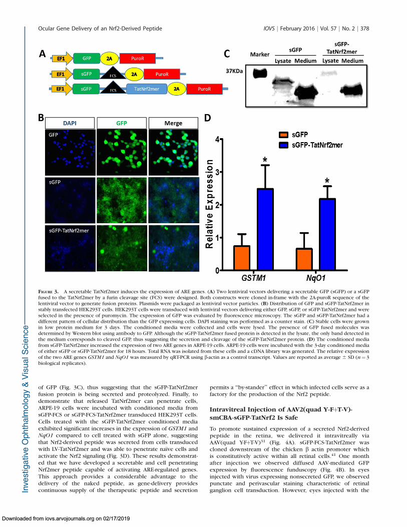
of GFP (Fig. 3C), thus suggesting that the sGFP-TatNrf2merfusion protein is being secreted and proteolyzed. Finally, todemonstrate that released TatNrf2mer can penetrate cells,ARPE-19 cells were incubated with conditioned media fromsGFP-FCS or sGFP-FCS-TatNrf2mer transduced HEK293T cells.Cells treated with the sGFP-TatNrf2mer conditioned mediaexhibited significant increases in the expression of GSTM1 andNqO1 compared to cell treated with sGFP alone, suggestingthat Nrf2-derived peptide was secreted from cells transducedwith LV-TatNrf2mer and was able to penetrate naıve cells andactivate the Nrf2 signaling (Fig. 3D). These results demonstrat-ed that we have developed a secretable and cell penetratingNrf2mer peptide capable of activating ARE-regulated genes.This approach provides a considerable advantage to thedelivery of the naked peptide, as gene-delivery providescontinuous supply of the therapeutic peptide and secretion
permits a ‘‘by-stander’’ effect in which infected cells serve as afactory for the production of the Nrf2 peptide.
Intravitreal Injection of AAV2(quad Y-FþT-V)-smCBA-sGFP-TatNrf2 Is Safe
To promote sustained expression of a secreted Nrf2-derivedpeptide in the retina, we delivered it intravitreally viaAAV(quad Y-FþT-V)31 (Fig. 4A). sGFP-FCS-TatNrf2mer wascloned downstream of the chicken b actin promoter whichis constitutively active within all retinal cells.41 One monthafter injection we observed diffused AAV-mediated GFPexpression by fluorescence funduscopy (Fig. 4B). In eyesinjected with virus expressing nonsecreted GFP, we observedpunctate and perivascular staining characteristic of retinalganglion cell transduction. However, eyes injected with the
FIGURE 3. A secretable TatNrf2mer induces the expression of ARE genes. (A) Two lentiviral vectors delivering a secretable GFP (sGFP) or a sGFPfused to the TatNrf2mer by a furin cleavage site (FCS) were designed. Both constructs were cloned in-frame with the 2A-puroR sequence of thelentiviral vector to generate fusion proteins. Plasmids were packaged as lentiviral vector particles. (B) Distribution of GFP and sGFP-TatNrf2mer instably transfected HEK293T cells. HEK293T cells were transduced with lentiviral vectors delivering either GFP, sGFP, or sGFP-TatNrf2mer and wereselected in the presence of puromycin. The expression of GFP was evaluated by fluorescence microscopy. The sGFP and sGFP-TatNrf2mer had adifferent pattern of cellular distribution than the GFP expressing cells. DAPI staining was performed as a counter stain. (C) Stable cells were grownin low protein medium for 3 days. The conditioned media were collected and cells were lysed. The presence of GFP fused molecules wasdetermined by Western blot using antibody to GFP. Although the sGFP-TatNrf2mer fused protein is detected in the lysate, the only band detected inthe medium corresponds to cleaved GFP, thus suggesting the secretion and cleavage of the sGFP-TatNrf2mer protein. (D) The conditioned mediafrom sGFP-TatNrf2mer increased the expression of two ARE genes in ARPE-19 cells. ARPE-19 cells were incubated with the 3-day conditioned mediaof either sGFP or sGFP-TatNrf2mer for 18 hours. Total RNA was isolated from these cells and a cDNA library was generated. The relative expressionof the two ARE genes GSTM1 and NqO1 was measured by qRT-PCR using b-actin as a control transcript. Values are reported as average 6 SD (n¼ 3biological replicates).
Ocular Gene Delivery of an Nrf2-Derived Peptide IOVS j February 2016 j Vol. 57 j No. 2 j 378
Downloaded from iovs.arvojournals.org on 02/17/2019
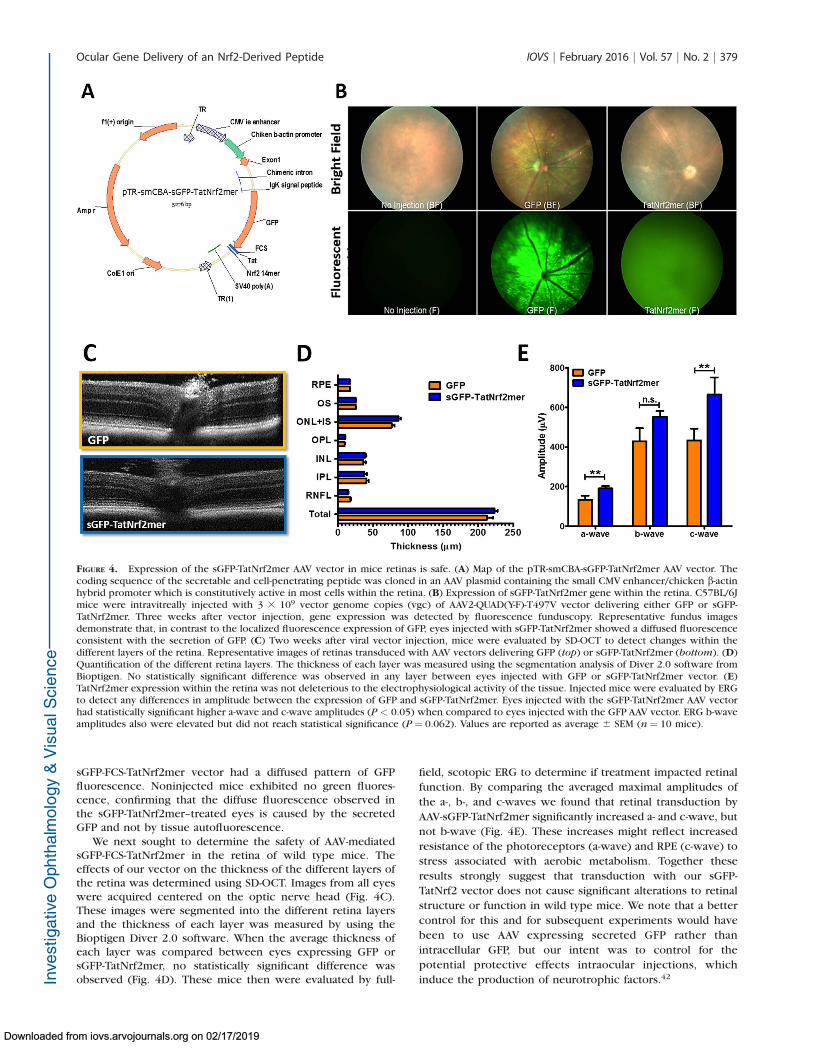
sGFP-FCS-TatNrf2mer vector had a diffused pattern of GFPfluorescence. Noninjected mice exhibited no green fluores-cence, confirming that the diffuse fluorescence observed inthe sGFP-TatNrf2mer–treated eyes is caused by the secretedGFP and not by tissue autofluorescence.
We next sought to determine the safety of AAV-mediatedsGFP-FCS-TatNrf2mer in the retina of wild type mice. Theeffects of our vector on the thickness of the different layers ofthe retina was determined using SD-OCT. Images from all eyeswere acquired centered on the optic nerve head (Fig. 4C).These images were segmented into the different retina layersand the thickness of each layer was measured by using theBioptigen Diver 2.0 software. When the average thickness ofeach layer was compared between eyes expressing GFP orsGFP-TatNrf2mer, no statistically significant difference wasobserved (Fig. 4D). These mice then were evaluated by full-
field, scotopic ERG to determine if treatment impacted retinal
function. By comparing the averaged maximal amplitudes of
the a-, b-, and c-waves we found that retinal transduction by
AAV-sGFP-TatNrf2mer significantly increased a- and c-wave, but
not b-wave (Fig. 4E). These increases might reflect increased
resistance of the photoreceptors (a-wave) and RPE (c-wave) to
stress associated with aerobic metabolism. Together these
results strongly suggest that transduction with our sGFP-
TatNrf2 vector does not cause significant alterations to retinal
structure or function in wild type mice. We note that a better
control for this and for subsequent experiments would have
been to use AAV expressing secreted GFP rather than
intracellular GFP, but our intent was to control for the
potential protective effects intraocular injections, which
induce the production of neurotrophic factors.42
FIGURE 4. Expression of the sGFP-TatNrf2mer AAV vector in mice retinas is safe. (A) Map of the pTR-smCBA-sGFP-TatNrf2mer AAV vector. Thecoding sequence of the secretable and cell-penetrating peptide was cloned in an AAV plasmid containing the small CMV enhancer/chicken b-actinhybrid promoter which is constitutively active in most cells within the retina. (B) Expression of sGFP-TatNrf2mer gene within the retina. C57BL/6Jmice were intravitreally injected with 3 3 109 vector genome copies (vgc) of AAV2-QUAD(Y-F)-T497V vector delivering either GFP or sGFP-TatNrf2mer. Three weeks after vector injection, gene expression was detected by fluorescence funduscopy. Representative fundus imagesdemonstrate that, in contrast to the localized fluorescence expression of GFP, eyes injected with sGFP-TatNrf2mer showed a diffused fluorescenceconsistent with the secretion of GFP. (C) Two weeks after viral vector injection, mice were evaluated by SD-OCT to detect changes within thedifferent layers of the retina. Representative images of retinas transduced with AAV vectors delivering GFP (top) or sGFP-TatNrf2mer (bottom). (D)Quantification of the different retina layers. The thickness of each layer was measured using the segmentation analysis of Diver 2.0 software fromBioptigen. No statistically significant difference was observed in any layer between eyes injected with GFP or sGFP-TatNrf2mer vector. (E)TatNrf2mer expression within the retina was not deleterious to the electrophysiological activity of the tissue. Injected mice were evaluated by ERGto detect any differences in amplitude between the expression of GFP and sGFP-TatNrf2mer. Eyes injected with the sGFP-TatNrf2mer AAV vectorhad statistically significant higher a-wave and c-wave amplitudes (P < 0.05) when compared to eyes injected with the GFP AAV vector. ERG b-waveamplitudes also were elevated but did not reach statistical significance (P ¼ 0.062). Values are reported as average 6 SEM (n¼ 10 mice).
Ocular Gene Delivery of an Nrf2-Derived Peptide IOVS j February 2016 j Vol. 57 j No. 2 j 379
Downloaded from iovs.arvojournals.org on 02/17/2019

Intravitreal Delivery of AAV-sGFP-TatNrf2merProtects the Retina Against Oxidative Stress
Knowing that AAV-sGFP-TatNrf2mer caused no overt retinaldamage when delivered intravitreally, we next asked if thesame dose of vector could induce the expression of ARE geneswithin the retina. C57BL/6J were injected intravitreally as inthe previous experiment, and the expression of ARE genes wasmeasured by qRT-PCR. In neural retinas of eyes injected in thevitreous with AAV-sGFP-TatNrf2mer we observed an increase inthe expression of HO-1, GSTM1, NqO1, and catalase relative toAAV-GFP treated eyes (Fig. 5). The level of induction amongthe four genes differed, suggesting that regulators other thanNrf2 impact the expression of these genes. NqO1, for example,also is under control of the aryl hydrocarbon receptor.43 Still,even in the absence of acute oxidative stress, expression ofTatNrf2mer led to increased expression of antioxidant genes.
To determine whether upregulation of ARE genes couldprotect the retina from oxidative stress, we evaluatedtreatment in the sodium iodate-inducible model of oxidativeinjury to the RPE.44 Mice were injected intravitreally in one eyewith AAV-GFP and with AAV-sGFP-TatNrf2mer in the contralat-eral eye. One month later, IP injections of NaIO3 (35 mg/kg)
were administered and, 1 week later, retinal function wasevaluated by full field scotopic ERG. Eyes treated with AAV-sGFP-TatNrf2mer showed a partial protection of the ERG a-
and b- wave amplitudes, suggesting protection of thephotoreceptors and bipolar cells (Figs. 6A, 6B). However,there was no protection of the c-wave amplitude relative tocontrol-treated contralateral control eyes at this dose (Fig. 6C).We decided to test a lower dose of NaIO3 to determine if it ispossible to protect the c-wave from a less severe injury. Asecond cohort treated with our vectors were injected IP with25 mg/kg NaIO3 and 7 days later were evaluated by ERG. In thiscohort we also observed a protection of a- and b-wave (Figs.6D, 6E), but this group of animals also showed a slight butsignificant protection of the c-wave (Fig. 6F).
Next, we sought to determine if this protection of the ERGat the lower dose of NaIO3 was associated with a protection ofthe retinal structure. Mice were evaluated by SD-OCT at day 9after injection with NaIO3. The thickness of the whole retina,the inner nuclear layer (INL), and the outer nuclear layer (ONL)was performed on SD-OCT images. Animals expressing thesGFP-TatNrf2mer showed a significantly thicker ONL whencompared to GFP-treated control eyes (Supplementary Fig.
FIGURE 5. Gene delivery of TatNrf2mer increases the expression of antioxidant genes within the retina. C57BL/6J mice were injected intravitreallywith 3 3 109 vgc/eye of AAV2-QUAD(Y-F) T497V delivering either GFP or sGFP-TatNrf2mer. One month after the injection, mice were euthanized,their retinas were harvested, and RNA isolated from them. A cDNA library from each treatment was prepared and qRT-PCR was performed tomeasure the relative levels of the ARE genes HO-1 and GSTM1 using b-actin as a control. Eyes treated with the sGFP-TatNrf2mer vector had higherlevels of HO-1, GSMT1, NqO1, and Catalase (all antioxidant genes) when compared to GFP injected control eyes. Values are reported as average 6SEM (n¼ 5 biological samples).
Ocular Gene Delivery of an Nrf2-Derived Peptide IOVS j February 2016 j Vol. 57 j No. 2 j 380
Downloaded from iovs.arvojournals.org on 02/17/2019

S2A), though overall retinal thickness and INL thickness werenot affected. To determine if TatNrf2mer-treated eye showedlower levels of oxidative stress compared to GFP-expressingeyes, the retinas were harvested and the amounts of nitro-tyrosine (a marker of protein oxidation) were quantified. Ourresults showed that eyes treated with the sGFP-TatNrf2mervector a greater than 50% reduction in the level of nitro-tyrosine-modified proteins when compared to eyes treatedwith AAV-GFP (Supplementary Fig. S2B). Finally, because of thepartial protection of the c-wave among these animals weexamined the effects on RPE architecture by RPE flat mount.Immunofluorescence analyses (ZO-1 staining) of the RPE layerrevealed comparable damage to the RPE structure in AAV-GFPand AAV-sGFP-TatNrf2mer–treated eyes (Supplementary Fig.S2C). However, at higher magnification (320), we observesome preservation of the RPE cells structure in the eyes treatedwith the AAV-sGFP-TatNrf2mer when compared to AAV-GFPtreated eyes, which exhibited many distorted and enlarged RPEcells. These results indicated that our TatNrf2mer vector didnot completely protect the RPE but did modulate the Nrf2signaling pathway in the retina and, in so doing, protected theretina from oxidative stress.
AAV-sGFP-TatNrf2mer Protects the Retina AgainstIntraocular Inflammation
Current research associates dry-AMD with oxidative stress andinflammation.45–48 We, therefore, tested the effect of intravit-real AAV-sGFP-TatNrf2mer on the modulation of proinflamma-tory signals in the NaIO3-treated mice. In retina and RPEextracts, we found that eyes treated with AAV-sGFP-TatNrf2merhad significantly lower quantities of IL-1b and IL-6 followingNaIO3 exposure (Fig. 7). The specificity of this effect wasdemonstrated by a lack of significant changes in the MCP-1(Ccl2) chemokine. This result suggested that the AAV-sGFP-TatNrf2mer vector has anti-inflammatory properties in the faceof acute oxidative stress.
The reduction of IL-1b level suggests an inhibition of theinflammasome signaling that regulates the activation andsecretion of this cytokine, which is produced in response tosignaling by the NLRP3 inflammasome. Inflammasome acti-vation recently has been associated with dry-AMD.49 To testthis hypothesis in vitro, we challenged stably transfectedARPE-19 cells expressing either LV-Puro or LV-TatNrf2mer-PuroR with the reactive aldehyde 4-hydroxynonenal (4-HNE),which is known to accumulate in eyes donated by patients
FIGURE 6. Expression of sGFP-TatNrf2mer partially protects the electrical activity of the retina from acute oxidative damage of NaIO3. C57BL/6Jmice were injected with the AAV vectors described in previous figures. One month after the intravitreal injection of the vector, mice were injectedintraperitoneally with 35 mg/kg sodium iodate to induce oxidative stress within the RPE. Seven days later mice were evaluated by ERG. One weekafter NaIO3 injury, eyes injected with the sGFP-TatNrf2mer had partial protection of the ERG a-wave (A) and b-wave (B) but not c-wave (P¼ 0.20;[C]), when compared to GFP injected eyes. (D–F) A second group of mice treated with the same AAV vectors received NaIO3 at a lower dose (25mg/kg) and evaluated as in the first cohort. At this lower dose, the amplitude of all the waves was significantly higher among the eyes treated withthe sGFP-TatNrf2mer. Histograms report the maximum amplitudes recorded at 0 dB using the UTAS BigShot Visual Diagnostic System (2.7 cd.s/m2).Values are reported as average 6 SEM (n¼ 11 mice in first cohort, n¼ 9 mice in second cohort).
Ocular Gene Delivery of an Nrf2-Derived Peptide IOVS j February 2016 j Vol. 57 j No. 2 j 381
Downloaded from iovs.arvojournals.org on 02/17/2019

with AMD.11 Treatment of control cells (LV-Puro) with 4-HNE
led to a 12-fold increase in IL-1b secretion, but cells
expressing TatNrf2mer peptide did not secrete significant
amounts of IL-1b into the media (Fig. 8A). This finding
suggested that TatNrf2mer can inhibit the inflammasome
signaling pathway. To test the effect of the AAV-sGFP-
TatNrf2mer on the recruitment of inflammatory cells, we
used the EIU mouse model50 as described in the Materials and
Methods section. Mice were treated with AAV-GFP in one eye
and with AAV-sGFP-TatNrf2mer in the other, as in the
FIGURE 8. The secretable TatNrf2mer has anti-inflammatory properties in a mouse model of ocular inflammation. (A) ARPE-19 stably expressingPuroR (Vector) or TatNrf2mer-PuroR (TatNrf2mer) were incubated with or without 30 lM 4-hydroxynonenal (4-HNE) for 18 hours. Theconcentration of IL-1b in the conditioned media was quantified in triplicate by ELISA. (B) C57BL/6J mice were injected intravitreally with 3 3 109
vgc of AAV vector delivering either GFP or sGFP-TatNrf2mer (TatNrf2mer). One month later mice were injected intravitreally with 25 ng LPS andthen were euthanized 24 hours later. Their eyes were harvested and analyzed by histology. Representative images of hematoxylin and eosin–stainedsections of eyes injected with either GFP (top) or TatNrf2mer vectors are shown (bottom). (C) The number of cells within the vitreous of at leasttwo sections per eye were quantified by two independent subjects who were not aware of the treatments. Eyes injected with the TatNrf2mer AAVvector had significantly lower numbers of infiltrating cells within the vitreous body than the eyes injected with the GFP AAV vector. Values arereported as average 6 SEM (n ¼ 2 biologic replicates in [A], n¼ 5 mice in [C]).
FIGURE 7. The secretable TatNrf2mer decreases inflammatory cytokines in the retinas of NaIO3 treated mice. Retinas and RPE from mice treatedwith 25 mg/kg NaIO3 were harvested and sonicated in PBS supplemented with protease inhibitors. The protein concentration in the cleared lysatewas determined and diluted to 400 ng/mL protein. The concentration of IL-1b, IL-6, and MCP-1 were determined by ELISA using 10 lg total proteinper sample. Biological samples were evaluated in triplicates. Values are reported as average 6 SEM (n¼ 7 samples).
Ocular Gene Delivery of an Nrf2-Derived Peptide IOVS j February 2016 j Vol. 57 j No. 2 j 382
Downloaded from iovs.arvojournals.org on 02/17/2019

oxidative stress experiments. One month later, they wereinjected in both eyes with E. coli lipopolysaccharide,euthanized 24 hours later, and their eyes harvested and fixedfor histologic analysis (Fig. 8B). The number of infiltratingcells within the vitreous was quantified by two independentmasked observers, and it was determined that eyes treatedwith the TatNrf2mer AAV vector showed significantly fewerinfiltrating cells within the vitreous body (Fig. 8C). Theseresults confirmed that AAV-sGFP-TatNrf2mer has anti-inflam-matory properties in the eye.
DISCUSSION
We developed an AAV vector that produces a secreted Nrf2peptide with cell-penetrating properties. We demonstratedthat our TatNrf2mer construct can induce the nucleartranslocation of endogenous Nrf2 based on immunofluores-cence and biochemical studies. Nuclear import of Nrf2resulted in the expression of ARE genes in vitro and in vivowhen measured by qRT-PCR. Furthermore, expression of ourTatNrf2mer sequence was sufficient to protect cells from thedamaging effects of potent oxidants, paraquat and H2O2 (bymeasuring cell viability in vitro) and NaIO3 (by measuring theelectrophysiological response of the retina in vivo). We notethat RPE cells were protected from oxidative damage in vitro(Figs. 1, 3C), but were not fully protected in the NaIO3 injurymodel in mice. There are several potential explanations for thisdiscrepancy. It is possible that the peptide did not reach theRPE following intravitreal injection of the virus. However, thisprotein (2.95 kDal) is much smaller than ranibizumab (48.3kDal) which effectively blocks choroidal neovascularizationfollowing intravitreal injection. Another interpretation is thatthe virally delivered peptide was not sufficient to protect the
RPE in this severe model of acute oxidative stress. We currentlyare attempting to distinguish between these alternatives bymeasuring TatNrf2mer protein levels in the neural retina andRPE and by testing AAV-sGFP-TatNrf2mer in a model of chronicRPE oxidative stress.51 When tested in models of inflammation,expression of TatNrf2mer significantly inhibited the secretionof IL-1b in vitro and LPS-induced recruitment of inflammatorycells within the vitreous humor of mice.
Our data suggested the following steps in the function ofour AAV-delivered Nrf2 peptide (Fig. 9): (1) SecretedTatNrf2mer penetrates nearby cells by virtue of its Tat-peptidesequence, (2) intracellular TatNrf2mer liberates endogenousNrf2 from Keap1, (3) Nrf2 translocates to the nucleus andinduces the expression of ARE genes, (4) these ARE genes aretranslated into the active enzymes within the cytoplasm of thecells, and (5) active antioxidant enzymes reduce the burden ofROS, thereby protecting the cells.
Although important for cell signaling events, when pro-duced in excess, ROS can damage otherwise stable macromol-ecules, particularly membrane lipids. This process can lead tothe generation of DAMP molecules that often trigger aninflammatory response accelerating the progression of thedisease. Furthermore, increased mitochondrial oxidative stresscan be sensed by the thioredoxin interacting protein (TXNIP)which can lead to activation of the inflammasome and to aproinflammatory response.52–54 The Nrf2 response can reducethe level of ROS, by increasing the level of antioxidantenzymes, such as heme oxygenase, catalase, and NqO1, andit can dampen inflammatory signaling by regenerating thio-redoxin.55 These mechanisms are of importance in tissues witha high metabolic rate, such as the retina.
Photo-oxidative damage to the RPE has been experimentallylinked to AMD.56,57 Brandstetter et al.58 provided an explana-tion of how exposure to high intensity light and accumulation
FIGURE 9. Mechanism of action of TatNrf2mer. The Nrf2mer peptide is derived from the Nrf2 gene domain that binds Keap-1 and penetrates cellswhen fused to the Tat peptide. Once inside the cell, the TatNrf2mer binds to Keap-1 liberating Nrf2, which can translocate into the nucleus where itinduces the expression of antioxidant genes (e.g., NqO1 and HO-1). These genes then reduce the ROS within therefore protecting the cell from thedeleterious effects of free radicals.
Ocular Gene Delivery of an Nrf2-Derived Peptide IOVS j February 2016 j Vol. 57 j No. 2 j 383
Downloaded from iovs.arvojournals.org on 02/17/2019
of lipofuscin could affect RPE viability. Interestingly, thecombination of oxidative stress and modified photoreceptorouter segments resulted not only in cell death, but also in theactivation of the inflammasome and secretion of IL-1b and IL-18. These cytokines also have been implicated in other oculardiseases, such as proliferative diabetic retinopathy and poly-poidal choroidal vasculopathy.59–62
Our work characterized a novel method for the delivery ofsmall peptides within the retina using AAV vectors. Bytransducing cells within the retina with a gene encoding theNrf2 peptide that can be secreted and taken up by other cells,we bypassed the need for the repeated administration of suchpeptides. Furthermore, the secretory and cell-penetratingproperties of our gene transfer approach potentially allowsthe use of a lower dose of vector to achieve therapeutic effectsdue to its ‘‘by-stander’’ effect when compared to a cell-autonomous version of the peptide. As noted above, Xiong etal.19 showed that increasing Nrf2 expression via subretinalinjection of AAV8-Nrf2 improved the electrophysiologicalresponse and the visual acuity in a mouse model of inheritedretinal degeneration. While subretinal injection currently isused in clinical trials for gene therapy, the injection of a virusproducing a secretable peptide is more readily translatable toclinical practice, because intravitreal injection of the virusshould permit the distribution of the short peptide to all layersof the neural retina and to the RPE. In addition, in a clinicalsetting intravitreal administration is less intrusive than thesubretinal injection.
Besides its antioxidant properties, our vector seems topossess the added benefit of being anti-inflammatory. Our AAV-sGFP-TatNrf2mer vector decreased the IL-1b and IL-6 cytokinesin the retinas of NaIO3 treated mice. Furthermore, when testedin the routinely used EIU mouse model, AAV-sGFP-TatNrf2merdecreased inflammation associated with the intravitreal deliv-ery of LPS. We currently are developing methods for analyzingmultiple cytokines in vitreous humor extracts. We also areaware of one potential limitation of this technology for thetreatment of ocular disease, the possibility that constitutivesuppression of the inflammatory response may make theposterior chamber more sensitive to infection. Before clinicalapplication, emphasis should be placed on methods to regulateexpression of secreted TatNrf2mer and on dose-response.Nevertheless, oxidative stress and inflammation are associateddiseases, such as Alzheimer’s disease and ALS, and, therefore,the vector characterized herein could be of significant usewithin and outside the eye.
Acknowledgments
The authors thank Vince Chiodo from the Retina Gene TherapyVector Core for production of AAV vectors, and James Thomas forhis assistance in the large-scale preparation of DNA.
Supported by a grant from the National Eye Institute (NEI;Bethesda, MD; R01 EY02025), a grant from the Florida BiomedicalResearch Foundation (e-10KG-s), a grant from the Macula VisionResearch Foundation, an AMD Research Fellowship from ARVO/Genentech, and the Shaler Richardson Professorship endowment.Core facilities were supported by NEI Grant P30 EY02172.
Disclosure: C.J. Ildefonso, P; H. Jaime, None; E.E. Brown,None; R. Iwata, None; C. Ahmed, None; M. Massengill, None;M.R. Biswal, None; S.E. Boye, None; W.W. Hauswirth, AGTC,Inc. (I); J.D. Ash, None; Q. Li, P; A.S. Lewin, P
References
1. Ikawa M, Okazawa H, Tsujikawa T, et al. Increased oxidativestress is related to disease severity in the ALS motor cortex: aPET study. Neurology. 2015;84:2033–2039.
2. Carrı MT, Valle C, Bozzo F, Cozzolino M. Oxidative stress andmitochondrial damage: importance in non-SOD1 ALS. Front
Cell Neurosci. 2015;9:41.
3. Hollyfield JG, Bonilha VL, Rayborn ME, et al. Oxidativedamage-induced inflammation initiates age-related maculardegeneration. Nat Med. 2008;14:194–198.
4. Beatty S, Koh HH, Phil M, Henson D, Boulton M. The role ofoxidative stress in the pathogenesis of age-related maculardegeneration. Surv Ophthalmol. 2000;45:115–134.
5. Hollyfield JG. Age-related macular degeneration: the molecularlink between oxidative damage, tissue-specific inflammationand outer retinal disease: the Proctor lecture. Invest Oph-
thalmol Vis Sci. 2010;51:1275–1281.
6. Gao H-M, Zhou H, Hong J-S. NADPH oxidases: noveltherapeutic targets for neurodegenerative diseases. Trends
Pharmacol Sci. 2012;33:295–303.
7. Uchida K. Aldehyde adducts generated during lipid peroxida-tion modification of proteins. Free Radic Res. 2015;49:896–904.
8. Terluk MR, Kapphahn RJ, Soukup LM, et al. Investigatingmitochondria as a target for treating age-related maculardegeneration. J Neurosci. 2015;35:7304–7311.
9. Ethen CM, Reilly C, Feng X, Olsen TW, Ferrington DA. Age-related macular degeneration and retinal protein modificationby 4-hydroxy-2-nonenal. Invest Ophthalmol Vis Sci. 2007;48:3469–3479.
10. Hollyfield JG, Perez VL, Salomon RG. A hapten generated froman oxidation fragment of docosahexaenoic acid is sufficient toinitiate age-related macular degeneration. Mol Neurobiol.2010;41:290–298.
11. Handa JT. How does the macula protect itself from oxidativestress? Mol Aspects Med. 2012;33:418–435
12. Huang HC, Nguyen T, Pickett CB. Regulation of theantioxidant response element by protein kinase C-mediatedphosphorylation of NF-E2-related factor 2. Proc Natl Acad Sci
U S A. 2000;97:12475–12480.
13. Kovac S, Angelova PR, Holmstrom KM, Zhang Y, Dinkova-Kostova AT, Abramov AY. Nrf2 regulates ROS production bymitochondria and NADPH oxidase. Biochim Biophys Acta.2015;1850:794–801.
14. Itoh K, Wakabayashi N, Katoh Y, et al. Keap1 represses nuclearactivation of antioxidant responsive elements by Nrf2 throughbinding to the amino-terminal Neh2 domain. Genes Dev. 1999;13:76–86.
15. Gonzalez-Rodrıguez A, Reibert B, Amann T, Constien R,Rondinone CM, Valverde AM. In vivo siRNA delivery of Keap1modulates death and survival signaling pathways and attenu-ates concanavalin-A-induced acute liver injury in mice. Dis
Model Mech. 2014;7:1093–1100.
16. Jang M, Cho I-H. Sulforaphane ameliorates 3-nitropropionicacid-induced striatal toxicity by activating the Keap1-Nrf2-AREpathway and inhibiting the MAPKs and NF-jB pathways. Mol
Neurobiol. 2015:1–17.
17. Pan H, He M, Liu R, Brecha NC, Yu ACH, Pu M. Sulforaphaneprotects rodent retinas against ischemia-reperfusion injurythrough the activation of the Nrf2/HO-1 antioxidant pathway.PLoS One. 2014;9:e114186.
18. Zhao X-D, Zhou Y-T, Lu X-J. Sulforaphane enhances the activityof the Nrf2–ARE pathway and attenuates inflammation inOxyHb-induced rat vascular smooth muscle cells. Inflamm
Res. 2013;62:857–863.
19. Xiong W, MacColl Garfinkel AE, Li Y, Benowitz LI, Cepko CL.NRF2 promotes neuronal survival in neurodegeneration andacute nerve damage. J Clin Invest. 2015;125:1433–1445.
20. Sachdeva MM, Cano M, Handa JT. Nrf2 signaling is impaired inthe aging RPE given an oxidative insult. Exp Eye Res. 2014;119:111–114.
Ocular Gene Delivery of an Nrf2-Derived Peptide IOVS j February 2016 j Vol. 57 j No. 2 j 384
Downloaded from iovs.arvojournals.org on 02/17/2019
21. Nagai N, Thimmulappa RK, Cano M, et al. Nrf2 is a criticalmodulator of the innate immune response in a model ofuveitis. Free Radic Biol Med. 2009;47:300–306.
22. Hancock R, Bertrand HC, Tsujita T, et al. Peptide inhibitors ofthe Keap1–Nrf2 protein–protein interaction. Free Radic Biol
Med. 2012;52:444–451.
23. Steel R, Cowan J, Payerne E, O’Connell MA, Searcey M. Anti-inflammatory effect of a cell-penetrating peptide targeting theNrf2/Keap1 interaction. ACS Med Chem Lett. 2012;3:407–410.
24. Boye SE, Boye SL, Lewin AS, Hauswirth WWA. Comprehensivereview of retinal gene therapy. Mol Ther. 2013;21:509–519.
25. Jacobson SG, Cideciyan AV, Ratnakaram R, et al. Gene therapyfor Leber congenital amaurosis caused by RPE65 mutations:safety and efficacy in 15 children and adults followed up to 3years. Arch Ophthalmol. 2012;130:9–24.
26. Jacobson SG, Acland GM, Aguirre GD, et al. Safety ofrecombinant adeno-associated virus type 2–RPE65 vectordelivered by ocular subretinal injection. Mol Ther. 2006;13:1074–1084.
27. Grote A, Hiller K, Scheer M, et al. JCat: a novel tool to adaptcodon usage of a target gene to its potential expression host.Nucleic Acids Res. 2005;33(suppl 2):W526–W531.
28. Ildefonso CJ, Jaime H, Rahman MM, et al. Gene delivery of aviral anti-inflammatory protein to combat ocular inflamma-tion. Hum Gene Ther. 2015;26:59–68.
29. Sanger F, Coulson AR. A rapid method for determiningsequences in DNA by primed synthesis with DNA polymerase.J Mol Biol. 1975;94:441–448.
30. Zolotukhin S, Potter M, Zolotukhin I, et al. Production andpurification of serotype 1, 2, and 5 recombinant adeno-associated viral vectors. Methods. 2002;28:158–167.
31. Kay CN, Ryals RC, Aslanidi GV, et al. Targeting photoreceptorsvia intravitreal delivery using novel, capsid-mutated AAVvectors. PLoS One. 2013;8:e62097.
32. Reed SE, Staley EM, Mayginnes JP, Pintel DJ, Tullis GE.Transfection of mammalian cells using linear polyethylenimineis a simple and effective means of producing recombinantadeno-associated virus vectors. J Virol Methods. 2006;138:85–98.
33. Livak KJ, Schmittgen TD. Analysis of relative gene expressiondata using real-time quantitative PCR and the 2(-Delta DeltaC(T)) method. Methods San Diego Calif. 2001;25:402–408.
34. Ildefonso CJ, Jaime H, Biswal MR, et al. Gene therapy with thecaspase activation and recruitment domain reduces the ocularinflammatory response. Mol Ther J Am Soc Gene Ther. 2015;23:875–884.
35. Justilien V, Pang J-J, Renganathan K, et al. SOD2 knockdownmouse model of early AMD. Invest Ophthalmol Vis Sci. 2007;48:4407–4420.
36. Mao H, James T, Schwein A, et al. AAV delivery of wild-typerhodopsin preserves retinal function in a mouse model ofautosomal dominant retinitis pigmentosa. Hum Gene Ther.2011;22:567–575.
37. Biswal MR, Prentice HM, Dorey CK, Blanks JC. A hypoxia-responsive glial cell-specific gene therapy vector for targetingretinal neovascularization. Invest Ophthalmol Vis Sci. 2014;55:8044–8053.
38. Ryan MD, Drew J. Foot-and-mouth disease virus 2A oligopep-tide mediated cleavage of an artificial polyprotein. EMBO J.1994;13:928–933.
39. Johnson JA, Johnson DA, Kraft AD, et al. The Nrf2-AREpathway: an indicator and modulator of oxidative stress inneurodegeneration. Ann N Y Acad Sci. 2008;1147:61–69.
40. Suzuki K, Bose P, Leong-Quong RY, Fujita DJ, Riabowol K.REAP: a two minute cell fractionation method. BMC Res Notes.2010;3:294.
41. Koilkonda RD, Hauswirth WW, Guy J. Efficient expression ofself-complementary AAV in ganglion cells of the ex vivoprimate retina. Mol Vis. 2009;15:2796–2802.
42. Wen R, Song Y, Cheng T, et al. Injury-induced upregulation ofbFGF and CNTF mRNAS in the rat retina. J Neurosci. 1995;15:7377–7385.
43. Wang L, He X, Szklarz GD, Bi Y, Rojanasakul Y, Ma Q. The arylhydrocarbon receptor interacts with nuclear factor erythroid2-related factor 2 to mediate induction of NAD(P)H:quino-neoxidoreductase 1 by 2,3,7,8-tetrachlorodibenzo-p-dioxin.Arch Biochem Biophys. 2013;537:31–38.
44. Enzmann V, Row BW, Yamauchi Y, et al. Behavioral andanatomical abnormalities in a sodium iodate-induced model ofretinal pigment epithelium degeneration. Exp Eye Res. 2006;82:441–448.
45. Ozaki E, Campbell M, Kiang A-S, Humphries M, Doyle SL,Humphries P. Inflammation in age-related macular degenera-tion. In: Ash JD, Grimm C, Hollyfield JG, Anderson RE, LaVailMM, Rickman CB, eds. Retinal Degenerative Diseases. NewYork, NY: Springer; 2014:229–235.
46. Campbell M, Doyle SL. An eye on the future of inflammasomesand drug development in AMD. J Mol Med. 2013;91:1059–1070.
47. Ambati J, Fowler BJ. Mechanisms of age-related maculardegeneration. Neuron. 2012;75:26–39.
48. Ambati J, Atkinson JP, Gelfand BD. Immunology of age-relatedmacular degeneration. Nat Rev Immunol. 2013;13:438–451.
49. Celkova L, Doyle SL, Campbell M. NLRP3 inflammasome andpathobiology in AMD. J Clin Med. 2015;4:172–192.
50. Yadav UC, Ramana KV. Endotoxin-induced uveitis in rodents.Methods Mol Biol. 2013;1031:155–162.
51. Mao H, Seo SJ, Biswal MR, et al. Mitochondrial oxidative stressin the retinal pigment epithelium leads to localized retinaldegeneration. Invest Ophthalmol Vis Sci. 2014;55:4613–4627.
52. Zhou R, Tardivel A, Thorens B, Choi I, Tschopp J. Thioredoxin-interacting protein links oxidative stress to inflammasomeactivation. Nat Immunol. 2010;11:136–140.
53. Devi TS, Lee I, Huttemann M, Kumar A, Nantwi KD, Singh LP.TXNIP links innate host defense mechanisms to oxidativestress and inflammation in retinal Muller glia under chronichyperglycemia: implications for diabetic retinopathy. Exp
Diabetes Res. 2012;2012:438238.
54. El-Azab MF, Baldowski BRB, Mysona BA, et al. Deletion ofthioredoxin-interacting protein preserves retinal neuronalfunction by preventing inflammation and vascular injury. Br
J Pharmacol. 2014;171:1299–1313.
55. Dinkova-Kostova AT, Abramov AY. The emerging role of Nrf2in mitochondrial function. Free Radic Biol Med. 2015;88:179–188.
56. Davies S, Elliott MH, Floor E, et al. Photocytotoxicity oflipofuscin in human retinal pigment epithelial cells. Free
Radic Biol Med. 2001;31:256–265.
57. Rozanowska M, Jarvis-Evans J, Korytowski W, Boulton ME,Burke JM, Sarna T. Blue light-induced reactivity of retinal agepigment. In vitro generation of oxygen-reactive species. J Biol
Chem. 1995;270:18825–18830.
58. Brandstetter C, Mohr LKM, Latz E, Holz FG, Krohne TU. Lightinduces NLRP3 inflammasome activation in retinal pigmentepithelial cells via lipofuscin-mediated photooxidative dam-age. J Mol Med. 2015;93:905–916.
59. Miao H, Tao Y, Li X. Inflammatory cytokines in aqueous humorof patients with choroidal neovascularization. Mol Vis. 2012;18:574–580.
60. Ongkosuwito JV, Feron EJ, Doornik CE van, et al. Analysis ofimmunoregulatory cytokines in ocular fluid samples frompatients with uveitis. Invest Ophthalmol Vis Sci. 1998;39:2659–2665.
Ocular Gene Delivery of an Nrf2-Derived Peptide IOVS j February 2016 j Vol. 57 j No. 2 j 385
Downloaded from iovs.arvojournals.org on 02/17/2019

61. Zhou J, Wang S, Xia X. Role of intravitreal inflammatory
cytokines and angiogenic factors in proliferative diabetic
retinopathy. Curr Eye Res. 2012;37:416–420.
62. Zhao M, Bai Y, Xie W, et al. Interleukin-1b level is increased in
vitreous of patients with neovascular age-related macular
degeneration (nAMD) and polypoidal choroidal vasculopathy
(PCV). PLoS One. 2015;10:e0125150.
63. Burgess A, Vigneron S, Brioudes E, Labbe J-C, Lorca T, CastroA. Loss of human Greatwall results in G2 arrest and multiplemitotic defects due to deregulation of the cyclin B-Cdc2/PP2A balance. Proc Natl Acad Sci U S A. 2010;107:12564–12569.
64. McCloy RA, Rogers S, Caldon CE, Lorca T, Castro A, Burgess A.Partial inhibition of Cdk1 in G 2 phase overrides the SAC anddecouples mitotic events. Cell Cycle. 2014;13:1400–1412.
Ocular Gene Delivery of an Nrf2-Derived Peptide IOVS j February 2016 j Vol. 57 j No. 2 j 386
Downloaded from iovs.arvojournals.org on 02/17/2019