targeting ripk1 for the treatment of human diseases · highly amenable for developing specific...
TRANSCRIPT

Targeting RIPK1 for the treatment of human diseasesAlexei Degtereva,1, Dimitry Ofengeimb,1, and Junying Yuanc,2
aDepartment of Developmental, Molecular and Chemical Biology, Sackler School of Graduate Biomedical Sciences, Tufts University, Boston, MA 02445;bRare and Neurologic Disease Research Therapeutic Area, Sanofi US, Framingham, MA 01701; and cDepartment of Cell Biology, Harvard Medical School,Boston, MA 02115
This contribution is part of the special series of Inaugural Articles by members of the National Academy of Sciences elected in 2017.
Edited by Don W. Cleveland, University of California, San Diego, La Jolla, CA, and approved April 8, 2019 (received for review January 21, 2019)
RIPK1 kinase has emerged as a promising therapeutic target forthe treatment of a wide range of human neurodegenerative,autoimmune, and inflammatory diseases. This was supported byextensive studies which demonstrated that RIPK1 is a key mediatorof apoptotic and necrotic cell death as well as inflammatory path-ways. Furthermore, human genetic evidence has linked the dysre-gulation of RIPK1 to the pathogenesis of ALS as well as otherinflammatory and neurodegenerative diseases. Importantly, uniqueallosteric small-molecule inhibitors of RIPK1 that offer high selectivityhave been developed. Thesemolecules can penetrate the blood–brainbarrier, thus offering the possibility to target neuroinflammation andcell death which drive various neurologic conditions including Alz-heimer’s disease, ALS, and multiple sclerosis as well as acute neuro-logical diseases such as stroke and traumatic brain injuries. We discussthe current understanding of RIPK1 regulatory mechanisms andemerging evidence for the pathological roles of RIPK1 in human dis-eases, especially in the context of the central nervous systems.
RIPK1 | necroptosis | apoptosis | inflammation | neurodegeneration
Cell death is a fundamental process that controls organismaldevelopment and homeostasis by regulating cell numbers
and eliminating damaged or infected cells. The discovery ofapoptosis as a regulated cell death mechanism suggested thepossibility of developing therapeutics to block pathologic celldeath, common in human degenerative diseases. Caspases, afamily of homologous mammalian cysteine proteases that are keymediators of apoptosis, emerged as attractive targets in pre-venting pathologic cell death (1). A major drug discovery efforttargeting caspases ensued in the mid-1990s (2). However, theseefforts largely failed to deliver new therapies. The reasons forthis failure include the presence of multiple members of caspaseswith somewhat redundant functions, the involvement of caspasesin important physiological functions, the activation of alternativecell death mechanisms upon inhibition of apoptosis and intrinsictoxicity-related challenges in inhibiting this class of cysteineproteases. As a result, it remained unclear whether blocking celldeath could be an effective strategy for the treatment of de-generative and inflammatory human diseases.Unexpectedly, apoptosis is not the only contributor to pa-
thology in human diseases; other forms of regulated cell deathalso play critical roles. In particular, necrosis, defined morpho-logically by cell lysis with the release of intracellular contents intothe extracellular space and traditionally believed to representpassive cell death, has attracted significant attention. Necrosiscan be induced by diverse events that disrupt normal cellularphysiological processes. However, a key question emerged whetherthere may also be a dedicated regulated mechanism that mediatesthe execution of necrosis, similar to that of caspases in mediatingapoptosis. The first established example of such a pathway is nec-roptosis, a regulated necrotic cell death mechanism that can beactivated under apoptosis-deficient conditions and is controlled bythe kinase activity of RIPK1 and its downstream mediators:RIPK3 kinase and the pseudokinase MLKL (3, 4). Interestingly,while mice carrying mutations in different caspases display signifi-cant developmental defects (5), necroptosis-resistant mutant mice
carrying different RIPK1 kinase dead knock-in mutations, includingD138N, K45A, K584R, and ΔG26F27, as well as RIPK3 or MLKLknockout mutations, show no abnormality in development or in theadult animals (6–10). Thus, necroptosis might be predominantlyactivated under pathological conditions, which makes inhibitingthis pathway an attractive option for the treatment of chronichuman diseases.Necroptosis was first defined by a series of small-molecule
inhibitors (necrostatins), including Nec-1/Nec-1s, Nec-3, Nec-4,and Nec-5, which blocked TNF-α–induced necrotic cell death(11). Subsequent studies of necrostatins revealed a key role ofRIPK1 as an important pharmacological target for inhibitingnecroptosis (12). Importantly, the kinase activity of RIPK1 playsa central role in mediating multiple deleterious inflammatorycell death mechanisms upon the activation of TNFR1 by TNF-α,which is known to be involved in the pathogenesis of varioushuman diseases (13). Anti-TNF agents have achieved majorclinical success for the treatment of human inflammatory dis-eases in the periphery, such as rheumatoid arthritis, colitis, andpsoriasis. However, anti-TNF strategy is not safe for the treat-ment of CNS diseases due to the involvement of TNFR2 inmediating neural regeneration (14). The specificity of RIPK1 inmediating TNFR1 signaling provides the possibility to safelyameliorate the deleterious TNF responses in the CNS withoutaffecting TNFR2.RIPK1 has become an important drug target in the pharma-
ceutical industry not only due to its key roles in TNF signalingresponses but also because the kinase structure of RIPK1 ishighly amenable for developing specific pharmacological small-molecule inhibitors. Nec-1/Nec-1s have been widely used to de-fine the role of necroptosis and RIPK1 in human diseases usinganimal models. This included studies examining the role of thiskinase in both acute injuries like ischemia as well as chronicneurodegenerative conditions such as multiple sclerosis (MS),ALS, and Alzheimer’s disease (AD) (13, 15, 16). To date, two
Significance
RIPK1 plays a critical role in mediating deleterious responsesdownstream of TNFR1. RIPK1 inhibitors have been progressedsuccessfully past human phase I clinical studies. This paperdiscusses why RIPK1 inhibitors present an opportunity for de-veloping oral drugs for a range of human degenerative andinflammatory diseases, especially CNS pathologies, includingALS, Alzheimer’s disease, Parkinson’s disease, traumatic braininjury, stroke, and lysosomal storage diseases.
Author contributions: A.D., D.O., and J.Y. wrote the paper.
Conflict of interest statement: J.Y. is a consultant for Denali Therapeutics. D.O. is anemployee of Sanofi.
This article is a PNAS Direct Submission.
This open access article is distributed under Creative Commons Attribution-NonCommercial-NoDerivatives License 4.0 (CC BY-NC-ND).1A.D. and D.O. contributed equally to this work.2To whom correspondence should be addressed. Email: [email protected].
Published online May 2, 2019.
9714–9722 | PNAS | May 14, 2019 | vol. 116 | no. 20 www.pnas.org/cgi/doi/10.1073/pnas.1901179116
Dow
nloa
ded
by g
uest
on
Mar
ch 6
, 202
0

different RIPK1 kinase inhibitor programs have progressed throughhuman phase I safety trials.
Regulation of Necroptosis by RIPK1 Kinase in Response toTNF-αRIPK1 is a multidomain protein comprising an N-terminal ki-nase domain, an intermediate domain, and a C-terminal deathdomain (DD). The intermediate domain of RIPK1 contains anRHIM [receptor interacting protein (rip) homotypic interactionmotif] domain which is important for interacting with otherRHIM-containing proteins such as RIPK3, TRIF, and ZBP1.The C-terminal DD mediates its recruitment by interacting withother DD-containing proteins, such as TNFR1 and FADD, andits homodimerization to promote the activation of the N-terminal kinase domain (10). In the case of TNF-α signaling,ligand-induced TNFR1 trimerization leads to the assembly of alarge receptor-bound signaling complex, termed Complex I,which includes multiple adaptors (TRADD, TRAF2, andRIPK1), and E3 ubiquitin ligases (cIAP1/2, LUBAC complex)(15, 17) (Fig. 1).RIPK1 is regulated by multiple posttranslational modifica-
tions, but one of the most critical regulatory mechanisms is viaubiquitination. The E3 ubiquitin ligases cIAP1/2 are recruitedinto Complex I with the help of TRAF2 to mediate RIPK1K63 ubiquitination. K63 ubiquitination of RIPK1 by cIAP1/2 promotes the recruitment and activation of TAK1 kinasethrough the polyubiquitin binding adaptors TAB2/TAB3 (18).K63 ubiquitination also facilitates the recruitment of theLUBAC complex, which in turn performs M1- type ubiquitina-tion of RIPK1 and TNFR1. M1 ubiquitination of Complex I isimportant for the recruitment of the trimeric IκB kinase complex
(IKK) through a polyubuiquitin-binding adaptor subunit IKKγ/NEMO (15, 17). The activation of RIPK1 is inhibited by directphosphorylation by TAK1, IKKα/β, MK2, and TBK1 (13).cIAP1 was also found to mediate K48 ubiquitination of RIPK1,inhibiting its catalytic activity and promoting degradation (19).When cells are defective in activating the apical apoptotic
mediator caspase-8, TNF-α stimulation promotes activation of asecondary cytosolic amyloid-like “necrosome” complex, alsoknown as Complex IIb. This complex contains a hetero-oligomerof RIPK1 and RIPK3 which interact through their cognateRHIM domains (20). The activation of RIPK1 kinase precedesand is essential for the formation of the necrosome. ActivatedRIPK1 undergoes autophosphorylation on multiple residues in-cluding Ser14/15, Ser20, and Ser161/166 in the activation seg-ment (12). Ser166 phosphorylation has emerged as a biomarkerfor RIPK1 activation (12, 21, 22). RIPK3 is phosphorylated innecrosomes on Ser227, and RIPK3 homo-oligomers eventuallyphosphorylate MLKL on the activation segment residues Thr357/Ser358 (23). The ensuing conformational change in MLKL leadsto the formation of disulfide bond dependent amyloid-like poly-mers (24, 25), which cause rapid plasma membrane lysis—a hall-mark of necrotic cell death.It should also be noted that RIPK1 is not absolutely required
for necroptosis beyond the ligands in the TNF family. For exam-ple, RHIM-domain-dependent activators TRIF and ZBP1 havebeen shown to engage and activate RIPK3 directly upon TLR3/4engagement and viral infection (26, 27). Thus, RIPK1 inhibitorsmay provide specificity toward sterile inflammation mediated byTNF involved in human pathology, without affecting host defensemechanisms activated by TLR pathways upon pathogen invasion.
Fig. 1. Schematic presentation of the RIPK1-dependent signaling events in response to TNF-α. Signaling bifurcates into poly-ubiquitin-dependent NF-κBactivation mediated by receptor-bound Complex I through TAK1 and IKK kinase complexes. Alternatively, RIPK1 kinase activation and deubiquitination/reubiquitinatiuon promotes formation of secondary cytosolic Complex IIa and IIb. Depending on the activity of caspase-8 and RIPK3, signaling by Complexes IImay lead to apoptosis, necroptosis, and increased inflammatory gene expression. The activating events are shown by arrows; however, it is not meant toindicate direct connections between signaling nodes. In many cases, mechanistic details remain to be uncovered.
Degterev et al. PNAS | May 14, 2019 | vol. 116 | no. 20 | 9715
CELL
BIOLO
GY
INAUGURA
LART
ICLE
Dow
nloa
ded
by g
uest
on
Mar
ch 6
, 202
0
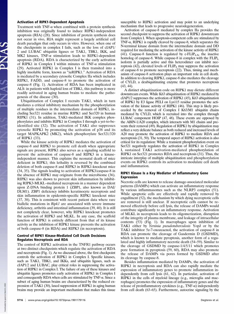
Activation of RIPK1-Dependent ApoptosisTreatment with TNF-α when combined with a protein synthesisinhibition was originally found to induce RIPK1-independentapoptosis (RIA) (28). Since inhibition of protein synthesis doesnot occur in vivo, RIA might represent a largely artificial celldeath paradigm that only occurs in vitro. However, when one ofthe checkpoints in complex I fails, such as the loss of cIAP1/2 and LUBAC ubiquitin ligases or TAK1, TBK1, IKK, andMK2 kinases, TNF-α stimulation leads to RIPK1-dependentapoptosis (RDA). RDA is characterized by the early activationof RIPK1 in Complex I within minutes of TNF-α stimulation(29). Activated RIPK1 in Complex I is then transited into ahighly insoluble form, known as “iuRIPK1.” Activation of RDAis mediated by a secondary cytosolic Complex IIa which includesRIPK1, FADD, and caspase-8 to promote the activation ofcaspase-8 (Fig. 1). Activation of RDA has been implicated inALS: in patients with haploid loss of TBK1, this pathway is morereadily activated in aging human brains to mediate the patho-genesis of the disease (30).Ubiquitination of Complex I recruits TAK1, which in turn
mediates a critical inhibitory mechanism by the phosphorylationof multiple residues in the intermediate domain of RIPK1, in-cluding Ser321 residue in murine RIPK1 and Ser320 in humanRIPK1 (31). In addition, TAK1-mediated IKK complex phos-phorylates and inhibits RIPK1 in Complex I through a yet-to-be-identified site (32). The activation of TAK1 also suppressescytosolic RIPK1 by promoting the activation of p38 and itstarget MAPKAPK2 (MK2), which phosphorylate Ser321/320of RIPK1 (33).While the kinase activity of RIPK1 mediates the activation of
caspase-8 and RIPK3 to promote cell death when appropriatesignals are present, RIPK1 also serves as a signaling scaffold toprevent the activation of caspase-8 and RIPK3 in a kinase-independent manner. This explains the neonatal death of micedeficient in RIPK1; this lethality is reversed by the combineddeletion of both caspase-8 and RIPK3 in RIPK1-knockout mice(34, 35). The signals leading to activation of RIPK3/caspase-8 inthe absence of RIPK1 may originate from the microbiome (36).RIPK1 was also shown to prevent skin inflammation by inhibit-ing RIPK3-MLKL–mediated necroptosis in a manner dependentupon Z-DNA binding protein 1 (ZBP1, also known as DAI/DLM1). ZBP1 deficiency inhibits keratinocyte necroptosis andskin inflammation in epidermis-specific RIPK1 knockout mice(37, 38). This is consistent with recent patient data where rarebiallelic mutations in Ripk1 are associated with severe immunedeficiency, arthritis and intestinal inflammation (39, 40). It is stillnot completely clear, however, why RIPK1 knockout promotesthe activation of RIPK3 and MLKL. In any case, the scaffoldfunction of RIPK1 is entirely different from that of its kinaseactivity as the inhibition of RIPK1 kinase prevents the activationof both caspase-8 (in RDA) and RIPK3 (in necroptosis).
Control of RIPK1 Kinase-Mediated Cell Death DecisionsRegulates Necroptosis and RDAThe control of RIPK1 activation in the TNFR1 pathway occursat two distinct checkpoints which regulate the activation of RDAand necroptosis (Fig. 1). As we discussed above, the first checkpointcontrols the activation of RIPK1 in Complex I. Specific kinases,such as TAK1, TBK1, and IKKs, and ubiquitin ligases, such ascIAP1/2 and LUBAC, play critical roles in suppressing the activa-tion of RIPK1 in Complex I. The failure of any of these kinases andubiquitin ligases promotes early activation of RIPK1 in Complex Iand consequently RDA when cells are stimulated by TNF-α. Since asubset of aging human brains are characterized by the reduced ex-pression of TAK1 (30), laxed suppression of RIPK1 in aging humanbrains may provide an important mechanism that makes this tissue
susceptible to RIPK1 activation and may point to an underlyingmechanism that leads to progressive neurodegeneration.Activation of caspase-8 mediated by complex IIa provides the
second checkpoint to suppress the activation of RIPK1 downstreamfrom Complex I. When apoptosis-competent cells are stimulated byTNF-α, RIPK1 is rapidly cleaved by caspase-8, which separates theN-terminal kinase domain from the intermediate domain and DDrequired for mediating the activation of the kinase activity of RIPK1(41). Caspase-8 function is regulated by c-FLIPL/S, the inactivehomolog of caspase-8. While caspase-8 in complex with the FLIPLisoform is partially active and this heterodimer can inhibit nec-roptosis (42), elevated levels of FLIPS may suppress the activationof caspase-8 to promote necroptosis (22). Thus, timing and mech-anism of caspase-8 activation plays an important role in cell death.In addition to cleaving RIPK1, caspase-8 also mediates the cleavageof CYLD, a deubiquitinating enzyme that promotes necroptosis(43, 44).A distinct ubiquitination code on RIPK1 may dictate different
downstream events. While K63 ubiquitination of RIPK1 mediated bycIAP1/2 suppresses the activation of RIPK1 (45), K63 ubiquitinationof RIPK1 by E3 ligase PELI on Lys115 residue promotes the acti-vation of the kinase activity of RIPK1 (46). This step is likely pro-ceeded by the removal of Complex I K63/M1 ubiquitin chainsmediated by CYLD, which is recruited into the complex throughLUBAC component HOIP (47, 48). These events are opposed bythe ABIN-1/A20 complex, which interacts with M1 chains and pre-vents their removal (49). Regulation of RIPK1 by ubiquitination mayreflect a very delicate balance as both reduced and increased levels ofA20 may promote the activation of RIPK1 to mediate RDA andnecroptosis (31, 50). The temporal aspect of RIPK1 activation is alsocritical for its regulation: While a transient phosphorylation event onSer321 negatively regulates the activation of RIPK1 in ComplexI, sustained TAK1 activation-mediated phosphorylation ofRIPK1 on Ser321 promotes RDA and necroptosis (31). Thus, theintricate interplay of multiple ubiquitination and phosphorylationevents on RIPK1 controls its activation to modulate cell deathand inflammation.
RIPK1 Kinase Is a Key Mediator of Inflammatory GeneExpressionNecrotic cells are known to release damage-associated molecularpatterns (DAMPs) which can activate an inflammatory responseby various inflammasomes such as the NLRP3 complex (51).While apoptotic cells are effectively and rapidly removed byengulfment (52), the mechanism by which dying necroptotic cellsare removed is still unclear. If necroptotic cells cannot be re-moved effectively before cell lysis, the release of DAMPs wouldcontribute significantly to an inflammatory response. Activationof MLKL in necroptosis leads to its oligomerization, disruptionof the integrity of plasma membrane, and leakage of intracellularcontents (53) (Fig. 1). In macrophages upon inhibition ofTAK1 either by Yersinia pseudotuberculosis YopJ or by theTAK1 inhibitor 5z-7-oxozeaenol, the activation of caspase-8 inRDA can promote the cleavage of Gasdermin D (GSDMD),which is known to mediate pyroptosis, another form of a regu-lated and highly inflammatory necrotic death (54–59). Similar tothe cleavage of GSDMD by caspase-1/4/5/11 which promotespore formation in pyroptosis (59, 60), RDA may also promotethe release of DAMPs via pores formed by GSDMD afterits cleavage by caspase-8.Besides inflammation mediated by DAMPs, the activation of
RIPK1 in necroptosis and RDA can also rapidly mediate theexpression of inflammatory genes to promote inflammation in-dependently from cell lysis (61, 62). In particular, activation ofRIPK1 in the cells of myeloid lineage (e.g., microglia and mac-rophages) promotes the expression of inflammatory genes and therelease of proinflammatory cytokines (e.g., TNF-α) independentlyfrom cell death (63–65). Furthermore, autocrine signaling by the
9716 | www.pnas.org/cgi/doi/10.1073/pnas.1901179116 Degterev et al.
Dow
nloa
ded
by g
uest
on
Mar
ch 6
, 202
0

released TNF family members, produced upon RIPK1 activation,is an important component of tissue injury. This has been shown inmice deficient in proteins that regulate RIPK1 activation, such asNEMO and Sharpin, the latter a component of the LUBACcomplex (21, 66, 67).Recent data delineated cell-death-independent signaling ac-
tivities of RIPK1 (sometimes along with RIPK3) leading totranscriptional up-regulation of a wide range of inflammatorymolecules (61–63, 65). Regulation of inflammatory gene ex-pression involves a range of MAPKs downstream from RIPK1,including p38 and Erk1/2, as well as TBK1/IKKe complex. Thesekinase circuits further activate the transcription factors AP1,Sp1, NF-κB, and IRF3/7. In the context of necroptosis, this mayprovide a burst of inflammatory signals in addition to the re-sponses elicited by DAMPs released from dead cells. However,there is also growing evidence that RIPK1-dependent proin-flammatory responses may occur independent from necroptosisin vivo. For example, RIPK1 kinase activity is required for in-flammatory gene expression in myeloid cells after mouse challengewith a sublethal dose of LPS (61). Similarly, RIPK1 exacerbateschronic inflammation during embryonic development when bothbranches of cell death, controlled by caspase-8 and RIPK3, areabsent (68). Pathologically, RIPK1-dependent inflammatory geneexpression has been linked to neutrophilic dermatitis in Ptpn6spin
mice (69), clearance of West Nile virus in mice (70), and neuro-inflammation in human diseases that will be discussed below.
Necrostatins and Other RIPK1 Kinase InhibitorsDeciphering the necroptosis pathway followed an unusual routeas it involved the extensive use of small-molecule tools to un-derstand the relevance and mechanistic underpinnings of thisprocess. A first set of selective necroptosis inhibitors, which wetermed necrostatins, was identified using a cell-based screen ofTNF-induced necroptosis in a human U937 monocytic cell line(11). Necrostatin-1 completely prevented the activation of nec-roptosis across a range of cellular models where TNF/FasL-induced activation of necrosis-like death had been observed(11). Importantly, Nec-1 was also shown to reduce oxygen/glu-cose deprivation-induced death in primary neurons and to de-crease the infarction size after transient middle cerebral arteryocclusion (MCAO), a mouse model of ischemic stroke, providingthe first indication for the pathologic relevance of necroptosis.Since then, necroptosis has been firmly established as a com-ponent of many ischemia-reperfusions injuries in the brain, ret-ina, heart, kidneys, and liver (71–75). Overall, these earlyfindings using necrostatins were significant because they dem-onstrated the existence of a common mechanism of necroticdeath across a range of cell types in response to TNF and underpathologic conditions in vivo.Subsequently, we identified RIPK1 as the direct target of Nec-
1 and other structurally distinct necrostatins (12). Importantly,we found that a mutation of Ser161 in the activation segment ofRIPK1 offered resistance to inhibition by Nec-1 and othernecrostatins. These data confirmed that RIPK1 is the soletarget of necrostatins in necroptosis. Furthermore, the fact thatall the hits in an unbiased cell-based screen were allRIPK1 inhibitors highlighted a unique significance of this kinasefor the effective and selective inhibition of necroptosis. To date,Nec-1, Nec-1s, and other recently developed RIPK1 inhibitorshave been instrumental in establishing the important contribu-tion of RIPK1 to a wide range of human pathologies, repre-senting areas of major unmet medical need, including braintrauma, sepsis, and neurodegenerative and autoimmune disor-ders (13, 16).Subsequent analyses revealed several remarkable features of
necrostatins. All necrostatins isolated in an unbiased cell-basedscreen display almost exclusive selectivity for RIPK1 (63), which isvery unusual for kinase inhibitors. The structural basis of inhibition
by Necs was revealed by the crystal structure of RIPK1 in complexwith three different necrostatins—Nec-1s, Nec-3, and Nec-4—reported by Shi and coworkers (76). All three necrostatinsbelong to a type III class of allosteric kinase inhibitors (77). Fur-thermore, all three Necs bind to the exact same allosteric site onRIPK1 formed behind the active center. This pocket becomesaccessible whenMg-binding DLGmotif assumes an inactive DLG-out conformation and αC helix is outwardly displaced to the Glu-out conformation with a characteristic loss of Glu-Lys ionic paircritical for catalysis (Fig. 2A). While inhibitors targeting eitherDFG-out (type II inhibitors) or Glu-out (αC helix displacing in-hibitors) conformations have been described previously, RIPK1 isthe only kinase for which doubly inactive DLG-out/Glu-out con-formation has been described to date.This unique allosteric pocket of RIPK1 is in part due to an
increased flexibility of the DLG motif in RIPK1, compared with
A
B
Fig. 2. Mode of inhibition of RIPK1 by necrostatins and other type III in-hibitors. (A) Rendering of the crystal structure of Nec-1s with RIPK1 (85). Nec-1s is occupying the back pocket localized behind the ATP binding center. Thispocket is created due to the outward movement of αC-helix, resulting in theloss of ionic pair between catalytic Lys45 and Glu63 of αC-helix. The otherside of the pocket is formed by the DLG motif (shown in green) in the in-active DLG-in conformation (catalytic Asp146 facing away from the activecenter) and the activation segment, which immediately follows the DLGmotif (shown in red). Ser161 residue of the activation segment, which formsa critical hydrogen bond with the indole of Nec-1s, is also shown. (B) Anumber of additional type III inhibitors have been recently developed byGlaxoSmithKline and Takeda, including clinical candidate GSK′772 (88–91).These molecules (rendered in red, based on published crystal structures)occupy the same back pocket as Nec-1s (shown in green) in the same Glu-out/DLG-out conformation but extend into the ATP binding pocket, whichmay contribute to the increased affinity.
Degterev et al. PNAS | May 14, 2019 | vol. 116 | no. 20 | 9717
CELL
BIOLO
GY
INAUGURA
LART
ICLE
Dow
nloa
ded
by g
uest
on
Mar
ch 6
, 202
0

a more rigid DFG motif found in most kinases (78). However, toengage this pocket necrostatins still need to fit into the allostericsite very precisely and tightly, resulting in a high degree ofspecificity. Overall, the unusual dynamics of RIPK1 and theunique binding mode of necrostatins most likely collectivelydictate an unprecedented selectivity of these molecules. Vali-dating this conclusion, GlaxoSmithKline and Takeda also suc-cessfully developed inhibitors targeting the same allostericpocket/inactive conformation of RIPK1 (Fig. 2B) and thesemolecules also display exclusive selectivity toward RIPK1 (79–82). Conversely, more typical type I inhibitors, targeting the ATPpocket (VX-680 and Pazopanib), and type II inhibitors, engagingresidues in the ATP pocket and back pocket in Glu-in/DLG-outconformation (Ponatinib and Rebastinib), were also found inscreening of kinase inhibitor collections, but these molecules arenot specific for RIPK1 (78, 83, 84).Development of type III RIPK1 inhibitors offers hope that in-
hibition of RIPK1 may prove a successful strategy in many chronicdegenerative and autoinflammatory diseases, where inhibitor selec-tivity is paramount to ensure safety. One of these molecules, a clinicalcandidate compound GSK2982772 developed by GlaxoSmithKline,has successfully completed phase I safety trials (85). Another mole-cule DNL747, developed by Denali Therapeutics, also successfullycompleted a phase I trial.
Developing RIPK1 Inhibitors for the Treatment of HumanDiseasesThe identification of RIPK1 kinase as a critical mediator of bothcell death and inflammation presents an exciting new opportu-nity for developing therapeutics for the treatment of humandiseases. This is particularly of interest in the context of neuro-degenerative diseases because the pathology in many of thesediseases is characterized by necrosis (86). New drug targets areneeded in the field as previous trials of potential therapies forneurodegenerative diseases such as AD, ALS, and Parkinson’sdisease (PD) have largely failed or demonstrated minimal effi-cacy. We have recently reviewed the mechanism of RIPK1 andits role in mediating CNS diseases in general (13). Our discussionhere will focus on developing RIPK1 inhibitors for the treatmentof specific human diseases with an emphasis on neurodegenerativediseases. While RIPK1 inhibitors are expected to be also effectivefor the treatment of peripheral human inflammatory diseases, thepossibility to develop highly specific RIPK1 inhibitors that canpass the blood–brain barrier (BBB) provides a special opportunityfor the treatment of CNS diseases.
RIPK1 in Acute Neuronal Injury. The involvement of necroptosis inacute neurological injuries (e.g., ischemic brain injury) was firstdemonstrated with Nec-1 before its targeting mechanism wasrevealed (11). Nec-1, but not inactive Nec-1i derivative, wasshown to attenuate infarct volume in the MCAO model of strokein a dose-dependent manner. It remained active when adminis-tered both prophylactically and therapeutically after the onset ofischemic insult with a significant time window. The identificationof RIPK1 as a specific target of Nec-1 revealed this kinase as anew mediator of acute ischemic neurological insult. Notably,RIPK1 also contributes to ischemic injuries of other organs in-cluding eye, heart, and kidney (71, 87–91). An increase inRIPK1 and RIPK3 and the formation of RIPK1/RIPK3 complexwere found following permanent MCAO (92). RIPK1 activationalso occurs after traumatic brain injury (TBI); similar to theMCAO model, both RIPK1 and RIPK3 are increased in thebrains of animals in the fluid percussion brain injury model inrats (93). Interestingly, posttraumatic hypothermia (33 °C),which is known to reduce brain injury following stroke and TBI,led to decreases in the levels of RIPK1, RIPK3, and MLKL.These data suggest that deleterious RIPK1-dependent signalingmay play a causal role in neuronal loss following TBI. Consistent
with this notion, mice administered Nec-1 before controlledcortical impact (CCI) had reduced neuronal death and improvedmotor and spatial memory outcomes. Furthermore, improvedspatial memory was observed even when necrostatin-1 wasadministered 15 min after CCI (94). These data implicateRIPK1 as a central mediator of neurodegeneration followingacute neuronal injury.
RIPK1 in MS. There is growing evidence that RIPK1 mediatesdeleterious processes in chronic neurodegeneration. A key sim-ilarity between acute injury and chronic neurodegeneration is thepresence of neuroinflammation. MS is an inflammatory diseaseof the CNS that leads to chronic neurodegeneration. TNF-αsignaling has been strongly linked to the pathophysiology ofMS (95, 96). The role of RIPK1 in mediating a deleterious re-sponse downstream of TNFR1 suggests the role of RIPK1 in thisdisease. In CNS lesions of both brain samples from MS patientsand in mouse models of MS, RIPK1 is relocalized to the in-soluble fraction (22), which occurs when RIPK1 is activated (97).Inhibition of RIPK1 by Nec-1s ameliorated disease pathology,improved animal behavior, and attenuated experimental allergicencephalomyelitis (EAE)-induced cytokine increase and re-cruitment of immune cells (22). The role of RIPK1 in EAE wasalso validated by the use of another RIPK1 inhibitor developedby Takeda (79). Mechanistically, RIPK1 is highly expressed inmacrophages and microglia in the EAE lesions, and Nec-1s maydampen the innate immune response in these cells. Consistently,blocking RIPK1 activity modulates the inflammatory and celldeath responses in microglia cells (62, 98).While RIPK1 and its downstream binding partners (RIPK3,
caspase-8, and MLKL) are highly expressed in immune cellsthey are also present in other cell types of the CNS. In partic-ular we identified a RIPK1-deleterious pathway in primary ol-igodendrocyte lineage cells, which are specifically killed by atoxin in the cuprizone model of demyelinating disease (22).Previous studies had shown that oligodendrocytes are sensitiveto TNF-α stimulation, in particular in the presence of otherCNS cells (99). Our study showed that this TNF-α–inducedoligodendrocyte cell death could be inhibited by either phar-macological inhibition of RIPK1 with Nec-1s or genetic de-letion of RIPK3. Consistent with this, RIPK3–MLKL interactionand necroptosis were shown to be critical for oligodendrocytenecroptosis following oxygen–glucose deprivation as well as inMCAO (71, 100).The deleterious role of both RIPK1-dependent inflammatory
signaling and necroptosis in MS is further supported by the ro-bust finding that RIPK1, RIPK3, and MLKL are activated intissues from postmortem pathological samples from MS patients(22). The beneficial effect of inhibiting RIPK1 is likely attribut-able to both modulation of inflammation as well as blocking nec-roptosis in susceptible oligodendrocytes and neurons (Fig. 3).RIPK1 may act in a cell-autonmous as well as non-cell-autonomousmanners to induce necroptosis susceptibility in oligodendrocytes.
RIPK1 and ALS. ALS is a progressive neurodegenerative diseasethat leads to degeneration of motor neurons and paralysis. Ac-tivation of RIPK1-mediated neuroinflammation and cell death isdirectly linked with the genetics of ALS in humans. Mutations inthe genes that lead to familial ALS in humans, including Optineurinand TBK1, have been shown to promote the onset of RIPK1-dependent necroptosis and RDA in the CNS (30, 101). Optn encodesa ubiquitin-binding protein that regulates RIPK1 ubiquitination anddegradation. Optn knockout mice developed neuropathology re-sembling ALS, including Wallerian-like axonal pathology and dys-myelination (101). The loss of optineurin in both oligodendrocytesand microglia, but not motor neurons or astrocytes, is sufficient topromote neuropathology. Inhibition of RIPK1 also protects againstthe degeneration of oligodendrocytes in SODG93A transgenic mice,
9718 | www.pnas.org/cgi/doi/10.1073/pnas.1901179116 Degterev et al.
Dow
nloa
ded
by g
uest
on
Mar
ch 6
, 202
0

which is known to occur before the onset of motor dysfunction (101,102). These results suggest that RIPK1 might promote axonal de-generation and neuroinflammation noncell autonomously in ALS.While Optn regulates the ubiquitination of RIPK1, TBK1
regulates RIPK1 through direct phosphorylation on multiplesites including Thr189 to suppress RIPK1 kinase activity byblocking the interaction with its substrates (30, 103). TBK1mutations are a major genetic cause of patients with ALS/fron-totemporal dementia (FTD) comorbidity (10.8%) and to a lesserextent causal in ALS alone (0.5%) (104, 105). Reduced expres-sion of TBK1 by mutant alleles associated with ALS/FTD has ledto the proposal that haploinsufficiency of TBK1 is a pathologicalmechanism. TBK1 knockout mice are embryonically lethal, andthis lethality is blocked in TBK1−/−; RIPK1D138N kinase-deadknock-in mutant mice, which provides a genetic verification forthe critical function of TBK1 in regulating the activation ofRIPK1 (30). This is consistent with in vitro observations thatTBK1 deficiency or pharmacological inhibition sensitizes cells toRIPK1-dependent cell death induced by TNF-α (30, 103). In-terestingly, while TBK1+/− mice are normal, the haploid loss ofTAK1, another RIPK1 suppressor, in the myeloid lineage ofTBK1+/− mice leads to many of the hallmarks of ALS/FTD, in-cluding dysmyelination, axonal degeneration, neuronal loss andcytoplasmic TDP-43 aggregates in the CNS (30). Thus, the ac-tivation of RIPK1 might be involved in mediating ALS pathologyin multiple lineages and CNS cell types (Fig. 3). Furthermore,reduced expression of TAK1 in the CNS of aging human brainsmay provide a common mechanism to promote not only ALS,but also other age-dependent neurodegenerative diseases in-cluding AD and PD.
RIPK1 in AD. AD is the most common age-related neurodegen-erative disorder. AD is characterized by cognitive impairmentwith loss of memory and neuropathological features such as theaccumulation of amyloid plaque deposits composed of amyloid-β(Aβ), hyperphosphorylation of microtubule-associated proteintau, neuroinflammation, cerebrovascular pathology, and neuro-nal loss. The amyloid hypothesis proposes that the accumulationof Aβ and formation of amyloid plaques is the main cause of ADpathogenesis. The amyloid hypothesis is supported by the earlyonset familial Alzheimer’s disease (FAD) mutations in the am-yloid precursor protein (APP) and the presenilin genes that driveamyloidosis (106). However, the poor performance of Aβ-directed therapies in clinical trials has forced rethinking of ad-ditional factors that can contribute to AD pathogenesis. Ahallmark of AD is chronic brain inflammation marked by in-creases in the levels of proinflammatory cytokines (107). Neu-roinflammation characterized by the overactivated dystrophicmicroglia is prevalent in late stage AD (LOAD) and may con-tribute significantly to dementia (108). The role of neuro-inflammation has also been supported by the genome-wideassociation studies on LOAD which have identified variants inmicroglia-associated genes, such as the genes encoding triggeringreceptor expressed on myeloid cells 2 (TREM2) and themicroglial surface receptor CD33, that confer higher risks fordeveloping LOAD.Under physiological conditions, microglia participate in reg-
ulating synaptic pruning and play a surveillance role in main-taining homeostasis by removing cellular debris, dying cells, ormisfolded proteins. During AD pathogenesis, microglia activa-tion may play a different role in early (preclinical) and late(clinical) AD stages. Acutely activated microglia can promotethe degradation of Aβ by phagocytosis, but sustained chronicallyactivated microglia may be deficient in degradation of Aβ andinstead contribute to neurotoxicity and synapse loss by triggeringproinflammatory cascades. Microglial activation triggered by thefocal aggregation of extracellular Aβ into insoluble amyloid mayoccur late during preclinical stages of AD and set the stage forthe onset of dementia (109). Activated microglia secrete proin-flammatory cytokines such as TNF-α, IL-1β, and IL-6, as well aschemokines that recruit and promote further activation of glialcells and neuronal damage. A genome-wide association study ofLOAD risk factors identified polymorphisms in the TNF pro-moter that are linked to increased TNF-α production (110).RIPK1 has a well-recognized role in mediating transcription of
neuroinflammatory genes in microglia (13, 101). RIPK1 is highlyexpressed in microglia in mouse and human brain samples (111).The levels of RIPK1, at both the mRNA and protein levels, inpostmortem samples from individuals with LOAD are increasedcompared with controls and positively correlate with the re-duction in brain weights and Braak stages (111, 112). RIPK1,MLKL, and pMLKL levels were significantly higher in the brainsof 11-mo-old 5xFAD mice compared with nontransgenic litter-mates, suggesting the involvement of necroptosis in AD (112).RIPK1 may be involved in regulating a transcriptional responsein AD as the set of genes regulated by RIPK1 overlapped sig-nificantly with multiple independent AD transcriptomic signa-tures, including multiple genes linked with the expression ofAD-risk variant genes and other CNS diseases (111, 112) (Fig.3). Inhibition of RIPK1 kinase activity has also been shown topromote the ability of microglia to degrade Aβ (111). The levelsof amyloid plaques in APP/PS1 mice are reduced upon phar-macological inhibition of RIPK1 using Nec-1s. Thus, inhibitionof RIPK1 may be able to reduce inflammatory microglia andrestore the phagocytic ability of microglia.The RIPK1-mediated gene expression signature in microglia
provides clues as to how RIPK1 activation may mediate in-flammation. Activation of RIPK1 in microglia regulates the ex-pression of important genes involved in mediating innate immune
Fig. 3. Bimodal RIPK1 activation in neurological disease leads to progressivedeleterious signaling loop to promote neuroinflammation and cell death.Activation of RIPK1 in either microglia oligodendrocytes or neurons caninitiate a degenerative signaling loop. This cascade relies on microglial-driven deleterious inflammation and necrotic cell death in the CNS. Micro-glial. RIPK1 regulates a degenerative neuroinflammatory milieu in the CNSthat can lead to necroptosis of oligodendrocytes and axonal degeneration.Neurons with damaged mitochondria and lysosomes may undergo nec-roptosis. In turn, necroptosis of either oligodendrocytes or neurons promotesinflammation by driving the cell-autonomous expression of proinflammatorycytokines in microglia as well as by releasing of the cellular content from ne-crotic cells (including DAMPs) into the CNS. This deleterious axis creates aprogressive inflammatory and degenerative environment in the brain topromote the progression of neurodegenerative disease.
Degterev et al. PNAS | May 14, 2019 | vol. 116 | no. 20 | 9719
CELL
BIOLO
GY
INAUGURA
LART
ICLE
Dow
nloa
ded
by g
uest
on
Mar
ch 6
, 202
0

response. In particular, Ch25h, Cst7, Clec7a, and Csf1 proteins areelevated in microglia of multiple mouse models of AD and ALS,for example 5xFAD mice and SOD1G93A mice, and in agingmicroglia (111). Among the genes regulated by RIPK1, the Ch25hgene encodes cholesterol 25-hydroxylase, which mediates theproduction of 25-hydroxycholesterol, a potent corepressor ofSREBP (sterol regulatory element binding protein) and animportant regulator of the gene expression involved in lipidmetabolism. The expression of the Ch25h gene can be inducedboth by infection and inflammation. Increased expression ofCh25h is found in the vulnerable brain regions of AD brainsamples and correlates with Braak (NFT) staging of diseaseprogression (113). The potential role of RIPK1 in regulating theexpression of Ch25h in AD suggests the possibility of lipid changesserving as a biomarker for RIPK1 activity. In addition to Ch25h,RIPK1 has also been shown to regulate the expression of Cst7,Csf1, and Clec7a, which have been shown recently as thebiomarkers of a microglial class known as disease-associatedmicroglia (DAM) (111, 114). It is believed that during AD path-ogenesis, DAM evolve from homeostatic microglia by pro-gressively acquiring a unique transcriptional profile. Up-regulatedCst7, which encodes an endosomal/lysosomal cathepsin inhibitorknown as cystatin F, is a biomarker for DAM at advanced stagesof AD (114) and is promoted by activation of RIPK1 (111).Elevated levels of Cst7 were found in microglia around Aβ
deposits in the APP/PS1 AD mouse model, whereas dispersedmicroglia were generally negative for Cst7. Thus, increased ex-pression of Cst7 might lead to lysosomal dysfunction in microgliaand contribute to reduced proteostasis in AD. Since inhibition ofRIPK1 reduces the microglial expression of CST7 in APP/PS1 mice and promotes the degradation of Aβ by microglia(111), the deleterious effect of RIPK1 activation in AD is likelydue at least in part to reducing the phagocytic activity of DAM.These results predict that inhibition of RIPK1 kinase activity inhuman AD patients should lead to reduced levels of Aβ accu-mulation and neuroinflammation, two hallmarks of AD.Notably, microglial CST7 expression is strongly induced across
the range of animal models of neurodegenerative diseases, in-cluding SOD1G93A AD transgenic mice, during demyelination, ina prion disease model, and with aging (115–117). Therefore,RIPK1 may be broadly involved in modulating DAM activity inneurodegenerative diseases.
RIPK1 in Lysosomal Storage Diseases and PD. The lysosomal storagediseases (LSDs) are a group of about 50 rare genetic disordersthat are characterized by lysosomal defects and an accumulationof waste products in the lysosomes. Other than these typical LSDs,aging and neurodegenerative diseases are also characterized bydefects in degradative mechanisms, including autophagy and ly-sosomal function, and the buildup of misfolded proteins. A recentstudy demonstrated an increase in the formation of insoluble,lipofuscin-like lysosomal inclusions in microglia, leading to lyso-somal storage and immune dysfunctions during aging (118). Inmicroglia, RIPK1 can be activated in response to both proteasomaland lysosomal inhibition (111). An enticing possibility is that pro-gressive alterations in the cellular degradative machinery maycontribute to RIPK1 activation in both aging neurodegenerativediseases and LSD. We already discussed the former, and in thecase of the latter, inhibition of RIPK1 both pharmacologically withthe RIPK1 inhibitor GSK′547 and genetically in the RIPK1 knock-down mice improves survival of a mouse model of Neimann–Pickdisease, a lysosomal storage disorder (119). NPC1 is a membraneprotein that regulates cholesterol trafficking, and it is remarkablethat inhibition of RIPK1 is sufficient to alter the progression ofdisease. Interestingly, combination of RIPK1 inhibition and 2-hydroxypropyl-β-cyclodextrin cholesterol-chelating therapy, whichhas been shown to slow neurological disease progression inNPC1 mice, cats, and patients, had additive effects (119). Sur-
prisingly, RIPK3 knockout animals did not show an improvedsurvival in the NPC model (119). This dichotomy may point to aclear role for RIPK1 either as a nonnecroptotic effector or asa neuroinflammatory mediator.These findings are in contrast to a study a mouse model of
Gaucher’s disease (GD). GD is the most common LSD causedby mutations in the glucocerebrosidase gene (GBA). Loss-of-function mutation in GBA leads to glucosylceramide and glu-cosylsphingosine accumulation in the brain and neuronal loss inpatients and in animal models of this disease. In the GBAknockout mouse model, RIPK3 deficiency substantially im-proved the clinical course of GD, with increased survival andmotor coordination and salutary effects on cerebral as well ashepatic injury (120). Since RIPK1 mediates the activation ofRIPK3, the role of RIPK1 should be tested in GD.While the complete loss of function in GBA leads to GD, a
partial loss of function is associated with PD (121). Similar to therole of necroptosis observed in a mouse model of GD, the in-creases in the levels of RIPK1, RIPK3, and MLKL were ob-served in the postmortem substantia nigra of individuals with PDcompared with controls. Furthermore, in a systematic RNAiscreen for regulators of RDA, leucine-rich repeat kinase 2(LRRK2), the gene which is often mutated in PD, was shown topromote the activation of Complex I-associated RIPK1 (29). Inaddition, Optic atrophy 1 (OPA1), a GTPase that regulates mi-tochondrial fission and fusion, is mutated in some PD patients.Neural cells differentiated from induced pluripotent stem cellsfrom OPA1-mutant PD patients displayed severe mitochondrialdysfunction and cell death that was inhibited by Nec-1s (122).These results suggest that dysfunction in lysosome and mito-chondria may promote the activation of RIPK1 and sensitizecells to the activation of necroptosis and inhibiting the kinaseactivity of this enzyme can change the disease course in LSDsand PD (Fig. 3).
Concluding RemarksRIPK1 has emerged as a promising target for a spectrum ofhuman CNS and peripheral pathologies. RIPK1 activity is tightlycontrolled by multiple layers of regulatory factors that we havesummarized as checkpoint I in Complex I and checkpoint II inComplex II. Disabling any one of these control points promotesthe activation of RIPK1-dependent cell death and inflammationand thus may provide a common pathological mechanism ofactivation of RIPK1 in human diseases. This is exemplified bythe loss of activity or haploinsufficiency in TAK1 and TBK1 thatpromote the development of ALS/FTD and ALS in aging (30,101). Similarly, down-regulation of caspase-8 was observed in thewhite matter lesions of MS patients (22). Insufficient caspase-8 activity may also contribute to RIPK1 activity in some cases ofAD (123). Conditions of acute energy loss and oxidative stressmay inhibit caspase activation, promoting necroptosis underacute neurologic stress conditions (124). On other side ofthe equation, overproduction of TNF-α and the loss of lyso-somal and nonlysosomal degradative functions may promoteRIPK1 activation in both LSDs and aging-related neurodegen-erative conditions. While we still do not fully understand thepositive and negative regulators of RIPK1 that operate in humandiseases, many critical controls on RIPK1 activity have beenuncovered in the last few years, and this presents putativemechanisms that need to be examined in human diseases inthe future.The activation of RIPK1 kinase mediates the majority of the
deleterious response downstream of TNFR1 (13). Just like anti-TNF has been proven to provide transformative therapies for thetreatment of peripheral inflammatory diseases, safe and BBB-permeable small-molecule RIPK1 inhibitors may provide anunique opportunity to develop oral drugs for a range of degen-erative CNS pathologies, including ALS, AD, PD, TBI, stroke,
9720 | www.pnas.org/cgi/doi/10.1073/pnas.1901179116 Degterev et al.
Dow
nloa
ded
by g
uest
on
Mar
ch 6
, 202
0

and LSDs as well as peripheral inflammatory diseases in a cost-effective manner. The proof-of-concept clinical trials for some ofthese indications are expected to commence in the near future. Ofcourse, there is a major stigma associated with the use of kinaseinhibitors in chronic CNS diseases, mainly due to their generallylimited selectivity and, therefore, safety. As we discussed above,the unusual conformation of the back pocket is uniquely suited forsmall-molecule inhibitors that, thus far, has only been describedfor RIPK1. Resulting inhibitors display exceptional specificity forRIPK1, significantly derisking targeting RIPK1 for the treatmentof chronical human diseases. Consistently, two phase I trials ofRIPK1 inhibitors were completed and did not show any significantadverse events.RIPK1 kinase was initially considered a specific activator of
necroptosis. This view is rapidly changing with the discovery ofRDA and the inflammatory gene expression networks controlledby RIPK1. At this point, however, there is still a limited un-derstanding of the specific modalities of RIPK1 involvement indifferent diseases. In a few cases the answer can be deducedbased on the additional analyses of the functional roles or the
lack thereof of different downstream effectors, especiallycaspase-8, RIPK3, and MLKL. For example, recent data suggestthat in age-related neurodegeneration RDA may be a primarymode of action (30), while in acute neurologic injuries nec-roptosis was proposed to be important (11). In yet another set ofpathologies, exemplified by MS, AD, or Niemann–Pick disease,cell-death-independent regulation of inflammatory gene ex-pression or other non-cell-death responses may be importantcontributors (22, 111, 119). Understanding the different modal-ities of the regulation is important for uncovering pathophysi-ology of human diseases and development of the specific predictivebiomarkers of the drug responses. At the same time, the majorpower of targeting RIPK1 is the promise of simultaneously target-ing all of these modalities with oral drugs in a sustained andsafe manner.
ACKNOWLEDGMENTS. Studies in the authors’ laboratories are supported by NIHGrants 1R01AG047231, RF1AG055521, and R21AG059073 (to J.Y.); and1R01CA190542, R21AI124049, and 1R56AG058642 (to A.D.).
1. Degterev A, Boyce M, Yuan J (2003) A decade of caspases. Oncogene 22:8543–8567.2. Callus BA, Vaux DL (2007) Caspase inhibitors: Viral, cellular and chemical. Cell Death
Differ 14:73–78.3. Christofferson DE, Yuan J (2010) Necroptosis as an alternative form of programmed
cell death. Curr Opin Cell Biol 22:263–268.4. Wallach D, Kang TB, Dillon CP, Green DR (2016) Programmed necrosis in in-
flammation: Toward identification of the effector molecules. Science 352:aaf2154.5. Zheng TS, Flavell RA (2000) Divinations and surprises: Genetic analysis of caspase
function in mice. Exp Cell Res 256:67–73.6. Shutinoski B, et al. (2016) K45A mutation of RIPK1 results in poor necroptosis and
cytokine signaling in macrophages, which impacts inflammatory responses in vivo.Cell Death Differ 23:1628–1637.
7. Duprez L, et al. (2011) RIP kinase-dependent necrosis drives lethal systemic in-flammatory response syndrome. Immunity 35:908–918.
8. Polykratis A, et al. (2014) Cutting edge: RIPK1 kinase inactive mice are viable andprotected from TNF-induced necroptosis in vivo. J Immunol 193:1539–1543.
9. Liu Y, et al. (2017) RIP1 kinase activity-dependent roles in embryonic development ofFadd-deficient mice. Cell Death Differ 24:1459–1469.
10. Meng H, et al. (2018) Death-domain dimerization-mediated activation ofRIPK1 controls necroptosis and RIPK1-dependent apoptosis. Proc Natl Acad Sci USA115:E2001–E2009.
11. Degterev A, et al. (2005) Chemical inhibitor of nonapoptotic cell death with thera-peutic potential for ischemic brain injury. Nat Chem Biol 1:112–119.
12. Degterev A, et al. (2008) Identification of RIP1 kinase as a specific cellular target ofnecrostatins. Nat Chem Biol 4:313–321.
13. Yuan J, Amin P, Ofengeim D (2019) Necroptosis and RIPK1-mediated neuro-inflammation in CNS diseases. Nat Rev Neurosci 20:19–33.
14. Arnett HA, et al. (2001) TNF alpha promotes proliferation of oligodendrocyte pro-genitors and remyelination. Nat Neurosci 4:1116–1122.
15. Ofengeim D, Yuan J (2013) Regulation of RIP1 kinase signalling at the crossroads ofinflammation and cell death. Nat Rev Mol Cell Biol 14:727–736.
16. Zhou W, Yuan J (2014) Necroptosis in health and diseases. Semin Cell Dev Biol 35:14–23.
17. Peltzer N, Darding M, Walczak H (2016) Holding RIPK1 on the ubiquitin leash inTNFR1 signaling. Trends Cell Biol 26:445–461.
18. Kanayama A, et al. (2004) TAB2 and TAB3 activate the NF-kappaB pathway throughbinding to polyubiquitin chains. Mol Cell 15:535–548.
19. Annibaldi A, et al. (2018) Ubiquitin-mediated regulation of RIPK1 kinase activityindependent of IKK and MK2. Mol Cell 69:566–580.e5.
20. Li J, et al. (2012) The RIP1/RIP3 necrosome forms a functional amyloid signalingcomplex required for programmed necrosis. Cell 150:339–350.
21. Berger SB, et al. (2014) Cutting Edge: RIP1 kinase activity is dispensable for normaldevelopment but is a key regulator of inflammation in SHARPIN-deficient mice. JImmunol 192:5476–5480.
22. Ofengeim D, et al. (2015) Activation of necroptosis in multiple sclerosis. Cell Rep 10:1836–1849.
23. Sun L, et al. (2012) Mixed lineage kinase domain-like protein mediates necrosissignaling downstream of RIP3 kinase. Cell 148:213–227.
24. Petrie EJ, Czabotar PE, Murphy JM (2019) The structural basis of necroptotic celldeath signaling. Trends Biochem Sci 44:53–63.
25. Liu S, et al. (2017) MLKL forms disulfide bond-dependent amyloid-like polymers toinduce necroptosis. Proc Natl Acad Sci USA 114:E7450–E7459.
26. Kaiser WJ, et al. (2013) Toll-like receptor 3-mediated necrosis via TRIF, RIP3, andMLKL. J Biol Chem 288:31268–31279.
27. Upton JW, Kaiser WJ, Mocarski ES (2012) DAI/ZBP1/DLM-1 complexes with RIP3 tomediate virus-induced programmed necrosis that is targeted by murine cytomega-lovirus vIRA. Cell Host Microbe 11:290–297.
28. Micheau O, Lens S, Gaide O, Alevizopoulos K, Tschopp J (2001) NF-kappaB signalsinduce the expression of c-FLIP. Mol Cell Biol 21:5299–5305.
29. Amin P, et al. (2018) Regulation of a distinct activated RIPK1 intermediate bridgingcomplex I and complex II in TNFα-mediated apoptosis. Proc Natl Acad Sci USA 115:E5944–E5953.
30. Xu D, et al. (2018) TBK1 suppresses RIPK1-driven apoptosis and inflammation duringdevelopment and in aging. Cell 174:1477–1491.e19.
31. Geng J, et al. (2017) Regulation of RIPK1 activation by TAK1-mediated phosphory-lation dictates apoptosis and necroptosis. Nat Commun 8:359.
32. Dondelinger Y, et al. (2015) NF-κB-independent role of IKKα/IKKβ in preventingRIPK1 kinase-dependent apoptotic and necroptotic cell death during TNF signaling.Mol Cell 60:63–76.
33. Jaco I, et al. (2017) MK2 phosphorylates RIPK1 to prevent TNF-induced cell death.Mol Cell 66:698–710.e5.
34. Rickard JA, et al. (2014) RIPK1 regulates RIPK3-MLKL-driven systemic inflammationand emergency hematopoiesis. Cell 157:1175–1188.
35. Dillon CP, et al. (2014) RIPK1 blocks early postnatal lethality mediated by caspase-8 and RIPK3. Cell 157:1189–1202.
36. Kaiser WJ, et al. (2014) RIP1 suppresses innate immune necrotic as well as apoptoticcell death during mammalian parturition. Proc Natl Acad Sci USA 111:7753–7758.
37. Lin J, et al. (2016) RIPK1 counteracts ZBP1-mediated necroptosis to inhibit in-flammation. Nature 540:124–128.
38. Newton K, et al. (2016) RIPK1 inhibits ZBP1-driven necroptosis during development.Nature 540:129–133.
39. Cuchet-Lourenço D, et al. (2018) Biallelic RIPK1 mutations in humans cause severeimmunodeficiency, arthritis, and intestinal inflammation. Science 361:810–813.
40. Li Y, et al. (2019) Human RIPK1 deficiency causes combined immunodeficiency andinflammatory bowel diseases. Proc Natl Acad Sci USA 116:970–975.
41. Lin Y, Devin A, Rodriguez Y, Liu ZG (1999) Cleavage of the death domain kinase RIPby caspase-8 prompts TNF-induced apoptosis. Genes Dev 13:2514–2526.
42. Oberst A, et al. (2011) Catalytic activity of the caspase-8-FLIP(L) complex inhibitsRIPK3-dependent necrosis. Nature 471:363–367.
43. O’Donnell MA, et al. (2011) Caspase 8 inhibits programmed necrosis by processingCYLD. Nat Cell Biol 13:1437–1442.
44. Hitomi J, et al. (2008) Identification of a molecular signaling network that regulatesa cellular necrotic cell death pathway. Cell 135:1311–1323.
45. Bertrand MJ, et al. (2008) cIAP1 and cIAP2 facilitate cancer cell survival by func-tioning as E3 ligases that promote RIP1 ubiquitination. Mol Cell 30:689–700.
46. Wang H, et al. (2017) PELI1 functions as a dual modulator of necroptosis and apo-ptosis by regulating ubiquitination of RIPK1 and mRNA levels of c-FLIP. Proc NatlAcad Sci USA 114:11944–11949.
47. Elliott PR, et al. (2016) SPATA2 links CYLD to LUBAC, activates CYLD, and controlsLUBAC signaling. Mol Cell 63:990–1005.
48. Kupka S, et al. (2016) SPATA2-mediated binding of CYLD to HOIP enables CYLDrecruitment to signaling complexes. Cell Rep 16:2271–2280.
49. Dziedzic SA, et al. (2018) ABIN-1 regulates RIPK1 activation by linking Met1ubiquitylation with Lys63 deubiquitylation in TNF-RSC. Nat Cell Biol 20:58–68.
50. Garcia-Carbonell R, et al. (2018) Elevated A20 promotes TNF-induced and RIPK1-dependent intestinal epithelial cell death. Proc Natl Acad Sci USA 115:E9192–E9200.
51. Saïd-Sadier N, Ojcius DM (2012) Alarmins, inflammasomes and immunity. Biomed J35:437–449.
52. Platt N, da Silva RP, Gordon S (1998) Recognizing death: The phagocytosis of apo-ptotic cells. Trends Cell Biol 8:365–372.
53. Dondelinger Y, et al. (2014) MLKL compromises plasma membrane integrity bybinding to phosphatidylinositol phosphates. Cell Rep 7:971–981.
54. Frank D, Vince JE (2019) Pyroptosis versus necroptosis: Similarities, differences, andcrosstalk. Cell Death Differ 26:99–114.
Degterev et al. PNAS | May 14, 2019 | vol. 116 | no. 20 | 9721
CELL
BIOLO
GY
INAUGURA
LART
ICLE
Dow
nloa
ded
by g
uest
on
Mar
ch 6
, 202
0

55. Man SM, Karki R, Kanneganti TD (2017) Molecular mechanisms and functions ofpyroptosis, inflammatory caspases and inflammasomes in infectious diseases. Im-munol Rev 277:61–75.
56. Shi J, Gao W, Shao F (2017) Pyroptosis: Gasdermin-mediated programmed necroticcell death. Trends Biochem Sci 42:245–254.
57. Orning P, et al. (2018) Pathogen blockade of TAK1 triggers caspase-8-dependentcleavage of gasdermin D and cell death. Science 362:1064–1069.
58. Sarhan J, et al. (2018) Caspase-8 induces cleavage of gasdermin D to elicit pyroptosisduring Yersinia infection. Proc Natl Acad Sci USA 115:E10888–E10897.
59. Kayagaki N, et al. (2015) Caspase-11 cleaves gasdermin D for non-canonical in-flammasome signalling. Nature 526:666–671.
60. Shi J, et al. (2015) Cleavage of GSDMD by inflammatory caspases determines py-roptotic cell death. Nature 526:660–665.
61. Najjar M, et al. (2016) RIPK1 and RIPK3 kinases promote cell-death-independentinflammation by Toll-like receptor 4. Immunity 45:46–59.
62. Zhu K, et al. (2018) Necroptosis promotes cell-autonomous activation of proin-flammatory cytokine gene expression. Cell Death Dis 9:500.
63. Christofferson DE, et al. (2012) A novel role for RIP1 kinase in mediating TNFαproduction. Cell Death Dis 3:e320.
64. McNamara CR, et al. (2013) Akt regulates TNFα synthesis downstream of RIP1 kinaseactivation during necroptosis. PLoS One 8:e56576.
65. Saleh D, et al. (2017) Kinase activities of RIPK1 and RIPK3 can direct IFN-β synthesisinduced by lipopolysaccharide. J Immunol 198:4435–4447.
66. Taraborrelli L, et al. (2018) LUBAC prevents lethal dermatitis by inhibiting cell deathinduced by TNF, TRAIL and CD95L. Nat Commun 9:3910.
67. Gerlach B, et al. (2011) Linear ubiquitination prevents inflammation and regulatesimmune signalling. Nature 471:591–596.
68. Kang TB, Jeong JS, Yang SH, Kovalenko A, Wallach D (2018) Caspase-8 deficiency inmouse embryos triggers chronic RIPK1-dependent activation of inflammatory genes,independently of RIPK3. Cell Death Differ 25:1107–1117.
69. Lukens JR, et al. (2013) RIP1-driven autoinflammation targets IL-1α independently ofinflammasomes and RIP3. Nature 498:224–227.
70. Daniels BP, et al. (2017) RIPK3 restricts viral pathogenesis via cell death-independentneuroinflammation. Cell 169:301–313.e11.
71. Chen Y, et al. (2018) Necrostatin-1 improves long-term functional recovery throughprotecting oligodendrocyte precursor cells after transient focal cerebral ischemia inmice. Neuroscience 371:229–241.
72. Hong JM, Kim SJ, Lee SM (2016) Role of necroptosis in autophagy signaling duringhepatic ischemia and reperfusion. Toxicol Appl Pharmacol 308:1–10.
73. Zhe-Wei S, Li-Sha G, Yue-Chun L (2018) The role of necroptosis in cardiovasculardisease. Front Pharmacol 9:721.
74. Rosenbaum DM, et al. (2010) Necroptosis, a novel form of caspase-independent celldeath, contributes to neuronal damage in a retinal ischemia-reperfusion injurymodel. J Neurosci Res 88:1569–1576.
75. von Mässenhausen A, et al. (2018) Phenytoin inhibits necroptosis. Cell Death Dis 9:359.
76. Xie T, et al. (2013) Structural basis of RIP1 inhibition by necrostatins. Structure 21:493–499.
77. Roskoski R, Jr (2016) Classification of small molecule protein kinase inhibitors basedupon the structures of their drug-enzyme complexes. Pharmacol Res 103:26–48.
78. Najjar M, et al. (2015) Structure guided design of potent and selective ponatinib-based hybrid inhibitors for RIPK1. Cell Rep 10:1850–1860.
79. Yoshikawa M, et al. (2018) Discovery of 7-oxo-2,4,5,7-tetrahydro-6 H-pyrazolo[3,4- c]pyridine derivatives as potent, orally available, and brain-penetrating receptor in-teracting protein 1 (RIP1) kinase inhibitors: Analysis of structure-kinetic relation-ships. J Med Chem 61:2384–2409.
80. Harris PA, et al. (2016) DNA-encoded library screening identifies benzo[b][1,4]oxazepin-4-ones as highly potent and monoselective receptor interacting protein1 kinase inhibitors. J Med Chem 59:2163–2178.
81. Harris PA, et al. (2017) Discovery of a first-in-class receptor interacting protein 1(RIP1) kinase specific clinical candidate (GSK2982772) for the treatment of in-flammatory diseases. J Med Chem 60:1247–1261.
82. Wang W, et al. (2018) RIP1 kinase drives macrophage-mediated adaptive immunetolerance in pancreatic cancer. Cancer Cell 34:757–774.e7.
83. Fauster A, et al. (2015) A cellular screen identifies ponatinib and pazopanib as in-hibitors of necroptosis. Cell Death Dis 6:e1767.
84. Martens S, et al. (2018) RIPK1-dependent cell death: A novel target of the Aurorakinase inhibitor Tozasertib (VX-680). Cell Death Dis 9:211.
85. Weisel K, et al. (2017) Randomized clinical study of safety, pharmacokinetics, andpharmacodynamics of RIPK1 inhibitor GSK2982772 in healthy volunteers. PharmacolRes Perspect 5:e00365.
86. Nicotera P, Leist M, Manzo L (1999) Neuronal cell death: A demise with differentshapes. Trends Pharmacol Sci 20:46–51.
87. Dvoriantchikova G, Degterev A, Ivanov D (2014) Retinal ganglion cell (RGC) pro-grammed necrosis contributes to ischemia-reperfusion-induced retinal damage. ExpEye Res 123:1–7.
88. Kim CR, Kim JH, Park HL, Park CK (2017) Ischemia reperfusion injury triggers TNFαinduced-necroptosis in rat retina. Curr Eye Res 42:771–779.
89. Smith CC, et al. (2007) Necrostatin: A potentially novel cardioprotective agent?Cardiovasc Drugs Ther 21:227–233.
90. Zhang S, et al. (2016) Necrostatin-1 attenuates inflammatory response and improvescognitive function in chronic ischemic stroke mice. Medicines (Basel) 3:E16.
91. Newton K, et al. (2016) RIPK3 deficiency or catalytically inactive RIPK1 providesgreater benefit than MLKL deficiency in mouse models of inflammation and tissueinjury. Cell Death Differ 23:1565–1576.
92. Ni Y, et al. (2018) RIP1K contributes to neuronal and astrocytic cell death in ischemicstroke via activating autophagic-lysosomal pathway. Neuroscience 371:60–74.
93. Liu T, et al. (2016) Therapeutic hypothermia attenuates tissue damage and cytokineexpression after traumatic brain injury by inhibiting necroptosis in the rat. Sci Rep 6:24547.
94. You Z, et al. (2008) Necrostatin-1 reduces histopathology and improves functionaloutcome after controlled cortical impact in mice. J Cereb Blood FlowMetab 28:1564–1573.
95. Pegoretti V, Baron W, Laman JD, Eisel ULM (2018) Selective modulation of TNF-TNFRs signaling: Insights for multiple sclerosis treatment. Front Immunol 9:925.
96. Gregory AP, et al. (2012) TNF receptor 1 genetic risk mirrors outcome of anti-TNFtherapy in multiple sclerosis. Nature 488:508–511.
97. Moquin DM, McQuade T, Chan FK (2013) CYLD deubiquitinates RIP1 in the TNFα-induced necrosome to facilitate kinase activation and programmed necrosis. PLoSOne 8:e76841.
98. Kim SJ, Li J (2013) Caspase blockade induces RIP3-mediated programmed necrosis inToll-like receptor-activated microglia. Cell Death Dis 4:e716.
99. Selmaj KW, Raine CS (1988) Tumor necrosis factor mediates myelin and oligoden-drocyte damage in vitro. Ann Neurol 23:339–346.
100. Fan H, et al. (2016) Reactive astrocytes undergo M1 microglia/macrohpages-inducednecroptosis in spinal cord injury. Mol Neurodegener 11:14.
101. Ito Y, et al. (2016) RIPK1 mediates axonal degeneration by promoting inflammationand necroptosis in ALS. Science 353:603–608.
102. Kang SH, et al. (2013) Degeneration and impaired regeneration of gray matter ol-igodendrocytes in amyotrophic lateral sclerosis. Nat Neurosci 16:571–579.
103. Lafont E, et al. (2018) TBK1 and IKKe prevent TNF-induced cell death byRIPK1 phosphorylation. Nat Cell Biol 20:1389–1399.
104. Cirulli ET, et al.; FALS Sequencing Consortium (2015) Exome sequencing in amyo-trophic lateral sclerosis identifies risk genes and pathways. Science 347:1436–1441.
105. Freischmidt A, et al. (2015) Haploinsufficiency of TBK1 causes familial ALS andfronto-temporal dementia. Nat Neurosci 18:631–636.
106. Walsh DM, Selkoe DJ (2004) Deciphering the molecular basis of memory failure inAlzheimer’s disease. Neuron 44:181–193.
107. Mandrekar-Colucci S, Landreth GE (2010) Microglia and inflammation in Alzheimer’sdisease. CNS Neurol Disord Drug Targets 9:156–167.
108. Sarlus H, Heneka MT (2017) Microglia in Alzheimer’s disease. J Clin Invest 127:3240–3249.
109. Streit WJ, et al. (2018) Microglial activation occurs late during preclinical Alzheimer’sdisease. Glia 66:2550–2562.
110. Collins JS, et al. (2000) Association of a haplotype for tumor necrosis factor in siblingswith late-onset Alzheimer disease: The NIMH Alzheimer Disease Genetics Initiative.Am J Med Genet 96:823–830.
111. Ofengeim D, et al. (2017) RIPK1 mediates a disease-associated microglial response inAlzheimer’s disease. Proc Natl Acad Sci USA 114:E8788–E8797.
112. Caccamo A, et al. (2017) Necroptosis activation in Alzheimer’s disease. Nat Neurosci20:1236–1246.
113. Papassotiropoulos A, et al. (2005) Cholesterol 25-hydroxylase on chromosome 10q isa susceptibility gene for sporadic Alzheimer’s disease. Neurodegener Dis 2:233–241.
114. Keren-Shaul H, et al. (2017) A unique microglia type associated with restrictingdevelopment of Alzheimer’s disease. Cell 169:1276–1290.e17.
115. Chiu IM, et al. (2009) Activation of innate and humoral immunity in the peripheralnervous system of ALS transgenic mice. Proc Natl Acad Sci USA 106:20960–20965.
116. Ma J, et al. (2011) Microglial cystatin F expression is a sensitive indicator for ongoingdemyelination with concurrent remyelination. J Neurosci Res 89:639–649.
117. Nuvolone M, et al. (2017) Cystatin F is a biomarker of prion pathogenesis in mice.PLoS One 12:e0171923.
118. Safaiyan S, et al. (2016) Age-related myelin degradation burdens the clearancefunction of microglia during aging. Nat Neurosci 19:995–998.
119. Cougnoux A, et al. (2018) Necroptosis inhibition as a therapy for Niemann-Pickdisease, type C1: Inhibition of RIP kinases and combination therapy with 2-hydrox-ypropyl-β-cyclodextrin. Mol Genet Metab 125:345–350.
120. Vitner EB, et al. (2014) RIPK3 as a potential therapeutic target for Gaucher’s disease.Nat Med 20:204–208.
121. Cullen V, et al. (2011) Acid β-glucosidase mutants linked to Gaucher disease, Par-kinson disease, and Lewy body dementia alter α-synuclein processing. Ann Neurol69:940–953.
122. Iannielli A, et al. (2018) Pharmacological inhibition of necroptosis protects fromdopaminergic neuronal cell death in Parkinson’s disease models. Cell Rep 22:2066–2079.
123. Rehker J, et al. (2017) Caspase-8, association with Alzheimer’s disease and functionalanalysis of rare variants. PLoS One 12:e0185777.
124. Chandra J, Samali A, Orrenius S (2000) Triggering and modulation of apoptosis byoxidative stress. Free Radic Biol Med 29:323–333.
9722 | www.pnas.org/cgi/doi/10.1073/pnas.1901179116 Degterev et al.
Dow
nloa
ded
by g
uest
on
Mar
ch 6
, 202
0