targeting mir-21 inhibits in vitro and in vivo multiple...
TRANSCRIPT

1
Targeting miR-21 inhibits in vitro and in vivo multiple myeloma cell growth
Emanuela Leone, PhD1*, Eugenio Morelli, MD1*, Maria T. Di Martino, PhD1, Nicola Amodio,
PhD1, Umberto Foresta, PhD1, Annamaria Gullà, MD1, Marco Rossi, MD1, Antonino Neri, MD2,
Antonio Giordano, MD3,6, Nikhil C. Munshi, MD4,5, Kenneth C. Anderson, MD4, Pierosandro
Tagliaferri, MD1, and Pierfrancesco Tassone, MD,1,6.
1Medical Oncology Unit, Department of Experimental and Clinical Medicine, Magna Graecia
University and T. Campanella Cancer Center, Italy; 2Department of Medical Sciences University of
Milan, Hematology 1, IRCCS Policlinico Foundation, Milan, Italy; 3Human Pathology and
Oncology Department, University of Siena, Siena, Italy; 4Department of Medical Oncology, Dana-
Farber Cancer Institute, Boston, MA, USA; 5Boston Veterans Administration Healthcare System,
West Roxbury, Boston, MA, USA; 6 Sbarro Institute for Cancer Research and Molecular Medicine,
Center for Biotechnology, College of Science and Technology, Temple University, Philadelphia,
PA, USA.
*: these authors equally contributed to the work
Running title: Antitumor activity of miR-21 inhibitors in multiple myeloma
Key words: multiple myeloma, plasma cell leukemia, miR-21, miR-21 inhibitor, microRNA,
Grant Support: This work has been supported by funds of Italian Association for Cancer
Research (AIRC), PI: PT. “Special Program Molecular Clinical Oncology - 5 per mille" n.
9980, 2010/15. KCA is an American Cancer Society Clinical Research Professor.
Corresponding author: Pierfrancesco Tassone, MD, Magna Graecia University, Viale Europa,
88100 Catanzaro, Italy; E-mail: [email protected], Phone: +39-0961-3697029, Fax: +39-0961-
3697341.
Conflicts- of- interest disclosure: The authors declare no competing financial interests.
Research. on June 13, 2019. © 2013 American Association for Cancerclincancerres.aacrjournals.org Downloaded from
Author manuscripts have been peer reviewed and accepted for publication but have not yet been edited. Author Manuscript Published OnlineFirst on February 27, 2013; DOI: 10.1158/1078-0432.CCR-12-3325

2
Abstract
Purpose: Deregulated expression of microRNAs (miRNAs) plays a role in the pathogenesis and
progression of multiple myeloma (MM). Among upregulated miRNAs, miR-21 has oncogenic
potential and therefore represents an attractive target for the treatment of MM.
Experimental design: Here, we investigated the in vitro and in vivo anti-MM activity of miR-21
inhibitors.
Results: Either transient enforced expression or lentivirus-based constitutive expression of miR-21
inhibitors triggered significant growth inhibition of primary patient MM cells or IL-6-
dependent/independent MM cell lines and overcame the protective activity of human bone marrow
stromal cells. Conversely, transfection of miR-21 mimics significantly increased proliferation of
MM cells, demonstrating its tumor promoting potential in MM. Importantly, upregulation of miR-
21 canonical validated targets (PTEN, Rho-B and BTG2), together with functional impairment of
both AKT and ERK signaling, were achieved by transfection of miR-21 inhibitors into MM cells. In
vivo delivery of miR-21 inhibitors in SCID mice bearing human MM xenografts expressing miR-21
induced significant anti-tumor activity. Upregulation of PTEN and downregulation of p-AKT were
observed in retrieved xenografts following treatment with miR-21 inhibitors.
Conclusions: Our findings show the first evidence that in vivo antagonism of miR-21 exerts anti-
MM activity, providing the rationale for clinical development of miR-21 inhibitors in this still
incurable disease.
Statement of translational relevance
Oncogenic miRNAs are an emerging target for the treatment of human cancer. We investigated the
therapeutic potential of miR-21 inhibitors in human multiple myeloma (MM). The translational
relevance of our study relies in the strong anti-MM activity of miR-21 inhibitors, which is not
abrogated by exogenous IL-6 or cell adherence to human bone marrow stroma, taking into account
the dependency of MM from the bone marrow milieu. Moreover, we provide evidence of a
successful in vivo treatment with miR-21 inhibitors in a murine xenograft model of human MM,
offering a framework for clinical development of miR-21 inhibitors in MM. Notably, this
therapeutic activity is dependent upon miR-21 expression, which may represent a predictive
biomarker to miR-21 inhibitors in this still incurable disease.
Research. on June 13, 2019. © 2013 American Association for Cancerclincancerres.aacrjournals.org Downloaded from
Author manuscripts have been peer reviewed and accepted for publication but have not yet been edited. Author Manuscript Published OnlineFirst on February 27, 2013; DOI: 10.1158/1078-0432.CCR-12-3325

3
Introduction
Multiple myeloma (MM) is a genetically complex hematologic malignancy characterized by
abnormal infiltration of clonal plasma cells in the bone marrow (BM) (1, 2). Despite novel insights
into the pathobiology of the disease and the availability of new research platforms and therapeutics
(3-6), innovative treatment strategies are urgently needed. A major area of investigation is the
human BM microenvironment (hBMM), which plays an essential role promoting growth, survival
and drug resistance in MM (7). Moreover, within the hBMM tumor cells progressively accumulate
genetic aberrations leading to relapsed and refractory disease (8). Currently, there is clear evidence
that changes in gene copy number, chromosomal translocations, mutations, as well as
transcriptional and epigenetic events during the evolutionary process of MM (9, 10) result in
profound deregulation of the microRNA (miRNA) network (11, 12), which in turn leads to aberrant
translation of messenger RNAs (mRNAs) and cell signaling.
miRNAs are small, non-coding RNAs of 19-25 nucleotides, which regulate gene expression
by inducing degradation or translation inhibition of target mRNAs primarily through base pairing to
partially or fully complementary sites in the 3’UTR (13). Aberrant expression of miRNAs occurs
widely in human cancers, including both solid tumors and hematological malignancies (11, 12, 14).
Deregulated miRNAs may act as oncogenes (Onco-miRNAs) or tumor-suppressors (TS-miRNAs)
(14-16), with the former generally upregulated and the latter downregulated in cancer cells. Since
miRNAs regulate several mRNAs relevant in cancer promotion, targeting of deregulated miRNAs
in cancer cells is emerging as a novel promising therapeutic approach in a variety of malignancies
(17, 18), including MM (19-25). Among those miRNAs involved in tumorigenesis, miR-21 plays a
key role in tumor progression and is significantly upregulated in several human cancers (26). It has
been recently shown that the Epstein–Barr virus (EBV)-encoded EBNA2, which is needed for the
transforming capacity of B cells in vitro, significantly increase miR-21 expression (27).
Interestingly, a genetically engineered mouse model showed that constitutive tissue-specific
overexpression of miR-21 resulted in a pre-B cell lymphoma (28). Conversely, inhibition of miR-21
induces antiproliferative and apoptotic effects as well as enhances sensitivity to anti-tumor agents
including gemcitabine, docetaxel, temozolomide and 5-fluorouracil (29-31). These findings strongly
support the notion that miR-21 represents a potential therapeutic target in human cancer. In the last
few years, there is increasing evidence for the role of miR-21 in pathogenesis of plasma cell
dyscrasias. Upregulation of miR-21 has been found in both MGUS and MM (32); in the latter, miR-
21 is regulated by IL-6 through Stat3-pathway activation (33), and a Stat3/miR-21 positive
feedback loop has been demonstrated (34). Moreover, miR-21 induces resistance to MM cell
Research. on June 13, 2019. © 2013 American Association for Cancerclincancerres.aacrjournals.org Downloaded from
Author manuscripts have been peer reviewed and accepted for publication but have not yet been edited. Author Manuscript Published OnlineFirst on February 27, 2013; DOI: 10.1158/1078-0432.CCR-12-3325

4
apoptosis triggered by dexamethasone, doxorubicin or bortezomib (35). Importantly, adhesion of
MM cells to human bone marrow stromal cells (hBMSCs) triggers upregulation of miR-21
expression, suggesting its role in BM-mediated growth, survival and drug resistance (35). This MM
cell growth and survival promoting activity of miR-21 suggests that it represents an attractive novel
therapeutic target.
In this report, we characterized the anti-MM activity and the molecular events triggered by
miR-21 inhibition in patient MM cells and MM cell lines, which were maintained even when tumor
cells were adherent to patient BMSCs in vitro. We then demonstrated the in vivo cytotoxicity and
mechanisms of action of miR-21 inhibitors in a murine xenograft model of human MM, providing
the framework for its clinical development.
Research. on June 13, 2019. © 2013 American Association for Cancerclincancerres.aacrjournals.org Downloaded from
Author manuscripts have been peer reviewed and accepted for publication but have not yet been edited. Author Manuscript Published OnlineFirst on February 27, 2013; DOI: 10.1158/1078-0432.CCR-12-3325

5
Materials and Methods
Reagents and cell culture
MM cell lines were cultured in RPMI-1640 (Gibco, Life Technologies, Carlsbad, CA)
supplemented with 10% fetal bovine serum (Lonza Group Ltd., Switzerland) and 1%
penicillin/streptomycin (Gibco, Life Technologies, Carlsbad, CA). The IL-6 dependent MM cell
line INA-6 (kindly provided from Dr Renate Burger, University of Erlangen-Nuernberg, Erlangen,
Germany) was cultured in the presence of rhIL-6 (R&D Systems, Minneapolis, MN), as previously
reported (36). Following informed consent approved by our University Hospital Ethical Commitee,
primary patient MM cells (ppMM cells) were isolated from BM aspirates by Ficoll-Hypaque
density gradient sedimentation followed by antibody mediated positive selection using anti-CD138
magnetic activated cell separation microbeads (Miltenyi Biotech, Gladbach, Germany). Purity of
immunoselected cells were assessed by flow cytometric analysis using a phicoeritrin conjugated
CD38 mAb (CD38-PE; Imgenex, San Diego, CA) by standard procedures (37, 38). Human BM
stromal cells (hBMSCs) were obtained by long-term culture of BM mononuclear cells (39). For co-
culture, 1 x 10⁵ ppMM cells were seeded on 2 x 10⁴ hBMSCs for 24 to 48 hours in 96-well plates.
RNA samples of normal healthy bone marrow-derived plasma cells (nPCs) were purchased
(AllCells, CA, US).
Overexpression and inhibition of miR-21 in MM cells
Pre-miR-21 miRNA precursor molecules and miR-21 inhibitors were purchased from Ambion
(Applied Biosystems, CA, US) and were used to enforce or to antagonize mir-21 expression,
respectively, at a final concentration of 100nM. Pre-miR precursor negative control and anti-miR
miRNA inhibitor negative control were obtained from Ambion (Applied Biosystem,CA, US). 1x10⁶
cells were transfected using Neon® Transfection System (Invitrogen, CA, US), (1 pulse at 1050 V,
30 ms), and the transfection efficiency evaluated by flow cytometric analysis relative to a FAM dye
labeled anti-miR negative control reached 85-90%. The same conditions were applied for
transfection of MM cells with 10 micrograms of the p3x FLAG-PTEN (kindly provided by Prof.
Giuseppe Viglietto, Magna Graecia University, Catanzaro, Italy) or with the same amount of the
empty p3x FLAG-CMV-7.1 vector. When co-transfected, we used 100nM of synthetic miR-21 or
miR-NC together with 10 micrograms of p3x FLAG--PTEN or the same amount of empty p3x
FLAG-CMV-7.1 vector.
Research. on June 13, 2019. © 2013 American Association for Cancerclincancerres.aacrjournals.org Downloaded from
Author manuscripts have been peer reviewed and accepted for publication but have not yet been edited. Author Manuscript Published OnlineFirst on February 27, 2013; DOI: 10.1158/1078-0432.CCR-12-3325

6
Cell proliferation assays
Cell growth was evaluated by Trypan blue exclusion cell count and BrdU proliferation assay.
Electroporated cells were incubated for 4 hours in 6 well plates; after harvesting, they were plated
in 24 well plates for Trypan blue exclusion cell count and in 96 well plates for BrdU proliferation
assay. Cells were counted at 24 hours intervals. BrdU uptake was measured every 24 hours by the
DELFIA cell proliferation assay, and luminescence was detected using a Victor 4 plate reader
(Perkin Elmer. Waltham, Massachusetts). Each sample was run at least in triplicate.
Survival assay
Cell survival was evaluated by MTT assay in 96-well plates. In brief, transfected cells were seeded
at a density of 1 x 10⁴ cells per well in 100 ul of culture medium. Every 24 hours, 10 ul of 5 mg/ml
MTT (Dimethyl thiazolyl diphenyl tetrazolium, Sigma) reagent were added to each well, and cells
were further incubated for 4 h at 37°C. Then medium was removed, and 100 ul of DMSO (dimethyl
sulfoxide) were added to each well to dissolve the formazan. The optical density (OD) was
evaluated at wave length of 560 nm. Wells without cells (DMSO alone) were used as blank, and
experiments were repeated at least three times. Data represent the mean ± SD of 3 independent
experiments.
Colony formation assay
Clonogenity was evaluated by a colony formation assay in methylcellulose-based medium
(Methocult H4100, StemCell Technologies), following manufacturer’s instructions. 2 x 10²
cells/mL electroporated cells were seeded in 24 well plates and incubated for three weeks; colony
formation was then evaluated by counting colonies of >100 cells. The experiments were repeated at
least three times. Data represent the mean ± SD of 3 independent experiments.
Quantification of IL-6 production
The concentration of IL-6 in culture medium was measured by enzyme-linked immunosorbent
assay (ELISA, Quantikine Rat IL-6; R&D Systems). hBMSCs were electroporated; following
incubation periods of 24-48 hours, aliquots of culture medium were collected and subjected to a
specific ELISA for IL-6.
Reverse transcription and quantitative real-time PCR
Total RNA containing miRNAs and mRNAs was extracted from cells with Trizol Reagent
(Invitrogen, CA, US), according to the manufacturer’s instructions. All RNA extractions were
Research. on June 13, 2019. © 2013 American Association for Cancerclincancerres.aacrjournals.org Downloaded from
Author manuscripts have been peer reviewed and accepted for publication but have not yet been edited. Author Manuscript Published OnlineFirst on February 27, 2013; DOI: 10.1158/1078-0432.CCR-12-3325

7
carried out in a sterile laminar flow hood using RNase/DNase-free laboratory ware. The integrity of
total RNA was verified by nanodrop (Celbio Nanodrop Spectrophotometer nd-1000). The single-
tube TaqMan miRNA assay (Applied Biosystems, CA, US) was used to detect and quantify mature
miR-21, using ViiA7 RT reader (Applied Biosystems, CA, US); the protocol was performed for 40
cycles at 95°C for 3 min, 95°C for 15 s, and 60°C for 30 s. miR-21 expression was normalized on
RNU44, and then expressed as fold change (2ΔΔCt). For mRNA dosage studies, oligo-dT-primed
cDNA was obtained through the High Capacity cDNA Reverse Transcription Kit (Applied
Biosystems), and then used as a template to quantify PTEN (Hs01026371_m1), Rho-B
(Hs1085292_m1), and BTG2 (Hs1105077_m1) levels by TaqMan assay (Applied Biosystems, CA,
US); normalization was performed with GAPDH (Hs03929097_g1). Comparative real-time
polymerase chain-reaction (RT-PCR) was performed in triplicate, including no-template controls.
Relative expression was calculated using the comparative cross threshold (Ct) method .
Protein extraction and western blotting analysis
Total proteins were extracted with a lysis buffer (Tris-HCl 15 mM pH7.5, NaCl 120 mM, KCl 25
mM, Tryton x-100 0.5 %) and addition of Halt Protease Inhibitor Single-Use Cocktail 1X (Thermo
SCIENTIFIC). After lysis in ice for 30 min, the solution was disrupted by gentle pipetting followed
by 3x 10 sonication cycles; the lysate was centrifuged at 12,000 rpm for 20 min, and the supernatant
was collected. For western blotting analysis, 60 µg of lysate were separated by electrophoresis on
Mini Protean TGX precast gels (4-20%) and electro-transferred onto a nitrocellulose membrane
(Invitrogen, Carlsbad, CA, USA). The membranes were blocked for 1 hour in 5% milk at room
temperature, and then incubated overnight at 4°C in milk 5% with the following antibodies: PTEN
(A2B1) (Santa Cruz); Phospho-Akt (Ser473) Rabbit mAb (Cell Signaling); AKT (pan) (11E7)
Rabbit mAb (Cell Signaling); Phospho-p44/42 MAPK (Erk1/2) (Thr202/Tyr204) Rabbit mAb
(Cell Signaling); p44/42 MAPK (Erk1/2) Rabbit mAb (Cell Signaling); γ Tubulin Antibody (C-20)
goat polyclonal; GAPDH-HRP rabbit polyclonal IgG (Santa Cruz). Membranes were washed 3
times in PBST, and then incubated with a secondary antibody conjugated with horseradish
peroxidase in 0.5% milk for 2 hours at room temperature. After 3 washes with PBSTween,
chemiluminescence was detected using Pierce ECL Western Blotting Substrate (cat. 32109, Pierce,
USA). Intensity of the bands was analyzed using Quantity One analyzing system (Bio-Rad, USA).
Virus Generation and Infection of MM cells
MM cells stably expressing miR-21 inhibitor were transduced by the lentiviral vector miRZip-21
anti-miR-21 construct (System Biosciences, CA, US). The supernatant was collected 48h post-
Research. on June 13, 2019. © 2013 American Association for Cancerclincancerres.aacrjournals.org Downloaded from
Author manuscripts have been peer reviewed and accepted for publication but have not yet been edited. Author Manuscript Published OnlineFirst on February 27, 2013; DOI: 10.1158/1078-0432.CCR-12-3325

8
transfection and was centrifuged (3,000xg for 15 min at 4°C) to remove cell debris; it was then
passed through a 0.45-µm filter and used for two rounds of transduction of U-266 and MM.1S cells
(1x10⁶) in the presence of 8 μg/ml polybrene (Sigma-Aldrich, US). MM cells underwent three
rounds of infection (8 hours each round), and transduced cells were selected in medium containing
1 μg/ml puromycin.
Animals and in vivo model of human MM
Male CB-17 severe combined immunodeficient (SCID) mice (6- to 8-weeks old; Harlan
Laboratories, Inc., Indianapolis) were housed and monitored in our Animal Research Facility. All
experimental procedures and protocols had been approved by the Institutional Ethical Committee
(Magna Graecia University) and conducted according to protocols approved by the National
Directorate of Veterinary Services (Italy). In accordance with institutional guidelines, mice were
sacrificed when tumors reached 2 cm in diameter or in the event of paralysis or major compromise,
to prevent unnecessary suffering. Animal experimental procedures have been performed as in
previous reports (40-42). Briefly, mice were sc inoculated with 1x10⁶ OPM-2 cells, and treatment
started when palpable tumors became detectable, approximately 3 weeks following injection of MM
cells. Each dose contained 20 μg of NLE-formulated (MaxSuppressor In Vivo RNA-LANCEr II)
synthetic oligo which equals 1 mg/kg per mouse, as previously described (21). Treatments were
performed intratumorally (i.t.) every two days for a total of 8 injections.
Statistical Analysis
Each experiment was performed at least 3 times and all values are reported as means ±SD.
Comparisons between groups were made with student’s t-test, while statistical significance of
differences among multiple groups was determined by GraphPad software (www.graphpad.com).
Graphs were obtained using SigmaPlot version 12.0. p-value of less than 0.05 was accepted as
statistically significant.
Research. on June 13, 2019. © 2013 American Association for Cancerclincancerres.aacrjournals.org Downloaded from
Author manuscripts have been peer reviewed and accepted for publication but have not yet been edited. Author Manuscript Published OnlineFirst on February 27, 2013; DOI: 10.1158/1078-0432.CCR-12-3325

9
Results
1. miR-21 expression in MM cell lines and primary patient MM cells
By real-time PCR, we evaluated the miR-21 expression in INA-6, MM.1S, NCI-H929, RPMI-8226,
OPM-2, U-266 and KMS-26 MM cell lines as compared to nPCs. Among these cell lines, we found
variable miR-21 expression: KMS-26, U-266 and OPM-2 showed the highest, whereas other cells
expressed very low levels of miR-21 (Fig. 1A). Notably, we found >3 fold increase in miR-21
expression when INA-6 cells, which are IL-6-dependent, were cultured adherent to hBMSCs (Fig.
1B). Consistent with data achieved in cell lines, miR-21 levels were upregulated in ppMM cells as
compared to nPCs; moreover, ppMM cells further increased miR-21 expression (> 4.18 fold) when
cells were cultured adherent to hBMSCs (Fig. 1C), demonstrating that miR-21 in MM is indeed
upregulated by the BM milieu.
2. Transfected miR-21 inhibitors exert anti-MM effects in vitro
To study the anti-MM activity of miR-21 inhibition, we transfected tumor cells with miR-21
inhibitors. By trypan blue exclusion cell count and BrdU proliferation assay, we found significantly
decreased cell growth of MM cell lines highly expressing miR-21 levels (KMS-26, U-266 and
OPM-2) (Fig. 2A-B). In these cells, we also observed reduction of cell survival by MTT assay (Fig.
2C). Conversely, miR-21 inhibitors did not affect cell proliferation or survival of MM cell lines
with low miR-21 expression (NCI-H929, MM.1S, RPMI-8226) (Fig. S2).
3. Lentiviral transduced miR-21 inhibitors affect MM cell proliferation and clonogenicity
We next evaluated the effects of constitutive inhibition of miR-21. U-266 cells (expressing high
miR-21) or MM.1S cells (expressing low miR-21) were transduced with a lentiviral vector carrying
a miR-21 inhibitory sequence or with a lentiviral empty vector. By BrdU proliferation assay, we
found significantly decreased cell growth of U-266 cells transduced with miR-21 inhibitors as
compared to controls (Fig. 3B). In contrast, no significant effects on cell proliferation were
observed in MM.1S cells transduced with miR-21 inhibitors (Fig. 3A). Constitutive expression of
miR-21 inhibitors in U-266 cells also significantly inhibited colony formation in methylcellulose
cultures (75% reduction in colonies, Fig. 3B), whereas clonogenicity of MM.1S cells was not
affected (Fig. 3A). Taken together, these results show that miR-21 inhibition obtained either by
transfection or transduction of miR-21 inhibitors exerts anti-proliferative activity in MM cells. The
growth-inhibitory activity strongly relies on high basal miR-21 expression, since MM cells with
low miR-21 were not affected.
Research. on June 13, 2019. © 2013 American Association for Cancerclincancerres.aacrjournals.org Downloaded from
Author manuscripts have been peer reviewed and accepted for publication but have not yet been edited. Author Manuscript Published OnlineFirst on February 27, 2013; DOI: 10.1158/1078-0432.CCR-12-3325

10
4. miR-21 inhibition modulates the expression and activity of several signaling molecules
miR-21 is known as a growth promoting and anti-apoptotic factor in several human cancers through
the targeting of multiple tumor suppressor genes (26). Among validated targets of miR-21, PTEN,
Rho-B and BTG2 are the most studied and involved in cell cycle progression and/or apoptosis
regulation (43-45). We therefore evaluated the effects of miR-21 inhibition on the expression of
these targets and found that these genes were upregulated at mRNA level after transfection of miR-
21 inhibitors, as compared to control cells transfected with scrambled sequences (miR-NC
inhibitors) (Fig. 3C). Moreover, by western blotting analysis, we found a marked upregulation of
PTEN protein expression 24-48 hours after cell transfection with miR-21 inhibitors (Fig. 3D).
Notably, we found that both p-AKT and p-ERK were reduced by miR-21 inhibitors, while total
AKT and ERK were unaffected (Fig. 3E).
5. Enforced expression of miR-21 mimics enhances proliferation of MM cells
To establish the oncogenic role of miR-21 in our model, we evaluated the effects of transiently
enforced expression of synthetic miR-21 mimics in tumor cells. Specifically, we examined whether
miR-21 mimics exerted a growth promoting activity in MM.1S (low miR-21) or in U-266 (high
miR-21) cells. We found that miR-21 mimics enhanced growth of MM.1S (Fig. 4B) cells, whereas
no effects were observed in U-266 cells (Fig. 4A). We then evaluated whether miR-21
overexpression downregulated canonical targets. As shown in Figure 4C, qRT-PCR analyses
demonstrated a decrease in PTEN, BTG2 and Rho-B mRNA expression (78%, 62%, and 42%,
respectively). Moreover, western blotting analysis showed that levels of PTEN protein were
reduced in MM.1S cells overexpressing miR-21 compared to controls (Fig. 4D). To investigate if
PTEN downregulation mediates the growth promoting activity of miR-21, we transfected MM.1S
cells with an expression vector encoding PTEN gene lacking regulatory 3’ UTR sequence. We
demonstrated that transfected cells indeed overexpressed PTEN gene which was not downregulated
by miR-21 (Fig. 4E). Importantly, the PTEN rescue completely abrogated the miR-21 growth
promoting activity (Fig. 4B). We conclude that the proliferative effect of miR-21 is dependent on
PTEN suppression in MM cells.
6. miR-21 inhibitors antagonize the hBMSCs protective role on MM cells
It is well known that the human BM milieu strongly support survival and proliferation of MM cells.
Since miR-21 expression in MM cells was significantly enhanced by adherence of cells to hBMSCs
(Fig. 1B-C), we next evaluated whether miR-21 inhibition could overcome the supportive effects of
the human BM milieu. To this aim, we evaluated the anti-tumor activity of miR-21 inhibition in a
Research. on June 13, 2019. © 2013 American Association for Cancerclincancerres.aacrjournals.org Downloaded from
Author manuscripts have been peer reviewed and accepted for publication but have not yet been edited. Author Manuscript Published OnlineFirst on February 27, 2013; DOI: 10.1158/1078-0432.CCR-12-3325

11
context closely resembling the intramedullary stage of MM. Specifically, we cultured IL-6-
dependent MM cell line INA-6 adherent to hBMSCs and enforced the expression of miR-21
inhibitors in in one or both cell types. As shown in Figure 6A, miR-21 inhibition affected viability
of MM cells to a similar extent as does hBMSCs deprivation. miR-21 inhibition was also observed
in INA-6 cells cultured in an IL-6-enriched culture medium (Fig. 5A). We wondered whether this
effect was due to miR-21 inhibition directly in INA-6 cells, in hBMSCs, or in both. Therefore, we
investigated the effects of miR-21 inhibition in INA-6 cells co-cultured with non transfected
hBMSCs and found that the anti-tumor effect was similar to that obtained when miR-21 inhibition
was induced in both cell types (Fig. 5B). In contrast, no effects were observed when miR-21 was
inhibited only in hBMSCs adherent to INA-6 cells (Fig.5B). Furthermore, viability and IL-6
secretion in culture medium were not affected by miR-21 inhibition in hBMSCs (data not shown).
Finally, ppMM cells were cultured adherent to hBMSCs and then exposed to miR-21 inhibitors,
which were able to overcome the supportive effect of human BM milieu (Fig. 5C). On these
findings, we conclude that miR-21 inhibitors abrogates the supporting activity of hBMSC on MM
cell lines and ppMM cells.
7. In vivo delivery of miR-21 inhibitors exert anti-MM activity against human MM xenografts
Finally, we studied the in vivo anti-tumor potential of miR-21 inhibitor oligonucleotides in
NOD/SCID mice bearing human MM xenografts. Based on in vitro findings, we induced OPM-2
xenografts into a cohort of 12 mice. When tumors became palpable, mice were randomized into 2
groups and treated intratumorally with miR-21 inhibitors or scrambled inhibitors (miR-NC
inhibitors). As shown in Figure 6A, repeated injection of miR-21 inhibitors (1mg/Kg; 8 injections, 2
days apart), significantly reduced growth of MM xenografts. Importantly, we next evaluated if the
anti-proliferative activity of miR-21 inhibitors was related to modulation of miR-21 canonical
targets. Consistent with our in vitro experiments, we found upregulation of PTEN both at mRNA
and protein levels in retrieved tumors from animals treated with miR-21 inhibitors (Fig. 6B-C).
Moreover, inhibition of miR-21 significantly reduced the phosphorylation of AKT, a down-stream
target of PTEN and key mediator of tumor cell survival (Fig. 6C), suggesting that the in vivo anti-
MM activity of miR-21 inhibitors is related to PTEN upregulation and AKT activation impairment
within tumors.
Research. on June 13, 2019. © 2013 American Association for Cancerclincancerres.aacrjournals.org Downloaded from
Author manuscripts have been peer reviewed and accepted for publication but have not yet been edited. Author Manuscript Published OnlineFirst on February 27, 2013; DOI: 10.1158/1078-0432.CCR-12-3325
12
Discussion
In this report, we demonstrate that antagonism of miR-21 by oligonucleotide inhibitors
exerts anti-tumor activity in vitro and in vivo against human MM xenografts in SCID/NOD mice.
To our knowledge, this is the first evidence of a successful in vivo treatment with miR-21 inhibitors
in a murine xenograft model of human MM, which has important potential clinical applications. We
show that efficacy of strategies based on miR-21 inhibition is dependent upon miR-21 expression
levels in MM cells. Indeed, in MM cells expressing high miR-21 levels (KMS-26, U-226 and OPM-
2), miR-21 inhibitors reduce cell proliferation, survival and clonogenicity. In contrast, no anti-MM
effects were observed in cells with low endogenous miR-21 expression. These data suggest that
miR-21 expression is a potential biomarker predictive of therapeutic response to miR-21 inhibitors
to be validated in future clinical trials.
The oncogenic role exerted by miR-21 in MM pathogenesis is predicted upon its
upregulated levels even at early stages of disease. This notion is further supported by the role of
miR-21 in IL-6/Stat3 signaling, a central pathway for MM cell growth and drug resistance (33, 46,
47). In this report, we demonstrate increased proliferation in MM cells transfected with miR-21
mimics, further supporting this view. Consistent with prior reports (35), we found that miR-21
expression in MM cells is upregulated by adherence to hBMSCs. Importantly, we demonstrate that
the anti-tumor properties of miR-21 inhibitors were not attenuated either by exogenous IL-6 or by
adherence of ppMM cells or MM cell lines to hBMSCs. These findings strongly suggest the clinical
potential of targeting miR-21. Specifically, the human BM milieu induces resistance to conventional
therapies including melphalan, doxorubicin and dexamethasone (7); in contrast, upregulation of
miR-21 promoted by the BM suggests that targeting this miRNA may be even more active against
MM cells in this milieu.
We investigated the molecular basis of miR-21 inhibitor anti-MM activity. It is well known
that tumor suppressor genes, including PTEN, BTG2 and Rho-B, are validated targets of miR-21
(26). Here we demonstrated that miR-21 inhibition triggered upregulation of these genes in MM
cell lines, whereas miR-21 mimics had the opposite effect. Modulation of PTEN expression and
activity may be of particular clinical relevance in MM, since PTEN mutations are rare events in this
disease (43) and PTEN downregulates AKT and ERK activity (48), thereby decreasing MM cell
proliferation, survival, and drug resistance (49). Importantly, upregulation of miR-21, p-AKT and
p-ERK in MM cells mediates hBMM-induced tumor cell growth and survival (7), and a p-
AKT/miR-21 positive feedback loop has been already suggested (50). It is therefore of note that
miR-21 inhibitors upregulate PTEN and decrease AKT and ERK phosphorylation in MM cells in
Research. on June 13, 2019. © 2013 American Association for Cancerclincancerres.aacrjournals.org Downloaded from
Author manuscripts have been peer reviewed and accepted for publication but have not yet been edited. Author Manuscript Published OnlineFirst on February 27, 2013; DOI: 10.1158/1078-0432.CCR-12-3325

13
our study. By transfection of a PTEN expression vector not containing 3’ miRNA target sequence
which could be not downregulated by miR-21, we demonstrated that PTEN suppression was indeed
essential for miR-21-induced proliferative activity. Moreover, while miR-21 inhibitors produced a
prolonged growth inhibitory activity with pERK impairment at 48h, pAKT was strongly suppressed
only at 24 h and almost recovered at 48h. We interpretate these findings as a rebound activation of
pAKT which does not translate in growth rescue.
In conclusion, our results both further highlight the oncogenic role of miR-21 in MM and
provide the framework for clinical development of miR-21 inhibitors to improve patient outcome in
MM.
Research. on June 13, 2019. © 2013 American Association for Cancerclincancerres.aacrjournals.org Downloaded from
Author manuscripts have been peer reviewed and accepted for publication but have not yet been edited. Author Manuscript Published OnlineFirst on February 27, 2013; DOI: 10.1158/1078-0432.CCR-12-3325

14
Figure legends
Figure 1. MiR-21 expression in MM patient cells and cell lines.
(A) Quantitative RT-PCR analysis of miR-21 expression using total RNA from 7 MM cell lines
and bone marrow-derived plasma cells (nPCs) from healthy donors. Raw Ct values were
normalized to RNU44 housekeeping snoRNA and expressed as ΔΔCt values relative to miR-21
levels in nPCs. Values represent mean ±SE of three different experiments. (B) Quantitative RT-
PCR of miR-21 expression in INA-6 cells cultured in the presence or absence of IL-6 (2.5
ng/mL) or co-cultured with hBMSCs, and then immunopurified by immunomagnetic sorting
with anti-CD138 beads. Raw Ct values were normalized to RNU44 housekeeping snoRNA and
expressed as ΔΔCt values calculated using the comparative cross threshold method. miR-21
expression levels in INA-6 cells cultured in the absence of IL-6 were set as an internal reference.
Values represent mean ±SE of three different experiments. (C) Quantitative RT-PCR of miR-21
expression in nPCs (n=3) and ppMM (n=3) cells before and 6 hours after seeding on hBMSCs,
and then immunopurified by immunomagnetic sorting with anti-CD138 beads. Raw Ct values
were normalized to RNU44 housekeeping snoRNA and expressed as ΔΔCt values calculated
using the comparative cross threshold method. MiR-21 expression levels in nPCs were used as
an internal reference. Data are the average of three independent experiments performed in
triplicate. P values were obtained using two-tailed t test.
Figure 2. Effects of ectopic expression of miR-21 inhibitors in MM cell lines.
(A) Trypan blue exclusion growth curves were generated from KMS-26, U-266 and OPM-2 cells
transfected with miR-21 inhibitors or scrambled controls (miR-NC inhibitors). (B) BrdU
proliferation assay was performed in KMS-26, U-266 and OPM-2 cells 48h after transfection
with miR-21 inhibitors or scrambled controls. (C) MTT survival assay was performed in KMS-
26, U-266 and OPM-2 cells 48h after transfection with miR-21 inhibitors or scrambled controls.
Averaged values ±SD of three independent experiments are plotted. P values were obtained
using two-tailed t test.
Figure 3. Activity of miR-21 inhibitors in MM cell lines.
BrdU proliferation assay and colony formation assay of (A) MM.1S and (B) U-266 cells
transduced with a lentivirus carrying either the empty vector or miR-21 inhibitory sequences.
Averaged values ±SD of three independent experiments are plotted. P values were obtained
Research. on June 13, 2019. © 2013 American Association for Cancerclincancerres.aacrjournals.org Downloaded from
Author manuscripts have been peer reviewed and accepted for publication but have not yet been edited. Author Manuscript Published OnlineFirst on February 27, 2013; DOI: 10.1158/1078-0432.CCR-12-3325

15
using two-tailed t test. (C) qRT-PCR of PTEN, BTG2 and Rho-B expression was performed in
U-266 cells 48 hours after transfection with either miR-21 inhibitors or scrambled controls (miR-
NC inhibitors). The results are shown as average mRNA expression after normalization with
GAPDH and ΔΔCt calculations. Data represent the average ±SD of 3 independent experiments.
(D) Western Blot of PTEN 24-48 hours after transfection of U-266 cells with miR-21 inhibitors
or scrambled controls. (E) Levels of total AKT, total ERK, p-AKT and p-ERK 24-48 hours after
transfection of U-266 cells with either miR-21 inhibitors or scrambled controls. Relative protein
level values are derived from densitometric scan.
Figure 4. Effects of transient enforced expression of miR-21 mimics in MM cell lines.
Growth curves and Brdu uptake (48 h time point) of (A) U-266 and (B) MM.1S cells transfected
with either miR-21 mimics or scrambled controls (miR-NC). Enforced expression of miR-21
mimics was also triggered in MM.1S cells overexpressing a miRNA insensitive PTEN construct
(mi-PTEN). (C) qRT-PCR of PTEN, BTG2 and Rho-B expression levels was done 48 hours
after transfection of MM.1S cells with either miR-21 mimics or scrambled controls. The results
shown are average mRNA expression after normalization with GAPDH and ΔΔCt calculations.
(D) Western Blot of PTEN 48 hours after transfection of MM.1S cells with either miR-21
mimics or scrambled controls. Relative protein level values are derived from densitometric scan.
(E) qRT-PCR of PTEN expression levels in MM.1S cells co-transfected with either mi-PTEN or
an empty vector and miR-21 mimics or scrambled controls (48h time point). The results shown
are average mRNA expression after normalization with GAPDH and ΔΔCt calculations. Data
represent the average ±SD of 3 independent experiments.
Figure 5. miR-21 inhibition antagonizes pro-survival effect of BM milieu.
(A) MTT assay of INA-6 cells cultured adherent to hBMSCs, in IL-6-enriched culture medium,
or in IL-6-free culture medium. The assay was performed 48 hours after transfection of co-
cultured cells with miR-21 inhibitors or scrambled controls (miR-NC inhibitors). (B) MTT assay
of INA-6 cells cultured with hBMSCs was performed 48 hours after either INA-6 cells or
hBMSCs were independently transfected with miR-21 inhibitors or scrambled controls. (C)
MTT assay performed in ppMM cells cultured in the presence or absence of hBMSCs. The assay
was performed 48 hours after transfection with miR-21 inhibitors or scrambled controls.
Averaged values ±SD of three independent experiments are plotted including. P values were
obtained using two-tailed t test.
Research. on June 13, 2019. © 2013 American Association for Cancerclincancerres.aacrjournals.org Downloaded from
Author manuscripts have been peer reviewed and accepted for publication but have not yet been edited. Author Manuscript Published OnlineFirst on February 27, 2013; DOI: 10.1158/1078-0432.CCR-12-3325

16
Figure 6. MiR-21 inhibition antagonizes MM tumor growth in vivo.
(A) In vivo tumor growth of OPM-2 xenografts intratumorally-treated with miR-21 inhibitors or
scrambled controls (miR-NC inhibitors). Palpable subcutaneous tumor xenografts were treated
every 2 days with 20 µg of miR-21 inhibitors for a total of 8 injections (indicated by arrows). A
separate control group of tumor-bearing animals was injected with scrambled controls. Tumors
were measured with an electronic caliper every 2 days, and averaged tumor volume of each
group ±SD are shown. P values were obtained using two-tailed t test. (B) Quantitative RT-PCR
of PTEN expression in lysates from retrieved OPM2 xenografts. The results shown are average
mRNA expression levels after normalization with GAPDH and ΔΔCt calculations. Data
represent the average ±SD of 3 independent experiments. (C) Western Blot of PTEN, total AKT
and p-AKT levels in lysates from a representative retrieved OPM-2 xenograft. Relative protein
level values are derived from densitometric scan.
Research. on June 13, 2019. © 2013 American Association for Cancerclincancerres.aacrjournals.org Downloaded from
Author manuscripts have been peer reviewed and accepted for publication but have not yet been edited. Author Manuscript Published OnlineFirst on February 27, 2013; DOI: 10.1158/1078-0432.CCR-12-3325
17
REFERENCES 1. Anderson KC, Carrasco RD. Pathogenesis of myeloma. Annu Rev Pathol 2011;6: 249-74. 2. Rajkumar SV. Treatment of multiple myeloma. Nat Rev Clin Oncol 2011;8: 479-91. 3. Tassone P, Neri P, Burger R, Di Martino MT, Leone E, Amodio N, et al. Mouse models as a translational platform for the development of new therapeutic agents in multiple myeloma. Curr Cancer Drug Targets 2012;12: 814-22. 4. Rossi M, Di Martino MT, Morelli E, Leotta M, Rizzo A, Grimaldi A, et al. Molecular targets for the treatment of multiple myeloma. Curr Cancer Drug Targets 2012;12: 757-67. 5. Hideshima T, Anderson KC. Novel therapies in MM: from the aspect of preclinical studies. Int J Hematol 2011;94: 344-54. 6. Podar K, Anderson KC. Emerging therapies targeting tumor vasculature in multiple myeloma and other hematologic and solid malignancies. Curr Cancer Drug Targets 2011;11: 1005-24. 7. Podar K, Chauhan D, Anderson KC. Bone marrow microenvironment and the identification of new targets for myeloma therapy. Leukemia 2009;23: 10-24. 8. Morgan GJ, Walker BA, Davies FE. The genetic architecture of multiple myeloma. Nat Rev Cancer 2012;12: 335-48. 9. Klein B, Seckinger A, Moehler T, Hose D. Molecular pathogenesis of multiple myeloma: chromosomal aberrations, changes in gene expression, cytokine networks, and the bone marrow microenvironment. Recent Results Cancer Res 2011;183: 39-86. 10. Tassone P, Tagliaferri P, Rossi M, Gaspari M, Terracciano R, Venuta S. Genetics and molecular profiling of multiple myeloma: novel tools for clinical management? Eur J Cancer 2006;42: 1530-8. 11. Benetatos L, Vartholomatos G. Deregulated microRNAs in multiple myeloma. Cancer 2012;118: 878-87. 12. Lionetti M, Agnelli L, Lombardi L, Tassone P, Neri A. MicroRNAs in the Pathobiology of Multiple Myeloma. Curr Cancer Drug Targets 2012;12: 823-37. 13. Bartel DP. MicroRNAs: genomics, biogenesis, mechanism, and function. Cell 2004;116: 281-97. 14. Medina PP, Slack FJ. microRNAs and cancer: an overview. Cell Cycle 2008;7: 2485-92. 15. Esquela-Kerscher A, Slack FJ. Oncomirs - microRNAs with a role in cancer. Nat Rev Cancer 2006;6: 259-69. 16. Croce CM. Causes and consequences of microRNA dysregulation in cancer. Nat Rev Genet 2009;10: 704-14. 17. Garzon R, Marcucci G, Croce CM. Targeting microRNAs in cancer: rationale, strategies and challenges. Nat Rev Drug Discov 2010;9: 775-89. 18. Trang P, Weidhaas JB, Slack FJ. MicroRNAs as potential cancer therapeutics. Oncogene 2008;27 Suppl 2: S52-7 19. Di Martino MT, Gullà A, Gallo Cantafio ME, Lionetti M, Leone E, Amodio N, et al. In Vitro and in Vivo Activity of miR-221/222 Inhibitors in Multiple Myeloma. Oncotarget 2013. 20. Tagliaferri P, Rossi M, Di Martino MT, Amodio N, Leone E, Gullà A, et al. Promises and Challenges of MicroRNA-based Treatment of Multiple Myeloma. Curr Cancer Drug Targets 2012;12: 838-46. 21. Di Martino MT, Leone E, Amodio N, Foresta U, Lionetti M, Pitari MR, et al. Synthetic miR-34a mimics as a novel therapeutic agent for Multiple Myeloma: in vitro and in vivo evidence. Clin Cancer Res 2012;18:6260-70. 22. Amodio N, Leotta M, Bellizzi D, Di Martino MT, D'Aquila P, Lionetti M, et al. DNA-demethylating and anti-tumor activity of synthetic miR-29b mimics in multiple myeloma. Oncotarget 2012;3:1246-58.
Research. on June 13, 2019. © 2013 American Association for Cancerclincancerres.aacrjournals.org Downloaded from
Author manuscripts have been peer reviewed and accepted for publication but have not yet been edited. Author Manuscript Published OnlineFirst on February 27, 2013; DOI: 10.1158/1078-0432.CCR-12-3325

18
23. Amodio N, Di Martino MT, Foresta U, Leone E, Lionetti M, Leotta M, et al. miR-29b sensitizes multiple myeloma cells to bortezomib-induced apoptosis through the activation of a feedback loop with the transcription factor Sp1. Cell Death Dis 2012;3:e436. Epub 2012 Nov 29. 24. Rossi M, Pitari MR, Amodio N, Di Martino MT, Conforti F, Leone E, et al. miR-29b negatively redulates human osteoclastic cell differentiation and function: Implications for the treatment of multiple myeloma-related bone disease. J Cell Physiol 2012. Epub Dec 18. 25. Tassone P, Tagliaferri P. Editorial: New Approaches in the Treatment of Multiple Myeloma: From Target-based Agents to the New Era of microRNAs. Curr Cancer Drug Targets 2012;12: 741-2. 26. Pan X, Wang ZX, Wang R. MicroRNA-21: a novel therapeutic target in human cancer. Cancer Biol Ther 2011;10: 1224-32. 27. Rosato P, Anastasiadou E, Garg N, Lenze D, Boccellato F, Vincenti S, et al. Differential regulation of miR-21 and miR-146a by Epstein-Barr virus-encoded EBNA2. Leukemia 2012;26:2343-52. 28. Medina PP, Nolde M, Slack FJ. OncomiR addiction in an in vivo model of microRNA-21-induced pre-B-cell lymphoma. Nature 2010;467: 86-90. 29. Giovannetti E, Funel N, Peters GJ, Del Chiaro M, Erozenci LA, Vasile E, et al. MicroRNA-21 in pancreatic cancer: correlation with clinical outcome and pharmacologic aspects underlying its role in the modulation of gemcitabine activity. Cancer Res 2010;70: 4528-38. 30. Shi L, Chen J, Yang J, Pan T, Zhang S, Wang Z. MiR-21 protected human glioblastoma U87MG cells from chemotherapeutic drug temozolomide induced apoptosis by decreasing Bax/Bcl-2 ratio and caspase-3 activity. Brain Res 2010;1352: 255-64. 31. Shi GH, Ye DW, Yao XD, Zhang SL, Dai B, Zhang HL, et al. Involvement of microRNA-21 in mediating chemo-resistance to docetaxel in androgen-independent prostate cancer PC3 cells. Acta Pharmacol Sin 2010;31: 867-73. 32. Corthals SL, Sun SM, Kuiper R, de Knegt Y, Broyl A, van der Holt B, et al. MicroRNA signatures characterize multiple myeloma patients. Leukemia 2011;25: 1784-9. 33. Loffler D, Brocke-Heidrich K, Pfeifer G, Stocsits C, Hackermüller J, Kretzschmar AK, et al. Interleukin-6 dependent survival of multiple myeloma cells involves the Stat3-mediated induction of microRNA-21 through a highly conserved enhancer. Blood 2007;110: 1330-3. 34. Xiong Q, Zhong Q, Zhang J, Yang M, Li C, Zheng P, et al. Identification of novel miR-21 target proteins in multiple myeloma cells by quantitative proteomics. J Proteome Res 2012;11: 2078-90. 35. Wang X, Li C, Ju S, Wang Y, Wang H, Zhong R. Myeloma cell adhesion to bone marrow stromal cells confers drug resistance by microRNA-21 up-regulation. Leuk Lymphoma 2011;52: 1991-8. 36. Fulciniti M, Hideshima T, Vermot-Desroches C, Pozzi S, Nanjappa P, Shen Z, et al. A high-affinity fully human anti-IL-6 mAb, 1339, for the treatment of multiple myeloma. Clin Cancer Res 2009;15: 7144-52. 37. Ditzel Santos D, Ho AW, Tournilhac O, Hatjiharissi E, Leleu X, Xu L, et al. Establishment of BCWM.1 cell line of Waldenstrom's macroglobulinemia with productive in vivo engrafment in SCID-hu mice. Exp Hematol 2007;35:1366-75. 38. Correale P, Tagliaferri P, Fioravanti A, Del Vecchio MT, Remondo C, Montagnani F, et al. Immunity feedback and clinical outcome in colon cancer patients undergoing chemoimmunotherapy with gemcitabine + FOLFOX followed by subcutaneous granulocyte macrophage colony-stimulating factor and aldesleukin (GOLFIG-1 Trial). Clin Cancer Res 2008;14:4192-9. 39. Calimeri T, Battista E, Conforti F, Neri P, Di Martino MT, Rossi M, et al. A unique three-dimensional SCID-polymeric scaffold (SCID-synth-hu) model for in vivo expansion of human primary multiple myeloma cells. Leukemia 2011;25: 707-11.
Research. on June 13, 2019. © 2013 American Association for Cancerclincancerres.aacrjournals.org Downloaded from
Author manuscripts have been peer reviewed and accepted for publication but have not yet been edited. Author Manuscript Published OnlineFirst on February 27, 2013; DOI: 10.1158/1078-0432.CCR-12-3325

19
40. Neri P, Tagliaferri P, Di Martino MT, Calimeri T, Amodio N, Bulotta A, et al. In vivo anti-myeloma activity and modulation of gene expression profile induced by valproic acid, a histone deacetylase inhibitor. Br J Haematol 2008;143: 520-31. 41. Neri P, Yasui H, Hideshima T, Tassone P, Raje N, Catley LP, et al. In vivo and in vitro cytotoxicity of R-etodolac with dexamethasone in glucocorticoid-resistant multiple myeloma cells. Br J Haematol 2006;134: 37-44. 42. Tassone P, Di Martino MT, Ventura M, Pietragalla A, Cucinotto I, Calimeri T, et al. Loss of BRCA1 function increases the antitumor activity of cisplatin against human breast cancer xenografts in vivo. Cancer Biol Ther 2009;8: 648-53. 43. Wang S, Cheng Z, Yang X, Deng K, Cao Y, Chen H, et al. Effect of wild type PTEN gene on proliferation and invasion of multiple myeloma. Int J Hematol 2010;92: 83-94. 44. Winkler GS. The mammalian anti-proliferative BTG/Tob protein family. J Cell Physiol 2010;222: 66-72. 45. Karlsson R, Pedersen ED, Wang Z, Brakebusch C. Rho GTPase function in tumorigenesis. Biochim Biophys Acta 2009;1796: 91-8. 46. Tassone P, Forciniti S, Galea E, Savino R, Turco MC, Iacopino P, et al. Synergistic induction of growth arrest and apoptosis of human myeloma cells by the IL-6 super-antagonist Sant7 and Dexamethasone. Cell Death Differ 2000;7: 327-8. 47. Tassone P, Galea E, Forciniti S, Tagliaferri P, Venuta S. The IL-6 receptor super-antagonist Sant7 enhances antiproliferative and apoptotic effects induced by dexamethasone and zoledronic acid on multiple myeloma cells. Int J Oncol 2002;21: 867-73. 48. Chetram MA, Hinton CV. PTEN regulation of ERK1/2 signaling in cancer. J Recept Signal Transduct Res 2012;32: 190-5. 49. Fresno Vara JA, Casado E, de Castro J, Cejas P, Belda-Iniesta C, Gonzalez-Baron M. PI3K/Akt signalling pathway and cancer. Cancer Treat Rev 2004;30: 193-204. 50. Sayed D, He M, Hong C, Gao S, Rane S, Yang Z, et al. MicroRNA-21 is a downstream effector of AKT that mediates its antiapoptotic effects via suppression of Fas ligand. J Biol Chem 2010;285: 20281-90.
Research. on June 13, 2019. © 2013 American Association for Cancerclincancerres.aacrjournals.org Downloaded from
Author manuscripts have been peer reviewed and accepted for publication but have not yet been edited. Author Manuscript Published OnlineFirst on February 27, 2013; DOI: 10.1158/1078-0432.CCR-12-3325

Figure 1
A B C
Research.
on June 13, 2019. © 2013 A
merican A
ssociation for Cancer
clincancerres.aacrjournals.org D
ownloaded from
Author m
anuscripts have been peer reviewed and accepted for publication but have not yet been edited.
Author M
anuscript Published O
nlineFirst on F
ebruary 27, 2013; DO
I: 10.1158/1078-0432.CC
R-12-3325

Figure 2KMS-26 U-266 OPM-2A
B
C
Research.
on June 13, 2019. © 2013 A
merican A
ssociation for Cancer
clincancerres.aacrjournals.org D
ownloaded from
Author m
anuscripts have been peer reviewed and accepted for publication but have not yet been edited.
Author M
anuscript Published O
nlineFirst on F
ebruary 27, 2013; DO
I: 10.1158/1078-0432.CC
R-12-3325
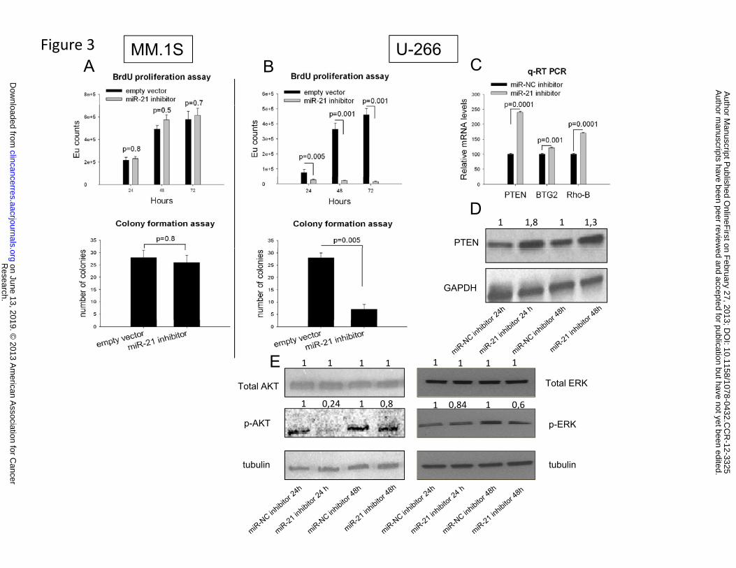
Figure 3A B
MM.1S U-266C
11 1,8 1,3
PTEN
D
GAPDH
1 1 1 1 1 11 1
Total AKT Total ERK
E
11 0,24 0,8 1 10,84 0,6
p-AKT
tubulin
p-ERK
tubulintubulin tubulin
Research.
on June 13, 2019. © 2013 A
merican A
ssociation for Cancer
clincancerres.aacrjournals.org D
ownloaded from
Author m
anuscripts have been peer reviewed and accepted for publication but have not yet been edited.
Author M
anuscript Published O
nlineFirst on F
ebruary 27, 2013; DO
I: 10.1158/1078-0432.CC
R-12-3325

Figure 4
BAMM.1SU-266
C D E
1 0,35
PTEN
C D E
GAPDH
Research.
on June 13, 2019. © 2013 A
merican A
ssociation for Cancer
clincancerres.aacrjournals.org D
ownloaded from
Author m
anuscripts have been peer reviewed and accepted for publication but have not yet been edited.
Author M
anuscript Published O
nlineFirst on F
ebruary 27, 2013; DO
I: 10.1158/1078-0432.CC
R-12-3325

Figure 5
A B C
Research.
on June 13, 2019. © 2013 A
merican A
ssociation for Cancer
clincancerres.aacrjournals.org D
ownloaded from
Author m
anuscripts have been peer reviewed and accepted for publication but have not yet been edited.
Author M
anuscript Published O
nlineFirst on F
ebruary 27, 2013; DO
I: 10.1158/1078-0432.CC
R-12-3325

Figure 6
1 1,7A B C
1
1 0,34
1
PTEN
Total AKT
p-AKT
tubulin
Research.
on June 13, 2019. © 2013 A
merican A
ssociation for Cancer
clincancerres.aacrjournals.org D
ownloaded from
Author m
anuscripts have been peer reviewed and accepted for publication but have not yet been edited.
Author M
anuscript Published O
nlineFirst on F
ebruary 27, 2013; DO
I: 10.1158/1078-0432.CC
R-12-3325

Published OnlineFirst February 27, 2013.Clin Cancer Res Emanuela Leone, Eugenio Morelli, Maria T. Di Martino, et al. cell growthTargeting miR-21 inhibits in vitro and in vivo multiple myeloma
Updated version
10.1158/1078-0432.CCR-12-3325doi:
Access the most recent version of this article at:
Material
Supplementary
http://clincancerres.aacrjournals.org/content/suppl/2013/02/27/1078-0432.CCR-12-3325.DC1
Access the most recent supplemental material at:
Manuscript
Authoredited. Author manuscripts have been peer reviewed and accepted for publication but have not yet been
E-mail alerts related to this article or journal.Sign up to receive free email-alerts
Subscriptions
Reprints and
To order reprints of this article or to subscribe to the journal, contact the AACR Publications
Permissions
Rightslink site. Click on "Request Permissions" which will take you to the Copyright Clearance Center's (CCC)
.http://clincancerres.aacrjournals.org/content/early/2013/02/27/1078-0432.CCR-12-3325To request permission to re-use all or part of this article, use this link
Research. on June 13, 2019. © 2013 American Association for Cancerclincancerres.aacrjournals.org Downloaded from
Author manuscripts have been peer reviewed and accepted for publication but have not yet been edited. Author Manuscript Published OnlineFirst on February 27, 2013; DOI: 10.1158/1078-0432.CCR-12-3325