target delineation for prostate cancer - nyp.org · target delineation for prostate cancer michael...
TRANSCRIPT

Target Delineation for Prostate Cancer
Michael J Zelefsky M.D.Professor of Radiation Oncology
Chief, Brachytherapy ServiceMemorial Sloan Kettering Cancer Center

Keys for Successful Contouring
• Familiarity with the target anatomy
• Understanding the extent of disease and areas at risk for microscopic extension
• Familiarity with the juxtaposed normal tissue structures

Zonal Anatomy

Saggital View

Levator Muscles
Anterior Vessels
Periprostaticspace
Obturator Internis

Anatomy of Imaging

IMRT @ MSKCC: Target Volume Definitions
Clinical Target Volume (CTV)Prostate and seminal vesicles (SV) Regional lymph nodes included for high risk disease
Planning Target Volume (PTV)1 cm margin around CTV Rectal-prostate interface - 6 mm margin onlySuperior border at tips of the SVInferior border above penile bulb
Treatment VolumePTV plus 5 mm except 10 mm at superior and inferior aspects to account for penumbra

IMRT @ MSKCC: Target Volume DefinitionsWith Fiducial Markers
Planning Target Volume (PTV)6mm margin around CTV
3mm posterior margin used for hypofractionated treated patients and seeded patients receiving supplemental EBRT
Superior border at tips of the SVInferior border above penile bulb
prostate apex generally 1.2 cm above the penile bulb
Simulation and Treatment Conditions
• Empty rectum (bowel prep for sim)• Empty bladder• Fiducial markers placed 3-5 days before
simulation• Calypso used for hypofractionated
patients• Aquaplast immobilization
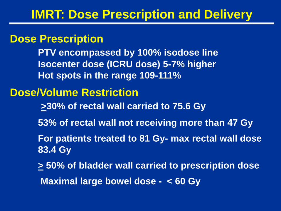
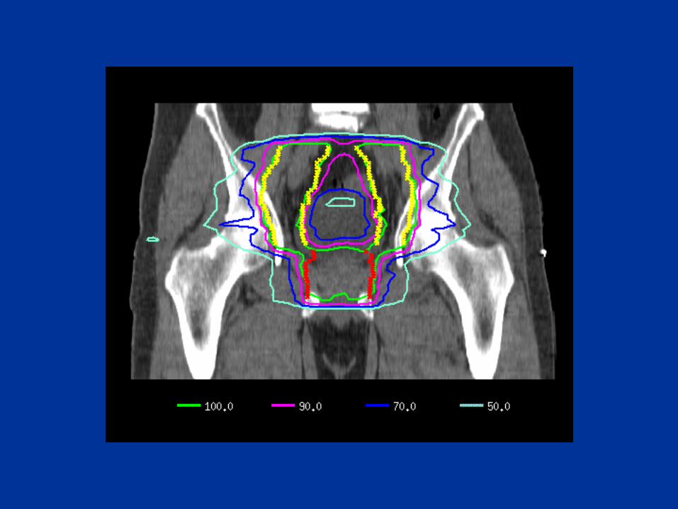
IMRT: Dose Prescription and Delivery
Dose PrescriptionPTV encompassed by 100% isodose line Isocenter dose (ICRU dose) 5-7% higher Hot spots in the range 109-111%
Dose/Volume Restriction >30% of rectal wall carried to 75.6 Gy
53% of rectal wall not receiving more than 47 GyFor patients treated to 81 Gy- max rectal wall dose 83.4 Gy> 50% of bladder wall carried to prescription doseMaximal large bowel dose - < 60 Gy
Full Bladder Conditions
• All post-op cases• Small bladder volume or small capacity
where greater bladder sparing is necessary

Mid-Gland- CTV Only

Mid-Gland- with PTV

Near Apex- CTV only
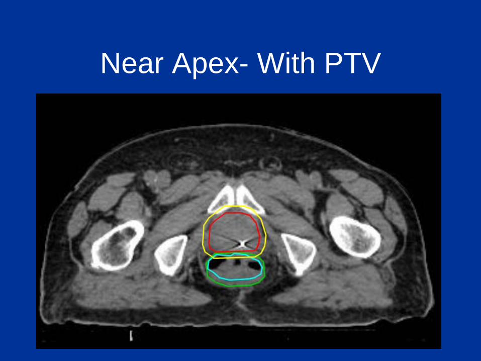
Near Apex- With PTV

Towards the Base- CTV Only

Towards the Base with PTV

At the Level of SV- CTV only

At the Level of SV-with PTV
Challenging Areas to Contour
•Apex
•Base
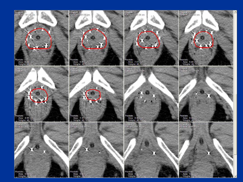
Errors in Measurement
• Cephalocaudad dimension– Identification of the first slice that the base
appears
– Identification of the last section that the apex appears
• Lateral and anteroposterior margins also source of error

Case #5-Apex
Common Errors in Prostate Contouring
• Overestimating the extent of the apex
• Underestimating the extent of the base
• Widening contours to include levator muscles or peri-prostatic tissues and ligaments
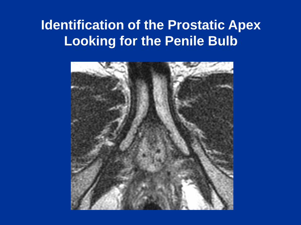
Identification of the Prostatic ApexLooking for the Penile Bulb

Apical Region
1.3 cm GU Diphragm

User Interface – “Look ahead – Look back”
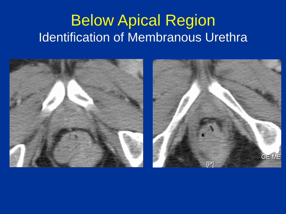
Below Apical RegionIdentification of Membranous Urethra

MRI anatomic correlation:can help improve accuracy of our target delineation for CT-based
treatment planning


Apical RegionTransition between Apex and GU Diaphragm

MRI Configuration at the Prostatic ApexMcLaughlin et al IJROBP 2009

CT Configuration at the Prostatic ApexMcLaughlin et al IJROBP 2009

• Use of IV contrast to delineate apex and base
• Utilization of other imaging modalities
– Image Fusion with registration
• User Interface – “Look ahead – look back”
– Real time 3D view / reconstruction
• Comparative with MRI – visual confirmation
– Most helpful at the base
Enhancing Visualization with CT

IV Contrast Enhancement
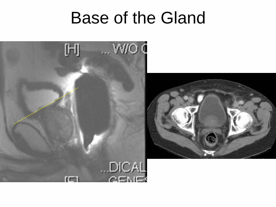
Base of the Gland

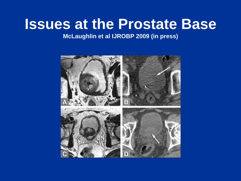
Issues at the Prostate BaseMcLaughlin et al IJROBP 2009 (in press)
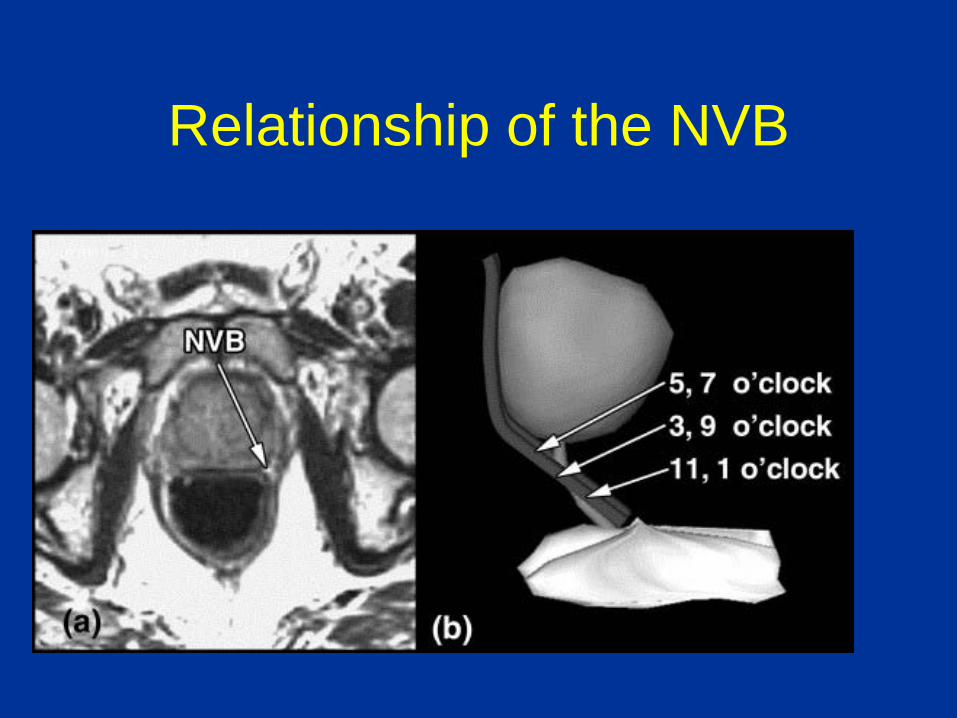
Relationship of the NVB
Defining the Prostate Target:Contouring Tips
• Start contouring prostate at mid-gland
• Avoid contouring levator muscles or obturator internis or venous plexi
• As you approach apex apply a “look forward and back approach”
• Identify penile bulb and in general the apex should be located 1-1.5 cm above this landmark

• For identification of apex and base CT contrast may be helpful
• If intravesical component is present, IV contrast cancan be particularly helpful
• Using beams eye views as you contour will eliminate contours that “jut out or in” too much which probably erroneous
Defining the Prostate Target:Contouring Tips
Contouring Post-Op CaseGeneral Comments
• Clips help define superior border.
• Volumes generally larger than typical definitive prostate.
• Urine or contrast can define the UV anastomosis which helps define the apex for CTV

Towards the Anastomosis-Apex
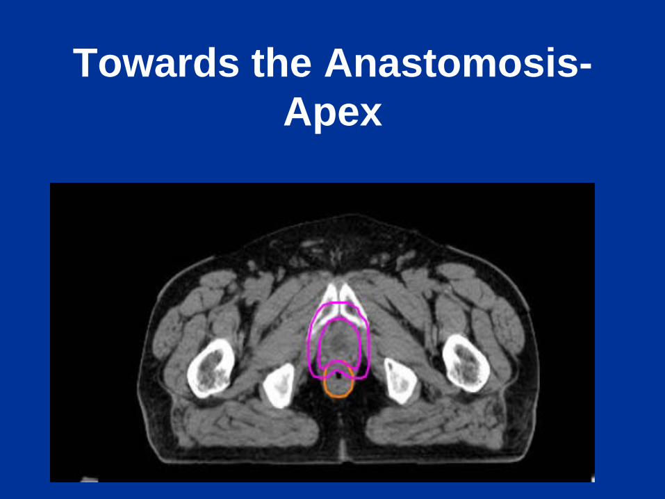
Towards the Anastomosis-Apex
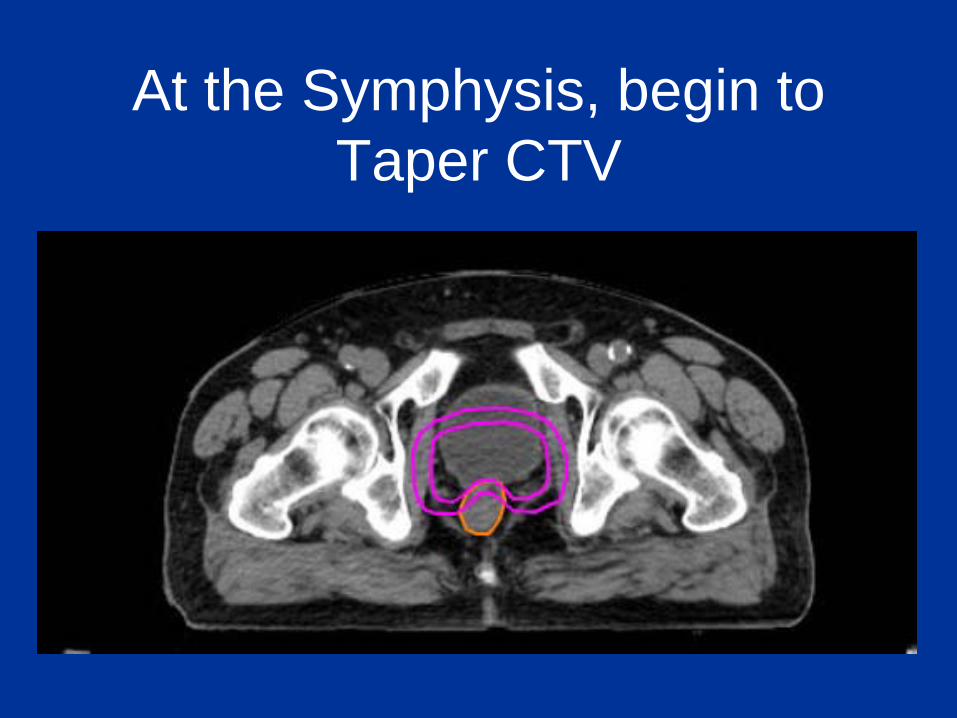
At the Symphysis, begin to Taper CTV

Status Post Robotic RPAt the Base- Covering SV
Remnants- No Clips Placed

Contouring Post-Op Cases• Superior Aspect: Include the clips and
any residual seminal vesicle remnants.
• Mid-Gland: – Include bladder up with lateral inclusion up
to the obturator internis muscles.– Start tapering the volume as you approach
the supeior aspect of the pubic symphysis
• Apex: 1 cm below the UV anastamosis

What about nodes?
3 options:1. Nodes+prostate to 45 with 3DCRT,
then prost with IMRT2. Nodes+prostate to 45 with IMRT, then
separate IMRT plan for prost3. Nodes+prostate in single IMRT plan,
but nodes at lower daily fraction size

Institution Nodes and IMRT?UM Option 1: 3DCRT then IMRTFCCC Option 3: Single plan IMRTWash U Option 2: Two IMRT plansWisconsin Option 3Duke Option 2Jefferson Option 1ROC Option 2MCW Option 2Mayo Option 2
MSKCC Option 2

Contouring of Pelvic Lymph Nodes
• Give IV contrast for target visualization
• Superior aspect at L5-S1
• At MSKCC 1-1.5 cm margin drawn for PTV around nodal or vascular structures

Conclusions• Familiarize yourself with the anatomic
borders of the prostate• After contouring mid-gland levels, turn
attention to apex and apply look ahead and back approach
• IV contrast especially for large intravesicle component can be helpful
• Knowing extent and location of intra-prostatic disease should fine tune the delineation of your PTV for more generous margin