take control with dynamic cell culture
TRANSCRIPT

EMD Millipore is a division of Merck KGaA, Darmstadt, Germany
CellASIC® ONIX Microfluidic Platform Take control with dynamic cell culture.
2
Biology is so much more than DMEM/FBS, 37 °C, 5% CO2. Living cells are constantly changing systems of interconnected mechanisms. Unlocking our understanding of these dynamic mechanisms requires real-time, instantaneous experimental control. With the flexible, intuitive CellASIC® ONIX Microfluidic Platform, you can easily take control of your cell culture. Simply program automated changes to culture media, gas and temperature, while tracking cell responses. By taking control of this truly in vivo -like environment, you’ll be able to perform dynamic, time-lapse experiments never before possible.
Think far beyond the limits of static cell culture.

Convective Transport
Flow BarrierDiffusive Transport
Cell/MatrixInteraction
Cell/Cell Interaction Cell/Cell
Signaling
Perfusion Channel Convective Transport
Perfusion Barrier
Cell Culture Chamber
Diffusive Transport
Convective Transport
Flow BarrierDiffusive Transport
Cell/MatrixInteraction
Cell/Cell Interaction Cell/Cell
Signaling
Perfusion Channel Convective Transport
Perfusion Barrier
Cell Culture Chamber
Diffusive Transport
3
Just as nutrients and gases are transported through blood vessels, culture media components and gases are transported through perfusion channels of the CellASIC® ONIX Microfluidic System. The perfusion barrier separating the cell culture from the channel (bottom) mimics the endothelial cell layer separating in vivo tissues from the blood (top).
What’s missing from traditional cell culture and analysis?
The analysis of living cells in vitro is critical to understanding basic biology, signaling pathways, drug effects, and disease models. But despite dramatic advances in detection methods, which have provided excellent means to interrogate living cells, the technology for controlling the environment of living cells during that analysis has not advanced far beyond the culture dish.
Because the cellular microenvironment, or “niche,” is as important as genetic factors for determining cell phenotype, a method for providing more accurate, dynamic control of living cells during experimental analysis can add a groundbreaking dimension to the science of cell biology.
The CellASIC® ONIX Microfluidic Platform was specifically designed to provide the dynamic cellular microenvironment control that has been missing until now.
In vivo
Microfluidic
Microfluidic perfusion mimics the in vivo cell environment

4
Delivering advanced control for live cell analysis experiments, the system integrates with your existing microscope to enable dynamic time-lapse experiments never before possible. Cutting-edge microfluidic technology provides an improved cell culture microenvironment, exceptional plate viewing
The CellASIC® ONIX Microfluidic Platform
“…We’ve been able to quickly and easily perform novel and technologically demanding experiments without any prior microfluidic experience. I’ve been able to focus on the fundamental biological questions while letting CellASIC® provide me with the tools I need to answer them.”
Maheshri Lab, MIT
quality for high magnification microscopy and superior media switching controls. An integrated Microincubator Controller maintains a temperature and gas environment directly on the microfluidic plate for long-term cell culture on any microscope stage.
Advanced control for live cell analysis. The system complements your microscope to provide a total solution for capturing the highest quality data with minimal effort.
Immunocytochemistry of primary neurons cultured, stained and imaged using the CellASIC® ONIX system. Primary rat cortical neurons were cultured to Day 15 and immunostained for MAP2 (Green, neurons) and GFAP (Red, astrocytes).
5
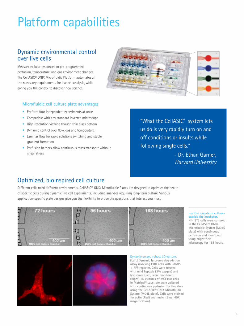
“What the CellASIC® system lets us do is very rapidly turn on and off conditions or insults while following single cells.”
- Dr. Ethan Garner, Harvard University
Dynamic environmental control over live cells Measure cellular responses to pre-programmed perfusion, temperature, and gas environment changes. The CellASIC® ONIX Microfluidic Platform automates all the necessary requirements for live cell analysis, while giving you the control to discover new science.
Platform capabilities
Dynamic assays, robust 3D culture. (Left) Dynamic lysosome degradation assay involving CHO cells with LAMP-1-RFP reporter. Cells were treated with mild hypoxia (3% oxygen) and lysosomes (Red) were monitored. (Right) 3D cultures of MCF10A cells in Matrigel® substrate were cultured with continuous perfusion for five days using the CellASIC® ONIX Microfluidic System (M04L plate). Cells were stained for actin (Red) and nuclei (Blue; 40X magnification).
Microfluidic cell culture plate advantages
• Perform four independent experiments at once
• Compatible with any standard inverted microscope
• High resolution viewing though thin glass bottom
• Dynamic control over flow, gas and temperature
• Laminar flow for rapid solutions switching and stable gradient formation
• Perfusion barriers allow continuous mass transport without shear stress
Optimized, bioinspired cell culture
Healthy long-term cultures outside the incubator. NIH 3T3 cells were cultured in the CellASIC® ONIX Microfluidic System (M04S plate) with continuous perfusion and monitored using bright field microscopy for 168 hours.
Different cells need different environments. CellASIC® ONIX Microfluidic Plates are designed to optimize the health of specific cells during dynamic live cell experiments, including analyses requiring long-term culture. Various application-specific plate designs give you the flexibility to probe the questions that interest you most.

6
Follow these simple steps
Prepare the microfluidic plate: Aspirate PBS from cell inlet well 6 and add 10 µL of desired cell suspension into the microfluidic plate. Cells will load automatically through capillary-driven cell loading.
Pipette reagents and media that will be used during your perfusion protocol into the four solution inlets (wells 2-5).
Seal plate to manifold by aligning the plate onto the manifold and turning on the vacuum switch on the CellASIC® ONIX Microfluidic Platform. The plate is sealed when the green “sealed” light is lit.
Place on inverted microscope stage and focus on the center of the viewing area. 4
3
You’re just minutes away from acquiring data using “load-and-go” CellASIC® ONIX Microfluidic Plates. Intuitive and easy-to-program CellASIC® ONIX FG Software automates your entire customizable protocol, so you can spend more time exploring the countless experimental possibilities enabled by this single platform.
1 Inlet for gravity driven continuous perfusion
2-5 Independent flow inlets for pressure driven flow
6 Inlet for cell loading
7-8 Outlets to waste wells
1
2
PerfusionBarrier
Diffusive mass exchange
between barriers
Microfluidic Flow Channels
Chamber Markers
Cell Culture Chamber
Automated integration into virtually any protocol
Cells
Recirculatingtemperature-controlledgas mixture
Manifold
Air pressure driven flow
Convective Peltierheat exchanger
#1.5 thickness170 µm thick glass
Microfluidic plate
Side view schematic of the microfluidic plate with microincubation manifold on a microscope stage.

7
Use the CellASIC® ONIX FG Software’s intuitive interface to program and monitor your experiment from one single view screen.
Valve on/off Buttons
Regulator Setpoints
Status Bar
Protocol Wizard:Click the valve and enter the time and flow rate for each
step. Design 5 steps or 15–it’s all under
your control.
Click “Run” to begin the program. Automate and perform live cell analysis using your microscope’s standard methods.
Run ProtocolOn this tab, you can save, change or add steps to the protocol you created using the “Create Protocol” Wizard.
Create ProtocolAn easy Wizard helps you set up an automated protocol for pre-programmed, walk-away perfusion changes over minutes, hours or days.
5
6
Manual OperationClick with your mouse to control inputs, outputs, gas and temperature in real time.
Three tabs, three easy programming options
‘‘Since I aim to quantify mitochondrial morphology, I require constant, stable imaging conditions that maintain the health of the cells, which the CellASIC® ONIX System does very well.”’
Marshall Lab, UCSF

Primary neuron culture over 21 daysPrimary rat cortical neurons cultured in the CellASIC® ONIX M04S Microfluidic Plate to Day 15 and immunostained in-plate for MAP2 (Green GFP, neurons) and GFAP (Red RFP, astrocytes; 40X).
8
Cell response to changing media conditionsLong-term live cell microscopy of cellular cytoskeletal changes in HeLa cells with precise microenvironment control. Tubulin (green) and actin (red) were stained using “in-plate” immunostaining with multi-solution, automated washing and exposure programs, using the CellASIC® ONIX M04S Microfluidic Plate. Image was acquired at 100X magnification.
Chemotaxis/migration in response to chemogradientHL-60 neutrophil migration in response to a chemokine. This frame from a live cell analysis video shows cells concentrating toward the chemokine in one chamber of a CellASIC® ONIX M04G Microfluidic Gradient Plate.
Courtesy of Jason Park, Wendell Lim Lab, UCSF.
Microscopy of 3D cell cultureObservation of multi-day morphology changes of 3D cancer spheroids cultured in extracellular matrix. MCF-10A breast cancer cells were suspended in Matrigel® substrate and grown in the CellASIC® ONIX M04S Microfluidic Plate. Cells were stained for actin (red) and nuclei (blue). Image was acquired at 40X magnification.
Popular applications of the CellASIC® ONIX PlatformWhat you’ve always imagined can now be reality, using the CellASIC® ONIX Platform to design dynamic cell biology experiments. It’s been demonstrated by our own scientists and loyal customers. The applications listed below are just a few of the exciting experiments you can perform with unprecedented precision.

9
Host-pathogen interactionsHost-pathogen assay monitoring Tuberculosis infection in macrophages. M. tuberculosis (RFP)
Bacteria single cell responseMeasurement of multi-generational response of live bacteria cells while maintaining cells in a single focal plane for days. A gene circuit in E. coli was induced and visualized for a time-lapse experiment in the CellASIC® ONIX B04 Microfluidic Plate. Images were acquired at 100X magnification.
Yeast single cell responseS. cerevisiae cells expressing GFP-tubulin and SPC42 mCherry during alpha-factor exposure and arrest. Images were acquired at 60X magnification. Courtesy of Soni Lacefield, University of Indiana.
Protein localization or translocationLocalization of actin (green) and microtubules (red) with respect to nuclei (blue) in the HT1080 human fibrosarcoma cell line immunofluorescently stained in the CellASIC® M04S Microfluidic Plate. Image was acquired at 40X magnification.
Dynamic autophagy assayLC3-GFP CHO reporter cells cultured on the CellASIC® ONIX system during perfusion of continuous chloroquine and starvation conditions to monitor the mechanisms of autophagy over 24 hours.

Burke TA, Christensen JR, Barone E, Suarez C, Sirotkin V, Kovar DR. Homeostatic actin cytoskeleton networks are regulated by assembly factor competition for monomers. Curr Biol. 2014 Mar 3;24(5):579-85.
Sieger B, Schubert K, Donovan C, Bramkamp M. The lipid II flippase RodA determines morphology and growth in Corynebacterium glutamicum. Mol Microbiol. 2013 Dec;90(5):966-82.
Gordon AJ, Satory D, Halliday JA, Herman C. Heritable change caused by transient transcription errors. PLoS Genet. 2013 Jun;9(6):e1003595.
Meyer RE, Kim S, Obeso D, Straight PD, Winey M, Dawson DS. Mps1 and Ipl1/Aurora B act sequentially to correctly orient chromosomes on the meiotic spindle of budding yeast. Science. 2013 Mar 1;339(6123):1071-4.
Donovan C, Schauss A, Kramer R, Bramkamp M. Chromosome segregation impacts on cell growth and division site selection in Corynebacterium glutamicum. PLOS One, February 2013; 8(2): eSS078.
Rafelski SM, Viana MP, Zhang Y, Chan YH, Thorn KS, Yam P, Fung JC, Li H, Costa L da F, Marshall WF. Mitochondrial network size scaling in budding yeast. Science. 2012 Nov 9;338(6108):822-4.
Ludington WB, Shi LZ, Zhu Q, Berns MW, Marshall WF. Organelle size equalization by a constitutive process. Curr Biol. 2012 Nov 20;22(22):2173-9.
Kraft C, Kijanska M, Kalie E, Siergiejuk E, Lee SS, Semplicio G, Stoffel I, Brezovich A, Verma M, Hansmann I, Ammerer G, Hofmann K, Tooze S, Peter M. Binding of the Atg1/ULK1 kinase to the ubiquitin-like protein Atg8 regulates autophagy. EMBO J. 2012 Sep 12;31(18):3691-703.
Sanchez-Diaz A, Nkosi PJ, Murray S, Labib K. The Mitotic Exit Network and Cdc14 phosphatase initiate cytokinesis by counteracting CDK phosphorylations and blocking polarised growth. EMBO J. 2012 Aug 29;31(17):3620-34.
Wei P, Wong WW, Park JS, Corcoran EE, Peisajovich SG, Onuffer JJ, Weiss A, Lim WA. Bacterial virulence proteins as tools to rewire kinase pathways in yeast and immune cells. Nature. 2012 Aug 16;488(7411):384-8.
Kono K, Saeki Y, Yoshida S, Tanaka K, Pellman D. Proteasomal degradation resolves competition between cell polarization and cellular wound healing. Cell. 2012 Jul 6;150(1):151-64.
Bermejo C, Haerizadeh F, Takanaga H, Chermak D, Frommer WB. Optical sensors for measuring dynamic changes of cytosolic metabolite levels in yeast. Nat Protoc. 2011 Oct 27;6(11):1806-17.
Eser U, Falleur-Fettig M, Johnson A, Skotheim JM. Commitment to a cellular transition precedes genome-wide transcriptional change. Mol Cell. 2011 Aug 19;43(4):515-27.
Dechant R, Binda M, Lee SS, Pelet S, Winderickx J, Peter M. Cytosolic pH is a second messenger for glucose and regulates the PKA pathway through V-ATPase. EMBO J. 2010 Aug 4;29(15):2515-26.
Manzoni R, Montani F, Visintin C, Caudron F, Ciliberto A, Visintin R. Oscillations in Cdc14 release and sequestration reveal a circuit underlying mitotic exit. J Cell Biol. 2010 Jul 26;190(2):209-22.
Furuya K, Niki H. The DNA damage checkpoint regulates a transition between yeast and hyphal growth in Schizosaccharomyces japonicus. Mol Cell Biol. 2010 Jun;30(12):2909-17. doi: 10.1128/MCB.00049-10.
Thorn KS. Spinning-disc confocal microscopy of yeast. Methods of Enzymology, 2010. vol 470: 581-602.
Octavio LM, Gedeon K, Maheshri N. Epigenetic and conventional regulation is distributed among activators of FLO11 allowing tuning of population-level heterogeneity in its expression. PLoS Genet. 2009 Oct;5(10):e1000673.
Key publications using the CellASIC® ONIX Microfluidic Platform
10
View the updated list of publications, review protocols and application data and watch video of live cells responding in real time by visiting:
www.emdmillipore.com/cellasic
Publication SpotlightTraditional cell signaling assays tell us that cells respond to stress by activating many interlinked pathways. But how can cells tell the difference between rapidly changing environmental conditions and gradually changing ones? Michael Elowitz’s lab realized the only way to ask this question was to grow their reporter strain of bacteria in the CellASIC® ONIX system, which enabled them to apply chemical stressors at different flow rates. Read their 2013 publication in PNAS (Proceedings of the National Academy of Sciences) to see how the authors discovered that cells turn on different sets of genes depending on the rate of stress increase:
Young JW, Locke JC, Elowitz MB. Rate of environmental change determines stress response specificity. Proc Natl Acad Sci U S A. 2013 Mar 5;110(10):4140-5.
11
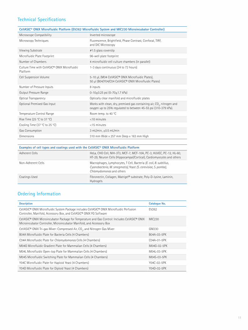
CellASIC® ONIX Microfluidic Platform (EV262 Microfluidic System and MIC230 Microincubator Controller)
Microscope Compatibility Inverted microscope
Microscopy Techniques Fluorescence, Brightfield, Phase Contrast, Confocal, TIRF, and DIC Microscopy
Viewing Substrate #1.5 glass coverslip
Microfluidic Plate Footprint 96-well plate footprint
Number of Chambers 4 microfluidic cell culture chambers (in parallel)
Culture Time with CellASIC® ONIX Microfluidic Platform
1-3 days continuous (24 to 72 hours)
Cell Suspension Volume 5-10 µL (M04 CellASIC® ONIX Microfluidic Plates), 50 µl (B04/Y04/C04 CellASIC® ONIX Microfluidic Plates)
Number of Pressure Inputs 8 inputs
Output Pressure Range 0-10±0.25 psi (0-70±1.7 kPa)
Optical Transparency Optically clear manifold and microfluidic plates
Optional Premixed Gas Input Works with clean, dry, premixed gas containing air, CO2, nitrogen and oxygen up to 25% regulated to between 45-55 psi (310-379 kPa).
Temperature Control Range Room temp. to 40 °C
Rise Time (25 °C to 37 °C) <10 minutes
Cooling Time (37 °C to 25 °C) <15 minutes
Gas Consumption 3 mL/min, ±0.5 mL/min
Dimensions 310 mm Wide x 257 mm Deep x 163 mm High
Examples of cell types and coatings used with the CellASIC® ONIX Microfluidic Platform
Adherent Cells HeLa, CHO Cell, NIH-3T3, MCF-7, MCF-10A, PC-3, HUVEC, PC-12, HL-60, HT-29, Neuron Cells (Hippocampal/Cortical), Cardiomyocytes and others
Non-Adherent Cells Macrophages, Lymphocytes, T Cell, Bacteria (E. coli, B. subtillus, Cyanobacteria, M. smegmatis), Yeast (S. cerevisiae, S. pombe), Chlamydomonas and others
Coatings Used Fibronectin, Collagen, Matrigel® substrate, Poly-D-lysine, Laminin, Hydrogels
Technical SpecificationsKey publications using the CellASIC® ONIX Microfluidic Platform
Description Catalogue No.
CellASIC® ONIX Microfluidic System Package includes CellASIC® ONIX Microfluidic Perfusion Controller, Manifold, Accessory Box, and CellASIC® ONIX FG Software
EV262
CellASIC® ONIX Microincubator Package for Temperature and Gas Control: Includes CellASIC® ONIX Microincubator Controller, Microincubator Manifold, and Accessory Box
MIC230
CellASIC® ONIX Tri-gas Mixer: Compressed Air, CO2, and Nitrogen Gas Mixer GM230
B04A Microfluidic Plate for Bacteria Cells (4 Chambers) B04A-03-5PK
C04A Microfluidic Plate for Chlamydomonas Cells (4 Chambers) C04A-01-5PK
M04G Microfluidic Gradient Plate for Mammalian Cells (4 Chambers) M04G-02-5PK
M04L Microfluidic Open-top Plate for Mammalian Cells (4 Chambers) M04L-03-5PK
M04S Microfluidic Switching Plate for Mammalian Cells (4 Chambers) M04S-03-5PK
Y04C Microfluidic Plate for Haploid Yeast (4 Chambers) Y04C-02-5PK
Y04D Microfluidic Plate for Diploid Yeast (4 Chambers) Y04D-02-5PK
Ordering Information

www.emdmillipore.com
EMD Millipore, the M mark, CellASIC, Calbiochem, Chemicon, Millex, Stericup, Steriflip, and Upstate are registered trademarks of of Merck KGaA, Darmstadt, Germany. SmartFlare and LentiBrite are trademarks of Merck KGaA, Darmstadt, Germany. Trademarks belonging to third parties are the properties of their respective owners.Lit. No. PB2444EN00 Rev. C BS-GEN-12-07257 Printed in the USA. 07/2014 © 2014 EMD Millipore Corporation, Billerica, MA, USA. All rights reserved.
To place an order or receivetechnical assistanceIn the U.S. and Canada, call toll-free 1-800-645-5476
For other countries across Europe and the world, please visit: www.emdmillipore.com/offices
For Technical Service, please visit: www.emdmillipore.com/techservice
Get connected! Join EMD Millipore Bioscience on your favorite social media outlet for the latest updates, news, products, innovations, and contests!
facebook.com/EMDMillipore
twitter.com/EMD_Millipore
Small Molecule Inhibitors, Activators, Libraries and Pathway PanelsPerturbing cellular pathways using small molecules and then using live cell analysis to analyze impacts on cells in real time can translate into powerful biological discoveries. EMD Millipore’s Calbiochem® libraries, pathway panels and individual reagents offer the widest and most cited selection of small molecule inhibitors and activators worldwide.
Learn more at www.emdmillipore.com/calbiochem
Live Cell RNA DetectionSmartFlare™ RNA Detection Probes quantitatively reveal expression of specific RNAs inside living cells. Following a single, nontoxic, overnight incubation, fluorescent signal corresponding to the presence of target RNAs can be detected using microscopy, flow cytometry or other detection platform. The same cells can be then used for further biochemical or functional analyses.
Choose from 1200+ probes or design your own: www.emdmillipore.com/smartflare
Related Products Get the most from your CellASIC® ONIX Microfluidic Platform by exploring EMD Millipore’s cell culture tools, antibodies, reagents, small molecules and kits for cell-based assays, including reagents specifically optimized for live cell analysis.
Cell CultureFor the most convenient, reliable, analysis-ready cell cultures, count on EMD Millipore’s wide variety of devices and surfaces to provide cell growth, structure, and function that more closely mimic what occurs in vivo. Spend less time growing cells and fumbling with clumsy devices and more time on your research.
Learn more at www.emdmillipore.com/cellculture
Sterile FiltrationEMD Millipore’s trusted line of sterile filtration tools have been specifically designed to eliminate contaminants and ensure the reproducibility of your downstream analyses.
For a complete listing of our sterile filtration products, including our Stericup® filters, Steriflip® filters and Millex® Syringe Filters, please visit www.emdmillipore.com/sterile
AntibodiesBased on the expertise of Chemicon® and Upstate®, EMD Millipore’s highly validated antibodies are guaranteed for quality performance. Many of our antibodies are conjugated to fluorophores and validated for immunocytochemistry.
Put the most reput(Ab)le antibodies to work for you: www.emdmillipore.com/antibodies