table of contents - university of torontodigital+assets/... · 1 joint neuroscience events june...
TRANSCRIPT


Table of Contents Schedule ……………………………………………………………………………………… 1 Event Organizers & Acknowledgements ………………………………………………. 3 Poster Judges ……………………………………………………………………………..... 3 CPIN Board of Directors & Committees ………………………………………………... 4 Abstracts …………………………………………………………………………………….. 5 Abstract Index ………………………………………………………………………………. 49 Event Sponsors …………………………………………………………………………….. 54

1
Joint Neuroscience Events June 2013
2013 Collaborative Program In Neuroscience Research Day
June 10, 2013
Medical Sciences Building 9:30 Pre-event: Career Workshop – Non-Conventional Careers (MSB2172) Dr. Christa Studzinski, Senior Program Lead of Research Programs, OBI Dr. Ruslan Dorfman, Co-Founder, Geneyouin Inc. Dr. Alison Denney, Senor Program Lead for Industry Relations, OBI Dr. Philip Caffrey, Public Policy and Programs Analyst, Alzheimer’s Society of Ontario Dr. Kirk Nylen, Acting Director of Operations and Outreach, OBI Theme: Alternative career options of Neuroscience Graduates
11:00 Meet with Workshop speakers (Stone Lobby)
11:30 Registration (Stone Lobby) Poster set-up
12:00 Opening remarks (Stone Lobby) Dr. Zhong-Ping Feng, Director, CPIN Dr. Alison Buchan, Vice Dean (Research), Faculty of Medicine Dr. Sandy Welsh, Vice Dean (Graduate Studies), Faculty of Arts and Science Dr. Peter Lewis, Vice President (Research), University of Toronto
12:15 Poster presentation grouping, judges, and judging process of Poster Presentation Awards Dr. Janice Robertson / Dr. Albert Wong
Rules of Student’s Choice Awards Dr. Lili-Naz Jazrati
Pick-up Lunch
12:30 CPIN Poster Presentation and evaluation (Refreshments are provided)
16:00 CPIN Group Photo (outside of MSB by JJR MacLeod Auditorium)
16:30 Raymond and Beverly Sackler Distinguished Lecture (JJR MacLeod Auditorium) (Co-organized with International Symposium on Structural Neurobiology, July 11,2013)
Welcome remarks
16:45 Professor Brian Kobilka (Stanford University, U.S.A.) Structural Insights into the Dynamic Process of G-protein-coupled Receptor Activation
2
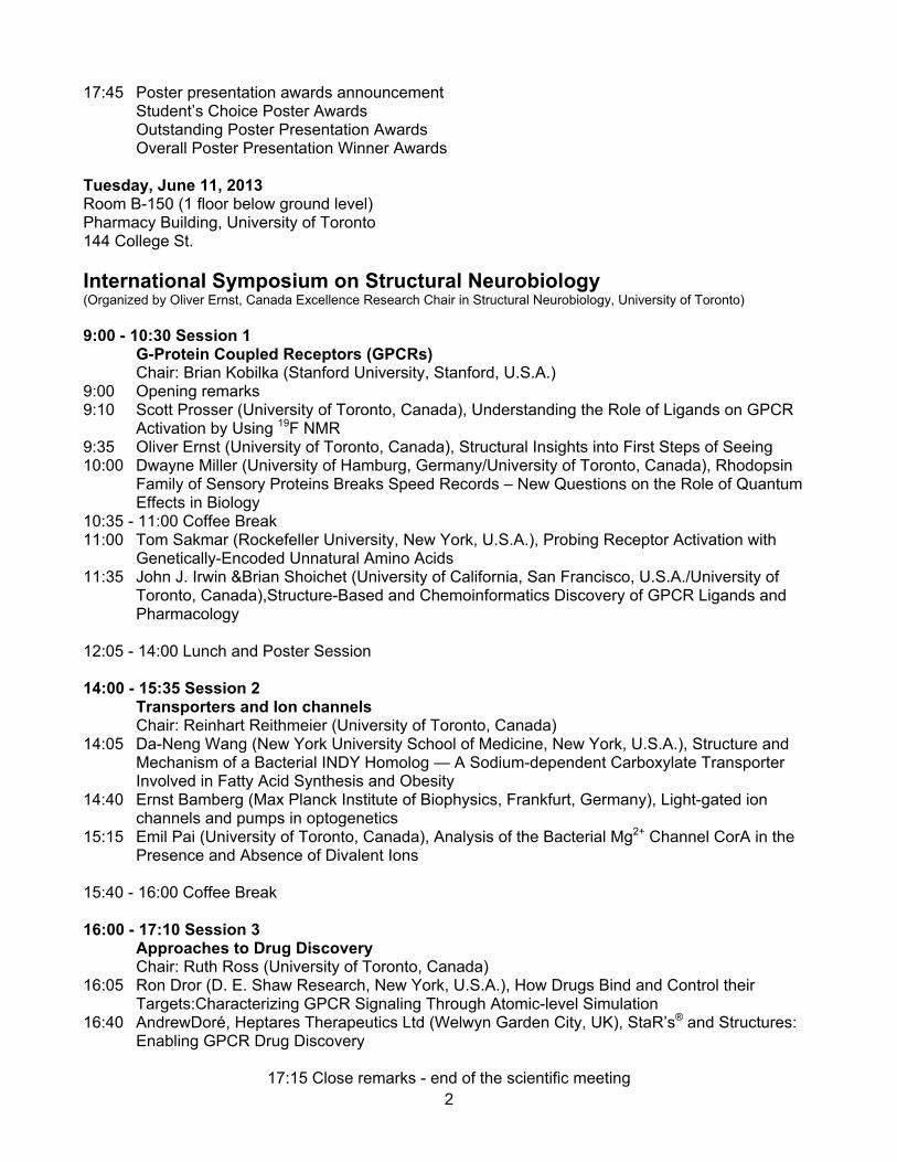
17:45 Poster presentation awards announcement Student’s Choice Poster Awards Outstanding Poster Presentation Awards Overall Poster Presentation Winner Awards Tuesday, June 11, 2013 Room B-150 (1 floor below ground level) Pharmacy Building, University of Toronto 144 College St.
International Symposium on Structural Neurobiology (Organized by Oliver Ernst, Canada Excellence Research Chair in Structural Neurobiology, University of Toronto) 9:00 - 10:30 Session 1 G-Protein Coupled Receptors (GPCRs) Chair: Brian Kobilka (Stanford University, Stanford, U.S.A.) 9:00 Opening remarks 9:10 Scott Prosser (University of Toronto, Canada), Understanding the Role of Ligands on GPCR Activation by Using 19F NMR 9:35 Oliver Ernst (University of Toronto, Canada), Structural Insights into First Steps of Seeing 10:00 Dwayne Miller (University of Hamburg, Germany/University of Toronto, Canada), Rhodopsin Family of Sensory Proteins Breaks Speed Records – New Questions on the Role of Quantum Effects in Biology 10:35 - 11:00 Coffee Break 11:00 Tom Sakmar (Rockefeller University, New York, U.S.A.), Probing Receptor Activation with Genetically-Encoded Unnatural Amino Acids 11:35 John J. Irwin &Brian Shoichet (University of California, San Francisco, U.S.A./University of
Toronto, Canada),Structure-Based and Chemoinformatics Discovery of GPCR Ligands and Pharmacology
12:05 - 14:00 Lunch and Poster Session 14:00 - 15:35 Session 2 Transporters and Ion channels Chair: Reinhart Reithmeier (University of Toronto, Canada) 14:05 Da-Neng Wang (New York University School of Medicine, New York, U.S.A.), Structure and Mechanism of a Bacterial INDY Homolog — A Sodium-dependent Carboxylate Transporter Involved in Fatty Acid Synthesis and Obesity 14:40 Ernst Bamberg (Max Planck Institute of Biophysics, Frankfurt, Germany), Light-gated ion channels and pumps in optogenetics 15:15 Emil Pai (University of Toronto, Canada), Analysis of the Bacterial Mg2+ Channel CorA in the Presence and Absence of Divalent Ions 15:40 - 16:00 Coffee Break 16:00 - 17:10 Session 3 Approaches to Drug Discovery Chair: Ruth Ross (University of Toronto, Canada) 16:05 Ron Dror (D. E. Shaw Research, New York, U.S.A.), How Drugs Bind and Control their Targets:Characterizing GPCR Signaling Through Atomic-level Simulation 16:40 AndrewDoré, Heptares Therapeutics Ltd (Welwyn Garden City, UK), StaR’s® and Structures: Enabling GPCR Drug Discovery
17:15 Close remarks - end of the scientific meeting

3
Event Organizers & Acknowledgements
CPIN Research Day Organizing Committee Zhong-Ping Feng (Chair) Oliver Ernst (also organizer Symposium SNB) Lili-Naz Hazrati Janice Robertson Albert Wong CPIN Graduate Student Executives: Andrew Barszczyk Ceilidh Cunningha Katie Ferguson (Liaison) Vivek Mahadevan Denis Osipov Vladislav Sekulic Bhanu Sharma Luka Srejic Sonia Sugumar Sackler Lecturer Invitation Oliver Ernst Workshop Zhong-Ping Feng Janice Robertson Poster Judge Organizing Committee Janice Robertson (Co-Chair) Albert Wong (Co-Chair) Lili-Naz Hazrati Zhong-Ping Feng Student’s Choice Poster Awards Committee Zhong-Ping Feng (Co-Chair) Lili-Naz Hazrati (Co-Chair) Andrew Barszczyk Luka Srejic
Event Program Design Katie Ferguson Vladislav Sekulic Sponsorships Zhong-Ping Feng (Chair) Oliver Ernst Lili-Naz Hazrati Janice Robertson Albert Wong Richard Zemel Poster Site Coordinators Luka Srejic Sonia Sugumar
Registration Desk Luka Srejic (Co-Chair) Bhanu Sharma (Co-Chair) Andrew Barszczyk Ceilidh Cunningha Katie Ferguson Vivek Mahadevan Vladislav Sekulic Sonia Sugumar (AV) Photographers Albert Wong Denis Osipov Hong-Shuo Sun
Administration Victoria Simpson Rob Reedijk
Poster Judges
Ana Andreazza Limor Avivi-Arbe Paul Boutros Sid Croul Suzanne Erb Jim Eubanks Paul Fletcher Susan George Karen Gordon David Hampson Jeff Handerson Zhengping Jia
Bill Ju Rasmus Kiehl Evelyn Lambe Robert Levitan Philippe Monnier Howard Mount Daniel Mueller Joanne Nash Jose Nobrega Janice Robertson Gerold Schmitt-Ulms Barry Sessle
Frances Skinner Shuzo Sugita Hong-Shuo Sun Franco Taverna Doug Tweed John Vincent Lu-Yang Wang John Yeomans Karl Zabjek Martin Zack Richard Zemel

4
CPIN Board of Directors and Committees (2012 – 2013) The CPIN Board of Directors reviews and signs the Memorandum of Agreement (MoA) and oversees the general direction of the program.
• Katherine Berg (Rehabilitation Science)
• Heather Boon (Pharmaceutical Sciences) • Peter Burns (Medical Biophysics)
• John Coleman (Interim, Cell and Systems Biology) • Sven Dickinson (Computer Science)
• Harry Elsholtz (Laboratory Medicine and Pathobiology) • Zhong-Ping Feng (Chair, CPIN Director; Physiology) • David R. Hampson (Honorary director, past CPIN director) • Ester Geva (Applied Psychology and Human Development)
• Avrum Gotlieb (Lead Faculty: Faculty of Medicine) • Allan Kaplan (Institute of Medical Science)
• Morris Manolson (Dentistry) • Stephen Matthews (Physiology) • Morris Moscovitch (Psychology)
• Reinhart Reithmeier (Biochemistry)
• Ruth Ross (Pharmacology) • Paul Santerre (Interim: Institute of Biomaterials and Biomedical Engineering)
The CPIN Academic Program Committee reviews admission and program requirements, program curriculum and completion, and program activities as per the MoA.
• Julie Audet (Institute of Biomaterials and Biomedical Engineering)
• Peter Carlen (Institute of Medical Science)
• Jonathan O. Dostrovsky (Honorary member: Physiology)
• Zhong-Ping Feng (Chair, CPIN Director)
• David R. Hampson (Honorary member: Pharmaceutical Sciences)
• Jeffrey Henderson (Pharmaceutical Sciences)
• Kang Lee (Applied Psychology and Human Development)
• Peter McPherson (Pharmacology)
• Angus McQuibban (Biochemistry) • John Peever (Cell and Systems Biology) • Janice Robertson (Laboratory Medicine and
Pathobiology) • Barry J. Sessle (Dentistry) • Bojana Stefanovic (Medical Biophysics) • Martin Wojtowicz (Physiology) • John S. Yeomans (Psychology) • Karl Zabjek (Rehabilitation Science) • Richard Zemel (Computer Science)
• CPIN Graduate Executives representative
The CPIN Executive Committee reviews and leads the program activities as per the MoA, including distinguished lectures, Research Day, outreach programs, sponsorships and faculty-student interactive events.
• Oliver Ernst (Biochemistry)
• Zhong-Ping Feng (Chair, CPIN Director) • David R. Hampson (Honorary member:
Pharmaceutical Sciences) • Lili-Naz Hazrati (Laboratory Medicine and
Pathobiology) • Jeffrey Henderson (Pharmaceutical Sciences)
• Kang Lee (Human Development and Applied Psychology)
• John Peever (Cell and Systems Biology) • Ruth Ross (Pharmacology & Toxicology)
• Barry J. Sessle (Dentistry) • Molly Shoichet (Institute of Biomaterials and
Biomedical Engineering) • Bojana Stefanovic (Medical Biophysics) • Lu-Yang Wang (Physiology) • Albert Wong (Institute of Medical Science) • John S. Yeomans (Psychology)
• Karl Zabjek (Rehabilitation Science) • Richard Zemel (Computer Science) • CPIN Graduate Executives representative

5
Abstracts Author:Elia Abi-Jaoude Affiliation: IMS Supervisor: Paul Sandor The Neural Correlates of Self-Regulatory Fatigue During Inhibitory Control of Eye Blinking Elia Abi-Jaoude MSc MD1, Barbara Segura PhD2, Sang Soo Cho PhD3, Antonio Strafella MD3 PHD, Paul Sandor MD1 1Department of Psychiatry, University Health Network, University of Toronto, Toronto, Ontario, Canada 2Department of Psychiatry and Clinical Psychobiology, University of Barcelona, Barcelona, Spain 3PET Imaging Centre, Centre for Addiction and Mental Health, University of Toronto, Toronto, Ontario, Canada INTRODUCTION: The capacity to regulate urges is an important human characteristic associated with a range of academic, financial, legal, physical and mental health outcomes (Mischel et al, 1989, 2011; Moffitt et al, 2011). Self-regulatory capacity has limited reserve, which, when depleted, leads to failure (Hagger et al, 2010). While the neural substrates of self-regulation are believed to entail a balance between prefrontal cortical control regions and subcortical limbic and affective areas (Heatherton & Wagner, 2011; Goldstein & Volkow, 2011), little is known about the neurobiology of self-regulatory fatigue. We set out to investigate the neural correlates of self-regulatory fatigue. We hypothesized this would involve altered activity in prefrontal cortical subregions and interoceptive processing areas. METHODS: Blood oxygen level-dependent functional magnetic resonance imaging was used to detect brain activations in 19 right-handed subjects during inhibition of eye blinking. A block design was used, consisting of one-minute blocks of effortful inhibition of eye blinking, alternating with one-minute blocks without such inhibition; there were 2 runs, 6 minutes each. A general linear model was used for contrast analyses using Statistical Parametric Mapping (SPM8). The increase in number of blinks during blink inhibition from the first to the last block was used as covariate of interest. In addition, tensorial independent component analysis, as implemented in the Multivariate Exploratory Linear Decomposition into Independent Components tool (MELODIC V.3.05), part of FMRIB Software Library (FSL), was carried out to identify relevant functional networks. RESULTS: There was an increase in the number of eye blinks escaping inhibitory control across blink inhibition blocks, whereas there was no change in the number of eye blinks occurring during rest blocks. Inhibition of blinking activated a wide network bilaterally including inferior frontal gyrus, dorsolateral prefrontal cortex, dorsal anterior cingulate cortex, frontal eye fields, supplementary motor area, and caudate. There were also bilateral activations in inferior parietal, anterior insula, precuneus, and sensory association areas. Deteriorating performance was associated with activity in orbitofrontal cortex, ventromedial prefrontal cortex, rostroventral anterior cingulate cortex, precuneus, somatosensory and parietal areas. Independent component analysis identified task-related synchronized activations and deactivations in the above areas. The strength of such networks decreased over time and this was associated with worsening performance. CONCLUSIONS: We demonstrated behavioral evidence of self-regulatory fatigue in an eye blink inhibition task. As anticipated, effortful eye blink control resulted in activation of prefrontal control areas and regions involved in urge and interoceptive processing. Worsening performance resulted in overlapping urge-related areas and in regions involved in affective salience attribution and craving. Our findings suggest that self-regulatory fatigue is associated with a decrease in the coordinated recruitment of prefrontal cortical control regions. References Hagger, M.S., Wood, C., Stiff, C., Chatzisarantis, N.L.D. (2010), ‘Ego depletion and the strength model of self-control: a meta-analysis’, vol. 136, no. 4, pp. 495-525. Heatherton, T.F., Wagner, D.D. (2011), ‘Cognitive neuroscience of self-regulation failure’, Trends in Cognitive Science, vol. 15, no. 3, pp. 132-139. Goldstein, R.Z., Volkow, N.D. (2011), ‘Dysfunction of the prefrontal cortex in addiction: neuroimaging findings and clinical implications‘, Nature Reviews Neuroscience, vol. 12, no. 11, pp. 652-669. Mischel, W., Shoda, Y., Rodriguez, M.L. (1989), ‘Delay of gratification in children’, Science, vol. 244, no. 4907, pp. 933-938. Mischel, W., Ayduk, O., Berman, M.G., Casey, B.J., Gotlib, I.H., Jonides, J., Kross, E., Teslovich, T., Wilson, N.L., Zayas, V., Shoda, Y. (2011), ‘’Willpower’ over the life span: decomposing self-regulation’, Social Cognition and Affective Neuroscience, vol. 6, no. 2, pp. 252-256.
6
Moffitt, T.E., Arseneault, L., Belsky, D., Dickson, N., Hancox, R.J., Harrington, H., Houts, R., Poulton, R., Roberts, B.W., Ross, S., Sears, M.R., Thomson, W.M., Caspi, A. (2011), ‘A gradient of childhood self-control predicts health, wealth, and public safety’, Proceedings of the National Academy of Sciences of the United States of America, vol. 108, no. 7, pp. 2693-2698. Author: Rylan Allemang-Grand Affiliation: Medical Biophysics Supervisor: Jason Lerch Altered brain development long before plaque deposition in the TgCRND8 mouse model of Alzheimer’s disease R. Allemang-Grand1,2, J.Scholz1, J.Ellegood1, C. Laliberté1, P.E. Fraser2,3, J.G. Sled1,2, J.P. Lerch1,2 1Mouse Imaging Centre, Toronto, Ontario 2Department of Medical Biophysics, University of Toronto 3Centre for Research in Neurodegenerative Diseases Introduction: Currently, cross-sectional magnetic resonance imaging (MRI) studies of transgenic mouse models of Alzheimer’s disease (AD) have demonstrated atrophy in similar regions of the brain implicated in human AD, suggesting that these models recapitulate disease progression [1,2]. However, these studies limit their analyses to a few regions of interest and monitor changes in anatomy with the onset of plaque deposition, the longstanding biological hallmark of AD. Although strongly implicated in the pathogenesis of AD, recent findings suggest that other biochemical events which precede plaque deposition drive disease onset [3]. Therefore, we set out to longitudinally track the anatomical changes across the entire brain before and after plaque deposition in the early-onset TgCRND8 mouse model of AD. Methods: We used MRI for longitudinally tracking regional brain atrophy in the TgCRND8 mouse model of AD [4]. For this study, transgenic and wild-type littermates were longitudinally scanned with a manganese-enhanced MRI (MEMRI) protocol before (4 & 9 weeks) and after (12, 16, 20 & 24 weeks) the onset of AD-related pathology in this model [5]. Additionally, we scanned a separate cohort of 1-week-old transgenic and wild-type mice with 3D diffusion-weighted imaging. Acquired images were registered and deformed to generate a consensus average. In-house MEMRI anatomical atlas labeled with 62 distinct structures was superimposed on the final non-linear images from the registration for volume computation of each brain region. Results: At 4, 9 and 12 weeks, transgenic mice had an 8% reduction in the volume of the cortex, hippocampus, olfactory bulbs and cerebellum. Localized expansion of the amygdala and periaquaductal gray were also apparent at this time. Analysis of the 3D diffusion weighted images demonstrate that many of the anatomical differences at the later time points were apparent at 1 week of age, 11 weeks before the onset of plaque deposition. These findings suggest that the drastic reductions in volume of the TgCRND8 brain are driven by a different biochemical phenomenon preceding plaque deposition, which occurs at 12 weeks of age in this model. Conclusion: Contemporary treatments for Alzheimer’s disease attempt to delay synaptic loss and brain atrophy associated with cognitive decline early in the disease. In order to properly test prevention-focused interventions in mice, experimentation would need to begin early before the onset of pathology. Our study demonstrates that brain development differences need to be accounted for when testing these early-life interventions due to the perinatal onset of pathology driving volume loss within the brain. 1. Delatour et al. (2005). Neurobiology of aging, 27(6), 835-47. 2. Redwine et al. (2003). PNAS, 100(3), 1381-1386. 3. Ferreira & Klein (2011). Neurobiology of Learning and Memory, 96, 529-543. 4. Chishti et al., (2001). Journal of Biological Chemistry, Vol. 276 (24). 5. Lau et al., (2008). Neuroimage, 42, 19-27. Author: Jessica Arsenault Affiliation: Psychology Supervisor: Bradley Buchsbaum An MVPA Analysis of the Role of the Motor Cortex During Speech Perception Jessica Arsenault and Bradley Buchsbaum Rotman Research Institute, University of Toronto Within the field of cognitive neuroscience, there is a debate regarding the role of the motor system in speech perception. Proponents of the motor theory of speech perception assume that the motor system is critically involved in the perception of speech (1, 2). On the other hand, it has been argued that the contribution of the motor system to speech perception plays

7
only a modulatory role, and is not necessary for speech perception in normal listening conditions (see ref. 3 for a review). While studies of speech perception using functional magnetic resonance imaging (fMRI) have often found elevated activity in the motor system (e.g. Broca’s area and premotor cortex) we know little about the representational code that is characterized in these regions. Recent advances in multivoxel pattern analysis (MVPA) of fMRI data allow one to ask just this sort of question: what information is being coded in these regions during auditory speech perception? Unlike the standard univariate approach to fMRI analysis, MVPA identifies reliable patterns of activity that are evoked by different stimuli or task conditions. Thus, MVPA can provide a detailed description of underlying neural codes. This study aims to explore the precise activity patterns associated with the auditory perception of consonants that share varying levels of feature similarity and acoustic confusability (4). Rather than simply identifying activation of the motor cortex during speech perception, the goal is to identify how the similarity of consonants as assessed by acoustic confusability (e.g. ‘m’ is confusable with ‘n’ but not ‘b’) is mirrored by the similarity among patterns of brain activation in the motor and auditory cortices, respectively. Sixteen syllables were aurally presented one at a time to participants in the fMRI scanner. Different speakers and different versions of each syllable were repeated such that 120 trials per syllable were collected for each participant over the course of a 1.5-hour scan. The data will be analyzed with MVPA and the similarity structure of consonant stimuli will be separately assessed in the auditory and motor cortices. If the similarity among the patterns of activation in motor cortex more closely resembles the acoustic confusability of the stimulus set than is observed in auditory cortex, this will provide strong support in favor of motor theories of speech perception. On the other hand, if acoustic confusability is better matched by the similarity among patterns of activation in auditory than motor cortex, this will provide evidence against motor theories of speech perception as it would suggest that the motor cortex is not playing a critical discriminatory role. References 1. Liberman, Am. M., Cooper, F. S., Shankweiler, D. P., & Studdert-Kennedy, M. (1967). Perception of the speech code. Psychological Review, 74 431-461. 2. D’Ausilio, A., Pulvermüller, F., Salmas, P., Bufalari, I., Begliomini, C., & Fadiga, L. (2009). The motor somatotopy of speech perception. Current biology : CB, 19(5), 381–5. doi:10.1016/j.cub.2009.01.017 3. Hickok, G. (2009). The functional neuroanatomy of language. Physics of Life Reviews, 6(3), 121–143. doi:10.1016/j.plrev.2009.06.001 4. Miller, G. A., & Nicely, P. E. (n.d.). An Analysis of Perceptual Confusions Among Some English Consonants HE over-all effects of noise and, 338–352. Author: Jura Augustinavicius Affiliation: Department of Cell and Systems Biology, University of Toronto Supervisor: Dr. Colin Shapiro The Role of Autonomic Nervous System Assessment by HRV in Depressed Adolescents Jura Augustinavicius1,2, Guido Simonelli5, Azmeh Shahid2,3,5, Daniel Vigo4,5, David Newman6, Colin Shapiro1,2,3,6 Department of Cell and Systems Biology, University of Toronto, Toronto, Canada1 Youthdale Child and Adolescent Sleep Centre, Toronto, Canada2 Toronto Western Hospital, University Health Network, Toronto, Canada3 Departamento de Docencia e Investigación, Facultad de Ciencias Médicas, Pontificia Universidad Católica Argentina, Buenos Aires, Argentina4 Consejo Nacional de Investigaciones Científicas y Técnicas, Argentina5 Faculty of Medicine, University of Toronto, Toronto, Canada6 Background: Major Depressive Disorder (MDD) is associated with disorders of autonomic function. Adolescent depression can be difficult to diagnose with current interview measures based on adult assessment parameters. We hypothesized that variables related to autonomic tone, measured by nocturnal heart rate variability (HRV) components, may have an important role in the diagnosis of depression. Additionally, we hypothesized that HRV over 24-hours with a focus on day versus night would be relevant in adolescents with early-onset depression. Methods: Adolescents between 12 and 18 years with MDD and healthy controls were recruited for the study. All participants were interviewed by a child psychiatrist and the presence of a clinical diagnosis of MDD was determined based on DSM-IV-TR criteria, the current gold standard for the diagnosis of depression. Holter monitors were worn for 24-hours. Day and night segments were identified per participant from a device that has been validated for sleep and wake assessment based on locomotor activity (Actigraphy). Time and frequency domain HRV measures were analyzed in one hour bins over 24-hours and for day and night segments. The wavelet transform was applied for frequency domain analyses.
8
Results: Sixteen depressed adolescents (5 males/11 females) with a mean age of 15.6 ± 1.46 years, and 11 healthy controls (4 males/7 females) with a mean age 14.2 ± 1.43 participated in the study. In both groups, HRV was greater at night compared to day: the mean duration of RR intervals was 719 ± 84ms during the day and 9223 ± 164ms during the night (p < .001), and the root mean square successive difference of RR intervals (RMSSD) was 44.8 ± 19.1ms and 65.1 ± 29.4ms, day versus night respectively (p = .004). During the night, depressed participants had less percentage low frequency (LF) HRV (3 ± 0.96 %) compared to controls (4.2 ± 1.14%), p = .01, without significant differences for high frequency (HF) measures. During the day, however, depressed participants had decreased percentage HF (0.31 ± 0.1% versus 0.46 ± 0.25%), p = 0.04, and percentage LF (2.19 ± 0.82% vs. 2.8 ± 0.68%), p = .05, compared to controls. Conclusion: Despite greater HRV at night in both groups, depressed adolescents show decreased nocturnal %LF and decreased diurnal %HF and %LF. These results may be useful for the diagnosis of adolescent depression. Author: Laith Awamleh Affiliation: Oral Physiology, Faculty of Dentistry Supervisor: Limor Avivi-Arber Decreased Excitability of Face Primary Motor Cortex (Face-M1) Induced by Mustard Oil Application to Rat Molar Tooth Pulp is Dependent on the Functional Integrity of Face-M1 Astrocytes L. Awamleh*, H. Pun*, B.J. Sessle, L. Avivi-Arber *Both authors contributed equally Department of Oral Physiology, Faculty of Dentistry, University of Toronto Acute dental pain is a common clinical occurrence that is often associated with altered sensory and motor orofacial functions. We have previously shown that application of the small-fiber excitant and inflammatory irritant mustard oil (MO) to the rat molar tooth pulp results in central sensitization of trigeminal medullary dorsal horn (MDH) (and thalamic) nociceptive neurons that can be modulated by MDH application of the astrocytic inhibitor methionine sulfoximine (MSO) (e.g. Chiang et al., J. Neuroscience, 2007). The objectives of the present study were to determine whether: 1) MO application to the rat molar tooth pulp also affects face-M1 excitability manifested as an altered intracortical microstimulation (ICMS) threshold required to evoke electromyographic (EMG) activity in the right anterior digastric (RAD) -a jaw-opening muscle-; and 2) MSO application to face-M1 can modulate the MO effect on face-M1 excitability. Under Ketamine general anaesthesia, the right maxillary first molar tooth pulp was exposed with a high speed dental drill, and EMG electrodes were implanted into the RAD of Sprague-Dawley male rats. Following surgical exposure of the left hemisphere, a microelectrode was positioned at a face-M1 site from which ICMS (35ms train, 12x0.2ms pulses, 333Hz) evoked low-threshold (≤30µA) RAD EMG activity. This baseline stimulation threshold was monitored every 15 min for 30 min; then MO (n=24) or saline (n=17) was applied to the exposed molar tooth pulp and ICMS thresholds were monitored every 5 min for 15 min. MSO (0.1mM, n=9) or saline (n=7) was then applied to the left face-M1 and ICMS thresholds were monitored every 10 min for 180 min. Data were analyzed by repeated-measures ANOVA followed by post-hoc Bonferroni as appropriate (p<0.05). Within 15 min of MO (but not saline) pulp application, RAD ICMS thresholds increased significantly as compared to baseline (49.9%±5.7%, Mean±SEM; p<0.001). One hour following MSO (but not saline) application to face-M1, elevated RAD ICMS thresholds decreased considerably towards baseline levels (14.2%±4.5%; p<0.05). These novel findings suggest that acute inflammatory dental pain is associated with decreased face-M1 excitability that is dependent on the functional integrity of face-M1 astrocytes and may be related to the mechanisms by which acute dental pain is associated with limited jaw movements. Support: CIHR MOP4918, University of Toronto Rosenstadt funding Author: Stephanie Baello Affiliation: Physiology Supervisor: Dr. Stephen Matthews TRANSFORMING GROWTH FACTOR-β1 IS A POTENT ACTIVATOR OF DRUG TRANSPORT IN THE FETAL BLOOD-BRAIN BARRIER (BBB) S. Baello1, M. Iqbal1, E. Bloise1, W.Gibb2 and SG. Matthews1 Physiology, University of Toronto1 and Obstetrics & Gynecology, University of Ottawa2. The developing brain is protected from a range of xenobiotics by multidrug resistance transporter, P-glycoprotein (P-gp). P-gp expression increases rapidly in the fetal brain BBB in late gestation. During this period, TGF-β1 is released by astrocytes in the developing brain. TGF-β1 has been shown to modulate P-gp activity in adult cell-types. However, little is known about how TGF-β1 affects P-gp in brain endothelial cell (BECs) in late gestation, when the brain is most vulnerable to teratogens. The objectives of this study were to determine the effect of TGF-β1 on P-gp expression and activity in the BBB at critical phases of brain development, and to determine the signaling pathways involved. We hypothesized that TGF-β1 will increase
9
P-gp expression and activity but that the magnitude of effect will change with age. BECs were isolated from gestational day(GD)40, GD50, GD65 and postnatal day(PND)14 guinea pigs (n=6-8). At confluence, BECs were treated with TGF-β1(0.001-10ng/ml) for 2-24h. To determine the signaling pathways involved, BECs were treated with ALK1 and ALK5 antagonists. P-gp activity was assessed using calcein-AM assay and abcb1 mRNA (encodes P-gp) by RT-PCR. Expression of TGF-β receptors were quantified. TGF-β1 dose-dependently increased abcb1 mRNA and P-gp activity in BECs derived at all ages. However, GD40 & GD50 BECs were more responsive than PND14 BECs. Betaglycan, which decreases responsiveness to TGF-β1, increased with age, correlating with the blunted response to TGF-1 in PND14 BECs. Analysis of signaling pathways involved revealed importance of the ALK1 pathway. TGF-β1 is a potent modulator of abcb1 expression and P-gp activity in the fetal BBB, with most pronounced at earlier stages of development. We have also identified the specific signaling pathways involved. These results indicate that TGF-β1 released from astrocytes upregulate P-gp at the BBB. However, aberrations in TGF-β1 levels in BECs, resulting from altered glial differentiation or fetal plasma TGF-β1, may lead to substantial changes in fetal brain exposure to xenobiotics and other P-gp substrates. Author: Andrew Barszczyk Affiliation: Physiology, University of Toronto Supervisor: Dr. Zhong-Ping Feng A Cell-Permeating Caltubin Peptide Enhances Neuronal Outgrowth To Promote In Vivo Recovery From Nerve Injury In A Mouse Model Andrew Barszczyk(1), Marielle Deurloo(1), Nasrin Nejatbakhsh(1), Jeffrey Lee(3), Hong-Shuo Sun(1,2), Zhong-Ping Feng(1) Department of Physiology (1), Surgery (2), Lab Medicine and Pathobiology (3), University of Toronto Neurite outgrowth is one of the essential properties of neurons during development or regeneration following nerve injury. Recently, our lab has identified a novel protein named caltubin that is endogenous to Lymnaea stagnalis (pond snail) and is required for both central neuron outgrowth and regeneration in that species (Nejatbakhsh et al., 2011 JNs). Expressing this protein in mammalian central neurons causes enhanced neurite outgrowth and reduces retraction following injury. It binds to neuronal tubulin in both Lymnaea and mice, suggesting that it affects outgrowth by modulating microtubule assembly. To establish the utility of this protein as a neuroregenerative tool, a fusion protein was synthesized consisting of caltubin affixed to an arginine-rich cell transduction domain for cell permeability. When applied to neuronal culture, this peptide enhanced neurite outgrowth of both murine primary cortical neurons and a human cortical cell line relative to the control groups. To determine whether this peptide enhances regenerative ability in vivo, mice underwent sciatic nerve crush and regeneration was evaluated using walking track and compound muscle action potential (CMAP) analysis. Preliminary data showed that mice treated with caltubin peptide tended to show earlier recovery than mice treated with vehicle alone. Taken together, caltubin peptide may serve as a tool for the enhancement of intrinsic outgrowth ability of neurons. Author: Pieter Beerepoot Affiliation: Pharmacology Supervisor: Dr. Ali Salahpour Identification and Characterization of Pharmacological Chaperones of the Dopamine Transporter. Beerepoot P., Ramsey A., Salahpour A.; Department of Pharmacology & Toxicology, University of Toronto, Toronto, ON, M5S 1A8 Background: Hereditary DAT deficiency syndrome is a recently discovered rare pediatric condition that is caused by loss-of-function mutations in the DAT. The disorder is characterized by parkinsonism-dystonia and raised CSF dopamine metabolites. When expressed in vitro, the DAT missense mutations reduce or eliminate dopamine uptake as well as preventing DAT protein maturation. We propose that the mutations result in ER retention of an otherwise functional DAT, which could potentially be rescued by using pharmacological chaperones. Methods: Compounds that increased surface expression of WT DAT and mutant DAT (G585A and D600A) HEK-293 cells were identified using a β-lactamase-reporter assay, after which effects on DAT protein and function were assessed using western blotting and a dopamine uptake assay respectively. Heterozygous DAT-knockout (DAT-HET, basal DAT levels 50% of DAT in WT mice) mice were treated daily with a putative pharmacological chaperone for a period of two weeks followed by a 1-day washout. Locomotor response to an amphetamine challenge was measured after which animals were sacrificed. DAT protein levels were assessed by performing western blotting on striatal tissue lysates. Results: We tested a number of known DAT ligands and have identified compounds that can promote maturation of both WT and mutant DAT in vitro, although DAT deficiency syndrome relevant mutations have so far not been tested. Subsequently, we examined the effect of a putative pharmacological chaperone in vivo and our data show that sub-chronic (2-week) treatment can increase striatal DAT protein in DAT-HET mice.

10
Discussion: Our data suggest that it is possible to increase DAT protein and function using a pharmacological chaperoning approach. Pharmacological chaperones for DAT could be used as a potential treatment to rescue DAT function in DAT deficiency syndrome. Author: Marie Kristel Bermejo Affiliation: Pharmacology & Toxicology Supervisor: Dr. Ali Salahpour Up-regulation of mitochondrial proteins in mice over-expressing the dopamine transporter Marie Kristel Bermejo1, Ana C. Andreazza2,3, and Ali Salahpour1 1Department of Pharmacology and Toxicology, University of Toronto 2Department of Psychiatry, University of Toronto 3Centre for Addiction & Mental Health Dopamine transporter transgenic mice (DAT-Tg) are mice with an over-expression of the dopamine transporter corresponding to a 2-fold increase in protein levels. These animals have a 40% reduction in extracellular dopamine (DA), and are classified as a genetic model of hypodopaminergia. The aim of this study is to identify postsynaptic protein changes in the striatum in response to reduced DA transmission. Postsynaptic density (PSD) of DAT-Tg and WT animals was isolated and 2D-difference gel electrophoresis (2D-DIGE) to separate proteins by isoelectric focusing followed by SDS-PAGE was conducted. Analytical gels were conducted and 58 protein spots were obtained (n=3; p<0.05). All proteins obtained in the 2D-DIGE were up-regulated in DAT-Tg. Fifty protein spots, identified by mass spectrometry, were found to be mitochondrial related proteins from Complex I, III, and IV of the electron transport chain. Three candidate proteins identified were verified using western blot approach. Immunoblot studies verified up-regulation of all three proteins in DAT-Tg animals. NDUFS2 was up-regulated by 35% (n=6; p<0.0001), NDUFS8 by 225% (n=3; p<0.05), and UQCRC2 by 152% (n=3; p<0.01). Preliminary experiments indicate that mitochondrial number is not increased in DAT-Tg animals (WT= 54.6 relative units, Tg= 60.0 relative units; n=6). From our initial observations, Complex I and III of the electron transport chain in DAT-Tg animals may be dysfunctional. Author: Hilary Bond Affiliation: Cell and Systems Biology Supervisor: Dr. Leslie Buck The seasonal reversal in GABA sensitivity of Lymnaea Stagnalis pedal ganglia neurons is photoperiod dependent Hilary C Bond1, Aqsa Malik3, and Leslie T Buck1,2. Departments of 1Cell and Systems Biology and 2Ecology and Evolutionary Biology, University of Toronto, Toronto, ON. 3Brain Research Centre, University of British Columbia, Vancouver, BC. GABA is the primary inhibitory neurotransmitter in the mature mammalian central nervous system. Activation of the GABAA receptor results in Cl¯ ion influx and neuronal inhibition. The Cl¯ ion gradient is established through the relative expression and efficacy of the K+/Cl¯ co-transporter 2 (KCC2) and the Na+/K+/2Cl¯ co-transporter 1 (NKCC1), which establishes the GABA reversal potential (EGABA) and determines whether GABA is excitatory or inhibitory. The role of GABA within the snail central ganglion consists of contradictory reports, citing both inhibitory and excitatory effects. Our lab has demonstrated a seasonal shift in the GABA response, with excitatory responses during the winter and inhibitory responses during the summer. It was the objective of this study to determine whether the changes in photoperiod associated with seasonality were responsible for this seasonal shift in GABAergic polarity. Using intracellular sharp recordings from cluster F neurons within the pedal ganglia of the central ganglion we determined that snails exposed to a 8h:16h light dark (LD) cycle exhibited more than a two fold increase in action potential frequency (APf) and a GABA-mediated depolarization of membrane potential (Vm). Conversely, in snails exposed to a 16h:8h LD cycle exhibited more than a 50% decrease in APf and GABA application induced a hyperpolarization of Vm. We conclude that the seasonal shift in GABA response results from a shift in EGABA which is photoperiod dependent and is likely mediated through a KCC2/NKCC1 mechanism.
11
Author: Monique Budani Affiliation: Laboratory Medicine and Pathobiology Supervisor: Dr. Clifford Lingwood IDENTIFICATION OF GLUCOSYL CERAMIDE FLIPPASE IN GLYCOSPHINGOLIPID BIOSYNTHESIS Monique Budani2,3, Murugesapillai Mylvaganam3,and Clifford Lingwood1,2,3 Departments of 1Biochemistry and 2Laboratory Medicine and Pathobiology University of Toronto, and the Division of 3Molecular Structure and Function, Research Institute, Hospital for Sick Children Abnormal glycosphingolipid (GSL) metabolism is involved in GSL storage diseases such as Type II and III Gaucher’s disease which result in neurological complications. GSLs participate in cell signalling, apoptosis, differentiation, proliferation, adhesion and pathogen entry. Glucosyl ceramide (GlcCer), the major GSL precursor, is synthesized on the outer Golgi membrane leaflet by GlcCer synthase. Complex GSLs are made within the Golgi; however the means of GlcCer Golgi lumenal access remains unknown. Hypothesis: There is a major undiscovered ATP-dependent flippase(s) which translocates GlcCer into the Golgi providing precursors for complex GSL biosynthesis. Objectives: a) synthesize photoaffinity GlcCer analogs, b) validation of GlcCer analogs, c) determine the identity of the GlcCer flippases. Methods: Bovine and plant GlcCer were deacylated to make lysoGlcCer. C16:0, C17:0 and C24:0 fatty acids were coupled to plant lysoGlcCer using BOP reagent. Bovine and plant lysoGlcCer were coupled to succinimidyl 6-(N-(7-nitrobenz-2-oxa-1,3-diazol-4-yl)amino)hexanoate (NBD) to make NBD-GlcCer, which were compared using a lactosyl ceramide synthase (LCS) assay. The amine group of 2-aminohexadecanoic acid was protected with t-Boc, coupled to the amino group of bovine lysoGlcCer, and then deprotected to make a GlcCer analog. Conclusions: Plant NBD-GlcCer was converted to NBD-Lactosyl ceramide(LacCer), confirming that it is a viable substrate for LCS, analogous to its mammalian counterpart. The synthesis of the GlcCer analog with a 2-amino fatty acid resulted in two compounds, possibly diastereomers that ran very differently on thin-layer chromatography but were identical in mass, which has not been previously observed. Acetylation of the amine function considerably reduced this TLC separation, suggesting an intramolecular H-bond from the amino group of one isomer was responsible for the TLC separation. Author: Fernando Caravaggio Affiliation: Institute of Medical Science Supervisor: Ariel Graff-Guerrero Ventral Striatum Binding of a Dopamine D2/3 Receptor Agonist But Not Antagonist Predicts Normal Body Mass Index BACKGROUND: Positron emission tomography research has shown that dopamine D2/3 receptor (D2/3R) availability is negatively correlated with body mass index (BMI) in obese but not in healthy subjects. However, previous positron emission tomography studies have not looked specifically at the ventral striatum (VS), which plays an important role in motivation and feeding. Furthermore, these studies have only used antagonist radiotracers. Normal-weight rats given free access to high-fat diets demonstrate behavioral sensitization to D2/3R agonists but not to antagonists. Sensitization is associated with increased D2/3R affinity, which affects binding of agonists but not antagonists. METHODS: We examined the association between BMI within the nonobese range (18.6-27.8) and D2/3R availability in the VS with the use of the agonist radiotracer [11C]-(+)-PHNO (n = 26) and the antagonist [11C]-raclopride (n = 35) in healthy humans. RESULTS: In the VS, we found a positive correlation between BMI and [11C]-(+)-PHNO binding but no relationship with [11C]-raclopride binding. Secondary analyses revealed no relationship between BMI and binding in the dorsal striatum with either radiotracer. CONCLUSIONS: We propose that in nonobese individuals, higher BMI may be associated with increased D2R affinity in the VS. This increased affinity may potentiate the incentive salience of food cues and counteract the effects of satiety cues, thereby increasing feeding. Author: Jason Charish Affiliation: Department of Physiology Supervisor: Philippe Monnier The role of RGMa and Neogenin in central nervous system development J Charish1,2, N Tassew1, Philippe Monnier1,2,3
12
Genetics and Development1, Toronto Western Research Institute, Toronto, ON, Canada; Department of Physiology2, Department of Opthomology3, Faculty of Medicine, University of Toronto, Toronto, ON, Canada, The Repulsive Guidance Molecule a (RGMa) was identified as an extracellular guidance cue involved in chick retino-tectal axon guidance acting to provide positional information affecting the outgrowth and guidance of developing retinal ganglion cell axons. RGMa’s inhibitory activity on axon outgrowth is mediated by its transmembrane receptor Neogenin and these molecules were further implicated in inhibiting regeneration following injury in the adult CNS. However, much remains unknown about the precise function of these molecules during development. In Situ Hybridization experiments using digoxygenin-labeled riboprobes against Neogenin and RGMa on crysosections of developing chick embryos demonstrate that the mRNA of these proteins are expressed in a spatial and temporal manner suggestive of various possible roles in commissural neuron axon guidance during the time point when commissural neurons from the dorsal spinal cord project their axons towards and across the midline at the floor plate. The mRNA expression patterns were also suggestive of roles in neural crest cell migration/development and various roles in visual system development. In order to investigate these possibilities, synthetic miRNA constructs targeting chick RGMa and Neogenin were generated and their effectiveness at knocking down these proteins were evaluated via western blotting. Spatially and temporally selective silencing of these proteins via in vivo electroporation or viral injection of miRNA expressing constructs in the developing chick neural tube lead to abnormal axonal phenotypes of commissural neurons, while knockdown of these proteins in pre-migratory trunk neural crest cells lead to subsequent abnormalities in neural crest cell migration. Silencing of Neogenin in the chick retina followed by DiI tracing provides direct in-vivo evidence that Neogenin is involved in axon guidance in the visual system. Understanding the functions of RGMa and Neogenin during development can therefore provide enormous insight into the key developmental processes of axon guidance and neural crest migration and can also provide clues on how these molecules contribute to the growth inhibitory environment following CNS injuries. Author: Robert Chen Affiliation: Toronto Western Research Institute/Department of Physiology Supervisor: Elise F. Stanley Examination of Synaptosomal Membranes through Electron Microscopy R.H.C. Chen1,2 and E.F. Stanley1,2 Division of Genetics & Development1, Toronto Western Research Institute, 399 Bathurst St, Toronto, Ontario, Canada; Department of Physiology2, University of Toronto, 1 King's College Circle, Toronto, Ontario, Canada. Isolated presynaptic terminals, known as synaptosomes (SSMs), have been used to investigate neurotransmitter release due to their retention of critical release machinery in a purified preparation. However, electron microscopy of SSMs for the purpose of investigating synaptic membrane and/or its associated structures is obstructed by the clutter of electron-dense components of the cytosol and the sheer concentration of organelles present. It is well established that synaptic vesicles can be expelled from the SSMs by osmotic shock/lysis, leaving an ‘SSM ghost’. We are exploring the use of this preparation to examine presynaptic structures that remain associated with these ghosts and, hence, are likely attached to its surface membrane. As expected and shown previously, mitochondria and electron-dense cytosolic factors are lost while the postsynaptic apparatus remains attached to some of the SSM ghosts. While most of the synaptic vesicles are lost, a small fraction remains. We have also observed feathery structures protruding into the SSM ghost interior, which warrant further study. The specific method by which we generate these ghosts also allows us to immunogold label targets inside the ghosts without the use of permeabilizing agents such as saponin which visibly disrupts membranes. Thus, SSM ghosts provide an efficient model for investigating synaptic membrane-associated structures. Author: Zoey Wei-Chich Cheng Affiliation: IMS Supervisor: Dr. Shun Wong The Role of p53 in Radiation-Induced Inhibition of Hippocampal Neurogenesis BACKGROUND: Cranial irradiation ablates adult neurogenesis in the dentate gyrus. p53 activation following radiation triggers post-radiation events that help to maintain genomic integrity by activating cell-cycle arrest, DNA repair and/or cell death. OBJECTIVE: To investigate the role of p53 in neurogenesis after radiation in the dentate gyrus. METHODS: Adult male C57 mice with 0 (p53KO), 1 (p53+/-), 2 (p53+/+), or 3 (SP53) copies of p53 gene were given cranial radiation and neurogenesis in the dentate gyrus was assessed at 9 weeks following a BrdU incorporation assay. RESULTS: p53 deficiency was associated with increased neurogenesis in the dentate gyrus, whereas profound inhibition of neurogenesis was observed in p53 deficient mice after a single dose of 5Gy. Similar results were observed after a fractionated radiation schedule. The number of dual NeuN/BrdU-positive cells was lower in SP53 mice compared to p53+/+ mice, but the extent of neurogenesis inhibition after 5 Gy was not different between the two groups. No difference in the number of newborn type I cells and activated microglia was observed in control and irradiated p53KO compared to p53+/+ mice. CONCLUSION: The number of p53 gene copy correlates negatively with adult neurogenesis in the dentate gyrus. Deficiency in p53 is associated

13
with profound inhibition of neurogenesis after irradiation, and this does not appear to be related to changes in microglia activation or early neural progenitor populations. An additional p53 gene does not confer protection against radiation induced inhibition of neurogenesis. Author: Helen Chiang Affiliation: Laboratory Medicine and Pathobiology Supervisor: Dr. Janice Robertson Identifying the role of TDP-43 in Amyotrophic Lateral Sclerosis (ALS) through interactome analysis of pathogenic TDP-43 in a transgenic mouse model Helen Chiang1,2, Shangxi Xiao2, Beibei Zhao2, Densie Miletic2, Keith Ho2,3, Howard Mount2,3, Gerold Schmitt-Ulms1,2, Janice Robertson1,2 1Department of Laboratory Medicine and Pathobiology, University of Toronto 2Tanz Centre for Research in Neurodegenerative Diseases 3Department of Physiology, University of Toronto Background TAR DNA-binding protein 43 (TDP-43) has been identified as a major protein in pathological inclusions of ALS and frontotemporal lobar degeneration (FTLD). A biochemical signature of TDP-43 proteinopathy is the presence of lower molecular weight (LMW) TDP-43 fragments. In understanding the origin of these LMW TDP-43 species, we have identified an abnormal splice variant of TDP-43 migrating at 35kD, herein referred to as TDP-35. TDP-35 expression is elevated in ALS tissues, and overexpression of TDP-35 in cell culture induces aggregate formation and cellular toxicity. Objectives To identify the role of TDP-35 in the pathogenesis of ALS, we have characterized transgenic mice overexpressing TDP-35 and performed interactome analysis of TDP-35 in order to elucidate disease-associated cellular pathways. Method Transgenic mice overexpressing human TDP-35 under the hamster prion promoter were characterized using protein biochemistry, immunohistochemistry, motor and cognitive function tests. Interactome analysis was performed by perfusion crosslinking of animals, co-immunoprecipitation of TDP-35 complexes from brain homogenate, and use of mass spectrometry with iTRAQ labeling to identify proteins that co-purify with TDP-35. Candidates were validated using reciprocal IP and immunohistochemistry. Results Human TDP-35 is overexpressed in the brain and to a lesser extent in the spinal cord of transgenic animals. Younger transgenic mice exhibit a predominantly nuclear localization of TDP-35 while older mice show rare cytoplasmic inclusions and increased gliosis. No axonal loss, weight change, or overt motor phenotype is observed. Novel object recognition test revealed that at 11 months of age, but not at 6 months of age, transgenic animals exhibit significantly lower memory score than non-transgenic animals. Interactome analysis of TDP-35 in 12 month-old mice reveal several potential candidates. One of which, a protein involved in the transport of an excitatory neurotransmitter, has been validated by reciprocal IP while the remaining are undergoing validation. Discussion and Conclusion Mice overexpressing TDP-35 exhibit progressive cognitive dysfunction accompanied by increased gliosis in the brain. The lack of motor dysfunction and presence of cognitive phenotype may be attributed to the preferred expression of the hamster prion promoter in the brain versus the spinal cord. Given the presence of TDP-43 proteinopathy in FTLD and cognitive symptoms in a subset of ALS patients, TDP-35 may play a role in the cognitive aspect of these neurodegenerative diseases. In conclusion, overexpression of TDP-35, an abnormal splice variant of TDP-43, is associated with cognitive dysfunction in mice and may underlie cognitive phenotypes in ALS and FTLD. The disease mechanism may involve abnormal transport of an excitatory neurotransmitter. Current investigation into TDP-35 interactome will shed light on the cellular pathways involved in neurodegeneration and provide additional insight into the role of excitotoxicity in ALS pathogenesis.
14
Author: Ceilidh Cunningham Affiliation: Physiology/Brain Research and Integrated Neurophysiology Supervisor: Dr. William Trimble/Dr. Lu-Yang Wang Mechanisms of Septin 5-Mediated Inhibition of Neurotransmitter Release C. Cunningham1,3, W.Reginold3, C. Froese3, L.Y. Wang1,4, W. Trimble1,2,3 Departments of Physiology1 and Biochemistry2, University of Toronto, Ontario, Canada; Programs in Cell Biology3 and Neurosciences and Mental Health4, The Hospital for Sick Children, Ontario, Canada Neurons communicate at chemical synapses via exocytosis of synaptic vesicles containing neurotransmitter. Exocytosis occurs when vesicle and plasma membranes fuse, a process mediated by the interaction of SNARE proteins. Protein interactions with SNARE proteins can therefore influence exocytosis. Septin 5, a filamentous cytoskeletal protein, binds the SNARE protein syntaxin 1A. Septin 5 is expressed predominantly in the brain where it associates with synaptic vesicles, prevents close docking of synaptic vesicles at the plasma membrane, and inhibits exocytosis. However, the specific mechanism underlying the inhibition of exocytosis by septin 5 is unknown. The current study aims to map the region(s) of septin 5 responsible for binding to syntaxin 1A. Intriguingly, two sequences found within septin 5 resemble sequences found in the SNARE-binding protein complexin. Once the binding regions have been characterized, mutant septin 5 lacking the binding region will be expressed in septin 5 -/- neurons to examine the role of this interaction in the regulation of SNARE mediated neurotransmission. This study will provide important advances in our understanding of the mechanisms regulating exocytosis and neurotransmitter release. Author: Danielle DeSouza Affiliation: Institute of Medical Science Supervisor: Karen Davis Altered Peripheral Nerve and Brain White Matter Microstructure in Trigeminal Neuralgia Danielle D. DeSouza1,3 Mojgan Hodaie1,2,4 Karen D. Davis1,2,3,4
1Institute of Medical Science, University of Toronto; 2Toronto Western Hospital; 3Toronto Western Research Institute, Division of Brain, Imaging & Behaviour - Systems Neuroscience; 4Division of Neurosurgery, Department of Surgery, University of Toronto Background: Idiopathic trigeminal neuralgia (TN), a severe neuropathic pain disorder affecting the trigeminal nerve, is commonly associated with neurovascular compression of the trigeminal root entry zone (REZ). Previous studies reported abnormal microstructure in the affected REZ of TN patients. However, it is not known if TN patients also have white matter (WM) abnormalities in the brain. Purpose: To determine if TN patients exhibit both central WM and trigeminal nerve abnormalities as measured by multiple diffusion tensor imaging (DTI) metrics. Methods: Retrospective MR analyses were done in 18 patients with right-sided TN and 18 healthy controls. DWI were acquired on a 3T GE MRI system (60 directions, voxel size= 0.97 x 0.97mm x 3mm, TE= 86.6ms, TR= 12000ms, b-value= 1000s/mm2, ASSET). We extracted FA, RD, AD and MD from the trigeminal REZ using individual masks. Group FA analyses of brain WM matter were done using Tract-Based Spatial Statistics (TBSS). Regions of significant group differences in FA were used as a mask in a secondary ROI analysis to examine focal WM alterations in RD, AD and MD within these ROIs. Results: The patients’ affected trigeminal nerve was abnormal compared to the unaffected side and to the healthy control nerves as exhibited by significantly lower FA, and higher RD, AD, and MD. Group TBSS results (significant at p<0.05, corrected) showed that patients had significantly lower FA in central WM including the corpus callosum, right cingulum, and posterior corona radiata. Patients also showed increased RD, AD, and MD in the corpus callosum and significantly higher RD in the cingulum and the posterior corona radiata. Conclusions: These data indicate that patients with TN have abnormal tissue microstructure in the affected trigeminal REZ, and also have, as a possible consequence, WM microstructural alterations in the brain. Furthermore, the finding of increased MD and RD suggest that neuroinflammation and edema may also contribute to TN pathophysiology.

15
Author: Marielle Deurloo Affiliation: Physiology Supervisor: Zhong-Ping Feng Identification of Neuronal Maturation and Synapse Development in Williams-Beuren Syndrome Model MARIELLE H.S. DEURLOO1, LIYAN ZHAO1, LUCY R. OSBORNE2,3, & ZHONG-PING FENG1 Departments of Physiology1, Medicine2, Molecular Genetics3, University of Toronto Williams-Beuren syndrome (WBS) and 7q11.23 Duplication syndrome are neuro-developmental disorders caused by the deletion and duplication, respectively, of 26 genes on chromosome 7 including the general transcription factor 2I, GTF2I. The syndromes are both associated with neurocognitive and behavioral features with contrasting phenotypes. To test the role of duplication or deletion of this gene, Osborne’s group previously generated mice with decreased (Gtf2iDel) or increased (Gtf2iDup) genomic copy number of Gtf2i, a strong candidate for the neurobehavioral features of these disorders. The copy number of this gene has been linked to separation anxiety in both mice and humans. Thus, Gtf2iDel and Gtf2iDup provide a useful model for understanding the molecular basis of both the 7q11.23 disorders and anxiety. TFII-I negatively regulates membrane targeting of TRPC3 by competing with TRPC3 for binding to phospholipase C-γ. These findings suggest a role for TFII-I as a negative regulator of agonist-induced calcium entry (ACE), which may be associated with the cognitive defects of WBS. However, the cellular effects of TFII-I deletion and duplication have not been tested. In this study we investigated the regulatory function of TFII-I on TRPC3 channels and neuronal morphology in vitro in our Gtf2iDup and Gtf2iDel mouse models. Using primary cell culture we found significant differences in total neurite length and axonal branching between and Gtf2iDup, Gtf2iDel and their wild type (WT) siblings. Furthermore we found differential distribution of TRPC3 channels in cell bodies and neurites in the three groups supporting the notion that TFII-I regulates the cellular localization of the TRPC3 channel. Interestingly, we show differences in ACE. Thus, TFII-I regulated ACE may play a critical role in neuronal maturation in the cortical region. Together our results using the genetic models provide functional insight into the cellular mechanisms of the 7q11.23 syndromic disorders and perhaps anxiety disorders. Author: Nancy Dong Affiliation: Physiology Supervisor: Dr. Zhong-Ping Feng LCABP, A NOVEL PUTATIVE CA2+-BINDING PROTEIN REQUIRED FOR LONG-TERM MEMORY FORMATION IN LYMNAEA STAGNALIS Neuronal calcium-binding proteins are critical to the activity-dependent modification of synaptic efficacy and neuronal excitability required for long-term memory (LTM) formation. Reflective of their importance, declines in calcium-binding protein expression has been shown to parallel aging and neurodegeneration-related memory deficits. Therefore, the identification and characterization of calcium-binding proteins involved in LTM formation is not only critical to furthering our understanding of the neural basis of memory formation, but also to the development of therapeutic tools. Recently, our lab has identified a novel EF-hand containing protein, LCaBP, that is required for LTM formation in an aversive operant conditioning paradigm of the freshwater pond snail Lymnaea stagnalis. Given previous demonstration that LCaBP is a positive regulator of cAMP and CREB1 expression, we employed double-stranded siRNA knockdown and intracellular sharp electrode recording to examine the possibility that LCaBP may be a novel neuronal calcium sensor involved in the activity-dependent modification of neural activity required for LTM formation. We first confirm that LCaBP is indeed required for LTM formation after aversive operant conditioning. Subsequently, we demonstrate that LCaBP knockdown prevents the induction and/or expression of enhancement of both synaptic efficacy and neuronal excitability that underlie the LTM behavioural phenotype. Finally, we present evidence that LCaBP knockdown modifies RPeD1 action potential waveform in both naïve and conditioned animals. Together, the findings suggest LCaBP may indeed be a novel molecular player involved in the activity-dependent modification of neural activity during LTM formation in L. stagnalis that warrants further study. Author: Danielle Douglas Affiliation: Psychology Supervisor: Morgan Barense Parametric differences in feature overlap between objects is correlated with perirhinal cortex activity Convergent evidence suggests that the perirhinal cortex (PRC) is involved in perception, in addition to long-term memory, by representing higher-order object feature conjunctions. Recent functional magnetic resonance imaging (fMRI) investigations have shown greater PRC activity during the processing of objects with a higher versus lower degree of features in common, but notably, these studies have been limited to examining only two levels of feature overlap. To address this limitation, we scanned neurologically healthy participants with fMRI during a 1-back working memory task for objects that possessed a very low, low, medium or high degree of feature overlap. We found that PRC activity was greatest in the high-feature overlap
16
condition, where-as PRC activity was not correlated with three lower feature-overlap conditions. Our findings support a role for the PRC in object perception, and further our understanding of the degree of complexity of the representations supported by the PRC. Author: David Dukoff Affiliation: Cell and Systems Biology Supervisor: Les Buck The contribution of decreasing ROS levels in anoxia mediated changes within turtle brain David Dukoff and Les Buck University of Toronto: Cell and Systems Biology Extended periods of oxygen deprivation cause brain death in mammals but the western painted turtle overwinters in anoxic mud for months without damage. Neural protection is achieved through decreases in the whole cell currents of N-methyl-D-aspartate and alpha-amino-3-hydroxy-5-methyl-4-isoxazolepropionic acid receptors (NMDAR and AMPAR) and associated with a mild increase in intracellular calcium from the mitochondria believed to be neuroprotective. Natural anoxic decreases in reactive oxidative species (ROS) may serve to trigger changes in receptor currents and intracellular calcium. NMDAR and AMPAR evoked whole cell currents were observed under varying conditions of oxygen and ROS manipulation. Receptor current amplitudes did not change during 90 minutes of normoxia but decreased during anoxia by about 50%. Anoxic decreases were not reversed by H2O2 addition and ROS scavenging during normoxia with N-2-mercaptopropionylglycine and n-acetylcysteine increased NMDAR amplitudes by approximately 100%. Normoxic H2O2 application decreased NMDAR amplitudes by 19% while normoxic ROS manipulations had no effect on AMPAR currents. Fluorescent investigation of intracellular calcium levels revealed no changes over a 30 minute normoxic time course, but transition to anoxia produced increases of about 15%. ROS scavenging did not produce any changes in intracellular calcium levels. In conclusion, ROS decreases affect receptor activity but do not directly mediate anoxic decreases in NMDAR and AMPAR currents or increases in intracellular calcium. Author: Katie Ferguson Affiliation: Physiology Supervisor: Dr. Frances Skinner Mathematical models of inhibitory networks predict possible mechanisms underlying high frequency rhythms in the hippocampus K.A. Ferguson (1,2), C.Y.L. Huh (4), B. Amilhon (4), S. Williams (4), F.K. Skinner (1,3,2) (1) Toronto Western Research Institute, University Health Network, Ontario, Canada; (2) Departments of Physiology and (3) Medicine (Neurology), University of Toronto, Ontario, Canada; (4) Department of Psychiatry, Douglas Mental Health University Institute, McGill University, Quebec, Canada Hippocampal high frequency oscillations (HFOs: >100 Hz) have been proposed to play an important role in decision making and memory processing. These rhythms are found nested in slower theta rhythms (4-12 Hz) during REM sleep and spatial navigation of rats, but the mechanism(s) by which they are generated remain unclear. We use mathematical models to investigate the conditions under which parvalbumin-positive (PV+) fast-firing inhibitory cell networks can produce these high frequency oscillations. Experimental data from an intact hippocampus in vitro was used to obtain a clear biological context. Importantly, cellular characteristics and the amount of input that these cells receive during an ongoing theta rhythm was estimated and used to constrain our models. Each cell in the network received excitatory input and synaptic inhibition from presynaptic interneurons, and were systematically varied within experimentally determined ranges. For each combination of these values, the coherence of the network population firing and the network frequency was determined. For each of these network simulation sets, we also varied the connectivity probability and network size to explore how they affect the network’s ability to produce coherent firing. We find that when our connection probability is experimentally constrained, our networks exhibit a sharp transition from random firing to network coherence with only a small change in synaptic input. However, as connectivity in the network is increased (or as network size is decreased) beyond experimentally estimated values, a larger window of coherence is achieved with a smooth transition from random to coherent firing. Our work indicates that fast-spiking PV+ networks can produce high frequency population rhythms, and that perturbation in and out of coherent states can occur abruptly. We propose that gating in and out of coherence could underlie mechanisms controlling the generation of HFOs in hippocampal circuits.
17
Author: Laura Finkelberg Affiliation: Institute of Medical Science Supervisor: Dr. Carol Westall Impact of Type 1 Diabetes on Cone Photoreceptor Function and Structure in Adolescents and Young Adults Introduction: Diabetic retinopathy (DR) is a complication of diabetes that currently afflicts at least 500,000 Canadians, and is diagnosed in 23% of all patients with type 1 diabetes (T1D). Clinically, retinal fundoscopy is used to detect microvascular signs which demarcate the initial stages of DR. However, human and animal studies have demonstrated that neuroretinal changes precede microvascular changes in early DR. The mechanisms by which these changes occur, and the order in which specific neuroretinal cell populations are affected, remain unclear. Hypothesis: We predicted that individuals ages 12-25 with T1D for 5 or more years, who had no signs of DR on funduscopy, would demonstrate functional and structural alterations to cone photoreceptors as compared to age-matched controls. Methods: Patients (T1D group) and typically developing age-matched participants (control group) were recruited from the Endocrinology Clinic at the Hospital for Sick Children in Toronto. Parameters of L-cone and M-cone function were measured using a full-field electroretinogram (ERG). In the ERG, a series of photopic red flashes were delivered at six incrementally increasing intensities, and the participants’ resultant neuroretinal responses were recorded. In addition, cone densities were measured in vivo using adaptive optics scanning laser ophthalmoscopy (AO-SLO). Square areas (2x2 degrees) of the photoreceptor layer were imaged, 7 degrees eccentric from the fovea, along the diagonal meridians (superior nasal, inferior nasal, superior temporal, and inferior temporal). Results: Patients do not differ from controls in terms of cone sensitivity (p=0.091) or maximal response (p=0.084). However, patients do have relative reductions in cone densities in the nasal retina, when compared with age-matched controls (p=0.005). Moreover, controls exhibit a nasal-temporal asymmetry in cone density (nasal retina more cone dense; p=0.009) that does not exist in patients (p=1). Conclusions: T1D affects the cone photoreceptor mosaic prior to the appearance of clinically detectable microvascular signs of DR. Local reductions in cone density may serve as biomarkers of sublinical DR. It is possible that preservation of cone photoreceptors will be a critical component of targeted DR prevention in the future. Author: Trehani Fonseka Affiliation: Institute of Medical Science Supervisor: Dr. Sidney Kennedy Antipsychotic-induced weight gain and the role of Histamine receptor H1 and H3 variants Purpose: In this study, we investigate whether variants in the genes coding for the Histamine receptors H1 (HRH1) and H3 (HRH3) are associated with antipsychotic-induced weight gain (AIWG). Weight gain and development of metabolic syndrome are the most common deleterious side effects following treatment with antipsychotic drugs. Clozapine and olanzapine, the antipsychotics associated with the highest risk of weight gain, have high affinity for HRH1. Methods: We investigated 40 tag and/or putative functional SNPs (HRH1=34 and HRH3=6) in 219 schizophrenia or schizoaffective disorder patients treated mainly with clozapine and olanzapine for up to 14 weeks. Overall, these SNPs cover almost 100% of the common variation in the HRH1 and HRH3 receptors. Results: We observed significant association of an intronic SNP, rs7639145, in HRH1 with AIWG (p=0.021). Carriers of the GG genotype gained more weight when treated with clozapine or olanzapine (GG vs. GA+AA, 5.2kg ±4.8 vs. 2.9kg ±3.9, p=0.026). In HRH3, trends of association were observed for rs1615746 (p=0.057) and rs6587299 (p=0.06). However, none of the other SNPs were significantly associated with AIWG. A limitation is that the above associations do not remain significant after correcting for multiple testing. Conclusions: We have carried out a comprehensive analysis of genetic variation in HRH1 and HRH3 genes with AIWG that yielded some interesting findings. However, our observations suggest that SNPs in the HRH1 and HRH3 genes may not play a major role in AIWG. Potential remote regulatory variants and downstream pathways require further investigation.
18
Author: Beverly Francis Affiliation: Physiology Supervisor: Dr. Howard Mount Noradrenergic deficits contribute to impairment in the TgCRND8 mouse model of Alzheimer’s disease Beverly M. Francis1,3, Margaret Fahnestock2, Brian Robinson4, JoAnne McLaurin5, and Howard T.J. Mount1,3,6 Tanz Ctr. Res. Neurodegenerative Dis.1, Psychiatry & Behavioural Neurosciences2, McMaster Univ., Hamilton, Physiol.3, Biochem.4, Lab. Med. & Pathobiol.5, Medicine (Div. Neurol.)6, Univ. Toronto, ON Autosomal-dominant mutations in the amyloid precursor protein (APP) gene increase the production and/or aggregation of toxic amyloid-β (Aβ) peptides and cause early-onset Alzheimer’s disease (AD). Noradrenergic cell loss is well documented in AD and has been posited to play a role in cognitive symptoms as well as disease progression. We investigated memory and affect, tissue levels of catecholamines, brain-derived neurotrophic factor (BDNF) mRNA and bioenergetic homeostasis in TgCRND8 mice that express a double mutant (K670N/M671L + V717F) human APP transgene. We found that TgCRND8 mice develop object memory impairment and behavioural despair, as well as reductions in noradrenaline and BDNF expression in the hippocampus and cortex, before the appearance of Aβ plaques. Animals with more advanced Aβ pathology exhibit disruptions in energetic status, along with diminished complex I+III activity in the electron transport chain. To test whether the AD-like phenotypes of TgCRND8 mice might be due to altered noradrenergic tone, pre-plaque mice were treated with dexefaroxan, an antagonist of presynaptic inhibitory α2-adrenoceptors that are highly expressed on both noradrenergic and cholinergic terminals. Effects of dexefaroxan were compared to those of rivastigmine, a cholinesterase inhibitor. Both dexefaroxan and rivastigmine improved behavioural phenotypes and BDNF expression without affecting tissue Aβ load. Drug treatments also restored complex I+III mitochondrial activity and increased ATP levels. Reductions in noradrenergic tone appear to underlie Aβ-induced functional impairment in TgCRND8 mice, in addition to BDNF deficits and bioenergetic stress. These studies suggest that α2-adrenoceptor targeting may warrant consideration as a therapeutic strategy in AD. Author: Vanessa Ghosh Affiliation: Psychology Supervisor: Asaf Gilboa Involvement of the ventromedial prefrontal cortex in representing schemas Authors:Ghosh, V.1,2, Moscovitch, M.1,2, Gilboa, A.1,2,3s 1University of Toronto 2Rotman Research Institute, Baycrest 3Centre for Stroke Recovery Abstract: While the ventromedial prefrontal cortex (vmPFC) has been shown to be involved in representing schemas (Kumaran et al., 2009; Tse et al., 2011), the nature of the involvement is unclear. This study tested whether vmPFC lesions would differentially impact activation of a relevant schema and inhibition of an irrelevant one. As vmPFC damage is sufficient to cause confabulation (Gilboa & Moscovitch, 2002), a measure of confabulation was included. Patients and healthy adults made speeded decisions about whether words were closely related to a schema (visiting a doctor). Ten minutes later they repeated the task for a new schema (going to bed) with some words related to the first schema included as lures. The non-confabulating patients performed comparably to healthy adults: high accuracy overall and longer response latencies to reject lures related to the irrelevant schema than lures unrelated to both schemas. Patients with confabulation were less efficient in rejecting irrelevant schema lures. Damage to a vmPFC sub-region—the sub-callosal cingulate cortex—may have in part been responsible for their differing performance, as this region was spared in the non-confabulating patients. An additional experiment comparing task performance across age corroborated this idea. It had previously been shown that grey matter volume in this region is reduced with age (Mann et al., 2011), and older adults exhibited greater reaction time differences to reject irrelevant schema lures compared to lures unrelated to both schemas, suggesting less efficient inhibition of the irrelevant schema. Author: Susan Gillingham Affiliation: Department of Psychology Supervisor: - Similarities and differences of the dorsal and ventral pathways in audition and vision: A meta-analysis Gillingham, Susan M.1,2, Arnott, Stephen R.2, & Alain, Claude1,2 1Department of Psychology, University of Toronto 2Rotman Research Institute at Baycrest Centre
19
Functional and anatomical evidence both in animals and humans suggests that non-spatial and spatial auditory information are processed via specialized ventral and dorsal cortical pathways, respectively, in a manner similar to the “what” and “where” segregation defined for the visual system. The current study compares the anatomical-functional relationship during the processing of non-spatial and spatial information between the auditory and visual modalities. Neuroimaging data from 74 fMRI and PET studies were examined through a coordinate-based quantitative method that mapped the activation likelihood estimation, or the above-chance clustering between studies. Depending on the stimulus modality, both non-spatial and spatial information activated their respective primary sensory cortices. Within modalities, auditory spatial activity was more posteriorly located along the superior temporal gyrus relative to non-spatial auditory activity, whereas visual spatial activity tended to be more dorsally focused in the cuneus compared to the non-spatial visual activation (i.e., in the lingual gyrus and posterior fusiform gyrus). In addition, auditory more so than visual non-spatial information activated areas within the inferior parietal lobe (IPL), with these latter auditory IPL activations overlapping with some (i.e., Brodmann Area, BA, 40) but not all (i.e., BA 7) auditory spatial areas. Visual spatial activations were distinctly more medial to the auditory spatial activations in BA7. With respect to the frontal lobes, bilateral superior medial frontal gyri (BA6) were activated regardless of task or modality. Activation in the middle frontal gyrus (BA6) was segregated based upon task but not modality; spatial tasks activated frontal eye field areas whereas non-spatial tasks generated activity more ventrolaterally. More activation in response to auditory compared to visual tasks occurred in the right frontal lobe (BA 9, 46), spatial being more anterior to non-spatial. These results will be discussed in context of understanding both the functional networks underlying the processing of spatial and non-spatial information as well as the unique considerations for each of the auditory and visual modalities. Author: Erika Harding Affiliation: Physiology Supervisor: - Characterization of dendritic spines in rat spinal cord lamina I and II neurons Authors: 1,2 Erika Harding, [email protected] 1,2 Michael E. Hildebrand, [email protected] 1,2 Simon Beggs, [email protected] 1,2 Michael W. Salter, [email protected] Author Addresses: 1. Program in Neurosciences and Mental Health, Hospital for Sick Children, Toronto, ON, Canada 2. Department of Physiology, University of Toronto, Toronto, ON, Canada Abstract: Dendritic spines serve not only as points of contact for presynaptic axons to communicate with the postsynaptic neuron, but also as computational units critical to synaptic plasticity. In the brain there are well-characterized differences in the presence and density of spines across different types of neurons. Here we studied dendritic spines in neurons in the superficial layers of the spinal dorsal horn. We used acute spinal cord slices from adult rats and filled neurons in lamina I and II with Lucifer yellow through a patch-clamp pipette. Slices were fixed with PFA, and imaged using 2-photon microscopy to build 3D reconstructions of filled neurons. Slices were then labelled for CGRP and NeuN to confirm laminar localization. 21 of 23 neurons injected with Lucifer yellow were adequately filled and included for analysis. 12 neurons were categorized as being lamina I, while 9 neurons were categorized as lamina II. Dendritic spine density of lamina II neurons was significantly greater than that of lamina I neurons. Lamina I neurons were then classified morphologically as fusiform (n=4), flattened (n=2), pyramidal (n=3) or multipolar (n=3). We found that multipolar neurons had the greatest dendritic spine density of all lamina I neurons, and flattened neurons were aspiny. Moreover, differences in dendritic arbour were found between lamina I and II neurons, with lamina II neurons containing a larger dendritic arbour. Additionally, differences in dendritic arbour between lamina I morphological classifications were quantified. Results suggest that in the superficial dorsal horn there are laminar and morphological cell-type differences in spine density and dendritic arbour. These differences may contribute to functional differences across populations of lamina I and II neurons.

20
Author: Nicholas Howell Affiliation: Institute of Medical Science Supervisor: William D. Hutchison NEURAL REPRESENTATIONS OF VISUAL-SENSORY AND REWARD INFORMATION IN THE HUMAN GLOBUS PALLIDUS PARS INTERNA. NA Howell, V Voon, I Prescott, A Lozano, M Hodaie, WD Hutchison BACKGROUND: The basal ganglia are a critical component of limbic, cognitive and motor processing. Primate electrophysiological studies suggest that sensory and motivational signals are represented by phasic changes in the firing rate of globus pallidus pars interna (GPi) neurons. Human imaging experiments have also shown changes in GPi hemodynamic responses during reward tasks but to date, no single cell recordings have addressed whether this information is represented similarly in humans. HYPOTHESIS: GPi neurons will display a phasic rise in activity to visual stimuli and a bidirectional phasic response to reward stimuli dependent on their value (gain or loss). METHODS: Patients undergoing neurosurgery targeting the GPi were recruited for participation. Microelectrodes were used to record single neuron activity intraoperatively while participants completed a version of the Monetary Incentive Delay task (Knutson et al., 2000). Off line, averaged firing rates were divided into 50 ms bins. Data were aligned to the presentation of visual reward cues and outcomes; activity ± 2 SD from baseline over 2 bins was considered a response. Response periods were then tested using a one-way ANOVA with reward condition as a factor. If testing was not significant and there was a consistent phasic response across conditions, activity was assessed using a one-way ANOVA with visual-stimulus type as a factor. RESULTS: 28 cells were tested from 6 patients. Three cells indicated phasic increases to visual stimuli. Post hoc testing revealed that all 3 had selective responses to particular types of stimuli. One cell showed significant modulation by reward information and a further 4 showed trends towards significance (0.05 < p < 0.1). CONCLUSIONS: The GP has previously been implicated in working memory and hedonic functions. These sensory responses may be used for updating memories and reward expectancies essential for task completion. Future work will examine the role of dopamine in these signals. Author: Sabah Husain Affiliation: Institute of Medical Science, University of Toronto Supervisor: Dr. Cathy Barr INVESTIGATION OF THE GENETIC FACTORS ASSOCIATED WITH ATTENTION DEFICIT HYPERACTIVITY DISORDER (ADHD) AND LANGUAGE ABILITY Sabah Husain1; Karen Wigg2; Lissette Gomez2; Yu Feng2; Tasha Cate-Carter3; Elizabeth Kerr3; Maureen Lovett3; Molly Malone4; Abel Ickowicz3,5; Cathy Barr1,2,3,5
1Institute of Medical Science, University of Toronto; 2Division of Genetics andDevelopment, Toronto Western Hospital Research Institute, 3Program in Neurosciences and Mental Health, SickKids Hospital; 4Holland Bloorview Kids Rehabilitation Hospital 5Department of Psychiatry, University of Toronto Attention deficit/hyperactivity disorder (ADHD) is one of the most common childhood-onset psychiatric disorders world-wide and frequently co-occurs with language disorders including reading disabilities (RD) and language impairment (LI). Results from recent genetic studies indicate genomic regions of overlap between ADHD and LI suggesting a shared genetic etiology for both disorders. Cadherin 13 (CDH13) and contactin associated protein-like 2 (CNTNAP2) have emerged as strong candidate genes in linkage and association studies of ADHD and language impairment, respectively. We investigated if CDH13 and CNTNAP2 are associated to ADHD and language ability. We hypothesize that CDH13 and CNTNAP2 are genetic risk factors that contribute to an increased susceptibility to both ADHD and language difficulties. We selected the most strongly implicated single nucleotide polymorphisms (SNPs) from association studies of ADHD and language disorders for our current study: 9 SNPs within CDH13 and 11 SNPs within CNTNAP2. These markers were tested for both categorical association to ADHD and RD and quantitative association to language measures in both ADHD and RD samples. 3 markers in CDH13 displayed a trend towards association to ADHD or language measures in our ADHD sample with one marker showing a significant association to expressive language, specifically. 4 marker in CNTNAP2 showed nominal association to both RD and language measures. Future plans are to study the genomic region containing nominally associated markers for gene regulatory elements.

21
Author: Sarah Hutchings Affiliation: Department of Physiology (U of T), Toronto Western Research Institute Supervisor: Dr. Lyanne Schlichter RECOMBINANT INTERLEUKIN-4 INJECTION INTO THE BRAIN ALTERS THE INFLAMMATORY RESPONSE IN A RAT MODEL OF ISCHEMIC STROKE S. Hutchings1,2 and L.C. Schlichter1,2 Department of Physiology, University of Toronto1, Toronto Western Research Institute2 Stroke is the second leading cause of death worldwide, 87% of which are ischemic. There is currently one treatment available for ischemic stroke patients, and it is limited by the need for its administration within hours of stroke onset. Following stroke, there is an inflammatory response involving activation of CNS-resident microglia, and infiltration of peripheral innate immune cells. The inflammatory response can last for hours to days, making it more amenable to treatment. Activated CNS-resident microglia can exist in many different states. In vitro, alternative activation of microglia can be induced by the cytokine, IL-4. Alternative activation results in an anti-inflammatory phenotype that opposes processes induced by the better characterized classical activation state (induced by LPS). Here, we have begun to evaluate the outcome of administering recombinant IL-4 within the brain, on the inflammatory response following stroke. To model transient, focal ischemia, the vasoconstrictor peptide, endothelin-1 (ET-1), was injected into the rat striatum alone (untreated) or together with 500 ng of recombinant IL-4 (treated). Using Nanostring technology to analyze gene expression of many individual mRNA transcripts, we characterized changes in inflammatory genes in the whole striatum of treated and untreated rats at 1, 3, and 7 days after stroke. Since significant changes in gene expression were likely widespread, we are currently using immunohistochemistry to evaluate where in the striatum these changes occurr. At 1 day after ischemia, IL-4 treatment increased expression of the alternative activation markers, CCL22 and CD163, and decreased expression of CD200. In vitro, CCL22 and CD163 expression levels are increased after IL-4 treatment, and an anti-inflammatory function for CD163 has been reported. ARG1 competes with the pro-inflammatory mediator, iNOS, for the substrate, L-arginine. ARG1 significantly increased 3 days after stroke in IL-4 treated animals. At 7 days, treated rats showed increased expression of the IL-4α receptor, which exerts an immunosuppressive function, and NG2, a proteoglycan expressed on specialized glial cells (NG2 glia) in the mature brain. Treatment with IL-4 did not change the expression levels of several pro-inflammatory markers. Ongoing studies are evaluating damage in the brains of IL-4-treated versus untreated rats, to delineate whether alternative activation early after ischemia is beneficial or detrimental. Author: Stefanie Hutka Affiliation: Department of Psychology/Rotman Research Institute Supervisor: Dr. Sylvain Moreno Evidence for the bidirectionality of music-to-language transfer effects Psychophysiological evidence suggests that music and language are intimately coupled such that experience/training in one domain can influence processing required in the other domain. While the influence of music on language processing is now well-documented, evidence of language-to-music effects have yet to be firmly established. Here, using a cross-sectional design, we compared the performance of musicians to that of tone-language (Cantonese) speakers on tasks of auditory pitch acuity, music perception, and general cognitive ability (e.g., fluid intelligence, working memory). While musicians demonstrated superior performance on all auditory measures, comparable perceptual enhancements were observed for Cantonese participants, relative to English-speaking nonmusicians. These results provide evidence that tone-language background is associated with higher auditory perceptual performance for music listening. Musicians and Cantonese speakers also showed superior working memory capacity relative to nonmusician controls, suggesting that in addition to basic perceptual enhancements, tone-language background and music training might also be associated with enhanced general cognitive abilities. Our findings support the notion that tone language speakers and musically trained individuals have higher performance than English-speaking listeners for the perceptual-cognitive processing necessary for basic auditory as well as complex music perception. These results illustrate bidirectional influences between the domains of music and language. Author: Alexandra Kentebe Affiliation: Institute of Medical Science Supervisor: Dr. Cathy L Barr DETERMINING THE CONTRIBUTION OF SLC1A1, TO TOURETTE SYNDROME, ATTENTION-DEFICIT/HYPERACTIVITY DISORDER, AND OBSESSIVE-COMPLUSIVE Alexandra Kentebe1, Karen Wigg1, Yu Feng1, Lissette Gomez1, Abel Icowicz2,3, Molly Malone4, Paul Sandor3,5, Cathy L. Barr1,2,3 1Genetics and Development Division, Toronto Western Research Institute, University Health Network, Toronto, ON, Canada

22
2Neuroscience and Mental Health Program, The Hospital for Sick Children, Toronto, ON, Canada 3Department of Psychiatry, Department of Psychiatry, University of Toronto, ON, Canada 4Holland Bloorview Kids Rehabilitation Hospital 5Division of Brain, Imaging and Behaviour – Systems Neuroscience, Toronto Western Research Institute, University Health Network, Toronto, ON, Canada Background: Over 50% of individuals affected with Tourette syndrome (TS) are also affected with attention-deficit/hyperactivity disorder (ADHD) and/or obsessive-compulsive disorder (OCD). Despite this comorbidity few susceptibility genes have been identified that contribute to all three disorders. One method of identifying susceptibility genes is to test putative OCD genes for association with TS and ADHD. A putative OCD gene that is a strong functional candidate for all three disorders is SLC1A1. This gene encodes for a neuronal glutamate transporter, which is responsible for removing glutamate from the synaptic cleft, terminating excitatory action. This is relevant as glutamate signaling is thought to play a role in the pathogenesis of TS, OCD, and ADHD. Despite being a strong candidate there are no published association studies that investigate the contribution of SLC1A1 to TS and ADHD. Purpose: The purpose of this study is to determine whether SLC1A1, a glutamate transporter gene, contributes to TS and ADHD. Hypothesis: We hypothesize that TS, ADHD, and OCD share susceptibility genes, and that SLC1A1 is one of many genes that contribute to vulnerability. Methods: To determine whether SLC1A1 also contributes to TS and ADHD, we began our study by selecting six single nucleotide polymorphisms (SNPs) that had been previously associated with OCD, and genotyped these SNPs in our TS and ADHD samples. To date, twelve SNPs have been genotyped in our TS sample and four have been genotyped in our ADHD sample. The design of our study is family-based. This method was used instead of a case-control design, because it controls for population stratification. Sample: Our TS sample consists of DNA from 303 nuclear families with at least one child meeting the DSM (III or IV-R) criteria for Tourette syndrome or chronic multiple tics (CMT) (including 74 TS/CMT affected siblings). TS affected children who were also affected with ADHD and/or OCD were not excluded from this study. Our ADHD sample consists of DNA from 238 families with at least one child who met the DSM-IV criteria for ADHD (including 57 ADHD affected siblings). Materials & Statistical Analysis: We genotyped with Applied Biosystems Inc. probes and used a TaqMan assay for allelic discrimination. The statistical analyses used for this family-based association study were an extended transmission disequilibrium test (ETDT) and Haploview. ETDT allowed us to determine whether biased transmission of alleles from parents to children affected with TS/CMT was occurring. Haploview was used to provide information on linkage disequilibrium between SNPs. Results: One SNP, rs301430, was significantly associated with our TS sample (p=0.014). We also found a significant association of another SNP, rs301435 (p=0.006) with our ADHD sample. Both rs301430 and rs301435 had been genotyped in our TS and ADHD samples, although neither was significant in both samples. Conclusion: To our knowledge, this study is the first to test and find association of SLC1A1 with TS and ADHD. These findings provide evidence that SLC1A1 contributes to not only OCD, but also TS and ADHD. The future direction of this study is to identify functional variants that account for both of these findings. Author: Ammar Khairullah Affiliation: IMS Supervisor: Dr. Tomas Paus Testosterone Exposure Over Adolescence Predicts Volume of White Matter in Males Authors: Ammar Khairullah, Tomáš Paus Affiliations: Institute of Medical Science, Rotman Research Institute, University of Toronto Background: Numerous studies implicate abnormalities of the brain’s white matter (WM) in psychiatric disorders (1). The literature also shows associations between levels of sex hormones and psychopathology (2). A cross-sectional study from our laboratory shows a relationship between testosterone levels and volume of WM in male adolescents between the ages of 12 and 18 years (3). This finding has potential implications for the development of psychopathology.
23
Purpose: To investigate the role of pubertal timing and testosterone exposure over adolescence on volume of WM in males in a longitudinal study. Hypothesis: Earlier pubertal timing and larger exposure to testosterone over adolescence predicts higher volume of WM at the end of adolescence. Methods: A sample of 500 males from the Avon Longitudinal Study of Parents and Children (ALSPAC) participated in the magnetic resonance imaging (MRI) protocol between 18 – 20 years of age. Volume of WM for each participant was obtained using an in-house image-processing analysis pipeline. Indices of pubertal timing and testosterone exposure over adolescence were assessed using longitudinal height measurements and blood samples, respectively. Results: Earlier pubertal timing is associated with an increased measure of testosterone exposure (r2=0.23; p<.0001). Nonetheless, when these two variables are used in the same model, they explain different proportions of variance in WM volume. Independent of testosterone exposure, early pubertal timing is associated with a higher WM volume. Independent of pubertal timing, increased testosterone exposure is associated with a lower WM volume. Conclusions: Our hypothesis has been partially confirmed by the association between pubertal timing and WM volume. On the other hand, the finding of an association between increased testosterone exposure and lower WM volume is unexpected. A greater testosterone exposure may cease WM growth through the aromatization of testosterone into estradiol, known to play a role in fusion of epiphyseal plates in bone growth, which may have an analogous effect on WM. This hypothesis can be tested using functional polymorphisms in the aromatase gene. 1. Paus T. Growth of white matter in the adolescent brain: myelin or axon? Brain and Cognition. 2010;72(1):26–35. 2. Schweiger U, Deuschle M, Weber B, et al. Testosterone, gonadotropin, and cortisol secretion in male patients with major depression. Psychosomatic Medicine. 1999;61(3):292–296. 3. Perrin JS, Hervé P-Y, Leonard G, et al. Growth of white matter in the adolescent brain: role of testosterone and androgen receptor. The Journal of Neuroscience. 2008;28(38):9519–9524. Author: Nathan Kolla Affiliation: IMS/Psychiatry Supervisor: Jeffrey Meyer Nathan J. Kolla1,2; Alan Wilson1,2; Sylvain Houle1,2; Paul Links3; Shelley McMain2; Michael Bagby2; Paraskevi V. Rekkas1; Jeffrey H. Meyer1,2 1CAMH Research Imaging Centre; 2Department of Psychiatry, University of Toronto; 3Department of Psychiatry, University of Western Ontario Elevated Monoamine Oxidase-A Binding in Highly Symptomatic Borderline Personality Disorder: An [11C] Harmine PET Investigation Introduction: Borderline personality disorder (BPD) is a common yet serious psychiatric condition. Evidence suggests that greater BPD symptomatology is associated with poorer outcomes and altered neurobiology. Monoamine oxidase-A (MAO-A) is an oxidative enzyme shown to be elevated in dysphoric mood states. Whether MAO-A is elevated in BPD has not yet been studied. We hypothesized that MAO-A would be increased in BPD with high symptoms relative to healthy subjects. Methods: We scanned 14 healthy females, 14 females with BPD and low/moderate symptoms, and 14 females with BPD and high symptoms using [11C] harmine PET to measure MAO-A total distribution volume (MAO-A VT). Results: MAO-A VT showed a striking 50% increase in BPD with high symptoms in all brain regions sampled, including prefrontal cortex and anterior cingulate cortex, compared to healthy controls. Conclusions: This is the first study to demonstrate increased MAO-A binding in BPD. These findings argue for developing therapeutics combining MAO-A inhibition with monoamine reuptake inhibitors to combat highly symptomatic BPD.
24
Author: Aaron Kucyi Affiliation: Institute of Medical Science Supervisor: Karen Davis Dynamic fluctuations in intrinsic functional connectivity within and between resting state networks Introduction: Brain regions dynamically communicate with one another on multiple temporal scales, both in the presence and absence of sensory stimulation or cognitive engagement. Currently, there is no gold standard approach to characterize the intrinsic temporal variability in BOLD connectivity, which can then be used to examine the behavioural significance of dynamic connectivity in groups of subjects (Chang and Glover, 2010, Handwerker et al., 2012, Hutchison et al., 2012). Here, using resting state fMRI, we evaluated the correspondence between BOLD connectivity variability and overall connectivity within and between the default mode network and salience network. Methods: Resting state fMRI scans (T2*-weighted images using EPI sequences, TR = 2000 ms, TE = 40 ms, flip angle = 45°, 64 x 64 matrix, 40 axial slices per volume, voxel dimensions 3.125 x 3.125 x 4 mm, 9 min 14 s duration) were acquired in 51 healthy adult subjects (26 females; mean age = 25.0 ± 2.68). We extracted the time courses of the posterior cingulate cortex (PCC) (Greicius et al., 2003) and right temporoparietal junction (TPJ) (Kucyi et al., 2012) as seed regions for the default mode and salience networks respectively. Individual-level connectivity maps were then used in a mixed-effects group-level analysis in FSL (v5.0.1), to define the major nodes in the default mode and salience networks (FWE-corrected threshold: Z > 2.3, cluster-based threshold: P < 0.05). Sliding time-window correlation analyses were then applied to all node pairs, using window durations ranging from 30 to 180 seconds. We derived a new metric, denoted “functional connectivity variability” (FCV) to characterize connectivity variability, simply as the average standard deviation of sliding-window correlations for network ROI pairs. This FCV within and between networks was then compared to overall connectivity measured with standard correlation. The FCV within-network was compared to the between-network FCV across subjects using a paired t-test. Results: Six nodes within the default mode network (PCC, medial prefrontal cortex, bilateral lateral parietal cortex, and bilateral lateral temporal cortex) and seven nodes within the salience network (bilateral TPJ, bilateral anterior insula, bilateral dorsolateral prefrontal cortex, and mid-cingulate cortex) were selected for connectivity variability analysis. Within and between both default mode and salience networks, there were marked fluctuations in connectivity across different time-windows found in all subjects. Within both networks there was a negative correlation between FCV and overall connectivity for all window durations studied. Across individuals, FCV within networks was significantly more stable than that between networks for all window durations studied. Conclusions: Our study demonstrates that brain networks identified with resting state fMRI have dynamic fluctuations in functional connectivity on time scales ranging from seconds to minutes. We introduce a new metric, FCV, to describe resting state functional connectivity variability that is related but not equivalent to overall connectivity over the time-course of a resting state scan. We propose that this metric may have utility for assessing the capacity for flexible connectivity within and between brain networks. Future studies are needed to determine the behavioural significance of individual differences in FCV. References Chang C, Glover GH (2010) Time-frequency dynamics of resting-state brain connectivity measured with fMRI. NeuroImage 50:81. Greicius MD, Krasnow B, Reiss AL, Menon V (2003) Functional connectivity in the resting brain: a network analysis of the default mode hypothesis. ProcNatlAcadSciUSA 100:253-258. Handwerker DA, Roopchansingh V, Gonzalez-Castillo J, Bandettini PA (2012) Periodic changes in fMRI connectivity. NeuroImage 63:1712-1719. Hutchison RM, Womelsdorf T, Gati JS, Everling S, Menon RS (2012) Resting-state networks show dynamic functional connectivity in awake humans and anesthetized macaques. Hum Brain Mapp [Epub ahead of print]. Kucyi A, Hodaie M, Davis KD (2012) Lateralization in intrinsic functional connectivity of the temporoparietal junction with salience- and attention-related brain networks. J Neurophysiol 108:3382-3392.

25
Author: Ananthavalli Kumarappah Affiliation: Institute of Medical Science Supervisor: Dr. Carol Westall Association Between Vigabatrin Toxicity Identified by ERG and Subsequent Visual Field Reduction Authors: Ananthavalli Kumarappah, Y. Arun Reginald, Aparna Bhan, J. Raymond Buncic, O. Carter Snead, Carol A. Westall Introduction: Vigabatrin is an antiepileptic drug approved for pediatric patients with infantile spasms (IS). Vigabatrin is known to be associated with visual field reductions in 30-50% of adults taking the drug (vigabatrin attributed field loss, VAFL). The amplitude of the 30-Hz flicker electroretinogram (ERG) is recommended for screening young children taking the drug. Vigabatrin ERG reduction (VAER) is reported if there has been significant reduction in age-corrected 30-Hz flicker amplitude from the baseline measurement on two consecutive visits. The purpose of this study is to determine if the VAER in a pediatric population is correlated with VAFL later in life. Methods: A prospective cross-sectional study assessing visual fields (Goldmann kinetic perimetry or confrontational methods) and retinal nerve fibre layer (RNFL) thickness (spectral domain optical coherence tomography, SD-OCT, Cirrus; Carl Zeiss Meditec) in participants older than 6 years. Results: 10 participants (4 male, 6 female; age: 8-23 years; duration of vigabatrin treatment: 3 months-9 years) who developed VAER while on vigabatrin (toxicity identified 7-14 years ago) were examined. 8 participants (6 male, 2 female; age: 9-12 years; duration of vigabatrin treatment:3 months-26 months) who did not develop VAER while on vigabatrin were also examined. For those with VAER, Goldmann perimetry was possible in 40% of the subjects; one subject showed severe restriction of the visual field, two had mild restriction of the visual fields and one had fields within normal limits. The RNFL was attenuated in all children who showed a reduction in the visual fields. Successful OCTs and visual fields were performed in 7 of 8 subjects without VAER; RNFL thickness and visual fields were within normal limits in all cases. Conclusions: Visual field testing is difficult in this population due to developmental and cognitive delays, however successful testing can be performed in some patients. The light-adapted 3.0 flicker amplitude has a sensitivity of 75% and a specificity of 100% in predicting vigabatrin-associated visual field loss in a pediatric population. Retinal nerve fibre layer attenuation correlates strongly with reductions in visual fields suggesting that OCT imaging may be a potential alternative to ERG testing for monitoring vigabatrin toxicity. Author: Doris Lam Affiliation: Physiology/ Toronto Western Hospital Supervisor: Dr. Lyanne Schlichter Comparing the functional expression of K+ channels in rat and mouse microglia Doris Lam & Lyanne C Schlichter Department of Physiology, Univ. of Toronto; and Toronto Western Res. Inst. The past two decades have seen a growing interest in the physiological roles of microglia, resident immune cells, in the CNS immune response. Our lab has demonstrated that K+ channels contribute to a number of fundamental cellular processes in resting and activated microglia in vitro and in situ. Our lab has previously characterized the biophysical and pharmacological properties of the inward rectifier K+ channel, Kir2.1, and the voltage dependent K+ (Kv) channel, Kv1.3. However, we only found functional expression of small-conductance Ca2+-activated K+ (SK) channels in a rat microglial cell line, and have not detected BK (big-conductance Ca2+-activated K+ channels). In contrast to rat, the literature on mouse microglia suggests that both SK and BK are functionally expressed. Moreover, there is a consensus that Kv1.3 channels are not functionally expressed in resting mouse microglia, but only in activated cells. To determine whether there is a species difference, this study compared the expression and functionality of the above-mentioned K+ channels in resting rat and mouse microglia in vitro. Highly purified cultures of microglia cells were isolated from neonatal Sprague Dawley rats and C57BL6 mouse brains. Using the patch-clamp technique, K+ channels from rodent microglia were examined using in the whole-cell mode. Recordings from rat microglia showed a Kv1.3 current and an inward Kir2.1 current, while mouse microglia expressed only the Kir2.1 current. Strong depolarization of mouse microglia revealed an additional outward rectifying current (at above +100 mV) and pharmacological profiling revealed both agitoxin- and TRAM-34-sensitive currents, suggesting Kv1.3 and SK4 channels, respectively. An important and novel finding are that the iberiotoxin-sensitive current (BK) was expressed in mouse and not rat microglia, and was enhanced by Ba2+ and Zn2+. These divalent cations have previously been used to block Kir2.1 (Ba2+), and voltage-gated proton (Hv) and some Cl- channels (Zn2+). Based on our results, we urge caution in interpreting functional assays that use these divalent cations to examine role of Kir2.1, Hv and Cl- channels in mouse microglia. This is the first study to directly address the similarities and differences between these rodent models that have been used extensively to understand the role of microglia physiology and how it contributes to the CNS immune response.
26
Author: Irene Lecker Affiliation: Physiology Supervisor: Dr. Beverley Orser Potentiation of GABAA receptor activity by volatile anesthetics is reduced by α5GABAA receptor-preferring inverse agonists Background: Animal studies have shown that memory deficits in the early post-anesthetic period can be prevented by pre-treatment with an inverse agonist that preferentially inhibits α5 subunit–containing γ-aminobutyric acid type A (α5GABAA) receptors. The goal of this in vitro study was to determine whether inverse agonists that inhibit α5GABAA receptors reduce anesthetic potentiation of GABAA receptor activity. Methods: Cultures of hippocampal neurons were prepared from Swiss white mice, wild-type mice (genetic background C57BL/6J and Sv129Ev) and α5GABAA receptor null mutant (Gabra5–/–) mice. Whole-cell voltage clamp techniques were used to study the effects of the α5GABAA receptor–preferring inverse agonists, L-655,708 and MRK-016, on anesthetic potentiation of GABA-evoked currents. Results: L-655,708 reduced sevoflurane potentiation of GABA-evoked current in wild-type neurons but not Gabra5–/– neurons, and produced a rightward shift in the sevoflurane concentration–response plot (sevoflurane EC50: 1.9±0.1 mM; sevoflurane + L-655,70850 nM EC50: 2.4±0.2 mM, P<0.05). Similarly, L-655,078 reduced isoflurane potentiation of GABA-evoked current (isoflurane: 4.0±0.6 pA/pF; isoflurane + L-655,70850 nM: 3.1±0.5 pA/pF, P<0.01). MRK-016 also reduced sevoflurane and isoflurane enhancement of GABA-evoked current (sevoflurane: 1.5±0.1 pA/pF; sevoflurane + MRK-01610 nM: 1.2±0.1 pA/pF, P<0.05; isoflurane: 3.5±0.3 pA/pF; isoflurane + MRK-0161 nM: 2.9±0.2 pA/pF, P<0.05). Conclusions: L-655,708 and MRK-016 reduced the potentiation by inhaled anaesthetics of GABAA receptor activated by low concentration of GABA. Future studies are required to determine whether this effect contributes to the memory preserving properties of inverse agonists after anesthesia. Authors: Daniel Lee Affiliation: Psychology Supervisor: Adam Anderson Reading what the mind thinks from how the eye sees The eyes can convey a variety of complex social and emotional information. However, it remains unknown which specific features of the eyes convey such complex states, and how that came to be. Here we posit that this ability originates from simple optical principles related to how facial expressive eye-widening versus narrowing gathers and focuses light, optimizing visual sensitivity versus discrimination. We first grounded our investigation in basic expressions of fear, disgust, and neutral. Using standard tests of visual sensitivity and acuity, we show that fear eye-widening enhanced light gathering for greater sensitivity relative to neutral and disgust, while disgust eye-narrowing more sharply focused light to enhance discrimination relative to neutral and fear, in direct trade-off with one another as optics predict. Next, using multivariate analyses relating perceived mental states based on structural eye features, we show that these opposing optical effects also communicate opposing mental states denoting sensitivity versus discrimination (e.g., awe versus suspicion). These results show that emotional expressions are specifically shaped to serve opposing visual functions and that one organizing principle of the eyes’ external reflection of internal mental states, and the human capacity to read them, arises from how the eye sees. Author: Celeste Leung Affiliation: Physiology Supervisor: Dr. Zhengping Jia The role of PAK signaling in synaptic transmission and plasticity using a tetracycline inducible system in mice Neurodevelopmental disorders including autism, Alzheimer's disease and intellectual disability are among the most devastating deficits of mental and neurological diseases. These brain diseases are associated with a diversity of potential causes, including single gene mutations. PAKs (p21-activated kinases) 1-3 are a family of serine/ threonine protein kinases that are target enzymes of Rho small family GTPases and central regulators of actin cytoskeleton and neuronal morphology. In vivo studies reveal that PAKs are involved in synaptic and behavioural plasticity. Mutations in the PAK gene are implicated in various brain diseases however we do not understand how these mutations cause synaptic and behavioural deficit. We employ a tetracycline inducible system where the dominant negative PAK3 mutation can be spatiotemporally modulated. We found that mutant PAK3 mice had profound impairments in spatial and associative memory. Furthermore, the learning deficit in the mutant mice can be rescued with a tetracycline analog that blocks the expression of the mutant PAK3 transgene,

27
which suggests that the memory impairments are not perturbed at development and are caused by deficits in mature synapses. We showed that mutant mice had reduced basal synaptic strength and plasticity that were not due to alterations in presynaptic function. Our data indicate that the molecular pathways through which PAK3 may mediate the Rho signalling process through cofilin dependent actin regulation in the cortex and hippocampus has a central role in the regulation of cognitive and synaptic function. Author: Zhong Xu Liu Affiliation: Applied Psychology & Human Development at OISE; Rotman Research Institute at Baycrest Hospital Supervisor: Dr. Morris Moscovitch Neural bases for subjective sense of task difficulty during associative encoding Subjective sense of cognitive-task difficulty can have profound effects on how similar tasks will be approached by the perceiver in future occasions (e.g., through affecting task motivations or strategies). One type of cognitive task - forming memory associations (associative memory) - is believed to be important for building relational knowledge systems, which may serve as the foundation of human higher cognition. However, the neural activity that possibly supports the subjective sense of difficulty during associative processing is not known. In this fMRI study, we asked participants to associate a face with a house at encoding. Immediately after each face-house encoding trial, participants were asked to indicate whether it was easy (or difficult) to make that association. In a control perceptual condition, participants were asked to differentiate patterns of two scrambled pictures and similarly indicate whether it was easy (or difficult) to do the differentiation. The results showed an interesting disassociation: in the associative encoding task, many brain regions, including the medial temporal lobe, ventral occipital/temporal cortices, ventral medial prefrontal cortex, left inferior frontal gyrus, and bilateral parietal regions, were more active in the trials that were judged as "easy" than "difficult" trials. Very few brain regions were more active in the "difficult" than "easy" trials. However, an opposite pattern was found in the perceptual task: there was more activation in many brain regions, including the right occipital, bilateral parietal and prefrontal cortices, in the trials that were judged as "difficult" than as "easy". Therefore, contrary to perceptual tasks (as well as working memory tasks investigated by previous studies), greater brain activation in the memory network during associative encoding was actually associated with subjective sense of "easiness". This may indicate that for the trials judged as "difficult", participants failed to sufficiently activate brain regions that have been found important for associative processing. Moreover, considering that the subjective judgement of difficulty does not necessarily lead to a successful memory encoding, this study also provided useful information for future studies to better delineate brain patterns that are associated with subjective sense of difficulty versus successful associative encoding. Author: Laura MacNair Affiliation: Laboratory Medicine and Pathobiology Supervisor: Dr. Janice Robertson Translational profiling in a mouse model of ALS Laura MacNair1,2, Beibei Zhao1,2, Denise Miletic2, Jean-Pierre Julian3, Janice Robertson1,2 1Department of Laboratory Medicine and Pathobiology, University of Toronto 2Tanz Centre for Research in Neurodegenerative Diseases, University of Toronto 3Centre de Recherche du Centre Hospitalier Universitaire du Quebec, Department of Psychiatry and Neuroscience, Laval University Amyotrophic lateral sclerosis (ALS), is an adult-onset neurodegenerative disease caused by the progressive loss of motor neurons in the CNS. A major feature of degenerating motor neurons in ALS is the mislocalization of the transactive response DNA-binding protein of 43kDa (TDP-43) from the nucleus to the cytoplasm, forming ubiquitinated inclusions. Mutations in TDP-43 account for a small portion of ALS cases, however, TDP-43 pathology is observed in over 90% of cases, indicating that abnormalities in TDP-43 are an important contributor to ALS pathogenesis. Since TDP-43 is a nuclear DNA and RNA binding protein that has known functions in regulating RNA metabolism it is likely that abnormalities in TDP-43 will be reflected in changes in RNA processing and expression. Our objective is to identify these changes as a means to understanding how TDP-43 contributes to ALS pathogenesis. Typical approaches to identifying changes in RNA expression (transcriptional profiles) rely on analyzing total mRNA pools from a tissue region or cell type. We used a novel technique entitled Translating Ribosome Affinity Purification (TRAP) to obtain mRNA directly being translated from spinal cord motor neurons of TDP-43A315T mice. Mice expressing an EGFP-tagged ribosomal protein, L10a under control of the choline acetyltransferase promoter were crossed with TDP-43A315T mice to facilitate affinity purification of translating mRNAs from motor neuron polysomes. Translational profiles were obtained via microarray analysis in symptomatic mice and compared to wildtype (WT) littermates. The Database for Annotation, Visualization and Integrated Discovery (DAVID) was used to identify overrepresented GO terms. Genes with a ≥ 2 fold change between TDP-43A315T and WT will be validated using immunofluorescence.

28
Translational profiles showed that 28 genes were significantly misregulated. DAVID analysis demonstrated that there was overrepresentation of genes involved in immune response, cellular respiration, and contain an RNA recognition motif in the TDP-43A315T mice, all which are highly implicated in ALS pathogenesis and other neurodegenerative diseases. Of the 28 misregulated genes, 20 are mapped and 7 had a fold change of ≥ 2, to be validated. This discovery-based approach has, for the first time revealed translational changes in motor neurons of a TDP-43 mouse model and will provide a greater understanding of the mechanistic basis of ALS. Author: Samantha Mahabir Affiliation: Cell and Systems Biology Supervisor: Robert Gerlai The Effect of embryonic alcohol on amino acid neurotransmitters in zebrafish Samantha Mahabir1, Diptendu Chatterjee2, Robert Gerlai1,2
1Department of Cell and Systems Biology, University of Toronto, Toronto, ON; 2Department of Psychology, University of Toronto Mississauga, Mississauga, ON The zebrafish is a useful vertebrate model for studying Fetal Alcohol Spectrum Disorder (FASD). The biological mechanisms underlying the teratogenic effects of alcohol in vertebrates are complex and not well understood. Exposure of zebrafish embryos to low levels of alcohol leads to disruption of the dopaminergic and serotonergic systems in the developing fish and the alcohol exposure also causes significant and long lasting behavioural alterations that are observable even in the adult fish. Recently, we have shown that the effect of embryonic alcohol treatment is strain (i.e. genetic background) dependent. These alcohol effects may include a number of neurotransmitter systems other than the dopaminergic and serotoninergic, a question that has not been investigated. Here we analyze the effect of embryonic alcohol exposure on amino acids in the brain of adult zebrafish from two strains, AB and TU. We expose the zebrafish to ethanol 24 hours post-fertilization (hpf) for 2 hours using five concentrations, 0.00%, 0.25%, 0.50%, 0.75%, 1.00% (EtOH vol/vol %). We analyze the levels of glutamate, aspartate, glycine, taurine and GABA (gamma-aminobutyric acid) from whole brain extracts of the treated fish. We are completing these analyses and will report on their results. Finding a change in the level of amino acids due to embryonic alcohol can provide deeper insight into the mechanisms underlying embryonic alcohol induce behavioural abnormalities. Given the translational relevance of the zebrafish, our studies may also be relevant for FASD as they may facilitate the identification of genes underlying the effects of embryonic alcohol exposure in humans, the ultimate goal of our studies. Author: Vincent Man Affiliation: Psychology Supervisor: William Cunningham Amygdala response to trustworthy versus untrustworthy faces is modulated by social context Authors: Vincent Man (1), Daniel L. Ames (2), Alexander Todorov (2), & William A. Cunningham (1, 3) Affiliations: (1) Department of Psychology, University of Toronto. (2) Department of Psychology, Princeton University. (3) Rotman School of Management, University of Toronto Abstract: The prominent perspective on amygdala function has emphasized the processing of threatening or negative information in a bottom-up manner, its function in providing cues that direct attention to such information, and consequently, its relation to negative affective experiences (Adolphs et al. 1999; LeDoux 1996). Yet, amygdala activation has been found to vary as a function of context. For example, the amygdala has been shown to respond most highly to information that is relevant to chronic goals (Cunningham, Raye, & Johnson, 2005) or related to situational motives (Cunningham, Van Bavel, & Johnsen, 2008). To examine this more directly, participants were presented with scenarios in which they were told to identity people who could help them with a goal (self-related), or to identify people that they could help (other-related). In some scenarios, the relatively more trustworthy people would be useful for a self-related goal or be the appropriate person to help, whereas in other scenarios, the relatively more untrustworthy people would be useful for a self-related goal or appropriate to help. Consistent with the idea that context shapes amygdala response, activation in both the right and left amygdala differed as a function of the contextual goal. This pattern of response was found for both self- and other-related goals. Author: Chloe McDonald Affiliation: Institute of Medical Science Supervisor: Dr. Kevin C. Kain

29
Experimental placental malaria induces neurocognitive injury in uninfected offspring through a C5a-C5aR dependent pathway The in utero environment profoundly impacts subsequent childhood neurodevelopment and behaviour. Approximately 25% of pregnancies in Africa are complicated by placental malaria (PM). However, little is known about the impact of in utero exposure to PM on fetal neurodevelopment. Dysregulated complement activation, in particular production of C5a, may contribute to neuropathology and to adverse outcomes during PM. We used an experimental model of PM and standardized neurocognitive testing, MRI and HPLC analysis of neurotransmitter levels, to test the hypothesis that in utero exposure to malaria alters neurodevelopment through a C5a-C5aR dependent pathway. We show that malaria-exposed offspring have persistent neurocognitive deficits in memory and affective-like behaviour compared to unexposed controls. Exposed offspring showed impaired novel object recognition (NOR) in the NOR test of non-spatial learning and memory (p < 0.0005), and increased immobility in the tail suspension test (TST), a test of depressive-like behaviour (p < 0.005) at 4 weeks of age. The behavioural phenotype persisted to adulthood in EPM-exposed offspring. Exposed mice tested at 20 weeks of age showed impaired performance in the NOR test ( p < 0.005) and increased immobility in the TST (p = 0.0005). Genetic and pharmacological blockade of maternal C5a-C5a receptor (C5aR) signaling rescued EPM-induced neurocognitive injury in exposed offspring (p > 0.05 across all tests). The behavioural deficits were associated with reduced regional brain levels of major biogenic amines including dopamine (p < 0.01) and serotonin (p < 0.005) in the frontal cortex, norephinephrine in the temporoparietal cortex (p < 0.05) and serotonin in the striatum (p < 0.05). The reduction in major biogenic amines in malaria-exposed offspring was completely rescued by blockade of C5-C5aR signaling. Our results demonstrate that experimental placental malaria induces neurocognitive deficits in exposed offspring via a C5a-dependent mechanism. Author: Mohadeseh Mehrabian Affiliation: Laboratory Medicine and Pathobiology Supervisor: Dr. Gerold Schmitt-Ulms The functional significance of ZIP-PrP connection Mohadeseh Mehrabian1,2, Sepehr Ehsani1,2, Cosmin L. Pocanschi1, Hezhen Ren1,2, Hansen Wang1, David Westaway3, George A. Carlson4, and Gerold Schmitt-Ulms1,2* 1Tanz Centre for Research in Neurodegenerative Diseases. 2Department of Laboratory Medicine and Pathobiology, University of Toronto, Toronto, ON, Canada. 3Centre for Prions and Protein Folding Diseases, University of Alberta, Edmonton, AB, Canada. 4McLaughlin Research Institute, Great Falls, MT, USA. Background: Despite more than 20 years of research into the normal function of the cellular prion protein (PrPC) there still is little agreement on its biological role. Often, the function of a molecule can be inferred from the known function of its molecular interactors. Using this approach, we recently documented the co-purification of members of the LIV-1 subfamily of ZIP (Zrt‐, Irt‐ like Protein) zinc transporters (LZTs) with the cellular prion protein and subsequently, established that the prion gene family descended from an ancestral LZT gene. Our investigations further revealed a yet more coordinated biology of ZIP10 (an LZT member) and prion protein that involves alterations to N-glycosylation and endoproteolysis in response to manipulations to the extracellular divalent cation milieu. Method: In order to investigate the functional significance of the LZT-PrP connection and determine the role of the PrP-like domain within LZTs for their contribution to the cellular zinc homeostasis, we cloned HA-tagged constructs of relevant LZTs in which the N-terminal PrP-like domain was truncated to varying degrees. The boundaries of these expression constructs coincided with the boundaries of the cysteine-flanked core domain (CFC), the most conserved stretch of sequence in multiple alignments of PrP and LZT genes. An additional construct eliminated a highly conserved CHELPHEL sequence in LZT transmembrane domain 5. To assess the metal uptake capability of each of these constructs, a 65Zn uptake assay was generated that, in addition to revealing absolute quantitative data on metal uptake, can monitor levels of cell-surface expressed LZTs. Result and conclusion: The application of this assay led to the conclusion that the N-terminal disordered domain of ZIP10 facilitates zinc uptake whereas the CFC domain has an inhibitory role. The data will be discussed in the context of prior data that documented shedding of the PrP-like domain in cells when starved of divalent cations or infected with the prion disease agent. Grant acknowledgement: Canadian Institutes of Health Research PrioNet Canada Garfield Wetson Foundation University of Toronto Graduate Fellowship Ontario Trillium Scholarship
30
Author: Catharine Mielnik Affiliation: Pharmacology and Toxicology Supervisor: Dr. Amy Ramsey Inducible rescue of NMDA receptor deficiency and characterization of schizophrenia endophenotypes. Authors: Catharine A. Mielnik, Wendy Horsfall, and Amy J. Ramsey University of Toronto, Faculty of Medicine, Department of Pharmacology & Toxicology The etiology of schizophrenia remains unknown, however there is strong scientific support that the pathophysiology of the disease includes a dysfunction of glutamate neurotransmission during the development of the brain. While the pathology of schizophrenia may arise during brain development, symptoms of schizophrenia do not emerge until adulthood. This raises the question of whether therapeutic interventions in adults can reverse symptoms that arise from neurodevelopmental deficits. To address this question, we have generated a mutant mouse line (NR1-IR) capable of inducible rescue of NMDA receptor deficiency (deficit in glutamate signalling). The mouse model is based on a previously developed mutation, the NR1 knockdown (NR1-KD), which expresses low levels of NMDA receptors due to an intronic insertion (Mohn et al., 1999). In the new model, the intronic insertion is loxP flanked, allowing reversal of the mutation and restoration of gene function by Cre recombinase. Taking advantage of tamoxifen-inducible activity of Cre recombinase, we will remove the foreign DNA at two key timepoints; 6 weeks or 12 weeks, ultimately restoring expression of the Grin1 gene. The NR1-IR mouse model recapitulates the previously described behaviours of the NR1-KD mouse prior to treatment with tamoxifen. Once treated with tamoxifen, we will assess the extent of phenotype reversal by examining biochemical, cellular and behavioural phenotypes. This work helps us better understand the plasticity of the brain, and can also indicate pivotal developmental periods for treatment and intervention. Author: Tatyana Mollayeva Affiliation: Graduate Department of Rehabilitation Science Supervisor: Angela Colantonio Screening for Sleep Dysfunction after Traumatic Brain Injury Tatyana Mollayeva, MD, (PhD cand), University of Toronto, Toronto Rehabilitation Institute Angela Colantonio, PhD , Saunderson Family Chair in Acquired Brain Injury Research, University of Toronto Shirin Mollayeva, BSc (cand), University of Toronto, ABI Research Lab Colin Shapiro, FRCPC, MBBS, PhD , University of toronto, Toronto Western Hospital BACKGROUND: Numerous studies have been conducted in the past few decades on the high prevalence of sleep disorders in individuals with traumatic brain injury (TBI). These disorders can accentuate other consequences of TBI, negatively impacting mood, exacerbating pain, heightening irritability, and diminishing cognitive abilities and the potential for recovery. Nevertheless, sleep is not routinely assessed in this population. OBJECTIVE: 1) To review the selective screening criteria and examine the scientific evidence regarding screening for post-TBI sleep disorders; 2) to identify gaps in our knowledge that are in need of resolution. METHODS: Papers written in English before June 2012 pertinent to the discussion on sleep after TBI, found through PubMed search. RESULTS: 1) Sleep dysfunction is highly burdensome after TBI; 2) treatment interventions for some sleep disorders result in favorable outcomes; 3) sensitive and specific tests to detect sleep disorders are available; 4) the cost-effectiveness and sustainability of screening have been determined from other populations. CONCLUSIONS: The current evidence supports screening for post-TBI sleep dysfunction. This approach has the potential to improve the outcomes and reduce the risks of post-TBI adverse health and non-health effects (e.g., secondary injuries). A joint sleep and brain injury collaboration focusing on outcomes is needed to improve our knowledge.

31
Author: Adam Mosa Affiliation: Physiology Supervisor: Dr. Martin Wojtowicz GABAergic Trophic Signaling in Hippocampal Neurogenesis: THIP promotes neuron survival in organotypic hippocampal slice cultures Adam J. Mosa(1), Yao Fang Tan(1), Sabrina Wang(1), J. Martin Wojtowicz(1) Department of Physiology (1), University of Toronto, 1 King’s College Circle, Toronto, Ontario, Canada Mammalian adult neurogenesis (AN) occurs throughout life in distinct neurogenic microenvironments. One such region is the dentate gyrus (DG) in the hippocampus. Neural progenitor cells (NPCs) in the DG undergo a choreographed process while maturing into excitatory dentate granule cells (DGCs). Under the right conditions, newborn neurons in the hippocampus mature into integrated, functional participants in the hippocampal memory circuit. For this reason neurogenesis in the adult brain represents one of the most remarkable examples of neural plasticity. GABA signaling through extrasynaptic GABAAR generates tonic depolarizing current in immature neurons, which is critical for the progression of NPCs into integrated, electrophysiologically viable, adult DGCs. Direct activation via GABAARs exerts regulatory effects on proliferation, survival, and maturation. Organotypic hippocampal slice-cultures were used to characterize the action of THIP and investigate δGABAAR influence on hippocampal AN. The selective GABA agonist, THIP, acts with superagonist properties on δ-subunit-containing GABAAR's (δGABAAR). Past experiments have shown THIP promotes increased neuronal survival and maturation in rat brain in vivo. We hypothesize that THIP promotes neuronal maturation and survival by directly activating newborn DGCs in-vitro. The present study was initiated using hippocampal slice cultures from P7 rats. Hippocampal cultures were maintained for 2 weeks and upon analysis the characteristic topography of the DG was shown to be intact. Application of the thymidine analog Chlorodeoxyuridine (CldU), and subsequent immunohistochemical staining for CldU and the immature neuronal marker Doublecortin (DCX) allowed for quantification of new neuron production. Confocal analysis revealed that 78% of CldU+ cells co-localized with DCX compared to 54% in control cultures (p<0.05, THIP n= 8, Control n= 10). Reduced neurogenesis impairs hippocampus-dependent learning and aberrant neurogenesis is involved in clinical conditions such as epilepsy, depression, and Alzheimer’s. Additionally, many diseases affecting AN are associated with concomitant cognitive disabilities. Understanding the influence of GABAergic signaling on the maturation and survival of adult-born DGCs in the hippocampus may lead to insights that guide novel therapies for neurological diseases in both pediatric and adult populations. Author: Christina Nona Affiliation: Pharmacology and Toxicology Supervisor: Jose Nobrega Sensitization to Ethanol is Not Associated with Increased NR2B Gene Expression in the Mouse Brain Nona CN1, 2; Nobrega JN1, 2, 3, 4
1Department of Pharmacology and Toxicology, University of Toronto; 2Behavioural Neurobiology Research Section, Centre for Addiction and Mental Health; 3Department of Psychiatry, University of Toronto; 4Department of Psychology, University of Toronto Repeated exposure to ethanol (EtOH) in mice leads to behavioural sensitization, a progressive increase in locomotor response that is common to drugs of abuse. Behavioural sensitization is a long-lasting phenomenon which appears to be mediated by neuroplastic changes in the brain. Much evidence implicates glutamate N-methyl-D-aspartate receptors (NMDARs) in sensitization, although the individual subunit compositions, their expression, and their involvement during EtOH sensitization has not been systematically studied. The NR2B subunit of the NMDAR is associated with greater sensitivity to glutamate, increased channel conductance and enhanced neuroplasticity. Therefore, the purpose of this study was to examine if mice who sensitize to the stimulant effects of EtOH (High sensitized, HS) show enhanced NR2B mRNA expression throughout the brain, compared to mice who do not show this heightened behavioural response (Low-sensitized, LS). Male DBA mice received 6 biweekly EtOH (2.2g/kg, i.p.) or saline (SAL) injections and locomotor activity (LMA) was assessed immediately after. Mice were classified as High sensitized (HS) or Low-sensitized (LS) on the basis of final LMA scores. Brains were removed immediately following the last injection for in situ hybridization analysis of NR2B. In contrast to our initial hypothesis, HS mice did not show greater NR2B mRNA expression. Rather, LS mice showed an increase in NR2B expression in the CA1 region of the hippocampus (p=0.05), and trends for greater expression in the dentate gyrus (p=0.06), CA2 region (p=0.08), nucleus accumbens shell (p=0.07) and the bed nucleus of the stria terminalis (p=0.09) when compared to SAL –treated mice, but not HS mice. These findings show that the sensitization response to EtOH is not associated with increased NR2B gene expression, and suggest that increased expression of this gene might instead be associated with resistance to developing EtOH sensitization.

32
Author: Laura Seohyun Park Affiliation: Institute of Medical Science Supervisor: Paul Arnold Dimensions and Heritability of Obsessive-Compulsive Traits in a Population Sample of Children and Adolescents Authors: Laura S. Park1,2, Christie Burton2, Janet Shan2, Annie Dupuis2, Nadine Forget-Dubois3, Jennifer Crosbie2,4, Russell J. Schachar1,2,4, Paul D. Arnold1,2,4 1Institute of Medical Science, Faculty of Medicine, University of Toronto 2The Hospital for Sick Children 3School of Psychology, Laval University 4Department of Psychiatry, Faculty of Medicine, University of Toronto Background & Purpose: Obsessive-compulsive disorder (OCD) is a common neuropsychiatric disorder affecting 1-3% of the population. Recent studies support a multidimensional model of OCD in which patients with distinct symptom presentations (e.g. contamination/cleaning, hoarding) rather than a unitary diagnosis, and suggest that distinct symptom dimensions of OCD are heritable. OCD represents the high extreme of obsessive-compulsive (OC) traits, which occur on a continuum of severity. While most studies examining dimensions of OC traits and their heritability have been performed in clinical samples and a few in adult populated-based twin samples, the properties and heritability of OC traits in children and adolescents are unknown. Therefore, we aimed to examine the nature, dimensions, and heritability of OC traits in a large population sample of children and adolescents. Methods: We collected phenotype and genotype information from a total of 16,381 children and adolescents (6 to 18 years) at the Ontario Science Centre in Toronto, ON. OC traits were measured by a 21-item questionnaire developed by our group (the Toronto Population-based Obsessive Compulsive Scale, TPOCS). We conducted a principal components factor analysis of the 21 TPOCS items to identify OC trait dimensions. Subsequently, in a subset of 217 twin pairs and 4 triplets, we conducted heritability analyses with Mx using the total OC trait score and the identified dimensions, to investigate their underlying genetic and environmental contributions. Results: Our principal component factor analysis yielded a six-factor model: (1) contamination/cleaning, (2) hoarding, (3) thoughts, (4) superstition, (5) checking, and (6) perfectionism. Total OC traits, when controlled for age and gender, were highly heritable with an additive genetic influence estimate of 73.49% (95% confidence interval [CI]), and unique environmental influence of 26.51% (95% CI), and a negligible shared environmental contribution. Among the dimensions, checking was most heritable at 76% (95%CI), while contamination/cleaning was least heritable at 37% (95%CI). Conclusions: Our results demonstrate that OC traits in a pediatric population cluster into distinct, heritable dimensions. Both genetic factors and unique environmental factors affect overall OC traits and distinct OC dimensions. Results from this study provide a powerful framework for future genetic analyses in the same population sample. Author: Melissa Polonenko Affiliation: Institute of Medical Sciences Supervisor: Dr. Karen Gordon Auditory brainstem development with combined acoustic and electric hearing in children with asymmetric hearing loss Melissa J. Polonenko1,2, Blake C. Papsin1,2, & Karen A. Gordon1,2 1The Hospital for Sick Children 2The University of Toronto Purpose: We aim to restore binaural hearing in children who have asymmetric hearing loss by providing them with bilateral input from one cochlear implant and one hearing aid. Background: Children who are deaf develop speech and language with unilateral auditory input through one cochlear implant; however, lack of auditory stimulation to the non-implanted ear causes asymmetric development in the brainstem and cortex, and disrupts binaural processing. Recent evidence shows that the brain is protected from this abnormal development when a second cochlear implant is provided within a sensitive period of 1.5 years (Gordon et al., 2013). We thus ask whether bilateral auditory development is at risk in children who receive one cochlear implant but whose hearing loss in the other ear is not sufficiently severe to qualify for implantation.
33
Hypothesis: Bilateral input from a cochlear implant in one ear (electrical hearing) and a hearing aid in the other (acoustical hearing) will mitigate asymmetric development in the auditory brain. Methods: Auditory brainstem responses (ABR) were recorded in 5 children who used one cochlear implant and one hearing aid for at least 9 months. Responses were evoked by 11 Hz acoustic clicks to the non-implanted ear and biphasic electrical pulses to the implanted ear. Absolute latencies of response peaks were compared for asymmetries in neural conduction and auditory development, and inter-wave latencies were calculated and compared for development and peripheral delays resulting from the different acoustic versus electric input. Results: Preliminary data suggest that children using a cochlear implant and a hearing aid in the opposite ear experience asymmetric neural conduction through the auditory brainstem. Responses will be further analyzed to determine whether both acoustic and electric pathways develop similarly on each side. Conclusion: If auditory brainstem pathways fully develop on both sides, this would suggest that bimodal stimulation protected the bilateral pathways from asymmetric development. Asymmetries resulting from acoustic relative to electric input delays could then be compensated for in order to optimize binaural hearing. Author: Eliane Proulx Affiliation: Department of Physiology Supervisor: Dr. Evelyn Lambe Early stress prevents the potentiation of muscarinic excitation by calcium release in adult prefrontal cortex The experience of early stress contributes to the etiology of several psychiatric disorders and leads to lasting cognitive deficits, particularly in executive function. The modulation of the prefrontal cortex by muscarinic M1 acetylcholine (ACh) receptors is essential to these functions. These Gαq-protein coupled receptors trigger the release of calcium (Ca2+) ions from internal stores in addition to eliciting prolonged neuronal excitation. We used multiphoton Ca2+ imaging simultaneously with whole-cell electrophysiological recordings to demonstrate that ACh-induced Ca2+ release potentiates ACh-elicited excitatory currents in pyramidal neurons of prefrontal brain slice. The enhancement was sensitive to manipulations of intracellular Ca2+ as well as to interference with electrogenic Na+/Ca2+ exchange. This phenomenon was found to emerge in young adulthood, at a time when executive function typically reaches maturity. However, such developmental consolidation of muscarinic ACh signaling was abolished subsequent to the early stress of repeated maternal separation. Under these conditions, the adolescent phenotype was retained and developmental disruptions in the expression of multiple relevant genes were observed. Taken together, this work illustrates cellular mechanisms that may allow early stress to disrupt cognitive performance on tasks requiring mature executive function. Author: Henry Pun Affiliation: Dentistry Supervisor: Dr. Avivi Arber Noxious tooth pulp stimulation decreases rat face primary motor cortex (face-m1) excitability by modulating medullary astrocyte Authors: Henry Pun*, Laith Awamleh*, Limor Avivi-Arber, Barry J Sessle *Both authors contributed equally Affiliation: Faculty of Dentistry, University of Toronto M5G 1G6 Canada Dental pain is a common symptom forcing patients to seek dental or medical treatment, and is often associated with disruption of orofacial motor function. Application of the inflammatory irritant mustard oil (MO) to the molar tooth pulp in rats has been shown to induce central sensitization in medullary dorsal horn (MDH) nociceptive neurons that is dependent on MDH astrocyte function (e.g. Chiang et al., 2007). We hypothesized that such noxious stimulation of the tooth pulp would also result in altered face-M1 excitability that is dependent on the functional integrity of medullary astrocytes. To test our hypothesis, we examined if MO application to the rat molar tooth pulp affects face-M1 excitability manifested as an altered intracortical microstimulation (ICMS) threshold required to evoke electromyographic (EMG) activity in the right anterior digastric (RAD), a jaw-opening muscle, and if application of an astrocyte inhibitor (methionine sulfoximine, MSO) to MDH can modulate the MO effect. Under ketamine general anaesthesia, male Sprague-Dawley rats were implanted with EMG electrodes in the RAD. A high-speed dental drill was used to expose the right maxillary first molar tooth pulp. Following a craniotomy and cervical laminectomy, a microelectrode was positioned in the left face-M1 (<2.4mm depth) at a site from which ICMS (35ms train, 12x0.2ms pulses, 333Hz) evoked low-threshold (≤30µA) RAD EMG responses. Baseline ICMS thresholds were monitored for 30 min, then MO (n=26) or vehicle (n=17) was applied to the tooth pulp. In some of these rats, 0.1 mM MSO (n=8) or vehicle (n=5) was applied to the MDH 15 min after MO application to the pulp. ICMS thresholds were monitored every 10 min for 200 min after MO or vehicle pulpal application. Changes in ICMS thresholds were analyzed by

34
repeated-measures ANOVA followed by post-hoc Bonferroni-adjusted pairwise comparisons as appropriate, and are reported as mean ± standard error (p<0.05). RAD ICMS thresholds significantly increased within 15 min of MO pulp application (49.9% ± 5.7%, p<0.001) compared to vehicle application or baseline levels. However, within 15 min of MDH application of MSO, but not vehicle, the MO-induced elevated RAD ICMS thresholds decreased significantly towards baseline thresholds (9.7% ± 5.2%, p<0.02). Our findings suggest that motor disturbances arising from dental pain may involve decreased face-M1 excitability that is modulated by MDH astrocytes. Support: CIHR MOP4918; University of Toronto Rosenstadt Funding Author: Durgaa Rajendran Affiliation: Institute of Medical Science Supervisor: Dr. Carol Westall Association between Inner Retinal Layer Thickness and Neuroretinal Function in Adolescents with Type 1 Diabetes and no Diabetic Retinopathy Presenting Author: Durgaa Rajendran, Institute of Medical Science, University of Toronto, and Department of Neurosciences and Mental Health, Hospital for Sick Children Co-Author(s): Y Garcia Sanchez, Department of Ophthalmology and Vision Sciences, Hospital for Sick Children; W Tan, School of Optometry and Vision Sciences, University of Waterloo; T Wright, Department of Ophthalmology and Vision Sciences, Hospital for Sick Children; C Westall, Department of Ophthalmology and Vision Sciences and Department of Neurosciences and Mental Health, Hospital for Sick Children and Institute of Medical Science, University of Toronto Introduction & Rationale Diabetic retinopathy (DR) is the primary cause of severe vision loss in type 1 diabetes (T1D), and will develop in 95% of individuals with T1D over time. Puberty is associated with increased risk of DR, and as such adolescents with diabetes are more vulnerable to developing DR. It is therefore imperative that signs of changes in retinal structure and function prior to the clinical diagnosis of DR be identified. Our lab has previously found functional abnormalities using multifocal electroretinography (mfERG), specifically significant delays in the implicit time of multifocal oscillatory potentials (mfOPs) in adolescents with T1D compared with control participants. In addition, there is a growing body of clinical and experimental evidence of neuronal and glial abnormalities in the early stages of diabetes, resulting in the dysfunction and degeneration of retinal cells. Therefore, the purpose of this study is to identify group differences and spatial associations between retinal areas with mfOP abnormalities (functional change) and inner retinal layer thickness (structural change) in adolescents with T1D. Methods In this hypothesis building cross-sectional pilot study, 7 adolescents with T1D for five years or more and no signs of DR (17.5 ± 1.4 years old, 3 males, 4 females) and 7 age-similar participants without diabetes (16.3 ± 1.5 years old, 2 males, 5 females) were examined. Cross-sectional images of the retina were captured using high-resolution, adaptive optics-enhanced optical coherence tomography (Physical Sciences Inc., Andover, MA, USA) at four quadrants 7 degrees eccentric from the fovea along the oblique meridians. The thickness of the inner retina, from the ganglion cell layer to the outer plexiform layer, was measured at each quadrant. mfOPs within the central 20 degrees of the retina were recorded using the slow-flash mfERG paradigm (VERIS EDI), and the implicit time of mfOPs were measured manually and averaged for each quadrant. Participant group and quadrant differences for inner retinal thickness and for mfOP implicit time were examined using repeated-measures ANOVA. Results There were no significant quadrant differences observed in mfOP implicit time or inner retinal thickness. There was delay in the implicit time of mfOPs in adolescents with T1D (20.6 ± 0.8 milliseconds) compared with control participants (20.3 ± 0.7 milliseconds) for quadrants in the nasal retina. Adolescents with T1D presented with a thinner inner retina (69.6 ± 15.3 pixels) compared with control participants (83.0 ± 10.2 pixels) (p = 0.04). Conclusions Data from this pilot study are suggestive of differences in localized neuroretinal function and the thickness of specific retinal layers in individuals with T1D prior to the onset of DR. The pilot study shows enough evidence to warrant a study investigating spatial associations between retinal areas with functional change and inner retinal layer change (structural change) in adolescents withT1D, the results of which may provide insight into the biological mechanisms of early DR progression, and direction for the development of novel therapeutic interventions.
35
All stated authors of this abstract have NO financial or other conflict of interest to disclose. Author: Gabriela Rozanski Affiliation: Genetics and Development, TWRI; Physiology, UofT FacMed Supervisor: Dr. Elise Stanley THE DORSAL ROOT GANGLION SANDWICH SYNAPSE: NOVEL TRANSGLIAL SIGNALING BETWEEN NEURONAL SOMATA G. M. Rozanski1,2, Q. Li2, E.F. Stanley1,2 Department of Physiology1, Faculty of Medicine, University of Toronto, Ontario, Canada; Department of Genetics and Development2, Toronto Western Research Institute, Ontario, Canada The dorsal root ganglion (DRG) contains a subset of closely-apposed neuronal somata (NS) that are separated solely by a thin satellite glial cell (SGC) membrane septum to form a NS-glial cell-NS (NGlN) cell trimer. We recently reported that stimulation of one NS evokes a delayed, noisy and long-duration inward current in both itself and its passive partner that was blocked by suramin, a general purinergic antagonist. Here we test the hypothesis that NGlN transmission involves purinergic activation of the SGC and its release of an excitatory transmitter. Stimulation of the NS triggered a sustained current noise in the SGC. Block of transmission through the NGlN by reactive blue 2 or thapsigargin, a Ca2+ store-depletion agent, implicated a Ca2+ store discharge-linked P2Y receptor. P2Y2 was identified by simulation of NGlN-like transmission by puff of UTP onto the SGC. Block of the UTP effect by BAPTA, an intracellular Ca2+ scavenger, supported the involvement of SGC Ca2+ stores in the signaling pathway. The response to UTP was also blocked by AP5, which, along with the NR2B subunit-specific antagonist ifenprodil, inhibited NGlN transmission, implicating a glutamatergic pathway via postsynaptic NMDA receptors. Puff of glutamate could evoke transmission-like current in the NS. Immunocytochemistry localized the NMDA receptor subunit NR2B to the NS membrane, abutting staining for P2Y2 on the SGC septum. We infer that NGlN transmission involves secretion of ATP from the NS, SGC Ca2+ store discharge via P2Y2 receptors and release of glutamate to activate NS postsynaptic NMDA receptors. Thus, the NS of the NGlN trimer communicate via a Sandwich Synapse transglial pathway, a novel signaling mechanism that may contribute to information transfer in other regions of the nervous system. Author: Michael Sage Affiliation: Graduate Department of Rehabilitation Science Supervisor: William McIlroy Temporal profile of alterations to cortical excitability and cognition following a single bout of aerobic exercise 1,2Sage, M.D., 1,3,4Middleton, L.E., 1,2,5Brooks, D., 2,3,4Roy, E.A., 1,2,3,4McIlroy, W.E. 1Toronto Rehabilitation Institute; 2Graduate Department of Rehabilitation Studies, University of Toronto, Toronto, Canada; 3Department of Kinesiology, University of Waterloo, Waterloo, Canada; 4Heart and Stroke Foundation Centre for Stroke Recovery, Sunnybrook Research Institute, Toronto, Canada; 5Department of Physical Therapy, University of Toronto; Background: Cortical activity and cognitive function are altered by exercise; however, the persistence post-exercise is relatively unknown. The objective of the current study was to examine the temporal profile of alterations in cortical activity and cognitive function, using behavioural and electrophysiological measures, after a single bout of aerobic exercise. Methods: Ten healthy adults (age=23.1) performed 20 minutes of aerobic exercise at 70% of age-calculated maximum heart rate. A modified Flanker task was completed pre-exercise and 5, 15, and 30 minutes post-exercise with concurrent electroencephalography (EEG) recording. A two-minute eyes-closed resting quantitative EEG (qEEG) was recorded before each Flanker task. Behavioural performance (reaction time, accuracy), event-related potentials (P300 amplitude & latency), and qEEG power (total & relative bands) were evaluated for change over time. Results: Reaction time was faster at all post-exercise time points (p<.02), with no loss in accuracy (p>.05). P300 amplitude was augmented for 30 minutes post-exercise (p<.007), while latency was delayed after exercise for incongruent trials only (p<.03). Total qEEG power was increased 5 and 15 minutes post-exercise (p<.035) and returned to pre-exercise levels after 30 minutes. A shift in relative EEG power towards higher frequency bands was also noted (p<.04). Conclusions: The benefits to cognition and cortical activity extend for at least 30 minutes after a bout of aerobic exercise. The extended influence of a single bout of exercise provides the opportunity to use this approach to prime CNS state prior to rehabilitation sessions.
36
Author: Cristina Saverino Affiliation: Psychology Supervisor: Cheryl Grady Aging and the associative deficit: Neural correlates of item and associative encoding Authors: Saverino, C. & Grady, C. Affiliations: Department of Psychology, University of Toronto; Rotman Research Institute, Baycrest Abstract: Numerous studies have shown that memory performance declines with age. Relative to younger adults, older adults show impairment in the recollection of associations despite having intact memory for items. The purpose of the present study was to investigate the neural correlates of age-related memory decline during item and associative encoding. Healthy young (N=14) and older adults (N=16) were scanned using functional magnetic resonance imaging while performing an incidental encoding task under item and associative instructions. A recognition test was then administered to participants outside the scanner. No significant difference in item memory was observed across age groups despite the fact that younger adults had better memory than older adults for associative information. Multivariate analysis of the fMRI data revealed that encoding of items successfully remembered was accompanied by increased activity in occipital regions while successful associative encoding was associated with increased hippocampal and medial temporal lobe activity. No age difference was observed in the pattern of recruitment across encoding tasks. However, an analysis examining correlations of individual differences in memory performance with brain activity showed that older adults had a more distributed set of brain regions that correlated with memory performance compared to younger adults. Greater activity in frontal areas at encoding related to better recognition in older individuals for both item and associative memory. These results suggest that despite similar recruitment at encoding, older adults in general rely on different regions for successful memory than do younger adults. Author: Ner Mu Nar Saw Affiliation: Physiology Supervisor: Dr. Z. P. Jia N.M.N. Saw1,3, S.P. Zhang3, Y. Meng3, X. Gao, and Z.P. Jia1,2,3 Departments of Physiology1 and Medicine2, Faculty of Medicine, University of Toronto, 1 King's College Circle, Toronto, Ontario, Canada; Department of Neurosciences and Mental Health3, Hospital for Sick Children, 555 University Avenue, Toronto, Ontario, Canada
Schizophrenia is a severe psychological disorder that affects about 1% of the population. One of the key characteristics of this disorder is a deficit in cognitive function such as learning and memory. Synaptic plasticity is a feature of the brain that is believed to be the basis for learning and memory and is represented by changes in synaptic strengths between presynaptic and postsynaptic neurons. Increases in postsynaptic response, termed ‘potentiation’, or decreases in this response, termed ‘depression’, underlie the mechanisms of synaptic plasticity and are often dependent on postsynaptic receptors such as N-methyl-D-aspartate receptors (NMDARs) and metabotropic glutamate receptors (mGluRs). Disrupted-in-Schizophrenia 1 (DISC1) has emerged as a strong genetic risk factor for psychological disorders such as schizophrenia, bipolar disorder, and major depression. The DISC1 protein consists of 854 amino acids and is known to interact with many proteins including Kalirin-7 (Kal-7), phosphodiesterase 4B (PDE4B), and glycogen synthase kinase-3b (GSK-3b). DISC1 has been found to be important in neurodevelopment through its involvement in neuronal proliferation and migration. This protein may also play a role in the regulation of synaptic plasticity as suggested by its interaction with proteins that are known to be involved in actin cytoskeleton regulation. For example, the guanine exchange factor, Kal-7, has been shown to play a role Rac1 signaling which is involved in actin cytoskeleton remodeling. Although DISC1 may potentially function in synaptic plasticity the exact mechanisms involved is still largely unexplored. In this project I will investigate, using DISC1 mutant mice (L100P and Q31L), whether DISC1 is important for the regulation of synaptic plasticity and to determine the molecular pathways involved in this regulation. I hypothesize that DISC1 is important for hippocampal synaptic plasticity acting through the Rac/Pak/Cofilin signaling pathway. Author: Vladislav Sekulić Affiliation: Toronto Western Research Institute / Dept. of Physiology Supervisor: Dr. Frances K. Skinner Using model databases to determine dendritic distributions of Ih channels in oriens-lacunosum/moleculare hippocampal interneurons The hippocampus is a brain region that is critically involved in memory formation and spatial navigation. Inhibitory interneurons, such as the stratum oriens-lacunosum/moleculare (O-LM) cell, are known to play dominant roles in the generation of population rhythms that are expressed during these behaviours. To better understand O-LM cell contribution to hippocampal output, we have developed multi-compartment computational models of them. However, due to the variability and incompleteness of experimental details, we are developing a database of models that collectively captures O-LM cell
37
behaviour. In this work we aim to examine the distribution of hyperpolarization-activated cation currents (Ih) in O-LM cell dendrites, which is currently unknown. We have generated an O-LM model database by varying the conductance densities of each model along physiologically plausible ranges. The resulting models were simulated within NEURON on a supercomputer cluster. The models were then ranked against electrophysiological recordings of O-LM cells using automated custom code in MATLAB. The appropriate electrophysiological characteristics to use for the ranking were determined through analysis of the experimental datasets. Our work to date indicates that models with Ih in somatodendritic regions, rather than just in the soma, are ranked more highly since they more closely conform to the experimental recordings. As such, our work suggests that dendritic Ih could directly modulate incoming synaptic input onto O-LM cells, thus affecting their contribution to hippocampal network rhythms. Author: Soaleha Shams Affiliation: Cell & Systems Biology Supervisor: Dr. Robert Gerlai Early Social Deprivation alters Adult Behaviour and Brain Function in Zebrafish Authors: Soaleha Shams1, Diptendu Chatterjee2, & Robert Gerlai1,2 Affiliation: 1Department of Cell & Systems Biology, University of Toronto Mississauga, 2Department of Psychology, University of Toronto Mississauga Given the already accumulated knowledge on its genetics and neurobiology, the zebrafish serves as an excellent translational model for the analysis of vertebrate physiology and behaviour. It is a highly social species with a wide range of quantifiable social and non-social behaviours. Similar to what has been found in mammals, early social environment of zebrafish can significantly affect subsequent brain development and expression of behaviour. However, little is known about the effects of social deprivation on zebrafish behaviour past the larval stage. To address this, we generated socially deprived zebrafish (complete isolation or partial isolation) and compared their individual locomotor activity and anxiety-related responses (thigmotaxis or wall-hugging), quantified using video-tracking, with socially raised control fish. In adulthood, we exposed groups of socially deprived and control fish to a social stimulus and analyzed the behavioural and neurochemical responses (whole brain dopamine, serotonin, and their metabolites) using high precision liquid chromatography (HPLC). We found that early social deprivation could alter locomotor activity, anxiety-like behaviour, social behaviour, and the functioning of the dopaminergic neurotransmitter system in the adult brain. We also mapped brain activation (using c-fos immunoreactivity) following social stimulus and compared various brain nuclei in socially deprived and control zebrafish. Results will be discussed in relation to other animal models of social deprivation. Author: Mark Shaw Affiliation: Department of Psychology Supervisor: Dr. Susanne Ferber Sustained electrophysiological activity reflects perceptual awareness for different object categories Recently, we have demonstrated how a percept fading in and out of consciousness can be tracked with an electrophysiological correlate of visual working memory (VWM). This particular component - the contralateral delay activity (CDA) – typically tracks the number of items maintained in (VWM), irrespective of the identity of these items. Here, we manipulate the type of object which participants see in a bilateral shape-from-motion display to test whether the CDA as a measure of object awareness is content-specific or content-invariant. The display involves a line drawing of an object moving in counter-phase to randomly oriented background lines. When in motion, the object can be easily segregated from the background. When the motion stops, the percept persists for a little while in the observer's conscious experience before it fades from awareness. The recruitment of the contralateral delay activity during the perceptual persistence phase suggests that visual working memory may play an important role in subjective awareness. By manipulating the type of object which participants view, we demonstrate that the recruitment of this ERP component is invariant with respect to object, animal and human face stimuli. This suggests that the CDA reflects whether a visual item moves in or out of conscious awareness, irrespective of object category.

38
Author: Sanjana Shellikeri Affiliation: Speech-Language Pathology Supervisor: Yana Yunusova Compensatory Articulation in Amyotrophic Lateral Sclerosis: Tongue and Jaw in Speech Shellikeri, S.1, Yunusova, Y.1,2, Thomas, D.1,2, Green, J.3,4, & Zinman, L.1,2 1University of Toronto; 2Sunnybrook Research Institute; 3University of Nebraska-Lincoln; 4Nebraska Medical Center Rationale: In Amyotrophic Lateral Sclerosis (ALS), different structures of the articulatory subsystem (e.g., jaw, tongue, lips) are presumed to be affected at a non-uniform rate. The tongue is affected earlier and to a greater extent than the jaw and the lips. The non-uniform rate of deterioration leads to compensatory interactions between structures. Previous studies demonstrated that those at more advanced stages of disease show slower and smaller tongue movement. In other studies, the jaw showed larger and faster movements. No study to date has described the compensatory interactions between the tongue and jaw in the same group of talkers and as a function of disease progression. Methods: This study reports kinematic measures of the peak speed and range of motion of tongue and jaw at three stages of disease. Participants with ALS (n=56) repeated Say that you owe me a yoyo today and Buy Bobby and Puppy ten times. All results were compared to healthy controls (n=56). Results: As disease progresses, 1) tongue speed and range is reduced 2) jaw speed and range is increased. There was a significant main effect of severity for average speed of the tongue and average range of the jaw. Pairwise comparisons revealed the average tongue speed was significantly lower in the severe group compared to the mild group; a significantly larger jaw movement for the severe group as compared to the mild group. Conclusions and Discussion: As disease progresses, peak speed of tongue movements has a tendency to decrease while the same measure for the jaw tends to increase, specifically in the moderate phase. Range of movements changes only minimally with time for both articulators. Both tongue and jaw movements increase in duration. There was a negative correlation between the tongue and jaw movement measures at the moderate stage of the disease. Changes in jaw movement may be compensatory in nature. The jaw may act to counteract the loss of tongue function. However, changes in jaw movements may be disease-related and not compensatory. Author: Lorelei Silverman Affiliation: Physiology Supervisor: - Calcium, calcineurin, and calpain in synaptic depression Low-frequency depression (LFD) of transmitter release at phasic synapses of crayfish neuromuscular junctions (NMJ) occurs with stimulation at 0.2Hz in both isolated neuromuscular junction preparations and in intact animals. LFD is regulated by presynaptic activity of the Ca2+ dependent phosphatase calcineurin (Silverman-Gavrila and Charlton, 2009). Since the fast Ca2+ chelator BAPTA-AM inhibits LFD but the slow chelator EGTA-AM does not, the Ca2+ sensor for LFD may be close to a Ca2+ source at active zones. Calcineurin can be activated by the Ca2+-activated protease calpain and immunostaining showed that both proteins are present at nerve terminals. Three calpain inhibitors, calpain inhibitor I, MDL-28170, and PD150606, but not the control compound PD145305 inhibit LFD both in the intact animal as shown by electromyograms and by intracellular recordings at neuromuscular junctions. Analysis of miniEPSPs indicated that these inhibitors had minimal postsynaptic effects. Proteolytic activity in CNS extract, detected by a fluorescent calpain substrate, was modulated by Ca2+ and calpain inhibitors. Western blot analysis of CNS extract showed that proteolysis of calcineurin to a fragment consistent with the constitutively active form required Ca2+ and was blocked by calpain inhibitors. Inhibition of LFD by calpain inhibition blocks the reduction in phosphoactin and the depolymerization of tubulin that normally occurs in LFD probably by blocking the dephosphorylation of cytoskeletal proteins by calcineurin. In contrast, high frequency depression (HFD) does not involve protein phosphorylation- or calpain-dependent mechanisms. LFD may involve a specific pathway in which local Ca2+ signaling activates presynaptic calpain and calcineurin at active zones and causes changes of tubulin cytoskeleton. Author: Patrick Steadman Affiliation: Medical Biophysics Supervisor: Jason Lerch The importance of structure in Autism: the cerebellum's morphology in three genetic mouse models Genetic animal models, which recapitulate a mutation associated with autism, when imaged with MRI quantify the impact of genetics on brain morphology. To investigate the neuroanatomical changes associated with single mutations implicated in autism, three genetic mouse models were imaged and compared to controls: Neuroligin 3 R451C substitution, Methyl-CpG

39
binding protein-2 (MECP2) 308-truncation and Integrin-β 3 homozygous knockout. This study identified the morphological differences specific to the cerebellum, a recurrent structure identified in human neuroimaging and post-mortem studies. To accomplish a comparative analysis a segmented cerebellum template was created and used to automatically segment each study image. For Neuroligin 3 R451C male mutants of the structures that were larger in volume were the grey and white matter of crus II lobule. The MECP2 mutant mouse had a larger cerebellar volume, including the several lobules of the posterior vermis. The Integrin-β3 mutant mouse had 29 out of 39 cerebellar structures smaller than controls. These imaging results will be discussed in relation to repetitive and social behaviours, and learning in the context of autism. In summary, MRI and image analysis applied to the cerebellum of genetic mouse models further illuminates the cerebellum’s role in autism. Author: Sonia Sugumar Affiliation: Department of Physiology Supervisor: Peter Carlen The role of gap junctional communication in hypoglycemic and glucose reperfusion seizures Miss. Sonia Sugumar , Alex Tonkikh , Jane Zhang , Carlos Florez , Dr. Liang Zhang , Dr. Peter Carlen 1. The Department of Physiology, University of Toronto 2. Collaborative Program in Neuroscience, University of Toronto 3. Division of Fundamental Neurobiology, Toronto Western Research Institute 4. Institute of Medical Sciences, University of Toronto Severe brain hypoglycemia, as the result of an insulin overdose in diabetic patients, can cause serious clinical complications such as seizures and coma. However, the mechanisms of hypoglycemic seizure generation and propagation remain unclear. Moreover, preliminary studies have shown that reperfusion with normal glucose after a period of severe hypoglycemia can cause neuronal hyperexcitability that can have further damaging effects. Gap-junctional communication plays a critical role in the genesis of hypoglycemia related injury by engaging astrocytic networks in metabolic compensation under low glucose conditions. We found that mouse brain slices perfused with low-glucose (0.5 mM) artificial cerebral spinal fluid (aCSF) typically displayed one seizure-like event (SLE), after which they experienced an irreversible loss of evoked potentials within 30 minutes unless they were immediately rescued by normal glucose aCSF. When gap junction blockers were added to the hypoglycemic perfusate, the slices had several SLEs before evoked potentials were lost. We found that 100% (n=7) of the brain slices that showed SLEs during hypoglycemia also showed subsequent SLEs during glucose reperfusion if the rescue was immediate. The addition of gap junction blockers into the aCSF during glucose reperfusion resulted in the cessation of SLEs and normal evoked responses. These data suggest that blockade of gap junctional communication plays a neuroprotective role, both during hypoglycemic conditions, where it maintains evoked potentials for a longer period of time, and during glucose reperfusion. Supported by JDRF and CIHR. Author: Steven Tran Affiliation: Cell and Systems Biology Supervisor: Robert Gerlai Temporal analysis of behaviour and brain alcohol content in response to an acute ethanol exposure in zebrafish (Danio rerio) Authors: Tran S.1, Chatterjee D.2, & Gerlai R.1,2 Affiliations: 1Cell and Systems Biology, University of Toronto; 2Psychology, University of Toronto Alcohol exerts a biphasic effect on behavior and underlying neuronal activity. It initially acts as a stimulant and is commonly associated with increased activity and euphoria. The initial stimulating effect of alcohol accompanies the rising phase of the blood alcohol concentration (BAC) curve. Continued exposure to alcohol increases BAC and causes inhibition marked by sedation and dysphoria. The zebrafish is a novel animal model with an increasing popularity in the field of alcohol research. Although significant advances in the identification of different molecular targets underlying alcohol’s actions in zebrafish have been made, a temporal analysis of brain alcohol concentrations and its subsequent effect on behavior has not been characterized. In the current study we exposed zebrafish to different doses of alcohol (0.00%, 0.50%, and 1.00% vol/vol) and recorded their locomotor activity over a 60 minute period as alcohol was entering the brain of the subjects. We also measured alcohol concentrations in whole brain tissue in a separate group of zebrafish at 1, 5, 10, 15, 20, 40, and 60 minutes after the initial exposure (0.50% and 1.00%). The results demonstrate a dose-dependent increase in locomotor activity in response to alcohol, with the biphasic effect of alcohol becoming apparent in the higher concentration (1.00%). Time-dependent increases in brain alcohol concentrations were seen for both concentration groups. The characterization of temporal changes in brain alcohol concentrations and in behavior during acute alcohol exposure in zebrafish will allow investigators to carefully manipulate the dose and length of exposure for precise experimental control.
40
Author: Ekaterina Turlova Affiliation: Physiology Supervisor: Dr. Hong-Shuo Sun Neuroprotection by Novel Molluscan Peptide in MPP+-induced Parkinson's Disease Model Parkinson’s disease (PD) is the second most common neurodegenerative disorder, affecting approximately 1% of the population over the age of 60 and 4% of the population over 80. The principal basis of PD is the degeneration of dopaminergic cells in the substantia nigra pars compacta (SNpc). Currently, there is neither a definitive cure for PD nor a treatment available to slow down the degeneration of SNpc dopamine-containing neurons. Existing management strategies, such as monoamine oxidase-B inhibitors (MAOBIs), Levodopa, or dopamine agonists decline in effectiveness as PD progresses and may cause a range of side effects. This pathology can be modeled experimentally by in vitro administration of 1-methyl-4-phenylpyridinium (MPP+), a highly toxic heroin analogue that induces cell death through inhibition of complex I of the mitochondrial electron transport chain and subsequent obstruction of ATP production. Some evidence also implicates MPP+ in microtubule dysfunction, production of Lewy bodies and free radicals. This study demonstrates that a novel molluscan calcium-binding protein, ubiquitously expressed in central neurons of Lymnaea stagnalis, but with no mammalian homologues or orthologues provides neuroprotection in MPP+-induced PD model in undifferentiated PC12 cells. Cultured PC12 cells were treated with only MPP+ or MPP+ together with novel protein, positive or negative controls. Both quantitative and qualitative cell viability assays revealed a significant decrease of cell death in cells treated with MPP+ together with the novel protein compared to cells treated with only MPP+ or MPP+ together with positive or negative controls. This study may implicate this novel molluscan peptide as a potential strategy for one of the world’s most widespread neurodegenerative diseases. Author: Robin Vigouroux Affiliation: Physiology Supervisor: Dr. Philippe Monnier Identification of intracellular kinases involved in the pro-apoptotic stimulus of Neogenin. Axons of the adult central nervous system (CNS) show no functional recovery following injury. Severalneurite outgrowth inhibitors have been associated with this phenomenon, such as myelin associated glyco-proteins (MAG), oligodendrocytes-myelin glycoproteins (OMgp), and more recently Repulsive Guidance Molecule (RGMa). In addition to inhibiting axon outgrowth, RGMa acts to prevent cell death due to its interaction with a well-known dependence receptor, Neogenin. When unbound to their specific ligands, these receptors induce a state of apoptosis in the cells, which results in cell death. The pro-apoptotic activity of Neogenin is believed to be regulated by caspases. However, the kinases involved in this signaling pathway remain poorly understood. Here, it is hypothesized that, in its unbound state, Neogenin triggers a pro-apoptotic stimulus by activating specific intracellular kinases. Isolating these kinases will allow for a novel therapeutic target which will both rescue cell death and promote axonal regeneration following CNS injury. In order to isolate specific kinases involved in the downstream pro-apoptotic signaling cascade of Neogenin an inducible stable cell line expressing Neogenin will be generated. The expression of Neogenin will be monitored followed by silencing the expression of specific kinases in a kinome assay using an RNA-intereference technique. Cell viability will be assessed 48 hours post-transfection using cellTiter Blue. The elucidation of specific downstream Neogenin-induced pro-apoptotic kinases will provide a potent therapeutic target for drug development due to the availability of kinases inhibitors. Therapeutic agents targeting these specific kinases will potentially serve as a recovery treatment in CNS injury (i.e. stroke). Author: Dulcie Vousden Affiliation: Medical Biophysics Supervisor: Jason Lerch Whole-brain mapping of behaviorally-induced neural activation in mice Authors: D.A. Vousden, J. Epp, H. Okuno, M. van Eede, B.Nieman, J. Dazai, T. Ragan, H. Bito, P.W. Frankland, J.P. Lerch, R.M. Henkelman Background: A central aim of neuroscience is to understand the brain-wide neural networks that give rise to particular behaviours. This requires the ability to visualize network activity following behavioural stimulation. However current imaging methods are limited in either their spatial resolution or their whole-brain coverage. Here we describe a novel automated method for obtaining cellular-level, whole-brain maps of behaviourally-induced neural activation patterns in the mouse. This method combines the use of a new transgenic immediate-early gene reporter mouse, whole-brain serial two-photon tomography, and advanced image processing algorithms. We demonstrate the use of this method to determine the neural networks involved in the retrieval of fear memories.

41
Methods: We used a transgenic Arc-Venus mouseline to visualize induction of the activity-dependent Arc immediate-early gene, which serves as a reporter of sustained synaptic activity. Mice were trained using a fear conditioning paradigm in which they received 5 tone and shock pairings. Following training, memory retrieval was induced by exposing the animals to either the tone (n=8) or the training context (n=8), while a control group remained in their home cage during the retrieval session(n=8). Following testing, the mice were anaesthetized and transcardially perfused, and their brains dissected and embedded in agar for imaging. We imaged the brains using a new tool for tissue imaging, serial two photon tomography (STPT), which combines two-photon microscopy with an integrated vibratome for sectioning, producing well-aligned, high-resolution 3D datasets (Ragan et al, 2012). Each brain was imaged as a set of 200 coronal sections, evenly spaced by 75 μm. To identify the brain networks involved in memory recall, we used open-source image analysis and registration tools to automatically quantify fluorescent Arc-Venus expressing neurons in each sample, and to align the samples so the activation patterns could be compared. Finally, to localize fluorescence to particular brain regions, a segmented atlas was aligned to each sample. Results: We were able to identify whole-brain networks involved in recalling contextual and cued fear memories. As in previous work we found that compared to untested controls, mice recalling contextual and cued fear memories showed greater activation in the amygdala and hippocampus (Maren, 2001). In addition, we observed activation in a number of previously unreported brain areas, including the somatosensory cortex. Conclusions: Here we demonstrate a novel automated method to detect behaviorally-evoked neural activation patterns across the mouse brain. In the future, this approach could be used to understand how neural circuits are disrupted in the pathogenesis and treatment of mouse models of complex brain disorders, such as Alzheimer's disease, schizophrenia, and autism. The combination of transgenic reporter of IEG expression, serial two photon tomography for high resolution whole-brain imaging, and automated image registration and segmentation techniques provides a powerful means of mapping functional circuitry across the mouse brain. Author: Gang Wang Affiliation: Institute of Medical Science Supervisor: Karen D Davis Sex Differences in Functional Connectivity of the Subgenual ACC: Implications for Pain Habituation Wang G1,2; Erpelding N2; Davis KD1,2,3 1Institute of Medical Science, University of Toronto, Toronto, ON 2Division of Brain, Imaging and Behaviour – Systems Neuroscience, Toronto Western Research Institute, Toronto, ON 3Department of Surgery, University of Toronto, Toronto, ON Introduction: Psychophysical studies in our lab have shown that women exhibit greater heat pain adaptation to a prolonged painful stimulus and greater habituation to repeated painful stimuli than men1. The neural mechanism underlying this sex difference is unknown. However, an fMRI study has shown that pain habituation after eight days of daily pain testing is associated with an increase in pain-evoked activity of the subgenual anterior cingulate cortex (sgACC, area 25) that then resolves after 1 year2, 3. These findings indicate a possible connection between the sgACC and the descending pain antinociceptive system mediating pain habituation. Therefore, we hypothesized that women have stronger functional connectivity (FC) than men between the sgACC and key nodes of the descending antinociceptive system including the periaqueductal gray (PAG), nucleus raphe magnus (NRM), insula (INS), amygdala, and hypothalamus. Methods: Eighty healthy subjects (40 females; 40 males) provided informed consent to procedures approved by the local institution research ethics board. MRI images were obtained on a 3 T MRI scanner and included a T1-weighted high resolution anatomical scan (flip angle = 15°; TE = 3 ms; TR = 7.8 ms; TI = 450 ms; FOV = 25.6 cm; matrix = 256x256; 1mm x1mm x 1mm voxels) and a T2*-weighted echo-planar imaging (EPI) resting-state scan (TE = 30 ms; TR = 2000 ms; FOV = 20 cm; matrix = 64x64; 3.2mm x 3.2mm x 4 mm voxels). To assess FC of the sgACC, the CONN Toolbox software in MatLab was used to preprocess and analyze fMRI data. Preprocessing included temporal filtering (0.01 – 0.1 Hz) and spatial smoothing (6 mm Gaussian kernel). Next, we defined bilateral sgACC seeds, overlapping with regions previously linked to pain habituation3. The CompCor strategy4 was implemented to denoise the data. At the individual level, we ran a general linear model with motion parameters and signal noise as covariates. Sex was used as a regressor at the group level to evaluate sex differences. Results: Our analysis revealed that the sgACC was functionally connected to the INS, midcingulate and posterior cingulate cortices (MCC and PCC), parahippocampal gyrus, temporal pole, fusiform gyrus, medial frontal lobe, and precuneus in both men and women (p < 0.05 corrected). However, women exhibited greater FC between the sgACC and the PAG, NRM, medial dorsal thalamus (MD), and anterior MCC than men (p < 0.05 corrected). Furthermore, men had stronger sgACC FC with the orbital area 47, Broca’s area, and the anterior INS (p < 0.05 corrected) than women.

42
Conclusion: Our results provide a possible neural mechanism for the role of the sgACC in mediating sex differences in pain habituation. Specifically, in women, their stronger sgACC FC with the PAG and NRM may enable them to more effectively modulate pain, and their stronger FC with the MD suggests a greater involvement of affective dimensions of pain5. In contrast, stronger sgACC FC in men with nodes of the salience network (TPJ, aINS, and orbital area 47)6 could sustain their attention to pain, thereby preventing habituation. 1. Hashmi, J. A. and Davis, K. D. (2009), Women experience greater heat pain adaptation and habituation than men, Pain, 145: 350-7. 2. Bingel, U., Herken, W., Teutsch, S. and May, A. (2008), Habituation to painful stimulation involves the antinociceptive system--a 1-year follow-up of 10 participants, Pain, 140: 393-4. 3. Bingel, U., Schoell, E., Herken, W., Buchel, C. and May, A. (2007), Habituation to painful stimulation involves the antinociceptive system, Pain, 131: 21-30. 4. Behzadi, Y., Restom, K., Liau, J. and Liu, T. T. (2007), A component based noise correction method (CompCor) for BOLD and perfusion based fMRI, Neuroimage, 37: 90-101. 5. Treede, R. D., Kenshalo, D. R., Gracely, R. H. and Jones, A. K. (1999), The cortical representation of pain, Pain, 79: 105-11. 6. Seeley, W. W., Menon, V., Schatzberg, A. F., Keller, J., Glover, G. H., Kenna, H., Reiss, A. L. and Greicius, M. D. (2007), Dissociable intrinsic connectivity networks for salience processing and executive control, J Neurosci, 27: 2349-56. Author: Amy Wilkinson Affiliation: Psychology Supervisor: Dr. Maureen Supervisor Parent- and Teacher-Rated Attention in the First Year Following Pediatric Traumatic Brain Injury (TBI) Authors: Wilkinson A1,2, Simic N2, Nenadovic V2, Hussain S2,3, Van Huyse J2,3, Guerguerian AM2,3, Raybaud C2,4, Hahn C2,5, Dennis M2,6, Choong K7, Fraser D8, Campbell C8, Doherty D9, Dhanani S9, Lacroix J10, Schachar R*2,11, Hutchison J*2,3 for the Attention and Traumatic Brain injury Investigators and the Canadian Critical Care Trials Group. 1 University of Toronto 2Neuroscience and Mental Health Research Program, SickKids Research Institute 3 Department of Critical Care, SickKids 4 Division of Neuroradiology, Department of Radiology, SickKids 5 Division of Neurology, Department of Paediatrics, SickKids 6 Department of Psychology, SickKids 7 Children’s Hospital of Hamilton, Hamilton 8 Children’s Hospital of Western Ontario, London 9 Children’s Hospital of Eastern Ontario, Ottawa 10 Ste Justine’s Hospital, Montreal 11 Department of Psychiatry *Co-principal investigators Abstract: Attention problems are consistently over-represented in children with traumatic brain injury (TBI). 20% of children who sustain a TBI meet the Diagnostic and Statistical Manual of Mental Disorders – Fourth Edition (DSM-IV) criteria for attention deficit hyperactivity disorder (ADHD) before the injury (Gerring et al., 1998), while an additional 18-20% of children with TBI experience de novo attention problems after the injury occurs (Gerring et al., 1998; Max et al., 2004). These attention difficulties have a negative impact on every day function and are represented in patients with all severities of TBI (Catroppa et al., 2007). Purpose: To better understand the trajectory of attention difficulties after childhood TBI throughout the first year post-injury Participants: 53 TBI participants, between the ages of 5-17 years (mean age = 12.21 years (SD = 3.46), 73.6% male) were recruited over a two year time period from five children’s hospitals in Ontario. Patients were asked to participate if they had sustained a complicated mild to severe TBI (Mild: n =7; Moderate: n = 14; Severe: n = 32). Exclusion criteria were: penetrating injuries to the brain, pre-injury history of neurological disorder, psychosis, or mental retardation. Outcome Measures: Glasgow Coma Scale (GCS). Severity of TBI is determined through the use of GCS (mild = 13-15, moderate = 9-12, severe = 3-8). Connors Parent and Teacher Rating Scale-Revised (Conners-3). The Conners-3 is a questionnaire that assesses ADHD and the most common co-morbid problems over the past six months. Different versions are used by parents and teachers based on home and school environments respectively. Four index scores from the Conners-3 were used to assess symptoms of attention. Inattention was measured as a trait by number of symptoms

43
(inattention symptom count) and as a diagnostic category using DSM-IV (ADHD Inattention). Hyperactivity/Impulsivity was measured as a trait by number of symptoms (Hyperactivity/Impulsivity symptom count) and as a diagnostic category using DSM-IV (ADHD Hyperactive-Impulsive). Procedure: Parents and teachers were asked to complete the Conners-3 as soon after the injury as possible in order to assess for pre-injury ADHD and baseline attention. They were then asked to complete the Conners-3 at three more time points (3, 6, and 12 months post-injury) in order to measure changes in attention over the first year post-injury. Results: Following a TBI, it appears that hyperactivity/impulsivity increases and then improves over time to baseline as rated by teachers, or remains elevated as rated by parents. Inattention appears to increase and then improves over time but remains elevated according to both parents and teachers. Conclusion: The Conners-3 results are consistent with previous findings (Levin et al., 2007). Children with attention problems post TBI appear to have elevated symptoms shortly after injury and then these symptoms abate with increasing time post injury. The difference in ratings between parents and teachers is an interesting observation and warrants further consideration, but is also consistent with previous research (Hartman et al., 2007; Tripp et al., 2006). Attention symptoms do not appear to be as severe as children with ADHD without TBI, but are still above the impaired level. Author: Angelita Pui-Yee Wong Affiliation: Psychology Supervisor: Tomas Paus Estimating pituitary volume from T1-weighted MR images: effects of age, puberty, and testosterone Authors: Angelita Pui-Yee Wong(1,2), Jon Pipitone(3,4), Min Tae Park(3,4), Erin Dickie(2), Gabriel Leonard(5), Michel Perron(6,7), G. Bruce Pike(5), Louis Richer(6), Suzanne Veillette(6,7), Mallar Chakravarty(2,3,4,8,9), Zdenka Pausova(10), Tomas Paus(1,2,5,8) Institutions: 1 . Department of Psychology, University of Toronto, Toronto, Canada, 2. Rotman Research Institute, University of Toronto, Toronto, Canada, 3. Centre for Addiction and Mental Health, Toronto, Canada, 4. Kimel Family Translational Imaging Genetics Research Laboratory, Toronto, Canada, 5. Montréal Neurological Institute, McGill University, Montréal, Canada, 6. Université du Québec à Chicoutimi, Chicoutimi, Canada, 7. ECOBES, Research and transfert, Cegep of Jonquiere, Jonquiere, Canada, 8. Department of Psychiatry, University of Toronto, Toronto, Canada, 9. Institute of Biomaterials and Biomedical Engineering, University of Toronto, Toronto, Canada, 10. The Hospital for Sick Children, Toronto, Canada Introduction: The pituitary gland is the key structure in the hypothalamic-pituitary-gonadal (HPG) and the hypothalamicpituitary-adrenal (HPA) axes; both are of critical importance during puberty (sexual maturation) and adolescence (stress). Even though the pituitary gland is a small structure, its volume can be quantified using magnetic resonance imaging (MRI) (Takano et al., 1999). In this study, we performed a multi-atlas segmentation (Chakravarty et al, 2012) to quantify the volume of the pituitary gland in an automated fashion. We then examined the relationship between development-related variables, namely age, puberty stage and level of total testosterone, in a large sample of adolescents. Methods: Structural brain MRI data were collected in 1,024 typically developing adolescents as part of the Saguenay Youth Study (Pausova et al., 2007). Using T1-weighted MR images (1-mm isotropic), we produced 45 pituitary atlases by segmenting manually the pituitary gland in 45 adolescents. We used a novel automatic segmentation algorithm of brain structures (Chakravarty et al, 2012), the Multiple Automatically Generated Templates (MAGeT Brain), to segment the rest of the dataset by using the pituitary atlases as input. To validate the accuracy of MAGeT Brain segmentation algorithm for the pituitary gland, 12 of these atlases were used as input for MAGeT Brain to segment the remaining 33 subjects that were labeled manually. The overlap between the manual segmentations and the automatically generated labels was high (mean Dice Kappa of 0.915). Given this high accuracy of the segmentation algorithm, we used this method to generate pituitary labels and pituitary volumes for all the adolescents in the study. In addition, the total brain volume of each adolescent was estimated using a pipeline based on a modified version of the ANIMAL algorithm (Collins et al., 1995). A total of 979 adolescents passed quality control and were included in this analysis. To control for global differences in brain growth, we adjusted pituitary volume by the total brain volume; the residuals from this regression served as the dependent variable (adjusted pituitary). For each sex, we then examined how age (in months), puberty stage, and total testosterone predict these adjusted pituitary volumes.

44
The puberty stage of the adolescents was assessed with the Puberty Development Scale, and the level of total testosterone was measured from blood samples taken in the morning (as described in Perrin et al., 2008). Results: In males, we found that each of the three variables (analyzed separately) explained a large percentage of variance in the adjusted pituitary volumes: age: 21.8%; puberty: 25.7%; and testosterone: 33.6% (ps <. 001). In females, age explained 10.7% and puberty 15.4% of variance in the adjusted pituitary volumes (ps< .001). Conclusions: The MAGeT Brain segmentation algorithm can estimate the volume of the pituitary gland with high accuracy. Consistent with past research, the pituitary gland undergoes dramatic growth during puberty (Takano et al., 1999). Although this growth is likely driven by the activation of the HPG axis during puberty, it is important to note that stressors during adolescence may also influence the activity of the HPA axis, and as a result, the size of the pituitary gland. References: Chakravarty, M.M., et al. (2012) ‘Performing Label-Fusion-Based Segmentation Using Multiple Automatically Generated Templates’, Human Brain Mapping, doi: 10.1002/hbm.22092. Collins, D.L., et al. (1995) ‘Automatic 3-D model-based neuroanatomical segmentation’ Human Brain Mapping, vol. 3, pp. 190-208. Pausova, Z., et al. (2007) ‘Genes, Maternal Smoking, and the Offspring Brain and Body During Adolescence: Design of the Saguenay Youth Study’, Human Brain Mapping, vol. 28, pp. 502-518. Perrin, J., et al. (2008) ‘Growth of White Matter in the Adolescent Brain: Role of Testosterone and Androgen Receptor’, The Journal of Neuroscience, vol. 28, pp. 9519-9524. Takano, K., et al. (1999) ‘Normal Development of the Pituitary Gland: Assessment with Three-dimensional MR Volumetry’, American Journal of Neuroradiology, vol. 20, pp. 312-315. Author: Raymond Wong Affiliation: Physiology Supervisor: Lyanne Schlichter The intermediate-conductance Ca2+-activated K+ channel, SK4, is regulated by protein kinase A (PKA) R Wong, LC Schlichter. Toronto Western Research Institute, & Dep’t Physiology, University of Toronto PKA regulates several ion channels, and this is an important determinant of physiological control in a range of cells. The effect of PKA on cellular functions is cell-dependent. For instance, in cardiac myocytes, β-adrenergic receptor activation prolongs cardiac action potentials largely through PKA regulation of Ca2+ channels; whereas, in macrophages, β-adrenergic receptors up-regulate cytokine production through a PKA-independent mechanism. PKA inhibits phagocytosis in mouse macrophages and microglia, but not in rat macrophages. In most migratory cells, PKA helps maintain cell polarity and establishes a signaling loop that modulates protrusion-retraction cycles. However, PKA reduces adhesion and migration of rat microglia, and when these cells are activated by plasminogen or gangliosides, PKA plays a role in up-regulating IL-1β, TNFα and iNOS. Our lab has found that PKA regulates the voltage-gated K+ channel, Kv1.3. Our lab has also found that both Kv1.3 and the intermediate-conductance Ca2+-activated K+ channel (SK4) are expressed in rat microglia, and both channels contribute to the neurotoxic capacity of activated microglia. Here, our aim was to determine whether PKA regulates the SK4 channel. The small number of previous studies has yielded conflicting results. Most used forskolin, which activates adenylyl cyclase (AC) and catalyzes conversion of ATP to 3’,5’-cyclic AMP (cAMP), which then can activate PKA. cAMP binding to the regulatory subunit of PKA releases the catalytic subunit, which can then phosphorylate proteins with an exposed Arg-Arg-X-Ser/Thr motif. The outcome of forskolin treatment is difficult to interpret because AC plays multiple roles in nearly all cells. Moreover, an increase in cAMP evoked by forskolin is often rapidly reversed by phosphodiesterases. To overcome these limitations, we used Sp-8-Br-cAMPS. This membrane-permeant cAMP analogue bypasses AC and is resistant to degradation by phosphodiesterases, thus allowing a sustained cAMP elevation. We used the rat microglia cell line, MLS-9, because it displays many similarities to primary rat microglia, and the SK4 current can be robustly activated by the positive gating modulator, 1-EBIO. Using patch-clamp recordings, we show that Sp-8-Br-cAMPS reduced the SK4 currents by ~70%. Next, to determine whether this cAMP-dependent channel regulation is conserved across species, we expressed the cloned human isoform of SK4 (hSK4) in HEK293 cells. Following break-in with 1 µM free Ca2+, Sp-8-Br-cAMPS reduced hSK4 currents by ~60%, which argues for a conserved mechanism. To further
45
investigate if SK4 is directly regulated by cAMP through PKA, we mutated the sole PKA site in hSK4 (S334A). Regulation by Sp-8-Br-cAMPS was lost. In being the first report of a PKA site having a direct role in SK4 channel function, this study contributes to the limited information about regulatory mechanisms of SK4. (Supported by an HSF grant to LCS) Author: Ingrid Xuan Affiliation: Pharmaceutical Sciences Supervisor: David R Hampson MODELLING AUTISM SPECTRUM DISORDER THROUGH MATERNAL IMMUNE ACTIVATION IN MICE Autism spectrum disorder (ASD) is a neurodevelopmental disorder characterized by impairments in social interaction and communication as well as ritualistic repetitive behaviours. Apart from genetic predisposition, epidemiological studies suggest that maternal immune activation (MIA) during pregnancy may also be a risk factor for ASD. This theory is supported by the presence of activated astrocytes and microglia in human post-mortem brain samples and changes in cytokine levels in the sera of ASD patients. To study MIA in a laboratory setting, we injected mouse dams (C57BL/6) with lipopolysaccharide (LPS) or polyinosinic:polycytidylic acid (Poly I:C) during mid-gestation to mimic a bacterial or viral infection, respectively. The offspring produced (i.e. LPS 1X or Poly IC 1X) were compared with offspring from dams that were injected during two consecutive pregnancies (i.e. LPS 2X or Poly IC 2X). Once the offspring reached adulthood, male and female mice were analyzed separately and compared with saline controls. We investigated ASD-associated behaviours including motor activity, social interaction and repetitive behaviour. The results indicate the presence of several ASD-like behaviours in the MIA offspring, some of which are sex and/or treatment-dependent. Author: Annette Ye Affiliation: Institute of Medical Science Supervisor: Dr. Margot Taylor Altered spontaneous gamma-band functional network in adolescents with autistic spectrum disorder Authors: Ye, A. X. (1,2,3); Leung, R. C. (1,3,4); Schaefer, C. B. (1); Taylor, M. J. (1,2,3,4); Doesburg, S. M. (1,2) Affiliations: 1-Diagnostic Imaging, Neurosciences and Mental Health, Research Institute, Hospital for Sick Children; 2-Institute of Medical Science, University of Toronto; 3-Collaborative Program in Neuroscience, University of Toronto; 4-Department of Psychology, University of Toronto. Abstract: Individuals with autism spectrum disorders (ASD) demonstrate social communication deficits, including difficulty recognizing and reacting to emotions and social gestures. These deficits could reflect a dysmaturation in functional and structural brain connectivity through development. Systems-level approaches, such as network analysis using graph theory, are well suited to assessing disrupted connectivity. In resting-state fMRI paradigms with ASD populations, disruptions in frontal circuits related to social cognition and executive function have been reported . Magnetoencephalography (MEG) measures synchrony of fast neural oscillations known to be critical for cognition, and can provide complementary information to fMRI-based approaches. The present research used MEG imaging to test the hypothesis that inter-regional synchronization of neural oscillations is altered in adolescents with ASD. We used a 151-channel whole-head CTF system and recorded resting state brain activity for five minutes while participants (16 adolescents with ASD and 15 typically developing controls) viewed a centrally presented fixation cross. Each subject’s MEG data was co-registered to their anatomical MRI. Atlas-guided reconstruction of spontaneous activity was performed for 90 brain regions using beamformer analysis, data were frequency filtered into five bands (θ, α, β, low γ, high γ) and inter-regional synchronization of brain activity was calculated using the weighted phase lag index . We employed Network Based Statistic (NBS) to determine group differences in inter-regional connectivity. Using graph theoretical measures, the regions of statistical significance were treated as nodes and network measures were quantified. With this methodology, we observed increased functional connectivity in the low gamma-band (30-80 Hz) in adolescents with ASD. Fifteen brain regions were determined to be significantly hyper-connected in the ASD group, the majority of which were located in the frontal lobes. Graph analysis demonstrated an increase in regional connectivity in numerous frontal regions in adolescents with ASD, as reflected by higher values in network strength and clustering coefficient. This study is the first demonstration of altered functional connectivity in ASD using a phase coherence analyses approach in MEG. Considered in concert with the recent literature, our finding of altered frontal lobe connectivity may be related to deficits in social cognition in the adolescent ASD population.

46
Author: Zeynep Yilmaz Affiliation: Institute of Medical Science Supervisors: Allan Kaplan & James Kennedy The Role of Leptin, Melanocortin, and Neurotrophin System Genes in Weight Regulation in Anorexia Nervosa Anorexia nervosa (AN) is a serious eating disorder with substantial morbidity and a lifetime of mortality as high as that associated with any psychiatric illness. Low weight or body mass index (BMI) is the sine qua non of AN and the primary target of initial treatment. Low weight and behaviours associated with reaching it are also the primary reason for the high morbidity and mortality in this illness. The purpose of this study is to determine the genetic factors that contribute to patients with anorexia nervosa’s capacity to maintain an abnormally low body weight. The sample consisted of 787 AN probands with no history of BN, 267 BN probands with no history of AN, and 322 female nonpsychiatric controls. We conducted an analysis of candidate genes selected from the leptin, melanocortin and neurotrophin systems. Preliminary results indicated that an MC4R genetic variant previously linked to antipsychotic medication-induced weight gain may be underrepresented in AN probands compared to controls. Furthermore, AGRP gene was associated with lowest lifetime BMI in AN, and an NTRK3 risk variant was linked to highest lifetime BMI in BN. To our knowledge, this is the first study to explore the role of various markers with known or putative function in genetic systems known to regulate appetite and weight in AN and BN. These genetic findings associated with low body weight may serve as an important first step toward gaining a better understanding of weight regulation in AN, BN, and healthy populations, including the possible identification of genetic protecting factors. These findings have the potential for developing more effective treatment options and more specifically for providing a highly specific target for the development of novel medications. Author: Julia Young Affiliation: Psychology Supervisor: Margot Taylor Subcortical growth rates and psychological outcomes in very preterm born children Authors: JM Young, BR Morgan, TL Powell, JG Sled, ML Smith, MJ Taylor Affiliations: Psychology Department, University of Toronto; Diagnostic Imaging, Physiology and Experimental Medicine, Hospital for Sick Children Objective: Infants born very preterm (VPT) comprise over 1 percent of all live births. Developmental difficulties in cognitive, language and motor skills are experienced in the majority of this population, driving a need to identify early neural contributors of later cognitive outcomes. This longitudinal study examined the early development of deep grey matter structures at preterm and term age as predictors of cognitive abilities at 4 years of age. Methods: T2- weighted MRIs were obtained in 96 VPT infants at birth and 70 of those infants at term equivalent age. All of the T2- weighted images were co-registered to generate an average template at each time point. Manual segmentation of the thalamus and basal ganglia was performed on each template and transformed back to each subject’s scan to acquire individual volumetric measures. Psychological measures obtained from 24 children (15 males and 9 females) at 4 years of age (currently ongoing) included subtests from the Wechsler Preschool and Primary Scales of Intelligence (WPPSI-III), Clinical Evaluation of Language Fundamentals – Preschool (CELF-Pre-2), and Developmental Test of Visual-Motor Integration (Beery VMI). Results: At preterm and term ages, volumes of the basal ganglia and thalamus were highly correlated with gestational age at birth (p < 0.001 for both). Of those with psychological measures at 4 years of age, growth rate of the basal ganglia significantly predicted scores on the core language subtest of the CELF-Pre-2 (p = 0.019) as well as the visual motor integration subtest of the Beery VMI (p = 0.013). The growth rate of the thalamus was also positively correlated with the WPPSI-III full scale IQ (p = 0.050) and the Beery VMI (p=0.051). Conclusions: Dynamic subcortical growth within the preterm period is critical for the formation of important cortical connections, especially thalamocortical sensorimotor circuitries. Reduced volumes of the subcortical structures are apparent in children born VPT compared to those born at term, which has been associated with negative effects on cognitive function. Our results indicate that higher growth rate of the basal ganglia and thalamic structures predict greater ability in various cognitive skills. Infants born VPT who have slower basal ganglia and thalamic growth during the preterm period may be more susceptible to adverse cognitive functioning, potentially requiring more care. Studying the subcortical development within the preterm period may help elucidate important neural mechanisms underlying the long-term outcomes of these vulnerable infants.
47
Author: John Zawadzki Affiliation: Institute of Medical Science Supervisor: Dr. Albert Wong SPATIAL NAVIGATION IN SCHIZOPHRENIA USING A REALISTIC VIRTUAL CITY J. A. Zawadzki (1, 2); G. Foussias (1, 2); A. Rodrigues (3); I. Siddiqui (2, 4); J. P. Lerch (5, 6); T. A. Girard (7); G. Remington (1, 2); C. Grady (8, 9); A. H. C. Wong (1, 2). 1. Institute of Medical Science, University of Toronto; 2. Centre for Addiction & Mental Health, Campbell Family Mental Health Research Institute; 3. Collaborative Program in Neuroscience, University of Toronto; 4. Psychology, University of Toronto Scarborough; 5. Medical Biophysics, University of Toronto; 6. Program in Neuroscience and Mental Health, The Hospital for Sick Children; 7. Psychology, Ryerson University; 8. Psychology and Psychiatry, University of Toronto; 9. Rotman Research Institute, Baycrest Hospital. Background: Various brain regions have been shown to be necessary for successful goal-directed navigation in humans including the hippocampus and striatum, two structures strongly implicated in the pathophysiology of schizophrenia. The hippocampus responds to distal cues and boundaries to incidentally form an allocentric, “cognitive” map. The striatum responds to egocentric, proximal landmark cues, with initial activation of the dorsomedial striatum, followed by gradual engagement of the dorsolateral striatum with training. Optimum navigation is associated with the ability to flexibly switch between hippocampal and striatal networks. Purpose: Virtual reality navigation paradigms used for schizophrenia research have used rodent models or circumscribed environments based on trial and error learning or extensive exploratory activity prior to testing. We sought to extend this work by designing realistic navigation tasks within a naturalistic city environment using an incidental, single-trial learning paradigm, much like going to a shopping mall and trying to find the shop you spotted on your way to the drug store, or trying to find your way back to your parked car. In addition, by incorporating an open-source software design for the virtual city we sought to build a platform that could incorporate other virtual tests of every day functioning in schizophrenia. Methods: Thirty-three patients with schizophrenia and 33 healthy controls matched for age, sex, video gaming experience and education completed eight navigation trials designed to challenge single-trial, hippocampal-based way-finding in a 6 x 6 block virtual city. Four trials tested their ability to find a target seen during the passive viewing of a closed path that led them around several city blocks (closed-loop trials); four trials tested their ability to return to a starting point after viewing a path that took them several blocks away from the starting position (return-path trials). In addition, participants completed one striatal-based trial designed to test use of a proximal landmark to guide navigation. Results: Across the four closed-loop trials, patients travelled significantly further (median = 9564) than their matched healthy controls (median = 5467), as analyzed by the Wilcoxon Signed-ranks test, Z = -4.05, p < 0.001, r = .50. Across the four return-path trials the patient group again travelled significantly further (median = 19214) than controls (median = 12579), Z = -3.58, p < 0.001, r = .44. Patients were also more likely to fail to notice the target during passive viewing, χ2(1, n = 66) = 8.25, p = 0.004, phi = -0.35, and fail to recognize the navigational salience of a prominent landmark, χ 2(1, n = 44) = 4.54, p = 0.03, phi = -0.32). Conclusion: Our results support the use of single-trial, goal-directed navigation in a naturalistic virtual environment as a novel way to probe hippocampal and striatal function in schizophrenia. Author: Timothy Zeyl Affiliation: Institute of Biomaterials & Biomedical Engineering (IBBME) Supervisor: Tom Chau Event-related potential differences in response to cursor movement errors made under brain-computer interface and manual control Event-related potentials (ERPs) elicited in response to randomly generated computer mistakes during a cued cursor movement task have been differentiated from ERPs in response to correct cursor movements. These so called interaction error-related potentials consist of a post-stimulus negativity at approximately 250 ms and a positivity at approximately 320 ms causing some to suggest these ERPs are related to the feedback error-related negativity commonly associated with performance monitoring. These similarities, as well as source localization studies, have suggested that similar neural mechanisms are involved in monitoring one’s own mistakes as in monitoring the mistakes made by an autonomous agent (e.g. a computer). Detection of interaction error-related potentials has been proposed as a mechanism to correct for brain-computer interface mistakes and to adapt brain-computer interface parameters for improving future performance. Error-related potential detectors generally require many trials for sufficient training and thus manual control of the BCI has been proposed with randomly generated mistakes to expedite error-related potential data collection. We ask whether the internal performance monitoring aspect that is present in the BCI task and absent in the manual task affects the shape of the error-
48
related potential, and hence online error-related potential detection. Preliminary results suggest that the negativity appearing at 250 ms is more prominent during the BCI task than the manual task when the number of errors was matched. The results suggest error-related potential detectors trained on manual task errors are poor predictors of error-related potentials in a BCI task, potentially requiring errors generated during a BCI calibration session before they can be used for adaptation. Author: Liyan Zhao Affiliation: Department of Physiology Supervisor: Hongshuo Sun DCPIB protects neonatal hypoxic-ischemic injury through volume-regulated anion channel(VRAC) Swelling-induced activation of Volume Regulated Anion Channels (VRACs) during ischemia mediates excitatory amino-acid (EAA) release and takes part in ischemic-induced damage. In this project, we evaluate the role of these channels in neonatal hypoxic-ischemic injury model using a selective VRAC blocker which is an ethacrynic acid derivative, 4-(2-butyl-6,7-dichloro-2-cyclopentyl-indan-1-on5-y1) oxobutyric acid (DCPIB). Previously, DCPIB demonstrated a promising neuroprotective effect in adult brain ischemia only when given intracisternally. Unlike adult animals with matured blood-brain-barrier (BBB) that prevents the blocker from entering the brain when given intravenously, perinatal and neonatal animals have immature formations of BBB. Hence, the intravenous or intraperitoneal application of DCPIB in these young animals may even be sufficient to provide neuroprotection during hypoxic-ischemic conditions. In this study, cerebral hypoxic-ischemic injury was induced in seven-day-old mouse pups, and DCPIB-treated mice showed a significant reduction in hemispheric corrected infarct volume (26.65 ± 2.23%) compared to that of the vehicle-treated mice (45.52 ± 1.45%) (P<0.001, n=5/group). DCPIB-treated mice also showed better functional recovery as they were more active than vehicle-treated mice at 4 and 24-hour post injury. In addition, DCPIB also reduced the OGD-induced cell death in vitro. These experiments further support the pathophysiological role of VRACs in ischemic brain injury, and suggest DCPIB as a potential, easily administrable therapeutic drug targeting VRACs in the context of perinatal and neonatal hypoxic-ischemic brain injury.

49
Abstract Index Poster First Name Last Name Abstract Title
A.1 (1) Ananthavalli Kumarappah Association Between Vigabatrin Toxicity Identified by ERG and Subsequent Visual Field Reduction
A.2 (4) Tatyana Mollayeva Screening for Sleep Dysfunction after Traumatic Brain Injury
A.3 (6) Nicholas Howell Neural Representations of Visual-Sensory and Reward Information in the Human Globus Pallidus Pars Interna
A.4 (7) Patrick Steadman The importance of structure in Autism: the cerebellum's morphology in three genetic mouse models
A.5 (9) Gang Wang Sex Differences in Functional Connectivity of the Subgenual ACC: Implications for Pain Habituation
A.6 (12) Gabriela Rozanski The Dorsal Root Ganglion Sandwich Synapse: Novel Transglial Signaling Between Neuronal Somata
B.1 (19) Zeynep Yilmaz The Role of Leptin, Melanocortin, and Neurotrophin System Genes in Weight Regulation in Anorexia Nervosa
B.2 (36) John Zawadzki Spatial Navigation in Schizophrenia using a Realistic Virtual City
B.3 (41) Sabah Husain Investigation of the Genetic Factors Associated with Attention Deficit Hyperactivity Disorder (ADHD) and Language Ability
B.4 (44) Angelita Pui-Yee Wong
Estimating pituitary volume from T1-weighted MR images: effects of age, puberty, and testosterone
B.5 (46) Sonia Sugumar The role of gap junctional communication in hypoglycemic and glucose reperfusion seizures
B.6 (48) Alexandra Kentebe Determining the Contribution of SLC1A1, to Tourette Syndrome, Attention-Deficit/Hyperactivity Disorder, and Obsessive-Compulsive
C.1 Laura MacNair Translational Profiling in a TDP-43 mouse model of Amyotrophic Lateral Sclerosis
C.2 (54) Laura Seohyun Park Dimensions and Heritability of Obsessive-Compulsive Traits in a Population Sample of Children and Adolescents
C.3 (55) Laith Awamleh
Decreased Excitability of Face Primary Motor Cortex (Face-M1) Induced by Mustard Oil Application to Rat Molar Tooth Pulp is Dependent on the Functional Integrity of Face-M1 Astrocytes
C.4 (56) Henry Pun Noxious tooth pulp stimulation decreases rat face primary motor cortex (face-m1) excitability by modulating medullary astrocyte
C.5 (57) Michael Sage Temporal profile of alterations to cortical excitability and cognition following a single bout of aerobic exercise
C.6 (58) Celeste Leung The role of PAK signaling in synaptic transmission and plasticity using a tetracycline inducible system in mice
D.1 (63) Robin Vigouroux Identification of intracellular kinases involved in the pro-apoptotic stimulus of Neogenin.
D.2 (70) Eliane Proulx Early stress prevents the potentiation of muscarinic excitation by calcium release in adult prefrontal cortex
D.3 (71) Irene Lecker Potentiation of GABAA receptor activity by volatile anesthetics is reduced by α5GABAA receptor-preferring inverse agonists
D.4 (73) Melissa Polonenko Auditory brainstem development with combined acoustic and electric hearing in children with asymmetric hearing loss
D.5 (77) Adam Mosa GABAergic Trophic Signaling in Hippocampal Neurogenesis: THIP promotes neuron survival in organotypic hippocampal slice cultures
D.6 (80) Sanjana Shellikeri Compensatory Articulation in Amyotrophic Lateral Sclerosis: Tongue and Jaw in Speech
E.1 (84) Erika Harding Characterization of dendritic spines in rat spinal cord lamina I and II neurons
E.2 (89) Christina Nona Sensitization to Ethanol is Not Associated with Increased NR2B Gene Expression in the Mouse Brain
E.3 (91) Ceilidh Cunningham Mechanisms of Septin 5-Mediated Inhibition of Neurotransmitter Release
E.4 (93) Jura Augustinavicius The Role of Autonomic Nervous System Assessment by HRV in Depressed Adolescents
E.5 (95) Pieter Beerepoot Identification and Characterization of Pharmacological Chaperones of the Dopamine Transporter.
E.6 (96) Catharine Mielnik Inducible rescue of NMDA receptor deficiency and characterization of schizophrenia endophenotypes.
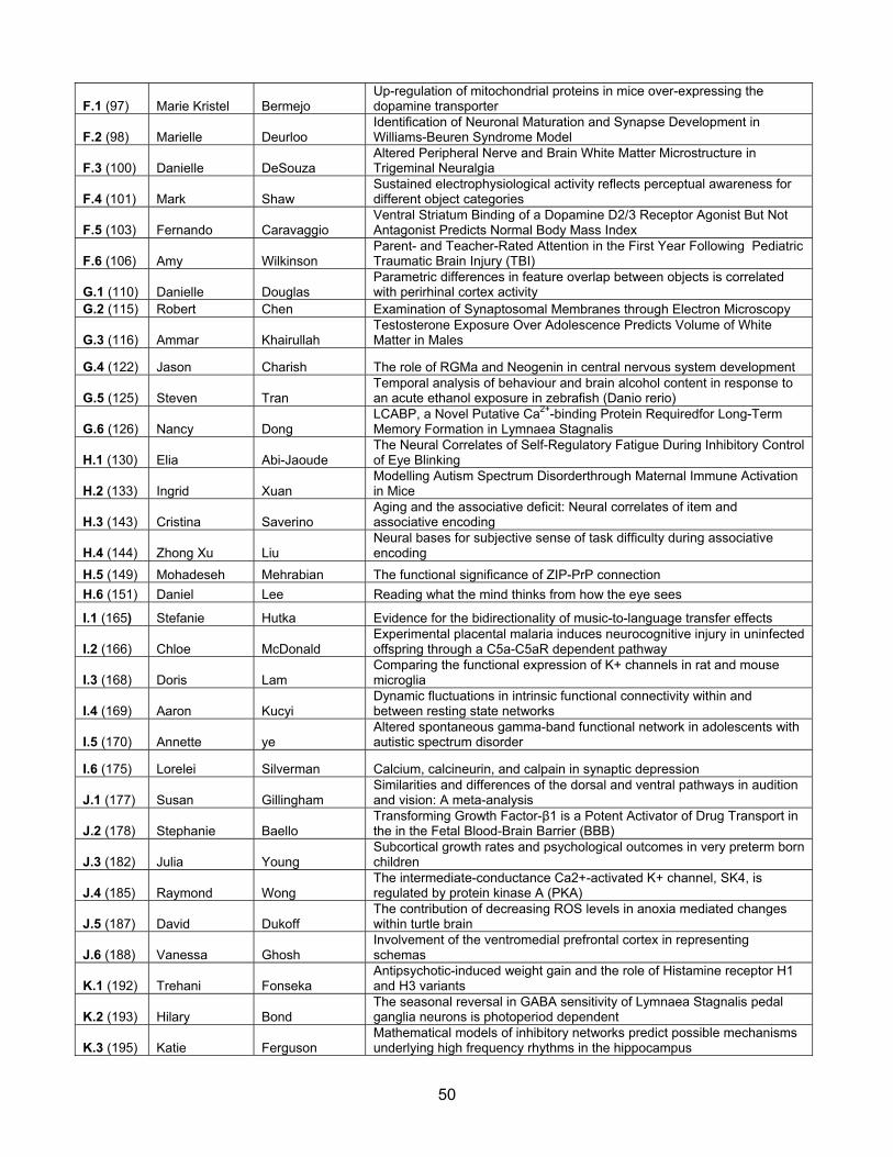
50
F.1 (97) Marie Kristel Bermejo Up-regulation of mitochondrial proteins in mice over-expressing the dopamine transporter
F.2 (98) Marielle Deurloo Identification of Neuronal Maturation and Synapse Development in Williams-Beuren Syndrome Model
F.3 (100) Danielle DeSouza Altered Peripheral Nerve and Brain White Matter Microstructure in Trigeminal Neuralgia
F.4 (101) Mark Shaw Sustained electrophysiological activity reflects perceptual awareness for different object categories
F.5 (103) Fernando Caravaggio Ventral Striatum Binding of a Dopamine D2/3 Receptor Agonist But Not Antagonist Predicts Normal Body Mass Index
F.6 (106) Amy Wilkinson Parent- and Teacher-Rated Attention in the First Year Following Pediatric Traumatic Brain Injury (TBI)
G.1 (110) Danielle Douglas Parametric differences in feature overlap between objects is correlated with perirhinal cortex activity
G.2 (115) Robert Chen Examination of Synaptosomal Membranes through Electron Microscopy
G.3 (116) Ammar Khairullah Testosterone Exposure Over Adolescence Predicts Volume of White Matter in Males
G.4 (122) Jason Charish The role of RGMa and Neogenin in central nervous system development
G.5 (125) Steven Tran Temporal analysis of behaviour and brain alcohol content in response to an acute ethanol exposure in zebrafish (Danio rerio)
G.6 (126) Nancy Dong LCABP, a Novel Putative Ca2+-binding Protein Requiredfor Long-Term Memory Formation in Lymnaea Stagnalis
H.1 (130) Elia Abi-Jaoude The Neural Correlates of Self-Regulatory Fatigue During Inhibitory Control of Eye Blinking
H.2 (133) Ingrid Xuan Modelling Autism Spectrum Disorderthrough Maternal Immune Activation in Mice
H.3 (143) Cristina Saverino Aging and the associative deficit: Neural correlates of item and associative encoding
H.4 (144) Zhong Xu Liu Neural bases for subjective sense of task difficulty during associative encoding
H.5 (149) Mohadeseh Mehrabian The functional significance of ZIP-PrP connection
H.6 (151) Daniel Lee Reading what the mind thinks from how the eye sees
I.1 (165) Stefanie Hutka Evidence for the bidirectionality of music-to-language transfer effects
I.2 (166) Chloe McDonald Experimental placental malaria induces neurocognitive injury in uninfected offspring through a C5a-C5aR dependent pathway
I.3 (168) Doris Lam Comparing the functional expression of K+ channels in rat and mouse microglia
I.4 (169) Aaron Kucyi Dynamic fluctuations in intrinsic functional connectivity within and between resting state networks
I.5 (170) Annette ye Altered spontaneous gamma-band functional network in adolescents with autistic spectrum disorder
I.6 (175) Lorelei Silverman Calcium, calcineurin, and calpain in synaptic depression
J.1 (177) Susan Gillingham Similarities and differences of the dorsal and ventral pathways in audition and vision: A meta-analysis
J.2 (178) Stephanie Baello Transforming Growth Factor-β1 is a Potent Activator of Drug Transport in the in the Fetal Blood-Brain Barrier (BBB)
J.3 (182) Julia Young Subcortical growth rates and psychological outcomes in very preterm born children
J.4 (185) Raymond Wong The intermediate-conductance Ca2+-activated K+ channel, SK4, is regulated by protein kinase A (PKA)
J.5 (187) David Dukoff The contribution of decreasing ROS levels in anoxia mediated changes within turtle brain
J.6 (188) Vanessa Ghosh Involvement of the ventromedial prefrontal cortex in representing schemas
K.1 (192) Trehani Fonseka Antipsychotic-induced weight gain and the role of Histamine receptor H1 and H3 variants
K.2 (193) Hilary Bond The seasonal reversal in GABA sensitivity of Lymnaea Stagnalis pedal ganglia neurons is photoperiod dependent
K.3 (195) Katie Ferguson Mathematical models of inhibitory networks predict possible mechanisms underlying high frequency rhythms in the hippocampus
51
K.4 (198) Timothy Zeyl Event-related potential differences in response to cursor movement errors made under brain-computer interface and manual control
K.5 Ne Mu Nar Saw
The role of disrupted-in-schizophrenia 1 (DISC1), a key risk factor in major psychological disorders, in hippocampal synaptic palsticity and cognition
K.6 (203) Sarah Hutchings Recombinant Interleukin-4 Injectioninto the Brain Alters the Inflammatory Response in a Rat Model of Ischemic Stroke
L.1 (205) Durgaa Rajendran
Association between Inner Retinal Layer Thickness and Neuroretinal Function in Adolescents with Type 1 Diabetes and no Diabetic Retinopathy
L.2 (208) Zoey Wei-Chih Cheng The Role of p53 in Radiation-Induced Inhibition of Hippocampal Neurogenesis
L.3 (209) Nathan Kolla Elevated Monoamine Oxidase-A Binding in Highly Symptomatic Borderline Personality Disorder: An [11C] Harmine PET Investigation
L.4 (212) Monique Budani Identification ofGlucosylCeramideFlippase in Glycosphingolipid Biosynthesis
L.5 (213) Vincent Man Amygdala response to trustworthy versus untrustworthy faces is modulated by social context
L.6 (215) Rylan Allemang-Grand Altered brain development long before plaque deposition in the TgCRND8 mouse model of Alzheimer’s disease
M.1 (216) Laura Finkelberg Impact of Type 1 Diabetes on Cone Photoreceptor Function and Structure in Adolescents and Young Adults
M.2 (217) Dulcie Vousden Whole-brain mapping of behaviorally-induced neural activation in mice
M.3 (220) Andrew Barszczyk A Cell-Permeating Caltubin Peptide Enhances Neuronal Outgrowth To Promote In Vivo Recovery From Nerve Injury In A Mouse Model
M.4 Helen Chiang Interactome analysis of TDP-43 in a mouse model of amyotrophic lateral sclerosis
M.5 (233) Vladislav Sekulić Using model databases to determine dendritic distributions of Ih channels in oriens-lacunosum/moleculare hippocampal interneurons
M.6 (235) Samantha Mahabir The Effect of embryonic alcohol on amino acid neurotransmitters in zebrafish
N.1 (236) Soaleha Shams Early Social Deprivation alters Adult Behaviour and Brain Function in Zebrafish
N.2 (242) Liyan Zhao DCPIB protects neonatal hypoxic-ischemic injury through volume-regulated anion channel(VRAC)
N.3 (250) Beverly Francis Noradrenergic deficits contribute to impairment in the TgCRND8 mouse model of Alzheimer’s disease
N.4 (269) Sacha Larda A new lysine tagging approach for structure-function studies of GPCRs and large protein complexes by NMR
N.5 (270) Nicole Law Reciprocal Cerebrocerebellar Pathways in Pediatric Medulloblastoma
N.6 (273) Paul Whissell δGABAA receptors promote memory and neurogenesis in the dentate gyrus
O.1 (277) Jessica Arsenault Does the premotor cortex map articulatory features during speech perception?
O.2 (279) Sofia Raitsin
Effects of Glucocorticoids and Oxidative Stress on Monoamine Oxidase A Activity in the Prefrontal and Anterior Cingulate Cortex in Rodents: Implications for Major Depressive Disorder
O.3 (280) Michael Chang An optogenetic approach to investigate the role of interneurons in seizureinitiation in in vitro and in vivo models of epilepsy
O.4 (284) Danielle Cha The Rationale for Evaluating the Effect of Intranasal Insulin on Emotional Processing in Treatment Resistant MDD
O.5 (288) Jackie Liu The role of PAK signalling in the regulation of structural plasticity of dendritic spines
O.6 (224) Ekaterina Turlova Neuroprotection by Novel Molluscan Peptide in MPP+-induced Parkinson's Disease Model

52

53

The CPIN Participating Academic Units
Faculty of Arts and Science Cell & System Biology Computer Science Psychology Faculty of Dentistry Graduate Department of Dentistry Faculty of Medicine Biochemistry Laboratory Medicine & Pathobiology Medical Biophysics
Pharmacology & Toxicology Physiology Rehabilitation Science Institute of Biomaterials & Biomedical Engineering Institute of Medical Science Faculty of Pharmacy Graduate Department of Pharmaceutical Sciences Ontario Institute for Studies in Education Applied Psychology & Human Development
Thanks to our Sponsors
Raymond and Beverly Sackler Foundation