table of contents - college of pharmacy - university of
TRANSCRIPT


Table of Contents
Program Announcement 1
Alphabetical List of Patient Advocacy Groups & Sponsors 2
Floor Plan 3
Alphabetical List of Poster Presenters 4 - 5
Poster Abstracts 6 - 43
FRIDAY, FEB 28, 2020 | MCNAMARA ALUMNI CENTER | 9 a.m. – 2:30 p.m.
#RareDiseaseDay #UMNResearchz.umn.edu/rare2020
R A R E D I S E A S E D A Y
COLLEGE OF PHARMACY MEDICAL SCHOOL
9 a.m. – 10:30 a.m. Patient Advocacy Group Breakfast & Networking Session10:30 a.m. – 12:00p.m. Poster Symposium12 p.m. – 2:30 p.m. Formal Program
Video GreetingVideo GreetingIntroduction of Rare Disease Advisory CouncilKeynote
UMN President Joan T.A. GabelSenator Amy Klobuchar
Erica Barnes, RDAC AdministratorSarah Wicks, JD, MPH
FORMAL PROGRAM
EVENT AGENDA
The Rare Disease Treatment Approval Process: Balancing Gold Standard Evidence with Patient-Centered Flexibility
Keynote: Sarah Wicks, JD, MPH
Sarah Wicks is an associate at Hyman, Phelps & McNamara, P.C. where she works with sponsors to bring life-changing therapies to market, particularly for persons with rare disease. She is a graduate of the University of Maryland School of Law and Johns Hopkins School of Public Health. Through her efforts, Sarah has gained a growing appreciation for the value of patients’ experiences and the need for the patient voice to inform prod-uct development. She is dedicated to furthering the involvement of patients in rare disease drug development as well as advancing and accelerating the development of new breakthrough therapies for those with rare conditions.
1
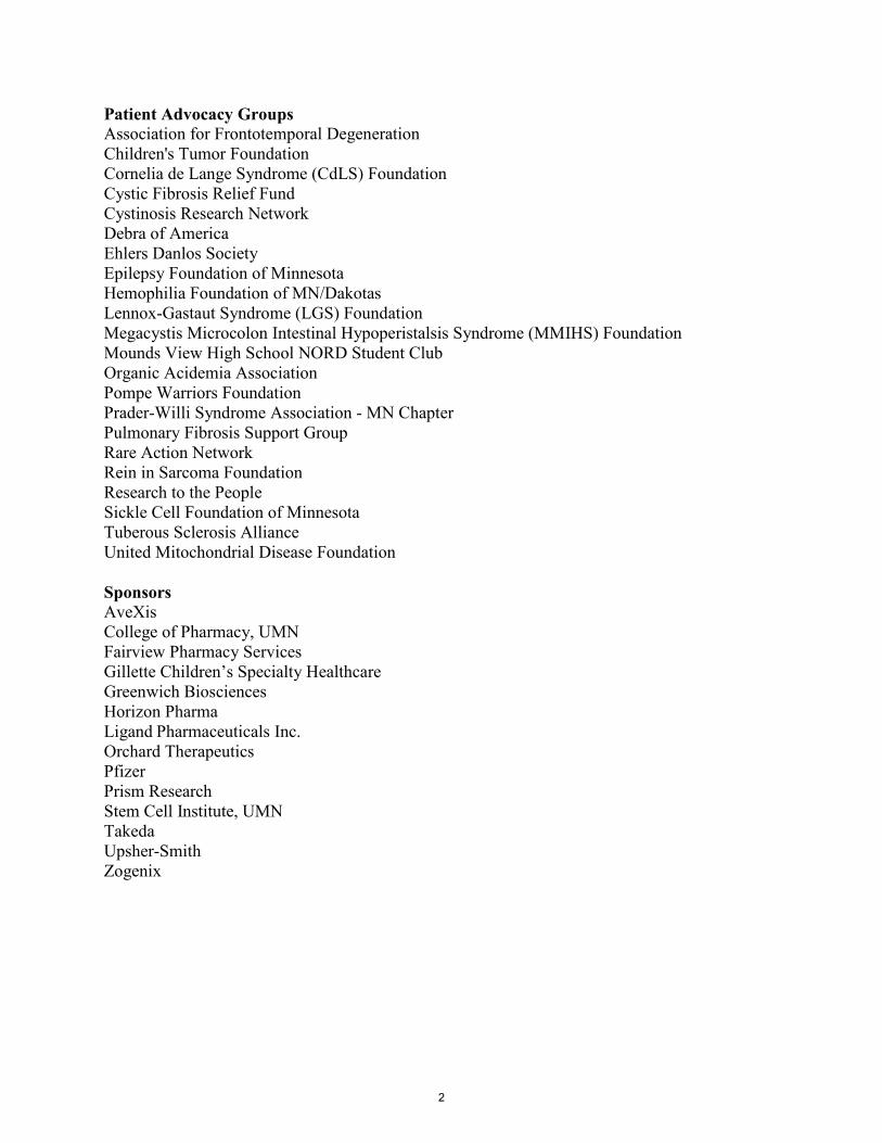
Patient Advocacy Groups Association for Frontotemporal Degeneration Children's Tumor Foundation Cornelia de Lange Syndrome (CdLS) Foundation Cystic Fibrosis Relief Fund Cystinosis Research Network Debra of America Ehlers Danlos Society Epilepsy Foundation of Minnesota Hemophilia Foundation of MN/Dakotas Lennox-Gastaut Syndrome (LGS) Foundation Megacystis Microcolon Intestinal Hypoperistalsis Syndrome (MMIHS) Foundation Mounds View High School NORD Student Club Organic Acidemia Association Pompe Warriors Foundation Prader-Willi Syndrome Association - MN Chapter Pulmonary Fibrosis Support Group Rare Action Network Rein in Sarcoma Foundation Research to the People Sickle Cell Foundation of Minnesota Tuberous Sclerosis Alliance United Mitochondrial Disease Foundation Sponsors AveXis College of Pharmacy, UMN Fairview Pharmacy Services Gillette Children’s Specialty Healthcare Greenwich Biosciences Horizon Pharma Ligand Pharmaceuticals Inc. Orchard Therapeutics Pfizer Prism Research Stem Cell Institute, UMN Takeda Upsher-Smith Zogenix
2

Ski-U-Mah Room A. The Association for Frontotemporal Degeneration B. Cystinosis Research Network C. Epilepsy Foundation of MN D. Tuberous Sclerosis Alliance E. Debra of America F. Greenwich Biosciences G. Pulmonary Fibrosis Support Group H. Lennox-Gastaut Syndrome (LGS) Foundation I. Zogenix J. Orchard Therapeutics
Memorial Hall K. Prader-Willi Syndrome Association - MN Chapter L. Prism Research M. Hemophilia Foundation of MN/Dakotas N. Sickle Cell Foundation of MN O. Organic Acidemia Association P. Cornelia de Lange Syndrome (CdLS) Foundation Q. Research to the People R. AveXis S. Ligand
Johnson Great Room T. Mounds View High School NORD Student Club U. Rare Action Network V. Fairview Pharmacy Services W. Gillette Children’s Specialty Healthcare X. Megacystis Microcolon Intestinal Hypoperistalsis Syndrome (MMIHS) Foundation Y. The Ehlers Danlos Society Z. Pompe Warrior Foundation Aa. Children’s Tumor Foundation Ab. Takeda Ac. Rein in Sarcoma Ad. The Cystic Fibrosis Relief Fund Ae. United Mitochondrial Disease Foundation
4 3 2 1 6 5
8 7
17
18
19
20
28
27
33
34
32
31 35
3637
38
Johnson Great Room Ski-U-Mah Room
29
30
3

Poster # First Name Last Name Abstract Title
28 Kelsie Becklin
Application of the Conditional Activation of Somatic Heterozygous Ewing Sarcoma (CASHEWS) system to model Ewing sarcoma in human iPSC
4 Lester Drewes Unexplained Ketoacidosis in Patients Under the Age of Five: Failure of Ketone Body Transport?
22 Patti Engel Rare Disease Qualitative Research: Methodologies for Conceptual Strength and Representativeness
19 Timothy Gauntner Hirschsprung Disease in an Infant with L1 Syndrome: Report of a New Case and a Novel L1CAM Variant
38 Dominic Gessler
rAAV Mediated Gene Therapy Completely Prevents and Reverses Canavan Disease at Early and Late Treatment Time Points in Different Mouse Models
37 Collin Gradin The Importance of Prompt Recognition of Neuromyelitis Optica Spectrum Disorder: A Case Report
7 Katie Gundry Antisense oligonucleotide (ASO) gene silencing reverses neurochemical abnormalities in SCA3 mice
1 Sara Healy Advancing Adult Leadership in Cystinosis Advocacy and Support
16 Robert Kaiser Preclinical Safety and Efficacy of in vivo Gene Therapy 17 Robert Kaiser A large animal model of PKU for gene editing
18 Robert Kaiser Activation of homology directed DNA repair plays key role in CRISPR mediated genome correction in HT1
21 Pete Kane Creating collaborative patient focused medical research cases
32 Sarah Kim
Quantification of cerebrospinal fluid chitotriosidase in a clinical laboratory is validated for use in diagnosis and clinical trials
36 Eric Klee
RNA Sequencing to Facilitate Genetic Diagnoses and Identify Putative Antisense Oligo Therapeutic Targets in Rare Disease Patients
34 Hongxin Lang Deletion of aryl hydrocarbon receptor in satellite cells affects diaphragm muscle in muscular dystrophic mouse
33 Sarah Lemmage Alport Syndrome Treatments and Outcomes Registry 27 Rebecca Madden Osteosarcoma modeling using induced pluripotent stem cells
8 Patty Maglalang The Evolution of Rescue Therapy for the Treatment of Seizure Emergencies
12 Santiago Martinez Cifuentes
Modeling progeroid cardiovascular disease using patient-derived induced pluripotent stem cells
31 Sean McSweeney
An international Challenge to use Artificial Intelligence to define the state of the art in kidney and kidney tumor segmentation in CT imaging
11 D'anna Nelson
Eccentric contraction causes rapid loss of microtubule organization in mdx skeletal muscle expressing mini- or micro-dystrophins
14 Michelle Ng MRI Gadolinium Volume in Cerebral Adrenoleukodystrophy: A Novel Biomarker to Quantify Cerebral Inflammation
15 Michelle Ng
Impact of extended post-HSCT enzyme replacement therapy (ERT) on linear growth in mucopolysaccharidosis type-1H (MPS-1H)
4
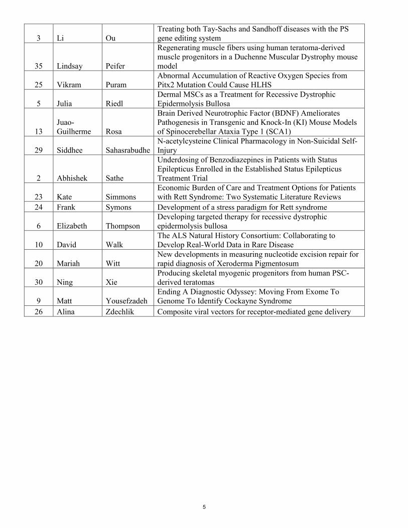
3 Li Ou Treating both Tay-Sachs and Sandhoff diseases with the PS gene editing system
35 Lindsay Peifer
Regenerating muscle fibers using human teratoma-derived muscle progenitors in a Duchenne Muscular Dystrophy mouse model
25 Vikram Puram Abnormal Accumulation of Reactive Oxygen Species from Pitx2 Mutation Could Cause HLHS
5 Julia Riedl Dermal MSCs as a Treatment for Recessive Dystrophic Epidermolysis Bullosa
13 Juao-Guilherme Rosa
Brain Derived Neurotrophic Factor (BDNF) Ameliorates Pathogenesis in Transgenic and Knock-In (KI) Mouse Models of Spinocerebellar Ataxia Type 1 (SCA1)
29 Siddhee Sahasrabudhe N-acetylcysteine Clinical Pharmacology in Non-Suicidal Self-Injury
2 Abhishek Sathe
Underdosing of Benzodiazepines in Patients with Status Epilepticus Enrolled in the Established Status Epilepticus Treatment Trial
23 Kate Simmons Economic Burden of Care and Treatment Options for Patients with Rett Syndrome: Two Systematic Literature Reviews
24 Frank Symons Development of a stress paradigm for Rett syndrome
6 Elizabeth Thompson Developing targeted therapy for recessive dystrophic epidermolysis bullosa
10 David Walk The ALS Natural History Consortium: Collaborating to Develop Real-World Data in Rare Disease
20 Mariah Witt New developments in measuring nucleotide excision repair for rapid diagnosis of Xeroderma Pigmentosum
30 Ning Xie Producing skeletal myogenic progenitors from human PSC-derived teratomas
9 Matt Yousefzadeh Ending A Diagnostic Odyssey: Moving From Exome To Genome To Identify Cockayne Syndrome
26 Alina Zdechlik Composite viral vectors for receptor-mediated gene delivery
5

Poster Number: 1 First Author: Maya Doyle MSW, PhD Contributing Authors: Maya Doyle MSW, PhD Author Affiliations: Quinnipiac University Subject Area: Cystinosis Keywords: Cystinosis Abstract Title: Advancing Adult Leadership in Cystinosis Advocacy and Support Abstract: Cystinosis is a rare metabolic disease that causes the amino acid, cystine, to
accumulate throughout the body. The disease affects every cell in the body, with particular impact on kidneys, eyes, and muscles. It is now typically diagnosed in early childhood. Thus, diagnosed patients have been aware of their illness from as early as they can remember. Earlier diagnosis and improved treatments over the last 35 years - including kidney transplant, immunosuppression drugs, and cysteamine treatments - have extended the lifespans of individuals with Cystinosis from adolescence well into adulthood.(Doyle & Werner-Lin, 2015; Nesterova & Gahl, 2013) A clinical trial of gene therapy for Cystinosis is now in progress (CITE). In the past, individuals with Cystinosis often died in childhood or adolescence. There is now a generation of Cystinosis patients who are surviving and thriving into adulthood, yet still face many challenges including disease progression and complications, and the impact of the disease and its treatments on their daily lives and decision-making. Advocacy for support, improved treatments, and ongoing research has until now been the task of caregivers. Adults with Cystinosis are now taking an active role in advocacy for themselves and for their community.
6

Poster Number: 2 First Author: Abhishek G. Sathe1,2 Contributing Authors: Lisa D. Coles1,2; Ellen Underwood3; Jordan J. Elm3; Robert Silbergleit4; James
Chamberlain5; Jaideep Kapur6,7; Hannah R. Cock8; Nathan B. Fountain6; Shlomo Shinnar9; Daniel H. Lowenstein10; Robin A. Conwit11; Thomas P. Bleck12; and James C. Cloyd1,2
Author Affiliations: 1: Department of Experimental and Clinical Pharmacology, University of Minnesota; 2: Center for Orphan Drug Research, University of Minnesota; 3: Department of Public Health Science, Medical University of South Carolina; 4: Department of Emergency Medicine, University of Michigan; 5: Division of Emergency Medicine, George Washington University; 6: Department of Neurology, University of Virginia; 7: Department of Neuroscience, University of Virginia; 8: St. Georges University of London and St. Georges University Hospitals NHS Foundation Trust; 9: Montefiore Medical Center, Albert Einstein College of Medicine; 10: Department of Neurology, University of California; 11: National Institute of Neurological Disorders and Stroke, National Institutes of Health; 12: Feinberg School of Medicine, Northwestern University.
Subject Area: Neurological Keywords: Status Epilepticus, Benzodiazepines, Dose, Emergency Medicine Abstract Title: Underdosing of Benzodiazepines in Patients with Status Epilepticus Enrolled
in the Established Status Epilepticus Treatment Trial Abstract: Early termination of status epilepticus (SE) requires administration of an adequate
benzodiazepine dose by an appropriate route. Prior reports suggest that benzodiazepine dosing in SE often differs from published guidelines. We describe patterns of diazepam (DZP), lorazepam (LZP), and midazolam (MDZ) use in prehospital and emergency department (ED) settings prior to enrollment in a trial of patients with benzodiazepine-refractory status epilepticus. Benzodiazepine dosing information was collected from adults and children prior to enrollment in the Established Status Epilepticus Treatment Trial (ESETT), a multicenter study of 2nd-line drug therapy in benzodiazepine-refractory SE. Data on individual and cumulative benzodiazepine doses, number of doses, timing, setting, and routes of administration were analyzed. Settings included prior to ambulance arrival, emergency medical service (EMS), and ED. Published guidelines served as the basis for defining recommended doses. There were 1217 benzodiazepine administrations (693 LZP, 415 MDZ, 109 DZP) given to 478 patients. Benzodiazepine use varied by setting and route: 71.6% of DZP doses were given prior to EMS arrival of which 92.3% were by the rectal route, 67.7% of MDZ doses were administered by EMS personnel either by the intramuscular (40.6%) or intravenous (34.5%) route, while 90.3% of LZP doses were administered in the ED mainly intravenously (97%). For the first administered dose of the first benzodiazepine, 86.6% of DZP, 19.3% of MDZ and 23.9% of LZP met the minimum dose recommended by published guidelines. Subsequent administrations continued the pattern of smaller than recommended and fragmented dosing.
Benzodiazepine underdosing in the EMS and ED was common in this geographically diverse set of patients who failed benzodiazepine treatment and were subsequently enrolled in ESETT. This phenomenon may contribute to reduced efficacy, potentially resulting in prolongation of SE.
7
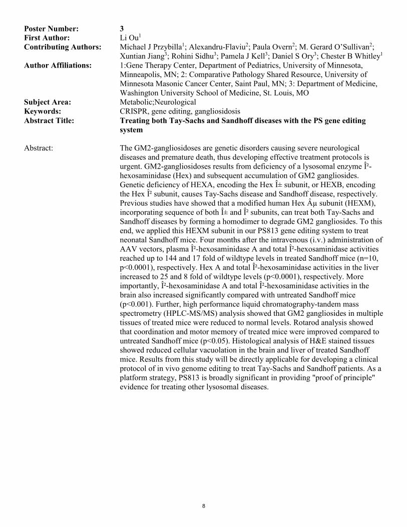
Poster Number: 3 First Author: Li Ou1 Contributing Authors: Michael J Przybilla1; Alexandru-Flaviu2; Paula Overn2; M. Gerard O’Sullivan2;
Xuntian Jiang3; Rohini Sidhu3; Pamela J Kell3; Daniel S Ory3; Chester B Whitley1 Author Affiliations: 1:Gene Therapy Center, Department of Pediatrics, University of Minnesota,
Minneapolis, MN; 2: Comparative Pathology Shared Resource, University of Minnesota Masonic Cancer Center, Saint Paul, MN; 3: Department of Medicine, Washington University School of Medicine, St. Louis, MO
Subject Area: Metabolic;Neurological Keywords: CRISPR, gene editing, gangliosidosis Abstract Title: Treating both Tay-Sachs and Sandhoff diseases with the PS gene editing
system Abstract: The GM2-gangliosidoses are genetic disorders causing severe neurological
diseases and premature death, thus developing effective treatment protocols is urgent. GM2-gangliosidoses results from deficiency of a lysosomal enzyme β-hexosaminidase (Hex) and subsequent accumulation of GM2 gangliosides. Genetic deficiency of HEXA, encoding the Hex α subunit, or HEXB, encoding the Hex β subunit, causes Tay-Sachs disease and Sandhoff disease, respectively. Previous studies have showed that a modified human Hex µ subunit (HEXM), incorporating sequence of both α and β subunits, can treat both Tay-Sachs and Sandhoff diseases by forming a homodimer to degrade GM2 gangliosides. To this end, we applied this HEXM subunit in our PS813 gene editing system to treat neonatal Sandhoff mice. Four months after the intravenous (i.v.) administration of AAV vectors, plasma β-hexosaminidase A and total β-hexosaminidase activities reached up to 144 and 17 fold of wildtype levels in treated Sandhoff mice (n=10, p<0.0001), respectively. Hex A and total β-hexosaminidase activities in the liver increased to 25 and 8 fold of wildtype levels (p<0.0001), respectively. More importantly, β-hexosaminidase A and total β-hexosaminidase activities in the brain also increased significantly compared with untreated Sandhoff mice (p<0.001). Further, high performance liquid chromatography-tandem mass spectrometry (HPLC-MS/MS) analysis showed that GM2 gangliosides in multiple tissues of treated mice were reduced to normal levels. Rotarod analysis showed that coordination and motor memory of treated mice were improved compared to untreated Sandhoff mice (p<0.05). Histological analysis of H&E stained tissues showed reduced cellular vacuolation in the brain and liver of treated Sandhoff mice. Results from this study will be directly applicable for developing a clinical protocol of in vivo genome editing to treat Tay-Sachs and Sandhoff patients. As a platform strategy, PS813 is broadly significant in providing "proof of principle" evidence for treating other lysosomal diseases.
8

Poster Number: 4 First Author: Alek Rudstrom1 Contributing Authors: Lester R. Drewes2 Author Affiliations: 1: MS2, Medical School Duluth, University of Minnesota, Duluth, MN;
2: Department of Biomedical Sciences, Medical School Duluth, University of Minnesota, Duluth, MN
Subject Area: Metabolic;Neurological Keywords: Ketoacidosis, Monocarboxylate Transporter-1, Neuropathology, DNA
Sequencing, Children Abstract Title: Unexplained Ketoacidosis in Patients Under the Age of Five: Failure of
Ketone Body Transport? Abstract: Monocarboxylate Transporter 1 (MCT1, member of the SLC16A transporter gene
family) is a carrier responsible for the movement of ketone bodies (b-hydroxybutyrate, acetoacetate), lactate and similar molecules across cell membranes. MCT1 is particularly abundant in the brain endothelium and astrocytes (neurovascular unit, blood-brain barrier) as well as localized around axons and in oligodendrocytes. While glucose is the brains primary fuel, during a starved metabolic state over half of the brains energy may come from ketones generated in the liver, distributed through the blood, and transported into the brain by MCT1. Additionally, MCT1 is central to the lactate-shuttle in which lactate from glia becomes an additional critical neuronal fuel during periods of acute demand. Dysfunctional MCT1 is associated with epilepsy, cognitive defects, neurodegeneration and neuroimaging abnormalities, suggesting an important, but poorly understood, neuropathological role. MCT1 deficiency was first identified by a Dutch group as a rare inborn error of metabolism causing non-diabetic ketoacidosis. In pediatric patients with dysfunctional MCT1 transport, increased liver ketogenesis during catabolic stress (flu, intestinal virus, food restriction) exceeded ketone transport and consumption and led to exceedingly elevated serum and urine ketones and a drastic drop in blood pH. Unlike diabetic ketoacidosis, blood glucose levels were normal or low and insulin was also normal. This unique and specific clinical presentation provides the opportunity to screen for likely MCT1-deficient patients using electronic health records (EHR) for cohort identification. Our IRB-approved project will use the TriNETx application with Fairviews EHR database (via the CTSI) to identify candidates, collect DNA samples, sequence the SLC16A1 gene, and identify possible MCT1 abnormalities. Our preliminary search has generated ~75 subjects meeting the phenotypic criteria. Through this study, we hope to identify a new cohort of MCT1-deficient patients, develop new diagnostic screening methods and novel therapies, and further research the metabolic basis for this disease.
9
Poster Number: 5 First Author: Julia Riedl1,3 Contributing Authors: Cindy Eide3, Megan Riddle3, Kirk Twaroski3, Markus Frank4, Natasha Frank4,
Jakub Tolar2,3 Author Affiliations: 1: Medical Scientist Training Program (MD/PhD), University of Minnesota,
Minneapolis, MN; 2: Department of Pediatrics, Division of Blood and Marrow Transplantation, University of Minnesota, Minneapolis, MN; 3: Stem Cell Institute, University of Minnesota, Minneapolis, MN 55455, USA; 4: Department of Dermatology, Harvard, Boston, MA
Subject Area: Connective Tissue and Musculoskeletal;Immunological and Auto-inflammatory Keywords: Recessive Dystrophic Epidermolysis Bullosa (RDEB), skin, mesenchymal stem
cells Abstract Title: Dermal MSCs as a Treatment for Recessive Dystrophic Epidermolysis
Bullosa Abstract: Recessive dystrophic epidermolysis bullosa (RDEB) is a blistering skin disease
caused by mutations in the COL7A1 gene encoding type VII collagen (C7). Mesenchymal Stromal Cells (MSCs) are currently being explored as a C7 therapeutic vector for these patients as they contribute to wound healing and C7 deposition. The mechanisms of MSC migration and immunomodulation in injured skin, as well as C7 deposition in RDEB models, is not completely understood. Specifically, ABCB5+ MSCs have shown promise in this area due to their immunoregulatory and anti-inflammatory markers such as the programmed-cell death protein PD-1 and anti-inflammatory cytokine IL-1RA. Our work has focused on elucidating the immunomodulation mechanisms of ABCB5+ MSCs via systemic injection into wounded, immunodeficient mice. Two-long term survivors were seen in the ABCB5+ group with significantly lower levels of macrophage marker CD68, which could contribute to an anti-inflammatory phenotype. Further in vitro characterization of the ABCB5+ cells will uncover a broader picture of secreted factors and genes expressed while future in vivo studies will focus on systemic injection of ABCB5+ cells into an RDEB mouse model to assess viability in treating this disease. In combination, these studies will uncover mechanisms of their immunomodulation and homing properties to ultimately improve and refine treatment for RDEB patients.
10

Poster Number: 6 First Author: Elizabeth Thompson Contributing Authors: Michael Pickett-Leonard; Jakub Tolar Author Affiliations: Department of Pediatrics, University of Minnesota Medical School Subject Area: Connective Tissue and Musculoskeletal Keywords: Epidermolysis Bullosa, targeted therapy, CRISPR screen Abstract Title: Developing targeted therapy for recessive dystrophic epidermolysis bullosa Abstract: Background: Recessive dystrophic epidermolysis bullosa (RDEB) is an inherited
genetic skin disorder that results in painful and debilitating skin blistering. RDEB results from biallelic mutation of the COL7A1 gene encoding type VII collagen (C7). Currently, there is no cure for RDEB and palliative care is the only option for most RDEB patients. Of further consequence, RDEB patients are prone to developing aggressive squamous cell carcinomas (SCC) at sites of chronic infection. For some RDEB patients, the upregulation of C7 expression can reduce blistering and potentially reduce the risk of SCC. Therefore, to identify methods of increasing C7 expression in the skin, we engineered a keratinocyte cell line with a fluorescent reporter for C7 expression. Methods: CRISPR/Cas9 gene editing was used to knock-in the tdTomato gene into the C-terminus of the COL7A1 gene. To identify genes that increase C7 expression, we performed a CRISPR activation (CRISPRa) screen to test the overexpression of 18,000 genes using 3 guide RNAs for each gene. The CRISPRa screen was followed by FACS analysis to collect high tdTomato expressing cells, and sequencing used to identify the genes that activated C7 expression. Validation of genes that upregulate C7 will be confirmed in RDEB patient keratinocytes using CRISPRa with the identified guide RNAs and Western blot for C7 expression. Results and Conclusion: This research has created a novel reporter cell line for investigating C7 expression and testing potential therapies for RDEB. In addition, we have identified new gene targets for increasing C7 expression. It is expected that this research will identify targetable genes or pathways to reduce blistering and even prevent SCC development in RDEB patients.
11

Poster Number: 7 First Author: Katie Gundry1 Contributing Authors: Orion Rainwater2, Annie Zalon3, Nick Stec3, Lynn Eberly4, Hayley McLoughlin3,
Gulin Oz1 Author Affiliations: 1: Center for Magnetic Resonance Research, University of Minnesota,
Minneapolis, MN; 2: Department of Laboratory Medicine and Pathology, University of Minnesota, Minneapolis, MN; 3: Department of Neurology, University of Michigan, Ann Arbor, MI; 4: School of Public Health, University of Minnesota, Minneapolis, MN
Subject Area: Neurological;Neuromuscular Keywords: Spinocerebellar Ataxia 3, Antisense Oligonucleotide (ASO) treatments, Gene
Silencing, Magnetic Resonance Spectroscopy Abstract Title: Antisense oligonucleotide (ASO) gene silencing reverses neurochemical
abnormalities in SCA3 mice Abstract: Background and Objective: Biomarkers in SCA3 are needed to monitor disease
progression and treatment response. ASO treatment mitigates neuropathology and rescues the motor phenotype in SCA3 mice. Here, we investigated if repeated ASO administration reverses neurochemical abnormalities in the cerebellum and brainstem of SCA3 YAC-Q84.2 mice expressing human mutant ATXN3 disease gene by 9.4 tesla (T) magnetic resonance spectroscopy (MRS). Methods: At 8 weeks of age, SCA3Q84/Q84 mice were treated with intracerebroventricular injection of ASO or vehicle solution; wild-type littermates (WT) were injected with vehicle solution. This procedure was repeated at 20 and 32 weeks of age. At 16 and 34 weeks, mice underwent longitudinal MRS of cerebellar and brainstem at 9.4T and metabolites quantified as described previously. MR data were acquired and analyzed in a blinded fashion. Group means were compared with one-way ANOVA with Tukey adjustment for multiple comparisons. Results: High-quality MR spectra yielded 15-16 reliably-quantified metabolites in both regions. Higher glutamine, and lower total N-acetylaspartate (tNAA) and total Choline (tCho) (p<0.05) in the vehicle treated SCA3Q84/Q84 mice vs WT mice were consistent with prior observations in patients with SCAs. ASO treated SCA3Q84/Q84 mice showed significant (p<0.05) rescue of tCho and Taurine relative to WT mice in the cerebellum at both time points. Partial reversals of abnormal Glutamine and tNAA level in the cerebellum and brainstem of the ASO treated mice towards WT levels were also observed. Discussion and Conclusion: Cerebellar and brainstem neurochemical trends in the SCA3Q84/Q84 mouse line were similar to those in patients. Changes in tCho may reflect oligodendrocyte and myelination abnormalities; tNAA concentration reflects neuronal health and high glutamine levels may indicate gliosis. ASO treatment fully or partially reversed select neurochemical abnormalities in SCA3Q84/Q84 mice, indicating that these measures may provide non-invasive biomarkers of treatment effects in future gene silencing trials in SCA3.
12

Poster Number: 8 First Author: Patty Maglalang Contributing Authors: James C. Cloyd Author Affiliations: Center for Orphan Drug Research, College of Pharmacy, University of
Minnesota, Minneapolis, MN Subject Area: Neurological Keywords: Rescue therapy; seizure clusters; status epilepticus; intranasal benzodiazepines Abstract Title: The Evolution of Rescue Therapy for the Treatment of Seizure Emergencies Abstract: Some patients with drug resistant-epilepsy have seizure clusters, which can
progress to prolonged seizures or status epilepticus. When patients or caregivers can identify such episodes, they could administer a therapy that interrupts seizure progression. In the 1980s, a team of clinical pharmacists, nurses, and epileptologists identified an unmet need for out-of-hospital management of seizure clusters. They designed an off-label therapy with rectally administered diazepam that triggered a new approach to treating seizure emergencies, now known as rescue therapy. Following reports on the off-label use of rectal diazepam, an industry-academic collaboration began development of a commercial rectal diazepam product in 1987. They obtained FDA orphan product designation in 1991 and completed an NIH-funded Phase III trial in 1996. The FDA approved diazepam rectal gel, Diastat, in 1997. Diastat approval launched a new approach to managing seizure emergencies. Given the objections to the use of rectal diazepam, clinicians began exploring off-label use of benzodiazepines by other routes. At the same time, industry and academic groups undertook formal development of rescue therapies. This led to the recent approval of an intranasal midazolam product, Nayzilam, and an NDA for an intranasal diazepam formulation is under review. Further, investigational intrapulmonary, intramuscular, buccal, and subcutaneous benzodiazepine products are in development.Rescue therapy has advanced the management of epilepsy, improved patient/family quality of life, and reduced health care costs. However, several barriers to effective use of rescue therapy remain. A 2014 Epilepsy Foundation survey found that only 30% of patients had a seizure emergency plan. The number of rescue therapies is expanding. Non-rectal routes of administration offer convenience and may lead to faster seizure cessation resulting in fewer emergency department visits and improved quality of life. Further improvements in drug delivery coupled with seizure prediction technology hold the promise of fundamentally changing our approach to treating epilepsy.
13

Poster Number: 9 First Author: Matt Yousefzadeh1,2,3
Contributing Authors: Jennifer Friedman4, Mariah Witt2,3, Lynne Bird5, Richard Haas4, Shira Robbins6, David Dimmock5, Shareef Nahas2, Shimul Chowhury5, Laura Niedernhofer1,2,3
Author Affiliations: 1: Department of Molecular Medicine and Center on Aging, The Scripps Research Institute, Jupiter, FL; 2: Institute on the Biology of Aging and Metabolism, University of Minnesota, Minneapolis, MN; 3: Department of Biochemistry, Molecular Biology and Biophysics, University of Minnesota, Minneapolis, MN; 4: Department of University of California System, San Diego, CA; 5: Rady Children's Institute for Genomic Medicine, San Diego, CA; 6: University of California System, San Diego, CA
Subject Area: Neurological Keywords: Cockayne Syndrome, DNA repair, DNA damage Abstract Title: Ending A Diagnostic Odyssey: Moving From Exome To Genome To Identify
Cockayne Syndrome Abstract: Molecular diagnosis of children with rare neurodegenerative and complex multi-
system disease is challenging especially when the phenotypic presentation deviates from what is reported in the literature. Next generation sequencing (NGS) techniques have improved diagnostic rates by providing an unbiased diagnostic approach guided, but not limited by, phenotype. Nevertheless, for rare disorders for which few variants may be reported and the phenotypic spectrum may not yet be fully elucidated, variants of uncertain significance (VUS) remain a vexing problem for NGS interpretation. Functional studies are often unavailable to investigate VUSs and even when available, are often beyond the diagnostic scope of a clinical testing lab. Cockayne Syndrome is an ultra-rare autosomal recessive disorder characterized by growth failure and multisystemic degeneration. Mutations in excision repair cross-complementation group 6 (ERCC6) accounts for the majority of cases. We report a child (INE4CC) with pre- and post-natal growth failure, progressive neurologic deterioration with multi-system involvement with bi-allelic novel variants in the ERCC6 gene. Pathogenicity of these variants was established by demonstrating reduced CSB protein levels, modest but significant reduction in unscheduled DNA synthesis following ultraviolet irradiation, and significant impairments in recovery of RNA synthesis post-UV irradiation of patient fibroblasts. The study confirms the pathogenicity of a previously undescribed upstream intronic variant, highlighting the power of genome sequencing plus functional studies to provide diagnosis in a child for whom a lengthy diagnostic odyssey, including exome sequencing, was previously unrevealed.
14

Poster Number: 10 First Author: David Walk1
Contributing Authors: Somers M1; Ferment V1; Maniak J1; Arcila-Londono X2; Olney N3; Gwathmey K4; Sherman A5, Macklin E5, Sinani E5, La T5, Hayat G6, Lunetta C7, Wymer J8, Heimann-Patterson T9, Ajroud-Driss S10
Author Affiliations: 1: University of Minnesota; 2: Henry Ford Hospital, Detroit MI; 3: Providence Health, Portland OR; 4: Virginia Commonwealth University, Richmond VA; 5. Neurological Clinical Research Institute, Boston MA; 6. St Louis University, St Louis MO; 7. NEMO Clinical Center, Milan; 8. University of Florida, Gainesville FL; 9. Temple University, Philadelphia PA; 10. Northwestern University, Chicago IL
Subject Area: Neurological Keywords: ALS; real-world data; natural history; clinical trial design Abstract Title: The ALS Natural History Consortium: Collaborating to Develop Real-World
Data in Rare Disease Abstract: The natural history of ALS is notably heterogeneous, complicating the design of
clinical trials and generalizability of their results. There is a recognized need for real-world data to better understand the natural history of ALS and provide real-world evidence to accelerate therapeutic approval. The most commonly referenced natural history data in ALS, the PRO-ACT database, is compiled from clinical trial data, and hence is limited by both selection bias and relatively short periods of observation. To better characterize the natural history of ALS, the ALS Natural History Consortium (ALS NHC) combines natural history data from all consenting people living with ALS (PALS) across multidisciplinary ALS treatment centers (MDCs). Nine participating sites in the US and Italy approach all PALS seen at their ALS MDCs with an opportunity to participate in ALS natural history data collection. Consenting PALS are assigned a global unique identifier (GUID) to allow de-identification of data. Demographic and longitudinal data from all clinic visits are recorded. Outcomes include ALS Functional Rating Scale Revised (ALSFRS-R), a validated functional outcome measure, and time to disease milestones, including loss of ambulation, loss of useful speech, loss of independence in activities of daily living (ADLs), gastrostomy, and nocturnal non-invasive ventilation. Over 1,000 PALS have been enrolled with over 90% participation of PALS seen in participating MDCs. Current data will be presented, including demographic data, rate of progression of ALSFRS-R, and time to milestones. Rate of progression will be compared with PRO-ACT data. Rare disease natural history data are needed for clinical trial design, real-world evidence collection, prognostication, and quality improvement. Clinic-based registries that endeavor to enroll all patients provide more representative information than clinical trial databases. The ALS NHC is a model for investigating rare diseases in the real-word setting.
15

Poster Number: 11 First Author: D'anna M Nelson1 Contributing Authors: A. Lindsay1,2; D.A. Lowe2; and J.M. Ervasti1 Author Affiliations: 1: Department of Biochemistry, Molecular Biology and Biophysics, University of
Minnesota, Minneapolis, MN 55455 2: Department of Rehabilitation Medicine, University of Minnesota, Minneapolis, MN 55455
Subject Area: Connective Tissue and Musculoskeletal Keywords: Duchenne muscular dystrophy, dystrophin, microtubules, gene therapy Abstract Title: Eccentric contraction causes rapid loss of microtubule organization in mdx
skeletal muscle expressing mini- or micro-dystrophins Abstract: Absence of dystrophin causes Duchenne muscular dystrophy, which is modeled
by the mdx mouse. Compared to wild type muscle, mdx muscle is highly susceptible to eccentric contraction-induced torque loss. Lack of dystrophin also influences the subsarcolemmal microtubule lattice. Wild type muscle has a rectilinear microtubule lattice of 90° intersections while mdx muscle most prominently shows loss of transverse microtubules. Transgenic expression of mini- or micro-dystrophin protects mdx skeletal muscle from torque loss caused by in vivo eccentric contractions and significantly improves microtubule organization. We now demonstrate that the microtubule lattice in transgenic mdx muscle expressing several different mini- and micro-dystrophins is modified soon after eccentric contractions. Mini-dystrophin mdx show partial, but significant loss of transverse microtubules immediately following eccentric contraction while micro-dystrophin mdx show complete loss of transverse microtubules. Eccentric contraction is necessary for loss of transverse microtubules in mdx skeletal muscle expressing mini- or micro-dystrophin because neither isometric contractions nor passive stretch affect microtubule organization. The microtubule lattice remains disorganized in transgenic mdx muscle expressing micro-dystrophin for up to ten days post-eccentric contraction. Most interestingly, the antioxidant N-acetylcysteine completely prevents eccentric contraction-induced loss of transverse microtubules in mdx muscle expressing micro-dystrophin. Our new data demonstrate that the microtubule lattice, restored in mdx muscle through expression of mini- or micro-dystrophins, can be rapidly disrupted by mechanical and redox perturbations.
16

Poster Number: 12 First Author: Santiago Martinez Cifuentes1 Contributing Authors: Claudia Trevilla-Garcia; Juan Carlos Rivera-Mulia Author Affiliations: Department of Biochemistry, Molecular Biology and Biophysics, University of
Minnesota Medical School, Minneapolis, MN Subject Area: Cardiac;Premature aging Keywords: Progeria, Replication Timing, hESCs, iPSCs Abstract Title: Modeling progeroid cardiovascular disease using patient-derived induced
pluripotent stem cells Abstract: Premature aging diseases are rare genetic disorders with features that resemble
natural aging. Hutchinson-Gilford Progeria Syndrome (HGPS) is one of the most studied and is caused by a point mutation in the LMNA gene that causes expression of a truncated lamin A protein, called progerin. Progerin accumulates at the nuclear membrane disrupting its morphology and is associated with alterations in 3D chromosome organization and temporal order of DNA replication (replication timing). Atherosclerosis and cardiac electrical alterations are late-onset pathologies of HGPS which lead to premature death. HGPS induced pluripotent stem cells (iPSCs) display a normal phenotype and replication timing program indistinguishable from normal human embryonic stem cells (hESCs) and iPSCs from healthy donors. However, upon differentiation, altered replication timing is recapitulated as well as expression of progerin and other cellular alterations (altered nuclear morphology and premature senescence). I will differentiate hESCs, iPSCs from healthy donors and HGPS patients towards cardiomyocytes and endothelial cells. Alterations in replication timing, gene expression and 3D genome organization will be explored by genome-wide next-generation sequencing. The success of this project will provide a better understanding of cellular alterations and insights about the alterations that can cause cardiovascular diseases in normal aging.
17
Poster Number: 13 First Author: Juao-Guilherme Rosa Contributing Authors: Juao-Guilherme Rosa, Carrie Sheeler, Ella Borgenheimer, Austin Ferro, Aaron
Mellesmoen, Marija Cvetanovic Author Affiliations: University of Minnesota, Minneapolis, MN Subject Area: Neurological Keywords: SCA1, BDNF Abstract Title: Brain Derived Neurotrophic Factor (BDNF) Ameliorates Pathogenesis in
Transgenic and Knock-In (KI) Mouse Models of Spinocerebellar Ataxia Type 1 (SCA1)
Abstract: Spinocerebellar ataxia type 1 (SCA1) is a fatal neurodegenerative disease caused
by a CAG trinucleotide repeat expansion in the Ataxin-1 (ATXN1) gene. SCA1 is characterized by progressive loss of motor coordination, cognitive decline, and premature death 10-20 years following clinical onset. Though disease onset usually occurs in patientâs mid-thirties, mutant ATXN1 is expressed in utero and throughout adulthood. This suggests that compensatory biological mechanisms limit the toxic effects of mutant ATXN1 in early life. We have previously shown that the transgenic Purkinje-cell specific SCA1 mouse model ATXN1[82Q] demonstrates elevated expression of brain derived neurotrophic factor (BDNF) in the cerebellum prior to motor symptom onset and decreased expression in later disease stages. Here, we investigate the therapeutic potential of chronic intraventricular (ICV) BDNF delivery in protecting against cognitive and motor deficits as well as pathology in SCA1 mouse models.
18

Poster Number: 14 First Author: Michelle Ng1
Contributing Authors: Paul J. Orchard1; Ashish Gupta1; Daniel Kenney-Jung2; David R. Nascene3; Troy C. Lund1
Author Affiliations: 1: Division of Pediatric Blood and Marrow Transplant, Department of Pediatrics, University of Minnesota, Minneapolis, MN 2: Department of Neurology, University of Minnesota Medical Center, Minneapolis, MN 3: Department of Diagnostic Radiology, University of Minnesota Medical Center, Minneapolis, MN
Subject Area: Metabolic Keywords: cALD; Gadolinium volume; Biomarker; HSCT Abstract Title: MRI Gadolinium Volume in Cerebral Adrenoleukodystrophy: A Novel
Biomarker to Quantify Cerebral Inflammation Abstract: Adrenoleukodystrophy (ALD) is caused by mutations within the X-linked
ABCD1 gene resulting in the inability to transport acylated very long chain fatty acids (VLCFA) into the peroxisome for degradation. VLCFA subsequently accumulate in tissues, including the central nervous system. Up to 40% of boys develop a severe, progressive demyelinating form of ALD, cerebral ALD (cALD), characteristically associated with a garland ring of gadolinium contrast enhancement on brain MRI. Gadolinium enhancement indicates blood-brain-barrier disruption and active inflammation. Hematopoietic cell transplant (HCT) is the only accepted therapy sufficient to halt neurologic progression and resolve the observed gadolinium enhancement, although the mechanism of disease arrest is unknown. We have previously shown that rapid donor neutrophil recovery after HCT facilitates MRI gadolinium resolution and early (30 days post HCT). We developed a method to quantify gadolinium on imaging and calculate a gadolinium volume (cm3). We evaluated gadolinium volume and other previously described imaging and transplant related biomarkers in 66 boys who underwent HCT. Univariate analysis showed day 30 gadolinium resolution was associated with the following pre-transplant parameters: lower gadolinium volume (7.3cm3 versus 2.7cm3, p=0.01), less gadolinium intensity (p=0.04), less extensive demyelination as determined by the Loes MRI scoring system (p=0.03) as well as lower pre-HCT plasma and cerebral spinal fluid chitotriosidase activity (p=0.04 for both), and faster neutrophil recovery post HCT (16 days vs. >16 days, p=0.03). Multivariate analysis revealed that only lower gadolinium volume was predictive of rapid gadolinium resolution at 30 days post-HCT (p=0.02). This is the first data to quantify the extent of cerebral inflammation in cALD as defined by gadolinium positivity and its resolution after HCT. Future studies should consider use of gadolinium volume prior to HCT as a useful biomarker for time to resolution.
19

Poster Number: 15 First Author: Michelle Ng1
Contributing Authors: Ashish Gupta1; Troy C. Lund1; Bradley S. Miller2; Paul J. Orchard1 Author Affiliations: 1: Department of Pediatric Blood and Marrow Transplant, University of
Minnesota, Minneapolis, MN 2: Department of Pediatric Endocrinology, University of Minnesota, Minneapolis, MN
Subject Area: Metabolic Keywords: MPS-1H; Post HSCT ERT; Growth; Anto-drug antibodies Abstract Title: Impact of extended post-HSCT enzyme replacement therapy (ERT) on linear
growth in mucopolysaccharidosis type-1H (MPS-1H) Abstract: In MPS-1H, a deficiency of the L-iduronidase enzyme leads to accumulation of
glycosaminoglycans (GAGs) in tissues and progressive multi-organ dysfunction. Hematopoietic stem cell transplant (HSCT) attenuates disease due to functional enzyme production and metabolic cross-correction from donor-derived hematopoietic cells. Short stature and growth delay are characteristic of MPS-1H. It has been previously shown that while HSCT improves linear growth potential, growth parameters still decelerate with increasing age, and that lower enzyme levels post-HSCT are associated with poorer growth. It is yet unknown whether augmentation with enzyme replacement therapy (ERT) post-HSCT has the potential to positively impact growth in these patients. Our aim was to evaluate the impact of ongoing ERT post-HSCT on linear growth in children with MPS-1H. We performed a retrospective analysis of patients with MPS-1H who underwent HSCT at our institution between 2012 and 2018 and continued ERT for at least 12 months post-HSCT. Growth velocity, height and height corrected for target height were compared to age matched controls at 6 months, 1 year and then annually post-HSCT, to last follow up. Secondary outcomes included development of anti-drug antibodies (ADA) to iduronidase. 9 patients met inclusion criteria, median age at HSCT was 12 months (4.2-23.5 months) and median duration of follow up was 3.5 years (range 2-7.6 years). All patients in the ERT group were continuing to receive ERT at last follow up. Those in the ERT group who were transplanted at <6 months of age appeared to have a growth advantage with growth velocity declining at a later age and slower rate, compared to controls. Additionally, they maintained their height trajectory compared to controls who had marked reduction in height z-scores over time. No patients developed persisting ADA. Though numbers are limited, our results suggest that post-HSCT ERT in MPS-1H may be beneficial in improving growth outcomes.
20

Poster Number: 16 First Author: Joseph B Lillegard1,2
Contributing Authors: Robert A Kaiser1,2, Clara T Nicolas1, Caitlin J VanLith1, Kari L Allen1, Raymond D Hickey2, Zeji Du1, Lori G Hillin1, Rebekah M Guthman1, Aditya Bhagwate1, Daniel O’Brien1, Jean-Pierre Kocher1
Author Affiliations: 1: Mayo Clinic, Department of Surgery, Rochester, MN 2: Midwest Fetal Care Center, Childrens Hospital of Minnesota, Minneapolis, MN
Subject Area: Metabolic Keywords: Tyrosinemia, gene therapy, lentiviral vector Abstract Title: Preclinical Safety and Efficacy of in vivo Gene Therapy Cure for Abstract: Hereditary tyrosinemia type 1 (HT1) is an autosomal recessive inborn error of
metabolism that is characterized by the inability to breakdown tyrosine, causing an accumulation of toxic metabolites in the liver and resulting in severe oxidative damage. These patients develop fibrosis, cirrhosis, high rates of hepatocellular carcinoma and ultimately liver failure at a very young age if left untreated. Our group has previously demonstrated that ex vivo lentiviral (LV) gene transfers followed by autologous transplantation of corrected hepatocytes is curative in both mouse and pig models of HT1. The ex vivo approach, although effective, requires a partial hepatectomy in liver-based diseases. In vivo liver-directed gene therapy is an attractive non-surgical option for the treatment of genetic hepatic disease. In this study, we evaluated both effectiveness and safety profile of in vivo liver-directed LV gene therapy for the treatment of HT1 in pigs. Pigs dosed with LV-FAH (n=4) showed only a transient immune response to the lentiviral vector and transgene. They were able to become weight stable after only four cycles on the maintenance medication, NTBC. Blood data from 150 days post treatment showed normalized tyrosine and liver function enzyme levels. No increase in alpha-fetoprotein was seen, indicating a lack of hepatocellular carcinoma. Immunohistochemistry of livers from dosed pigs showed nearly complete repopulation of the liver with FAH+ hepatocytes at 6 month post treatment, with a reversal of fibrosis by 12 months. An analysis of the lentivirus integration profile showed integration in the livers of all animals, in addition to a number of other tissues, such as lung, colon, and heart. Intronic integration seemed to be favored, with a lack of preference for highly expressed genes, CpG islands, or tumor coding genes.
21

Poster Number: 17 First Author: Joseph Lillegard, MD, PhD1,3
Contributing Authors: Lori G Hillin1, Kari L Allen1, Clara T Nicolas1, Daniel F Carlson2, Zeji Du1, Catherine W Kaiser1, Robert A Kaiser1,3, Raymond D Hickey1,4, Dennis A Webster2, Cary Harding5
Author Affiliations: 1: Division of Surgery, Mayo Clinic, Rochester, MN; 2: Recombinetics, Inc, Minneapolis, MN, 3: Midwest Fetal Care Center, Childrens Hospitals and Clinics of Minnesota, Minneapolis, MN, 4: Division of Molecular Medicine, Mayo Clinic, Rochester, MN, 5: Division of Molecular and Medical Genetics, Oregon Health and Science University, Portland, OR
Subject Area: Metabolic Keywords: Phenylketoniuria, CRISPR, animal model Abstract Title: A large animal model of PKU for gene editing Abstract: We aimed to create and characterize a humanized porcine model of
phenylketonuria (PKU) utilizing TALEN-mediated gene editing. An R408W mutation was introduced into the PAH gene of porcine fibroblasts to create homozygote clones and somatic cell nuclear transfer was used to create the first PAHR408W/R408W pigs. In the 5 and 3 direction of the point mutation we also inserted 250 bp of human sequence to allow for the first time ever a large animal model to be utilized for the development of a gene-editing construct for human use. Biochemical and histological analysis were performed to evaluate for the presence and activity of PAH. Three pregnancies resulted in six live born PAHR408W/R408W piglets that were hand-reared on a combination of commercially available bovine colostrum/milk replacer and a phenylalanine-free human infant formula (Phenex-1). Plasma phenylalanine concentrations were elevated at birth and severe hyperphenylalaninemia and neurologic abnormalities were observed by 36 hours of age in piglets consuming colostrum only. Treatment with Phenex-1 resulted in normalization of neurologic function and a decrease in plasma phenylalanine levels. Amino acid analysis on lysates of brain cortex showed lower concentrations of phenylalanine in the brain of animals treated with Phenex-1. Liver homogenates revealed no PAH enzyme activity in PAHR408W/R408W tissues (0.07±0.11nmol tyrosine/mg protein/hr) compared to wild-type controls (90.96±18.85 nmol tyrosine/mg protein/hr). Western blot analysis of the same homogenates detected normal amounts of PAH protein. Immunohistological analysis of PAHR408W/R408W piglet liver also revealed minimal background PAH expression compared to wild-type controls. Preliminary data suggests that the R408W mutation in pigs has resulted in the expression of nonfunctional PAH leading to complete PAH deficiency similar to what is seen in human PKU patients with the same mutation. Future work will include further characterization of the model as well as the development of translational gene editing techniques.
22

Poster Number: 18 First Author: Joseph Lillegard1,3,4
Contributing Authors: Caitlin J VanLith1, Clara T Nicolas1,2, Robert A Kaiser1,3, Whitney S Thompson1, Kari Allen1, Lori Hillin1, William Cao1, Raymond D Hickey4 and Gourish Mondal1
Author Affiliations: 1. Department of Surgery, Mayo Clinic, Rochester, MN. 2. Department of Surgery, University of Alabama Birmingham, Birmingham, AL 3. Midwest Fetal Care Center, Childrens Hospital of Minnesota, Minneapolis, MN. 4. Pediatric Surgical Associates, Minneapolis, MN.
Subject Area: Metabolic Keywords: Tyrosinemia, CRISPR, in utero Abstract Title: Activation of homology directed DNA repair plays key role in CRISPR
mediated genome correction in HT1 Abstract: Inborn errors of metabolism (IEMs) resulting from genetically inherited single
gene mutations cause liver dysfunction in neonates leading to chronic liver disease in adults. Approaches to gene therapy for the cure of IEMs have produced encouraging results, however high rates of off-target effects and overall inefficacy of fully differentiated adult hepatocytes for genome manipulation pose significant challenges. In this study, we provide evidence that in utero genome editing in a mouse model of hereditary tyrosinemia type-1 (HT1) can repopulate the liver with corrected hepatocytes and provide stable cure of the disease phenotype. An extensive gene expression analysis revealed the inherent characteristics of fetal liver versus adult liver that allows for such genomic correction. Fetal liver is comprised of proliferative hepatocytes with abundant expression of components that are involved in homology directed repair (HDR) of damaged DNA, which also serves a key role during genome correction by CRISPR editing. Homology directed genome correction requires the replicative phase of an actively proliferating cell that is abundant in the fetal liver. Injury to adult hepatocytes via controlled partial hepatectomy can trigger similar cell proliferative activity and expression of HDR components, thereby allowing successful HDR mediated genome correction following CRISPR editing in adult liver. This study signifies that triggering cell proliferation and HDR activity through partial hepatectomy may serve as a tool to improve CRISPR-mediated genome correction and provide the potential for curative therapy of inborn liver disease in adults.
23

Poster Number: 19 First Author: Timothy Gauntner1
Contributing Authors: Teresa Andreone2 Author Affiliations: 1: Internal Medicine Residency Program, Department of Medicine, University of
Minnesota, Minneapolis, MN; Department of Pediatrics, Cardinal Glennon Childrens Hospital, St. Louis University School of Medicine, St. Louis, MO
Subject Area: Neurological;Gastrointestinal Keywords: L1 syndrome; L1CAM; X-linked congenital hydrocephalus; Hirschsprung
disease; Genetics; Pediatrics Abstract Title: Hirschsprung Disease in an Infant with L1 Syndrome: Report of a New Case
and a Novel L1CAM Variant Abstract: L1 syndrome is an X-linked disorder that manifests across a wide spectrum of
phenotypes, including congenital hydrocephalus, adducted thumbs, spasticity, intellectual disability, spastic paraplegia, aphasia and agenesis of the corpus callosum. Mutations in the X-linked L1CAM gene, which encodes a neural cell adhesion molecule, are responsible for L1 syndrome. There are only 13 reported cases of patients with L1 syndrome who have confirmed L1CAM mutations and coincident Hirschsprung disease. Here we present a rare case of an infant with clinically diagnosed L1 syndrome who was found to have Hirschsprung disease during admission for a ventriculoperitoneal shunt infection. Subsequent genetic confirmation of L1 syndrome by targeted sequencing of the L1CAM gene revealed a novel pathogenic variant (c.934T>C; p.Cys312Arg). This case adds to a small number of reports of L1 syndrome with coincident HD, which further supports the notion that L1CAM gene mutations may be involved in the pathogenesis of HD. Furthermore, this case exemplifies that clinicians should have a high index of suspicion for HD when caring for patients with any form of L1 syndrome.
24
Poster Number: 20 First Author: Mariah Witt2,3
Contributing Authors: Matt Yousefzadeh1,2,3; Tania Rozgaja1; Amira Bargouthy1; Erin Wade1; Aimee Milota2,3; Laura Niedernhofer1,2,3
Author Affiliations: 1: Department of Molecular Medicine and Center on Aging, The Scripps Research Institute, Jupiter, FL 2: Institute on the Biology of Aging and Metabolism, University of Minnesota, Minneapolis, MN 3: Department of Biochemistry, Molecular Biology and Biophysics, University of Minnesota, Minneapolis, MN
Subject Area: Cancer;Neurological;Diagnostic Keywords: Xeroderma Pigmentosum, DNA repair, nucleotide excision repair Abstract Title: New developments in measuring nucleotide excision repair for rapid
diagnosis of Xeroderma Pigmentosum Abstract: Xeroderma Pigmentosum (XP) is an autosomal recessive disorder caused by a
defect in various genes which encode the Nucleotide Excision Repair (NER) complex, a DNA repair mechanism solely responsible for fixing bulky lesions in DNA which most commonly result from UV induced damage. Affecting 1 in 1,000,000 people, XP is an extremely rare disease which occurs throughout the world in men and women of all ethnicities. Patients with XP can experience a complicated combination of symptoms due to its pleiotropic nature. XP patients typically present with photosensitivity and hyperpigmentation in response to even minimal sun exposure. XP patients have a 10,000-fold increase in the incidence of skin cancer. Progressive neurological complications including weakened or missing reflexes, hearing loss, difficulties with speech, trouble with balance and walking, and cognitive decline can be present in some XP patients. Currently, there is no cure or treatment for XP let alone a national diagnostic center, thus an early diagnosis and aggressive sun avoidance is critical to manage the disease and reduce risk of skin cancer. Measurement of unscheduled DNA synthesis (UDS) after UV irradiation of patient cells is the gold standard for diagnosing NER-deficiency disorders. Historically, UDS was measured in patient dermal fibroblasts produced from a skin biopsy using radiolabeled nucleotides. More recently fluorescently-labelled nucleotides and microscopy are used to measure NER. However, both of these approaches are laborious, slow and require specialized equipment, impeding clinical utility of the assay. We have developed a UDS assay able to test a patient’s NER capacity by flow cytometry analysis of lymphocytes from blood, creating a rapid, sensitive and readily available assay to measure NER capacity and expedite diagnosis of suspected XP patients.
25

Poster Number: 21 First Author: Peter Kane Contributing Authors: Nina Sardesh Author Affiliations: Research to the People Community Subject Area: Connective Tissue and Musculoskeletal;Metabolic;Immunological and Auto-
inflammatory;Cancer;Neurological;Kidney Keywords: Bioinformatics, Genetic Sequencing, Open Source Collaboration Abstract Title: Creating collaborative patient focused medical research cases Abstract: Research to the People is a research initiative by SVAI, a 501(c)(3) non-profit
based in San Francisco, California. We host platforms for open collaboration for biomedicine research and gather bright minds to contribute to rare and complex disease cases Our community includes computational scientists, biologists, doctors, data analysts. We partner with patients to create robust datasets from tissue and blood samples. Tests and analyses through Research to the People and our partners are free. In most cases, a group of this community gathers together for a weekend to focus on problem tracks related to the case in focus. Research teams have delivered a wide range of results including: identifying new drug treatment pathways, gene mutations, and potential therapies. We also aim to work with genome sequencing partners to produce novel, open datasets for our research community. We take advantage of public resources like the NIH's Cancer Genome Atlas to supplement our research efforts.
26

Poster Number: 22 First Author: Patti Engel1
Contributing Authors: Skyler Jackson1, James E. Valentine2 Author Affiliations: 1: Engage Health, Inc., Eagan MN, 2:Phelps & McNamara PC, Washington DC. Subject Area: Qualitative Research Methodology Keywords: Qualitative Methodology, Saturation, Patient Experience, Quantifying Qualitative
Data, Like-concepts, FDA Guidance Abstract Title: Rare Disease Qualitative Research: Methodologies for Conceptual Strength
and Representativeness Abstract: Qualitative research data in patient voice is important in rare disease therapy
development and patients are increasingly called upon to define aspects of therapy that are important to them. Caution must be paid, as it is difficult to determine common concepts of importance in heterogenous populations and small sample sizes in rare conditions make it difficult to ensure that concepts of importance provided by research respondents are representative of the opinions of the larger group. In a rare degenerative neuromuscular condition, the authors attempted to address these issues through a novel methodology. Respondents were assessed by functional classification of disease to ensure similar physical abilities. Disease experts were consulted to provide concepts that were likely to be raised as important to each subgroup. Interviews were conducted with a baseline cohort of 5 patients for each functional category, eliciting important concepts in an open-ended manner, which were collected using patient voice. Concepts were reviewed by independent coders, who grouped like-concepts together, and added any that were provided by respondents that were not anticipated by disease experts. Subsequent cohorts of 5 patients for each functional category were recruited, and results compared to the baseline cohort. If a new concept was raised, content experts were consulted to determine if the concept was expected or important. If it was, it was deemed saturation had not been met, and an additional cohort was recruited for this functional category. Recruitment continued in this purposive manner until saturation had been met for each functional category. Using this method, the authors were able to gather qualitative data in the patient voice that showed like-concepts of importance to patients within functional subgroups of a heterogeneous rare neurodegenerative disease. Recruiting to saturation of response for each subgroup provided confidence that responses were representative of the larger population.
27

Poster Number: 23 First Author: Katrina Simmons Contributing Authors: Omar Dabbous1, Vanessa Taieb2, Emna Abdennadher3, Meryem Bouchemi3,
Justyna Chory4, Katarzyna Borkowska4, Veneta Georgieva5, Bryan E. McGill1, Thomas A. Macek1, Benit Maru1, Ramesh Arjunji1
Author Affiliations: 1: AveXis, Inc., Bannockburn, IL, United States; 2: Creativ-Ceutical, London, United Kingdom; 3: Creativ-Ceutical, Tunis, Tunisia; 4: Creativ-Ceutical, Krakow, Poland; 5: Creativ-Ceutical, Sofia, Bulgaria
Subject Area: Neuromuscular Keywords: Rett, burden, economic, treatment pattern Abstract Title: Economic Burden of Care and Treatment Options for Patients with Rett
Syndrome: Two Systematic Literature Reviews Abstract: Rett syndrome, a rare X-linked disorder caused by mutations in the MECP2 gene,
occurs predominantly in girls and causes severe developmental impairment. To assess the economic burden of care imposed on patients with Rett syndrome and their families, and clinical trials on therapeutic approaches for Rett syndrome.Two systematic literature reviews related to Rett syndrome were performed in June 2018: 1). Related to economic burden (Medline, Embase, Cochrane Library, and Database of Abstracts of Reviews and Effects); 2). Clinical trials (Medline, Embase, ClinicalTrials.gov, and Cochrane Library). The search on economic burden yielded 133 articles; intervention type and costs were extracted from 9, representing 4 studies. In the economic burden studies, enteral feeding and assisted walking increased the risk of respiratory-related hospital admissions, while length-of-stay was lower in younger patients. Mean recovery-stay after scoliosis-correcting surgery was 18.2 days and 12.3 days in each of two studies. Care integration improved outcomes and reduced costs. The search of clinical trials yielded 652 articles; efficacy and safety were extracted from 28, representing 20 studies (15 randomized controlled trials, 5 single-arm; N=882; follow-up 1-26 months). Of these, 19 focused on pharmacological symptom treatment; 1 examined environmental enrichment effects; none targeted the underlying cause. The most common primary endpoints were Rett syndrome Gross Motor Scale, Clinical Severity Score, Motor and Behavioral Assessment, and the Anxiety Depression and Mood Scale. Naltrexone, trofinetide, and mecasermin demonstrated clinical benefits versus placebo, but most treatments yielded no significant improvement. Clinical practice guidelines and treatment patterns data were limited. There are little data on costs of managing patients with Rett syndrome. Because available therapies manage only symptoms, there is demand for safe, effective treatments targeting the underlying cause (eg, gene therapy), which could improve quality of life and prognosis for patients with Rett syndrome.
28
Poster Number: 24 First Author: Frank Symons1
Contributing Authors: Breanne Byiers1; Alyssa Merbler1; Kristin Frenn2; Chantel Barney2 Author Affiliations: 1: Department of Educational Psychology, University of Minnesota, Minneapolis,
MN 2: Gillette Children's Specialty Healthcare, St. Paul, MN
Subject Area: Neurological Keywords: Rett syndrome, salivary biomarkers, heart rate variability, stress physiology Abstract Title: Development of a stress paradigm for Rett syndrome Abstract: Rett syndrome (RTT) is a neurodevelopmental disorder associated with severe
communication and motor impairments, and comorbid health problems. Studies in animal models of RTT have shown abnormal stress responses, particularly elevated cortisol responses. There is limited information on stress physiology in the clinical population, however, as identifying stress paradigms for this population is challenging due to ethical and practical limitations. In the current study, we investigated changes in three salivary biomarkers (cortisol [CORT], alpha amylase [sAA], and betaendorphin [BE] and concurrent changes in heart rate (HR) and heart rate variability (HRV) during a research visit consisting of two standardized physical examinations. The goal of the study was to determine whether this protocol would elicit detectable physiological responses in this population. Data from 22 participants with RTT (aged 4- 38 years) were included. Saliva samples were collected at three timepoints and HR/HRV were collected continuously. The results showed that CORT and BE increased significantly during the visit, whereas sAA did not show consistent changes. Changes in CORT, but not BE, differed significantly by age. Changes in CORT were significantly associated with changes in HR, whereas changes in BE were significantly associated with changes HRV. These preliminary results show that this protocol may be a useful tool for evaluating stress physiology in RTT. Additional work is needed to replicate these effects and to determine the independent effects of the two examinations.
29

Poster Number: 25 First Author: Vikram Puram1
Contributing Authors: Anant Naik2; Jack Hedberg3; Dr. James Harmon4 Author Affiliations: 1: University of Minnesota Medical School, Minneapolis, MN
2: The Carle Illinois College of Medicine, University of Illinois at Urbana-Champaign, Champaign, IL 3: The Ohio State University College of Medicine, Columbus, OH 4: Department of Surgery, University of Minnesota, Minneapolis, MN
Subject Area: Cardiac Keywords: Hypoplastic Left Heart Syndrome (HLHS), Congenital Heart Disease, Cardiac
Embryology, Genetics. Abstract Title: Abnormal Accumulation of Reactive Oxygen Species from Pitx2 Mutation
Could Cause HLHS Abstract: Hypoplastic Left Heart Syndrome (HLHS) is a highly rare congenital heart defect
in which the left side of the heart is critically underdeveloped. At the cellular level, changes in ventricular structure include myocyte growth and myocyte apoptosis. Ultimately, this progressive remodeling can result in heart failure and, in HLHS, premature death. Recent progress in understanding these mechanisms of myocardial remodeling has led to evidence that reactive oxygen species (ROS) and oxidative stress play a central role in regulating the phenotype of cardiac myocytes. We hypothesize that ROS-mediated developmental destabilization due to pitx2 dysfunction in the left ventricle, coupled with the low and high strain placed on the interventricular septum (IV) and left ventricular (LV) wall during cardiac contraction, respectively, causes HLHS. Genetic tests of mouse models and in vitro studies of cardiomyocytes confirm this proposed progression. Further investigation could open the door to curative gene therapy and prevent many years of suffering and complications with surgical procedures for affected infants.
30

Poster Number: 26 First Author: Alina C. Zdechlik1
Contributing Authors: Yungui He2, Eric J. Aird1, Wendy R. Gordon1, Daniel Schmidt2 Author Affiliations: 1: Dept. of Biochemistry, Molecular Biology & Biophysics; 2: Dept. of Genetics,
Cell Biology & Developmental Biology, University of Minnesota, Minneapolis, MN 55455
Subject Area: Connective Tissue and Musculoskeletal Keywords: AAV, gene therapy Abstract Title: Composite viral vectors for receptor-mediated gene delivery Abstract: Modern gene therapy is limited by lack of specific delivery vectors, rather than
lack of therapeutic targets. Viral vectors currently used in clinical trials, such as adeno-associated virus (AAV), infect a broad range of tissues naturally targeted by the wild-type virus. In the clinic, this can result in ectopic expression in undesirable cell types and/or low expression in target tissues. To remedy this, engineering infection to be antigen-specific has been a goal of the field for almost 30 years. Past engineering efforts have grafted a targeting moiety (i.e. scFv, DARPin) into the protein capsid. While this method has resulted in targeted infection, each new antigen requires a new engineered capsid. We have developed a method of creating antigen-specific AAVs using a single engineered capsid containing a DNA binding domain (HUH tag). These HUH-AAVs can then be attached to DNA-conjugated antibodies with high efficiency. Our strategy is additionally unique in that it uses a novel flexible surface loop, is compatible with all AAV capsid proteins, and is fully covalent. We have shown that a single HUH-AAV can be retargeted to generically any antigen; each of the antibodies we have used thus far has worked as expected in immortalized and primary cells. I will present data showing targeted delivery of fluorescent markers to multiple subtypes of blood and neural cells. Future work will utilize pre-existing complex genomes to perform targeted editing and cellular logic entirely in vivo. Modern gene therapy is limited by lack of specific delivery vectors, rather than lack of therapeutic targets. Viral vectors currently used in clinical trials, such as adeno-associated virus (AAV), infect a broad range of tissues naturally targeted by the wild-type virus. In the clinic, this can result in ectopic expression in undesirable cell types and/or low expression in target tissues. To remedy this, engineering infection to be antigen-specific has been a goal of the field for almost 30 years. Past engineering efforts have grafted a targeting moiety (i.e. scFv, DARPin) into the protein capsid. While this method has resulted in targeted infection, each new antigen requires a new engineered capsid. We have developed a method of creating antigen-specific AAVs using a single engineered capsid containing a DNA binding domain (HUH tag). These HUH-AAVs can then be attached to DNA-conjugated antibodies with high efficiency. Our strategy is additionally unique in that it uses a novel flexible surface loop, is compatible with all AAV capsid proteins, and is fully covalent. We have shown that a single HUH-AAV can be retargeted to generically any antigen; each of the antibodies we have used thus far has worked as expected in immortalized and primary cells. I will present data showing targeted delivery of fluorescent markers to multiple subtypes of blood and neural cells. Future work will utilize pre-existing complex genomes to perform targeted editing and cellular logic entirely in vivo.
31

Poster Number: 27 First Author: Rebecca Madden1
Contributing Authors: Kelsie Becklin1; Tony DeFeo1; Branden Moriarity1,2,3; Beau Webber1,2,3,4 Author Affiliations: 1: Department of Pediatrics, University of Minnesota, Minneapolis, MN, USA; 2:
Masonic Cancer Center, University of Minnesota, Minneapolis, MN, USA; 3: Center for Genome Engineering, University of Minnesota, Minneapolis, MN, USA; 4: Stem Cell Institute, University of Minnesota, Minneapolis, MN, USA
Subject Area: Cancer Keywords: Osteosarcoma; Disease models; Induced pluripotent stem cells Abstract Title: Osteosarcoma modeling using induced pluripotent stem cells Abstract: Osteosarcoma (OS) is the most common bone cancer in adolescents and young
adults. Long term survival is 70% for localized disease; however, if distal metastases are present at the time of diagnosis 5-year survival drops to <30%. Despite progress in our understanding of OS biology, current treatments have changed little in decades and continue to rely on tumor resection and combination chemotherapy. Only prototypical cancer genes, such as TP53, RB1, and MYC have been definitely implicated in OS; with TP53 being dysregulated in >90% of all OS patients. Although a number of other candidate genes have been nominated it is difficult to confirm their role without proper validation. A pressing problem in developing new treatments for OS is a lack of disease models. To surmount these challenges our lab is developing a new model for OS using human induced pluripotent stem cells (iPSCs). Using CRISPR-Cas, we engineered iPSCs to carry TP53 and RB1 mutations and differentiated these cells into mesenchymal stromal cells (MSCs) and then into osteoblasts (OB), the suspected cell(s) of origin for OS. We observed increased proliferation of MSC and OB carrying TP53-null and TP53/RB1-null mutations which was further increased by overexpression of cMYC/hRAS oncogenes. In soft agar assays of anchorage independent growth, we observed colony formation in TP53/RB1-null mutant MSC and OB, and colony formation was further enhanced in TP53/RB1-null MSC and OB expressing cMYC/hRAS. To test the tumorigenicity, the engineered MSC and OB were injected into the calcaneal bones of immunodeficient mice. Tumors were observed in mice that received TP53/RB1-null or TP53/RB1-null + cMYC/hRAS cells, with the latter showing accelerated tumor formation and growth. Studies are underway to characterize these tumors and determine how closely they mimic patient-derived OS tumors.
32

Poster Number: 28 First Author: Kelsie L. Becklin1,2,3,4
Contributing Authors: Anthony DeFeo1,2,3; Branden S. Moriarity1,2,3,4,5; Beau R. Webber1,2,3,4,5 Author Affiliations: 1: Department of Pediatrics, University of MN, 2: Center for Genome
Engineering, University of MN 3: Masonic Cancer Center, University of MN 4: College of Veterinary Medicine, University of MN 5:Stem Cell Institute, University of MN
Subject Area: Cancer Keywords: Ewing sarcoma modeling in iPSCs Abstract Title: Application of the Conditional Activation of Somatic Heterozygous Ewing
Sarcoma (CASHEWS) system to model Ewing sarcoma in human iPSC Abstract: Ewing sarcoma (ES) is a rare, deadly bone cancer that occurs nearly exclusively
in adolescents and young adults. Five-year survival rates have plateaued, calling for a desperate need in new strategies to address basic questions surrounding ES etiology, which may illuminate therapeutic vulnerabilities. Unusual in its pathogenesis, ES is driven primarily by an aberrant transcription factor resulting from the fusion of the EWSR1 and FLI1 genes, also known as EWS/FLI. Despite the driving factor of ES being identified, little has been elucidated about the cellular state required for ES permissibility. This discrepancy is largely due to the inherent toxicity of the EWS/FLI fusion protein in most cell types. It is thought that ES cell of origin is either neural crest cells (NCC) or mesenchymal stromal cells (MSC), but conclusive evidence to either has been yet to be produced. To better understand the surrounding cellular and genomic context required for ES initiation and development we have developed a genetrap system, Conditional Activation of Somatic Heterozygous Ewing Sarcoma (CASHEWS) which allows for conditional expression of the EWS/FLI fusion protein using the native EWSR1 promoter. The goals of this project are to develop a new ES model using induced pluripotent stem cells (iPSC) and the CAHSEWS system to create a ‘bottom-up’ model that will provide a new platform for the in-depth study of ES initiation and development as it relates to permissible cell types, specifically MSC and NCC. To date we have generated one iPSC line containing the native translocation with the CASHEWS system. Current efforts are focusing on differentiating the cell line into MSC and NCC and performing functional and molecular tests on each cell type to compare the iPSC derived ES model to current model systems and we aim to identify the developmental context surrounding permissibility to the EWS/FLI transcription factor.
33

Poster Number: 29 First Author: Siddhee A. Sahasrabudhe1,2
Contributing Authors: Coles LD1,2, Mishra U1,2, Cloyd JC1,2, Cullen KR3, Kartha RV1,2 Author Affiliations: 1: Center for Orphan Drug Research, University of Minnesota; 2: Department of
Experimental and Clinical Pharmacology, College of Pharmacy, University of Minnesota; 3: Department of Psychiatry, Medical school, University of Minnesota
Subject Area: Neurological Keywords: X-Linked Adrenoleukodystrophy, N-acetylcysteine, Pharmacokinetic analysis Abstract Title: N-acetylcysteine Clinical Pharmacology in Non-Suicidal Self-Injury Abstract: Non-suicidal self-injury (NSSI) is a psychiatric disorder described as intentional
injuring of one’s own body without suicidal intent. In Minnesota, about 22% adolescent girls have a tendency to self-harm. Unfortunately, there is no approved treatment for NSSI. N-acetylcysteine (NAC) has been used in psychiatric disorders, although there is no consensus regarding its clinical benefits. We hypothesize that oral NAC in patients with NSSI can reduce episodes of self-harm in relation to NAC exposure. Toward this end we have developed an analytical method to measure expected range of blood NAC concentrations. Methods A double-blind, randomized, placebo-controlled study designed to enroll 12 adolescent girls in each of the treatment arms (3600mg/day or 5400 mg/ day) and placebo, is underway. Blood samples from research participants will be collected and analyzed for NAC using the developed LC/MS/MS assay. Population PK model using concentration-time profiles obtained from a previous study, was developed. This model was then used to predict NAC concentrations for this study. These simulations were used to help set the lower and upper limit of quantitation for our analytical method. Results Based on the pharmacokinetic simulations, we developed a method that can reliably measure NAC concentrations in patients with NSSI in the range 200-20,000 ng/mL. Conclusion This work demonstrates amalgamation of pharmacometric and analytical techniques for development of reliable assay of NAC concentrations in patient derived blood samples. This study is a first step in evaluating the effectiveness of NAC in the management of NSSI.
34
Poster Number: 30 First Author: Ning Xie
Contributing Authors: Lindsay Peifer; Sabrina Chu; Michael Kyba; Sunny Chan Author Affiliations: Department of Pediatrics and Lillehei Heart Institute, University of Minnesota,
Minneapolis, MN Subject Area: Connective Tissue and Musculoskeletal Keywords: Muscular dystrophy, skeletal myogenic progenitors, pluripotent stem cell Abstract Title: Producing skeletal myogenic progenitors from human PSC-derived
teratomas Abstract: Stem cell therapy has become an attractive strategy to treat muscular dystrophies
by producing new healthy muscles in patients. Pluripotent stem cells (PSCs) have unlimited proliferative capability and can differentiate into many different cell types, making them an attractive source for cell therapy applications. Previous studies showed, by adding growth factors and small molecules, that PSCs were capable of differentiation into skeletal myogenic progenitors in a cell culture dish. However, these cells did not reliably engraft after transplantation. We have recently developed an innovative method to generate myogenic progenitors from mouse PSCs via teratoma formation, and these cells showed robust regenerative potency. In this study, we extended our findings with human PSCs and showed that engraftable myogenic progenitors can be obtained from human teratomas. We have identified three surface markers that specify myogenic cells from teratomas. Upon transplantation, these myogenic cells engrafted and formed dystrophin+ muscle fibers in a Duchenne muscular dystrophy mouse model. Therefore, we have established a robust method to produce skeletal myogenic progenitors from human PSCs.
35

Poster Number: 31 First Author: Sean McSweeney1
Contributing Authors: Nicholas Heller1, Matthew Peterson1, Sarah Peterson2, Jack Rickman1, Bethany Stai1, Resha Tejpaul1, Makinna Oestreich1, Paul Blake1, Joel Rosenberg1, Keenan Moore3, Edward Walczak1, Zachary Rengel1, Zach Edgerton1, Ranveer Vasdev1, Arveen Kalapara4, Niranjan Sathianathen4, Nikolaos Papanikolopoulos1, Christopher Weight1
Author Affiliations: 1: University of Minnesota, Minneapolis, Minnesota United States; 2:Brigham Young University, Provo, Utah, United States; 3:Carleton College, Northfield, Minnesota, United States; 4: University of Melbourne, Melbourne, Australia
Subject Area: Cancer;Kidney Keywords: Artificial Intelligence, Kidney Cancer, Segmentation, Imaging Abstract Title: An international Challenge to use Artificial Intelligence to define the state of
the art in kidney and kidney tumor segmentation in CT imaging. Abstract: The 2019 Kidney and Kidney Tumor Segmentation challenge (KiTS19) was an
international competition held in conjunction with the 2019 International Conference on Medical Image Computing and Computer Assisted Intervention (MICCAI) and sought to stimulate progress on automatic segmentation frontier. Growing rates of kidney tumor incidence led to research into the use of artificial intelligence (AI) to radiographically differentiate and objectively characterize these tumors. Automated segmentation using AI objectively quantifies complexity and aggression of renal tumors to better differentiate and describe the tumors for improved treatment decision making. A training set of over 31,000 CT images from 210 patients with kidney tumors was publicly released with corresponding semantic segmentation masks. 106 teams from five continents used this data to develop automated deep learning systems to predict the true segmentation masks on a test set of an additional 13,500 CT images in 90 patients for which the corresponding ground truth segmentations were private. Predictions were scored and ranked according to their average¸rensen-Dice coefficient between kidney and tumor across the 90 test cases. The winning team achieved a Dice of 0.974 for kidney and 0.851 for tumor, approaching the human inter-annotator performance on kidney (0.983) but falling short on tumor (0.923). The challenge is now an open leaderboard where it serves as a benchmark in 3D semantic segmentation. Results of the KiTS19 challenge show deep learning methods are fully capable of reliable segmentation of kidneys and kidney tumors. The KiTS19 challenge attracted a high number of submissions and serves as an important and challenging benchmark in 3D segmentation. The publicly available data will further propel the use of automated 3D segmentation analysis. Fully segmented kidneys and tumors allow for automated calculation of nephrometry, tumor textural variation and discovery of new predictive features important for personalized medicine and prediction of patient relevant outcomes.
36

Poster Number: 32 First Author: Sarah Kim1,2
Contributing Authors: Chester B. Whitley1,2,3; Jeanine R. Jarnes1,2,3 Author Affiliations: 1: Gene Therapy and Diagnostic Laboratory, University of Minnesota,
Minneapolis, MN; 2: Department of Experimental and Clinical Pharmacology, University of Minnesota, Minneapolis, MN; 3: Advanced Therapies Program, University of Minnesota, Fairview, Minneapolis, MN;
Subject Area: Metabolic;Immunological and Auto-inflammatory;Neurological Keywords: Lysosomal diseases, gangliosidoses, neuroinflammation, chitotriosidase,
biomarker Abstract Title: Quantification of cerebrospinal fluid chitotriosidase in a clinical laboratory is
validated for use in diagnosis and clinical trials Abstract: In an ongoing natural history study of Tay-Sachs disease, Sandhoff disease, and
GM1-gangliosidosis, chitotriosidase activity (chito) was included in a screening of almost 200 analytes as biomarker candidates. While chito levels in the plasma are well-established biomarkers for non-neuronopathic Gaucher disease, chito was not anticipated as a candidate biomarker for gangliosidoses. Yet, a summary of the candidate analytes found that elevated chito in the cerebrospinal fluid (CSF) may be related to the most severe forms of gangliosidoses (Utz 2019). However, there were no clinical laboratories positioned to pursue a more rigorous evaluation. To investigate CSF chito as a useful biomarker, this study aimed to develop a clinical laboratory for CSF chito and validate chito levels for important clinical outcomes. A new method of quantifying 4-methylumbelliferyl-beta-D-N-triacetylchitotrioside was developed that yielded maximal activity under conditions comparable with concurrent measurements in serum or plasma. A total of 134 CSF and serum specimens were collected from 32 patients with GM1-gangliosidosis (n=8) and GM2-gangliosidosis (n=12) of various ages and severity, as well as mucopolysaccharidoses (MPS, n=11) and multiple sulfatase deficiency (n=1). CSF chito levels were significantly higher in more neuroinflammatory diseases: GM1-gangliodosis vs MPS (p<0.0001); GM2-gangliosidosis vs MPS (p<0.0001). In GM1-gangliosidosis and GM2-gangliosidosis, CSF chito levels were higher in more severe phenotypes than in milder phenotypes. Moreover, higher CSF chito levels were significantly associated with worsened cognition in patients with GM1-gangliosidosis, GM2-gangliosidosis, and MPS (p=1.12*10-5, R2=0.72). These results support using CSF chito to diagnose different disease subtypes and for monitoring disease progression. These results also motivate the inclusion of CSF and serum chito levels in future studies, including in clinical trials with new therapeutic agents. This clinical laboratory (CLIA certification pending) is prepared to receive specimens to further assess the relationship between chito and disease type, severity, rate of progression, and response to therapies.
37

Poster Number: 33 First Author: Clifford Kastan, MD
Contributing Authors: Michelle Rheault; Sarah Lemmage Author Affiliations: Department of Pediatrics, Division of Nephrology, University of Minnesota,
Minneapolis, MN Subject Area: Kidney Keywords: Alport Syndrome, Registry, Kidney Disease, Genetics, Hearing loss, Hematuria Abstract Title: Alport Syndrome Treatments and Outcomes Registry Abstract: The University of Minnesota's Department of Pediatrics has created the Alport
Syndrome Treatments and Outcomes Registry (ASTOR). ASTOR's primary purpose is to enroll families and patients with a history of Alport syndrome in a central registry. The information we gather will be used as a basis for studies designed to test potential treatments for Alport syndrome. ASTOR also aims to provide patients, families and physicians with the most up-to-date information about Alport syndrome.
38

Poster Number: 34 First Author: Hongxin Lang1,2
Contributing Authors: Ning Xie1; Michael Kyba1 Author Affiliations: 1: Lillehei Heart Institute and Department of Pediatrics, University of Minnesota,
Minneapolis, MN; 2: Department of Stem Cells and Regenerative Medicine, China Medical University, Shenyang , Liaoning, China
Subject Area: Connective Tissue and Musculoskeletal Keywords: Aryl hydrocarbon receptor, satellite cells, diaphragm, muscular dystrophy Abstract Title: Deletion of aryl hydrocarbon receptor in satellite cells affects diaphragm
muscle in muscular dystrophic mouse Abstract: Muscle satellite cells are the quiescent muscle stem cells required for adult
skeletal muscle regeneration. The diaphragm is the primary muscle of ventilation and pivotal physical barrier. The aryl hydrocarbon receptor (AhR) is a ligand-activated transcription factor that is widely studied for mediation of environmental toxicity and tumor-promoting properties of dioxin and involved in the function of immune and cardiovascular system. We have conducted in vivo testing to investigate the role AhR on diaphragm muscle. Since satellite cell turnover is a relatively slow process, we performed a long-term loss-of-function assay. After AhRPax7KO/mdx4Cv mice were treated with tamoxifen for 5 months, expression of AhR was efficiently deleted in satellite cells as evident by mRNA level dropped. AhR mutant satellite cells were reduced in number in diaphragm muscle based on FACS analysis, indicating that long-term blocking AhR activity is deleterious to satellite cell pool. AhRPax7KO/mdx4Cv mice had a markedly lower body weight after 5-month tamoxifen diet, as did the diaphragm. We also found that the diaphragm was significantly thinner compared to wild-type controls. These results suggest that long-term deletion of AhR impairs satellite cell self-renewal and turnover in uninjured conditions. We present a hypothesis that AhR may contribute to muscle dysfunction recovery.
39

Poster Number: 35 First Author: Lindsay Peifer Contributing Authors: Jacqueline Penaloza; Ning Xie; Sunny Chan Author Affiliations: Department of Pediatrics, University of Minnesota, Minneapolis, MN Subject Area: Connective Tissue and Musculoskeletal Keywords: muscular dystrophy, stem cells, teratoma, myogenic progenitors Abstract Title: Regenerating muscle fibers using human teratoma-derived muscle
progenitors in a Duchenne Muscular Dystrophy mouse model Abstract: Duchenne muscular dystrophy (DMD) is a genetic disease characterized by
weakness and degeneration of muscle due to the absence of the dystrophin protein in skeletal muscles. There is current interest in restoring lost muscle mass in DMD patients through the use of stem cell derivatives due to their infinitely proliferative property. We have investigated a method of obtaining muscle progenitors from human pluripotent stem cells via teratomas purposefully grown in a mouse host that will then engraft and differentiate into functional muscle fibers. Several combinations of antibodies were utilized during FACS to determine the best way to identify and purify the muscle progenitor population from teratomas of different developmental stages. The myogenic potential of the sorted cells was then investigated immediately after harvest through qPCR of myogenic genes, and visualization after further culture through immunohistochemistry.
40

Poster Number: 36 First Author: Eric W. Klee Contributing Authors: Margot A. Cousin; Tanya L. Schwab; Gavin Oliver; Garrett Jenkinson Author Affiliations: Center for Individualized Medicine, Mayo Clinic, Rochester, MN Subject Area: ASO Therapeutics Keywords: Antisense Oligo, Diagnostic Odyssey, RNA sequencing Abstract Title: RNA Sequencing to Facilitate Genetic Diagnoses and Identify Putative
Antisense Oligo Therapeutic Targets in Rare Disease Patients Abstract: Technological advancements have had a disruptive effect on human genetics,
enabling genome wide testing allowing the genetic factors causing disease to be increasingly recognized and more patients suffering with undiagnosed rare disease to receive a diagnosis. Unfortunately, despite these transformative gains, genetic testing is challenged by the complexity of the patients being tested and the ambiguity of variants of uncertain significance, with a majority of patients remaining undiagnosed after exome or genome sequencing. Many of these patients have reported variants of uncertain significance, but DNA testing alone is not sufficient to disambiguate these findings and clearly define pathogenicity. Transcriptomic sequencing complementary to the DNA testing has the opportunity to increase the diagnostic yield in these patients. It many cases RNA data can clarify the ambiguity in variant interpretation and offer a high-throughput method to profile and ultimately diagnose these difficult cases and improve patient outcome and experiences. We have applied RNA sequencing to over 200 patients to facilitate diagnosis and treatment. Mayo Clinic is piloting a project using RNA sequencing to facilitate genetic diagnoses and identification of patients amenable to ASO therapeutics. Transcriptome modulation using chemically synthesized anti-sense oligonucleotides (ASOs) is regaining attention as an impactful way of addressing certain genetic defects. ASOs can correct or restore function of a gene by targeting the genetic defect in the RNA transcript. We have used research grade oligo therapeutics to test efficacy in patient cells. Here we report on the initial findings from this work targeting genetic defects in two patients, and discuss future plans to expand this work to targeting other genetic defects.
41

Poster Number: 37 First Author: Collin Gradin1
Contributing Authors: Taylor Argo, MD2; BrieAnna Bloomquist, MD3 Author Affiliations: 1: University of Minnesota Medical School; 2: Resident Physician, University of
Minnesota; 3: Pediatric Hospitalist, Minneapolis Children's Hospital Subject Area: Immunological and Auto-inflammatory;Neurological Keywords: Neuromyelitis Optica Spectrum Disorder, Autoimmune Disorder, Axonal
Damage, Optic Nerve Abstract Title: The Importance of Prompt Recognition of Neuromyelitis Optica Spectrum
Disorder: A Case Report Abstract: A 14-year-old male presents to the hospital with six months of progressive vision
loss, increased fatigue, headaches and gait instability. Upon presentation he was hemodynamically stable with abnormal neurological findings including decreased visual acuity bilaterally L>R, sluggishly reactive right pupil, disconjugate gaze, pain with eye movements, unstable gait and excessive daytime drowsiness. Reflexes were brisk and symmetric with normal muscle tone. Differential at this time included ADEM, multiple sclerosis, viral encephalitis, Behcet disease and other autoimmune pathologies. Initial labs, including routine CSF studies were unremarkable. MRI of the brain and spinal cord showed multiple lesions involving the optic nerves, brain parenchyma, basal ganglia, hypothalamus, thalamus and midbrain as well as a T9-T11 hyperintense lesion. He was ultimately positive for the MOG FACS antibody, confirming the diagnosis. Discussion Neuromyelitis Optica Spectrum Disorders (NMOSD) is a class of autoimmune disorders characterized by axonal damage of the optic nerves, brain, brainstem and spinal cord. MRI imaging with confirmatory antibody testing is necessary for the diagnosis. Prompt diagnosis and treatment is crucial as this disease is often progressive with remitting and relapsing symptoms causing severe impairment. Treatment for an acute flare includes steroids and sometimes plasmaphoresis for severe persistent symptoms. Immune modulating therapy is often used long term under the direction of a neurologist in an attempt to prevent flares and preserve neurologic function. Conclusion It is imperative for primary care providers to identify acute neurologic changes and obtain appropriate imaging and neurology evaluation urgently as outcomes are improved with early intervention.
42

Poster Number: 38 First Author: Dominic Gessler1,2
Contributing Authors: Anoushka Lotun1; Danning Li1; Qin Su1; Guangping Gao1 Author Affiliations: 1: Horae Gene Therapy Center, UMass Medical School, Worcester, USA
2: Department of Neurosurgery, University of Minnesota, Minneapolis, USA Subject Area: Metabolic;Neurological Keywords: rAAV, Canavan disease, rare disease, gene therapy, metabolome Abstract Title: rAAV Mediated Gene Therapy Completely Prevents and Reverses Canavan
Disease at Early and Late Treatment Time Points in Different Mouse Models Abstract: Canavan disease (CD) is a severe leukodystrophy caused by autosomal recessive
mutations in the aspartoacylase (ASPA) gene leading to the accumulation of N-acetylaspartate (NAA) and spongy degeneration of the central nervous system. No treatment is available, but gene replacement therapy is an attractive treatment-approach for this devastating disease. Using intravenously (i.v.) delivered recombinant adeno-associated virus (rAAV) expressing ASPA, our gene therapy cures the disease in the CD KO mouse, which usually dies around post-natal day (p) 28 when untreated. This is further supported by normalized motor function, CNS pathology, and magnet resonance imaging (MRI) at p25 and p365. To further support the treatment efficacy, we performed neurometabolome profiling with over 400 characterized metabolites that showed reversal of the Canavan disease-related metabolic changes, including myelin-associated lipids. Next, we moved to the Nur7 mouse model that displays similar symptoms as the CD KO mouse but survival closer to wild-type mice. We treated mice i.v. with a single dose of rAAVhASPA at neonatal, juvenile, and adult age to determine the therapeutic window. Of note, the juvenile treatment group recovered within four weeks post-treatment. The Adult group showed a delayed recovery but still demonstrated significant improvement over Nur7 mutants. This recovery was also correlated by motor function, CNS pathology, electron microscopy (EM), and MRI. Overall, our data show clear evidence for the rescue of the disease at early and late stages in two different mouse models. This is confirmed on different levels of cellular complexity by MRI, CNS pathology, EM, and neurometabolic profiling.
43